Embed Size (px)
Citation preview

COPDCHRONIC
OBSTRUCTIVE
PULMONARY DISEASE

SYNONYMS
• Chronic obstructive pulmonary disease(COPD) is also called as chronic obstructive lung disease(COLD), chronic obstructive airway disease(COAD),chronic airflow obstruction(CAO).

DEFINITION• Chronic obstructive pulmonary disease is a
progressive disease that makes it hard to breath. “progressive” means the disease get worse over time.
In COPD, less air flows in and out of the airways because of one or more of the following:
• The airways and air sacs lose their elastic quality.• The walls between many of the air sacs are
destroyed.• The walls of the airways become thick and
inflamed.• The airways make more mucus than usual, which
can clog them.


RISK FACTORS
Exposures:• Cigarette smoking, pipe & cigar smoking is the
most important risk factor.
• Breathing in second hand smoke, Air pollution, or chemical fumes or dust from the environment or workplace.
• Previous Infectious diseases like HIV, tuberculosis
• Poverty & malnutrition

Host factors:• Genetic condition called alpha-1 antitrypsin
deficiency.(alpha-1 anti protease deficiency)
• age:>40yrs
• Airway hyperactivity as in asthma

Chronic bronchitis Emphysema
Incidence:
• middle & late adult life• More in male than females• More in smokers than non smokers• More in urban than in rural dwellers

CHRONIC BRONCHITIS
• Chronic bronchitis is defined as condition associated with excessive tracheobronchial mucus production to cause productive cough for at least 3 months of the year for more than 2 consecutive years.
• Chronic bronchitis is characterized by inflammation of airways extended from trachea to small airways, alveoli.

EMPHYSEMA
• Emphysema is defined as abnormal permanent distension of air spaces distal to the terminal bronchiole with destruction of alveolar septa.
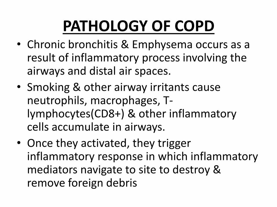
PATHOLOGY OF COPD• Chronic bronchitis & Emphysema occurs as a
result of inflammatory process involving the airways and distal air spaces.
• Smoking & other airway irritants cause neutrophils, macrophages, T-lymphocytes(CD8+) & other inflammatory cells accumulate in airways.
• Once they activated, they trigger inflammatory response in which inflammatory mediators navigate to site to destroy & remove foreign debris

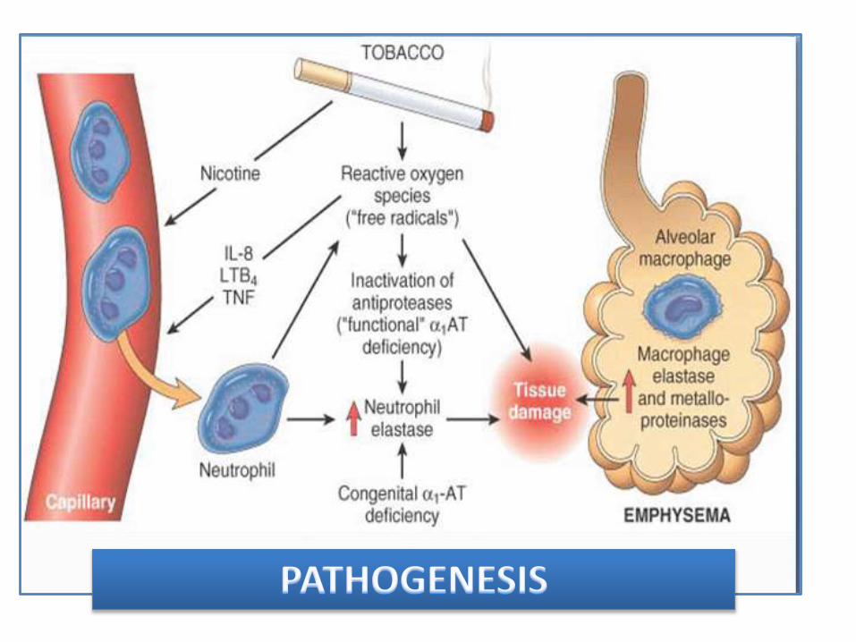
• Repeated exposure to airway irritants cause ongoing inflammatory response which causes permanent inflammation of airways, chronic bronchitis.
• In emphysema, oxidants produced by smoking & proteases produced by inflammatory macrophages, epithelial cell cause protease & anti protease imbalance which is responsible for breakdown of lungs fragile elastic lamina, destruction of alveolar septa.
• Mediators: LTb4( attracts neutrophils, lymphocytes),TNFα, IL1β, IL6(amplify inflammatory response) & TGFβ(induce fibrosis in small airways)

s
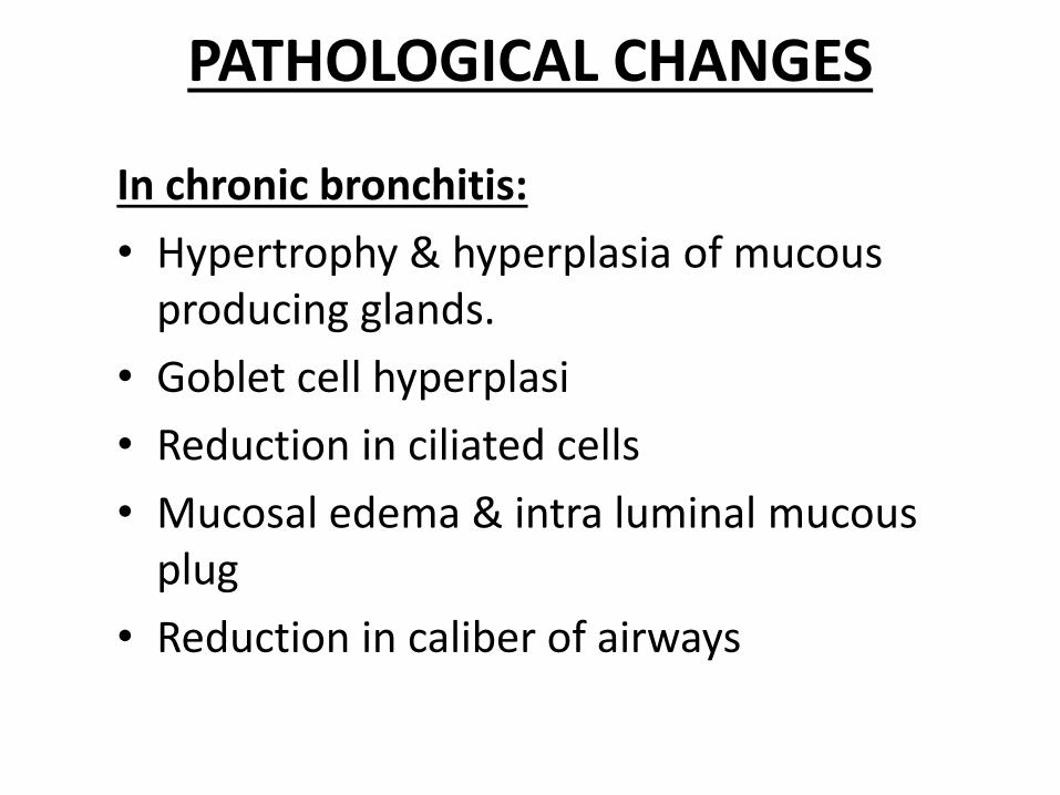
PATHOLOGICAL CHANGES
In chronic bronchitis:
• Hypertrophy & hyperplasia of mucous producing glands.
• Goblet cell hyperplasi
• Reduction in ciliated cells
• Mucosal edema & intra luminal mucous plug
• Reduction in caliber of airways

Normal reid index 0.44±0.09Chronic bronchitis>0.51

In Emphysema:
• Severe destruction of small airways, alveoli.
• Destruction of alveolar septa.
• Formation of large pockets called bullae.


CLASSIFICATION OF EMPHYSEMA
Centriacinar:
Destruction and distension are limited to respiratory bronchiole and alveoli closely related to them(with sparing of the periphery).
Predominantly found in upper lobe & superior segments of lower lobe, most frequently associated with cigarette smoking.

• Panacinar:
Generalized destruction of both central & peripheral portion of acinus. Predominant in lower half of the lungs, mostly associated with alpha 1 antitrypsin deficiency.
• Paraseptal:
Involves only distal acinus. It involves distal airway structures, alveolar ducts & sacs. Localized around septa of the lungs.


SYMPTOMSChronic bronchitis Emphysema
•Ongoing cough with mucus production(called smoker’s cough)
•Copious purulent sputum
•Breathlessness relatively late in onset
•Mucopurulent relapses more frequent
•Wheezing
•Chest tightness
•Minimal cough with expectoration
•Scanty mucoid sputum
•Breathlessness insidious in onset initially on Exertional, gradually even at rest
•Mucopurelent relapses less frequent
• Generalised Weakness
•lethargy
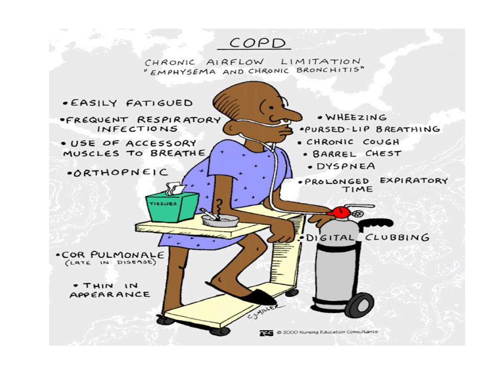

On examination:Inspection:• Pt looks dyspnic• Use of accessory muscles• Barrel shaped chest (due to hyper inflation)• Prolonged expiration• Tripod positioning to facilitate action of accessory muscles.• advanced disease patients have paradoxical inward movement
of the rib cage with inspiration (Hoover's sign), • Patients with advanced disease have paradoxical inward
movement of the rib cage with inspiration (Hoover's sign).
• Emphysema patients, termed "pink puffers" are thin and non cyanotic at rest and have prominent use of accessory muscles
• Chronic bronchitis patients are more likely to be heavy and cyanotic & termed as “blue bloaters”.


Palpation:
-decreased chest expansion
Percussion:
- hyper resonance on lung fields
Auscultation:
–Decreased breath sounds
–Normal vesicular breathing but prolonged expiration
– Expiratory ronchi
–Coarse crepitatons on both phases

INVESTIGATIONS
Chest X-Ray:
– Not sensitive for Dx
– To exclude other diseases
– Hyper-inflation signs
– Low set flat diaphragm
ABG: important for assessing patients with severe COPD.
Detect acute & chronic hypercapnia
Respiratory acidosis(pco2 raised, hypercarbia)
Pao2 is markedly reduced (hypoxemia) Measurement of serum α1AT level, normal level 2-4 g/l.
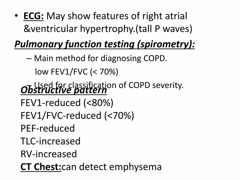
• ECG: May show features of right atrial &ventricular hypertrophy.(tall P waves)
Pulmonary function testing (spirometry):
– Main method for diagnosing COPD.
low FEV1/FVC (< 70%)
– Used for classification of COPD severity.Obstructive patternFEV1-reduced (<80%)FEV1/FVC-reduced (<70%)PEF-reducedTLC-increasedRV-increasedCT Chest:can detect emphysema

COMPLICATIONS OF COPD
• Carbon dioxide narcosis: Persistant Co2 retention causes increase Paco2; hypercarbia, causes drowsiness, altered sensorium, headache.
• Respiratory failure:
Type Ι respiratory failure(low Pao2, normal Paco2) occurs in mild to moderate COPD.
Acute or Chronic Type ΙΙ respiratory failure occurs in severe COPD.
• Secondary polycythemia: Results from hypoxemia stimulating erythropoiesis.

COPD
HypoxaemiaHypercarbia
Pulmonary vasoconstriction
Pulmonary HTN
Chronic after load to R ventricle
RVH
RVF
Corpulmonale:

• Clinical findings: peripheral edema
raised JVP
tender hepatomegaly
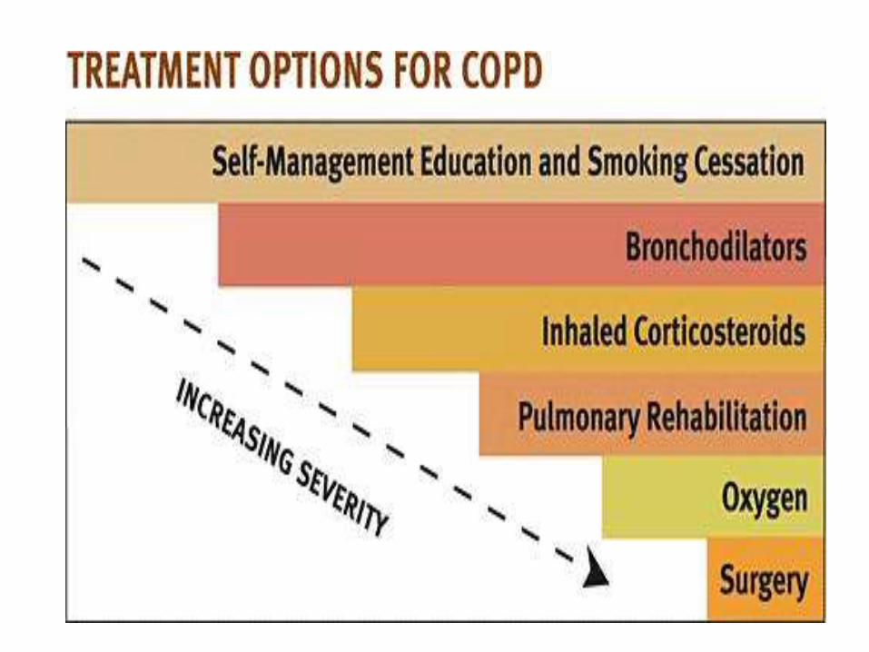

MANAGEMENTGeneral measures:
• Cessation of smoking
• Avoid lung irritants
• Improve nutrition
• Regular exercises
• Chest physiotherapy.

Specific management:
• Mild COPD: Add short acting β2 agonists like salbutamol 2-4mg or terbutaline 2.5-5mg 6 hourly.
• Moderate COPD: Add long acting β-stimulants like salmetrol,2puffs of 25 mcg each 2-3 times a day, formetrol 2 puffs of 6 mcg each 1-3 times a day with short acting anti cholinergic like ipratropiumbromide 40-80μg 6 hourly or long acting anti cholinergic like tiotropium bromide 18μg once a day.
• Severe COPD: Add inhaled glucocorticosteroids like beclomethasone/budesonide/fluticasone. If response is not satisfactory add systemic glucocorticosteroids like prednisolone/methyl prednisolone.

• Very severe COPD: Long term O2, ventilatory support, management of cardiac failure, may consider surgical management.
Methylxanthines like aminophylline or theophylline can be administered when necessary.
Long term domiciliary oxygen therapy should be administered to patients with PaO2<55 mm hg, >16 hrs/day, 2-3ltr/min to maintain SaO2>90%. This reduces pulmonary hypertension, polycythemia, dyspnoea, hypoxemia.
Antibiotics like tetracycline or ampicillin is needed when respiratory infection present, if no response sputum culture sensitivity to be done & antibiotic changed accordingly.

For out patients management doxycycline, amoxicilline-clavulanate can be given. Patients older than 65 years fluoroquinolones like levofloxacin, gemifloxacin, moxifloxacincan be given.
For hospitalized patients IV antibiotics like azithromycin or fluoroquinolones or third generation cephalosporins like ceftriaxone or cefotaxime should be administered.

Severe acute exacerbation of COPD:
Oxygen: Initial therapy should be maintaining SaO2 >90%, can be ).
Bronchodilators: Nebulisation of β2 agonists like salbutamol 2.5mg every 20 min & anti cholinergic agents like ipratropium bromide to be administered.
Antibiotics: Indicated if sputum volume & purulence is increased.
Most common organisms incluceS.pneumoniae, H.influenzae, M,catarrhalis.
patients with severe exacerbation third generation cephalosporins & fluoroquinolones or an aminoglycoside to be given.

Corticosteroids: IV or oral corticosteroids to be administered as they improve lung function & hypoxemia.
Aminophylline: should be administered if patient fails to respond to initial treatment with Nebulization of β2 agonists. . Given as a
loading dose of 5mg/kg/hr as an infusion.
Diuretics: should be administered to patients with gross cardiac failure.
Respiratory stimulants: Like Doxapram can be if patient is not responding to conventional agents. Dose 1.5-4mg/min as infusion.
Non-invasive positive pressure Ventilation(NIPPV): Ventilation should be tried with tight fitting face mask.
This is used in patient with normal mental status, stable cardiovascular function.
Indications- severe dyspnoea, use of accessory muscles, paradoxical abdominal motion, PH<7.35 mmhg, PaCo2> 45 mmhg, Respiratory rate>25/min.

Invasive mechanical ventilation: When NIV fails patient should be intubated for mechanical ventilation.
Indications- severe dyspnoea, use of accessory muscles, paradoxical abdominal motion, Respiratory rate>35/min
Severe acidosis-PH<7.25
Hypercapnia>60 mmhg
Hypoxia<40 mmhg
Altered sensorium
Respiratory arrest, unstable cardiovascular function, sepsis, hypotension, shock.

Surgical management:
• Bullectomy
• Lung volume reduction surgery(LVRS)-resection of damaged portion of lung, improves exercise tolerance but doesn’t improve life expectancy.
• Lung transplantation- If FEV1<35% (PaO2< 60 mmhg), & PaCo2>50 mmhg.

GOLD Stage
Severity Spirometry Management
0 At Risk Normal Avoid risk factors
I Mild FEV1/FVC <0.7 and FEV1 80% predicted
Short acting β- 2 agonist
II Moderate FEV1/FVC <0.7 and 50% FEV1
<80% predictedLong acting β- 2 agonist/short or long acting anti cholinergics
III Severe FEV1/FVC <0.7 and 30% FEV1
<50% predictedAdd inhaled steroids/methylxanthines
IV Very Severe FEV1/FVC <0.7 and FEV1 <30% predictedorFEV1 <50% predicted with respiratory failure or signs of right heart failure
Add oxygen, ventilatory support, management of RHF


THANK YOU

![Chronic Obstructive Pulmonary Diseaseopenaccessebooks.com/chronic-obstructive-pulmonary...Chronic Obstructive Pulmonary Disease 5 a-MCI is made [32]. COPD patients without significant](https://img.pdfslide.us/doc/110x75/5f853ccf82a2412fd65b9e28/chronic-obstructive-pulmonary-dis-chronic-obstructive-pulmonary-disease-5-a-mci.jpg)