Embed Size (px)
Citation preview


P1: PBU
LWBK192-FM LWBK192-Hubbard December 2, 2008 13:43
A Concise Review
of Clinical
Laboratory Science
Second Edition
i

P1: PBU
LWBK192-FM LWBK192-Hubbard December 2, 2008 13:43
ii
P1: PBU
LWBK192-FM LWBK192-Hubbard December 2, 2008 13:43
A Concise Review
of Clinical
Laboratory Science
Second Edition
Joel D. Hubbard, PhD, MT (ASCP)Associate Professor, Program of Clinical Laboratory ScienceDepartment of Laboratory Sciences and Primary CareSchool of Allied Health SciencesTexas Tech University Health Sciences CenterLubbock, Texas
iii
P1: PBU
LWBK192-FM LWBK192-Hubbard December 2, 2008 13:43
Acquisitions Editor: John Goucher
Managing Editor: Meredith Brittain
Project Manager: Rosanne Hallowell
Manufacturing Manager: Margie Orzech
Marketing Manager: Allison Noplock
Cover Designer: Melissa Walter
Design Coordinator: Stephen Druding
Production Services: Aptara, Inc.
c© 2010 by LIPPINCOTT WILLIAMS & WILKINS, a Wolters Kluwer business
c© 1997 by WILLIAMS & WILKINS
530 Walnut Street
Philadelphia, PA 19106
All rights reserved. This book is protected by copyright. No part of this book may be reproduced in any form or by
any means, including photocopying, or utilized by any information storage and retrieval system without written
permission from the copyright owner, except for brief quotations embodied in critical articles and reviews. To
request permission, please contact Lippincott Williams & Wilkins at 530 Walnut Street, Philadelphia, PA 19016, via
email at [email protected] or via our website at lww.com (products and services).
Printed in the United States of America.
Library of Congress Cataloging-in-Publication DataA concise review of clinical laboratory science / [edited by] Joel Hubbard.—2nd ed.
p. ; cm.
Includes index.
ISBN 978-0-7817-8202-9
1. Medical laboratory technology—Examinations, questions, etc. 2. Medical laboratory technology—Outlines,
syllabi, etc. I. Hubbard, Joel D. (Joel David), 1952-
[DNLM: 1. Laboratory Techniques and Procedures–Examination Questions. QY 18.2 C744 2010]
RB38.25.H83 2010
616.07′56—dc22
2008043981
DISCLAIMER
Care has been taken to confirm the accuracy of the information presented and to describe generally accepted
practices. However, the authors, editors, and publisher are not responsible for errors or omissions or for any
consequences from application of the information in this book and make no warranty, expressed or implied, with
respect to the currency, completeness, or accuracy of the contents of the publication. Application of this information
in a particular situation remains the professional responsibility of the practitioner; the clinical treatments described
and recommended may not be considered absolute and universal recommendations.
The authors, editors, and publisher have exerted every effort to ensure that drug selection and dosage set forth in this
text are in accordance with recommendations and practice at the time of publication. However, in view of ongoing
research, changes in government regulations, and the constant flow of information relating to drug therapy and drug
reactions, the reader is urged to check the package insert for each drug for any change in indications and dosage and
for added warnings and precautions. This is particularly important when the recommended agent is a new or
infrequently employed drug.
Some drugs and medical devices presented in this publication have Food and Drug Administration (FDA) clearance
for limited use in restricted research settings. It is the responsibility of the clinician to ascertain the FDA status of
each drug or device planned for use in their clinical practice.
The publishers have made every effort to trace copyright holders for borrowed material. If they have inadvertently
overlooked any, they will be pleased to make the necessary arrangements at the first opportunity.
To purchase additional copies of this book, call our customer service department at (800) 638-3030 or fax orders to
(301) 223-2320. International customers should call (301) 223-2300. Visit Lippincott Williams & Wilkins on the
Internet at: LWW.com. Lippincott Williams & Wilkins customer service representatives are available from 8:30 am
to 6:00 pm, EST.
10 9 8 7 6 5 4 3 2 1
iv
P1: PBU
LWBK192-FM LWBK192-Hubbard December 2, 2008 13:43
This book is dedicated to clinical laboratory science students everywhere.In your upcoming role as professionals, remember that your job isimportant to the medical world as well as to the individual patient.
Be proud of the fact that you will be making a differencein people’s lives. Always be excited about the unlimited opportunities
available in your chosen profession and help lead the fieldof Clinical Laboratory Science well into the 21st century.
v

P1: PBU
LWBK192-FM LWBK192-Hubbard December 2, 2008 13:43
vi
P1: PBU
LWBK192-FM LWBK192-Hubbard December 2, 2008 13:43
PREFACE
The arrival of the second edition of A Concise Review of Clinical Laboratory Sciencehas long been anticipated by students and educators alike. This review text is a valuableeducational tool for both the novice and the experienced clinical laboratory scientist. Itis designed to be an updated and concise review of all disciplines of clinical laboratoryscience and will also serve as a tool for students of clinical laboratory science studying fornational certification examinations, including the American Society of Clinical PathologistsBoard of Registry exam, the National Certification Agency (NCA) exam, and the AmericanMedical Technologist (AMT) exam. Practicing clinical laboratory scientists and medicalresidents will also find this book to be an excellent source for review.
This book represents a culmination of the efforts and expertise of the faculty of theClinical Laboratory Science program at Texas Tech University Health Sciences Center inLubbock, Texas, and reflects over 100 years of combined medical technology experience.All contributing authors reflect their professional excellence in their contributed chapters,not only as educators, but also as outstanding professionals in their field. I encourage readersto send me feedback on this book at the following email address: [email protected].
Text Format and Features
Each chapter presents a concise summary of the most important facts and concepts in thatsubject area in an outline format. Key points appear in bold for easy reference. Boxes,tables, and figures throughout distill concepts and make them easier to comprehend. Onlinemenus at the end of each chapter point readers to supplementary Web-based materials.
What’s New in This Edition
The second edition includes the most current and updated information. An expanded chap-ter dealing with laboratory operations (Chapter 11) addresses topics such as managementand organizational theory, professionalism, quality assurance, laboratory regulations, anddelivery of an educational unit. In addition, a new chapter on molecular pathology (Chapter10) focuses on molecular laboratory methods and an overview on the testing of geneticdiseases.
Additional Resources
A Concise Review of Clinical Laboratory Science, second edition, includes additional re-sources for both instructors and students that are available on the book’s companion Website at thePoint.lww.com/Hubbard2e.
vii
P1: PBU
LWBK192-FM LWBK192-Hubbard December 2, 2008 13:43
viii Preface
Instructors
Approved adopting instructors will be given access to the following additional resources:
� Image Bank (including color images referenced in the text)� Quiz Bank� Web Case Studies (including those referenced in the text)
Students
Students who have purchased A Concise Review of Clinical Laboratory Science, secondedition, have access to the following additional resources:
� Image Bank (including color images referenced in the text)� Quiz Bank� Web Case Studies (including those referenced in the text)
In addition, purchasers of the text can access the searchable Full Text Online by goingto the A Concise Review of Clinical Laboratory Science, second edition, Web site atthePoint.lww.com/Hubbard2e. See the inside front cover of this text for more details, in-cluding the pass code you will need to gain access to the Web site.

P1: PBU
LWBK192-FM LWBK192-Hubbard December 2, 2008 13:43
ACKNOWLEDGMENTS
I would like to thank all of the following contributing authors—Dr. Lynne Hamilton, Dr.Hal Larsen, Dr. Barbara Sawyer, Mr. Wade Redman, Ms. Lori Rice-Spearman, and Dr.Tootie Tatum—for making this book possible. Their individual expertise, willingness topresent the highest quality of material, and high level of professionalism made the task ofproducing this text easy. I would also like to thank my wife, Kathy, who patiently listenedto my endless rambling about the project.
ix

P1: PBU
LWBK192-FM LWBK192-Hubbard December 2, 2008 13:43
x
P1: PBU
LWBK192-FM LWBK192-Hubbard December 2, 2008 13:43
CONTRIBUTORS
Lynne Hamilton, PhD, MT (ASCP)Assistant Professor, Program of Clinical Laboratory
ScienceDepartment of Laboratory Sciences and Primary
CareSchool of Allied Health SciencesTexas Tech University Health Sciences CenterLubbock, Texas
Joel D. Hubbard, PhD, MT (ASCP)Associate Professor, Program of Clinical Laboratory
ScienceDepartment of Laboratory Sciences and Primary
CareSchool of Allied Health SciencesTexas Tech University Health Sciences CenterLubbock, Texas
Hal S. Larsen, MT (ASCP), CLS (NCA), PhDProfessor and Chair, Department of Diagnostic and
Primary CareSchool of Allied Health SciencesTexas Tech University Health Sciences CenterLubbock, Texas
Wade Redman, MT (ASCP), MBAAssistant Professor, Program of Clinical Laboratory
ScienceDepartment of Laboratory Sciences and Primary
CareSchool of Allied Health SciencesTexas Tech University Health Sciences CenterLubbock, Texas
Lori Rice-Spearman, MS, MT (ASCP)Associate Professor andProgram Director of Clinical Laboratory ScienceDepartment of Laboratory Sciences and Primary
CareSchool of Allied Health SciencesTexas Tech University Health Sciences CenterLubbock, Texas
Barbara Sawyer, PhD, MT (ASCP),CLS (NCA), CLSp (MB)
Professor, Department of Laboratory Sciences andPrimary Care
School of Allied Health SciencesTexas Tech University Health Sciences CenterLubbock, Texas
Owatha L. Tatum, PhD, CLSp (MB),MP (ASCP)
Assistant Professor, Program of Clinical LaboratoryScience
Department of Laboratory Sciences and PrimaryCare
School of Allied Health SciencesTexas Tech University Health Sciences CenterLubbock, Texas
xi

P1: PBU
LWBK192-FM LWBK192-Hubbard December 2, 2008 13:43
xii
P1: PBU
LWBK192-FM LWBK192-Hubbard December 2, 2008 13:43
CONTENTS
CHAPTER 1 Clinical Chemistry. . . . . . . . . . . . . . . . . . . . . . . . . . . . . . . . . . . . . . . . 1
Barbara SawyerI. Clinical Chemistry Basics . . . . . . . . . . . . . . . . . . . . . . . . . . . . . . . . . . . . . . . . . . . . . . . . . 1
II. Special Methods in Clinical Chemistry . . . . . . . . . . . . . . . . . . . . . . . . . . . . . . . . . . . . . . 7III. Basic Anatomy and Physiology . . . . . . . . . . . . . . . . . . . . . . . . . . . . . . . . . . . . . . . . . . . . 10IV. Analytes and Pathophysiology . . . . . . . . . . . . . . . . . . . . . . . . . . . . . . . . . . . . . . . . . . . . . 14V. Enzymology . . . . . . . . . . . . . . . . . . . . . . . . . . . . . . . . . . . . . . . . . . . . . . . . . . . . . . . . . . . . . 30
VI. Endocrinology . . . . . . . . . . . . . . . . . . . . . . . . . . . . . . . . . . . . . . . . . . . . . . . . . . . . . . . . . . . 35VII. Toxic and Therapeutic Drugs . . . . . . . . . . . . . . . . . . . . . . . . . . . . . . . . . . . . . . . . . . . . . . 46
CHAPTER 2 Hemostasis and Coagulation. . . . . . . . . . . . . . . . . . . . . . . . . . . . . . 52
Joel HubbardI. Platelet Physiology . . . . . . . . . . . . . . . . . . . . . . . . . . . . . . . . . . . . . . . . . . . . . . . . . . . . . . . 52
II. Platelet Pathophysiology . . . . . . . . . . . . . . . . . . . . . . . . . . . . . . . . . . . . . . . . . . . . . . . . . . 55III. Blood Coagulation and Fibrinolysis . . . . . . . . . . . . . . . . . . . . . . . . . . . . . . . . . . . . . . . . 66IV. Coagulation Disorders . . . . . . . . . . . . . . . . . . . . . . . . . . . . . . . . . . . . . . . . . . . . . . . . . . . . 76
CHAPTER 3 Routine Hematology . . . . . . . . . . . . . . . . . . . . . . . . . . . . . . . . . . . . . . 85
Joel HubbardI. Laboratory Analysis . . . . . . . . . . . . . . . . . . . . . . . . . . . . . . . . . . . . . . . . . . . . . . . . . . . . . . 85
II. Hematopoietic Tissues . . . . . . . . . . . . . . . . . . . . . . . . . . . . . . . . . . . . . . . . . . . . . . . . . . . . 94III. Hemoglobin Synthesis, Structure, and Function . . . . . . . . . . . . . . . . . . . . . . . . . . . . . . 99IV. Erythrocytes and Erythropoiesis . . . . . . . . . . . . . . . . . . . . . . . . . . . . . . . . . . . . . . . . . . . . 104
CHAPTER 4 Hematologic Disorders . . . . . . . . . . . . . . . . . . . . . . . . . . . . . . . . . . . 117
Joel HubbardI. Red Blood Cell Indices and Their Use in the Diagnosis of Anemia . . . . . . . . . . . . . 117
II. Red Blood Cell (RBC) Disorders . . . . . . . . . . . . . . . . . . . . . . . . . . . . . . . . . . . . . . . . . . . 118III. Anemias and Polycythemias . . . . . . . . . . . . . . . . . . . . . . . . . . . . . . . . . . . . . . . . . . . . . . . 120IV. Leukocyte Disorders . . . . . . . . . . . . . . . . . . . . . . . . . . . . . . . . . . . . . . . . . . . . . . . . . . . . . . 145V. Lymphocyte Physiology and Disorders . . . . . . . . . . . . . . . . . . . . . . . . . . . . . . . . . . . . . . 164
xiii
P1: PBU
LWBK192-FM LWBK192-Hubbard December 2, 2008 13:43
xiv Contents
CHAPTER 5 Immunology and Serology . . . . . . . . . . . . . . . . . . . . . . . . . . . . . . . . 177
Wade Redman and Joel HubbardI. Introduction . . . . . . . . . . . . . . . . . . . . . . . . . . . . . . . . . . . . . . . . . . . . . . . . . . . . . . . . . . . . . . 177
II. Cells and Tissues of the Immune System . . . . . . . . . . . . . . . . . . . . . . . . . . . . . . . . . . . . 181III. Immunity . . . . . . . . . . . . . . . . . . . . . . . . . . . . . . . . . . . . . . . . . . . . . . . . . . . . . . . . . . . . . . . . 183IV. Immune Response (IR) . . . . . . . . . . . . . . . . . . . . . . . . . . . . . . . . . . . . . . . . . . . . . . . . . . . . 183V. Major Histocompatibility Complex . . . . . . . . . . . . . . . . . . . . . . . . . . . . . . . . . . . . . . . . . 184
VI. Hypersensitivity . . . . . . . . . . . . . . . . . . . . . . . . . . . . . . . . . . . . . . . . . . . . . . . . . . . . . . . . . . 187VII. Autoimmunity. . . . . . . . . . . . . . . . . . . . . . . . . . . . . . . . . . . . . . . . . . . . . . . . . . . . . . . . . . . . 188
VIII. Immunodeficiencies . . . . . . . . . . . . . . . . . . . . . . . . . . . . . . . . . . . . . . . . . . . . . . . . . . . . . . 191IX. Techniques in Immunology and Serology . . . . . . . . . . . . . . . . . . . . . . . . . . . . . . . . . . . 193X. Syphilis Serology . . . . . . . . . . . . . . . . . . . . . . . . . . . . . . . . . . . . . . . . . . . . . . . . . . . . . . . . . 196
XI. Acute Phase Proteins . . . . . . . . . . . . . . . . . . . . . . . . . . . . . . . . . . . . . . . . . . . . . . . . . . . . . . 197XII. Hepatitis . . . . . . . . . . . . . . . . . . . . . . . . . . . . . . . . . . . . . . . . . . . . . . . . . . . . . . . . . . . . . . . . . 198
XIII. Streptococcal Serology . . . . . . . . . . . . . . . . . . . . . . . . . . . . . . . . . . . . . . . . . . . . . . . . . . . . 200XIV. Epstein-Barr Virus (EBV) Serology . . . . . . . . . . . . . . . . . . . . . . . . . . . . . . . . . . . . . . . . 201XV. Rubella Serology . . . . . . . . . . . . . . . . . . . . . . . . . . . . . . . . . . . . . . . . . . . . . . . . . . . . . . . . . 202
XVI. Febrile Disease Serology . . . . . . . . . . . . . . . . . . . . . . . . . . . . . . . . . . . . . . . . . . . . . . . . . . 202XVII. Borrelia Burdorferi Serology. . . . . . . . . . . . . . . . . . . . . . . . . . . . . . . . . . . . . . . . . . . . . . . 202
XVIII. Transplant Immunology . . . . . . . . . . . . . . . . . . . . . . . . . . . . . . . . . . . . . . . . . . . . . . . . . . . 203XIX. Tumor Immunology. . . . . . . . . . . . . . . . . . . . . . . . . . . . . . . . . . . . . . . . . . . . . . . . . . . . . . . 205
CHAPTER 6 Immunohematology . . . . . . . . . . . . . . . . . . . . . . . . . . . . . . . . . . . . . . . 207
Wade RedmanI. Introduction . . . . . . . . . . . . . . . . . . . . . . . . . . . . . . . . . . . . . . . . . . . . . . . . . . . . . . . . . . . . . . 207
II. Blood Group Systems . . . . . . . . . . . . . . . . . . . . . . . . . . . . . . . . . . . . . . . . . . . . . . . . . . . . . 208III. Donor Selection and Blood Collection . . . . . . . . . . . . . . . . . . . . . . . . . . . . . . . . . . . . . . 217IV. Donor Processing . . . . . . . . . . . . . . . . . . . . . . . . . . . . . . . . . . . . . . . . . . . . . . . . . . . . . . . . . 219V. Blood Components and Component Therapy . . . . . . . . . . . . . . . . . . . . . . . . . . . . . . . . 221
VI. Antiglobulin Testing . . . . . . . . . . . . . . . . . . . . . . . . . . . . . . . . . . . . . . . . . . . . . . . . . . . . . . 224VII. Unexpected Antibodies . . . . . . . . . . . . . . . . . . . . . . . . . . . . . . . . . . . . . . . . . . . . . . . . . . . . 225
VIII. Compatibility Testing . . . . . . . . . . . . . . . . . . . . . . . . . . . . . . . . . . . . . . . . . . . . . . . . . . . . . 228IX. Transfusion Reactions . . . . . . . . . . . . . . . . . . . . . . . . . . . . . . . . . . . . . . . . . . . . . . . . . . . . . 230X. Hemolytic Disease of the Newborn (HDN) . . . . . . . . . . . . . . . . . . . . . . . . . . . . . . . . . . 231
XI. Autoimmune Hemolytic Anemias (AIHA) . . . . . . . . . . . . . . . . . . . . . . . . . . . . . . . . . . 233XII. Transfusion-Transmitted Diseases . . . . . . . . . . . . . . . . . . . . . . . . . . . . . . . . . . . . . . . . . . 234
XIII. The Major Histocompatibility Complex (MHC) and Human LeukocyteAntigens (HLA) . . . . . . . . . . . . . . . . . . . . . . . . . . . . . . . . . . . . . . . . . . . . . . . . . . . . . . . . . . 235
XIV. Alternative Methodologies in Blood Bank Testing. . . . . . . . . . . . . . . . . . . . . . . . . . . . 236
CHAPTER 7 Clinical Microbiology . . . . . . . . . . . . . . . . . . . . . . . . . . . . . . . . . . . 238
Lynne Hamilton and Hal LarsenI. Bacteria . . . . . . . . . . . . . . . . . . . . . . . . . . . . . . . . . . . . . . . . . . . . . . . . . . . . . . . . . . . . . . . . . 238
II. Bacterial Cell Structure . . . . . . . . . . . . . . . . . . . . . . . . . . . . . . . . . . . . . . . . . . . . . . . . . . . 239III. Stains . . . . . . . . . . . . . . . . . . . . . . . . . . . . . . . . . . . . . . . . . . . . . . . . . . . . . . . . . . . . . . . . . . . 241IV. Normal Flora . . . . . . . . . . . . . . . . . . . . . . . . . . . . . . . . . . . . . . . . . . . . . . . . . . . . . . . . . . . . . 241V. Pathogenesis of Infection . . . . . . . . . . . . . . . . . . . . . . . . . . . . . . . . . . . . . . . . . . . . . . . . . . 242
VI. Collection and Handling of Clinical Specimens . . . . . . . . . . . . . . . . . . . . . . . . . . . . . . 243VII. Micrococcaceae . . . . . . . . . . . . . . . . . . . . . . . . . . . . . . . . . . . . . . . . . . . . . . . . . . . . . . . . . . 246
VIII. Streptococcus, Enterococcus, and Related Genera . . . . . . . . . . . . . . . . . . . . . . . . . . . . 249IX. Aerobic Gram-Positive Bacilli . . . . . . . . . . . . . . . . . . . . . . . . . . . . . . . . . . . . . . . . . . . . . 254X. Neisseria and Moraxella Catarrhalis . . . . . . . . . . . . . . . . . . . . . . . . . . . . . . . . . . . . . . . 256
XI. Miscellaneous Gram-Negative Bacilli . . . . . . . . . . . . . . . . . . . . . . . . . . . . . . . . . . . . . . . 258XII. Enterobacteriaceae . . . . . . . . . . . . . . . . . . . . . . . . . . . . . . . . . . . . . . . . . . . . . . . . . . . . . . . . 262
P1: PBU
LWBK192-FM LWBK192-Hubbard December 2, 2008 13:43
Contents xv
XIII. Campylobacter, Helicobacter, and Vibrionaceae . . . . . . . . . . . . . . . . . . . . . . . . . . . . . 265XIV. Gram-Negative Nonfermentative Bacilli . . . . . . . . . . . . . . . . . . . . . . . . . . . . . . . . . . . . 268XV. Miscellaneous Genera . . . . . . . . . . . . . . . . . . . . . . . . . . . . . . . . . . . . . . . . . . . . . . . . . . . . . 271
XVI. Mycoplasma, Ureaplasma, and the Chlamydiaceae . . . . . . . . . . . . . . . . . . . . . . . . . . . 272XVII. Spirochetes . . . . . . . . . . . . . . . . . . . . . . . . . . . . . . . . . . . . . . . . . . . . . . . . . . . . . . . . . . . . . . 273
XVIII. Mycobacteria . . . . . . . . . . . . . . . . . . . . . . . . . . . . . . . . . . . . . . . . . . . . . . . . . . . . . . . . . . . . 274XIX. Anaerobes . . . . . . . . . . . . . . . . . . . . . . . . . . . . . . . . . . . . . . . . . . . . . . . . . . . . . . . . . . . . . . . 277XX. Zoonotic and Rickettsial Infections . . . . . . . . . . . . . . . . . . . . . . . . . . . . . . . . . . . . . . . . . 282
XXI. Agents of Bioterrorism . . . . . . . . . . . . . . . . . . . . . . . . . . . . . . . . . . . . . . . . . . . . . . . . . . . . 283XXII. Antimicrobial Susceptibility Testing . . . . . . . . . . . . . . . . . . . . . . . . . . . . . . . . . . . . . . . . 284
XXIII. Disinfection and Sterilization . . . . . . . . . . . . . . . . . . . . . . . . . . . . . . . . . . . . . . . . . . . . . . 287XXIV. Molecular Testing . . . . . . . . . . . . . . . . . . . . . . . . . . . . . . . . . . . . . . . . . . . . . . . . . . . . . . . . 288
CHAPTER 8 Clinical Parasitology, Mycology, and Virology . . . . . . . . 289
Lori Rice-Spearman and Lynne HamiltonI. Parasitology . . . . . . . . . . . . . . . . . . . . . . . . . . . . . . . . . . . . . . . . . . . . . . . . . . . . . . . . . . . . . . 289
II. Mycology. . . . . . . . . . . . . . . . . . . . . . . . . . . . . . . . . . . . . . . . . . . . . . . . . . . . . . . . . . . . . . . . 297III. Virology . . . . . . . . . . . . . . . . . . . . . . . . . . . . . . . . . . . . . . . . . . . . . . . . . . . . . . . . . . . . . . . . . 305
CHAPTER 9 Urinalysis and Body Fluids Analysis . . . . . . . . . . . . . . . . . . . . . 313
Barbara SawyerI. The Renal System . . . . . . . . . . . . . . . . . . . . . . . . . . . . . . . . . . . . . . . . . . . . . . . . . . . . . . . . 313
II. The Urine Specimen . . . . . . . . . . . . . . . . . . . . . . . . . . . . . . . . . . . . . . . . . . . . . . . . . . . . . . 314III. Physical Examination of Urine . . . . . . . . . . . . . . . . . . . . . . . . . . . . . . . . . . . . . . . . . . . . . 319IV. Chemical Examination of Urine . . . . . . . . . . . . . . . . . . . . . . . . . . . . . . . . . . . . . . . . . . . . 322V. Microscopic Examination of the Urine . . . . . . . . . . . . . . . . . . . . . . . . . . . . . . . . . . . . . . 327
VI. Automation in the Urinalysis Laboratory . . . . . . . . . . . . . . . . . . . . . . . . . . . . . . . . . . . . 335VII. Metabolic Products in the Urine . . . . . . . . . . . . . . . . . . . . . . . . . . . . . . . . . . . . . . . . . . . . 335
VIII. Diseases of the Kidney . . . . . . . . . . . . . . . . . . . . . . . . . . . . . . . . . . . . . . . . . . . . . . . . . . . . 338IX. Renal Synthetic Products . . . . . . . . . . . . . . . . . . . . . . . . . . . . . . . . . . . . . . . . . . . . . . . . . . 343X. Urine Pregnancy Testing . . . . . . . . . . . . . . . . . . . . . . . . . . . . . . . . . . . . . . . . . . . . . . . . . . 344
XI. Serous Body Fluids . . . . . . . . . . . . . . . . . . . . . . . . . . . . . . . . . . . . . . . . . . . . . . . . . . . . . . . 345XII. Synovial Fluid . . . . . . . . . . . . . . . . . . . . . . . . . . . . . . . . . . . . . . . . . . . . . . . . . . . . . . . . . . . 347
XIII. Seminal Fluid Analysis . . . . . . . . . . . . . . . . . . . . . . . . . . . . . . . . . . . . . . . . . . . . . . . . . . . . 348XIV. Cerebrospinal Fluid (CSF) Analysis . . . . . . . . . . . . . . . . . . . . . . . . . . . . . . . . . . . . . . . . 351XV. Gastric Fluid Analysis . . . . . . . . . . . . . . . . . . . . . . . . . . . . . . . . . . . . . . . . . . . . . . . . . . . . 354
XVI. Fecal Analysis . . . . . . . . . . . . . . . . . . . . . . . . . . . . . . . . . . . . . . . . . . . . . . . . . . . . . . . . . . . 355
CHAPTER 10 Molecular Diagnostics . . . . . . . . . . . . . . . . . . . . . . . . . . . . . . . . . . . 360
Tootie TatumI. Biology of the Cell . . . . . . . . . . . . . . . . . . . . . . . . . . . . . . . . . . . . . . . . . . . . . . . . . . . . . . . 360
II. Molecular Diagnostic Methods . . . . . . . . . . . . . . . . . . . . . . . . . . . . . . . . . . . . . . . . . . . . . 362III. Inherited Genetic Disease . . . . . . . . . . . . . . . . . . . . . . . . . . . . . . . . . . . . . . . . . . . . . . . . . 365IV. Molecular Oncology . . . . . . . . . . . . . . . . . . . . . . . . . . . . . . . . . . . . . . . . . . . . . . . . . . . . . . 365V. Molecular Infectious Disease . . . . . . . . . . . . . . . . . . . . . . . . . . . . . . . . . . . . . . . . . . . . . . 366
VI. DNA-Based Human Identification . . . . . . . . . . . . . . . . . . . . . . . . . . . . . . . . . . . . . . . . . . 366
CHAPTER 11 Current Issues in Laboratory Management . . . . . . . . . . . . . 368
Wade Redman, Lori Rice-Spearman, and Hal S. LarsenI. Management and Organizational Theory . . . . . . . . . . . . . . . . . . . . . . . . . . . . . . . . . . . . 368
II. Professionalism . . . . . . . . . . . . . . . . . . . . . . . . . . . . . . . . . . . . . . . . . . . . . . . . . . . . . . . . . . 370III. Quality Assurance . . . . . . . . . . . . . . . . . . . . . . . . . . . . . . . . . . . . . . . . . . . . . . . . . . . . . . . . 371IV. Laboratory Regulations . . . . . . . . . . . . . . . . . . . . . . . . . . . . . . . . . . . . . . . . . . . . . . . . . . . 372

P1: PBU
LWBK192-FM LWBK192-Hubbard December 2, 2008 13:43
xvi Contents
V. Financial Management . . . . . . . . . . . . . . . . . . . . . . . . . . . . . . . . . . . . . . . . . . . . . . . . . . . . 381VI. Laboratory Information Systems (LIS) . . . . . . . . . . . . . . . . . . . . . . . . . . . . . . . . . . . . . . 382
VII. Instrument Selection Process . . . . . . . . . . . . . . . . . . . . . . . . . . . . . . . . . . . . . . . . . . . . . . . 382VIII. Problem Solving . . . . . . . . . . . . . . . . . . . . . . . . . . . . . . . . . . . . . . . . . . . . . . . . . . . . . . . . . . 383
IX. Delivery of Education Unit . . . . . . . . . . . . . . . . . . . . . . . . . . . . . . . . . . . . . . . . . . . . . . . . 383X. Outreach Program . . . . . . . . . . . . . . . . . . . . . . . . . . . . . . . . . . . . . . . . . . . . . . . . . . . . . . . . 383
Index 385

P1: PBU
LWBK192-FM LWBK192-Hubbard December 5, 2008 4:24
A Concise Review
of Clinical
Laboratory Science
Second Edition
xvii

P1: PBU
LWBK192-FM LWBK192-Hubbard December 5, 2008 4:24
xviii
P1: OSO
LWBK192-01 LWBK192-Hubbard November 23, 2008 17:19
CHAPTER 1
Clinical Chemistry
BARBARA SAWYER, PhD, MT (ASCP), CLS (NCA)
I. CLINICAL CHEMISTRY BASICS
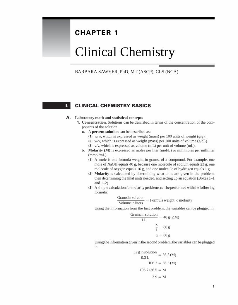
A. Laboratory math and statistical concepts1. Concentration. Solutions can be described in terms of the concentration of the com-
ponents of the solution.a. A percent solution can be described as:
(1) w/w, which is expressed as weight (mass) per 100 units of weight (g/g).(2) w/v, which is expressed as weight (mass) per 100 units of volume (g/dL).(3) v/v, which is expressed as volume (mL) per unit of volume (mL).
b. Molarity (M) is expressed as moles per liter (mol/L) or millimoles per milliliter(mmol/mL).(1) A mole is one formula weight, in grams, of a compound. For example, one
mole of NaOH equals 40 g, because one molecule of sodium equals 23 g, onemolecule of oxygen equals 16 g, and one molecule of hydrogen equals 1 g.
(2) Molarity is calculated by determining what units are given in the problem,then determining the final units needed, and setting up an equation (Boxes 1–1and 1–2).
(3) A simple calculation for molarity problems can be performed with the followingformula:
Grams in solution
Volume in liters= Formula weight × molarity
Using the information from the first problem, the variables can be plugged in:
Grams in solution
1 L= 40 g (2 M)
x
1= 80 g
x = 80 g
Using the information given in the second problem, the variables can be pluggedin:
32 g in solution
0.3 L= 36.5 (M)
106.7 = 36.5 (M)
106.7/36.5 = M
2.9 = M
1
P1: OSO
LWBK192-01 LWBK192-Hubbard November 23, 2008 17:19
2 A Concise Review of Clinical Laboratory Science
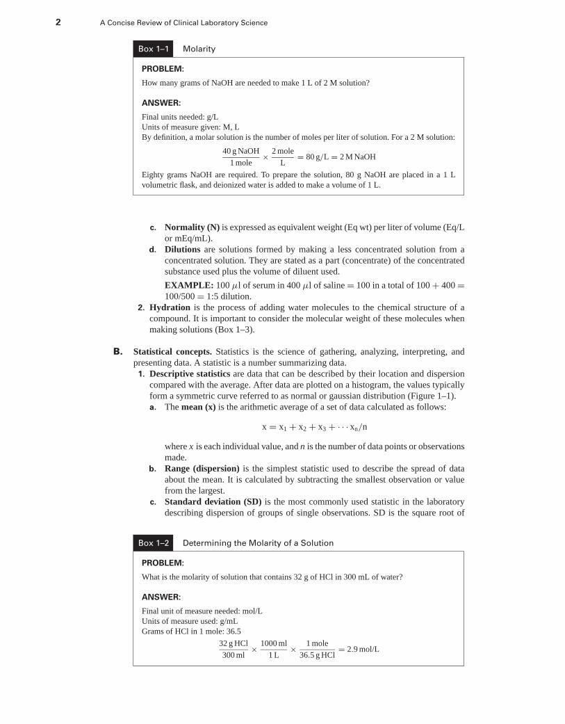
Box 1–1 Molarity
PROBLEM:
How many grams of NaOH are needed to make 1 L of 2 M solution?
ANSWER:
Final units needed: g/L
Units of measure given: M, L
By definition, a molar solution is the number of moles per liter of solution. For a 2 M solution:
40 g NaOH
1 mole× 2 mole
L= 80 g/L = 2 M NaOH
Eighty grams NaOH are required. To prepare the solution, 80 g NaOH are placed in a 1 L
volumetric flask, and deionized water is added to make a volume of 1 L.
c. Normality (N) is expressed as equivalent weight (Eq wt) per liter of volume (Eq/Lor mEq/mL).
d. Dilutions are solutions formed by making a less concentrated solution from aconcentrated solution. They are stated as a part (concentrate) of the concentratedsubstance used plus the volume of diluent used.
EXAMPLE: 100 μl of serum in 400 μl of saline = 100 in a total of 100 + 400 =100/500 = 1:5 dilution.
2. Hydration is the process of adding water molecules to the chemical structure of acompound. It is important to consider the molecular weight of these molecules whenmaking solutions (Box 1–3).
B. Statistical concepts. Statistics is the science of gathering, analyzing, interpreting, andpresenting data. A statistic is a number summarizing data.
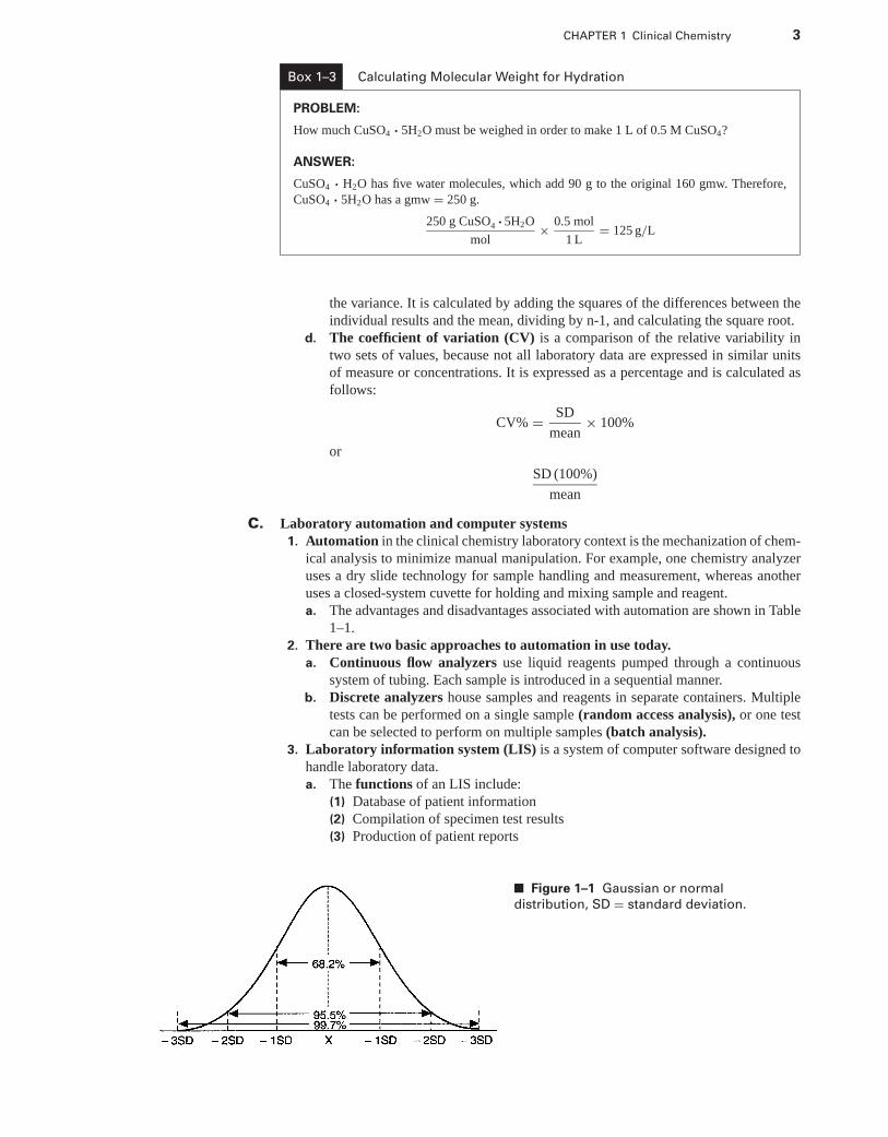
1. Descriptive statistics are data that can be described by their location and dispersioncompared with the average. After data are plotted on a histogram, the values typicallyform a symmetric curve referred to as normal or gaussian distribution (Figure 1–1).a. The mean (x) is the arithmetic average of a set of data calculated as follows:
x = x1 + x2 + x3 + · · · xn/n
where x is each individual value, and n is the number of data points or observationsmade.
b. Range (dispersion) is the simplest statistic used to describe the spread of dataabout the mean. It is calculated by subtracting the smallest observation or valuefrom the largest.
c. Standard deviation (SD) is the most commonly used statistic in the laboratorydescribing dispersion of groups of single observations. SD is the square root of
Box 1–2 Determining the Molarity of a Solution
PROBLEM:
What is the molarity of solution that contains 32 g of HCl in 300 mL of water?
ANSWER:
Final unit of measure needed: mol/L
Units of measure used: g/mL
Grams of HCl in 1 mole: 36.5
32 g HCl
300 ml× 1000 ml
1 L× 1 mole
36.5 g HCl= 2.9 mol/L
P1: OSO
LWBK192-01 LWBK192-Hubbard November 23, 2008 17:19
CHAPTER 1 Clinical Chemistry 3
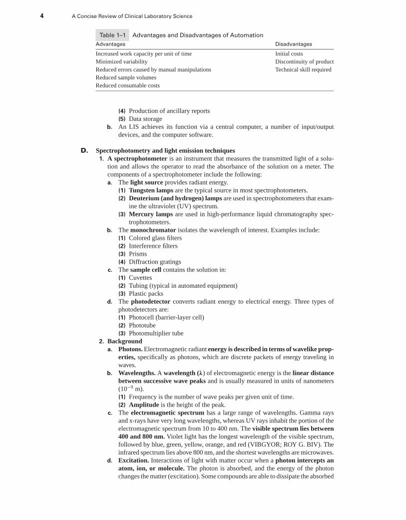
Box 1–3 Calculating Molecular Weight for Hydration
PROBLEM:
How much CuSO4 · 5H2O must be weighed in order to make 1 L of 0.5 M CuSO4?
ANSWER:
CuSO4 · H2O has five water molecules, which add 90 g to the original 160 gmw. Therefore,
CuSO4 · 5H2O has a gmw = 250 g.
250 g CuSO4 · 5H2O
mol× 0.5 mol
1 L= 125 g/L
the variance. It is calculated by adding the squares of the differences between theindividual results and the mean, dividing by n-1, and calculating the square root.
d. The coefficient of variation (CV) is a comparison of the relative variability intwo sets of values, because not all laboratory data are expressed in similar unitsof measure or concentrations. It is expressed as a percentage and is calculated asfollows:
CV% = SD
mean× 100%
or
SD (100%)
mean
C. Laboratory automation and computer systems1. Automation in the clinical chemistry laboratory context is the mechanization of chem-
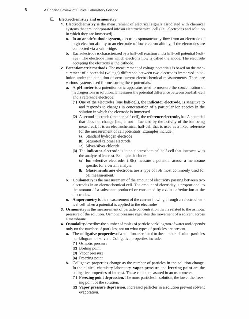
ical analysis to minimize manual manipulation. For example, one chemistry analyzeruses a dry slide technology for sample handling and measurement, whereas anotheruses a closed-system cuvette for holding and mixing sample and reagent.a. The advantages and disadvantages associated with automation are shown in Table
1–1.2. There are two basic approaches to automation in use today.
a. Continuous flow analyzers use liquid reagents pumped through a continuoussystem of tubing. Each sample is introduced in a sequential manner.
b. Discrete analyzers house samples and reagents in separate containers. Multipletests can be performed on a single sample (random access analysis), or one testcan be selected to perform on multiple samples (batch analysis).
3. Laboratory information system (LIS) is a system of computer software designed tohandle laboratory data.a. The functions of an LIS include:
(1) Database of patient information(2) Compilation of specimen test results(3) Production of patient reports
■ Figure 1–1 Gaussian or normal
distribution, SD = standard deviation.
P1: OSO
LWBK192-01 LWBK192-Hubbard November 23, 2008 17:19
4 A Concise Review of Clinical Laboratory Science
Table 1–1 Advantages and Disadvantages of Automation
Advantages Disadvantages
Increased work capacity per unit of time Initial costs
Minimized variability Discontinuity of product
Reduced errors caused by manual manipulations Technical skill required
Reduced sample volumes
Reduced consumable costs
(4) Production of ancillary reports(5) Data storage
b. An LIS achieves its function via a central computer, a number of input/outputdevices, and the computer software.
D. Spectrophotometry and light emission techniques1. A spectrophotometer is an instrument that measures the transmitted light of a solu-
tion and allows the operator to read the absorbance of the solution on a meter. Thecomponents of a spectrophotometer include the following:a. The light source provides radiant energy.
(1) Tungsten lamps are the typical source in most spectrophotometers.(2) Deuterium (and hydrogen) lamps are used in spectrophotometers that exam-
ine the ultraviolet (UV) spectrum.(3) Mercury lamps are used in high-performance liquid chromatography spec-
trophotometers.b. The monochromator isolates the wavelength of interest. Examples include:
(1) Colored glass filters(2) Interference filters(3) Prisms(4) Diffraction gratings
c. The sample cell contains the solution in:(1) Cuvettes(2) Tubing (typical in automated equipment)(3) Plastic packs
d. The photodetector converts radiant energy to electrical energy. Three types ofphotodetectors are:(1) Photocell (barrier-layer cell)(2) Phototube(3) Photomultiplier tube
2. Backgrounda. Photons. Electromagnetic radiant energy is described in terms of wavelike prop-
erties, specifically as photons, which are discrete packets of energy traveling inwaves.
b. Wavelengths. A wavelength (λ) of electromagnetic energy is the linear distancebetween successive wave peaks and is usually measured in units of nanometers(10−9 m).(1) Frequency is the number of wave peaks per given unit of time.(2) Amplitude is the height of the peak.
c. The electromagnetic spectrum has a large range of wavelengths. Gamma raysand x-rays have very long wavelengths, whereas UV rays inhabit the portion of theelectromagnetic spectrum from 10 to 400 nm. The visible spectrum lies between400 and 800 nm. Violet light has the longest wavelength of the visible spectrum,followed by blue, green, yellow, orange, and red (VIBGYOR; ROY G. BIV). Theinfrared spectrum lies above 800 nm, and the shortest wavelengths are microwaves.
d. Excitation. Interactions of light with matter occur when a photon intercepts anatom, ion, or molecule. The photon is absorbed, and the energy of the photonchanges the matter (excitation). Some compounds are able to dissipate the absorbed
P1: OSO
LWBK192-01 LWBK192-Hubbard November 23, 2008 17:19
CHAPTER 1 Clinical Chemistry 5
energy as radiant energy upon return to a nonexcited state. Excitation can involveany of the following:(1) Movement of an electron to a higher energy state(2) Change in covalent bond vibrations(3) Change in covalent bond rotations
e. Beer’s law states that the concentration of a substance is directly proportional tothe amount of radiant energy absorbed:
A = abc or ebcwhere a (or e) is molar absorptivity (a constant for a given molecule); b is thelength of the path traveled by the light; and c is the concentration of absorbingmolecules.
f. Standard curve. In clinical chemistry, concentrations of unknown solutions are de-termined by plotting the absorbance of standard solutions (concentrations known)versus the concentrations of the standard solution, which creates a standard curve.
3. Types of spectrophotometrya. Absorption spectrophotometry is defined as the measurement of radiant energy
absorbed by a solution. This measurement can be related to the concentration of asubstance in the solution.(1) Every solution has an ability to absorb and transmit light, and only transmitted
light can be measured. Transmittance is defined as the proportion of incidentlight that is transmitted and is usually expressed as a percentage:
%T = I/I0 × 100
where I is the transmitted radiant energy, and I0 is the original incident radiation.Transmittance varies inversely and logarithmically with the concentration
of the solution.(2) Absorbance is calculated as follows:
A = 2 − log% T
The absorbance is the critical measure used in the calculation of concentration(Beer’s law).
b. Atomic absorption spectrophotometry (AAS) measures concentration throughthe detection of absorbance of electromagnetic radiation by atoms instead ofmolecules. It is used to measure concentration of metals that are not easily ex-cited.(1) Principle. An element of interest is dissociated from its chemical bonds in the
flame; then it is in an unexcited state. At this low energy, the atom can absorbradiation at a narrow specific bandwidth. A wavelength of light (emitted bya light source) specific for the atom is absorbed by the low-energy atoms inthe flame, resulting in a decrease in the intensity of the light measured by thedetector.
(2) Components(a) The light source (hollow cathode lamp)(b) Flame (produced by a burner head)(c) Monochromator(d) Photodetector (photomultiplier tube)
c. Nephelometry is a method of measuring concentration in terms of light energyscattered in a forward direction by small particles in solution. The intensity of thescattered light is directly proportional to the number of particles in solution.
d. Turbidimetry is a photometric measurement of unscattered light passing througha colloidal solution of small particles. It is essentially a measurement of blockedlight, and the amount of blocked light is directly proportional to the number ofparticles in solution.
e. Fluorometry is the photometric measurement of light emitted by a substance thathas been previously excited by a source of UV light. After it is excited and driveninto a higher energy state, a molecule loses energy by fluorescing. The amount oflight emitted is proportional to the concentration of the substance in solution.
P1: OSO
LWBK192-01 LWBK192-Hubbard November 23, 2008 17:19
6 A Concise Review of Clinical Laboratory Science
E. Electrochemistry and osmometry1. Electrochemistry is the measurement of electrical signals associated with chemical
systems that are incorporated into an electrochemical cell (i.e., electrodes and solutionin which they are immersed).a. In an anode/cathode system, electrons spontaneously flow from an electrode of
high electron affinity to an electrode of low electron affinity, if the electrodes areconnected via a salt bridge.
b. Each electrode is characterized by a half-cell reaction and a half-cell potential (volt-age). The electrode from which electrons flow is called the anode. The electrodeaccepting the electrons is the cathode.
2. Potentiometric methods. The measurement of voltage potentials is based on the mea-surement of a potential (voltage) difference between two electrodes immersed in so-lution under the condition of zero current electrochemical measurements. There arevarious systems used for measuring these potentials.a. A pH meter is a potentiometric apparatus used to measure the concentration of
hydrogen ions in solution. It measures the potential difference between one half-celland a reference electrode.(1) One of the electrodes (one half-cell), the indicator electrode, is sensitive to
and responds to changes in concentration of a particular ion species in thesolution in which the electrode is immersed.
(2) A second electrode (another half-cell), the reference electrode, has A potentialthat does not change (i.e., is not influenced by the activity of the ion beingmeasured). It is an electrochemical half-cell that is used as a fixed referencefor the measurement of cell potentials. Examples include:(a) Standard hydrogen electrode(b) Saturated calomel electrode(c) Silver/silver chloride
(3) The indicator electrode is in an electrochemical half-cell that interacts withthe analyte of interest. Examples include:(a) Ion-selective electrodes (ISE) measure a potential across a membrane
specific for a certain analyte.(b) Glass-membrane electrodes are a type of ISE most commonly used for
pH measurement.b. Coulometry is the measurement of the amount of electricity passing between two
electrodes in an electrochemical cell. The amount of electricity is proportional tothe amount of a substance produced or consumed by oxidation/reduction at theelectrodes.
c. Amperometry is the measurement of the current flowing through an electrochem-ical cell when a potential is applied to the electrodes.
3. Osmometry is the measurement of particle concentration that is related to the osmoticpressure of the solution. Osmotic pressure regulates the movement of a solvent acrossa membrane.
4. Osmolality describes the number of moles of particle per kilogram of water and dependsonly on the number of particles, not on what types of particles are present.a. The colligative properties of a solution are related to the number of solute particles
per kilogram of solvent. Colligative properties include:(1) Osmotic pressure(2) Boiling point(3) Vapor pressure(4) Freezing point
b. Colligative properties change as the number of particles in the solution change.In the clinical chemistry laboratory, vapor pressure and freezing point are thecolligative properties of interest. These can be measured in an osmometer.(1) Freezing point depression. The more particles in solution, the lower the freez-
ing point of the solution.(2) Vapor pressure depression. Increased particles in a solution prevent solvent
evaporation.
P1: OSO
LWBK192-01 LWBK192-Hubbard November 23, 2008 17:19
CHAPTER 1 Clinical Chemistry 7
c. Osmolal gap is the difference between the calculated osmolality and the actualmeasured osmolality.(1) The formula for calculated plasma osmolality is:
2 × Na (mEq/L) + Glucose (mg/dL)
18+ BUN (mg/dL)
2.8= mOsm/kg
(2) If the osmolal gap is >0, there is an indication of an abnormal concentration ofunmeasured substances (typically, ethanol) in the blood.
F. Pre-analytical variables in laboratory testing affect the outcome of specimen analysis andincludes any event that affects specimen integrity, its collection, transport, or handling priorto analysis. Within a laboratory and phlebotomy area, approved procedure manuals thataddress patient identification (usually two types are required) and collection of each typeof specimen that is tested by that laboratory must be available.
1. Specimen collection. Inappropriate specimen type and mislabeled specimens are themost common pre-analytical variables encountered in the laboratory.a. Evacuated blood tubes. Order of the draw is critical to avoid cross-contamination
with anticoagulants (typically sterile tubes, then sodium citrate tubes followed byserum collection tubes, then heparin tubes, EDTA and glycolysis-inhibiting tubesare collected in that order). Samples collected for blood gas analysis have veryspecific requirements (see Section IV, C 5 f below).
b. Urine specimens have specific collection requirements (see Chapter 9) as do spec-imens for bacteriological studies (see Chapter 7). Other specimen types requireunique collection, transport, and storage.
2. Specimen transport is important in cases when samples must remain cold or on ice,such as samples required for blood gas analysis. Sample storage and preservationprior to analysis is also an important pre-analytical variable, particularly for urinespecimens or samples that must be stored long term before testing.
G. Post-analytical interpretation is an essential component of quality analytical outcome.Use of appropriate control samples are the first step to quality postanalytical interpretation(see Chapter 11). Interpretation of laboratory results is typically the role of the physician;however, the quality of results that the physician sees remains the responsibility of thelaboratory.
II. SPECIAL METHODS IN CLINICAL CHEMISTRY
A. Electrophoresis is the migration of charged particles in some medium (either liquid or solid)when an electrical field is applied. Depending on the charge of the molecules, negativelycharged particles migrate toward the positive electrode (anode), and positively chargedparticles migrate toward the negative electrode (cathode).
1. Migration rate depends on:a. Charge of the molecule, which is directly proportional to rate of movementb. Size of the molecule, which is inversely proportional to rate of movementc. Electrical field, in which increased current increases migration rated. Ionic strength of buffer, in which increased ionic strength decreases migration
ratee. pH of buffer, in which decreased pH slows migrationf. Viscosity of supporting medium, which is inversely proportional to migration
rateg. System temperature, in which high temperature can denature protein and slow
migration2. Analytic electrophoretic procedures include protein electrophoresis and isoenzyme
electrophoresis.a. Protein electrophoresis
(1) The principle of protein electrophoresis
P1: OSO
LWBK192-01 LWBK192-Hubbard November 23, 2008 17:19
8 A Concise Review of Clinical Laboratory Science
(a) Proteins are amphoteric (i.e., they can have positive or negative chargebecause of their acidic and basic side chains).
(b) The isoelectric point of protein is the pH at which a protein has no netcharge.
(c) At pH 8.6, proteins are negatively charged and migrate toward the anode.(d) If the buffer pH is higher than the isoelectric point of protein, the protein
carries a negative charge and migrates toward the anode.(2) The methodology of electrophoresis
(a) A support medium (agarose gel or cellulose acetate) is put in contact withthe buffer.
(b) A sample is applied to the medium.(c) A constant current or voltage is applied, and particles are allowed to
migrate and separate.(d) The support is fixed and stained to visualize protein bands.
b. Isoenzyme electrophoresis is typically performed to visualize the isoenzymes ofsome clinically relevant enzymes.(1) The principle of isoenzyme electrophoresis is similar to that of protein elec-
trophoresis because isoenzymes are proteins. The procedure is performed at apH of 8.6, and the most negatively charged particles migrate toward the anode.
(2) The methodology involved in isoenzyme electrophoresis is similar to that usedfor protein electrophoresis.
B. Immunoassay is a chemical assay based on the highly specific and tight, noncovalentbinding of antibodies to target molecules (antigens). Immunoassay is typically useful whenthe endogenous concentration of an analyte is very low.
1. Components in the immunoassay system include antigens and antibodies.a. An antigen (ag) is a substance that can elicit an immune response (production
of a specific antibody) when injected into an animal. The antigen is typically theanalyte of interest.
b. An antibody (ab) is an immunoglobulin formed in response to a foreign substance(antigen). The antibody is the most important component of this system, becauseit determines the sensitivity (ability to detect small amounts) and specificity (thedegree of uniqueness of the ag-ab reaction) of the procedure.
2. Immunochemical labels are necessary to detect the ag-ab reaction.a. Enzyme labels are attached to the antibody. With the addition of a Chromagen,
they allow the immunoassay results to be quantitated colorimetrically.b. Fluorescent labels are attached to the antibody and are detected when a photon
is released from a fluorescent molecule that is excited from its ground state to ahigher state and then returns to the ground state. A drawback of this system lieswith the autofluorescence of serum.
c. Chemiluminescent labels are compounds that undergo a chemical reaction andform an unstable derivative. Upon return to the ground state, they release energy inthe form of visible light. The light is measured by a luminometer, and light intensityis related directly to the concentration of the reactants.
d. Radioisotope labels are compounds that have the same atomic number but differentweights than the parent nuclide (e.g., 125I, 14C). Radioisotopes decay to form amore stable isotope. In the process, they emit energy in the form of radiation(electromagnetic gamma rays) that can be detected and quantitated.
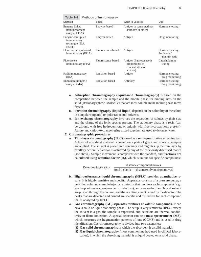
3. Immunoassay methodologies are based on the label attached to the antigen or antibody(Table 1–2).
C. Chromatography is a technique used to separate complex mixtures on the basis of differentphysical interactions between the individual compounds and the stationary phase of thesystem (a solid or a liquid - coated solid). The goal of this technique is to produce “fractions”for quantitation.
1. Mechanisms of separation are based on the interactions of solutes with mobile andstationary phases.
P1: OSO
LWBK192-01 LWBK192-Hubbard November 23, 2008 17:19
CHAPTER 1 Clinical Chemistry 9
Table 1–2 Methods of Immunoassay
Method Basis What is Labeled Use
Enzyme-linkedimmunosorbentassay (ELISA)
Enzyme-based Antigen in some methods;antibody in others
Hormone testing
Enzyme-multipliedimmunoassaytechnique (EIA,EMIT)
Enzyme-based Antigen Drug monitoring
Fluorescence-polarizedimmunoassay (FPIA)
Fluorescence-based Antigen Hormone testingSurfactant/
albumin ratio
Fluorescentimmunoassay (FIA)
Fluorescence-based Antigen (fluorescence isproportional toconcentration ofanalyte)
Catecholaminetesting
Radioimmunoassay(RIA)
Radiation-based Antigen Hormone testing;drug monitoring
Immunoradiometricassay (IRMA)
Radiation-based Antibody Hormone testing;drug monitoring
a. Adsorption chromatography (liquid-solid chromatography) is based on thecompetition between the sample and the mobile phase for binding sites on thesolid (stationary) phase. Molecules that are most soluble in the mobile phase movefastest.
b. Partition chromatography (liquid-liquid) depends on the solubility of the solutein nonpolar (organic) or polar (aqueous) solvents.
c. Ion-exchange chromatography involves the separation of solutes by their sizeand the charge of the ionic species present. The stationary phase is a resin (canbe cationic with free hydrogen ions or anionic with free hydroxyl ions present).Anion- and cation-exchange resins mixed together are used to deionize water.
2. Chromatographic proceduresa. Thin-layer chromatography (TLC) is used as a semi-quantitative screening test.
A layer of absorbent material is coated on a plate of glass, and spots of samplesare applied. The solvent is placed in a container and migrates up the thin layer bycapillary action. Separation is achieved by any of the previously discussed modes(see above). Sample movement is compared with the standard, and fractions arecalculated using retention factor (Rf), which is unique for specific compounds:
Retention factor (Rf) = distance component moves
total distance − distance solvent front moves
b. High-performance liquid chromatography (HPLC) provides quantitative re-sults. It is highly sensitive and specific. Apparatus consists of a pressure pump; agel-filled column; a sample injector; a detector that monitors each component (e.g.,spectrophotometers, amperometric detectors); and a recorder. Sample and solventare pushed through the column, and the resulting eluent is read by the detector. Thepeaks that are detected and printed are specific and distinctive for each compoundthat is analyzed by HPLC.
c. Gas chromatography (GC) separates mixtures of volatile compounds. It canhave a solid or liquid stationary phase. The setup is very similar to HPLC, exceptthe solvent is a gas, the sample is vaporized, and detectors are thermal conduc-tivity or flame ionization. A special detector can be a mass spectrometer (MS),which measures the fragmentation patterns of ions (GCMS) and is used in drugidentification. Gas chromatography is divided into two categories:(1) Gas-solid chromatography, in which the absorbent is a solid material;(2) Gas-liquid chromatography (most common method used in clinical labora-
tories), in which the absorbing material is a liquid coated on a solid phase.
P1: OSO
LWBK192-01 LWBK192-Hubbard November 23, 2008 17:19
10 A Concise Review of Clinical Laboratory Science
III. BASIC ANATOMY AND PHYSIOLOGY
A. Kidney1. Renal structure can be viewed both macroscopically and microscopically.
a. The macroscopic structure of the kidney consists of the cortex, medulla, andpelvis.
b. The microscopic structure of the kidney includes the nephron, which is consideredto be the functional unit of the kidney, and consists of the:(1) Glomerulus (made of arterioles surrounded by the distended end of a renal
tubule in the renal cortex)(2) Proximal tubules (located in the cortex)(3) Henle’s loop (descending and ascending limbs in the renal medulla)(4) Distal tubules (in the cortex)(5) Collecting tubules (collect urine from distal tubules to drain into the renal
pelvis)2. Renal physiology is based on the function of each microscopic component.
a. Glomerular function is to strain proteins from the plasma and produce a “protein-free” filtrate that becomes urine.(1) The glomerular filtration rate (GFR) equals 125 to 130 mL protein-free fluid
formed per minute.(2) Clearance indicates the number of milliliters of plasma from which the kidney
can remove all of a given substance in 1 minute. A request for “clearance” isa request for assessment of glomerular filtration rate.
(3) Plasma renal flow is the number of milliliters of plasma passing through thekidney in 1 minute; normal is 625 mL/min.
b. Tubular function is to resorb certain substances back into the body. The proxi-mal tubule resorbs 75% of water, sodium, much of glucose, amino acids, certainions, and small molecules. Some substances have a maximum concentration inplasma, so the tubule cannot resorb it all. Excess substance spills over into urine(e.g., glucose). The proximal tubule allows for the elimination of urea and creati-nine.
c. The Loop of Henle adjusts urine osmolality to keep the urine watery.d. The distal tubule resorbs some salt, water, and bicarbonate, but eliminates uric
acid, ammonia, and hydrogen ions. The distal tubule is under hormonal control.e. The collecting ducts are under hormonal control for resorption of water and
sodium.3. The renal system functions to maintain a balance of water, ions, and pH; to eliminate
nonprotein nitrogens; and to synthesize certain hormones.a. Water balance is maintained by ingestion of water (controlled by the brain thirst
center) and excretion/resorption of water in the renal tubules under hormonal con-trol by antidiuretic hormone (ADH).
b. Ionic balance of sodium, potassium, phosphate, calcium, and magnesium is main-tained by tubule resorption under hormonal control (aldosterone). Chloride is pas-sively resorbed with sodium.
c. Acid-base balance is controlled by kidney conservation of bicarbonate ions andremoval of metabolic acids (H+) to conserve blood pH level.
d. Nonprotein nitrogen (e.g., urea, creatinine, uric acid) is eliminated or filtered bythe glomerulus. Some urea and uric acid is reabsorbed into the blood.
e. The kidneys synthesize three hormones and one enzyme. Kidneys also serve asa site for the hormonal action of aldosterone and ADH.(1) Renin is a vasoconstrictor synthesized in the renal medulla.(2) Prostaglandins are synthesized in the kidney and affect renal blood flow.(3) Erythropoietin increases heme production and iron insertion into red blood
cells (RBCs) and is formed in conjunction with an enzyme made in thekidney.
(4) Dihydroxycholecalciferol hydrolase activates Vitamin D into a usable form.
P1: OSO
LWBK192-01 LWBK192-Hubbard November 23, 2008 17:19
CHAPTER 1 Clinical Chemistry 11
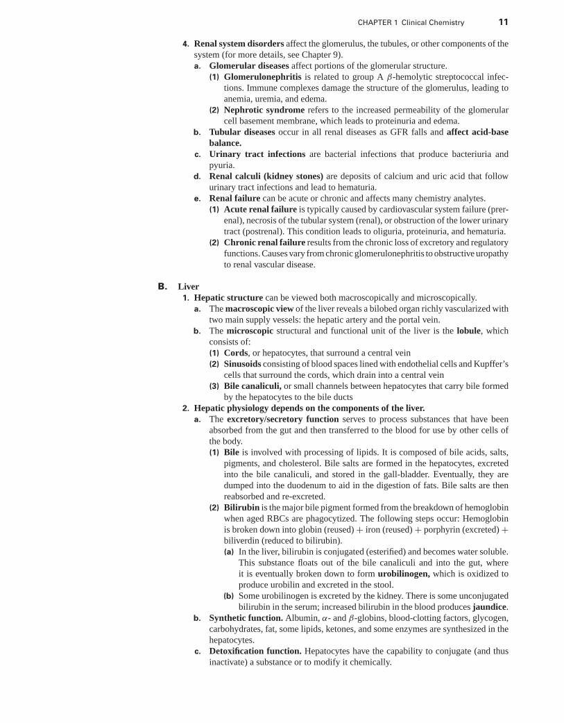
4. Renal system disorders affect the glomerulus, the tubules, or other components of thesystem (for more details, see Chapter 9).a. Glomerular diseases affect portions of the glomerular structure.
(1) Glomerulonephritis is related to group A β-hemolytic streptococcal infec-tions. Immune complexes damage the structure of the glomerulus, leading toanemia, uremia, and edema.
(2) Nephrotic syndrome refers to the increased permeability of the glomerularcell basement membrane, which leads to proteinuria and edema.
b. Tubular diseases occur in all renal diseases as GFR falls and affect acid-basebalance.
c. Urinary tract infections are bacterial infections that produce bacteriuria andpyuria.
d. Renal calculi (kidney stones) are deposits of calcium and uric acid that followurinary tract infections and lead to hematuria.
e. Renal failure can be acute or chronic and affects many chemistry analytes.(1) Acute renal failure is typically caused by cardiovascular system failure (prer-
enal), necrosis of the tubular system (renal), or obstruction of the lower urinarytract (postrenal). This condition leads to oliguria, proteinuria, and hematuria.
(2) Chronic renal failure results from the chronic loss of excretory and regulatoryfunctions. Causes vary from chronic glomerulonephritis to obstructive uropathyto renal vascular disease.
B. Liver1. Hepatic structure can be viewed both macroscopically and microscopically.
a. The macroscopic view of the liver reveals a bilobed organ richly vascularized withtwo main supply vessels: the hepatic artery and the portal vein.
b. The microscopic structural and functional unit of the liver is the lobule, whichconsists of:(1) Cords, or hepatocytes, that surround a central vein(2) Sinusoids consisting of blood spaces lined with endothelial cells and Kupffer’s
cells that surround the cords, which drain into a central vein(3) Bile canaliculi, or small channels between hepatocytes that carry bile formed
by the hepatocytes to the bile ducts2. Hepatic physiology depends on the components of the liver.
a. The excretory/secretory function serves to process substances that have beenabsorbed from the gut and then transferred to the blood for use by other cells ofthe body.(1) Bile is involved with processing of lipids. It is composed of bile acids, salts,
pigments, and cholesterol. Bile salts are formed in the hepatocytes, excretedinto the bile canaliculi, and stored in the gall-bladder. Eventually, they aredumped into the duodenum to aid in the digestion of fats. Bile salts are thenreabsorbed and re-excreted.
(2) Bilirubin is the major bile pigment formed from the breakdown of hemoglobinwhen aged RBCs are phagocytized. The following steps occur: Hemoglobinis broken down into globin (reused) + iron (reused) + porphyrin (excreted) +biliverdin (reduced to bilirubin).(a) In the liver, bilirubin is conjugated (esterified) and becomes water soluble.
This substance floats out of the bile canaliculi and into the gut, whereit is eventually broken down to form urobilinogen, which is oxidized toproduce urobilin and excreted in the stool.
(b) Some urobilinogen is excreted by the kidney. There is some unconjugatedbilirubin in the serum; increased bilirubin in the blood produces jaundice.
b. Synthetic function. Albumin, α- and β-globins, blood-clotting factors, glycogen,carbohydrates, fat, some lipids, ketones, and some enzymes are synthesized in thehepatocytes.
c. Detoxification function. Hepatocytes have the capability to conjugate (and thusinactivate) a substance or to modify it chemically.
P1: OSO
LWBK192-01 LWBK192-Hubbard November 23, 2008 17:19
12 A Concise Review of Clinical Laboratory Science
d. Storage function. Iron, glycogen, amino acids, and some lipids are stored in hep-atocytes.
3. Hepatic disordersa. Jaundice, which causes yellowish discoloration of skin, is caused by abnormal
bilirubin metabolism or by retention of bilirubin.(1) Prehepatic jaundice is the result of excessive bilirubin presented to the liver.
It can occur in newborns and in people with hemolytic anemia or ineffectiveerythropoiesis. This condition produces increased serum unconjugated biliru-bin.
(2) Hepatic jaundice is present in people with hepatobiliary disease. This disorderexhibits increases in both unconjugated and conjugated bilirubin levels.
(3) Posthepatic jaundice is produced by obstruction of the flow of bile into the guteither by gallstones or a tumor, which causes increased conjugated bilirubin lev-els in serum and urine, but low urobilinogen levels in urine and colorless stool.
b. Cirrhosis is defined as destruction of the liver’s architecture. The leading cause ofthis condition is alcohol abuse.
c. Reye’s syndrome is liver destruction caused by viral infection, although the etiol-ogy of this disease is unknown. Ammonia accumulates in the liver and blood.
d. Hepatitis is defined as inflammation of the liver and subsequent hepatocellulardamage caused by bacterial infection, drugs, toxins, or viral infections. Types ofviral hepatitis include:(1) Hepatitis A (“infectious” hepatitis), also known as hepatitis A virus (HAV), is
transmitted by contamination of food and water.(2) Hepatitis B (“serum” hepatitis), or hepatitis B virus (HBV), has an outer coat
called the HBV surface antigen (HBsAg) that covers the HBV core antigen(HBcAg). Hepatitis B is transmitted through parenteral injection or throughexchange of bodily secretions, as occurs during sexual intercourse.
(3) Hepatitis C (HCV) is a non-A, non-B hepatitis that is transmitted parenterallythrough blood transfusions, body piercings, and inoculations and has becomemore common. It is the leading cause of chronic liver disease.
(4) Delta hepatitis can cause infection only in patients infected with hepatitis B.
C. Gastrointestinal (GI) tract and pancreas. Anatomically, the GI tract is composed of fiveregions: the mouth, stomach, duodenum, jejunum-ileum, and large intestine.1. Gastric and GI functions are important to consider in the diagnosis of digestive disor-
ders.a. Digestion is the chemical processing of food into an absorbable substance. It begins
in the mouth and continues in the stomach and duodenum.(1) Gastric fluid in the stomach is composed of hydrochloric acid, pepsin, intrinsic
factor, and mucus. The pH of this fluid is <3. The secretion of gastrin by gastriccells stimulates gastric fluid secretion.
(2) Intrinsic factor, produced in the parietal cells of the stomach, is required forthe transport of vitamin B12 across the intestinal wall.
b. Absorption is the process that allows digested food to enter the body. This processoccurs in the small intestine.
2. GI function tests evaluate the level of function and determine the primary cause ofmalabsorption syndrome.a. Gastric fluid analysis serves to:
(1) Determine pH of gastric fluid, with low pH (achlorhydria) indicative of perni-cious anemia
(2) Detect hypersecretion of gastric fluid caused by a secreting tumor (e.g.,Zollinger-Ellison syndrome)
(3) Check acid secretion in treatment of ulcers(4) Verify vagotomy (i.e., severing nerves to stomach for treatment of ulcers)
b. Lactose intolerance test examines whether lactose is formed normally in gastriccells. The procedure involves ingestion of a lactose cocktail followed by glucoseanalysis. Little or no increase in serum glucose indicates lactase deficiency.
P1: OSO
LWBK192-01 LWBK192-Hubbard November 23, 2008 17:19
CHAPTER 1 Clinical Chemistry 13
D. The pancreas is a highly vascularized organ connected to the small intestine by the ampullaof Vater. It is considered both an endocrine gland that synthesizes hormones and an exocrinegland that provides digestive enzymes to aid in digestion.
1. Pancreatic functionsa. Endocrine function is performed in the islets of Langerhans. These cell groups
are composed of three types of cells.(1) α cells produce glucagon, which stimulates the conversion of glycogen into
glucose (glycogenolysis).(2) β cells are responsible for making insulin, which functions to promote glyco-
genesis and thereby lowers glucose levels.(3) δ cells produce gastrin and somatostatin.
b. Exocrine function is performed by the acinar cells. These cells produce the fol-lowing enzymes:(1) Amylase, which breaks down starch and glycogen and is used to diagnose
acute pancreatitis;(2) Lipase, which hydrolyzes fats to produce alcohols and fatty acids with elevated
levels present in people who have acute pancreatitis; and(3) Trypsin, which is a proteolytic enzyme (functions in protein breakdown).
2. Pancreatic disorders typically result in decreased secretion of enzymes or hor-mones.a. Cystic fibrosis is an autosomal recessive genetic disorder characterized by pul-
monary disease and intestinal malabsorption caused by lack of pancreatic enzymesecretion.
b. Pancreatitis (inflammation of the pancreas) is associated with alcohol abuse orgallbladder disease and also occurs in patients with lipid disorders and is causedby the release of pancreatic enzymes from cells into the surrounding pancreatictissue.
c. Diabetes mellitus is a multifactorial disease that occurs when the pancreas can nolonger produce insulin, which leads to hyperglycemia. This disorder almost alwaysdestroys the β cells in the islets. In type II diabetes mellitus, cells no longer aresensitive to insulin and glucose remains in the blood.
d. Pancreatic cancer is a fatal disease that affects the ducts in the pancreas. Insuli-noma is a tumor of the β cells in the islets that leads to increased circulating insulinand hypoglycemia.
3. Tests of exocrine pancreatic functiona. Secretin test determines the secretory capacity of the pancreas. It involves intuba-
tion and gathering of pancreatic fluid after stimulation with secretin, followed bymeasurement of fluid volume.
b. Quantitative fecal fat examination determines the presence of increased fats infeces (steatorrhea), which is a disorder almost always associated with exocrinepancreatic insufficiency. A 72-hour fecal specimen is collected, and the fats ex-tracted with ether and weighed. A screening procedure involves mixing a smallamount of fecal specimen with a fat-soluble stain and examining the specimenmicroscopically for lipid droplets.
c. Sweat electrolytes are measured to diagnose cystic fibrosis. Pilocarpine nitrateis used to stimulate sweating on skin which is collected on a small disc. Sweatis eluted from the disc and analyzed for chloride and sodium content. Newbornscreening programs and genetic tests that assess the presence of genetic alterationsin a number of genes related to cystic fibrosis are also available (see Chapter 10).
d. Enzyme testing for amylase and lipase is performed using a variety of method-ologies. These are listed below.
E. The cardiovascular system is composed of the heart and blood vessels. Some include thepulmonary system because of the extensive connections between the heart and lungs.
1. The heart is a four-chambered muscular organ. Blood passes first through the right, orpulmonary, side of the heart to be oxygenated in the lungs and then is returned to theleft, or systemic, side that boosts pressure for the circuit of blood around the body.
P1: OSO
LWBK192-01 LWBK192-Hubbard November 23, 2008 17:19
14 A Concise Review of Clinical Laboratory Science
2. The microscopic anatomy of the heart includes the myocardium that is made up ofcardiac muscle fibers.a. Myocardium is the muscular tissue of the heart. Myocardium is composed of
cardiac muscle fibers interspersed with blood vessels, lymphatics, and nerves. Thefuel of the heart muscle tissue is free fatty acid.
b. Cardiac muscle is found only in the heart. Cardiac muscle fibers synthesize specificproteins (troponin for example) that can be assessed in blood following muscle cellinjury. Myoglobin acts as the storage vessel for oxygen in muscle cells.
3. Cardiac dysfunction involves many parts of the heart and can begin in an area otherthan the heart itself. Heart failure takes many forms, such as congestive heart failure,coronary artery disease, and myocardial infarction (heart attack). Heart failure is basedthe functional anatomy of the heart. Failing hearts do not pump enough blood to sustainthe body with oxygen.
IV. ANALYTES AND PATHOPHYSIOLOGY
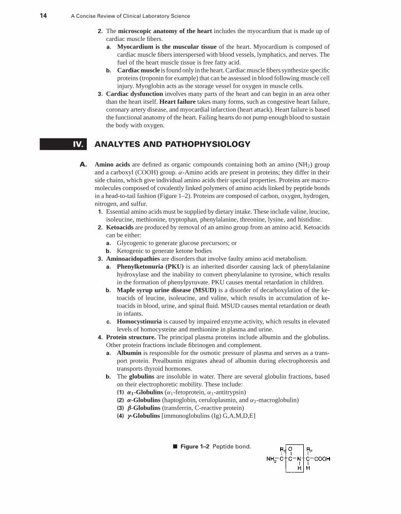
A. Amino acids are defined as organic compounds containing both an amino (NH2) groupand a carboxyl (COOH) group. α-Amino acids are present in proteins; they differ in theirside chains, which give individual amino acids their special properties. Proteins are macro-molecules composed of covalently linked polymers of amino acids linked by peptide bondsin a head-to-tail fashion (Figure 1–2). Proteins are composed of carbon, oxygen, hydrogen,nitrogen, and sulfur.
1. Essential amino acids must be supplied by dietary intake. These include valine, leucine,isoleucine, methionine, tryptophan, phenylalanine, threonine, lysine, and histidine.
2. Ketoacids are produced by removal of an amino group from an amino acid. Ketoacidscan be either:a. Glycogenic to generate glucose precursors; orb. Ketogenic to generate ketone bodies
3. Aminoacidopathies are disorders that involve faulty amino acid metabolism.a. Phenylketonuria (PKU) is an inherited disorder causing lack of phenylalanine
hydroxylase and the inability to convert phenylalanine to tyrosine, which resultsin the formation of phenylpyruvate. PKU causes mental retardation in children.
b. Maple syrup urine disease (MSUD) is a disorder of decarboxylation of the ke-toacids of leucine, isoleucine, and valine, which results in accumulation of ke-toacids in blood, urine, and spinal fluid. MSUD causes mental retardation or deathin infants.
c. Homocystinuria is caused by impaired enzyme activity, which results in elevatedlevels of homocysteine and methionine in plasma and urine.
4. Protein structure. The principal plasma proteins include albumin and the globulins.Other protein fractions include fibrinogen and complement.a. Albumin is responsible for the osmotic pressure of plasma and serves as a trans-
port protein. Prealbumin migrates ahead of albumin during electrophoresis andtransports thyroid hormones.
b. The globulins are insoluble in water. There are several globulin fractions, basedon their electrophoretic mobility. These include:(1) α1-Globulins (α1-fetoprotein, α1-antitrypsin)(2) α-Globulins (haptoglobin, ceruloplasmin, and α2-macroglobulin)(3) β-Globulins (transferrin, C-reactive protein)(4) γ-Globulins [immunoglobulins (Ig) G,A,M,D,E]
■ Figure 1–2 Peptide bond.
P1: OSO
LWBK192-01 LWBK192-Hubbard November 23, 2008 17:19
CHAPTER 1 Clinical Chemistry 15
5. Synthesis. Proteins are synthesized in the liver (serum proteins) or by B-cells (im-munoglobulins). Protein formation is specified by the DNA in each type of cell (hepaticor plasma).
6. Protein catabolism takes place in the gastrointestinal tract, kidneys, and liver. Proteindisintegrates into constituent amino acids, which are further deaminated into ketoacidsand ammonia. Ammonia is used in the formation of urea.
7. Classification. Proteins are classified as simple or conjugated.a. Simple proteins are peptide chains that hydrolyze to amino acids.b. Conjugated proteins are composed of protein (apoprotein) and a nonprotein sub-
stance (referred to as prosthetic groups), such as lipid (forms lipoproteins), carbo-hydrate (forms glycoproteins), or metals (form metalloproteins).
8. Functions of protein include tissue nutrition, water distribution, plasma buffer, sub-stance transport, and structural support.
9. Protein analysis determines either the total nitrogen content of the sample or the totalprotein. The normal reference range of serum protein is 6.5 to 8.3 g/dL.a. Total nitrogen determination analyzes both protein and nonprotein nitrogen.b. Kjeldahl’s method involves the conversion of protein nitrogen into ammonium
ion. This classic method is not practical for routine use.c. Refractometry methods use the refractive index of a solution in the determination
of solute concentration.(1) The principle states that the velocity of light changes when it passes between
air and water, causing light to bend (refract). Therefore, the refractive index ofwater increases proportionally to the concentration of a solute in solution.
(2) The solutes present in greatest concentration in serum are proteins. Therefore,this method provides an approximation useful as a rapid test, but error canoccur in patients with hyperglycemia, lipemia, or azotemia.
d. The biuret method is the most widely used method of protein determination.Analysis depends on the presence of peptide bonds.(1) The principle states that cupric (Cu2+) ions react with peptide bonds to form
a violet color proportional to the number of peptide bonds present.(2) This method is widely used and easily automated.
e. The dye-binding method is based on the ability of proteins to bind dyes. Albuminbinds dye with the strongest affinity.(1) The principle of this method is that dye binds to protonated amine groups of
amino acids, with absorption at 595 nm.(2) Dye-binding methods are used chiefly for albumin analysis. When albumin is
positively charged, it binds to certain dyes, causing a shift in absorbance fromfree dye. Brom-cresol green is the most widely used dye, although brom-cresolpurple is considered more specific in binding albumin.
f. UV absorption is based on absorption of UV light by peptide bonds at 210 and280 nm.
g. Serum protein electrophoresis (SPE) relies on the separation of proteins based ontheir net electrical charges, size, properties of the support medium, and temperatureof operation. This is a semi-quantitative method.(1) The principle of SPE states that when an electric field is applied to a medium
containing charged particles, the negatively charged particles migrate to-ward the positive electrode (anode), and the positively charged particles mi-grate toward the cathode. At pH 8.6, most serum proteins have a negativecharge. This separates the protein fractions in serum when an electrical field isapplied.
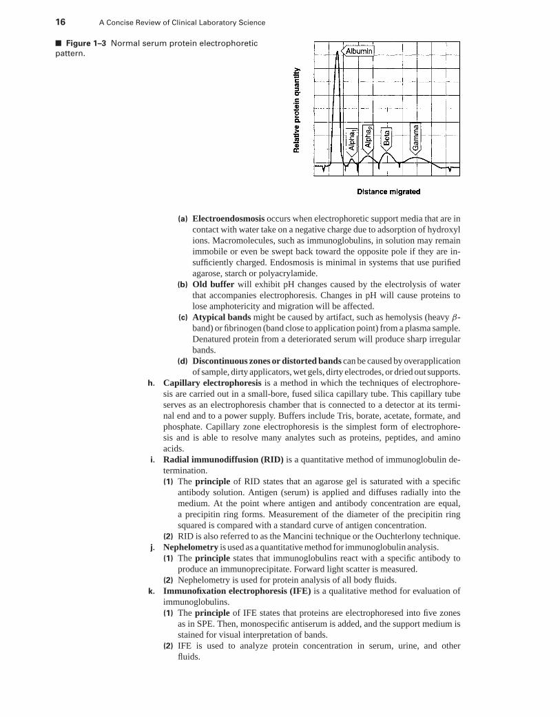
(2) On the support medium (cellulose acetate or agarose gel), the pattern of migra-tion is as follows: Albumin is most anodic (because of its small size and largenumber of negative charges), then α1-globulins, α2-globulins, β-globulins; λ-globulins are most cathodic (Figure 1–3).
(3) Technical issues with electrophoresis include endosmosis, inappropriatebuffer pH, unusual or atypical bands, distorted protein bands, and discontinuouszones.
P1: OSO
LWBK192-01 LWBK192-Hubbard November 23, 2008 17:19
16 A Concise Review of Clinical Laboratory Science
■ Figure 1–3 Normal serum protein electrophoretic
pattern.
(a) Electroendosmosis occurs when electrophoretic support media that are incontact with water take on a negative charge due to adsorption of hydroxylions. Macromolecules, such as immunoglobulins, in solution may remainimmobile or even be swept back toward the opposite pole if they are in-sufficiently charged. Endosmosis is minimal in systems that use purifiedagarose, starch or polyacrylamide.
(b) Old buffer will exhibit pH changes caused by the electrolysis of waterthat accompanies electrophoresis. Changes in pH will cause proteins tolose amphotericity and migration will be affected.
(c) Atypical bands might be caused by artifact, such as hemolysis (heavy β-band) or fibrinogen (band close to application point) from a plasma sample.Denatured protein from a deteriorated serum will produce sharp irregularbands.
(d) Discontinuous zones or distorted bands can be caused by overapplicationof sample, dirty applicators, wet gels, dirty electrodes, or dried out supports.
h. Capillary electrophoresis is a method in which the techniques of electrophore-sis are carried out in a small-bore, fused silica capillary tube. This capillary tubeserves as an electrophoresis chamber that is connected to a detector at its termi-nal end and to a power supply. Buffers include Tris, borate, acetate, formate, andphosphate. Capillary zone electrophoresis is the simplest form of electrophore-sis and is able to resolve many analytes such as proteins, peptides, and aminoacids.
i. Radial immunodiffusion (RID) is a quantitative method of immunoglobulin de-termination.(1) The principle of RID states that an agarose gel is saturated with a specific
antibody solution. Antigen (serum) is applied and diffuses radially into themedium. At the point where antigen and antibody concentration are equal,a precipitin ring forms. Measurement of the diameter of the precipitin ringsquared is compared with a standard curve of antigen concentration.
(2) RID is also referred to as the Mancini technique or the Ouchterlony technique.j. Nephelometry is used as a quantitative method for immunoglobulin analysis.
(1) The principle states that immunoglobulins react with a specific antibody toproduce an immunoprecipitate. Forward light scatter is measured.
(2) Nephelometry is used for protein analysis of all body fluids.k. Immunofixation electrophoresis (IFE) is a qualitative method for evaluation of
immunoglobulins.(1) The principle of IFE states that proteins are electrophoresed into five zones
as in SPE. Then, monospecific antiserum is added, and the support medium isstained for visual interpretation of bands.
(2) IFE is used to analyze protein concentration in serum, urine, and otherfluids.
P1: OSO
LWBK192-01 LWBK192-Hubbard November 23, 2008 17:19
CHAPTER 1 Clinical Chemistry 17
l. Immunoelectrophoresis (IEP) is a qualitative method for the evaluation of im-munoglobulins. Capillary electrophoresis can be used for IEP.(1) The principle of IEP states that proteins are electrophoresed into five zones
similar to SPE. Then, antibody is added to produce precipitin arcs that arevisually interpreted.
(2) IEP is used to analyze serum, cerebrospinal fluid (CSF), and urine.10. Other methods. Urinary proteins (for details, see Chapter 9) are analyzed by:
a. Qualitative dipstick tests for proteinuria, glycosuria, and other abnormal sub-stances in the urine that rely on color change for interpretation.
b. Quantitative turbidimetric methods require that a protein precipitant is added tourine. The resulting turbidity is measured photometrically.
11. Protein disorders are the result of high or low serum protein levels or a dysfunctionof the immunoglobulins.a. Hypoproteinemia can be caused by kidney disease, blood loss, malnutrition, and
liver disease.b. Hyperproteinemia is observed in people with dehydration or excess production
of γ -globulins.c. Decreased serum albumin is caused by a variety of disorders, including poor diet,
liver dysfunction, GI inflammation, and renal disease.d. Specific globulin disorders include the following:
(1) α1-Antitrypsin deficiency is caused by pulmonary disease, and increased lev-els are caused by inflammation or pregnancy.
(2) Elevated levels of α1-fetoprotein during pregnancy may indicate neural tubedefects, spina bifida in the fetus, or twins. Very high levels are found in indi-viduals with liver cancer. Decreased levels during pregnancy indicate a risk offetal Down’s syndrome.
(3) Haptoglobin, an α2-macroglobulin, is increased in inflammatory conditions,burns, and rheumatic disease. Decreased levels are seen in people with trans-fusion reactions or hemolytic disease.
(4) Ceruloplasmin, an α2-macroglobulin, is decreased in people with Wilson’sdisease and states of malnutrition.
(5) Transferrin levels are increased during iron deficiency anemia.(6) Immunoglobulin increases indicate infection, liver disease, Waldenstrom’s
macroglobulinemia (IgM), multiple myeloma, or autoimmune reactions. Theseincreases appear as a spike, either monoclonal or polyclonal, in the gammaregion of SPE.
B. Water and electrolyte balance. Electrolytes are ions capable of carrying an electric charge.They can be either anions (negatively charged) or cations (positively charged). Total bodywater (TBW) makes up about 60% of male body weight and 50% of female weight. Twothirds of TBW is in the intracellular compartment and the other third is extracellular fluid(ECF); 25% of the extracellular fluid is plasma.
1. Sodium is the most abundant extracellular cation. It contributes to the osmolality ofextracellular fluid and maintains the volume of ECF and cell size and shape. Sodiumis essential for transmitting nerve impulses.a. Regulation of sodium is performed by the renal system in two ways.
(1) The renin-ADH system. Low blood volume (as in cardiac failure) inducessecretion of renin, a vasoconstrictor, from the kidney, which raises blood pres-sure and causes production of ADH. In turn, fluid volume is increased by theretention of sodium.
(2) The renin-aldosterone system. Low blood volume induces secretion of renin,which induces production of aldosterone by the adrenal glands. In turn, kidneyreabsorption of sodium and retention of water increase.
b. Sodium disorders(1) Hyponatremia is low serum sodium caused by gastrointestinal loss, burns,
or renal problems. Dilutional hyponatremia is a relative decrease of sodiumcaused by excess body water, as in nephrotic syndrome or cirrhosis.
P1: OSO
LWBK192-01 LWBK192-Hubbard November 23, 2008 17:19
18 A Concise Review of Clinical Laboratory Science
(2) Hypernatremia is increased sodium caused by excess water loss, as in sweat-ing or diarrhea. Dehydration causes a relative increase in sodium.
2. Potassium is the major intracellular cation that regulates activity at the neuromuscularjunction, as well as cardiac muscle contraction and pH.a. Regulation of potassium occurs in two ways:
(1) Dietary intake, which controls the amount of potassium in the circulation(2) The renal system, which controls potassium by:
(a) Aldosterone induces potassium reabsorption and secretion by the renaltubules by exchanging it for sodium.
(b) A high concentration of hydrogen ions keeps potassium out of cellsand induces its renal retention. Low hydrogen ion concentration allowsmore potassium ions to enter cells, which lowers serum potassium. Lowhydrogen ion concentration also allows more potassium to be excreted bythe kidney.
b. Potassium disorders include:(1) Hypokalemia (low serum potassium), which is a result of decreased dietary
intake, gastrointestinal loss, or renal dysfunction, can produce irregular heart-beat.
(2) Hyperkalemia (high serum potassium) is rare. It occurs following excessivedietary intake, adrenal failure, blood transfusions, or crush injuries.
3. Chloride is the major extracellular anion that acts to maintain osmotic pressure, keepsthe body hydrated, and maintains electric neutrality via interaction with sodium orcarbon dioxide.a. Regulation of chloride depends on dietary intake, sodium concentration, and the
chloride shift, which moves chloride into RBCs as bicarbonate diffuses out toproduce electroneutrality.
b. Chloride disorders include:(1) Hypochloremia (low serum chloride) is caused by salt loss during renal dis-
ease, diabetic ketoacidosis, or prolonged vomiting.(2) Hyperchloremia (elevated serum chloride) is caused by dehydration, acute
renal failure, prolonged diarrhea with loss of sodium bicarbonate, and salicylateintoxication.
4. Bicarbonate is the second most abundant anion in the extracellular fluid. It is a majorcomponent of the blood buffering system, accounts for 90% of total blood carbondioxide, and maintains charge neutrality in the cell.a. Regulation of bicarbonate is achieved by the kidneys, which are responsible for
reabsorbing all bicarbonate as carbon dioxide.b. Bicarbonate (or total carbon dioxide) disorders include decreased levels ob-
served during metabolic acidosis, renal failure, or diarrhea, and elevated levels dueto carbon dioxide retention, as is observed during respiratory acidosis.
5. Primary methods of electrolyte determination include:a. Ion-selective electrodes that use a semipermeable membrane to develop a potential
between two different ion concentrationsb. Amperometric-coulometric titration for chloride determination, in which silver
ions are combined with chloride; when excess free silver ions are noted, elapsedtime is relative to the chloride concentration
c. Atomic absorption spectrophotometry6. Anion gap is the difference between unmeasured anions and unmeasured cations. The
normal reference range for the anion gap is 6 to 18 mmol or mEq/L. It is useful indetermining increases in unmeasured anions and is calculated as follows:
Anion gap = (Na+ + K+) − (Cl− + HCO−3)
C. Acid-base balance assures the maintenance of a constant blood pH (7.4) through physio-logic buffers, the respiratory system, and the renal system.
P1: OSO
LWBK192-01 LWBK192-Hubbard November 23, 2008 17:19
CHAPTER 1 Clinical Chemistry 19
1. Buffer systems protect the body against changes in hydrogen ion concentration. Threephysiologic buffers act to maintain a constant pH:a. The carbonic acid-bicarbonate systemb. Hemoglobinc. The phosphoric acid-phosphate system
2. The respiratory system acts to maintain acid-base balance. Oxygen is removed fromoxyhemoglobin in the tissues. This allows for the acceptance of hydrogen ions, formingcarboxyhemoglobin. In the lungs, carboxyhemoglobin recombines with bicarbonate toform carbonic acid, which breaks down to form carbon dioxide and water. The carbondioxide is then expired by respiration. Thus, ventilation affects the pH of the blood;this system is called the “respiratory” component of acid-base balance.
3. The renal system controls bicarbonate concentration. The overall reaction results inthe reabsorption of sodium and bicarbonate in the kidney tubules. These substancespick up excess hydrogen ions. This system is called the “nonrespiratory” or “metabolic”component of acid-base balance.
4. Acid-base disorders are considered in terms of the Henderson-Hasselbalch equation,which states acid-base relationships:
pH = pK + log(
cAcHA
)
where A is the proton acceptor (base) and HA is the proton donor (acid).5. Blood gas analyzers determine acid-base balance through the measurement of par-
tial pressure of oxygen, carbon dioxide, and pH. Analyzers use electrodes as sensingdevices, and bicarbonate and other parameters are calculated from the previously men-tioned measurements using the Henderson-Hasselbalch equation. Oxygen saturation iscalculated as well. Analyzers typically measure arterial blood gases (ABG).a. Specimen requirements for blood gas analysis include the following.
(1) Arterial blood is collected in a glass or plastic syringe. Capillary specimenscan also be used (blood must be “arterialized”).
(2) Lyophilized or liquid heparin is the preferred anticoagulant.(3) No air bubbles should exist in the sample because they lower the pCO2 value.(4) The specimen must be placed on ice and transported to the laboratory in 15
minutes at 4◦C and tested immediately. Otherwise, pH values decrease, andpCO2 values increase.
(5) Blood clots are unacceptable.6. Acid-base disorders are classified according to their cause. Compensation occurs when
pH becomes abnormal.a. Respiratory acidosis results from hypoventilation, which causes a decrease in
carbon dioxide elimination. Compensation occurs by the kidneys increasing theresorption of bicarbonate.
b. Respiratory alkalosis results from an increase in ventilation, resulting in excessiveelimination of carbon dioxide. Compensation occurs by the kidneys excreting morebicarbonate.
c. Nonrespiratory (metabolic) acidosis occurs in many disorders and results in adecrease in bicarbonate levels. The lungs compensate by hyperventilating.
d. Nonrespiratory (metabolic) alkalosis is produced in many disorders and resultsin an increase in bicarbonate levels. The lungs compensate by hypoventilation.
7. The clinical significance of blood gas levels is as follows.a. pCO2 levels increase with administration of 100% oxygen or following exercise.
Decreases (hypoxemia) indicate pulmonary difficulties, exposure to carbon monox-ide, or improper anesthesia. Hypoxemia also may occur at high altitudes.
b. pCO2 levels increase with respiratory acidosis. Decreased levels indicate respira-tion that is too rapid.
D. Nonprotein nitrogen measurements monitor and assess renal function. These substancesarise from the breakdown of proteins and nucleic acids.
P1: OSO
LWBK192-01 LWBK192-Hubbard November 23, 2008 17:19
20 A Concise Review of Clinical Laboratory Science
1. Urea is the major excretory product of protein metabolism and is synthesized in theliver from carbon dioxide and ammonia arising from the deamination of amino acids.It is excreted by the kidney.a. All analytic methods include, in the initial step, the hydrolysis of urea by urease
and subsequent production of ammonium. Most techniques are sensitive to excessammonia contamination.(1) The coupled enzymatic assay is a kinetic assay that involves a second enzyme.(2) A pH indicator dye detects the presence of ammonium and causes a color
change, which is read by a spectrophotometer.(3) The direct method does not use urease. It measures urea directly using diacetyl
monoxime.b. Urea disorders typically involve an elevated level of urea in blood, which is
referred to as azotemia. Disorders are named in association with the location of thedysfunction.(1) Prerenal azotemia is typically caused by decreased renal blood flow to
the kidneys from congestive heart failure, shock, dehydration, decrease inblood volume, greater protein breakdown (as in major illness), or high-proteindiet.
(2) Renal azotemia is produced by renal failure.(3) Postrenal azotemia is caused by an obstruction anywhere in the renal system
(e.g., tubules, ureter).(4) Uremia is a toxic condition involving a very high serum level of urea accom-
panied by renal failure.2. Creatine is made in the liver from amino acids and used in muscle as an energy source.
In its anhydrous state, it is called creatinine, which is excreted into the plasma in anamount proportional to muscle mass and then excreted in the urine. Because the levelof creatinine is unaffected by diet, its level reflects the GFR.a. Analytic methods for creatinine
(1) The Jaffe reaction involves the reaction of creatinine with picric acid to forma reddish chromogen. The absorbance is measured colorimetrically.
(2) The kinetic reaction uses various enzymes and hydrogen peroxide to form acolored product.
(3) Creatinine clearance is an estimate of the GFR obtained by measuring plasmacreatinine and its rate of excretion into urine. This test requires a 24-hour urinespecimen and blood sample for serum creatinine determination. The formulafor calculation is:
UVP
× 1.73A
with urinary creatinine as U (mg/L), V as volume of urine (mL/min), P asplasma creatinine (mg/L), and 1.73
Aas normalization factor for body surface
area.b. Abnormal creatinine levels are typically elevated because of abnormal renal func-
tion, such as reduced GFR. Creatinine levels are examined in conjunction with ureato determine the cause of azotemia. The normal blood urea nitrogen (BUN): cre-atinine ratio is 10 to 20:1. Higher ratios indicate that the elevation of BUN iscaused by prerenal rather than renal causes.
3. Uric acid is synthesized in the liver from the breakdown of nucleic acids (DNA andRNA) and transported to the kidney for resorption.a. Analytic methods are based on the same initial reaction involving the oxidation
of uric acid by uricase to allantoin and hydrogen peroxide. There are two coupledenzymatic methods that involve either:(1) The measurement of peroxide production following reaction with phenol and
4-amino phenazone (4-AP)(2) The measurement of peroxide production by the catalyzed oxidation of ethanol
coupled to the production of acetate
P1: OSO
LWBK192-01 LWBK192-Hubbard November 23, 2008 17:19
CHAPTER 1 Clinical Chemistry 21
b. Abnormal uric acid levels are either:(1) Elevated because of gout, increased nuclear breakdown (due to increased cell
destruction, as in chemotherapy), renal disease, or toxemia of pregnancy(2) Decreased primarily because of severe liver or kidney disease
4. Ammonia is formed by the deamination of amino acids. It is used by the liver toproduce urea and is not excreted by the kidney.a. Analytic methods include:
(1) The coupled enzymatic method, in which ammonium reacts with α-ketoglutarate and reduced nicotinamide adenine dinucleotide phosphate(NADPH) to form L-glutamate and nicotinamide adenine dinucleotide phos-phate (NADP), with changes in absorbance measured spectrophotometrically
(2) The ammonia electrode, which is based on diffusion of NH3 through a selec-tive membrane
b. Increased ammonia levels are typically caused by severe liver dysfunction (e.g.,liver failure, Reye’s syndrome) or inadequate blood circulation through the liver.
E. Carbohydrates are polyhydroxyl aldehydes or ketones that, on hydrolysis, yield one ofthese compounds. Carbohydrates are a major source of energy for the body, and starch isthe major source of carbohydrate.
1. Classification is based on the structure of carbohydrates.a. Monosaccharides are simple sugars that contain four to eight carbons and only
one aldehyde or ketone group. These are reducing sugars (i.e., they can give upelectrons). Examples include glucose and fructose.
b. Oligosaccharides are formed by the interaction of two monosaccharides with theloss of a water molecule and are sometimes referred to as disaccharides. Examplesinclude maltose, lactose, and sucrose.
c. Polysaccharides are formed by interactions between many units of simple sugars.Examples are starch and glycogen.
2. Carbohydrate metabolism begins in the mouth.a. Salivary amylase breaks down ingested starches into disaccharides, and these are
further broken down into monosaccharides by disaccharides and absorbed intointestinal cells.
b. Monosaccharides are then transported to the liver and converted to glucose. Someglucose is released into the blood, and the rest is stored as glycogen in the liver andskeletal muscle.
c. Glycogenesis is the process of glycogen formation by enzyme action on glucoseto eventually form glycogen.
d. Glycogenolysis is the breakdown of glycogen, with the eventual formation ofglucose-6-phosphate or free glucose that can be used for energy production.
e. Glycolysis is the catabolism of glucose to pyruvate or lactate for adenosine triphos-phate (ATP) production (Embden-Meyerhof pathway and Krebs’ cycle).
f. Gluconeogenesis is the formation of glucose from amino acids and lipids thatoccurs when carbohydrate intake decreases.
3. Factors that affect glucose levels include:a. Insulin, which is a pancreatic hormone that decreases glucose levels by increasing
cellular uptake of glucose and promoting glycogenesis and lipogenesis (formationof fat from carbohydrates)
b. Glucagon, which is a pancreatic hormone that increases glucose levels by stimu-lating glycogenolysis and gluconeogenesis
c. Epinephrine, which is an adrenal hormone that elevates glucose levelsd. Growth hormone and adrenocorticotropic hormone (ACTH), which are pitu-
itary hormones that increase glucose levelse. Glucocorticoids (e.g., cortisol), which are adrenal hormones that increase gluco-
neogenesis and eventually elevate blood glucosef. Thyroid hormones, which stimulate glycogenolysis and increase blood glucose
levels
P1: OSO
LWBK192-01 LWBK192-Hubbard November 23, 2008 17:19
22 A Concise Review of Clinical Laboratory Science
4. Glucose disorders depend on serum glucose levels.a. Hyperglycemia occurs when the fasting blood sugar level rises higher than
110 mg/dL due to a pathologic disorder, such as diabetes mellitus or liver fail-ure.
b. Hypoglycemia occurs when the fasting blood glucose level is <70 mg/dL. Thistypically occurs as a result of hormone deficiency, drug reaction, insulin excess (asin insulinoma), or a genetic disorder.
c. Glycosuria (sugar in the urine) occurs when the renal threshold for glucose isexceeded (160–180 mg/dL) during hyperglycemia.
d. Diabetes mellitus is a genetic disorder of glucose metabolism that results in insulindeficiency and lack of carbohydrate tolerance. There are two classifications:(1) Type 1, formerly insulin-dependent diabetes mellitus. The individual
presents in an acute state with hyperglycemia and ketosis. This type is causedby an autoimmune destruction of pancreatic β cells and is usually juvenileonset. However, onset is often around the age of 20.
(2) Type 2, formerly non-insulin-dependent diabetes mellitus. This is amultifactorial disease with possible causes including genetics, environment(lifestyle), and autoimmunity. One cause of onset is thought to be cellularresistance to insulin or decreased insulin receptors on cell surfaces. Most indi-viduals with this disorder are obese or overweight, and onset is typically duringadulthood.
5. Methods of glucose analysis include:a. Glucose oxidase methods. Glucose is oxidized to gluconic acid and hydrogen
peroxide to eventually form a colored product. Falsely low results are caused byhigh serum levels of uric acid, bilirubin, or ascorbic acid.
b. Hexokinase methods. Glucose becomes phosphorylated and dehydrogenated toeventually form NADPH.
c. o-Toluidine (nonenzymatic method). o-Toluidine reacts with glucose in aceticacid to form a colored product. Falsely elevated glucose values are obtained byinterference of mannose and galactose, whereas bilirubin induces a false decreasein glucose values.
d. Glycated hemoglobin methods. The presence of glycated hemoglobins is ex-amined in diabetic patients. This test examines a patient’s compliance with aninsulin therapy regimen over a period of 8 to 10 weeks. Glycated hemoglobinsare formed when a glucose is attached to the N-terminal valine residue of either β
chain of the hemoglobin A molecule. Hemoglobin A1 (made up of several minorhemoglobins) is termed a fast hemoglobin based on its electrophoretic migration.Glycated hemoglobin is separated from nonglycated hemoglobin by affinity chro-matography. Interferences include the presence of hemoglobin S and hemoglobinF and may produce increased or decreased glycated hemoglobin values dependingon the specific methodology used.
6. Glucose metabolism tests examine a patient’s ability to metabolize glucose.a. Glucose tolerance test (GTT) evaluates the insulin response challenge. It is useful
in evaluating pregnancy-induced diabetes and involves drawing a fasting bloodspecimen, followed by patient ingestion of a 75-g oral dose of glucose in liquidwithin a 5-minute period. Blood samples are then taken at 30-, 60-, 120-, and180-minute intervals and tested for glucose. Urine is tested as well.(1) Nondiabetics have negative urine tests and show highest glucose levels at 30
to 60 minutes, with low and normal levels following.(2) Severe diabetics reach peak glucose levels after 30 to 60 minutes, and the
levels remain elevated.b. A 2-hour postprandial blood glucose test evaluates diabetes. A fasting blood
specimen and a specimen taken 2 hours after breakfast are taken. Normal patientshave no increase in serum glucose after 2 hours.
F. Lipids are substances that are insoluble in water and can be extracted from cells only by or-ganic solvents (e.g., ether or chloroform). Lipids include fats (most abundant), steroids,
P1: OSO
LWBK192-01 LWBK192-Hubbard November 23, 2008 17:19
CHAPTER 1 Clinical Chemistry 23
and terpenes. Fats are carboxylic esters derived from glycerol and are also known asglycerides.
1. Lipid composition of fooda. Triglycerides comprise 98% of fat found in food and are made up of 95% fatty acid
and 5% glycerol. Fatty acids are long carbon chains joined by single (saturated) ordouble bonds (unsaturated) and a terminal carboxyl group.
b. The remaining 2% of fat in food is composed of cholesterol, phospholipids, diglyc-erides, fat-soluble vitamins, steroids, and terpenes.
2. The physiology of lipids involves three phases.a. Digestive phase begins with chewing and swallowing. Triglycerides are digested
by lipase, other enzymes, bile salts, and acid in the gut to form monoglycerides anddiglycerides. Cholesterol becomes surrounded by bile to form a micelle packagethat is absorbed by the small intestine.
b. Absorptive phase occurs in the small intestine as triglycerides and cholesterol inthe micelles are absorbed and broken down into fatty acids.
c. Transport phase occurs as long fatty acids reassemble into chylomicrons (water-soluble macromolecules) and enter the lymphatic system. Short fatty acids enterthe blood bound to albumin, and these head to all tissues, including adipose tissue.
3. Specific lipid physiology determines the function of each lipid.a. Cholesterol is a sterol (steroid with long side chains), which is a four-ringed
structure made in liver hepatocytes from two acetate units. The process is long, and3-hydroxy-3-methylglutaryl coenzyme A (HMG-CoA) reductase is the committedstep. Cholesterol is an important constituent of cell membranes and a precursorof many hormones. Most serum cholesterol is in the form of cholesterol esters,which are transported through the blood by low-density lipoproteins (LDL)and high-density lipoproteins (HDL).
b. Triglyceride is made up of fatty acids and glycerol and is partly synthesized inthe liver hepatocyte. It is transported through the bloodstream by chylomicronsand very low-density lipoproteins (VLDLs). Triglyceride provides energy to cellsas it loses its fatty acid and forms ATP, thus acting as an energy store in the formof fat, and it insulates organs through fat deposits.
c. Phospholipids make up the bilayer of cell membranes and also form a coating thatsurrounds cholesterol and triglyceride and “glues” them to the lipoprotein core.
d. Sphingolipids are important in cell membrane composition and in nerve transmis-sion.
4. Lipoproteins are transport vehicles for lipids that contain varying amounts of specificlipid, phospholipid, and apoprotein.a. Chylomicrons are large molecules that contain mostly triglyceride. They originate
in the intestinal tract and travel through the blood and lymph to various tissues.They are degraded in the liver.
b. VLDLs are smaller than chylomicrons. They contain mostly endogenous triglyc-eride, are made in the liver, contain equal amounts of phospholipids and cholesterol,and degrade to LDLs in the circulation.
c. LDLs contain mostly cholesterol, with equal amounts of phospholipid and proteinand some triglyceride. They are taken into cells via a special cell-surface receptor—the apoprotein B (apoB) receptor—and are degraded into component parts. This isconsidered “bad” cholesterol.
d. HDLs contain mostly protein, some cholesterol, and a little triglyceride. They aremade in the liver, and they remove excess cholesterol from cells. HDL is consideredthe “good” lipoprotein.
5. Lipid disorders usually lead to abnormal lipid deposits on walls of vasculature(atherosclerosis) and skin (xanthomas). Hyperlipidemia obstruction leads to lack ofbile, so cholesterol cannot be adequately absorbed by the small intestine.a. Hyperlipidemia
(1) Triglyceride levels are most affected by diet, but high triglyceride levels areoften caused by diabetes or pancreatitis. Lipoprotein lipase (LPL), presentin the capillary wall, hydrolyzes triglycerides for use in the tissues and can
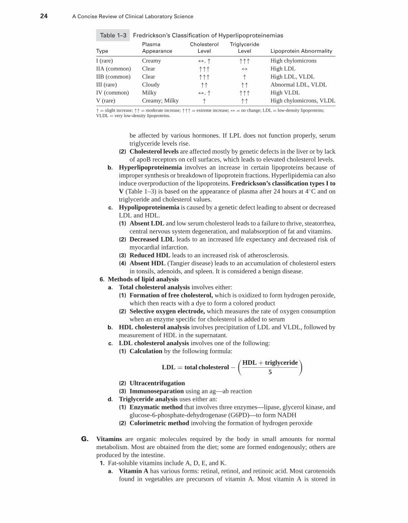
P1: OSO
LWBK192-01 LWBK192-Hubbard November 23, 2008 17:19
24 A Concise Review of Clinical Laboratory Science
Table 1–3 Fredrickson’s Classification of Hyperlipoproteinemias
Plasma Cholesterol Triglyceride
Type Appearance Level Level Lipoprotein Abnormality
I (rare) Creamy ↔, ↑ ↑↑↑ High chylomicrons
IIA (common) Clear ↑↑↑ ↔ High LDL
IIB (common) Clear ↑↑↑ ↑ High LDL, VLDL
III (rare) Cloudy ↑↑ ↑↑ Abnormal LDL, VLDL
IV (common) Milky ↔, ↑ ↑↑↑ High VLDL
V (rare) Creamy; Milky ↑ ↑↑ High chylomicrons, VLDL
↑ = slight increase; ↑↑ = moderate increase; ↑↑↑ = extreme increase; ↔ = no change; LDL = low-density lipoproteins;VLDL = very low-density lipoproteins.
be affected by various hormones. If LPL does not function properly, serumtriglyceride levels rise.
(2) Cholesterol levels are affected mostly by genetic defects in the liver or by lackof apoB receptors on cell surfaces, which leads to elevated cholesterol levels.
b. Hyperlipoproteinemia involves an increase in certain lipoproteins because ofimproper synthesis or breakdown of lipoprotein fractions. Hyperlipidemia can alsoinduce overproduction of the lipoproteins. Fredrickson’s classification types I toV (Table 1–3) is based on the appearance of plasma after 24 hours at 4◦C and ontriglyceride and cholesterol values.
c. Hypolipoproteinemia is caused by a genetic defect leading to absent or decreasedLDL and HDL.(1) Absent LDL and low serum cholesterol leads to a failure to thrive, steatorrhea,
central nervous system degeneration, and malabsorption of fat and vitamins.(2) Decreased LDL leads to an increased life expectancy and decreased risk of
myocardial infarction.(3) Reduced HDL leads to an increased risk of atherosclerosis.(4) Absent HDL (Tangier disease) leads to an accumulation of cholesterol esters
in tonsils, adenoids, and spleen. It is considered a benign disease.6. Methods of lipid analysis
a. Total cholesterol analysis involves either:(1) Formation of free cholesterol, which is oxidized to form hydrogen peroxide,
which then reacts with a dye to form a colored product(2) Selective oxygen electrode, which measures the rate of oxygen consumption
when an enzyme specific for cholesterol is added to serumb. HDL cholesterol analysis involves precipitation of LDL and VLDL, followed by
measurement of HDL in the supernatant.c. LDL cholesterol analysis involves one of the following:
(1) Calculation by the following formula:
LDL = total cholesterol −(
HDL + triglyceride5
)
(2) Ultracentrifugation(3) Immunoseparation using an ag—ab reaction
d. Triglyceride analysis uses either an:(1) Enzymatic method that involves three enzymes—lipase, glycerol kinase, and
glucose-6-phosphate-dehydrogenase (G6PD)—to form NADH(2) Colorimetric method involving the formation of hydrogen peroxide
G. Vitamins are organic molecules required by the body in small amounts for normalmetabolism. Most are obtained from the diet; some are formed endogenously; others areproduced by the intestine.1. Fat-soluble vitamins include A, D, E, and K.
a. Vitamin A has various forms: retinal, retinol, and retinoic acid. Most carotenoidsfound in vegetables are precursors of vitamin A. Most vitamin A is stored in
P1: OSO
LWBK192-01 LWBK192-Hubbard November 23, 2008 17:19
CHAPTER 1 Clinical Chemistry 25
the liver, and some is transferred to cells to promote mRNA synthesis. The bestunderstood physiology of vitamin A is in the visual system; a lack of vitamin Aleads to night blindness.
b. Vitamin D is a sterol derivative known as cholecalciferol. It is produced in the skinby absorption of UV light or obtained by dietary intake and stored in adipose tissue.Vitamin D is essential for bone mineralization and neuromuscular activity, andsome forms act to regulate uptake of calcium and phosphate. A lack of vitaminD leads to rickets (failure of bones to calcify) or osteomalacia (abnormal bonesynthesis).
c. Vitamin E (α-tocopherol) is available only from dietary intake and accumulatesin the liver, adipose tissue, and muscle. Vitamin E is critical for normal neuro-logic structure and function, and also functions as an antioxidant by preventingformation of free radicals. A deficiency of vitamin E produces a normocytic nor-mochromic anemia.
d. Vitamin K is supplied partly by diet and partly by flora in the bowel. Vitamin K isknown as phylloquinone and is essential in the formation of coagulation factors,particularly prothrombin (see Chapter 2, III D).
2. Water-soluble vitamins include vitamin C and the vitamin B complex.a. Vitamin C (ascorbic acid) is absorbed in the stomach and distributed to all tissues.
It is a potent reducing agent, functions in the synthesis of collagen, and aids inthe biosynthesis of some neurotransmitters. It is considered important in reducingthe risk of certain cancers and the common cold.
b. The B complex vitamins are grouped together because of their isolation from thesame sources. Most B vitamins are known to serve as enzyme cofactors, function-ing to transport atoms between molecules in enzyme-coupled reactions. Deficiencyof any B vitamin affects the body’s metabolism. The B vitamins include thiamine,riboflavin, niacin, pantothenic acid, biotin, pyridoxine (B6), the folates, andcyanocobalamin (B12). Lack of vitamin B12 produces pernicious anemia becauseof its function in erythropoiesis. Deficient folate leads to a megaloblastic anemia,and in pregnant women, the possibility of fetal neural tube defects.
H. Tumor markers are substances that are synthesized and released by cancer cells or madeby other tissues in response to the presence of cancer cells. These markers may be presentin the blood, in other fluids, on cells, or within cells.1. Clinically relevant (diagnostically accessible) tumor markers must have high disease
sensitivity and specificity, and levels should reflect the status of the disease process.These analytes are typically examined by some form of immunoassay.a. Oncofetal antigens exist as normal proteins in the embryo and fetus and are also
found in certain tumors.(1) Carcinoembryonic antigen (CEA) is an oncofetal antigen used to assess
tumors of the colon. It is not as specific as once thought, and not all coloncancers have elevated CEA levels.
(2) α-Fetoprotein (AFP) is an oncofetal antigen used to determine the presenceof hepatic tumors (hepatoma) and testicular tumors.
(3) Carbohydrate-associated antigen (CA-125) is a glycoprotein oncofetal anti-gen that appears in the serum of patients with ovarian cancer.
(4) CA-19 is another glycoprotein oncofetal antigen associated with gastrointesti-nal tumors.
(5) Prostate-specific antigen (PSA) is an oncofetal antigen that is important inscreening and monitoring patients with prostate carcinoma.
(6) Bladder tumor-associated antigens include fibrin degradation products andcomplement-related proteins.
b. Placental proteins are synthesized by placental trophoblasts and certain tumorsof the trophoblast.(1) Chorionic gonadotropin (CG) is typically used to determine pregnancy; how-
ever, high levels can indicate tumors of the testes or ovaries, as well as tro-phoblastic neoplasia.
P1: OSO
LWBK192-01 LWBK192-Hubbard November 23, 2008 17:19
26 A Concise Review of Clinical Laboratory Science
(2) Human placental lactogen (hPL) is used to monitor fetal well-being, but itslevels also can be elevated in patients with trophoblastic neoplasms.
c. Certain enzymes elevations can indicate the presence of cancer cells such as al-kaline phosphatase for bone cancer.
d. Increased levels of some hormones indicate malignancy of the organ that pro-duces the hormone. For example, extremely elevated growth hormone levels mightindicate a pituitary tumor.
2. DNA analysis determines the proliferative capacity of tumor cells including thoseinvolved in leukemia. Molecular diagnosis is used to identify oncogenes, the genesthat transform normal cells into malignant cells. This type of testing is becoming morewidespread with the advent of simplified molecular techniques. See Chapter 10 fordetails.
I. Porphyrin is a chemical intermediate in the synthesis of hemoglobin, myoglobin, andother respiratory pigments called cytochromes. Porphyrin measurement is important forthe determination of porphyria (a disturbance of heme synthesis). Heme synthesis beginsin the mitochondria.
1. Porphyrin chemistry involves the following principles.a. Synthesis. Porphyrin is synthesized from porphin (four pyrrole rings), but side
chains are substituted for the eight hydrogen atoms. A variety of substances makeup the side chains (magnesium makes up part of the side chains in chlorophyll,which is a porphyrin).(1) The main sites of porphyrin synthesis are bone marrow cells and liver cells.(2) The rate of synthesis is controlled through regulation of the enzyme
δ-aminolevulinic acid (ALA) synthase by the hemoproteins in theliver.
b. Clinical importance(1) Three porphyrin compounds are clinically important: protoporphyrin (ex-
creted in feces), uroporphyrin (excreted in urine), and coproporphyrin (ex-creted in both). All are intermediate products in the synthesis of heme that canbe assessed clinically.
(2) Porphobilinogen (PBG) and ALA are precursors of porphyrin and can accu-mulate in certain porphyrin disorders. These substances are typically found inthe urine of patients with acute porphyria.
(3) Free erythrocyte porphyrins (FEP) are porphyrins that can be extracted fromRBCs, the primary one being protoporphyrin. FEP concentration is increasedin persons with lead poisoning and iron deficiency anemia.
2. Porphyrias are inherited or acquired disorders of heme synthesis in which overproduc-tion of heme precursors in the bone marrow or the liver cause characteristic symptoms.The porphyrias are classified based on the signs and symptoms manifested by the patient(neurologic versus cutaneous). The primary cause of porphyrias is a specific enzymedeficiency.a. The neurologic porphyrias include the symptoms of abdominal pain, psychotic
behavior, and neuromuscular difficulties. Clinically, the three neurologic porphyr-ias have in common increased urinary ALA and porphobilinogen levels.(1) Acute intermittent porphyria (most common) has a clinical presentation of
increased uroporphyrin levels.(2) Variegate porphyria (rare) has a presentation of increased protoporphyrin and
coproporphyrin levels.(3) Coproporphyria (rare) has a presentation of increased coproporphyrin levels.
b. The cutaneous porphyrias are induced by the presence of excess porphyrins in theskin, which generate oxygen radicals that attack cells and produce photosensitivityand skin lesions. The three cutaneous porphyrias have in common normal urinaryALA and porphobilinogen levels.(1) Congenital erythropoietic porphyria (the rarest of the inherited porphyrin
disorders) is a severe disorder that has a presentation of increased uroporphyrinand coproporphyrin levels.
P1: OSO
LWBK192-01 LWBK192-Hubbard November 23, 2008 17:19
CHAPTER 1 Clinical Chemistry 27
(2) Protoporphyria (somewhat rare) has a presentation of increased protopor-phyrin and increased free erythrocyte protoporphyrin levels.
(3) Porphyria cutanea tarda (most common) appears in adults following liverdisease or excessive alcohol intake. It has a clinical presentation of increasedlevels of uroporphyrin.
c. Porphyrinuria is a moderate elevation of urine coproporphyrin secondary to anumber of disorders, including pregnancy, neoplasia, intoxication, and liver dis-ease.
d. Porphyrinemia is a moderate elevation in erythrocyte protoporphyrin secondaryto a number of disorders, including:(1) Iron deficiency states, caused by poor nutrition, malabsorption, poor iron
transport, or blood loss(2) Anemia (hemolytic, iron-deficiency, sideroblastic)(3) Lead poisoning
3. Methods of porphyrin analysis include qualitative and quantitative procedures. Spec-imen collection techniques are important. All samples must be protected from light;urinary porphyrin concentrations can decrease by up to 50% if kept in the light for24 hours. Urinary porphyrins and PBG are best analyzed in fresh, early morning(10–20 mL) urine specimens collected without preservative.a. Qualitative tests include those for:
(1) Urine porphobilinogen, which is a screening test whereby normal results arenegative (e.g., the Watson-Schwartz test, the Hoesch test)
(2) Urine porphyrins, which is a screening test performed to measure uropor-phyrin and coproporphyrin
b. Quantitative tests include a variety of procedures:(1) Urine tests for presence of porphobilinogen, uroporphyrin, coproporphyrin,
and ALA(2) Blood tests include analysis of ALA dehydratase (an enzyme in the blood that
breaks down ALA) and whole blood or erythrocyte protoporphyrin.(3) Fecal tests for coproporphyrin and protoporphyrin
J. Iron is contained in the porphyrin ring of heme in hemoglobin, myoglobin, catalase, andcytochromes. The reversible interaction of iron with oxygen and the ability of iron tofunction in electron transfer reactions make iron physiologically important. Bilirubin is anorange-yellow pigment derived from aged RBC and formed in the spleen. It is transportedto and transformed in the liver and is excreted in bile and urine.
1. Iron metabolism is controlled by its absorption in the intestine. Most circulating ironis derived from the release of iron following RBC destruction.a. Absorption of iron occurs mainly in the duodenum and includes only 5% to
10% of daily iron intake. The body’s iron supply is controlled by intestinal absor-ption.
b. Iron transport involves a transport protein, transferrin, which binds ferric iron.The bound iron either moves to the mitochondria for heme synthesis or is storedin cells as ferritin.
c. Iron storage includes storage as soluble ferritin (found in most cells) or relativelyinsoluble hemosiderin. One third of iron is stored in the liver, one third in the bonemarrow, and one third in the spleen.
2. Iron disease states include:a. Iron deficiency anemia, which is usually caused by blood loss (menstrual cycle,
ulcer, tumor)b. Iron overload, including:
(1) Hemosiderosis (no tissue injury)(2) Hemochromatosis
(a) Hereditary hemochromatosis is an autosomal recessive disease in whichiron is deposited directly in the liver, heart, and kidney, leading to organfailure.
(b) Sideroblastic anemia is caused by iron overload of unknown cause.
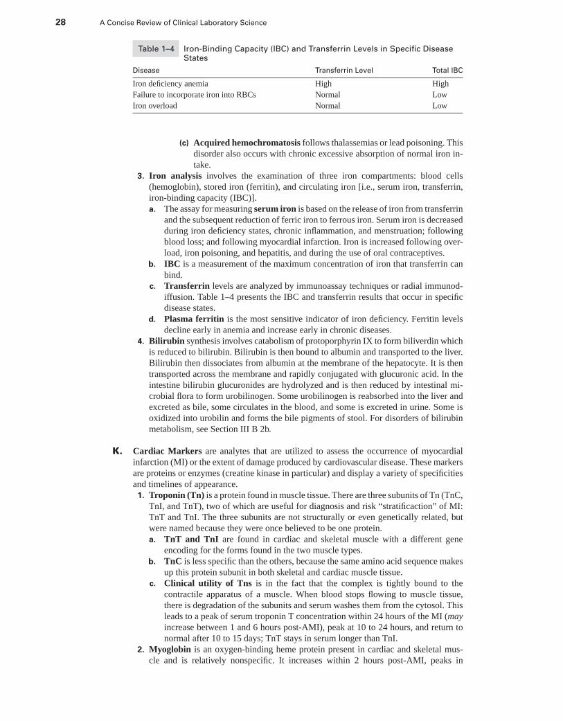
P1: OSO
LWBK192-01 LWBK192-Hubbard November 23, 2008 17:19
28 A Concise Review of Clinical Laboratory Science
Table 1–4 Iron-Binding Capacity (IBC) and Transferrin Levels in Specific DiseaseStates
Disease Transferrin Level Total IBC
Iron deficiency anemia High High
Failure to incorporate iron into RBCs Normal Low
Iron overload Normal Low
(c) Acquired hemochromatosis follows thalassemias or lead poisoning. Thisdisorder also occurs with chronic excessive absorption of normal iron in-take.
3. Iron analysis involves the examination of three iron compartments: blood cells(hemoglobin), stored iron (ferritin), and circulating iron [i.e., serum iron, transferrin,iron-binding capacity (IBC)].a. The assay for measuring serum iron is based on the release of iron from transferrin
and the subsequent reduction of ferric iron to ferrous iron. Serum iron is decreasedduring iron deficiency states, chronic inflammation, and menstruation; followingblood loss; and following myocardial infarction. Iron is increased following over-load, iron poisoning, and hepatitis, and during the use of oral contraceptives.
b. IBC is a measurement of the maximum concentration of iron that transferrin canbind.
c. Transferrin levels are analyzed by immunoassay techniques or radial immunod-iffusion. Table 1–4 presents the IBC and transferrin results that occur in specificdisease states.
d. Plasma ferritin is the most sensitive indicator of iron deficiency. Ferritin levelsdecline early in anemia and increase early in chronic diseases.
4. Bilirubin synthesis involves catabolism of protoporphyrin IX to form biliverdin whichis reduced to bilirubin. Bilirubin is then bound to albumin and transported to the liver.Bilirubin then dissociates from albumin at the membrane of the hepatocyte. It is thentransported across the membrane and rapidly conjugated with glucuronic acid. In theintestine bilirubin glucuronides are hydrolyzed and is then reduced by intestinal mi-crobial flora to form urobilinogen. Some urobilinogen is reabsorbed into the liver andexcreted as bile, some circulates in the blood, and some is excreted in urine. Some isoxidized into urobilin and forms the bile pigments of stool. For disorders of bilirubinmetabolism, see Section III B 2b.
K. Cardiac Markers are analytes that are utilized to assess the occurrence of myocardialinfarction (MI) or the extent of damage produced by cardiovascular disease. These markersare proteins or enzymes (creatine kinase in particular) and display a variety of specificitiesand timelines of appearance.
1. Troponin (Tn) is a protein found in muscle tissue. There are three subunits of Tn (TnC,TnI, and TnT), two of which are useful for diagnosis and risk “stratificaction” of MI:TnT and TnI. The three subunits are not structurally or even genetically related, butwere named because they were once believed to be one protein.a. TnT and TnI are found in cardiac and skeletal muscle with a different gene
encoding for the forms found in the two muscle types.b. TnC is less specific than the others, because the same amino acid sequence makes
up this protein subunit in both skeletal and cardiac muscle tissue.c. Clinical utility of Tns is in the fact that the complex is tightly bound to the
contractile apparatus of a muscle. When blood stops flowing to muscle tissue,there is degradation of the subunits and serum washes them from the cytosol. Thisleads to a peak of serum troponin T concentration within 24 hours of the MI (mayincrease between 1 and 6 hours post-AMI), peak at 10 to 24 hours, and return tonormal after 10 to 15 days; TnT stays in serum longer than TnI.
2. Myoglobin is an oxygen-binding heme protein present in cardiac and skeletal mus-cle and is relatively nonspecific. It increases within 2 hours post-AMI, peaks in
P1: OSO
LWBK192-01 LWBK192-Hubbard November 23, 2008 17:19
CHAPTER 1 Clinical Chemistry 29
approximately 6 to 9 hours, and becomes normal in approximately 24 hours. Manythings can increase serum myoglobin levels including strenuous exercise, surgery, re-nal failure, and muscular dystrophy. Myoglobin is quickly passed by the kidneys, andelevations of it in serum may be missed if blood is not drawn appropriately.
L. Bone is composed of two matrix components: minerals (calcium and phosphate) and anorganic (collagen) component. The cellular components of bone include osteoblasts (bone-forming), osteocytes, and osteoclasts (responsible for bone resorption).
1. Bone disease can be divided into metabolic disorders, disorders of mineral metabolism,and disease of unknown etiology.a. Metabolic bone disease includes the following.
(1) Osteoporosis is a decrease in the total amount of bone due to either decreasedosteoblastic activity or increased osteoclastic activity.
(2) Osteomalacia is the formation of “soft” bones due to inadequate calcificationof matrix components. It is typically caused by vitamin D deficiency leadingto hypocalcemia and elevated PTH.
(3) Rickets is a hereditary disorder with symptoms similar to osteomalacia; inchildren, this includes bowing of the extremities. It is caused by phosphatedepletion through renal wasting or by vitamin D deficiency or PTH excess.
(4) Scurvy is caused by lengthy deficiency of vitamin C that leads to inadequatecollagen synthesis and abnormal bone formation.
(5) Renal osteodystrophy is renal failure that induces hyperphosphatemia, lowvitamin D, etc. with osteomalacia as a complication. Any renal problem ac-companied by a bone disorder fits into this category.
b. Disorders of mineral metabolism include hyperparathyroidism, hyperpitu-itarism, hypo- and hypercalcemia, hypo- and hyperphosphatemia, and hypomag-nesemia. These are all discussed in previous lectures.
c. Disorders of unknown etiology include Paget’s Disease (osteitis deformans)which is loss of bone mass followed by replacement of bone in a chaotic archi-tecture. Bones are progressively enlarged and deformed. It is thought that a “slowvirus” affecting osteoclasts is the cause.
2. Laboratory analysis of bone disease consists of several approaches.a. Mineral concentration in serum is analyzed, including calcium, phosphate, and
magnesium. These values depend on bone deposition/resorption, renal clearance,and intestinal absorption.
b. Hormone concentration in serum is analyzed, including PTH, vitamin D, andcalcitonin. In addition, estrogen and testosterone are evaluated. In diagnosis ofosteoporosis particularly, serum estrogen levels are important.
c. Markers of bone metabolism, or bone markers, are of great importance whendiagnosing bone loss. These include osteocalcin, alkaline phosphatase, urinaryhydroxyproline, deoxypyridinoline, and N-telopeptide.(1) Markers of bone formation
(a) Osteocalcin is a non-collagenous protein in bone that is synthesized by os-teoblasts. Serum levels of this marker reflect osteoblast activity or bone for-mation/resorption. Synthesis of osteocalcin depends on vitamin K. Meth-ods of detection include immunoassays.
(b) Alkaline phosphatase (ALP) is elevated during bone growth, hyper-parathyroidism, renal osteodystrophy, osteomalacia, estrogen deficiency,and Paget’s disease. It is synthesized in part by osteoblasts and is a goodindicator of overall bone formation activity.
(2) Markers of bone resorption(a) Urinary hydroxyproline is found in collagen fibers. When collagen is de-
graded, urinary excretion is increased. This marker is considered a markerfor bone resorption. It is nonspecific, however.
(b) Deoxypyridinoline is a protein that stabilizes collagen chains during col-lagen synthesis. It is considered a highly specific marker of bone formation.Techniques used to assay this marker include HPLC and immunoassay.
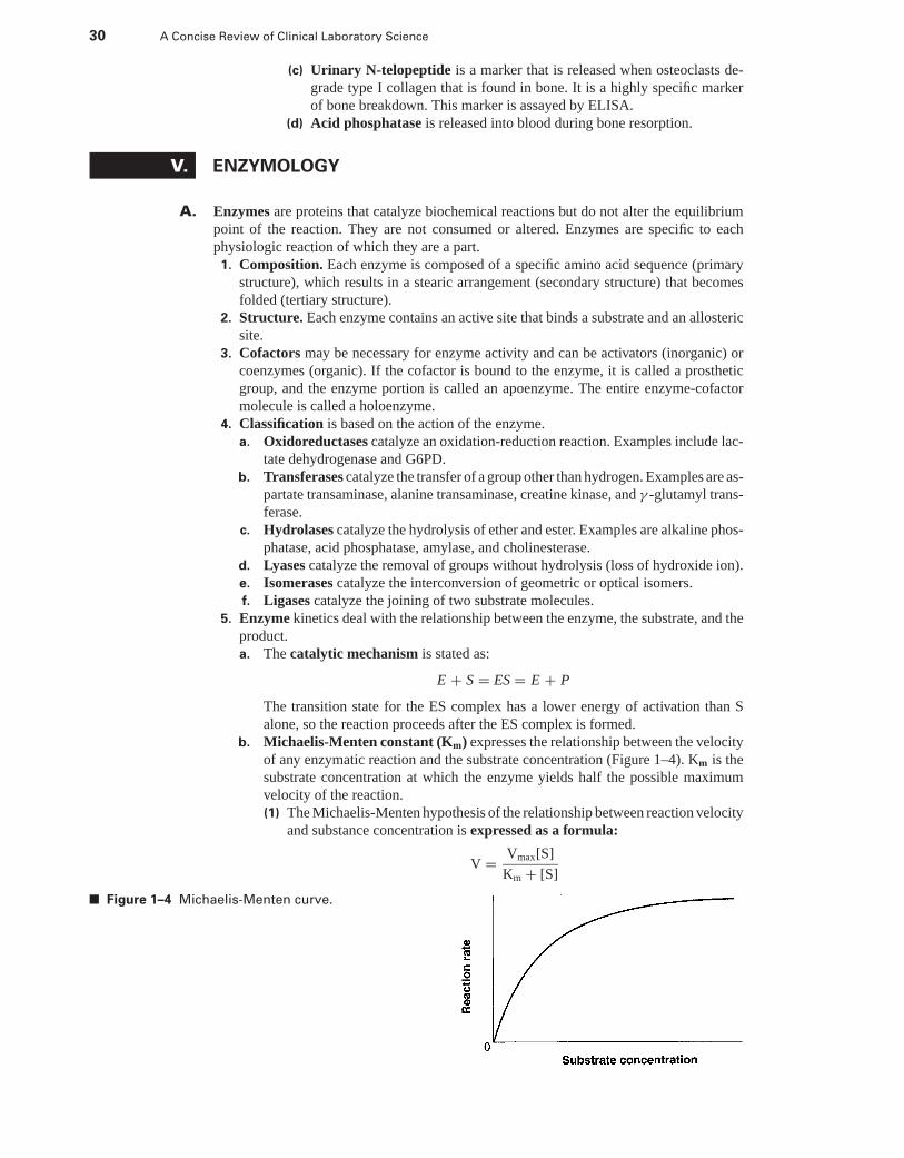
P1: OSO
LWBK192-01 LWBK192-Hubbard November 23, 2008 17:19
30 A Concise Review of Clinical Laboratory Science
(c) Urinary N-telopeptide is a marker that is released when osteoclasts de-grade type I collagen that is found in bone. It is a highly specific markerof bone breakdown. This marker is assayed by ELISA.
(d) Acid phosphatase is released into blood during bone resorption.
V. ENZYMOLOGY
A. Enzymes are proteins that catalyze biochemical reactions but do not alter the equilibriumpoint of the reaction. They are not consumed or altered. Enzymes are specific to eachphysiologic reaction of which they are a part.
1. Composition. Each enzyme is composed of a specific amino acid sequence (primarystructure), which results in a stearic arrangement (secondary structure) that becomesfolded (tertiary structure).
2. Structure. Each enzyme contains an active site that binds a substrate and an allostericsite.
3. Cofactors may be necessary for enzyme activity and can be activators (inorganic) orcoenzymes (organic). If the cofactor is bound to the enzyme, it is called a prostheticgroup, and the enzyme portion is called an apoenzyme. The entire enzyme-cofactormolecule is called a holoenzyme.
4. Classification is based on the action of the enzyme.a. Oxidoreductases catalyze an oxidation-reduction reaction. Examples include lac-
tate dehydrogenase and G6PD.b. Transferases catalyze the transfer of a group other than hydrogen. Examples are as-
partate transaminase, alanine transaminase, creatine kinase, and γ -glutamyl trans-ferase.
c. Hydrolases catalyze the hydrolysis of ether and ester. Examples are alkaline phos-phatase, acid phosphatase, amylase, and cholinesterase.
d. Lyases catalyze the removal of groups without hydrolysis (loss of hydroxide ion).e. Isomerases catalyze the interconversion of geometric or optical isomers.f. Ligases catalyze the joining of two substrate molecules.
5. Enzyme kinetics deal with the relationship between the enzyme, the substrate, and theproduct.a. The catalytic mechanism is stated as:
E + S = ES = E + P
The transition state for the ES complex has a lower energy of activation than Salone, so the reaction proceeds after the ES complex is formed.
b. Michaelis-Menten constant (Km) expresses the relationship between the velocityof any enzymatic reaction and the substrate concentration (Figure 1–4). Km is thesubstrate concentration at which the enzyme yields half the possible maximumvelocity of the reaction.(1) The Michaelis-Menten hypothesis of the relationship between reaction velocity
and substance concentration is expressed as a formula:
V = Vmax[S]
Km + [S]
■ Figure 1–4 Michaelis-Menten curve.
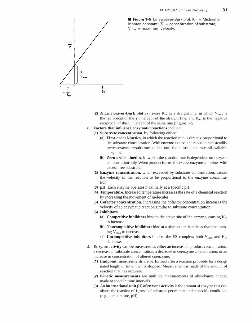
P1: OSO
LWBK192-01 LWBK192-Hubbard November 23, 2008 17:19
CHAPTER 1 Clinical Chemistry 31
■ Figure 1–5 Lineweaver-Burk plot. Km = Michaelis-
Menten constant; [S] = concentration of substrate;
Vmax = maximum velocity.
(2) A Lineweaver-Burk plot expresses Km as a straight line, in which Vmax isthe reciprocal of the y intercept of the straight line, and Km is the negativereciprocal of the x intercept of the same line (Figure 1–5).
c. Factors that influence enzymatic reactions include:(1) Substrate concentration, by following either:
(a) First-order kinetics, in which the reaction rate is directly proportional tothe substrate concentration. With enzyme excess, the reaction rate steadilyincreases as more substrate is added until the substrate saturates all availableenzymes.
(b) Zero-order kinetics, in which the reaction rate is dependent on enzymeconcentration only. When product forms, the excess enzyme combines withexcess free substrate.
(2) Enzyme concentration, when exceeded by substrate concentration, causesthe velocity of the reaction to be proportional to the enzyme concentra-tion.
(3) pH. Each enzyme operates maximally at a specific pH.(4) Temperature. Increased temperature increases the rate of a chemical reaction
by increasing the movement of molecules.(5) Cofactor concentration. Increasing the cofactor concentration increases the
velocity of an enzymatic reaction similar to substrate concentration.(6) Inhibitors
(a) Competitive inhibitors bind to the active site of the enzyme, causing Km
to increase.(b) Noncompetitive inhibitors bind at a place other than the active site, caus-
ing Vmax to decrease.(c) Uncompetitive inhibitors bind to the ES complex; both Vmax and Km
decrease.d. Enzyme activity can be measured as either an increase in product concentration,
a decrease in substrate concentration, a decrease in coenzyme concentration, or anincrease in concentration of altered coenzyme.(1) Endpoint measurements are performed after a reaction proceeds for a desig-
nated length of time, then is stopped. Measurement is made of the amount ofreaction that has occurred.
(2) Kinetic measurements are multiple measurements of absorbance changemade at specific time intervals.
(3) An international unit (U) of enzyme activity is the amount of enzyme that cat-alyzes the reaction of 1 μmol of substrate per minute under specific conditions(e.g., temperature, pH).
P1: OSO
LWBK192-01 LWBK192-Hubbard November 23, 2008 17:19
32 A Concise Review of Clinical Laboratory Science
B. Phosphatases promote the hydrolysis of phosphate esters.1. Acid phosphatase (ACP) promotes the hydrolysis of orthophosphate esters, although
the physiologic function of ACP is unknown.a. Almost every body tissue contains ACP, but significant levels are found in RBCs
and platelets. In adult men, 50% of ACP is found in the prostate gland. High levelsof ACP are found in semen as well.
b. In the measurement of total activity typical substrates acted upon by ACP in-clude p-nitrophenylphosphate or thymolphthalein monophosphate. All reactionsto measure ACP are carried out at a pH <6.0.(1) Isoenzyme tests include:
(a) Inhibition of prostate ACP by tartrate, followed by the determination ofthe serum level of other ACP fractions
(b) Immunoassay with antibodies specific to certain isoenzymes(c) Electrophoretic procedures
(2) Falsely low values are produced by improper anticoagulant (e.g., fluoride,oxalate, heparin) use, whereas hemolysis produces false elevations.
c. The major diagnostic significance of ACP is its aid in detecting metastaticcarcinoma. Other types of cancer and bone disease exhibit increases ofenzyme.
2. Alkaline phosphatase (ALP) hydrolyzes phosphate esters, but the function of thisenzyme is relatively unknown.a. ALP exists in a wide variety of tissues, with significant amounts found in liver,
bone, intestine, kidney, and placenta.b. In the measurement of total activity, the Bessey-Lowry-Brock reaction using p-
nitrophenylphosphate as the substrate for ALP is most commonly used with pH orreaction >8.0.
c. Improper anticoagulant use produces decreased values of ALP, and a hemolyzedspecimen demonstrates a false increase in ALP value.
d. The diagnostic significance of ALP is its elevation during the third trimester ofpregnancy and in persons with liver disease (hepatitis, cirrhosis), bone disease,hyperthyroidism, or diabetes mellitus.
e. There are four isoenzymes of ALP: bone, liver, intestinal, and placental fractions.In disease states, the fractions are affected as follows:(1) The bone fraction levels are elevated in persons with bone disease (osteoma-
lacia, rickets, Paget’s disease) and high in children.(2) The liver fraction level is greatly elevated in persons with hepatobiliary ob-
struction.(3) The placental fraction level becomes elevated between the 16th and 20th
weeks of pregnancy.
C. Transaminases catalyze the transfer of amino groups and acids. This is an importantreaction in the synthesis and degradation of amino acids.1. Aspartate transaminase (AST) transfers an amino group between aspartate and keto
acids (aspartate +α-ketoglutarateAST−→ oxaloacetate + glutamate). Pyridoxal phosphate
(vitamin B6) is the coenzyme in this reaction.a. AST exists in cardiac tissue, liver, skeletal muscle, and RBCs.b. Three major approaches are taken in the measurement of total activity.
(1) The reaction with dinitrophenylhydrazine couples color reagent with ketoacid product.
(2) The reaction with diazonium salts couples the salt with the keto acid productand forms a color.
(3) The coupled enzyme assay (Karmen method) involves NADH and malatedehydrogenase and keto acid to form NAD.
c. The clinical significance of AST elevations is that the elevations occur in patientswith myocardial infarction, viral hepatitis, and skeletal muscle disease.
2. Alanine transaminase (ALT) catalyzes the transfer of an amino group from alanineto α-ketoglutarate with the formation of glutamate and pyruvate.
P1: OSO
LWBK192-01 LWBK192-Hubbard November 23, 2008 17:19
CHAPTER 1 Clinical Chemistry 33
a. Distribution. ALT is localized in the liver, with some in the heart, skeletal muscle,and RBCs.
b. The measurement of total activity is similar to the reactions of AST, except thatthe coupled enzyme assay uses lactate dehydrogenase to reduce pyruvate to lactateand oxidize NADH.
c. The clinical significance of ALT is in the evaluation of hepatocellular disor-ders.
D. Creatine kinase (CK) catalyzes a reaction responsible for the formation of ATP in tissues,especially contractile systems. When muscle contraction occurs, ATP is hydrolyzed to ADPto produce chemical energy for the concentration process. CK is involved in the storage ofhigh-energy creatine phosphate (creatine + ATP
CK−→ creatine phosphate + ADP).1. Distribution. CK is localized in skeletal muscle, brain, and cardiac muscle in addition
to many other tissues.2. The measurement of total activity includes the following assays.
a. The Tanzer-Gilvarg assay involves the reaction stated previously coupled withother enzymes (pyruvate kinase and lactate dehydrogenase) to produce a changein absorbance when measured spectrophotometrically.
b. The Oliver-Rosalki assay is the reverse reaction of the one stated previously, inwhich creatine is produced from creatine phosphate. The enzymes used includehexokinase and G6PD.
c. Hemolysis must be avoided when analyzing because of the presence of adenylatekinase (AK) in RBCs. AK induces false elevations in CK assays. Because CKactivity is unstable due to the oxidation of its sulfhydryl groups, cysteine is addedto both of the previously mentioned assays.
3. The clinical significance of CK is in its elevation in muscle disorders. CK and oneof its isoenzymes is a sensitive indicator of acute myocardial infarction and musculardystrophy.
4. The structure of CK allows for the formation of three isoenzymes.a. CK-MM is the major form found in striated and cardiac muscle and normal serum.
Hypothyroidism and intramuscular injections result in CK-MM elevations.b. CK-MB is localized in cardiac muscle. Elevated levels are indicative of ischemic
heart disease. If the serum level of CK-MB is >6% of total CK, it is a highly specificindicator of myocardial damage. Following acute myocardial infarction, CK-MBlevels rise within 48 to 72 hours, peak at 12 to 24 hours, and return to normal within48 to 72 hours.
c. CK-BB is present in small quantities in normal serum and appears if extensivedamage to the brain occurs, but is present in many other tissues.
d. Assays used for measurement of isoforms include electrophoresis, ion-exchangechromatography, and immunoassay.
E. Lactate dehydrogenase (LD) catalyzes the interconversion of lactic and pyruvic acids. Itis a hydrogen-transfer enzyme that uses the coenzyme NAD.
1. Distribution. LD is widely distributed in all tissues, with high concentrations found inthe heart and liver and low levels in RBCs, skeletal muscle, and kidneys
2. Measurement of total activity is based on the detection of NADH in the assay ap-proaches.a. The Wacker method uses the lactate-to-pyruvate reaction with formation of
NADH.b. The Wroblewski-Ladue method employs the reverse reaction of the Wacker
method and measures the decrease in absorbance as NADH is consumed.3. The diagnostic significance of LD is in the diagnosis of cardiac, hepatic, skeletal
muscle, and renal disease. Highest LD levels are seen in persons with pernicious ane-mia, viral hepatitis, cirrhosis, and crush injuries. An elevated LD means little withoutisoenzyme studies.
4. There are, in normal serum, five different isoforms of LD (LD1 to LD5), with eachisoenzyme being composed of four protein subunits.
P1: OSO
LWBK192-01 LWBK192-Hubbard November 23, 2008 17:19
34 A Concise Review of Clinical Laboratory Science
a. LD1 is localized predominantly in heart muscle and RBCs and constitutes approx-imately 25% of total LD.
b. LD2 localization is similar to LD1, but it also is found in the kidneys and comprisesapproximately 35% of total LD.
c. LD3 is found in lung, lymphocytes, spleen, and pancreas. It constitutes approxi-mately 22% of total LD.
d. LD4 is found mostly in liver and skeletal muscle and comprises approximately10% of total LD.
e. LD5 is similar to LD4 in both localization and portion of total.f. Testing methodologies for isoforms include:
(1) Electrophoresis. LD1 migrates most quickly toward the anode, followed bythe others in numerical order. After myocardial infarction, LD1 concentrationis greater than LD2, which presents as a “flipped pattern” in electrophoresis.This pattern is also observed with hemolyzed specimens.
(2) Immunoinhibition for the measurement of LD1 activity. This procedureinvolves an antibody that binds all other isoforms, leaving LD1 to be assayedby chemical reaction.
F. Miscellaneous enzymes that are clinically significant include γ -glutamyl-transferase(GGT), amylase, cholinesterase, pseudocholinesterase, and 5′-nucleotidase.
1. GGT transfers a γ -glutamyl residue to an amino acid (transpeptidation). This functionis involved in peptide and protein synthesis.a. Distribution. Tissue sources of GGT include kidney, brain, prostate, pancreas, and
liver. Urine contains significant amounts of GGT.b. Measurement of total activity is by the Szasz assay. In this reaction, the substrate
is γ -glutamyl-p-nitroanilide, with the release of p-nitroaniline.c. The clinical significance of GGT is as follows.
(1) GGT levels are elevated in almost all hepatobiliary disorders or biliary tractobstructions as well as in patients taking enzyme-inducing drugs like warfarin,phenobarbital, and Dilantin.
(2) GGT levels are often examined with ALP levels. If both GGT levels and ALPvalues are high, some type of liver disorder is suspected. If GGT is normal andALP is high, bone disease is likely present.
(3) Patients with acute pancreatitis exhibit increased GGT levels. In persons withdiabetes, GGT is typically increased as triglyceride levels rise.
(4) Because alcohol has enzyme-inducing properties, the most useful applicationfor GGT is in the detection of alcoholism and the monitoring of alcohol intakeby patients during treatment.
2. Amylase (AMS) is a hydrolase that catalyzes the breakdown of starch and glycogenand produces products consisting of glucose, maltose, and dextrins. AMS is the smallestin size of all enzymes.a. Distribution. Tissue sources of AMS include the pancreas and salivary glands, as
well as skeletal muscle.b. The clinical significance of AMS is its use in the diagnosis of acute pancreatitis.
AMS levels rise 2 to 12 hours after onset of an attack and peak at 24 hours.Salivary gland lesions (e.g., mumps, parotiditis) also produce high levels of AMS.Patients with hyperlipidemia have low-to-normal AMS levels. Opiates constrictthe pancreatic sphincters, which causes false elevations of AMS.
3. Cholinesterase is an enzyme that hydrolyzes the esters of choline. There are two types:acetylcholinesterase (AChe) and pseudocholinesterase (PChe). The substrate for ACheis acetylcholine (a neurotransmitter). PChe acts on many different substrates, but mainlybutyryl esters.a. Distribution. AChe is found in RBCs, brain, and nerve cells. PChe is found mostly
in the serum, liver, pancreas, heart, and white matter of the nervous system.b. The measurement of total activity includes:
(1) Manometric techniques measure liberated carbon dioxide from the formationof acetic acid.
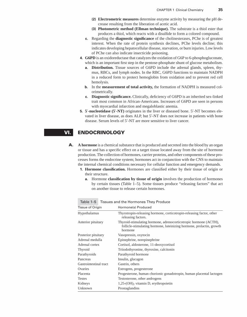
P1: OSO
LWBK192-01 LWBK192-Hubbard November 23, 2008 17:19
CHAPTER 1 Clinical Chemistry 35
(2) Electrometric measures determine enzyme activity by measuring the pH de-crease resulting from the liberation of acetic acid.
(3) Photometric method (Ellman technique). The substrate is a thiol ester thatproduces a thiol, which reacts with a disulfide to form a colored compound.
c. Regarding the diagnostic significance of the cholinesterases, PChe is of greatestinterest. When the rate of protein synthesis declines, PChe levels decline; thisindicates developing hepatocellular disease, starvation, or burn injuries. Low levelsof PChe can also indicate insecticide poisoning.
4. G6PD is an oxidoreductase that catalyzes the oxidation of G6P to 6-phosphogluconate,which is an important first step in the pentose-phosphate shunt of glucose metabolism.a. Distribution. Tissue sources of G6PD include the adrenal glands, spleen, thy-
mus, RBCs, and lymph nodes. In the RBC, G6PD functions to maintain NADPHin a reduced form to protect hemoglobin from oxidation and to prevent red cellhemolysis.
b. In the measurement of total activity, the formation of NADPH is measured col-orimetrically.
c. Diagnostic significance. Clinically, deficiency of G6PD is an inherited sex-linkedtrait most common in African-Americans. Increases of G6PD are seen in personswith myocardial infarction and megaloblastic anemia.
5. 5′-nucleotidase (5′-NT) originates in the liver or diseased bone. 5′-NT becomes ele-vated in liver disease, as does ALP, but 5′-NT does not increase in patients with bonedisease. Serum levels of 5′-NT are more sensitive to liver cancer.
VI. ENDOCRINOLOGY
A. A hormone is a chemical substance that is produced and secreted into the blood by an organor tissue and has a specific effect on a target tissue located away from the site of hormoneproduction. The collection of hormones, carrier proteins, and other components of these pro-cesses forms the endocrine system; hormones act in conjunction with the CNS to maintainthe internal chemical conditions necessary for cellular function and emergency demands.
1. Hormone classification. Hormones are classified either by their tissue of origin ortheir structure.a. Hormone classification by tissue of origin involves the production of hormones
by certain tissues (Table 1–5). Some tissues produce “releasing factors” that acton another tissue to release certain hormones.
Table 1–5 Tissues and the Hormones They Produce
Tissue of Origin Hormone(s) Produced
Hypothalamus Thyrotropin-releasing hormone, corticotropin-releasing factor, otherreleasing factors.
Anterior pituitary Thyroid-stimulating hormone, adrenocorticotropic hormone (ACTH),follicle-stimulating hormone, luteinizing hormone, prolactin, growthhormone
Posterior pituitary Vasopressin, oxytocin
Adrenal medulla Epinephrine, norepinephrine
Adrenal cortex Cortisol, aldosterone, 11-deoxycortisol
Thyroid Triiodothyronine, thyroxine, calcitonin
Parathyroids Parathyroid hormone
Pancreas Insulin, glucagon
Gastrointestinal tract Gastrin, others
Ovaries Estrogens, progesterone
Placenta Progesterone, human chorionic gonadotropin, human placental lactogen
Testes Testosterone, other androgens
Kidneys 1,25-(OH)2 vitamin D, erythropoietin
Unknown Prostaglandins
P1: OSO
LWBK192-01 LWBK192-Hubbard November 23, 2008 17:19
36 A Concise Review of Clinical Laboratory Science
b. Hormone classification by structure involves the specific chemical makeup ofhormones. Hormones of the same basic structure appear to produce the samefundamental biochemical changes.(1) Peptide hormones make up the majority of hormones. Releasing factors
are peptides. The hormones are water-soluble and do not require transportproteins to move through the blood. They are synthesized and stored withinthe cell and have a short half-life (5–60 minutes) in the circulation.
(2) Steroid hormones are involved in the regulation of sexual development andcharacteristics. All steroid hormones are synthesized from cholesterol asthe basic molecule. They are not water soluble, and they require a transportprotein to travel through the blood. Steroid hormones are synthesized in theadrenal gland, gonads, or placenta and have a long half-life (60–100 minutes)in the circulation.
(3) Amino acid derivatives (amines) have properties similar to those of bothsteroid and peptide hormones.
(4) Fatty acid derivatives include the prostaglandins.2. Mechanisms of hormone action typically depend on the type of hormone.
a. Hormone/receptor interaction involves the binding of hormone to a specificreceptor molecule on or within a cell.
b. Hormone binding occurs on or within a cell.(1) Binding at the cell surface produces a conformational change in the receptor
complex that leads to release of proteins and formation of AMP fromATP within the cell. Cyclic AMP (AMP-c) then causes cellular enzymesto be phosphorylated, and these enzymes can exert regulatory control onbiochemical reactions within the cell. Most peptide hormones interact withcells in this manner. However, insulin phosphorylates without AMP.
(2) Internal binding typically involves steroid hormones. Diffusion into thecell occurs before attachment to a specific receptor within the cell. Thesteroid/receptor complex then migrates to the DNA in the nucleus and exertscontrol over the type and amount of RNA produced and ultimately over whatproteins are synthesized.
3. Hormone release is controlled by a number of factors.a. Regulation by releasing factors involves a complex system of “factors” produced
by tissues that induce synthesis of a specific hormone. For example, a declinein blood pressure causes corticotropin-releasing factor to be released, which inturn induces the release of adrenocorticotropic hormone and constriction of bloodvessels.
b. Prohormones are precursors of peptide hormones. Following hydrolysis of aportion of its peptide chain, a prohormone becomes an active hormone. Thehydrolysis is controlled by the cell containing the prohormone.
c. Feedback control involves the release of a hormone that regulated prior steps inthe releasing process. For example, thyroid hormones feed back to the hypothala-mus to “shut off” thyrotropin-releasing hormone so excess thyroid hormone willnot be produced.
4. Hormone transport proteins affect both the concentration of a hormone and itsinfluence on the system. Peptide hormones are transported in the unbound state.a. Types of transport proteins are specific for each hormone. The attachment of
hormone to protein is through noncovalent linkages.(1) Steroid and thyroid hormones are bound to albumin, sex hormone-binding
protein, or cortisol-binding globulin.(2) Amines are transported by serum proteins and thyroxine-binding globulin.
b. Transport protein level can affect hormone action. Relative amounts of boundand free hormone determine the degree of stimulus provided by the hormone.Only the free (unbound) fraction of a hormone exhibits activity, so any situationthat affects the transport protein level or degree of binding has an impact on theconcentration of the free hormone. The measurement of the transport proteinconcentration is sometimes an integral part of the hormone assay.
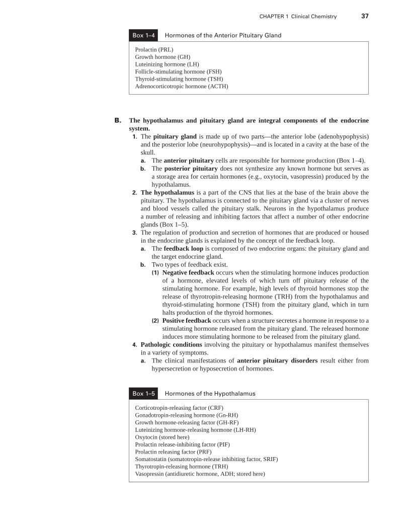
P1: OSO
LWBK192-01 LWBK192-Hubbard November 23, 2008 17:19
CHAPTER 1 Clinical Chemistry 37
Box 1–4 Hormones of the Anterior Pituitary Gland
Prolactin (PRL)
Growth hormone (GH)
Luteinizing hormone (LH)
Follicle-stimulating hormone (FSH)
Thyroid-stimulating hormone (TSH)
Adrenocorticotropic hormone (ACTH)
B. The hypothalamus and pituitary gland are integral components of the endocrinesystem.
1. The pituitary gland is made up of two parts—the anterior lobe (adenohypophysis)and the posterior lobe (neurohypophysis)—and is located in a cavity at the base of theskull.a. The anterior pituitary cells are responsible for hormone production (Box 1–4).b. The posterior pituitary does not synthesize any known hormone but serves as
a storage area for certain hormones (e.g., oxytocin, vasopressin) produced by thehypothalamus.
2. The hypothalamus is a part of the CNS that lies at the base of the brain above thepituitary. The hypothalamus is connected to the pituitary gland via a cluster of nervesand blood vessels called the pituitary stalk. Neurons in the hypothalamus producea number of releasing and inhibiting factors that affect a number of other endocrineglands (Box 1–5).
3. The regulation of production and secretion of hormones that are produced or housedin the endocrine glands is explained by the concept of the feedback loop.a. The feedback loop is composed of two endocrine organs: the pituitary gland and
the target endocrine gland.b. Two types of feedback exist.
(1) Negative feedback occurs when the stimulating hormone induces productionof a hormone, elevated levels of which turn off pituitary release of thestimulating hormone. For example, high levels of thyroid hormones stop therelease of thyrotropin-releasing hormone (TRH) from the hypothalamus andthyroid-stimulating hormone (TSH) from the pituitary gland, which in turnhalts production of the thyroid hormones.
(2) Positive feedback occurs when a structure secretes a hormone in response to astimulating hormone released from the pituitary gland. The released hormoneinduces more stimulating hormone to be released from the pituitary gland.
4. Pathologic conditions involving the pituitary or hypothalamus manifest themselvesin a variety of symptoms.a. The clinical manifestations of anterior pituitary disorders result either from
hypersecretion or hyposecretion of hormones.
Box 1–5 Hormones of the Hypothalamus
Corticotropin-releasing factor (CRF)
Gonadotropin-releasing hormone (Gn-RH)
Growth hormone-releasing factor (GH-RF)
Luteinizing hormone-releasing hormone (LH-RH)
Oxytocin (stored here)
Prolactin release-inhibiting factor (PIF)
Prolactin releasing factor (PRF)
Somatostatin (somatotropin-release inhibiting factor, SRIF)
Thyrotropin-releasing hormone (TRH)
Vasopressin (antidiuretic hormone, ADH; stored here)
P1: OSO
LWBK192-01 LWBK192-Hubbard November 23, 2008 17:19
38 A Concise Review of Clinical Laboratory Science
(1) Hypersecretion usually involves one hormone. It is not uncommon forhypersecretion to be associated with the hyposecretion of another tropichormone.(a) Primary factors center on disorders of the pituitary gland: either pituitary
adenomas or pituitary hyperplasia.(b) Secondary factors center on disorders of the hypothalamus or may relate
to ectopic production of pituitary hormones by nonendocrine tumors or tothe hyposecretion of hormone by the target tissue.
(2) Hyposecretion can be decreased secretion of one hormone, a group ofhormones, or all hormones. The latter condition is referred to as “panhypopi-tuitarism.”(a) Single hormone hyposecretion results from lesions on the hypothalamus.(b) Pituitary adenoma, causing hypersecretion of one hormone, is commonly
the cause of hyposecretion of remaining pituitary hormones because ofthe destruction of the pituitary gland by the growing tumor.
b. Specifics of pathologic conditions associated with dysfunction of the anteriorpituitary gland.(1) Growth hormone (GH)
(a) The effects of hypersecretion are age dependent. In adults, the resultingcondition is called acromegaly, which is the progressive enlargement ofthe distal parts of the extremities. In children, the resulting conditionis gigantism. Both conditions usually result from pituitary adenomassecreting GH and compression of adjacent tissues of the pituitary gland,causing hyposecretion of other tropic hormones.
(b) Hyposecretion in children leads to pituitary dwarfism (small stature butproportionally built).
(2) Prolaction (PRL)(a) Hypersecretion causes galactorrhea or lactation and is associated with
infertility and amenorrhea in women and impotence in men. It usually isinduced by pituitary adenoma.
(b) Hyposecretion leads to the lack of lactation in postpartum women.(3) ACTH
(a) Hypersecretion is referred to as Cushing’s disease, with symptomsincluding truncal obesity, hyperglycemia, hypertension, and proteinwasting. It is caused by pituitary adenoma, adrenal hyperplasia, or excessproduction by a nonendocrine tumor.
(b) Hyposecretion causes weight loss, weakness, and gastrointestinalproblems.
(4) TSH(a) Hypersecretion causes thyrotoxicosis and is the result of either hyper-
activity of the thyroid or hyperactivity of the pituitary gland, leading toincreased TSH release.
(b) Hyposecretion is difficult to differentiate from primary hypothyroidism.(5) Gonadotropins (FSH and LH)
(a) Hypersecretion results in sexual precocity and is usually a result of braintumors in the region of the hypothalamus.
(b) Hyposecretion results in sexual underdevelopment and infertility.c. The clinical manifestations of posterior pituitary dysfunction involve either vaso-
pressin or oxytocin. These disorders result from either hypothalamic dysfunctionor some peripheral disease.(1) Anti-diuretic hormone (ADH; also known as Vasopressin) regulates water re-
absorption and blood pressure by affecting the renal tubules and the arterioles.(a) Hypersecretion of ADH results in a condition referred to as syndrome of
inappropriate ADH secretion (SIADH). The disorder occurs in a widevariety of conditions, including meningitis, head injury, tuberculosis,hypoadrenalism, hypothyroidism, and cirrhosis. SIADH is associated withhyponatremia (low blood sodium) and hypertonic urine, despite normal
P1: OSO
LWBK192-01 LWBK192-Hubbard November 23, 2008 17:19
CHAPTER 1 Clinical Chemistry 39
renal and adrenal function. Symptoms include weakness, malaise, poormental status, convulsions, and coma. It is typically caused by release ofADH from ectopic tumors.
(b) Hyposecretion of ADH is associated with diabetes insipidus. Symptomsinclude insatiable thirst, polydipsia (excessive drinking), and polyuria(excessive urine volume). This disorder results from destruction of theposterior pituitary gland or the hypothalamus.
(2) No known disorders are associated with excess or deficient secretion ofoxytocin.
C. Adrenal glands. One adrenal gland is situated on top of each kidney. Each adrenal gland iscomposed of two distinct layers: the adrenal cortex (the outer-most region) and the adrenalmedulla (the innermost region).
1. The adrenal cortex is composed of three distinct tissues, the zona glomerulosa, thezona fasciculata, and the zona reticularis.
2. The adrenal medulla is composed of chromaffin cells.3. Adrenal hormones
a. Glucocorticoids are steroid hormones produced in the zona fasciculata andreticularis of the adrenal cortex.
b. Mineralocorticosteroids are steroid hormones produced in the zona glomerulosa.c. Catecholamines are amine hormones produced in the adrenal medulla.
4. Adrenal endocrine function includes regulation of proteins, carbohydrates, andmany other metabolic functions.a. Glucocorticoids are synthesized from cholesterol. They are transported bound
to plasma proteins [albumin and corticosteroid-binding protein (CBP)], arecatabolized by the liver and kidneys, and are regulated by the hypothalamus-pituitary-adrenal axis. The primary action of the glucocorticoids is catabolic (i.e.,promoting protein and lipid breakdown, inhibiting protein synthesis).(1) Cortisol is the primary glucocorticoid produced and secreted by the adrenal
cortex. Its functions include carbohydrate, lipid, and protein metabolism;suppression of inflammation; stimulating gluconeogenesis; increasing urineproduction; and stimulating erythropoiesis.
(2) Target tissues of glucocorticoid action include the kidney glomerulusand renal tubules, bone marrow stem cells, hepatocytes, and adiposetissue.
b. Mineral corticosteroids are synthesized from cholesterol, transported bound toCBP and albumin, and are catabolized by the liver and the kidney. The primaryaction is regulation of electrolytes.(1) Aldosterone is the primary mineralocorticoid produced and secreted by the
adrenal cortex. Its functions include:(a) Stimulating sodium resorption in the distal convoluted tubules in exchange
for potassium or hydrogen(b) Increasing blood volume (via renin/angiotensin system) and pressure(c) Regulating extracellular fluid volume
(2) Target tissues of mineralocorticoid action include the distal renal tubules andthe large intestine.
(3) Regulation of aldosterone secretion via the renin/angiotensin system isachieved as follows. Decreased blood volume or blood pressure induces therelease of kidney renin, which induces the production of angiotensin I and II.Angiotensin II affects release of aldosterone from the adrenal gland, whichultimately causes the kidney distal tubule to retain sodium, thereby raisingblood volume and blood pressure.
c. Catecholamine production is not limited to the adrenal medulla. Norepinephrineis predominantly synthesized in the CNS, whereas epinephrine is predominantlysynthesized by the adrenal medulla. Catecholamines are products of the hydrox-ylation of the amino acid tyrosine. They are transported free in the blood andregulated by feedback inhibition of synthesis.
P1: OSO
LWBK192-01 LWBK192-Hubbard November 23, 2008 17:19
40 A Concise Review of Clinical Laboratory Science
(1) Catecholamine functions include:(a) Mobilization of energy stores by increasing blood pressure, heart rate,
blood sugar level (by stimulating glycogenolysis)(b) Neurotransmitter actions(c) Release in response to pain and emotional disturbance (stress) to mobilize
organs(2) Tissue targets include the liver and adipose tissue.(3) Approximately 20% of catecholamines are excreted into the urine as
metanephrine and normetanephrine: approximately 80% are converted tovanillylmandelic acid (VMA) by the enzyme monoamine oxidase (MAO).
5. Diseases associated with the adrenal cortex center on hyperfunction (excess productionof bioactive molecules) or hypofunction.a. Hyperadrenalism involves three basic conditions:
(1) Cushing’s syndrome involves excess cortisol production, either at the levelof the adrenal gland or by increased release of ACTH.
(2) Hyperaldosteronism involves excess aldosterone production with symptomsof hypertension. There are two causes.(a) Conn’s syndrome is induced by an aldosterone-secreting adrenal
adenoma or adrenal hyperplasia and is a rare cause of hypertension.(b) Excess renin production leads to elevated aldosterone levels.
(3) Congenital adrenal hyperplasia is a genetic disorder causing a deficiency ofenzymes in the synthetic pathways that lead to cortisol and aldosterone pro-duction. ACTH levels are increased, and steroid hormones are hypersecreted.A common cause of this condition is a 21-hydroxylase deficiency.
b. Hypoadrenalism is caused by adrenal hypofunction or insufficiency and can beinduced by three conditions:(1) Primary disease involving the adrenal cortex (known as Addison’s disease),
which is relatively rare(2) Secondary adrenal insufficiency precipitated by decreased levels of
corticotropin-releasing hormone (CRH) or ACTH(3) Long-term suppression of the hypothalamic-pituitary-adrenal axis by gluco-
corticoids6. The major disorder of the adrenal medulla is pheochromocytoma. Pheochromo-
cytoma is a relatively rare, usually benign tumor arising in the chromaffin cells of theadrenal medulla that results in hypersecretion of catecholamines.
7. Laboratory analysis of adrenal function involves several different testing method-ologies.a. The laboratory investigation of Cushing’s syndrome proceeds in two stages.
(1) The fact that the patient has autonomous cortisol production must beestablished by using the dexamethasone suppression test. This test involvesthe injection of a synthetic steroid that acts like cortisol (dexamethasone) toinduce feedback inhibition of cortisol release at the level of the pituitary.
(2) The cause of the disease must be differentiated.b. Plasma cortisol values normally display diurnal variation, with the highest levels
occurring in the morning and the lowest levels in the early evening. Evening valuesare <50% of the early morning concentrations. Classically, samples are drawn at8 a.m. and 4 p.m.
c. Primary hyperaldosteronism is analyzed by examining serum and urine potas-sium. Values of serum potassium <3.5 mmol/L, coupled with a urine potassiumexcretion rate >30 mmol/24 hr, are commonly seen in persons with primaryhyperaldosteronism. The definitive test for primary hyperaldosteronism, however,is the measurement of serum or urine aldosterone following a high salt chal-lenge. Primary hyperaldosteronism caused by an aldosterone-producing tumordemonstrates no change in plasma aldosterone levels following salt challenge,whereas primary aldosteronism caused by adrenal hyperplasia demonstrates a risein plasma aldosterone. A simple method for investigating Conn’s syndrome is tomeasure serum and urine potassium when a patient is not receiving diuretics.
P1: OSO
LWBK192-01 LWBK192-Hubbard November 23, 2008 17:19
CHAPTER 1 Clinical Chemistry 41
d. Congenital adrenal hyperplasia is analyzed by investigating the possibility ofadrenal insufficiency. The patient may also suffer biochemical consequences ofexcess androgen, and measurements of testosterone in serum and pregnanetriol inurine may be requested.
e. In the analysis of Addison’s disease, patients typically exhibit postural hypoten-sion, hyponatremia, and hyperkalemia. The ACTH stimulation test is given; itinvolves injection of synthetic ACTH and subsequent measurement of ACTH andcortisol at 30 and 60 minutes. High ACTH with no cortisol response indicatesAddison’s disease.
f. There are many ways to investigate pheochromocytoma. Urinary metabo-lites (e.g., VMA, metanephrines) can be quantitated, or plasma epinephrineand norepinephrine can be measured. In difficult cases, when the patient hasepisodic hypertension of short duration, a clonidine suppression test combinedwith plasma catecholamine measurements is performed. Clonidine suppressescatecholamine release from the CNS but not from the adrenal gland.
D. The thyroid gland is a bilobed endocrine gland located in the lower part of the neck that iscomposed of groups of cells called follicles. This gland contains two cell types: Follicularcells produce the hormones thyroxine (T4) and triiodothyronine (T3), and parafollicularcells (lying adjacent to the follicles) produce the hormone calcitonin.
1. Thyroid hormones require iodine for their synthesis. The iodine combines with theprotein thyroglobulin to form hormone precursors that in turn combine to form T3 andT4.a. The hormones are either stored within the follicle or released into the bloodstream.
In the blood, most T4 eventually gives up an iodine molecule and forms T3. Thereis much more circulating T3 than T4.
b. Approximately 98% of circulating T3 and T4 is bound to protein, includingthyroxine-binding globulin (TBG) and thyroxine-binding albumin. Somehormone remains unbound or free, and this is the physiologically active fraction.
c. Thyroid hormone function includes action at the cellular level to regulatecarbohydrate, lipid, and protein metabolism. The hormones also act on the CNS,stimulate the heart, and have a role in physical growth and development.
d. The regulation of T3 and T4 occurs in the following manner.(1) Thyroid-releasing hormone (TRH) is released by the brain and stimulates the
release of TSH (thyrotropin) from the pituitary gland.(2) TSH stimulates iodine uptake by the thyroid gland and also causes the release
of T3 and T4 from the thyroid gland.(3) High serum levels of free T3 and T4 “shut off” the release of TSH from the
pituitary gland, whereas decreased levels induce TSH release.2. Thyroid disorders are caused by increased or decreased levels of the circulating
hormones T3 and T4. A wide variety of physical diseases can be traced back to adysfunctional thyroid gland.a. Hypothyroidism is a serum level of thyroid hormone that is insufficient to provide
for the metabolic needs of cells. This disorder affects women four times more thanmen between the ages of 30 and 60 years. Hypothyroidism is usually referred toas primary, secondary, or tertiary, depending on the site of the dysfunction.(1) The symptoms of hypothyroidism include an enlarged thyroid gland (goiter),
fatigue, impairment of mental processes, and loss of appetite. Myxedema(loss of hair, swelling of the hands and face, course skin) occurs as the diseaseprogresses.
(2) The causes of hypothyroidism relate to the area of tissue damage. In addition,hypothyroidism can be caused by lack of dietary iodine.(a) Primary hypothyroidism involves the inadequate secretion of thyroid
hormones caused by a damaged or surgically removed thyroid gland.Congenital hypothyroidism is caused by the absence of the thyroid gland.Laboratory results indicate decreased T3, T4, free thyroxine index (FT4I),T3 uptake (T3U), and increased TSH.
P1: OSO
LWBK192-01 LWBK192-Hubbard November 23, 2008 17:19
42 A Concise Review of Clinical Laboratory Science
(b) Secondary hypothyroidism involves decreased production of TSHcaused by pituitary disorder leading to low serum levels of the thyroidhormones. Laboratory results indicate all thyroid test values are decreased.
(c) Tertiary hypothyroidism is caused by hypothalamic failure leading to alack of TRH production.
(3) In the laboratory evaluation of hypothyroidism, the earliest abnormality isincreased TSH, followed by decreased serum levels of T4 and T3.
b. Chronic immune thyroiditis (Hashimoto’s disease) is caused by a geneticabnormality in the immune system and involves massive infiltration of the thyroidgland by lymphocytes. The symptoms match those of hypothyroidism.
c. Hyperthyroidism is caused by excessive thyroid hormone in the circulation.This causes cells to become overactive. The disorder is sometimes referred to asthyrotoxicosis.(1) The symptoms of hyperthyroidism include weight loss, loss of muscle
mass, hyperactivity yet, quick fatigability, insomnia, increased sweating,nervousness, palpitations, goiter, and bulging eyes.
(2) The causes of this disorder include pituitary tumors that cause excessive TSHsecretion, thyroid carcinoma, or toxic multinodular goiter (gland producesexcess hormones).
(3) The laboratory evaluation of hyperthyroidism in the initial evaluation revealselevated thyroid hormone serum levels and decreased serum TSH.
d. Graves’ disease is an autoimmune disorder that occurs six times more frequentlyin women than in men. In this disorder, immunoglobulins stimulate the thyroidgland by binding to TSH receptors. Symptoms are similar to those of hyper-thyroidism. Laboratory results indicate increased T3, T4, FT4I, and T3U, anddecreased or normal TSH.
e. Thyroiditis is an inflammation of the thyroid gland caused by either bacterial orviral infection.
3. Assays for thyroid function include testing for serum level of total (both boundand free) or free T3, total or free T4, TSH, and TBG. These tests are typicallyimmunoassays. Other thyroid tests include the following:a. T3 resin uptake analyzes the capacity of TBG to bind thyroid hormones. It is an
indirect measurement of the number of free binding sites on the TBG molecule.b. Free thyroxine index (FT4I) indirectly assesses the concentration of circulating
free T4. It is calculated by multiplying the value of the total T4 by the percentagevalue of the T3 resin uptake.
c. Thyroid antibody screens assay for the presence of thyroid-stimulatingimmunoglobulins, such as those in Graves’ disease and Hashimoto’s thyroiditis.
d. TRH stimulation test measures pituitary TSH stores and is considered con-clusive for hyperthyroidism, although it is not needed in most hyperthyroidpatients. In patients with slightly elevated hormone levels (but other symptomsof hyperthyroidism), TRH is injected, and blood samples are assayed for TSH.TSH levels rise rapidly in a normal person but will not rise in a hyperthyroidpatient.
E. Parathyroid hormone is produced by the parathyroid glands, which are small, pairedstructures located in the posterior thyroid capsule. They may, however, be located in otherparts of the neck or upper chest cavity. The sole function of the parathyroid gland is theproduction of parathyroid hormone.
1. Parathyroid hormone (PTH) synthesis begins with the precursor proparathyroidhormone.a. The major physiologic action of PTH is mineral homeostasis. Specifically, PTH
is involved in the metabolism of both calcium and phosphorus by the kidney andbone. A complex interrelationship exists between PTH, cholecalciferol (vitaminD), and calcitonin.
b. Transport. Parathyroid hormone, unlike many other proteins, is transported asa freely circulating, intact, active molecule. The biologically inactive amino acidfragments can be detected in the serum as well.
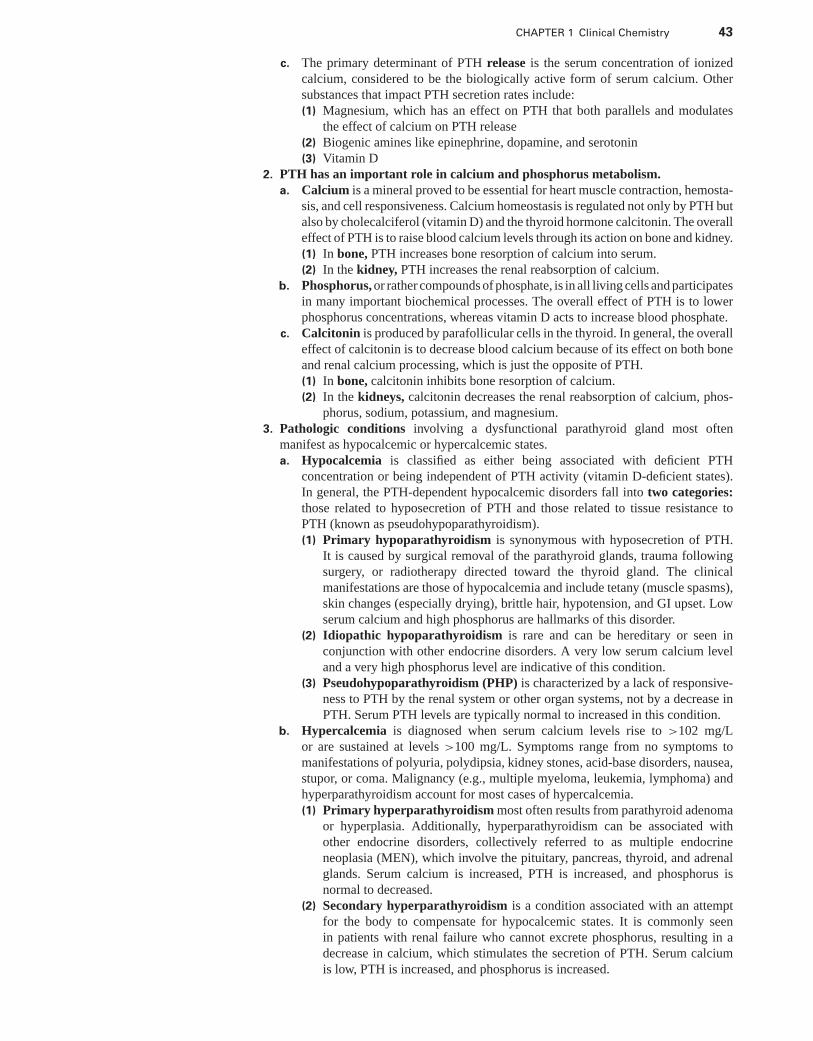
P1: OSO
LWBK192-01 LWBK192-Hubbard November 23, 2008 17:19
CHAPTER 1 Clinical Chemistry 43
c. The primary determinant of PTH release is the serum concentration of ionizedcalcium, considered to be the biologically active form of serum calcium. Othersubstances that impact PTH secretion rates include:(1) Magnesium, which has an effect on PTH that both parallels and modulates
the effect of calcium on PTH release(2) Biogenic amines like epinephrine, dopamine, and serotonin(3) Vitamin D
2. PTH has an important role in calcium and phosphorus metabolism.a. Calcium is a mineral proved to be essential for heart muscle contraction, hemosta-
sis, and cell responsiveness. Calcium homeostasis is regulated not only by PTH butalso by cholecalciferol (vitamin D) and the thyroid hormone calcitonin. The overalleffect of PTH is to raise blood calcium levels through its action on bone and kidney.(1) In bone, PTH increases bone resorption of calcium into serum.(2) In the kidney, PTH increases the renal reabsorption of calcium.
b. Phosphorus, or rather compounds of phosphate, is in all living cells and participatesin many important biochemical processes. The overall effect of PTH is to lowerphosphorus concentrations, whereas vitamin D acts to increase blood phosphate.
c. Calcitonin is produced by parafollicular cells in the thyroid. In general, the overalleffect of calcitonin is to decrease blood calcium because of its effect on both boneand renal calcium processing, which is just the opposite of PTH.(1) In bone, calcitonin inhibits bone resorption of calcium.(2) In the kidneys, calcitonin decreases the renal reabsorption of calcium, phos-
phorus, sodium, potassium, and magnesium.3. Pathologic conditions involving a dysfunctional parathyroid gland most often
manifest as hypocalcemic or hypercalcemic states.a. Hypocalcemia is classified as either being associated with deficient PTH
concentration or being independent of PTH activity (vitamin D-deficient states).In general, the PTH-dependent hypocalcemic disorders fall into two categories:those related to hyposecretion of PTH and those related to tissue resistance toPTH (known as pseudohypoparathyroidism).(1) Primary hypoparathyroidism is synonymous with hyposecretion of PTH.
It is caused by surgical removal of the parathyroid glands, trauma followingsurgery, or radiotherapy directed toward the thyroid gland. The clinicalmanifestations are those of hypocalcemia and include tetany (muscle spasms),skin changes (especially drying), brittle hair, hypotension, and GI upset. Lowserum calcium and high phosphorus are hallmarks of this disorder.
(2) Idiopathic hypoparathyroidism is rare and can be hereditary or seen inconjunction with other endocrine disorders. A very low serum calcium leveland a very high phosphorus level are indicative of this condition.
(3) Pseudohypoparathyroidism (PHP) is characterized by a lack of responsive-ness to PTH by the renal system or other organ systems, not by a decrease inPTH. Serum PTH levels are typically normal to increased in this condition.
b. Hypercalcemia is diagnosed when serum calcium levels rise to >102 mg/Lor are sustained at levels >100 mg/L. Symptoms range from no symptoms tomanifestations of polyuria, polydipsia, kidney stones, acid-base disorders, nausea,stupor, or coma. Malignancy (e.g., multiple myeloma, leukemia, lymphoma) andhyperparathyroidism account for most cases of hypercalcemia.(1) Primary hyperparathyroidism most often results from parathyroid adenoma
or hyperplasia. Additionally, hyperparathyroidism can be associated withother endocrine disorders, collectively referred to as multiple endocrineneoplasia (MEN), which involve the pituitary, pancreas, thyroid, and adrenalglands. Serum calcium is increased, PTH is increased, and phosphorus isnormal to decreased.
(2) Secondary hyperparathyroidism is a condition associated with an attemptfor the body to compensate for hypocalcemic states. It is commonly seenin patients with renal failure who cannot excrete phosphorus, resulting in adecrease in calcium, which stimulates the secretion of PTH. Serum calciumis low, PTH is increased, and phosphorus is increased.
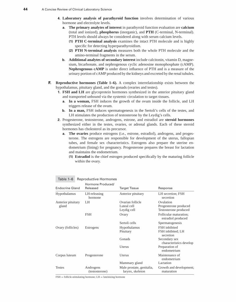
P1: OSO
LWBK192-01 LWBK192-Hubbard November 23, 2008 17:19
44 A Concise Review of Clinical Laboratory Science
4. Laboratory analysis of parathyroid function involves determination of varioushormone and electrolyte levels.a. The primary analytes of interest in parathyroid function evaluation are calcium
(total and ionized), phosphorus (inorganic), and PTH (C-terminal, N-terminal).PTH levels should always be considered along with serum calcium levels.(1) PTH C-terminal analysis examines the intact PTH molecule and is highly
specific for detecting hyperparathyroidism.(2) PTH N-terminal analysis measures both the whole PTH molecule and the
amino-terminal fragments in the serum.b. Additional analytes of secondary interest include calcitonin, vitamin D, magne-
sium, bicarbonate, and nephrogenous cyclic adenosine monophosphate (cAMP).Nephrogenous cAMP is under direct influence of PTH and is a measure of theurinary portion of cAMP produced by the kidneys and excreted by the renal tubules.
F. Reproductive hormones (Table 1–6). A complex interrelationship exists between thehypothalamus, pituitary gland, and the gonads (ovaries and testes).
1. FSH and LH are glycoprotein hormones synthesized in the anterior pituitary glandand transported unbound via the systemic circulation to target tissues.a. In a woman, FSH induces the growth of the ovum inside the follicle, and LH
triggers release of the ovum.b. In a man, FSH induces spermatogenesis in the Sertoli’s cells of the testes, and
LH stimulates the production of testosterone by the Leydig’s cells.2. Progesterone, testosterone, androgens, estrone, and estradiol are steroid hormones
synthesized either in the testes, ovaries, or adrenal glands. Each of these steroidhormones has cholesterol as its precursor.a. The ovaries produce estrogens (i.e., estrone, estradiol), androgens, and proges-
terone. The estrogens are responsible for development of the uterus, fallopiantubes, and female sex characteristics. Estrogens also prepare the uterine en-dometrium (lining) for pregnancy. Progesterone prepares the breast for lactationand maintains the endometrium.(1) Estradiol is the chief estrogen produced specifically by the maturing follicle
within the ovary.
Table 1–6 Reproductive Hormones
Hormone Produced/
Endocrine Gland Released Target Tissue Response
Hypothalamus LH-releasinghormone
Anterior pituitary LH secretion; FSHsecretion
Anterior pituitarygland
LH Ovarian follicleLuteal cellLeydig cell
OvulationProgesterone producedTestosterone produced
FSH Ovary Follicular maturation;estradiol produced
Sertoli cells Spermatogenesis
Ovary (follicles) Estrogens HypothalamusPituitary
FSH inhibitedFSH inhibited; LH
secretionGonads Secondary sex
characteristics developUterus Preparation of
endometrium
Corpus luteum Progesterone Uterus Maintenance ofendometrium
Mammary gland Lactation
Testes Androgens(testosterone)
Male prostate, genitalia,larynx, skeleton
Growth and development;maturation
FSH = follicle-stimulating hormone; LH = luteinizing hormone
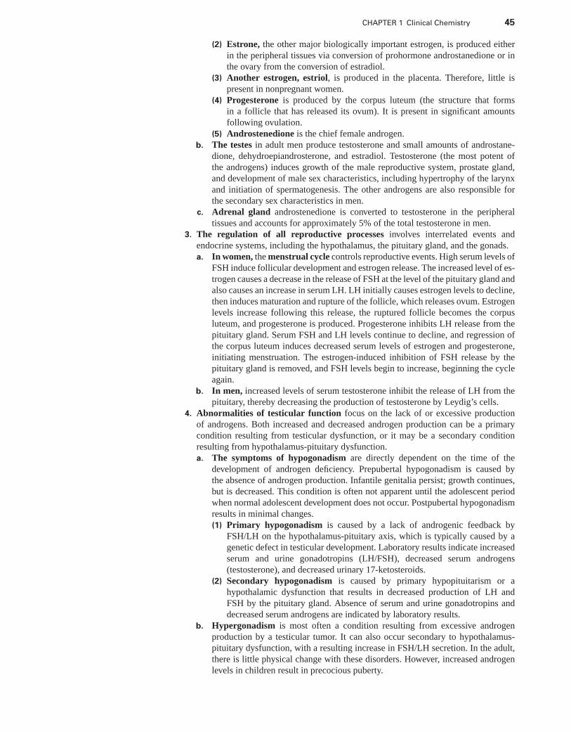
P1: OSO
LWBK192-01 LWBK192-Hubbard November 23, 2008 17:19
CHAPTER 1 Clinical Chemistry 45
(2) Estrone, the other major biologically important estrogen, is produced eitherin the peripheral tissues via conversion of prohormone androstanedione or inthe ovary from the conversion of estradiol.
(3) Another estrogen, estriol, is produced in the placenta. Therefore, little ispresent in nonpregnant women.
(4) Progesterone is produced by the corpus luteum (the structure that formsin a follicle that has released its ovum). It is present in significant amountsfollowing ovulation.
(5) Androstenedione is the chief female androgen.b. The testes in adult men produce testosterone and small amounts of androstane-
dione, dehydroepiandrosterone, and estradiol. Testosterone (the most potent ofthe androgens) induces growth of the male reproductive system, prostate gland,and development of male sex characteristics, including hypertrophy of the larynxand initiation of spermatogenesis. The other androgens are also responsible forthe secondary sex characteristics in men.
c. Adrenal gland androstenedione is converted to testosterone in the peripheraltissues and accounts for approximately 5% of the total testosterone in men.
3. The regulation of all reproductive processes involves interrelated events andendocrine systems, including the hypothalamus, the pituitary gland, and the gonads.a. In women, the menstrual cycle controls reproductive events. High serum levels of
FSH induce follicular development and estrogen release. The increased level of es-trogen causes a decrease in the release of FSH at the level of the pituitary gland andalso causes an increase in serum LH. LH initially causes estrogen levels to decline,then induces maturation and rupture of the follicle, which releases ovum. Estrogenlevels increase following this release, the ruptured follicle becomes the corpusluteum, and progesterone is produced. Progesterone inhibits LH release from thepituitary gland. Serum FSH and LH levels continue to decline, and regression ofthe corpus luteum induces decreased serum levels of estrogen and progesterone,initiating menstruation. The estrogen-induced inhibition of FSH release by thepituitary gland is removed, and FSH levels begin to increase, beginning the cycleagain.
b. In men, increased levels of serum testosterone inhibit the release of LH from thepituitary, thereby decreasing the production of testosterone by Leydig’s cells.
4. Abnormalities of testicular function focus on the lack of or excessive productionof androgens. Both increased and decreased androgen production can be a primarycondition resulting from testicular dysfunction, or it may be a secondary conditionresulting from hypothalamus-pituitary dysfunction.a. The symptoms of hypogonadism are directly dependent on the time of the
development of androgen deficiency. Prepubertal hypogonadism is caused bythe absence of androgen production. Infantile genitalia persist; growth continues,but is decreased. This condition is often not apparent until the adolescent periodwhen normal adolescent development does not occur. Postpubertal hypogonadismresults in minimal changes.(1) Primary hypogonadism is caused by a lack of androgenic feedback by
FSH/LH on the hypothalamus-pituitary axis, which is typically caused by agenetic defect in testicular development. Laboratory results indicate increasedserum and urine gonadotropins (LH/FSH), decreased serum androgens(testosterone), and decreased urinary 17-ketosteroids.
(2) Secondary hypogonadism is caused by primary hypopituitarism or ahypothalamic dysfunction that results in decreased production of LH andFSH by the pituitary gland. Absence of serum and urine gonadotropins anddecreased serum androgens are indicated by laboratory results.
b. Hypergonadism is most often a condition resulting from excessive androgenproduction by a testicular tumor. It can also occur secondary to hypothalamus-pituitary dysfunction, with a resulting increase in FSH/LH secretion. In the adult,there is little physical change with these disorders. However, increased androgenlevels in children result in precocious puberty.
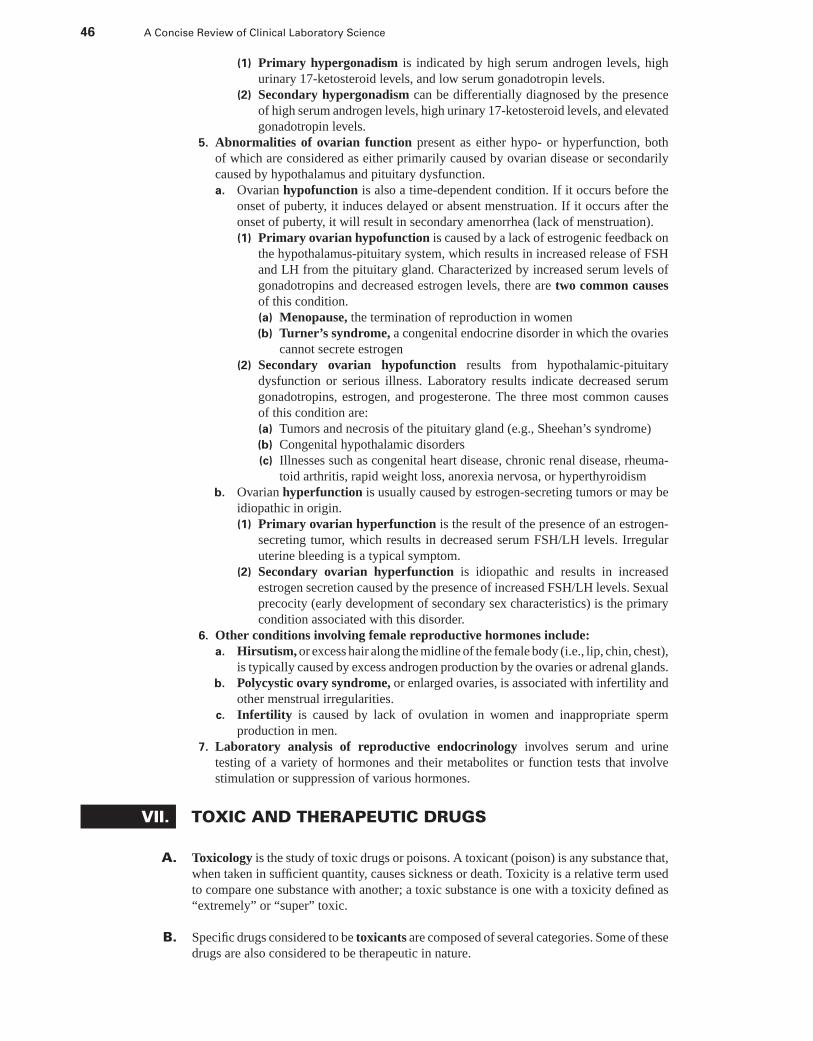
P1: OSO
LWBK192-01 LWBK192-Hubbard November 23, 2008 17:19
46 A Concise Review of Clinical Laboratory Science
(1) Primary hypergonadism is indicated by high serum androgen levels, highurinary 17-ketosteroid levels, and low serum gonadotropin levels.
(2) Secondary hypergonadism can be differentially diagnosed by the presenceof high serum androgen levels, high urinary 17-ketosteroid levels, and elevatedgonadotropin levels.
5. Abnormalities of ovarian function present as either hypo- or hyperfunction, bothof which are considered as either primarily caused by ovarian disease or secondarilycaused by hypothalamus and pituitary dysfunction.a. Ovarian hypofunction is also a time-dependent condition. If it occurs before the
onset of puberty, it induces delayed or absent menstruation. If it occurs after theonset of puberty, it will result in secondary amenorrhea (lack of menstruation).(1) Primary ovarian hypofunction is caused by a lack of estrogenic feedback on
the hypothalamus-pituitary system, which results in increased release of FSHand LH from the pituitary gland. Characterized by increased serum levels ofgonadotropins and decreased estrogen levels, there are two common causesof this condition.(a) Menopause, the termination of reproduction in women(b) Turner’s syndrome, a congenital endocrine disorder in which the ovaries
cannot secrete estrogen(2) Secondary ovarian hypofunction results from hypothalamic-pituitary
dysfunction or serious illness. Laboratory results indicate decreased serumgonadotropins, estrogen, and progesterone. The three most common causesof this condition are:(a) Tumors and necrosis of the pituitary gland (e.g., Sheehan’s syndrome)(b) Congenital hypothalamic disorders(c) Illnesses such as congenital heart disease, chronic renal disease, rheuma-
toid arthritis, rapid weight loss, anorexia nervosa, or hyperthyroidismb. Ovarian hyperfunction is usually caused by estrogen-secreting tumors or may be
idiopathic in origin.(1) Primary ovarian hyperfunction is the result of the presence of an estrogen-
secreting tumor, which results in decreased serum FSH/LH levels. Irregularuterine bleeding is a typical symptom.
(2) Secondary ovarian hyperfunction is idiopathic and results in increasedestrogen secretion caused by the presence of increased FSH/LH levels. Sexualprecocity (early development of secondary sex characteristics) is the primarycondition associated with this disorder.
6. Other conditions involving female reproductive hormones include:a. Hirsutism, or excess hair along the midline of the female body (i.e., lip, chin, chest),
is typically caused by excess androgen production by the ovaries or adrenal glands.b. Polycystic ovary syndrome, or enlarged ovaries, is associated with infertility and
other menstrual irregularities.c. Infertility is caused by lack of ovulation in women and inappropriate sperm
production in men.7. Laboratory analysis of reproductive endocrinology involves serum and urine
testing of a variety of hormones and their metabolites or function tests that involvestimulation or suppression of various hormones.
VII. TOXIC AND THERAPEUTIC DRUGS
A. Toxicology is the study of toxic drugs or poisons. A toxicant (poison) is any substance that,when taken in sufficient quantity, causes sickness or death. Toxicity is a relative term usedto compare one substance with another; a toxic substance is one with a toxicity defined as“extremely” or “super” toxic.
B. Specific drugs considered to be toxicants are composed of several categories. Some of thesedrugs are also considered to be therapeutic in nature.
P1: OSO
LWBK192-01 LWBK192-Hubbard November 23, 2008 17:19
CHAPTER 1 Clinical Chemistry 47
1. Analgesics are anti-inflammatory agents and painkillers. These drugs are also consid-ered to be therapeutic.a. Salicylate (aspirin) is considered toxic at a serum level of >90 mg/dL 6 hours
following ingestion. The time since ingestion must be known to determine severityof toxicity.(1) Salicylate intoxication results in the following: stimulation of the respiratory
system with initial respiratory alkalosis, conversion of pyruvate to lactate, in-hibition of oxidative phosphorylation, and breakdown of fatty acids to produceketoacids. Eventually, metabolic acidosis occurs.
(2) Renal clearance of salicylate can be increased by forced alkaline diuresis.b. Acetaminophen, if present in serum at 300 mcg/mL 2 hours after ingestion, in-
duces hepatic toxicity. Again, time since ingestion is critical in determining ac-etaminophen intoxication.(1) Intoxication results in hepatocystic necrosis 3 to 4 days after overdose be-
cause of the inability of the liver to adequately conjugate the metabolite ofacetaminophen (i.e., acetamidoquinone) to glutathione. High levels of acetami-doquinone in the liver induce hepatocyte death.
(2) Antidote. The effective antidote for acetaminophen overdose is N-acetylcysteine, which is thought to act as a glutathione substitute and binds tothe metabolite.
2. Barbiturates are short-acting (pento-, secobarbital); intermediate-acting (amo-, β-,butabarbital); and long-acting (phenobarbital) sedatives that exert a tranquilizing effectthrough their depressant effect on the CNS. The barbiturates can be considered assubstances of abuse or as analgesics.a. Barbiturate intoxication results in cardiac arrest and respiratory depression through
its effect on the CNS.b. There are no real antidotes for barbiturate overdose except the establishment of
an open airway, aiding respiration, and maintaining cardiac output.3. Narcotics (opioids) are compounds include heroin, morphine, codeine, and methadone.
Most of these drugs are habit forming and can be considered drugs of abuse.a. Intoxication. The toxic effects of overdose include depression of respiration caused
by a decreased sensitivity to carbon dioxide and coma. Heroin is metabolized bythe liver to form morphine and is excreted by the kidney as morphine glucuronide.
b. Antidote. The effective antidote for narcotic overdose is naloxone, a narcoticantagonist.
4. Pesticides exist as organic complexes, organophosphates (largest single group of pes-ticides), and carbamates. These compounds inhibit AChE, which results in specificeffects on the heart and respiratory centers, muscle cramps, and certain CNS effects.a. Intoxication. The method used for the determination of pesticide poisoning is the
examination of PChE (an isoenzyme of AChE found in serum).b. Antidote. The effective antidote for pesticide poisoning is atropine sulfate.
5. Carbon monoxide is a tasteless gas with 200-fold greater affinity for hemoglobin thanoxygen.a. Intoxication. During carbon monoxide poisoning, hemoglobin cannot adequately
exchange carbon dioxide for oxygen because of the increased amount of carboxy-hemoglobin present. Suffocation and death follow.
b. Antidote. The effective antidote for carbon monoxide poisoning is removal of thesource of carbon monoxide or removal of the victim from the source.
6. Metal poisoning includes intoxication by the heavy metals lead, arsenic, and mercury.a. Lead poisoning is most typically caused by lead paint ingestion or continuous
exposure to lead in the soil.(1) Intoxication. Lead is found primarily in RBCs in intoxicated victims but pro-
duces widespread effects, such as gastrointestinal irritation, weight loss, kidneydamage, convulsions, and in children, altered cognition and encephalopathy.Death occurs because of peripheral vascular collapse or brain involvement.
(2) Antidote. Administration of ethylenediaminetetraacetic acid (EDTA), penicil-lamine, and other lead chelates that bind the lead and allow it to be excreted by

P1: OSO
LWBK192-01 LWBK192-Hubbard November 23, 2008 17:19
48 A Concise Review of Clinical Laboratory Science
the kidney. Examination of urinary ALA levels and RBC protoporphyrins candetermine the occurrence of lead poisoning after serum and urine lead levelshave returned to normal.
b. Mercury (mercury salts) is found in antibacterial agents, photographic reagents,pesticides, and batteries. Poisoning by these agents is typically the result of over-exposure or ingestion.(1) Intoxication. Effects of mercury poisoning include gastrointestinal irritation,
severe kidney damage, or neurologic symptoms.(2) Antidote. As with lead poisoning, chelates, such as EDTA, penicillamine, or
dimercaprol [also referred to as British antilewisite (BAL)] bind the mercuryand render it able to be excreted.
c. Arsenic is found in pesticides, weed killer, and some paints.(1) Intoxication. Arsenic induces purging gastroenteritis, shredding of the stom-
ach lining, and causes Mees’ lines in the fingernails because of keratin binding.Death typically occurs because of hemorrhagic gastroenteritis. Because arsenicis cleared rapidly from the blood, urine is the specimen of choice for analysis.
(2) Antidote. Chelation therapy with penicillamine or BAL is effective.7. Substances of abuse include a variety of compounds that, when found in high levels in
the urine or serum, can incriminate an individual. It is mandatory in substance-of-abuseanalysis to obtain a satisfactory specimen and to process the specimen in a secure partof the laboratory.a. Ethanol is the most common toxicant and substance of abuse seen in patients in
emergency departments in the United States. It acts by depressing the CNS andincreasing the heart rate and blood pressure. Ethanol metabolism occurs in theliver by alcohol dehydrogenase, which converts alcohol to acetaldehyde, which isthen excreted to acetate. A blood level of 0.1 g/dL is defined as intoxicating.
b. Methanol is widely used in paints, solvents, antifreeze, and solid canned fuels.Poisoning with methanol is typically caused by ingestion.(1) Intoxication by methanol is considered more dangerous than ethanol poison-
ing because of the formation in humans of formaldehyde and formic acid asmetabolites. Formic acid formation results in metabolic acidosis, pancreaticnecrosis, and visual system impairment.
(2) Antidotes for methanol poisoning include the administration of sodium bi-carbonate to treat the acidosis and ethanol to bind alcohol dehydrogenase andblock the formation of the metabolites.
c. Amphetamines are CNS stimulants that block dopamine receptors in the brain.Amphetamine metabolism occurs in the liver and produces benzoic acid.(1) Intoxication by amphetamines can produce severe depression, respiratory dif-
ficulties, and episodes of paranoia.(2) Antidote. The antidote for amphetamine overdose is forced acid diuresis.
d. Cocaine is a CNS stimulant that is metabolized by cholinesterase to form ben-zoylecgonine, which is excreted by the kidney.(1) Intoxication. Cocaine overdose produces hypertension, myocardial infarction,
or seizure. Cardiotoxicity can occur and result in sudden death following co-caine use.
(2) Antidote. There is no antidote for cocaine overdose except the passage of timeand urinary excretion.
e. Cannabinoid compounds [tetrahydrocannabinol (THC), marijuana] producepsychologic effects and are stored in fat cells. THC is excreted in the urine for anextended period of time depending on use. Overdose of this drug is rare and is notsevere enough to be life-threatening.
f. Phencyclidine (PCP, angel dust) is an abused anesthetic that is illegally used asa hallucinogen.(1) Intoxication. This drug can produce violence, seizures, respiratory depression,
or death.(2) Antidote. Treatment for PCP overdose is diazepam. PCP is unmetabolized and
excreted in the urine as phencyclidine.
P1: OSO
LWBK192-01 LWBK192-Hubbard November 23, 2008 17:19
CHAPTER 1 Clinical Chemistry 49
C. The drug screen rapidly identifies a drug or drugs present in the blood, urine, or gas-tric contents of a patient suffering from toxicity. Neutral and basic drugs as well as drugmetabolites are best detected in urine, whereas acidic drugs are best found in detectableconcentrations in blood and serum. Following a positive drug screen, confirmatory methodsmust be used to quantitatively analyze drug levels in a patient.
1. Handheld immunoassays are the most common type of drug screen. They detect awide variety of drugs but cannot separate closely related compounds. Blood and urinecan both be analyzed with this method.
2. Gas-liquid chromatography allows for greater sensitivity in the identification ofdrugs. It can be used as a confirmatory technique for drugs detected by drug screen.
D. Confirmatory drug tests are required to confirm and quantify those drugs found in apatient’s serum or urine using drug-screening methods.
1. Gas chromatography/mass spectrometry is a sensitive technique used to confirmdrugs detected by screening techniques. Typically, urine samples are initially analyzedby gas chromatography to determine the presence of compounds, then reanalyzed bymass spectrometry to examine fragments of these compounds for relative abundancein the sample.
2. Immunoassay techniques use antibodies to detect drugs. These methods are usuallyautomated and in the form of enzyme immunoassays.
3. Ethanol testing is typically performed using gas chromatography. However, an enzymeassay using alcohol dehydrogenase and measuring the increase in NADH formationfollowing the reaction is widely used and can be automated.
4. Heavy metal testing is most often performed by atomic absorption spectrophotometry.
E. Therapeutic drugs must be monitored to determine what doses are inadequate or exces-sive in the treatment of the patient. Often, the ingested drug (called the “parent” drug)is metabolized to form an active metabolite that produces an effect similar to the parentdrug.
1. Cardioactive drugs are divided into two categories: the cardiac glycosides and theantiarrhythmic drugs. These agents serve to maintain normal heart function.a. Digoxin is the major cardiac glycoside and alters the force of contraction through
its effect on the ATPase pump in heart muscle. Blood specimens must be collected8 hours after a dose of digoxin is administered, because its peak concentrationin tissue occurs 6 to 10 hours after administration. Digoxin toxicity producessymptoms of nausea, rapid heart rate, and visual impairment. Digoxin is excretedas digoxigenin in the urine.
b. The antiarrhythmic drugs are prescribed to treat irregular heartbeat that producesinappropriate ventricular contraction or tachycardia (increased heart rate).(1) Lidocaine is used for the treatment of faulty ventricular contractions and ar-
rhythmias. It binds to α1-acid glycoprotein and is metabolized in the liver, pro-ducing two active metabolites, monoethylglycinexylidide and glycinexylide.
(2) Procainamide is used to treat inappropriate ventricular contractions and tachy-cardia. This drug is metabolized in the liver to form an active metabolite, N-acetylprocainamide, which produces the same effect as its parent drug. There-fore, serum levels of both drugs must be analyzed.
(3) Disopyramide stabilizes the heartbeat. It is both excreted by the renal systemas the unchanged drug and is metabolized in the liver to form an inactivemetabolite.
(4) Quinidine is a myocardial depressant that decreases the heart’s ability to con-duct current. It is metabolized in the liver to produce several active metabolites,including 3-hydroxyquinidine. If quinidine is added to a digoxin therapy regi-men, an interaction occurs that induces an increase in digoxin concentration.
(5) Propranolol is prescribed for atrial and ventricular arrhythmias and hyperten-sion. It is considered to be a beta-blocker.
2. Anticonvulsants function to alter transmission of nerve impulses within the brain tominimize the seizures of epilepsy.
P1: OSO
LWBK192-01 LWBK192-Hubbard November 23, 2008 17:19
50 A Concise Review of Clinical Laboratory Science
a. Phenobarbital is used to treat all types of seizures except absence seizures. Itis effective in children and neonates. It is metabolized in the liver, and serumconcentrations increase during the administration of valproic acid or salicylicacid.
b. Phenytoin corrects grand mal seizures. It is metabolized by the liver and caninteract with several drugs that induce increased serum concentration or increasedmetabolism of phenytoin.
c. Valproic acid is prescribed for absence (petit mal) seizures. Valproic acid affectsmany others anticonvulsants by inhibiting their metabolism in the liver, thus in-creasing serum concentration.
d. Primidone is metabolized in the liver to form phenobarbital. Therefore, dual anal-yses must be performed to determine the proper dosage of this drug. It is used totreat both grand mal and complex-partial seizures.
e. Carbamazepine is typically used for treatment of various seizures and facial pain.f. Ethosuximide is prescribed for the treatment of petit mal seizures.
3. Bronchodilators act to relax bronchial smooth muscle for relief or prevention ofasthma. Theophylline is the most common in this category of therapeutic drugs and ismetabolized in the liver to produce several metabolites, including caffeine.
4. Psychotropic or antipsychotic drugs are used to treat psychotic patients. They canbe categorized in two classes: lithium and the antidepressants.a. Lithium treats manic-depressive illness. The mechanism of action of lithium as
a mood stabilizer remains unknown, although effects on synaptic neurotransmis-sion are thought to be the cause. Lithium is filtered by the renal glomerulus andeliminated as the unchanged drug.
b. Antidepressants, or tricyclic antidepressants, are used to treat depression thathas no apparent organic or social cause. Antidepressants include imipramine, nor-triptyline, amitriptyline, and desipramine, all of which are metabolized by the liverto form active metabolites. The active metabolites include desipramine (parentis imipramine), nortriptyline (parent is amitriptyline), and 2-hydroxy-desipramine(parent is desipramine).
c. Fluoxetine is not chemically related to the tricyclic antidepressants, but has asimilar effect by blocking serotonin uptake by nerve terminals in the CNS and byplatelets.
5. Antineoplastic drugs are used in the management of certain tumors, including thosefound in breast, testicular, pharyngeal, and sometimes lung cancer. These agents workby inhibiting DNA synthesis.
F. Therapeutic drug monitoring (TDM) is performed to determine patient compliance tothe drug-taking regimen, to monitor drug interactions, and to monitor drugs that are usedfor a preventive effect.
1. Changes in drug concentrations in the body, which occur with time, are related to thecourse of the pharmacologic effects. The change in drug concentration over time isdescribed by the following steps.a. Liberation is the release of this ingredient, followed by the process of the drug
passing into solution.b. Absorption is the process by which the drug molecule is taken up into systemic
circulation. Following absorption through the intestinal mucosa, a drug traverses thehepatic system, where some drugs undergo substantial metabolism and elimination.This is called first-pass elimination or metabolism.
c. Drug molecules can be confined to the blood, leave the bloodstream, and enter theextravascular space, or they can migrate into various tissues. This is referred to asdistribution, a process that typically occurs between a period of 30 minutes and2 hours. The bioavailability of a drug is the amount of drug that is absorbed intothe system and is available for distribution.
d. Metabolism is the process of transformation of the parent drug molecule to itsmetabolite(s). Metabolites are usually water soluble and can be easily excreted.Most of the metabolism occurs in the liver, where enzymes catalyze oxidation,reduction, or hydrolysis of the drug.
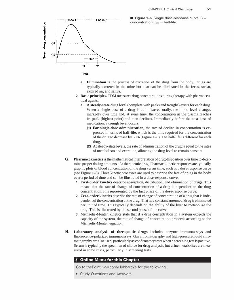
P1: OSO
LWBK192-01 LWBK192-Hubbard November 23, 2008 17:19
CHAPTER 1 Clinical Chemistry 51
■ Figure 1–6 Single dose-response curve. C =concentration; t1/2 = half-life.
e. Elimination is the process of excretion of the drug from the body. Drugs aretypically excreted in the urine but also can be eliminated in the feces, sweat,expired air, and saliva.
2. Basic principles. TDM measures drug concentrations during therapy with pharmaceu-tical agents.a. A steady-state drug level (complete with peaks and troughs) exists for each drug.
When a single dose of a drug is administered orally, the blood level changesmarkedly over time and, at some time, the concentration in the plasma reachesits peak (highest point) and then declines. Immediately before the next dose ofmedication, a trough level occurs.(1) For single-dose administration, the rate of decline in concentration is ex-
pressed in terms of half-life, which is the time required for the concentrationof the drug to decrease by 50% (Figure 1–6). The half-life is different for eachdrug.
(2) At steady-state levels, the rate of administration of the drug is equal to the ratesof metabolism and excretion, allowing the drug level to remain constant.
G. Pharmacokinetics is the mathematical interpretation of drug disposition over time to deter-mine proper dosing amounts of a therapeutic drug. Pharmacokinetic responses are typicallygraphic plots of blood concentration of the drug versus time, such as a dose-response curve(see Figure 1–6). Three kinetic processes are used to describe the fate of drugs in the bodyover a period of time and can be illustrated in a dose-response curve.1. First-order kinetics describe absorption, distribution, and elimination of drugs. This
means that the rate of change of concentration of a drug is dependent on the drugconcentration. It is represented by the first phase of the dose-response curve.
2. Zero-order kinetics describe the rate of change of concentration of a drug that is inde-pendent of the concentration of the drug. That is, a constant amount of drug is eliminatedper unit of time. This typically depends on the ability of the liver to metabolize thedrug. This is illustrated by the second phase of the curve.
3. Michaelis-Menten kinetics state that if a drug concentration in a system exceeds thecapacity of the system, the rate of change of concentration proceeds according to theMichaelis-Menten equation.
H. Laboratory analysis of therapeutic drugs includes enzyme immunoassays andfluorescence-polarized immunoassays. Gas chromatography and high-pressure liquid chro-matography are also used, particularly as confirmatory tests when a screening test is positive.Serum is typically the specimen of choice for drug analysis, but urine metabolites are mea-sured in some cases, particularly in screening tests.
Online Menu for this Chapter
Go to thePoint.lww.com/Hubbard2e for the following:
� Study Questions and Answers
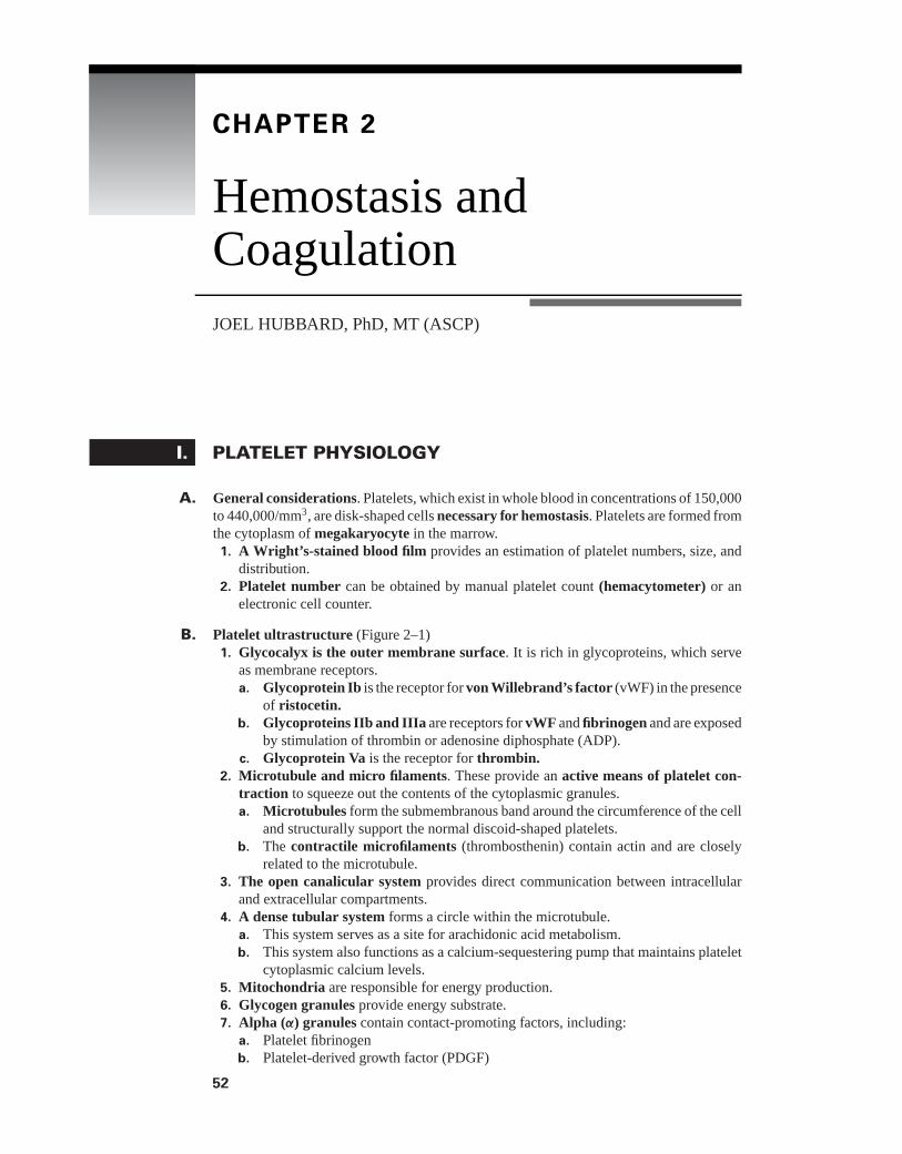
P1: OSO
LWBK192-02 LWBK192-Hubbard November 23, 2008 17:20
CHAPTER 2
Hemostasis andCoagulation
JOEL HUBBARD, PhD, MT (ASCP)
I. PLATELET PHYSIOLOGY
A. General considerations. Platelets, which exist in whole blood in concentrations of 150,000to 440,000/mm3, are disk-shaped cells necessary for hemostasis. Platelets are formed fromthe cytoplasm of megakaryocyte in the marrow.1. A Wright’s-stained blood film provides an estimation of platelet numbers, size, and
distribution.2. Platelet number can be obtained by manual platelet count (hemacytometer) or an
electronic cell counter.
B. Platelet ultrastructure (Figure 2–1)1. Glycocalyx is the outer membrane surface. It is rich in glycoproteins, which serve
as membrane receptors.a. Glycoprotein Ib is the receptor for von Willebrand’s factor (vWF) in the presence
of ristocetin.b. Glycoproteins IIb and IIIa are receptors for vWF and fibrinogen and are exposed
by stimulation of thrombin or adenosine diphosphate (ADP).c. Glycoprotein Va is the receptor for thrombin.
2. Microtubule and micro filaments. These provide an active means of platelet con-traction to squeeze out the contents of the cytoplasmic granules.a. Microtubules form the submembranous band around the circumference of the cell
and structurally support the normal discoid-shaped platelets.b. The contractile microfilaments (thrombosthenin) contain actin and are closely
related to the microtubule.3. The open canalicular system provides direct communication between intracellular
and extracellular compartments.4. A dense tubular system forms a circle within the microtubule.
a. This system serves as a site for arachidonic acid metabolism.b. This system also functions as a calcium-sequestering pump that maintains platelet
cytoplasmic calcium levels.5. Mitochondria are responsible for energy production.6. Glycogen granules provide energy substrate.7. Alpha (α) granules contain contact-promoting factors, including:
a. Platelet fibrinogenb. Platelet-derived growth factor (PDGF)
52
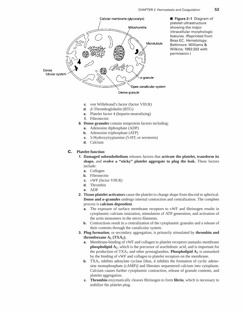
P1: OSO
LWBK192-02 LWBK192-Hubbard November 23, 2008 17:20
CHAPTER 2 Hemostasis and Coagulation 53
■ Figure 2–1 Diagram of
platelet ultrastructure
showing the major
intracellular morphologic
features. (Reprinted from
Besa EC. Hematology.
Baltimore: Williams &
Wilkins; 1992:202 with
permission.)
c. von Willebrand’s factor (factor VIII:R)d. β-Thromboglobulin (BTG)e. Platelet factor 4 (heparin-neutralizing)f. Fibronectin
8. Dense granules contain nonprotein factors including:a. Adenosine diphosphate (ADP)b. Adenosine triphosphate (ATP)c. 5-Hydroxytryptamine (5-HT; or serotonin)d. Calcium
C. Platelet function1. Damaged subendothelium releases factors that activate the platelet, transform its
shape, and evolve a “sticky” platelet aggregate to plug the leak. These factorsinclude:a. Collagenb. Fibronectinc. vWF (factor VIII:R)d. Thrombine. ADP
2. Tissue platelet activators cause the platelet to change shape from discoid to spherical.Dense and α-granules undergo internal contraction and centralization. The completeprocess is calcium dependent.a. The exposure of surface membrane receptors to vWF and fibrinogen results in
cytoplasmic calcium ionization, stimulation of ATP generation, and activation ofthe actin monomers in the micro filaments.
b. Contractions result in a centralization of the cytoplasmic granules and a release oftheir contents through the canalicular system.
3. Plug formation, or secondary aggregation, is primarily stimulated by thrombin andthromboxane A2 (TXA2).a. Membrane-binding of vWF and collagen to platelet receptors unmasks membrane
phospholipid A2, which is the precursor of arachidonic acid, and is important forthe production of TXA2 and other prostaglandins. Phospholipid A2 is unmaskedby the binding of vWF and collagen to platelet receptors on the membrane.
b. TXA2 inhibits adenylate cyclase [thus, it inhibits the formation of cyclic adeno-sine monophosphate (cAMP)] and liberates sequestered calcium into cytoplasm.Calcium causes further cytoplasmic contraction, release of granule contents, andplatelet aggregation.
c. Thrombin enzymatically cleaves fibrinogen to form fibrin, which is necessary tostabilize the platelet plug.
P1: OSO
LWBK192-02 LWBK192-Hubbard November 23, 2008 17:20
54 A Concise Review of Clinical Laboratory Science
4. Growth-limiting factors of the platelet aggregate include:a. Blood flow, which washes away coagulation-promoting factorsb. Release of prostaglandin PGI2 (prostacyclin) by the surrounding vascular
tissues5. Granular release of substances from dense granules, such as serotonin, prostaglandins
(except for TXA2), and lysozymes, causes local inflammation and vasodilation, whichincreases blood flow.
D. Kinetics1. Megakaryocytopoiesis development occurs by endomitosis (i.e., nuclear splitting
without cell division).a. A single megakaryoblast nucleus may contain 2 to 64 times the normal number of
chromosomes.b. Nuclear chromatin is densely staining, dispersed early, and more compact at later
stages.c. Nucleoli are small at all development stages.
2. Stages of megakaryocytic maturation (Figure 2–2)a. Megakaryoblasts descend from a unipotential stem cell (CFU-Meg). They are
characterized by overlapping nuclear lobes and basophilic staining cytoplasm; theirsize ranges from 6 to 24 μm.
b. Promegakaryocytes are larger than megakaryoblasts (14 to 30 μm in diameter)and have more cytoplasm.(1) The nucleus becomes increasingly lobulated and spreads out into a horseshoe
shape.(2) Red-pink granules are visible in the center of the cell.
c. Granular megakaryocytes (16 to 56 μm) are characterized by increased spreadingof nuclear lobes and spreading of pink granules throughout the cytoplasm.
d. Mature megakaryocytes (20 to 50 μm) have a compact nucleus, and the basophiliaof cytoplasm has disappeared.(1) Platelet fields are clusters of pink granules in the cytoplasm.
(a) Platelet fields are produced by an invagination of surface membrane, sep-arating the cytoplasm into individual platelets.
■ Figure 2–2 Morphologic characteristics of stages of megakaryocytic maturation comparing the
nuclear and cytoplasmic variations of each stage. N:C, nuclear-cytoplasmic ratio.
P1: OSO
LWBK192-02 LWBK192-Hubbard November 23, 2008 17:20
CHAPTER 2 Hemostasis and Coagulation 55
(b) Individual platelets are shed from the megakaryocyte cytoplasm into themarrow sinuses, and then are released into the vascular lumen.
(2) Each mature megakaryocyte produces from 2,000 to 7,000 platelets that eachrange in size from 2 to 3 μm.
3. Maturation time from the blast stage to platelet formation is typically 5 days.4. Normal marrow contains approximately 15 million megakaryocyte. This equates to
approximately 5 to 10 megakaryocytes per 10× power field when bone marrow smearsare microscopically examined.
5. Normal circulation life of a platelet is 8 to 10 days.6. Platelets are removed by macrophage in the liver and spleen or by active use in daily
coagulation mechanisms.7. Circulating platelets are distributed between the spleen and blood.
a. One third of the circulating platelets are always in the spleen.b. The platelet count is higher in patients without a spleen and lower in patients
with splenomegaly (enlarged spleen).8. Regulation of the platelet count
a. Under normal conditions the platelet count (or mass) is constant, even with activeuse. This indicates a feedback system that adjusts production to consumption.(1) Rebound thrombocytopenia occurs after platelet transfusion.(2) Rebound thrombocytosis occurs after platelet depletion.
b. Feedback stimulus results in an increased megakaryoblast endomitosis, whichincreases platelet volume and number. It also affects committed unipotential stemcells, which results in more megakaryoblast.
E. Laboratory measurements of platelet activities1. Initial evaluation includes a platelet count and slide estimate, with a reference value
of 150,000 to 440,000/mm3.2. Bleeding time is an effective in vivo screening test of platelet function by timing the
length it takes for platelets to plug broken capillaries after a small cut is made in theforearm.a. Reference values are approximately 3 to 8 minutes.b. Increased bleeding times are seen in:
(1) Patients taking drugs with antiplatelet action (e.g., aspirin)(2) Patients with von Willebrand’s disease (vWD)(3) Patients who suffer from congenital platelet abnormalities(4) Patients with platelet counts lower than 100,000/mm3
3. Platelet aggregation is measured with a platelet aggregometer.a. Basic principle. Citrated, platelet-rich plasma is stirred in the aggregometer while
a light beam is passed through the suspension.b. A chemical stimulus is added [e.g., collagen, epinephrine, ADP, ristocetin,
A23187 (calcium ionophore), arachidonic acid, γ -thrombin (partially trypsinizedthrombin)].
c. γ-Thrombin retains aggregating properties but lacks clotting ability.d. The shape change from discoid to spheroid is monitored as an initial decrease in
light transmittance.e. Subsequent aggregation allows an increase of light to pass through the suspension
to the photodetector and to be recorded as an increase in light transmittance.
II. PLATELET PATHOPHYSIOLOGY
A. Quantitative platelet disorders (Figure 2–3)1. Thrombocytopenia is characterized by a decrease in the number of circulating platelets
(i.e., <100,000/mm3). Clinical evidence of thrombocytopenia includes an increasednumber of petechiae, hemorrhages, prolonged bleeding time, and impaired clot retrac-tion. Decreased circulating platelet counts result from the following conditions:a. Low platelet counts can result from defective production in the bone marrow.
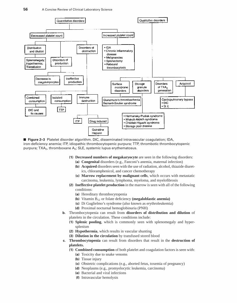
P1: OSO
LWBK192-02 LWBK192-Hubbard November 23, 2008 17:20
56 A Concise Review of Clinical Laboratory Science
■ Figure 2–3 Platelet disorder algorithm. DIC, disseminated intravascular coagulation; IDA,
iron-deficiency anemia; ITP, idiopathic thrombocytopenic purpura; TTP, thrombotic thrombocytopenic
purpura; TXA2, thromboxane A2; SLE, systemic lupus erythematosus.
(1) Decreased numbers of megakaryocyte are seen in the following disorders:(a) Congenital disorders (e.g., Fanconi’s anemia, maternal infection)(b) Acquired disorders seen with the use of radiation, alcohol, thiazide diuret-
ics, chloramphenicol, and cancer chemotherapy(c) Marrow replacement by malignant cells, which occurs with metastatic
carcinoma, leukemia, lymphoma, myeloma, and myelofibrosis(2) Ineffective platelet production in the marrow is seen with all of the following
conditions:(a) Hereditary thrombocytopenia(b) Vitamin B12 or folate deficiency (megaloblastic anemia)(c) Di Guglielmo’s syndrome (also known as erythroleukemia)(d) Proximal nocturnal hemoglobinuria (PNH)
b. Thrombocytopenia can result from disorders of distribution and dilution ofplatelets in the circulation. These conditions include:(1) Splenic pooling, which is commonly seen with splenomegaly and hyper-
splenism(2) Hypothermia, which results in vascular shunting(3) Dilution in the circulation by transfused stored blood
c. Thrombocytopenia can result from disorders that result in the destruction ofplatelets.(1) Combined consumption of both platelet and coagulation factors is seen with:
(a) Toxicity due to snake venoms(b) Tissue injury(c) Obstetric complications (e.g., aborted fetus, toxemia of pregnancy)(d) Neoplasms (e.g., promyelocytic leukemia, carcinoma)(e) Bacterial and viral infections(f) Intravascular hemolysis
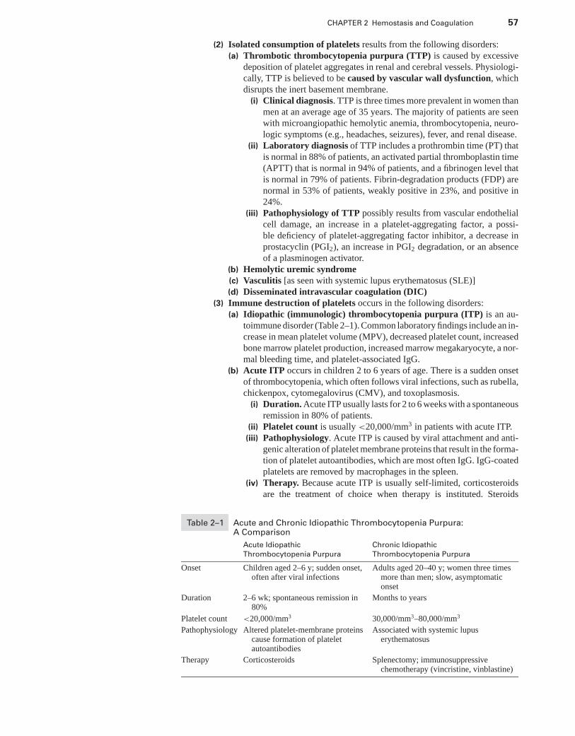
P1: OSO
LWBK192-02 LWBK192-Hubbard November 23, 2008 17:20
CHAPTER 2 Hemostasis and Coagulation 57
(2) Isolated consumption of platelets results from the following disorders:(a) Thrombotic thrombocytopenia purpura (TTP) is caused by excessive
deposition of platelet aggregates in renal and cerebral vessels. Physiologi-cally, TTP is believed to be caused by vascular wall dysfunction, whichdisrupts the inert basement membrane.
(i) Clinical diagnosis. TTP is three times more prevalent in women thanmen at an average age of 35 years. The majority of patients are seenwith microangiopathic hemolytic anemia, thrombocytopenia, neuro-logic symptoms (e.g., headaches, seizures), fever, and renal disease.
(ii) Laboratory diagnosis of TTP includes a prothrombin time (PT) thatis normal in 88% of patients, an activated partial thromboplastin time(APTT) that is normal in 94% of patients, and a fibrinogen level thatis normal in 79% of patients. Fibrin-degradation products (FDP) arenormal in 53% of patients, weakly positive in 23%, and positive in24%.
(iii) Pathophysiology of TTP possibly results from vascular endothelialcell damage, an increase in a platelet-aggregating factor, a possi-ble deficiency of platelet-aggregating factor inhibitor, a decrease inprostacyclin (PGI2), an increase in PGI2 degradation, or an absenceof a plasminogen activator.
(b) Hemolytic uremic syndrome(c) Vasculitis [as seen with systemic lupus erythematosus (SLE)](d) Disseminated intravascular coagulation (DIC)
(3) Immune destruction of platelets occurs in the following disorders:(a) Idiopathic (immunologic) thrombocytopenia purpura (ITP) is an au-
toimmune disorder (Table 2–1). Common laboratory findings include an in-crease in mean platelet volume (MPV), decreased platelet count, increasedbone marrow platelet production, increased marrow megakaryocyte, a nor-mal bleeding time, and platelet-associated IgG.
(b) Acute ITP occurs in children 2 to 6 years of age. There is a sudden onsetof thrombocytopenia, which often follows viral infections, such as rubella,chickenpox, cytomegalovirus (CMV), and toxoplasmosis.
(i) Duration. Acute ITP usually lasts for 2 to 6 weeks with a spontaneousremission in 80% of patients.
(ii) Platelet count is usually <20,000/mm3 in patients with acute ITP.(iii) Pathophysiology. Acute ITP is caused by viral attachment and anti-
genic alteration of platelet membrane proteins that result in the forma-tion of platelet autoantibodies, which are most often IgG. IgG-coatedplatelets are removed by macrophages in the spleen.
(iv) Therapy. Because acute ITP is usually self-limited, corticosteroidsare the treatment of choice when therapy is instituted. Steroids
Table 2–1 Acute and Chronic Idiopathic Thrombocytopenia Purpura:A Comparison
Acute Idiopathic Chronic Idiopathic
Thrombocytopenia Purpura Thrombocytopenia Purpura
Onset Children aged 2–6 y; sudden onset,often after viral infections
Adults aged 20–40 y; women three timesmore than men; slow, asymptomaticonset
Duration 2–6 wk; spontaneous remission in80%
Months to years
Platelet count <20,000/mm3 30,000/mm3–80,000/mm3
Pathophysiology Altered platelet-membrane proteinscause formation of plateletautoantibodies
Associated with systemic lupuserythematosus
Therapy Corticosteroids Splenectomy; immunosuppressivechemotherapy (vincristine, vinblastine)
P1: OSO
LWBK192-02 LWBK192-Hubbard November 23, 2008 17:20
58 A Concise Review of Clinical Laboratory Science
suppress macrophage phagocytic activity, decrease Fc-receptor func-tion, and decrease antibody platelet binding. Splenectomy is rarelyneeded, and platelet transfusion is ineffective.
(c) Chronic ITP occurs in adults 20 to 40 years of age. It is found in womenthree times more than in men and has a slow, asymptomatic onset of throm-bocytopenia.
(i) Duration. Chronic ITP can last from months to years.(ii) Platelet count usually ranges from 30,000 to 80,000/mm3.
(iii) Pathophysiology. Chronic ITP is often associated with SLE.(iv) Therapy. Splenectomy is the most common treatment because it
decreases the number of macrophage with Fc receptors. Immuno-suppressive chemotherapy with vincristine or vinblastine is used inseverely affected patients.
(d) Posttransfusion purpura occurs in 1% to 2% of persons who receiveblood transfusions. Production of antiplatelet antibodies by the recipientof platelet transfusions results in the destruction of platelets.
(e) Isoimmune neonatal purpura is caused by maternal viremia (e.g., CMVor rubella) or maternal drug ingestion.
(f) Drug-induced antibody formation is most commonly seen with the useof quinidine and heparin. The drugs function as haptens, combining witha serum protein and causing an antibody response. The drug-antibodycomplex attaches to platelets, which results in agglutination, complementfixation, and destruction by macrophages.
d. Thrombocytopenia can result from a combination of mechanisms (e.g., alco-holism, lymphoproliferative disorders).
e. Heparin-induced thrombocytopenia is observed in more than 10% of patientswho undergo heparin therapy.(1) Risk from thrombosis without heparin therapy is greater than the risk of bleed-
ing from heparin-induced thrombocytopenia.(2) The mechanism of thrombocytopenia is due to a direct platelet-aggregating
effect, as well as immune destruction by antiplatelet antibodies.(3) Laboratory diagnosis. A normal aggregation pattern in platelet aggregome-
ter studies is found, except that adding heparin as a stimulant will increaseaggregation instead of blunting the aggregation reaction.
f. Thrombocytopenia associated with human immunodeficiency virus (HIV) in-fection is severe but rarely hemorrhagic.(1) Characteristics similar to classic ITP include:
(a) Abundant megakaryocytes(b) Occasional giant platelets(c) Immune origin(d) Absence of splenomegaly
(2) Characteristics different from ITP include:(a) Greater levels of bound antibody(b) Involvement of immune complexes
2. Thrombocytosis is characterized by an increase in circulating platelet counts>450,000/mm3.a. Essential thrombocytosis is the result of a primary bone marrow disorder. Al-
though it is characterized by an increased number of platelets, it is caused by aclonal proliferation that affects all hemopoietic cells. Often, patients with a throm-bocytosis will have increased bleeding tendencies because of possible accompa-nying functional abnormalities. Essential thrombocytosis is most commonly seenin patients with the following disorders:(1) Hodgkin’s lymphoma(2) Polycythemia vera(3) Myelofibrosis(4) Chronic myelogenous leukemia(5) Thrombocythemia
P1: OSO
LWBK192-02 LWBK192-Hubbard November 23, 2008 17:20
CHAPTER 2 Hemostasis and Coagulation 59
b. Secondary thrombocytosis is a secondary response most commonly associatedwith the following disorders:(1) Iron-deficiency anemia associated with chronic blood loss(2) Chronic inflammatory disease may be associated with high platelet counts(3) Splenectomy-associated thrombocytosis(4) Rebound thrombocytosis, which may occur after a platelet depletion through
a massive blood loss
B. Qualitative platelet abnormalities1. Surface membrane defects that are genetically acquired
a. Glanzmann thrombasthenia(1) This disease is a homozygous autosomal recessive disorder in which one of
two genes coding for either the membrane receptor glycoprotein IIb (GPIIb)or for GPIIIa is affected. Both genes are found on chromosome 17. Males aswell as females are affected.
(2) Both receptors function in the aggregation process to anchor platelets to ex-posed collagen in the subendothelial tissue of damaged blood vessel walls. Theblood clot fails to form, because the dimer complex is no longer defective andplatelets can no longer bind to one another.
(3) Symptoms commonly present in infants and in early childhood.(a) Bruising (i.e., purpura)(b) Nose bleeds(c) Bleeding from the gums started by trauma or simple sneezing, crying,
coughing, or the eruption of a tooth(d) Female adolescents may experience heavy menorrhagia when they begin
their cycle.(e) In many, the severity of bleeding events lessens with age.
(4) Laboratory diagnosis of this disorder is for the most part straightforward.(a) Normal platelet count and morphology(b) Significantly prolonged bleeding time(c) Platelet aggregometer profile shows failure to aggregate with all agents
(i.e., ADP, adrenaline, thrombin, collagen), except von Willebrand factor.(5) Clinical variants of Glanzmann’s have been identified based on their patterns
of severity.(a) Type I is a severe deficiency of <5% of the glycoprotein complex.(b) Type II is considered a mild to moderate deficiency with 5% to 20% of
the complex remaining.(c) Clinical Type III is found to be a qualitative dysfunction.
(6) Prognosis and treatment is of a lifelong disorder. Glanzmann’s now is asso-ciated with a low death rate from hemorrhage with modern medical care andsupport.(a) The only curative treatment for Glanzmann’s disorder is the bone marrow
transplant.(b) Most treatments are supportive of the bleeding and preventive management.
The avoidance of trauma and drugs, such as aspirin, that have antiplateletactions are considered foremost.
(c) Treatment of bleeding episodes can include platelet transfusion or localapplication of thrombin and adrenaline solutions.
b. Bernard-Soulier syndrome (BSS)(1) Is directly due to an inherited quantitative or qualitative defect in the platelet
glycoprotein (GP)Ib/IX complex consisting of four platelet surface glyco-proteins (Ibα, Ibβ, IX, and V).(a) The GP Ibα gene is located on the short arm of chromosome 17, the GP
Ibβ gene is on the long arm of chromosome 22, and the GP IX and GPV genes are located on the long arm of chromosome 3.
(b) The Ib molecule carries both the high- and moderate-affinity receptor sitesfor thrombin.
P1: OSO
LWBK192-02 LWBK192-Hubbard November 23, 2008 17:20
60 A Concise Review of Clinical Laboratory Science
(c) This receptor complex mediates adhesion of platelets to the blood ves-sel wall at sites of injury by binding vWF and facilitates the ability ofthrombin to activate platelets.
(d) BSS is extremely rare, occurring <1 per 1 million.(2) Clinical symptoms can present early in life and most commonly include fre-
quent bouts of epistaxis, gingival and cutaneous bleeding, and hemorrhageassociated with trauma.
(3) Laboratory diagnosis commonly results in a prolonged Ivy bleeding time,giant platelets, and thrombocytopenia.(a) Platelet counts may range from very low (i.e., <30,000/mm3) to slightly
low or low normal.(b) Bleeding time results range from slightly prolonged (8–10 minutes) to
significantly prolonged (>20 minutes).(c) Examination of a blood smear and the MPV for giant platelets.(d) Platelet aggregation studies will show a reduced aggregation with risto-
cetin and a normal response to other agonists.(4) Because BSS involves gene mutations of several glycoproteins codes by several
different chromosomes, several clinical variants of the disorder have beenidentified.(a) Type A clinical variant is what is known as classic BSS due to mutation
in the GP Ibβ gene. A functional defect is often possible in which the GPIbα is expressed in a dysfunctional form that fails to bind ligand.
(b) Type B variants have also been identified where the genetic lesion may belocalized to the GP Ibα gene on chromosome 17, 22, or the GP IX geneon chromosome 3.
(c) Type C variants are due to GPV gene lesions on chromosome 3.(d) Rarely, cases of acquired BSS have been described in patients with
myelodysplasia and with acute myelogenous leukemia (M6).(5) Prognosis and treatment of these patients concentrates on supportive mea-
sures and the treatment of acute bleeding episodes.(a) Management should include the education of the patient and their fami-
lies about their condition and avoiding unnecessary risks, antiplatelet med-ication such as aspirin or a nonsteroidal anti-inflammatory, and adequatedental hygiene.
(b) Iron-deficiency anemia often develops due to chronic bleeding.(c) Splenectomy lessens the degree of thrombocytopenia and the severity of
some symptoms.(d) Often the transfusion of blood and platelets is necessary to control hem-
orrhage and thrombocytopenia with a traumatic event.(e) In future modes of treatment, BSS may be an ideal candidate for gene
therapy. Bone marrow or stem cell transplantation is still experimentalfor individuals with BSS.
c. Differential diagnosis of membrane-related disorders;(1) Glanzmann’s thrombasthenia and BSS have many clinical and physiological
similarities (Table 2–2).(a) Both result in chronic bleeding of mucous membranes. Treatments and
diagnosis will be different so it is important that a correct diagnosis ismade.
(b) Both disorders will have an increased bleeding time, but the plateletcount is commonly decreased in BSS with giant platelets.
(c) The aggregation patterns will typically give opposite results for eachdisorder.
(2) Bernard Soulier must be further differentiated from von Willebrand’s diseaseor ITP.(a) Von Willebrand’s disease also presents with an increased bleeding time
and impaired Ristocetin aggregation, but there is a decrease in vWF(VIII:R), not in the receptor binding site.
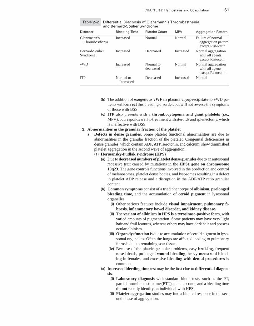
P1: OSO
LWBK192-02 LWBK192-Hubbard November 23, 2008 17:20
CHAPTER 2 Hemostasis and Coagulation 61
Table 2–2 Differential Diagnosis of Glanzmann’s Thrombastheniaand Bernard-Soulier Syndrome
Disorder Bleeding Time Platelet Count MPV Aggregation Pattern
Glanzmann’sThrombasthenia
Increased Normal Normal Failure of normalaggregation patternexcept Ristocetin
Bernard-SoulierSyndrome
Increased Decreased Increased Normal aggregationwith all agentsexcept Ristocetin
vWD Increased Normal todecreased
Normal Normal aggregationwith all agentsexcept Ristocetin
ITP Normal toIncreased
Decreased Increased Normal
(b) The addition of exogenous vWF in plasma cryoprecipitate to vWD pa-tients will correct this bleeding disorder, but will not reverse the symptomsof those with BSS.
(c) ITP also presents with a thrombocytopenia and giant platelets (i.e.,MPV), but responds well to treatment with steroids and splenectomy, whichis ineffective with BSS.
2. Abnormalities in the granular fraction of the plateleta. Defects in dense granules. Some platelet functional abnormalities are due to
abnormalities in the granular fraction of the platelet. Congenital deficiencies indense granules, which contain ADP, ATP, serotonin, and calcium, show diminishedplatelet aggregation in the second wave of aggregation.(1) Hermansky-Pudlak syndrome (HPS)
(a) Due to decreased numbers of platelet dense granules due to an autosomalrecessive trait caused by mutations in the HPS1 gene on chromosome10q23. The gene controls functions involved in the production and controlof melanosomes, platelet dense bodies, and lysosomes resulting in a defectin platelet ADP release and a disruption in the ADP/ATP ratio granularcontent.
(b) Common symptoms consist of a triad phenotype of albinism, prolongedbleeding time, and the accumulation of ceroid pigment in lysosomalorganelles.
(i) Other serious features include visual impairment, pulmonary fi-brosis, inflammatory bowel disorder, and kidney disease.
(ii) The variant of albinism in HPS is a tyrosinase-positive form, withvaried amounts of pigmentation. Some patients may have very lighthair and frail features, whereas others may have dark hair and possessocular albinism.
(iii) Organ dysfunction is due to accumulation of ceroid pigment in lyso-somal organelles. Often the lungs are affected leading to pulmonaryfibrosis due to remaining scar tissue.
(iv) Because of the platelet granular problems, easy bruising, frequentnose bleeds, prolonged wound bleeding, heavy menstrual bleed-ing in females, and excessive bleeding with dental procedures iscommon.
(c) Increased bleeding time test may be the first clue to differential diagno-sis.
(i) Laboratory diagnosis with standard blood tests, such as the PT,partial thromboplastin time (PTT), platelet count, and a bleeding timedo not readily identify an individual with HPS.
(ii) Platelet aggregation studies may find a blunted response in the sec-ond phase of aggregation.
P1: OSO
LWBK192-02 LWBK192-Hubbard November 23, 2008 17:20
62 A Concise Review of Clinical Laboratory Science
(iii) The most definite diagnosis can be found by electron microscopeobservation of the patient’s platelets and finding an absence of densegranules.
(2) Chediak-Higashi syndrome (CHS)(a) CHS is a generalized autosomal recessive genetic disorder with recurrent
infections in combination with ocular, neurological, and skin manifesta-tions.
(i) Onset in early childhood and death often occurs before the age of 7.(ii) Caused by mutations in the lysosomal trafficking regulator gene
(LYST) found on chromosome 1.(iii) Results in abnormal membrane fluidity, uncontrolled granule mem-
brane fusion, and formation of giant cytoplasmic granules, and alack of distinguishable dense granules in leukocytes, melanocytes,and platelets.
(b) The pathogenesis of CHS result in:(i) Decreased (i.e., partial) pigmentation of the hair and eyes
(ii) Photophobia(iii) Nystagmus(iv) Abnormal susceptibility to infection(v) Neutrophils demonstrate defective chemotaxis, degranulation, and
bactericidal activity.(vi) Platelets have decreased dense granule ADP resulting in an increased
ATP:ADP ratio.(vii) Neurological manifestations usually onset in adults with CHS and
include motor, sensory, and cranial nerve defects.(c) The laboratory diagnosis of CHS would include a CBC with platelet
count, Wright’s stained blood smear, bleeding time, platelet aggregationstudies, ATP:ADP ratio measurements, and bone marrow biopsy.
(i) A CBC will reflect a WBC count between 2,000 to 15,000/mm3 anda possible neutropenia.
(ii) Platelet counts are usually normal(iii) Large eosinophilic, peroxidase-positive inclusion bodies in the
myeloblasts and promyelocytes in the marrow(iv) Bleeding time will be prolonged.(v) Coagulation screening will be normal.(vi) Blood smear examination will reveal leukocytes with darkly stained
giant granulation.(vii) Platelet aggregation profile will reveal decreased platelet aggrega-
tion in response to collagen and ADP.(d) Prognosis and treatment of CHS in the chronic stable phase includes a
multidisciplinary approach.(i) High doses of vitamin C have been suggested to improve phagocytic
function.(ii) Treatment in the accelerated phase may include a combination of
corticosteroids, vincristine, and cyclophosphamide to partially ar-rest the lymphohistiocytic infiltration of the secondary lymphoid tis-sues but is not usually successful in arresting the progression of thedisease.
(iii) To date, bone marrow transplantation has been successful in some,of the reversal of the accelerated phase of CHS.
(3) Wiskott-Aldrich syndrome (WAS)(a) This disorder is a rare x-linked immunodeficiency disorder highlighted by
a thrombocytopenia, eczema, recurrent infections, and a predispositionfor secondary leukemia or lymphoma.
(b) The syndrome is caused by the deletion of a specific gene on the X-chromosome called the WAS protein gene (WASp). Gene expression islimited to cells of hematopoietic lineage.
P1: OSO
LWBK192-02 LWBK192-Hubbard November 23, 2008 17:20
CHAPTER 2 Hemostasis and Coagulation 63
(c) Although the function of WASp is not fully known as yet, there is evidencethat it seems to function as a bridge between signaling and movement of theactin filaments in the cytoskeleton of hematopoietic cells such as plateletsand lymphocytes.
(d) The deletion results an immunodeficiency that is caused by low productionof B and T lymphocytes. Immunodeficiency is found in the majority andcommonly includes IgM deficiency.
(i) Because both types of lymphs are affected, these young males aresusceptible to repeated infections from bacteria, fungi, and viruses.
(ii) Ear infections, meningitis, and pneumonia are common.(e) Diagnosis is usually made on the basis of the patient’s age and sex and
the demonstration of a thrombocytopenia, a weak antibody responseto certain specific polysaccharide antigens, and weak level of plateletaggregation response.
(f) The manifestations of the syndrome are localized hemorrhage, eczema,proneness to infection and bloody diarrhea.
(g) Laboratory diagnosis includes serum immunoglobulin levels, functionaltesting of humoral and cellular immunity, and CBC with cytoflow studies.
(i) In classic WAS, IgM levels are low and IgG levels are normal.(ii) A CBC with manual differential is helpful, paying special attention
to lymphocyte numbers and platelet size.(iii) Patients with WAS usually have a mean platelet volume (MPV) of
3.8 to 5.0 fl compared to a normal range of 7.1 to 10.5 fl.(iv) Counting T- and B-lymphocyte subsets by flow cytometry are useful.
T-cell deficiency may occur, but many times the B-cell number isnormal.
(v) Patients will demonstrate a weak response with CD43 and a lowCD8 count.
(h) Prognosis and treatment with standard supportive therapy is poor. Deathusually occurs before the age of 10 due to infection, malignancy, andbleeding.
(i) Standard treatments include antibiotics for infections and platelettransfusions to limit bleeding.
(ii) Immune globulin is sometimes given to strengthen the patient’s im-mune system.
(iii) If the thrombocytopenia becomes severe, splenectomy may be ben-eficial to reduce the risk of bleeding five to sixfold.
(iv) To date, the most successful form of treatment is a bone marrowtransplant from a compatible sibling. Marrow transplantation mayproduce a cure is an appropriate HLA compatible donor is found.
b. α-Granule deficiencies are rare platelet functional abnormalities in which bothaggregation and release properties are diminished.(1) Storage pool disease, also known as Grey platelet syndrome (GPS) is a con-
genital platelet disease which associates thrombocytopenia and aggregationabnormalities.(a) Inheritance is autosomal dominate.(b) The disorder is typically mild and patients commonly present with mild
epistaxis, easy bruising, and long-lasting hemorrhages after accidentalcuts.
(c) Is characterized by a marked decrease or absence of platelet α-granulesand specific α-granule proteins. Morphologically, platelets appear largeand contain few granules, giving them a grey appearance with Wright-Giemsa stained blood smears.(i) Analysis of platelet contents has showed an elevated mean ATP/ADP
ratio of 3.8 as compared to a normal of 1.6.(ii) Serotonin content was also decreased in the affected platelets as
compared to normal.
P1: OSO
LWBK192-02 LWBK192-Hubbard November 23, 2008 17:20
64 A Concise Review of Clinical Laboratory Science
(iii) Aggregation studies demonstrated that patient aggregation responseto ADP, collagen, or epinephrine is markedly impaired, but thrombinand arachidonic acid aggregation is normal.
(d) Laboratory diagnosis typically reveals a decreased platelet count withlow MPV and abnormal aggregation results as described previously.
(i) Other diagnostic criteria include an increased bleeding time.(ii) The diagnosis can be confirmed with transmission electron microscope
studies to reveal lack or decrease of α-granules.(iii) Newer methods have recently been reported for marking platelets with
a green fluorescent dye (Mepacrine) that is rapidly and selectivelytaken up by granules in platelets. Subsequent flow cytometric analysisof platelet rich plasma using mepacrine-stained platelets can measuregranule presence and compared to normal platelets.
(e) Prognosis of this disorder is mild and not life-threatening; treatment iscommonly supportive with platelet transfusion given in rare cases of severechronic thrombocytopenia.
3. Deficiencies of thromboxane generation can occur because of a genetic deficiency ofthe cyclo-oxygenase enzyme. Platelet aggregation tests are unresponsive to arachi-donic acid as a stimulator.a. Platelet secretion of granular content depends on the activation of a series of phos-
pholipases that catalyze the release of arachidonic acid.(1) This metabolite is converted to intermediate prostaglandins by the enzyme
cyclooxygenase, and then on to thromboxane A2 (TXA2) by another enzyme,thromboxane synthetase.
(2) TXA2 functions to stimulate a cascade of events resulting in secretion andplatelet aggregation by mobilization of iron calcium from internal storesinto the cytoplasm.
b. Deficiencies of thromboxane generation that occur due to a genetic deficiency ofthe cyclooxygenase enzyme are very rare. Only two families have been reportedwith a genetic TXA2 deficiency.
c. Several acquired or genetic disorders of platelet secretion are traced to structuraland functional modifications of arachidonic acid or pathway enzymes. Dysfunctionor deficiency of thromboxane receptors is also known.(1) Ingestion of aspirin and ibuprofen causes inhibition of arachidonic acid path-
way enzymes; therefore, these types of disorders are referred to as having“aspirin-like effects.”
d. In the absence of cyclooxygenase, secretion and secondary aggregation does notoccur.(1) Such deficiencies produce a platelet aggregation pattern that is similar to storage
pool disease.(2) In contrast to storage pool disorders, ultrastructural and granular contents are
normal.e. Laboratory diagnosis will find that the platelet count in such cases is normal, but
the bleeding time is increased.(1) Primary aggregation is normal.(2) Platelet aggregation tests will be unresponsive to arachidonic acid, ADP, and
collagen in the secondary aggregation stage.f. The prognosis of such disorders is not life-threatening and easy to manage. Patients
with these deficiencies present with a mild bleeding disorder and treatment usuallyconsists of avoiding antiplatelet drugs and in females controlling menorrhagiawith hormonal therapy.
4. Acquired disorders of platelet function exist that are secondary to the followingconditions:a. Thrombocytopenia and platelet function defects, caused by a depletion of α-
granules, are seen in some patients after cardiopulmonary bypass.(1) Functional defects result from platelet activation and fragmentation in the
bypass mechanical process.
P1: OSO
LWBK192-02 LWBK192-Hubbard November 23, 2008 17:20
CHAPTER 2 Hemostasis and Coagulation 65
(a) Causes the bleeding time to increase more than 30 minutes.(b) Causes of defects and granule depletion include:
(i) Aggregation of platelets by fibrinogen absorbed onto the surfaces ofthe bypass circuit material
(ii) Hypothermia(iii) Complement activation(iv) Mechanical trauma and shear stresses(v) Bypass pump-priming solutions
(c) Platelet fragments are commonly found in the blood of bypass patients.(2) Typically, after a normal bypass without complications, normal platelet func-
tion returns in about 1 to 3 hours after surgery, but platelet count of coursetakes several days for the marrow to correct for loss.
(3) Thrombocytopenia can also be amplified by hemodilution as the blood passesthrough the bypass mechanism and removal of damaged platelets by the patientsliver and spleen.
b. Disseminated intravascular coagulation (DIC) refers to a deposition of largeamounts of fibrin throughout the microcirculation, which results in a pathologicalactivation of platelet aggregation and coagulation pathways.(1) DIC is classified as a consumption coagulopathy because it results in depletion
of platelets, as well as plasma coagulation factors.(2) Plasminogen is activated on a wider scale to plasmin, resulting in an increase
in fibrin degradation products (FDP) in the plasma.(3) Extrinsic system activation by large amounts of tissue factor entering the
circulation and can result from hypofibrinogenemic states of pregnancy,metastatic carcinoma, or promyelocytic leukemia.
(4) Intrinsic system activation results from events that damage or alter thevascular endothelium, thereby exposing collagen (e.g., infectious diseases,antigen-antibody complexes, liver disease, snake venom poisoning, mas-sive trauma, or surgery).
(5) Common diagnostic test results include:(a) Thrombocytopenia(b) Prolonged coagulation screening tests(c) Elevated FDP or D-Dimer(d) Decreased plasma fibrinogen
c. Lupus-like anticoagulant is developed in 10% to 20% of people with SLE, asignificant number of patients taking the drug, phenothiazine, and occasionally incases of lymphoproliferative disorders.(1) Autoantibodies found in the patient’s plasma are directed against the phospho-
lipid portion of phospholipoprotein components found as part of the plateletmembrane surface, as well as the laboratory reagent used to perform theAPTT.
(2) Occasionally patients with lupus-like anticoagulant have a mild thrombocy-topenia and a functional defect of granular release.(a) The presence of clinical bleeding with lupus-like anticoagulant is only
found in patient’s with high titers of anticoagulant.(b) The antibody is first detected by a slightly prolonged APTT. Further testing
for a specific factor deficiency will prove to be normal.d. Heparin-induced thrombocytopenia occurs in a significant number of patients
who receive heparin demonstrate a slight transient decrease in platelet count.(1) The mechanism of thrombocytopenia is postulated as a direct platelet ag-
gregating effect as well as immune destruction by antiplatelet antibodies.Sequestration by the spleen is also a factor.
(2) Diagnostic criteria include;(a) Abnormal PT and APTT time(b) Low platelet count(c) Prolonged bleeding time(d) Positive antiheparin antibody
P1: OSO
LWBK192-02 LWBK192-Hubbard November 23, 2008 17:20
66 A Concise Review of Clinical Laboratory Science
(e) Normal aggregation pattern in platelet aggregometer studies, except thatadding heparin as stimulant will increase aggregation instead of bluntingthe aggregation reaction. Symptoms subside upon the discontinuation ofheparin.
e. Uremia due to kidney failure (Hemolytic-uremic syndrome) sometimes causesplatelet aggregation abnormalities because of prostacyclin production and de-creased platelet TXA2 production.(1) A qualitative abnormality of platelet function is the most common finding.(2) The defect is extrinsic and is proportional to the level of metabolite buildup
in the blood due to lack of renal function.(a) The most damaging metabolites to platelet function are thought to be urea,
guanidinosuccinic acid, and phenolic acid.(b) Endothelial cells in uremia demonstrate an increase in prostacyclin-like
activity.(c) Blood urea nitrogen (BUN) levels and bleeding time results are directly
proportional and inversely proportional to the hematocrit.(3) Diagnostic criteria include abnormal values for bleeding time and the PT.
Platelet retention is abnormal and aggregation studies show abnormal resultswith ADP, collagen, epinephrine and ristocetin.
f. Drug induced platelet abnormalities are the most common cause of acquiredplatelet dysfunction.(1) Functional inhibition is most common with the group of drugs that inhibit
platelet prostaglandin production, such as aspirin. Other classifications ofdrugs affect platelet function by altering membrane function.
(2) Chemical compounds may alter the bleeding time and platelet aggregationstudies.(a) The most common inhibitors are aspirin, alcohol, and certain antibiotic
agents such as penicillins and cephalosporins.(b) Aspirin can result in a platelet dysfunction by irreversible inactivation of
the cyclooxygenase enzyme and the resultant decrease in TXA2 forma-tion.
(c) Other nonsteroidal anti-inflammatory drugs, such as ibuprofen-relateddrugs, have the same effect.
(d) Chronic ingestion of large quantities of alcohol can lead to platelet dys-function. Studies of this dysfunction suggest a mechanism of an inhibi-tion of prostaglandin synthesis, the alteration of the storage pool ofADP/ATP, or an alteration of platelet membrane stabilization.
(e) Antibiotics that are designed to affect bacterial cell wall synthesis can alterplatelet function by decreasing the aggregation response with ADP inthe primary or secondary wave of aggregation.
(3) Diagnostic criteria commonly include an increase in the bleeding time.Platelet aggregation studies usually result in a pattern of abnormal aggre-gation in response to ADP, collagen, and epinephrine.
(4) In the majority of cases, full platelet function returns after the offending drugsare discontinued.
III. BLOOD COAGULATION AND FIBRINOLYSIS (Figure 2–4)
A. Initiating reactions (contact activation)1. The intrinsic system refers to the path of the coagulation cascade in which
prekallikrein, heavy-molecular-weight kininogen (HMWK), and factors XII, XI,X, IX, VIII, V, II, and I are involved in the formation of a fibrin clot. In the laboratory,the APTT is used to test the coagulation cascade.a. Initiation of the intrinsic system coagulation cascade is by the activation of factor
XII (Hageman factor).(1) Vascular damage exposes negatively charged subendothelial tissue.
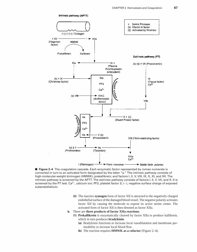
P1: OSO
LWBK192-02 LWBK192-Hubbard November 23, 2008 17:20
CHAPTER 2 Hemostasis and Coagulation 67
■ Figure 2–4 The coagulation cascade. Each enzymatic factor represented by roman numerals is
converted in turn to an activated form designated by the letter “a.” The intrinsic pathway consists of
high-molecular-weight kininogen (HMWK); prekallikrein; and factors I, II, V, VIII, IX, X, XI, and XII. The
intrinsic pathway is screened by the APTT. The extrinsic pathway consists of factors I, II, V, VII, and X. It is
screened by the PT test. Ca2+, calcium ion; PF3, platelet factor 3; (−), negative surface charge of exposed
subendothelium.
(2) The inactive zymogen form of factor XII is attracted to the negatively chargedendothelial surface of the damaged blood vessel. The negative polarity activatesfactor XII by causing the molecule to expose its active serine center. Theactivated form of factor XII is then denoted as factor XIIa.
b. There are three products of factor XIIa reactions.(1) Prekallikrein is enzymatically cleaved by factor XIIa to produce kallikrein,
which in turn produces bradykinin.(a) Bradykinin functions to increase local vasodilatation and membrane per-
meability to increase local blood flow.(b) The reaction requires HMWK as a cofactor (Figure 2–4).
P1: OSO
LWBK192-02 LWBK192-Hubbard November 23, 2008 17:20
68 A Concise Review of Clinical Laboratory Science
(2) Plasminogen is enzymatically cleaved by factor XIIa to functional plasmin,which initiates clot dissolution reactions.
(3) Factor XIIa enzymatically activates factor XI (plasma thromboplastin an-tecedent) to yield factor XIa. The activation of factor XIa will continue thecoagulation cascade.
2. The extrinsic systema. Initiation. Contact activation of the extrinsic system begins with the activation of
factor VII.(1) Factor III, known as tissue factor, is the primary activator of VII to VIIa,
which is a potent serine protease.(2) Tissue factor consists of lipoproteins, which are produced in many tissues.(3) Minor activation of factor VII can occur by proteolytic attack from factors
XIIa, Xa, IXa, or thrombin.b. In laboratory testing of the extrinsic system, lipoprotein-rich extracts are added
to citrated plasma as the PT reagent to support the activation of factor VII by tissuefactor.
B. Intermediate reactions1. Factor VIIa in the extrinsic pathway enzymatically alters factor X to yield Xa in the
presence of factor III. Factor VIIa has limited ability to activate the conversion of IXto its activated form (IXa).
2. Factor IX in the intrinsic pathway is most strongly activated by the direct enzymaticaction of XIa.a. Initiation. This reaction does not require tissue factor lipoprotein as extrinsic
pathway activation. It does require negatively charged membrane phospholipidsand ionized calcium. Platelets are the main source of in vivo phospholipidsurfaces.
b. In laboratory testing of the intrinsic system, phospholipid extracts are added tocitrated plasma as part of the APTT reagent to provide the activation for platelet-supported reactions.
3. Factor X (Stuart-Prower factor) is activated by two different pathways.a. In the extrinsic pathway, factor X is enzymatically activated by VIIa, with factor
III and calcium as cofactors.b. In the intrinsic pathway, factor X is activated by factor IXa. Factor IXa forms a
complex with a platelet phospholipid membrane surface and factor VIII (antihe-molytic factor) in the presence of calcium.
4. The factor VIII complex is a high-molecular-weight (MW) complex formed of twosubunits.a. VIII:C (antihemolytic factor) is synthesized in the liver and is genetically con-
trolled on the X-chromosome (sex-linked transmission).(1) Function. Factor VIII:C serves as a cofactor in the activation of factor X by
factor IXa. The presence of VIII:C accelerates the reaction rate by 500–1000times.
(2) Activator. Factor VIII:C is activated by thrombin.(3) Inactivator. Coagulation inhibitor protein C (PC) breaks down factor VIII:C
enzymatically.(4) Pathology. An inherited deficiency of factor VIII:C is known as hemophilia
A.b. Factor VIII:R (vWF) is synthesized by endothelial cells, megakaryocyte, and
platelet and demonstrates autosomal genetic expression.(1) Function. vWF supports the adhesion of platelets to the exposed suben-
dothelial surface of the blood vessel.(2) Activator. vWF activation occurs through the release of platelet aggregators
from damaged subendothelial tissue and from the release of platelet α-granulecontents.
(3) Pathology. An inherited deficiency of factor VIII: R is known clinically as vonWillebrand’s disease (vWD).
P1: OSO
LWBK192-02 LWBK192-Hubbard November 23, 2008 17:20
CHAPTER 2 Hemostasis and Coagulation 69
5. Factor Xa activation begins the common pathway, because the following enzymaticreactions are shared by both the intrinsic and the extrinsic pathways.a. Factor Xa enzymatically cleaves the zymogen prothrombin (factor II) to its acti-
vated form, thrombin.b. The activated form of factor V (proaccelerin) acts as a cofactor for factor Xa
activation of prothrombin. Factor V is converted to its active form by thrombin.c. The combination of phospholipid membrane surface, factor Xa, factor Va, and
calcium forms the receptor complex known as thrombomodulin, which supportsthe enzymatic conversion of prothrombin to the active enzyme thrombin (Figure2–4).
6. Thrombin (IIa) is a powerful enzyme with many functions, including:a. Enzymatic conversion of fibrinogen to fibrin monomerb. Activation of factor XIII (fibrin stabilizing factor)c. Activation of platelet aggregationd. Activation of factor V and factor VIII:Ce. Activation of PCf. Weak activation of factor VII to factor VIIa
C. Fibrin clot formation is the last step in the coagulation cascade.1. Thrombin enzymatically converts fibrinogen (factor I) to fibrin.2. Fibrinogen has the highest plasma concentration of any clotting factor, with a normal
range of 150 to 400 mg/dL. The molecule is produced in the liver and has a uniquemolecular structure.a. The fibrinogen monomer consists of two identical subunits bound together to pro-
duce a symmetric structure.b. Three nodular domains in the fibrinogen molecule have been identified as two
identical D regions at either carboxy-terminal end and a central E domain at theN-terminal end.
3. Thrombin enzymatically activates the fibrinogen monomer by splitting off the fib-rinopeptides Aα and Bβ from the N-terminals in the E domain.
4. The thrombin-exposed N-terminal peptides in the E domain react noncovalently byelectrostatic forces with polar D domain regions of adjacent fibrin molecules to form apolymer structure.
5. Formation of a fibrin polymer is the endpoint detected in the majority of in vitroclotting time tests.
6. Clot stabilization is achieved by factor XIIIa (fibrin stabilizing factor) from theformation of covalent bonds between chains of adjacent fibrin molecules.a. The inactive circulating zymogen form of factor XIII is activated by the proteolytic
action of thrombin with calcium and fibrinogen serving as cofactors.b. Factor XIIIa also covalently cross-links α2-antiplasmin to the fibrin clot, rendering
the clot less susceptible to lysis by plasmin.
D. Vitamin K–dependent factors are coagulation factors (i.e., factors II, VII, IX, X) andinhibitors [i.e., PC, protein S (PS)] that depend on vitamin K metabolism to be completelyfunctional.
1. Without vitamin K, the coagulation factors and inhibitors are nonfunctioning, evenwhen present in normal concentrations.
2. Coumarin anticoagulants inhibit vitamin K reduction from the epoxide form. Theend result is that factors II, VII, IX, and X are rendered inactive.a. Unlike heparin, coumarin is inactive as an in vitro anticoagulant and functions only
as a therapeutic in vivo anticoagulant.b. The PT test is the best screening method for coumarin therapy because factor VII
has the shortest half-life and is the most sensitive to levels of coumarin therapy.
E. Natural inhibitors of coagulation function to counterbalance the effects of coagulationfactors, provide limitations for the forming fibrin clot, and prevent systemic thrombusformation.
P1: OSO
LWBK192-02 LWBK192-Hubbard November 23, 2008 17:20
70 A Concise Review of Clinical Laboratory Science
1. Antithrombin III is the principal inhibitor of thrombin and factor Xa with limitedinhibitory activity against factors IXa, XIa, and XIIa.
a. Antithrombin III functions by binding with thrombin to form a 1:1 inactivecomplex.
b. Heparin serves as a cofactor in the inactivation, thereby increasing the reactionrate by more than 2,000 times.
2. α2-macroglobulin is a minor inhibitor of thrombin.3. Complement C1 inhibitor is a minor inhibitor of factors XIa and XIIa.4. α1-antitrypsin has limited inhibition of thrombin, kallikrein, and factor XIa.5. PC is a vitamin K-dependent inhibitor that circulates as an inactive zymogen.
a. Activator. PC is activated by thrombin as part of the thrombomodulin plateletreceptor complex.
b. Function. PC inactivates factors VIII: C and Va in the presence of cofactor PS.PS also depends on vitamin K, functions to enhance binding of PC to phospholipidsurfaces, and increases the rate of Va and VIIIa inactivation by PC.
F. Laboratory testing of coagulation depends on the quality and freshness of the plasmaspecimen obtained. Whole blood anticoagulated with sodium citrate is the specimen ofchoice. A 9:1 blood:citrate ratio is required for accurate coagulation testing, because aratio of <9:1 may falsely increase results. Conditions that can interfere with obtainingthe required 9:1 ratio are an abnormally high hematocrit, traumatic blood drawing, or ahemolyzed specimen. EDTA contamination can falsely increase PT and APTT results.Specimens must be assayed as soon as possible, and the plasma must be kept cold to avoidfactor deterioration.
1. PT tests for extrinsic pathway deficiencies in factors VII, X, V, II, and I (fibrinogen).a. Reagent. A lipoprotein tissue extract from brain or lung tissue serves as the
reagent source of tissue factor. An excess of calcium is also added to the PTreagent.
b. Principle. Citrated plasma is added to the lipoprotein reagent with calcium, andthe time required for fibrin clot formation is measured.
c. Reference range. Although the PT assay has an approximate normal range of 11to 13 seconds, it is important for each laboratory to establish its own range.
d. Variation. The addition of Russell’s viper venom (Stypven) instead of lipoproteinreagent activates factor X directly, bypassing factor VII as a necessary componentvariable. This variation of the common PT is known as the Stypven time.
2. The international normalized ratio (INR) by definition is the PT ratio that reflectsthe results that would have been obtained if the World Health Organization (WHO)international reference preparation (IRP) thromboplastin had been used to performthe test.a. The specific purpose of the INR is for reporting results on stable, orally antico-
agulated patients taking Coumarin.b. Calculation of the INR:
(1) In order to calculate the INR, it is necessary to have a PT ratio and the inter-national sensitivity index (ISI) value of the thromboplastin reagent used tomeasure the PT.
(2) The ISI value is assigned by the manufacture of the thromboplastin reagent,and attests to the purity of the reagent. The lower the manufacture ISI value,the more desirable the reagent for use. An ISI value of 1.0 would claim equalpurity with the WHO thromboplastin IRP.
(3) The PT ratio is calculated as follows:
Patient PTMean normal PT
(4) The INR is then derived by the following formula:
INR = [patient PT ÷ mean of normal PT]ISI
P1: OSO
LWBK192-02 LWBK192-Hubbard November 23, 2008 17:20
CHAPTER 2 Hemostasis and Coagulation 71
Alternatively, the INR can be calculated by the following formula:
log (INR) = ISI × log (patient PT/normal PT)
c. Therapeutic ranges of INR(1) Patients not on oral anticoagulants with normal coagulation should have low
PT values in the range of the INR.(2) The majority of patient’s INRs of 2.0 to 3.0 corresponds to PT ratio for
thromboplastin of 1.5 to 2.0.(3) When the INR is correctly used for monitoring a patient’s oral anticoagulant
therapy, the physician gives a standard dose of coumarin to achieve a targetINR between 2.0 and 3.0.
(4) If the physician chooses to use a high dose of coumarin, the target valueINR is between 2.5 and 3.5. Indications of high-dose anticoagulant therapyare mechanical heart valves.
(5) An INR value >3.5 constitutes a panic value and should be reported to thepatient’s nurse immediately.
3. APTT tests for intrinsic pathway deficiencies in factors prekallikrein, HMWK, andfactors XII, XI, IX, X, VIII, V, II, and I.a. Reagents. A phospholipid-rich preparation is used as a platelet-membrane substi-
tute. An activator, such as kaolin, ellagic acid, or celite, is also added to the APTTreagent to provide the negative surface charge required to activate factor XII andprekallikrein. Calcium chloride is used as an additional reagent to initiate clotting.
b. Principle. A citrated plasma sample is preincubated with the phospholipid reagentto initiate contact activation factors in the intrinsic pathway. Following incubation,calcium chloride reagent is added as a separate reagent to initiate the clottingcascade. The time required for fibrin clot formation to occur is measured.
c. Reference range. The APTT assay has an approximate normal range of 25 to40 seconds, but it is important for each laboratory to establish its own range.
4. Thrombin clotting time (TCT) tests for a deficiency or inhibition of fibrinogen.a. Principle. Commercially prepared thrombin reagent is added to citrated plasma,
and the time required for clot formation is measured.b. Reference range. The range is generally 10 to 20 seconds, but each laboratory
should establish its own range.c. A prolonged TCT can occur in patients receiving therapeutic heparin, patients
with increased fibrin degradation products, and patients with any disorder of hy-pofibrinogenemia.
5. Quantitative fibrinogen assay is an expansion of the TT methodology.a. Principle. A measured amount of commercially prepared thrombin reagent is
added to citrated plasma. The clotting time is measured and compared with theclotting times of plasma fibrinogen standards containing known amounts of fib-rinogen.
b. A standard curve is constructed, and the clotting time in seconds is plotted againstmilligrams per deciliter of fibrinogen. Patient unknown data can be quantitated forfibrinogen from a standard curve.
c. Reference range. The normal range for a quantitative fibrinogen is 200 to 400mg/dL.
6. Substitution tests (Table 2–3) can be adapted if primary tests like the PT or APTT areabnormally prolonged and indicate a factor deficiency. The patient’s deficient plasmais diluted 1:1 with a plasma or serum substitute, and the APTT or PT is repeated. Acorrection of the original prolonged APTT or PT indicates that the deficient factorhad been added to the patient’s plasma by the substitution solution. The preparedsubstitution solutions are as follows:a. Aged plasma lacks labile factors V and VIII, but retains normal activity of all
other coagulation factors. Normal plasma retains normal activity of all coagulationfactors.
b. Fresh absorbed plasma lacks vitamin K factors (i.e., factors II, VII, IX, X), butretains normal activity of all other coagulation factors.
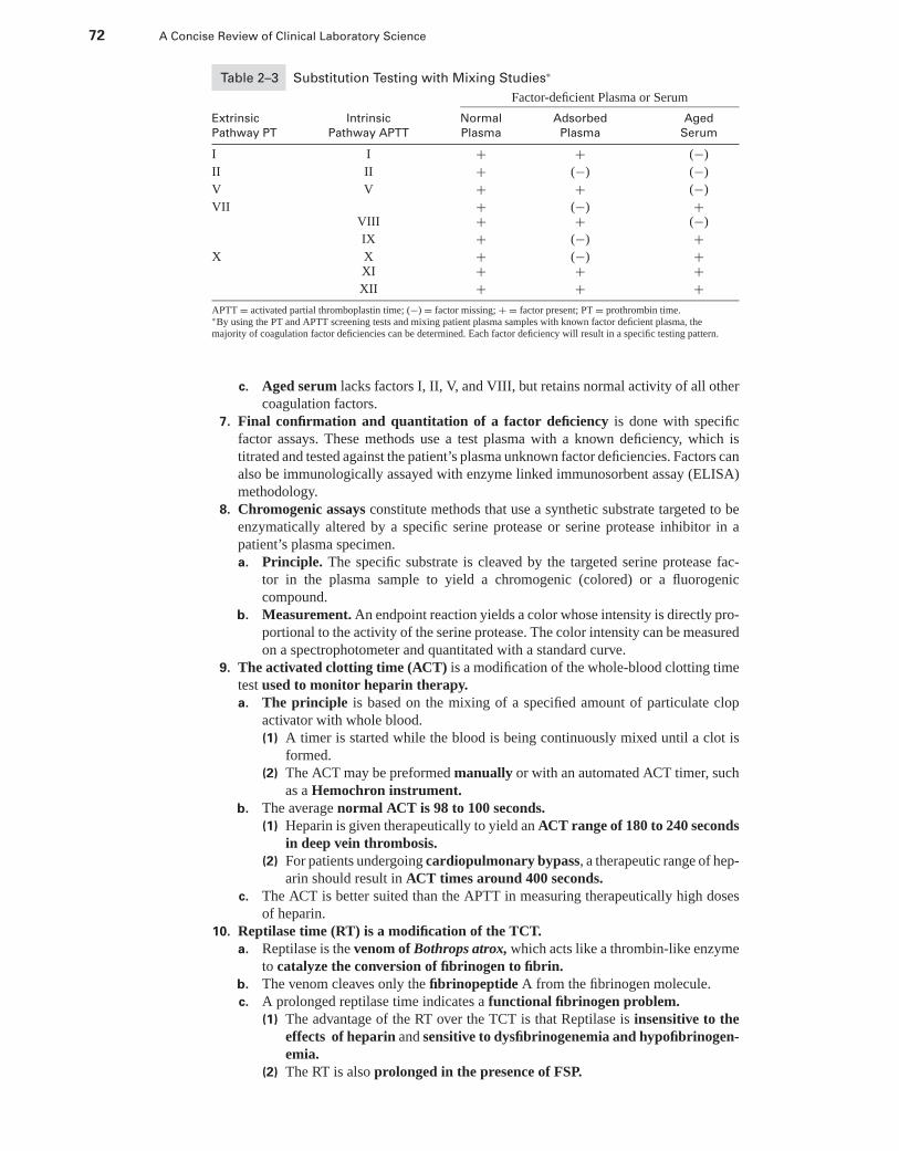
P1: OSO
LWBK192-02 LWBK192-Hubbard November 23, 2008 17:20
72 A Concise Review of Clinical Laboratory Science
Table 2–3 Substitution Testing with Mixing Studies∗
Factor-deficient Plasma or Serum
Extrinsic Intrinsic Normal Adsorbed Aged
Pathway PT Pathway APTT Plasma Plasma Serum
I I + + (−)
II II + (−) (−)
V V + + (−)
VII + (−) +VIII + + (−)
IX + (−) +X X + (−) +
XI + + +XII + + +
APTT = activated partial thromboplastin time; (−) = factor missing; + = factor present; PT = prothrombin time.∗By using the PT and APTT screening tests and mixing patient plasma samples with known factor deficient plasma, themajority of coagulation factor deficiencies can be determined. Each factor deficiency will result in a specific testing pattern.
c. Aged serum lacks factors I, II, V, and VIII, but retains normal activity of all othercoagulation factors.
7. Final confirmation and quantitation of a factor deficiency is done with specificfactor assays. These methods use a test plasma with a known deficiency, which istitrated and tested against the patient’s plasma unknown factor deficiencies. Factors canalso be immunologically assayed with enzyme linked immunosorbent assay (ELISA)methodology.
8. Chromogenic assays constitute methods that use a synthetic substrate targeted to beenzymatically altered by a specific serine protease or serine protease inhibitor in apatient’s plasma specimen.a. Principle. The specific substrate is cleaved by the targeted serine protease fac-
tor in the plasma sample to yield a chromogenic (colored) or a fluorogeniccompound.
b. Measurement. An endpoint reaction yields a color whose intensity is directly pro-portional to the activity of the serine protease. The color intensity can be measuredon a spectrophotometer and quantitated with a standard curve.
9. The activated clotting time (ACT) is a modification of the whole-blood clotting timetest used to monitor heparin therapy.a. The principle is based on the mixing of a specified amount of particulate clop
activator with whole blood.(1) A timer is started while the blood is being continuously mixed until a clot is
formed.(2) The ACT may be preformed manually or with an automated ACT timer, such
as a Hemochron instrument.b. The average normal ACT is 98 to 100 seconds.
(1) Heparin is given therapeutically to yield an ACT range of 180 to 240 secondsin deep vein thrombosis.
(2) For patients undergoing cardiopulmonary bypass, a therapeutic range of hep-arin should result in ACT times around 400 seconds.
c. The ACT is better suited than the APTT in measuring therapeutically high dosesof heparin.
10. Reptilase time (RT) is a modification of the TCT.a. Reptilase is the venom of Bothrops atrox, which acts like a thrombin-like enzyme
to catalyze the conversion of fibrinogen to fibrin.b. The venom cleaves only the fibrinopeptide A from the fibrinogen molecule.c. A prolonged reptilase time indicates a functional fibrinogen problem.
(1) The advantage of the RT over the TCT is that Reptilase is insensitive to theeffects of heparin and sensitive to dysfibrinogenemia and hypofibrinogen-emia.
(2) The RT is also prolonged in the presence of FSP.
P1: OSO
LWBK192-02 LWBK192-Hubbard November 23, 2008 17:20
CHAPTER 2 Hemostasis and Coagulation 73
11. Factor XIII Screening is a simple test using urea to dissolve clots.a. Factor XIII is responsible for converting the fibrin clot to a stable form.b. It is activated by thrombin during the fibrinogen-to-fibrin conversion.c. When factor XIII is present, a fibrin clot is insoluble in 5 M urea when left standing
for 24 hours. If factor XIII is deficient, 5 M urea will dissolve a fibrin clot within2 hours.
12. Lupus-like anticoagulant/antiphospholipid antibodies is developed in 10% to 20%of people with SLE, a significant number of patients taking the drug, phenothiazine,and occasionally in cases of lymphoproliferative disorders.a. Inhibitors most commonly are IgG and occasionally IgM. Autoantibodies found
in the patient’s plasma are directed against the phospholipid portion of phospho-lipoprotein components found in APTT reagent.
b. Occasionally patients with lupus-like anticoagulant have a mild thrombocytope-nia.
c. The presence of clinical bleeding with lupus-like anticoagulant is only found inpatient’s with high titers of anticoagulant.
d. The antibody is first detected by a slightly prolonged APTT with a normal PTand TT.(1) The APTT will not be corrected with a 1:1 dilution of patient and normal
plasma like a typical factor deficiency.(2) Increasing prolonged times are directly proportional to extended incuba-
tion times with the APTT phospholipid reagent.e. Further testing for a specific factor deficiency will prove to be normal.
13. Factor VIII assay and other specific factor assays are based on the ability of com-mercially prepared plasmas to correct a factor-deficient patient.a. The APTT assay is used to estimate the concentration of functional factor VIII by
serial diluting the patient’s plasma with a VIII-depleted plasma control.(1) Factor VIII-depleted plasma contains full activity of all factors except VIII,
which has been immunodepleted.(2) The factor-depleted plasma will have a prolonged APTT unless corrected by
diluting with a normal plasma control.(3) A prolonged APTT result on a mixture of the patient plasma and the factor-
deficient plasma implies that the patient is deficient in the same factor that ismissing in the factor-depleted control.
b. The APTT clotting time interval of a 1:10 dilution of the patient plasma and thefactor-depleted plasma is compared to a previously prepared reference curve toobtain the % activity of the patient plasma.(1) The reference graph is prepared with a series of five or more dilutions of
factor-depleted control plasma with a normal coagulation control.(2) The reference curve is plotted with clotting time on the y-axis against %
activity of factor VIII, or another factor, on the x-axis.14. Point of care testing (PCT) in coagulation has been developed for bed-side testing
of the PT, APTT, TCT, and the ACT to provide rapid turn-around-times for thephysician.a. An automated device such as the Hemochron Portable Blood Coagulation Tim-
ing System may be used.b. The methodology uses a technique similar to the automated ACT.
(1) Commercially prepared tubes containing thromboplastin and a magnetizedstir bar are used for the PT.
(2) Tubes containing diatomaceous earth (for an activator), partial thrombo-plastin, buffer, calcium chloride, and a magnetized stir bar are used forthe APTT.
(3) Tubes containing lyophilized thrombin, buffer, calcium chloride, and a mag-netized stir bar are used for the TCT.
c. Two milliliters of whole blood is collected and transferred to the reaction tube.(1) The timer is started after the tube is placed in the reaction well.(2) Whole blood PT and APTT results are interpreted by blood-to-plasma con-
version tables provided by the manufacturer.
P1: OSO
LWBK192-02 LWBK192-Hubbard November 23, 2008 17:20
74 A Concise Review of Clinical Laboratory Science
G. Normal fibrinolysis refers to the enzymatic pathway of cloth dissolution in which plasminis the key enzyme responsible for breaking down bonds in the fibrin polymer and releasingfibrin degradation products (FDP).
1. Plasmin activation results from proteolytic cleavage of a circulating inactive zymogenknown as plasminogen by either of two pathways:a. Extrinsic pathway activation in vivo involves a tissue proteolytic enzyme and
fibrin as a cofactor.(1) Urokinase can also convert plasminogen to plasmin and is used as a throm-
bolytic agent.(2) Streptokinase is a streptococcal-derived protein used as a therapeutic throm-
bolytic drug.b. Intrinsic pathway activation of plasmin involves factors XIIa, kallikrein, or
HMWK.2. Plasmin degradation of fibrin begins by the breaking down of the fibrin polymer into
a monomer form known as fragment X. This fragment is identical to a fibrin monomer,consists of one E domain and two D domains, and retains clotting properties.a. Fragment X is further cleaved to produce FDP, which consists of:
(1) Fragment Y, which consists of one E domain and one D domain(2) Fragment E, which consists of the E domain only(3) Fragment D, which consists of the D domain only(4) D dimer, which consists of two D fragments of linked monomers
b. The FDPs produced during fibrinolysis have anticoagulating effects of their ownbecause they inhibit the process of fibrin polymerization, demonstrate competi-tive inhibition of thrombin, and prolong the TT test.
3. Other functions of plasmin in addition to fibrin degradation include:a. Fibrinogen degradationb. The inactivation of factor VIII:Cc. The inactivation of factor Vd. Direct antiaggregation effect on platelets
4. α2-Antiplasmin inhibits the actions of plasmin by forming a 1:1 complex with plasmin,which prevents fibrin binding.a. α2-Antiplasmin is bound to the fibrin by factor XIIIa.b. α2-Macroglobulin also has weak antiplasmin activity.
H. Laboratory tests for fibrinolysis1. Latex agglutination can measure freely circulating FDP in the patient’s serum. Latex
coated with antifibrinogen detects the presence of increased levels of FDP. Only serumcan be used to avoid interference with endogenous fibrinogen.
2. Plasminogen orα2-antiplasmin can be measured serologically or with chromogenics.3. D dimer test is based on a highly specific, monoclonal antibody directed against a
unique neoantigen of covalently crosslinked D fragments resulting from fibrinolysis.The D dimer has several advantages in detecting degradation fragments when comparedwith the standard FDP assay.a. The FDP assay can be run on plasma, eliminating the risk of false positives.b. D dimer is superior in sensitivity and specificity as compared with the conven-
tional FDP assay (i.e., sensitive to as little as 20 ng/mL, as compared with 10 μ/mLfor conventional latex FDP).
4. Prothrombin fragment 1.2 (F-1.2) is a new assay that tests for the presence of thefragment generated when prothrombin is activated to thrombin (factor IIa).a. Prothrombin fragment serves as a sensitive biologic marker of thrombin gen-
eration and Xa activity because generation of F-1.2 precedes thrombus forma-tion.
b. Levels are elevated in persons predisposed to thrombotic risk (e.g., cancer, heartdisease, orthopedic surgery).
c. Levels are depressed in persons undergoing anticoagulant therapy.5. Euglobulin Lysis Time is used as a screening test for fibrinolytic activity.
a. Euglobulin fraction of plasma consists of plasminogen, fibrinogen, and activatorsof plasminogen.
P1: OSO
LWBK192-02 LWBK192-Hubbard November 23, 2008 17:20
CHAPTER 2 Hemostasis and Coagulation 75
b. The euglobulin fraction is precipitated with 1% acetic acid and resuspended in aborate solution(1) Euglobulins are clotted by the addition of thrombin (5 units/mL).(2) The resulting clot is incubated and the time of lysis is obtained.
c. Clot lysis in <2 hours is indicative of abnormal fibrinolytic activity.(1) A decreased time below normal is also caused by a decrease in fibrinogen.(2) A prolonged time >4 hours is caused by a decrease in plasminogen or acti-
vator.6. Protein-C (PC), and protein-S (PS), and antithrombin-III (ATIII) assays can all
be measured for degree of activity with chromogenic assays or with immunoassaysto measure the concentration of the molecule (i.e., antigen).a. PC can be accurately measured by chromogenics or enzyme immunoassay, but the
latter has a better shelf life.(1) In the chromogenic assay, the patient plasma sample is mixed with venom to
activate PC.(a) Added chromogenic substrate is hydrolyzed by the APC.(b) The amount of the hydrolyzed colored product is proportional to the
activity of PC in the patient’s sample.(2) The immunoassay method is based on using rabbit-derived antihuman PC-
coated microtiter plate wells that capture the PC in the added plasma sample.(a) Bound PC is conjugated with peroxidase, yielding a color product the
intensity of the of the PC in the sample.b. PS can be measured with similar methodologies.c. ATIII can be measured with chromogenics or immunoassay using similar method-
ology for each.(1) The chromogenic uses heparin as an ATIII activator.(2) Activated ATIII then binds with a serine protease, such as factor X, and con-
jugated with peroxidase to yield the colored end product.7. Activated protein-C resistance (APCR) is defined as the inability of APC to prolong
clotting tests when added to the APTTa. This phenomenon is characteristic of a genetic thrombotic disorder known as factor
V Leiden.(1) When APC is added to normal test plasma, inactivation of factors VIII
and V result in the prolongation of the APTT.(2) Patients with APCR demonstrate a diminished ability to inactivate V when
APC is added to the test sample.b. The assay method is based on the prolongation of the APTT when APC is added
to the test plasma.(1) The aPTT reagent is added to two aliquots of the patient’s plasma and prein-
cubated.(2) Calcium chloride is added to one aliquot and the time to clot is recorded.(3) To the second aliquot, a mixture of calcium chloride and APC is added and
clot formation is timed.(4) In a normal plasma without APCR, the APTT results of the first sample aliquot
without APC should be more than twice the clotting time of the second aliquotwith APC.
(5) If the patient has a genetic resistance to PC inactivation of V, then the secondAPTT is shortened in time and will be less than twice the clotting time of thesample without APC.
I. Vascular factors in coagulation1. Blood vessels are lined with a continuous monolayer of endothelial cells anchored to
inert basement membrane by a subendothelial matrix.2. In arteries and arterioles, the subendothelial layer is surrounded by layers of smooth
muscle cells and adventitia with fibroblasts.3. When vascular subendothelium is exposed by vessel damage, platelet aggregation
and coagulation can be directly initiated by collagen release and indirectly by tissue-released vWF.
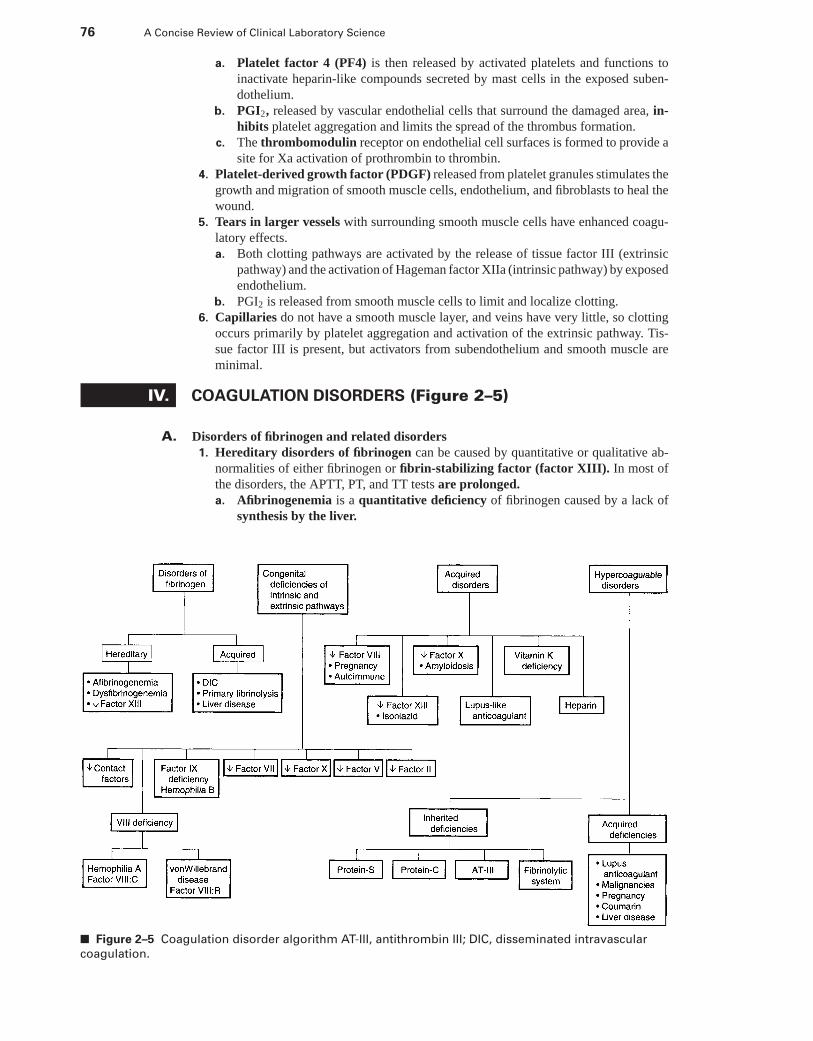
P1: OSO
LWBK192-02 LWBK192-Hubbard November 23, 2008 17:20
76 A Concise Review of Clinical Laboratory Science
a. Platelet factor 4 (PF4) is then released by activated platelets and functions toinactivate heparin-like compounds secreted by mast cells in the exposed suben-dothelium.
b. PGI2, released by vascular endothelial cells that surround the damaged area, in-hibits platelet aggregation and limits the spread of the thrombus formation.
c. The thrombomodulin receptor on endothelial cell surfaces is formed to provide asite for Xa activation of prothrombin to thrombin.
4. Platelet-derived growth factor (PDGF) released from platelet granules stimulates thegrowth and migration of smooth muscle cells, endothelium, and fibroblasts to heal thewound.
5. Tears in larger vessels with surrounding smooth muscle cells have enhanced coagu-latory effects.a. Both clotting pathways are activated by the release of tissue factor III (extrinsic
pathway) and the activation of Hageman factor XIIa (intrinsic pathway) by exposedendothelium.
b. PGI2 is released from smooth muscle cells to limit and localize clotting.6. Capillaries do not have a smooth muscle layer, and veins have very little, so clotting
occurs primarily by platelet aggregation and activation of the extrinsic pathway. Tis-sue factor III is present, but activators from subendothelium and smooth muscle areminimal.
IV. COAGULATION DISORDERS (Figure 2–5)
A. Disorders of fibrinogen and related disorders1. Hereditary disorders of fibrinogen can be caused by quantitative or qualitative ab-
normalities of either fibrinogen or fibrin-stabilizing factor (factor XIII). In most ofthe disorders, the APTT, PT, and TT tests are prolonged.a. Afibrinogenemia is a quantitative deficiency of fibrinogen caused by a lack of
synthesis by the liver.
■ Figure 2–5 Coagulation disorder algorithm AT-III, antithrombin III; DIC, disseminated intravascular
coagulation.
P1: OSO
LWBK192-02 LWBK192-Hubbard November 23, 2008 17:20
CHAPTER 2 Hemostasis and Coagulation 77
(1) Severe hemorrhages predominate in the umbilical, mucosal, gastrointestinal(GI), and intracranial regions.
(2) The most common treatment is replacement therapy with cryoprecipitate orfresh frozen plasma (FFP) to raise the blood fibrinogen level higher than60 mg/dL.
b. Dysfibrinogenemia is a qualitative abnormality in the structure and functionof the fibrinogen molecule.(1) Fibrinogen levels may be normal, and the bleeding time is usually normal.(2) Posttraumatic or postoperative bleeding of mucosal tissues is common.
c. Factor XIII deficiency can be clinically severe with moderate-to-severe bleeding.Delayed bleeding and wound healing is often observed after trauma.(1) The APTT, PT, and TT tests are normal.(2) Low factor XIII concentrations are detected by incubation of a fibrin clot
in a 5 M urea solution. A normal fibrin clot will not be dissolved in 5 Murea after 24 hours, but a clot deficient in factor XIII will be dissolved after24 hours.
2. Acquired disorders of fibrinogen occur secondary to other pathologic events.a. DIC refers to a deposition of large amounts of fibrin throughout the microcircu-
lation that results in a pathologic activation of coagulation pathways. DIC can befatal, self-limiting, acute, or chronic.(1) Classification. DIC is classified as a consumption coagulopathy, because it
results in a depletion of platelets as well as plasma coagulation factors.(2) Physiologic effects. Plasminogen is activated systemically to plasmin, result-
ing in an increase in FDP in the plasma. There is possible red blood cell (RBC)fragmentation caused by damage from multiple thromboemboli.
(3) Common coagulation test results include:(a) A decreased platelet count(b) A prolonged PT, APTT, and TT(c) An elevated FDP or D dimer(d) A decreased fibrinogen
(4) The causes of DIC are widespread and can occur from intrinsic or extrinsicpathway activation.(a) Extrinsic system activation occurs by large amounts of tissue factor en-
tering the circulation, and can result from hypofibrinogenemic states ofpregnancy, metastatic carcinoma, or promyelocytic leukemia.
(b) Intrinsic system activation results from events that damage or alter thevascular endothelium, thereby exposing collagen (e.g., infectious diseases,antigen-antibody complexes, liver disease, snake venom poisoning,massive trauma, or surgery).
(5) Treatment for DIC varies with the cause, but most often FFP-pooled platelet,cryoprecipitate, or low-molecular-weight heparin is used to treat the symptomsand break the cycle.
b. Primary fibrinolysis is symptomatically similar to DIC, but results from increasedlevels of plasmin.(1) Common causes include cirrhosis, shock, metastatic carcinoma of the
prostate, injury to the genitourinary tract, and leaking of urokinase fromthe urine into tissues.
(2) Common coagulation test results include decreased fibrinogen, prolonged PTor APTT, and increased FDP or D dimer. In contrast to DIC, primary fibrinolysisusually demonstrates a normal platelet count, and the RBC morphology doesnot show fragmentation.
(3) A test tube sample from a patient with primary fibrinolysis will initially forma clot that dissolves in 1 to 2 hours.
c. Liver disease can be associated with coagulation disorders because the majorityof the coagulation factors are synthesized in the liver.(1) A decrease in factor VII occurs first because factor VII has the shortest half-life
of the coagulation factors.
P1: OSO
LWBK192-02 LWBK192-Hubbard November 23, 2008 17:20
78 A Concise Review of Clinical Laboratory Science
Table 2–4 Comparison of the Thrombin and Reptilase∗ Times to DistinguishInherited Fibrin
Thrombin Time Reptilase Time Defect
Infinitely prolonged Infinitely prolonged Dysfibrinogenemia
Infinitely prolonged Infinitely prolonged Afibrinogenemia
Prolonged Equally prolonged Hypofibrinogenemia
Prolonged Normal Heparin
Prolonged Slight to moderate Fibrin degradation product
∗Reptilase activates fibrinogen in a manner similar to thrombin but is not sensitive to heparin and fibrin degradation productinhibition.
(2) In patients with severe liver disease, the PT usually is prolonged; in later stages,the APTT will also be prolonged.
(3) Severe liver disease often causes decreased fibrinogen production (hypofib-rinogenemia) or an abnormal fibrinogen molecule (dysfibrinogemia).
d. Therapeutic heparin administration, commonly used in post-surgical and car-diac patients, can prolong the APTT and TT acutely and can prolong the PT withchronic use. In a patient receiving heparin, abnormal coagulation results must bedistinguished from similar coagulopathies if the patient history is unknown. Thiscan be done with the reptilase-R time test (Table 2–4).
B. Inherited deficiencies of other factors in the intrinsic and extrinsic pathways1. Hemophilia A is an inherited deficiency or dysfunction of factor VIII:C (anti-
hemolytic factor).a. Inheritance pattern is sex-linked (i.e., on the X chromosome), and female ho-
mozygotes with a hemophiliac father and carrier mother are very rare.b. There are three patterns of severity.
(1) Severe hemophilia A is diagnosed in patients with spontaneous hemorrhagesand a factor VIII:C level <1% of the normal level.
(2) Moderate hemophilia A is diagnosed in patients who have VIII:C levels <5%of the normal level.
(3) Mild hemophilia A is seen in patients with 6% to 30% of the normal VIII:Clevels. Typically, these patients only bleed excessively in association withtrauma or surgical procedures.
c. Human alloantibodies to VIII:C are produced in a minority of severe hemophiliaA cases.(1) Approximately 10% of severe hemophiliacs are positive for cross-reacting
material (CRM+). CRM is an antigen that neutralizes anti-VIII:C antibodies.(2) Approximately 90% of hemophiliacs are negative for CRM.
d. Patients with moderate-to-severe hemophilia A demonstrate a clinical bleedingpattern of spontaneous bleeding into joints or muscles, excessive postoperativehemorrhage, and easy bruising.
e. Predicted coagulation profile results include:(1) Normal bleeding time (usually)(2) Normal PT and TT(3) Prolonged APTT if VIII:C levels are <0.2 U/mL (i.e., <20% activity)(4) Female heterozygote carriers will have 25% to 75% VIII:C activity that will
not usually show up on laboratory testing.f. Treatment of hemophilia A is based on replacement therapy.
(1) Cryoprecipitate is a plasma-fraction preparation prepared by thawing FFP at4◦C. It contains a concentrated portion of fibrinogen and VIII complex.
(2) Preparations rich in factor VIII:C are more costly, but are effective.2. Hemophilia B (Christmas disease) is an inherited deficiency in factor IX resulting
from a sex-linked (X-chromosome) mutation.a. Diagnosis. Hemophilia B is less common than hemophilia A. However, clinical
symptoms are virtually indistinguishable, and laboratory differential diagnosis isnecessary.
P1: OSO
LWBK192-02 LWBK192-Hubbard November 23, 2008 17:20
CHAPTER 2 Hemostasis and Coagulation 79
b. Treatment. A correct laboratory diagnosis must be made because treatment ofhemophilia B is different than treatment of hemophilia A.(1) Cryoprecipitate is deficient in factor IX, but rich in the factor VIII complex.(2) FFP replacement therapy is the treatment of choice for hemophilia B, because
FFP contains active factor IX. However, it is deficient in factor VIII.(3) The half-life of factor IX (24 hours) is longer than that for factor VIII (12
hours), so the administration of treatment for a factor IX deficiency is lessfrequent.
c. Coagulation profile results initially may not produce results that differ from apatient with hemophilia A because the bleeding time, PT, and TT will also benormal, and the APTT will be prolonged. If hemophilia is suspected, and the APTTis prolonged, substitution tests or assay for factors IX and VIII activity should beperformed.
3. vWD occurs almost as frequently as hemophilia A.a. Genetically, vWD is inherited as an autosomal dominant condition.b. The mutation is generally a defect in the VIII:R component of factor VIII, but
partial deficiencies in the VIII:C portion often occur.c. Patients with vWD characteristically bleed from mucous membranes and sub-
cutaneous tissues.(1) Easy bruising and GI bleeding is common.(2) Muscle hematomas and deep vessel hemorrhages are not as common as in
hemophilia A.d. Production of VIII:R and VIII:C are controlled by different tissues but circulate as
a VIII:C/VIII:R complex.e. The vWF molecule consists of a basic monomer form, but it also combines with
additional monomers to produce a series of multimers of increasing molecularweight.(1) Largest MW multimers possess the functional activities contributing to
platelet-mediated homeostatic events.(2) Antigenically, there appears to be a plasma vWF that is synthesized from a
tissue source and a platelet-derived vWF based on multimer size.(3) The variation of vWF size results in several clinical subclassifications of this
disorder.(a) Clinical Type I vWD is the most common “classic” form of vWD, in which
a partial decrease of all sizes of vWF MW multimers occurs.(b) Clinical Type II vWD patients have a selective absence of higher MW
multimers.(i) In patients with Type IIa vWD, ristocetin-induced activity is decreased
because both platelet and plasma vWF are absent.(ii) In patients with Type IIb vWD, the ristocetin-induced activity is nor-
mal or increased because only plasma high-MW multimers are de-creased.
(c) Patients with Type III vWD suffer from the most rare and severe type,because factor VIII:C is almost nondetectable.
(d) Some patients are classified as having a platelet-type variant, which issimilar to Type IIb, but is accompanied by a thrombocytopenia.
f. The common laboratory coagulation results for patients with vWD include:(1) Prolonged bleeding time and increased petechiae, because platelet adhesion
to capillaries is impaired(2) Normal PT time(3) Prolonged APTT test, only if VIII:C activity is <20%(4) Platelet aggregation reactions that are normal with ADP, epinephrine, colla-
gen, or thrombin activation, but there is impaired ristocetin-induced aggrega-tion
g. Treatment. Factor VIII concentrations are not useful, because they contain primar-ily factor VIII:C and are deficient in high MW multimers of vWF. The treatmentof choice, therefore, is cryoprecipitate.
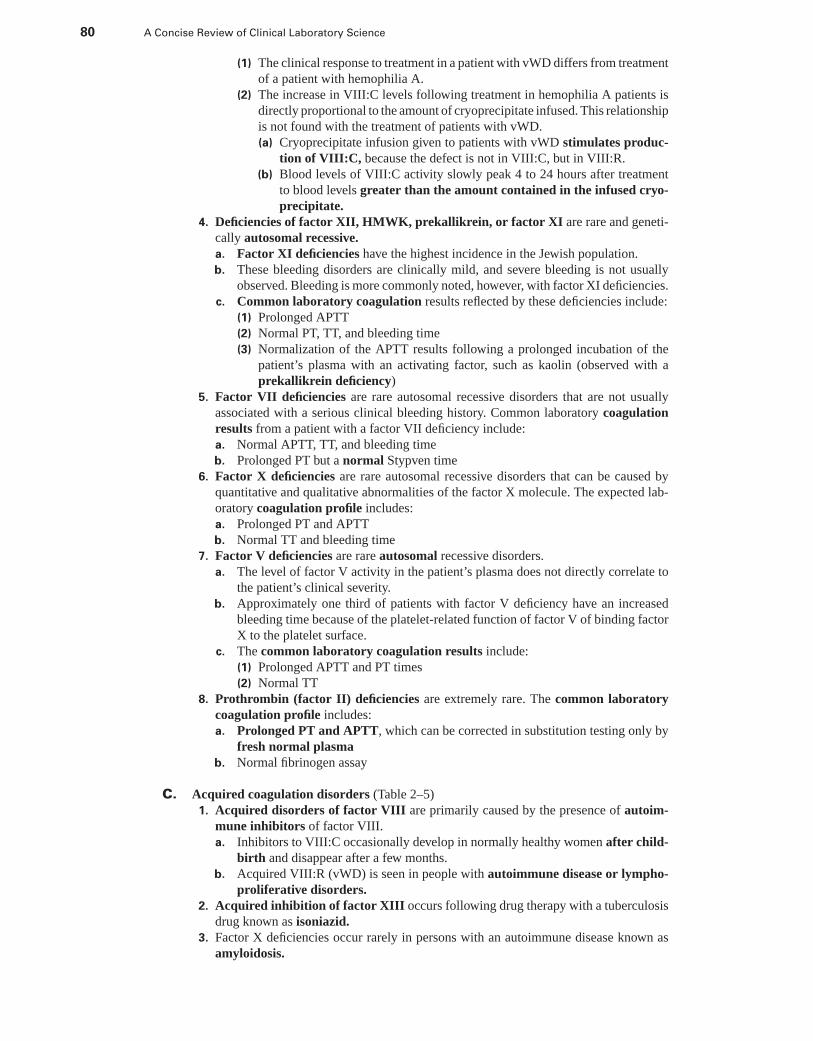
P1: OSO
LWBK192-02 LWBK192-Hubbard November 23, 2008 17:20
80 A Concise Review of Clinical Laboratory Science
(1) The clinical response to treatment in a patient with vWD differs from treatmentof a patient with hemophilia A.
(2) The increase in VIII:C levels following treatment in hemophilia A patients isdirectly proportional to the amount of cryoprecipitate infused. This relationshipis not found with the treatment of patients with vWD.(a) Cryoprecipitate infusion given to patients with vWD stimulates produc-
tion of VIII:C, because the defect is not in VIII:C, but in VIII:R.(b) Blood levels of VIII:C activity slowly peak 4 to 24 hours after treatment
to blood levels greater than the amount contained in the infused cryo-precipitate.
4. Deficiencies of factor XII, HMWK, prekallikrein, or factor XI are rare and geneti-cally autosomal recessive.a. Factor XI deficiencies have the highest incidence in the Jewish population.b. These bleeding disorders are clinically mild, and severe bleeding is not usually
observed. Bleeding is more commonly noted, however, with factor XI deficiencies.c. Common laboratory coagulation results reflected by these deficiencies include:
(1) Prolonged APTT(2) Normal PT, TT, and bleeding time(3) Normalization of the APTT results following a prolonged incubation of the
patient’s plasma with an activating factor, such as kaolin (observed with aprekallikrein deficiency)
5. Factor VII deficiencies are rare autosomal recessive disorders that are not usuallyassociated with a serious clinical bleeding history. Common laboratory coagulationresults from a patient with a factor VII deficiency include:a. Normal APTT, TT, and bleeding timeb. Prolonged PT but a normal Stypven time
6. Factor X deficiencies are rare autosomal recessive disorders that can be caused byquantitative and qualitative abnormalities of the factor X molecule. The expected lab-oratory coagulation profile includes:a. Prolonged PT and APTTb. Normal TT and bleeding time
7. Factor V deficiencies are rare autosomal recessive disorders.a. The level of factor V activity in the patient’s plasma does not directly correlate to
the patient’s clinical severity.b. Approximately one third of patients with factor V deficiency have an increased
bleeding time because of the platelet-related function of factor V of binding factorX to the platelet surface.
c. The common laboratory coagulation results include:(1) Prolonged APTT and PT times(2) Normal TT
8. Prothrombin (factor II) deficiencies are extremely rare. The common laboratorycoagulation profile includes:a. Prolonged PT and APTT, which can be corrected in substitution testing only by
fresh normal plasmab. Normal fibrinogen assay
C. Acquired coagulation disorders (Table 2–5)1. Acquired disorders of factor VIII are primarily caused by the presence of autoim-
mune inhibitors of factor VIII.a. Inhibitors to VIII:C occasionally develop in normally healthy women after child-
birth and disappear after a few months.b. Acquired VIII:R (vWD) is seen in people with autoimmune disease or lympho-
proliferative disorders.2. Acquired inhibition of factor XIII occurs following drug therapy with a tuberculosis
drug known as isoniazid.3. Factor X deficiencies occur rarely in persons with an autoimmune disease known as
amyloidosis.
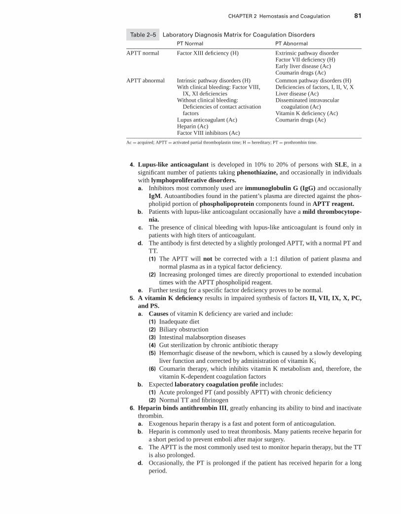
P1: OSO
LWBK192-02 LWBK192-Hubbard November 23, 2008 17:20
CHAPTER 2 Hemostasis and Coagulation 81
Table 2–5 Laboratory Diagnosis Matrix for Coagulation Disorders
PT Normal PT Abnormal
APTT normal Factor XIII deficiency (H) Extrinsic pathway disorderFactor VII deficiency (H)Early liver disease (Ac)Coumarin drugs (Ac)
APTT abnormal Intrinsic pathway disorders (H)With clinical bleeding: Factor VIII,
IX, XI deficienciesWithout clinical bleeding:
Deficiencies of contact activationfactors
Lupus anticoagulant (Ac)Heparin (Ac)Factor VIII inhibitors (Ac)
Common pathway disorders (H)Deficiencies of factors, I, II, V, XLiver disease (Ac)Disseminated intravascular
coagulation (Ac)Vitamin K deficiency (Ac)Coumarin drugs (Ac)
Ac = acquired; APTT = activated partial thromboplastin time; H = hereditary; PT = prothrombin time.
4. Lupus-like anticoagulant is developed in 10% to 20% of persons with SLE, in asignificant number of patients taking phenothiazine, and occasionally in individualswith lymphoproliferative disorders.a. Inhibitors most commonly used are immunoglobulin G (IgG) and occasionally
IgM. Autoantibodies found in the patient’s plasma are directed against the phos-pholipid portion of phospholipoprotein components found in APTT reagent.
b. Patients with lupus-like anticoagulant occasionally have a mild thrombocytope-nia.
c. The presence of clinical bleeding with lupus-like anticoagulant is found only inpatients with high titers of anticoagulant.
d. The antibody is first detected by a slightly prolonged APTT, with a normal PT andTT.(1) The APTT will not be corrected with a 1:1 dilution of patient plasma and
normal plasma as in a typical factor deficiency.(2) Increasing prolonged times are directly proportional to extended incubation
times with the APTT phospholipid reagent.e. Further testing for a specific factor deficiency proves to be normal.
5. A vitamin K deficiency results in impaired synthesis of factors II, VII, IX, X, PC,and PS.a. Causes of vitamin K deficiency are varied and include:
(1) Inadequate diet(2) Biliary obstruction(3) Intestinal malabsorption diseases(4) Gut sterilization by chronic antibiotic therapy(5) Hemorrhagic disease of the newborn, which is caused by a slowly developing
liver function and corrected by administration of vitamin K1
(6) Coumarin therapy, which inhibits vitamin K metabolism and, therefore, thevitamin K-dependent coagulation factors
b. Expected laboratory coagulation profile includes:(1) Acute prolonged PT (and possibly APTT) with chronic deficiency(2) Normal TT and fibrinogen
6. Heparin binds antithrombin III, greatly enhancing its ability to bind and inactivatethrombin.a. Exogenous heparin therapy is a fast and potent form of anticoagulation.b. Heparin is commonly used to treat thrombosis. Many patients receive heparin for
a short period to prevent emboli after major surgery.c. The APTT is the most commonly used test to monitor heparin therapy, but the TT
is also prolonged.d. Occasionally, the PT is prolonged if the patient has received heparin for a long
period.
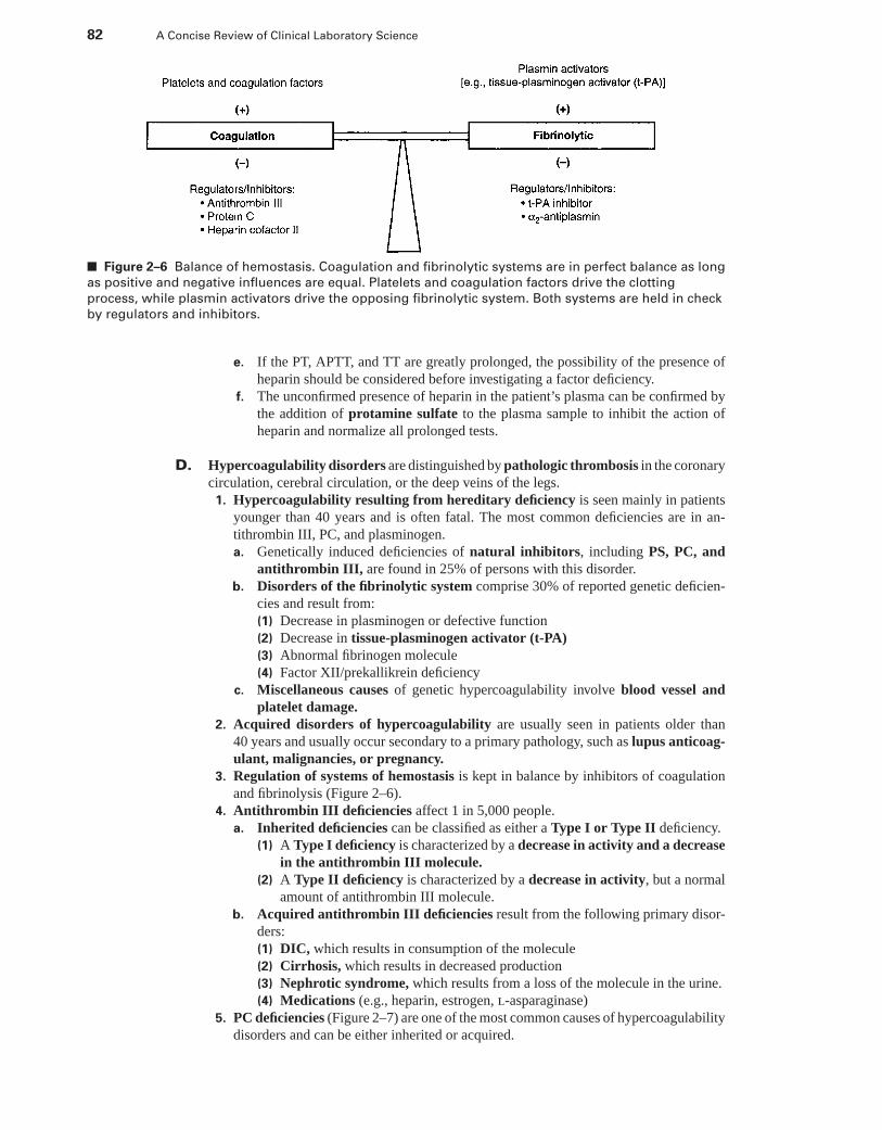
P1: OSO
LWBK192-02 LWBK192-Hubbard November 23, 2008 17:20
82 A Concise Review of Clinical Laboratory Science
■ Figure 2–6 Balance of hemostasis. Coagulation and fibrinolytic systems are in perfect balance as long
as positive and negative influences are equal. Platelets and coagulation factors drive the clotting
process, while plasmin activators drive the opposing fibrinolytic system. Both systems are held in check
by regulators and inhibitors.
e. If the PT, APTT, and TT are greatly prolonged, the possibility of the presence ofheparin should be considered before investigating a factor deficiency.
f. The unconfirmed presence of heparin in the patient’s plasma can be confirmed bythe addition of protamine sulfate to the plasma sample to inhibit the action ofheparin and normalize all prolonged tests.
D. Hypercoagulability disorders are distinguished by pathologic thrombosis in the coronarycirculation, cerebral circulation, or the deep veins of the legs.
1. Hypercoagulability resulting from hereditary deficiency is seen mainly in patientsyounger than 40 years and is often fatal. The most common deficiencies are in an-tithrombin III, PC, and plasminogen.a. Genetically induced deficiencies of natural inhibitors, including PS, PC, and
antithrombin III, are found in 25% of persons with this disorder.b. Disorders of the fibrinolytic system comprise 30% of reported genetic deficien-
cies and result from:(1) Decrease in plasminogen or defective function(2) Decrease in tissue-plasminogen activator (t-PA)(3) Abnormal fibrinogen molecule(4) Factor XII/prekallikrein deficiency
c. Miscellaneous causes of genetic hypercoagulability involve blood vessel andplatelet damage.
2. Acquired disorders of hypercoagulability are usually seen in patients older than40 years and usually occur secondary to a primary pathology, such as lupus anticoag-ulant, malignancies, or pregnancy.
3. Regulation of systems of hemostasis is kept in balance by inhibitors of coagulationand fibrinolysis (Figure 2–6).
4. Antithrombin III deficiencies affect 1 in 5,000 people.a. Inherited deficiencies can be classified as either a Type I or Type II deficiency.
(1) A Type I deficiency is characterized by a decrease in activity and a decreasein the antithrombin III molecule.
(2) A Type II deficiency is characterized by a decrease in activity, but a normalamount of antithrombin III molecule.
b. Acquired antithrombin III deficiencies result from the following primary disor-ders:(1) DIC, which results in consumption of the molecule(2) Cirrhosis, which results in decreased production(3) Nephrotic syndrome, which results from a loss of the molecule in the urine.(4) Medications (e.g., heparin, estrogen, l-asparaginase)
5. PC deficiencies (Figure 2–7) are one of the most common causes of hypercoagulabilitydisorders and can be either inherited or acquired.
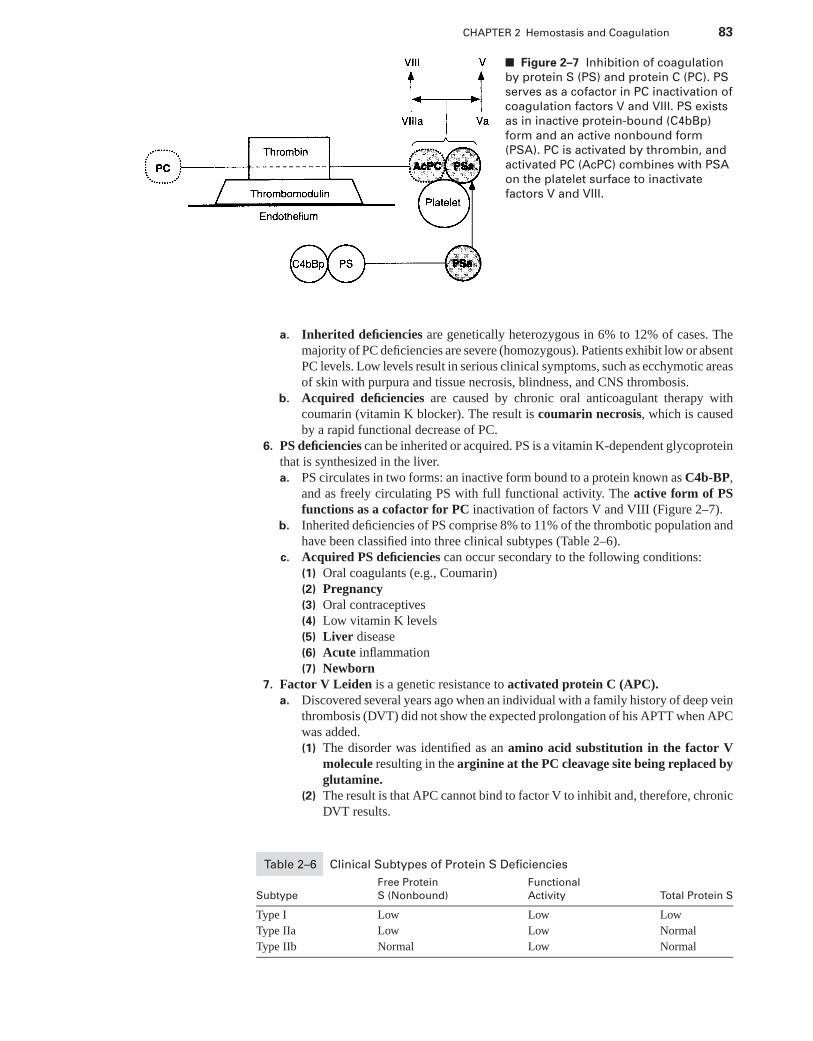
P1: OSO
LWBK192-02 LWBK192-Hubbard November 23, 2008 17:20
CHAPTER 2 Hemostasis and Coagulation 83
■ Figure 2–7 Inhibition of coagulation
by protein S (PS) and protein C (PC). PS
serves as a cofactor in PC inactivation of
coagulation factors V and VIII. PS exists
as in inactive protein-bound (C4bBp)
form and an active nonbound form
(PSA). PC is activated by thrombin, and
activated PC (AcPC) combines with PSA
on the platelet surface to inactivate
factors V and VIII.
a. Inherited deficiencies are genetically heterozygous in 6% to 12% of cases. Themajority of PC deficiencies are severe (homozygous). Patients exhibit low or absentPC levels. Low levels result in serious clinical symptoms, such as ecchymotic areasof skin with purpura and tissue necrosis, blindness, and CNS thrombosis.
b. Acquired deficiencies are caused by chronic oral anticoagulant therapy withcoumarin (vitamin K blocker). The result is coumarin necrosis, which is causedby a rapid functional decrease of PC.
6. PS deficiencies can be inherited or acquired. PS is a vitamin K-dependent glycoproteinthat is synthesized in the liver.a. PS circulates in two forms: an inactive form bound to a protein known as C4b-BP,
and as freely circulating PS with full functional activity. The active form of PSfunctions as a cofactor for PC inactivation of factors V and VIII (Figure 2–7).
b. Inherited deficiencies of PS comprise 8% to 11% of the thrombotic population andhave been classified into three clinical subtypes (Table 2–6).
c. Acquired PS deficiencies can occur secondary to the following conditions:(1) Oral coagulants (e.g., Coumarin)(2) Pregnancy(3) Oral contraceptives(4) Low vitamin K levels(5) Liver disease(6) Acute inflammation(7) Newborn
7. Factor V Leiden is a genetic resistance to activated protein C (APC).a. Discovered several years ago when an individual with a family history of deep vein
thrombosis (DVT) did not show the expected prolongation of his APTT when APCwas added.(1) The disorder was identified as an amino acid substitution in the factor V
molecule resulting in the arginine at the PC cleavage site being replaced byglutamine.
(2) The result is that APC cannot bind to factor V to inhibit and, therefore, chronicDVT results.
Table 2–6 Clinical Subtypes of Protein S Deficiencies
Free Protein Functional
Subtype S (Nonbound) Activity Total Protein S
Type I Low Low Low
Type IIa Low Low Normal
Type IIb Normal Low Normal
P1: OSO
LWBK192-02 LWBK192-Hubbard November 23, 2008 17:20
84 A Concise Review of Clinical Laboratory Science
b. Prevalence:(1) Approximately 5% of general population(2) Approximately 20% of all DVT cases(3) Approximately 50% of cases with a family history of DVT(4) Approximately 60% of pregnancy associated with thrombosis
c. Laboratory Screening:(1) Resistance to APC prolongation of the APTT has a sensitivity of 90% in
detecting the disorder(2) PCR identification of the abnormal GAGG sequence
Online Menu for This Chapter
Go to thePoint.lww.com/Hubbard2e for the following:
� Study Questions and Answers
P1: OSO
LWBK192-03 LWBK192-Hubbard November 23, 2008 17:20
CHAPTER 3
Routine Hematology
JOEL HUBBARD, PhD, MT (ASCP)
I. LABORATORY ANALYSIS
A. Electronic cell counting. Because of manufacturer diversity and the complexity (i.e.,performance characteristics) of cell counters that exists among laboratories, individuallaboratory instrumentation is not presented here.
1. Multiparameter cell counters provide 10 or more common parameters, includingcount of red blood cells (RBCs), white blood cells (WBCs), and platelets with a totallyautomatic diluting system. The most common parameters provided by current cellcounters include:a. RBC countb. WBC countc. Hemoglobin (Hb)d. Hematocrit (HCT)e. Mean corpuscular volume (MCV)f. Mean corpuscular hemoglobin (MCH)
g. Mean corpuscular hemoglobin concentration (MCHC)h. Platelet counti. Mean platelet volume (MPV) is determined from the platelet histogram curve.
The reference range is 6.5 to 12 fL.j. Red cell distribution width (RDW) provides an estimate of RBC anisocytosis
(size variation).(1) Calculation. The RDW (Figure 3–1) is calculated from the following formula:
RDW = (A − B) ÷ (A + B) × k
where A = the MCV, in which 20% of the RBCs are larger than the rest; B =the MCV, in which 80% of the RBCs are larger than the rest; and k = aconstant that represents the number that is required to give a normal value of10.
(2) The normal reference range for the RDW is 8.5 to 14.5. Samples showingvalues >14.5 should be carefully checked for anisocytosis.
(3) Diagnostic use. When considered with the MCV, the RDW can be of usediagnostically, as illustrated in Table 3–1.
2. Electrical impedancea. Particles (most often cells) are forced to flow through small openings (i.e., aper-
tures) between two electrodes in an ionic solution.
85
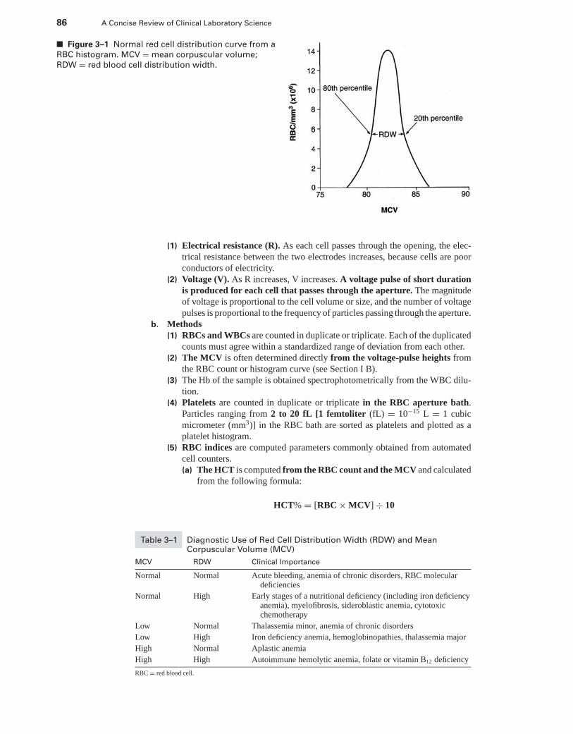
P1: OSO
LWBK192-03 LWBK192-Hubbard November 23, 2008 17:20
86 A Concise Review of Clinical Laboratory Science
■ Figure 3–1 Normal red cell distribution curve from a
RBC histogram. MCV = mean corpuscular volume;
RDW = red blood cell distribution width.
(1) Electrical resistance (R). As each cell passes through the opening, the elec-trical resistance between the two electrodes increases, because cells are poorconductors of electricity.
(2) Voltage (V). As R increases, V increases. A voltage pulse of short durationis produced for each cell that passes through the aperture. The magnitudeof voltage is proportional to the cell volume or size, and the number of voltagepulses is proportional to the frequency of particles passing through the aperture.
b. Methods(1) RBCs and WBCs are counted in duplicate or triplicate. Each of the duplicated
counts must agree within a standardized range of deviation from each other.(2) The MCV is often determined directly from the voltage-pulse heights from
the RBC count or histogram curve (see Section I B).(3) The Hb of the sample is obtained spectrophotometrically from the WBC dilu-
tion.(4) Platelets are counted in duplicate or triplicate in the RBC aperture bath.
Particles ranging from 2 to 20 fL [1 femtoliter (fL) = 10−15 L = 1 cubicmicrometer (mm3)] in the RBC bath are sorted as platelets and plotted as aplatelet histogram.
(5) RBC indices are computed parameters commonly obtained from automatedcell counters.(a) The HCT is computed from the RBC count and the MCV and calculated
from the following formula:
HCT% = [RBC × MCV] ÷ 10
Table 3–1 Diagnostic Use of Red Cell Distribution Width (RDW) and MeanCorpuscular Volume (MCV)
MCV RDW Clinical Importance
Normal Normal Acute bleeding, anemia of chronic disorders, RBC moleculardeficiencies
Normal High Early stages of a nutritional deficiency (including iron deficiencyanemia), myelofibrosis, sideroblastic anemia, cytotoxicchemotherapy
Low Normal Thalassemia minor, anemia of chronic disorders
Low High Iron deficiency anemia, hemoglobinopathies, thalassemia major
High Normal Aplastic anemia
High High Autoimmune hemolytic anemia, folate or vitamin B12 deficiency
RBC = red blood cell.
P1: OSO
LWBK192-03 LWBK192-Hubbard November 23, 2008 17:20
CHAPTER 3 Routine Hematology 87
(b) The MCH is computed from the MCV and the MCHC and calculatedfrom the following formula (note that 1 μg = 1 picogram [pg]):
MCH pg = [MCV × MCHC] ÷ 100
or
MCH = [Hb × 10] ÷ RBC count
(c) The MCHC is computed from the Hb and HCT and calculated fromthe following formula:
MCHC% = [Hb ÷ HCT] × 100
c. Common errors(1) Missing parameter(s)
(a) Any WBC or RBC count grossly out of the normal range must be treatedwith suspicion, and the sample must be repeated.
(b) The “rule of three” (i.e., three times the Hb value should agree ± 3%with the HCT value). If it does not, the sample must be repeated.
(c) Any one of the indices being “singly” out of range must be consideredsuspicious.
(2) Carryover from high to low WBC counts is a problem with some cell countersbecause it can amount to a 2% to 3% error. Carryover results from incompleteremoval of all WBCs from the counting chamber between counts.(a) If the ratio of successive counts exceeds 3:1, then the second count may be
in error by as much as 5%.(b) It may be necessary to repeat any low WBC count that follows a high
count.(3) Increased WBC counts >50,000/mm3 may produce a proportional elevation
in Hb values because of increased cellular turbidity in the WBC countingbaths. In addition, the MCV and HCT may also be increased because thenumber of WBCs is high enough to produce error when sized and counted inthe RBC aperture bath.
(4) Extremely microcytic MCVs may be overestimated by the instrument.(5) High patient glucose concentrations >400 mg/dL result in intracellular hy-
perosmolality in RBCs and may cause a high MCV and HCT, with a lowMCHC.
(6) Cold agglutinins in high titer give a falsely high MCV, low RBC counts, andvery high MCHCs.(a) A good clue for this problem is to look for RBC clumping in a thin area
on the blood smear.(b) Warming the blood or diluent to 37◦C may eliminate this problem.
(7) Very high plasma lipid levels resulting in lipemic plasma may produce tur-bidity in the WBC aperture bath and falsely increase the Hb, MCH, andMCHC.
B. Blood cell histograms are provided by many high-volume instruments to provide sizedistributions of the different cell populations. The volume, given in mm3 or fL, isplotted against the relative frequency for platelets, WBCs, and RBCs (Figure 3–2).1. The WBC histogram provides a count and plot of cells in the WBC aperture bath
larger than 45 fL.a. Normal WBC histograms have three distribution peaks:
(1) The first peak, ranging from 45 to 90 fL, represents a small mononuclearpopulation of cells (i.e., lymphocytes).
(2) The second peak, ranging from 90 to 160 fL, represents a minor population oflarger mononuclear cells (i.e., monocytes). An increase in the number of cellsin this size range can also represent abnormal cell types, such as the immatureprecursor cell types found in patients with leukemia.
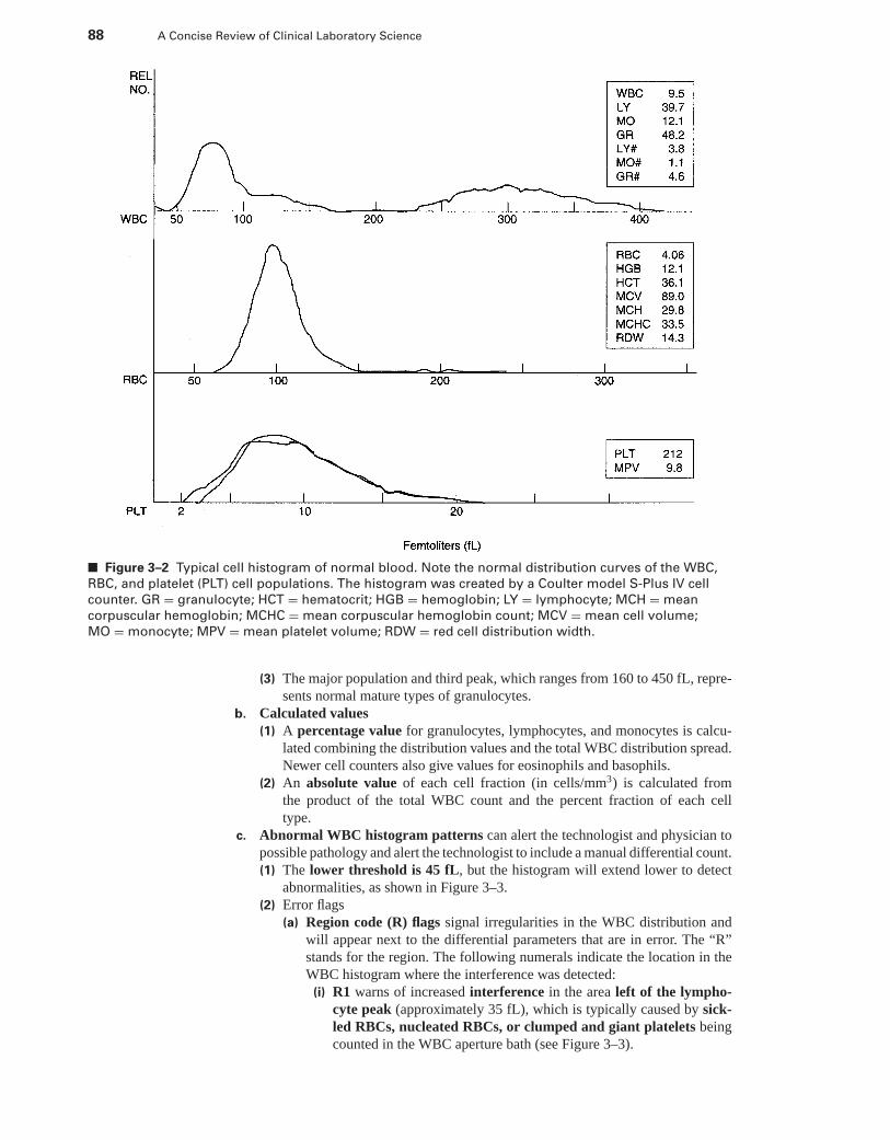
P1: OSO
LWBK192-03 LWBK192-Hubbard November 23, 2008 17:20
88 A Concise Review of Clinical Laboratory Science
■ Figure 3–2 Typical cell histogram of normal blood. Note the normal distribution curves of the WBC,
RBC, and platelet (PLT) cell populations. The histogram was created by a Coulter model S-Plus IV cell
counter. GR = granulocyte; HCT = hematocrit; HGB = hemoglobin; LY = lymphocyte; MCH = mean
corpuscular hemoglobin; MCHC = mean corpuscular hemoglobin count; MCV = mean cell volume;
MO = monocyte; MPV = mean platelet volume; RDW = red cell distribution width.
(3) The major population and third peak, which ranges from 160 to 450 fL, repre-sents normal mature types of granulocytes.
b. Calculated values(1) A percentage value for granulocytes, lymphocytes, and monocytes is calcu-
lated combining the distribution values and the total WBC distribution spread.Newer cell counters also give values for eosinophils and basophils.
(2) An absolute value of each cell fraction (in cells/mm3) is calculated fromthe product of the total WBC count and the percent fraction of each celltype.
c. Abnormal WBC histogram patterns can alert the technologist and physician topossible pathology and alert the technologist to include a manual differential count.(1) The lower threshold is 45 fL, but the histogram will extend lower to detect
abnormalities, as shown in Figure 3–3.(2) Error flags
(a) Region code (R) flags signal irregularities in the WBC distribution andwill appear next to the differential parameters that are in error. The “R”stands for the region. The following numerals indicate the location in theWBC histogram where the interference was detected:
(i) R1 warns of increased interference in the area left of the lympho-cyte peak (approximately 35 fL), which is typically caused by sick-led RBCs, nucleated RBCs, or clumped and giant platelets beingcounted in the WBC aperture bath (see Figure 3–3).
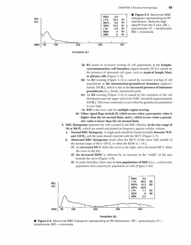
P1: OSO
LWBK192-03 LWBK192-Hubbard November 23, 2008 17:20
CHAPTER 3 Routine Hematology 89
■ Figure 3–3 Abnormal WBC
histogram representing an R1
distribution. Note the high
takeoff from the Y axis. GR =granulocyte; LY = lymphocyte;
MO = monocyte.
(ii) R2 warns of excessive overlap of cell populations at the lympho-cyte/mononuclear cell boundary (approximately 90 fL) caused bythe presence of abnormal cell types, such as atypical lymph, blast,or plasma cells (Figure 3–4).
(iii) An R3 warning (Figure 3–5) is caused by excessive overlap of cellpopulations at the mononuclear/granulocyte boundary (approxi-mately 160 fL), which is due to the increased presence of immaturegranulocytes (i.e., bands, metamyelocytes).
(iv) An R4 warning (Figure 3–6) is caused by the extension of the celldistribution past the upper end of the WBC threshold (approximately450 fL). This most commonly occurs when the granulocyte populationis very high.
(v) RM is the error code for multiple region overlap.(b) Other signal flags include H, which occurs when a parameter value is
higher than the set normal limit, and L, which occurs when a param-eter value is lower than the set normal limit.
2. RBC histograms represent the cells counted in the RBC dilution, in the size range of36 to 360 fL, which are sorted and plotted as frequency against cellular volume.a. Normal RBC histogram. A single peak should be found normally between 70 fL
and 110 fL, and the peak should coincide with the MCV (Figure 3–7).b. Abnormal RBC histograms result when the MCV of the curve falls outside of
the normal range of 80 to 100 fL or when the RDW is >14.5.(1) An increased MCV shifts the curve to the right, and a decreased MCV shifts
the curve to the left.(2) An increased RDW is reflected by an increase in the “width” of the area
beneath the curve (Figure 3–8).(3) In some disorders, there may be two populations of RBCs [i.e., a microcytic
population and a macrocytic population of cells (Figure 3–8)].
■ Figure 3–4 Abnormal WBC histogram representing an R2 distribution. GR = granulocyte; LY =lymphocyte; MO = monocyte.
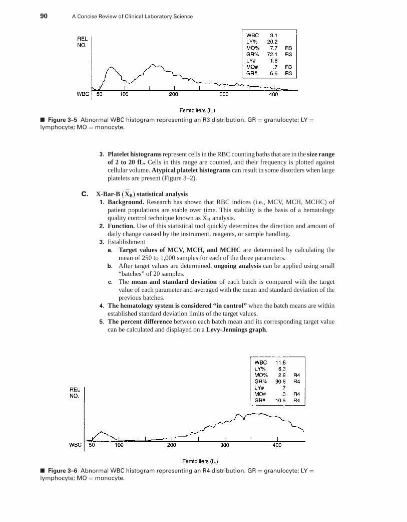
P1: OSO
LWBK192-03 LWBK192-Hubbard November 23, 2008 17:20
90 A Concise Review of Clinical Laboratory Science
■ Figure 3–5 Abnormal WBC histogram representing an R3 distribution. GR = granulocyte; LY =lymphocyte; MO = monocyte.
3. Platelet histograms represent cells in the RBC counting baths that are in the size rangeof 2 to 20 fL. Cells in this range are counted, and their frequency is plotted againstcellular volume. Atypical platelet histograms can result in some disorders when largeplatelets are present (Figure 3–2).
C. X-Bar-B (XB) statistical analysis1. Background. Research has shown that RBC indices (i.e., MCV, MCH, MCHC) of
patient populations are stable over time. This stability is the basis of a hematologyquality control technique known as XB analysis.
2. Function. Use of this statistical tool quickly determines the direction and amount ofdaily change caused by the instrument, reagents, or sample handling.
3. Establishmenta. Target values of MCV, MCH, and MCHC are determined by calculating the
mean of 250 to 1,000 samples for each of the three parameters.b. After target values are determined, ongoing analysis can be applied using small
“batches” of 20 samples.c. The mean and standard deviation of each batch is compared with the target
value of each parameter and averaged with the mean and standard deviation of theprevious batches.
4. The hematology system is considered “in control” when the batch means are withinestablished standard deviation limits of the target values.
5. The percent difference between each batch mean and its corresponding target valuecan be calculated and displayed on a Levy-Jennings graph.
■ Figure 3–6 Abnormal WBC histogram representing an R4 distribution. GR = granulocyte; LY =lymphocyte; MO = monocyte.
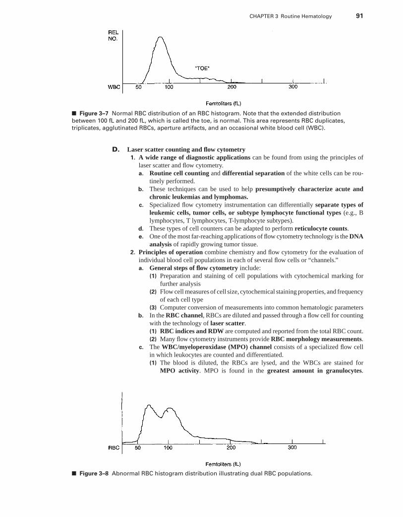
P1: OSO
LWBK192-03 LWBK192-Hubbard November 23, 2008 17:20
CHAPTER 3 Routine Hematology 91
■ Figure 3–7 Normal RBC distribution of an RBC histogram. Note that the extended distribution
between 100 fL and 200 fL, which is called the toe, is normal. This area represents RBC duplicates,
triplicates, agglutinated RBCs, aperture artifacts, and an occasional white blood cell (WBC).
D. Laser scatter counting and flow cytometry1. A wide range of diagnostic applications can be found from using the principles of
laser scatter and flow cytometry.a. Routine cell counting and differential separation of the white cells can be rou-
tinely performed.b. These techniques can be used to help presumptively characterize acute and
chronic leukemias and lymphomas.c. Specialized flow cytometry instrumentation can differentially separate types of
leukemic cells, tumor cells, or subtype lymphocyte functional types (e.g., Blymphocytes, T lymphocytes, T-lymphocyte subtypes).
d. These types of cell counters can be adapted to perform reticulocyte counts.e. One of the most far-reaching applications of flow cytometry technology is the DNA
analysis of rapidly growing tumor tissue.2. Principles of operation combine chemistry and flow cytometry for the evaluation of
individual blood cell populations in each of several flow cells or “channels.”a. General steps of flow cytometry include:
(1) Preparation and staining of cell populations with cytochemical marking forfurther analysis
(2) Flow cell measures of cell size, cytochemical staining properties, and frequencyof each cell type
(3) Computer conversion of measurements into common hematologic parametersb. In the RBC channel, RBCs are diluted and passed through a flow cell for counting
with the technology of laser scatter.(1) RBC indices and RDW are computed and reported from the total RBC count.(2) Many flow cytometry instruments provide RBC morphology measurements.
c. The WBC/myeloperoxidase (MPO) channel consists of a specialized flow cellin which leukocytes are counted and differentiated.(1) The blood is diluted, the RBCs are lysed, and the WBCs are stained for
MPO activity. MPO is found in the greatest amount in granulocytes.
■ Figure 3–8 Abnormal RBC histogram distribution illustrating dual RBC populations.
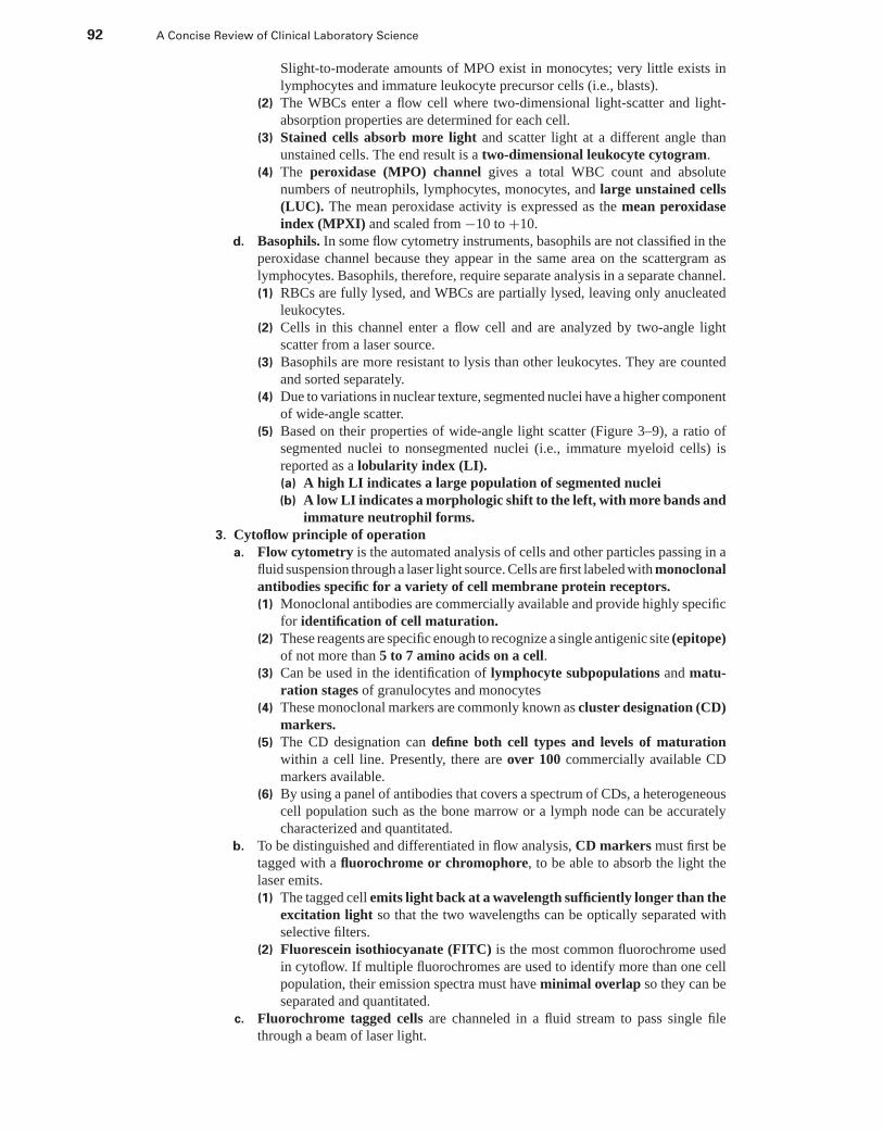
P1: OSO
LWBK192-03 LWBK192-Hubbard November 23, 2008 17:20
92 A Concise Review of Clinical Laboratory Science
Slight-to-moderate amounts of MPO exist in monocytes; very little exists inlymphocytes and immature leukocyte precursor cells (i.e., blasts).
(2) The WBCs enter a flow cell where two-dimensional light-scatter and light-absorption properties are determined for each cell.
(3) Stained cells absorb more light and scatter light at a different angle thanunstained cells. The end result is a two-dimensional leukocyte cytogram.
(4) The peroxidase (MPO) channel gives a total WBC count and absolutenumbers of neutrophils, lymphocytes, monocytes, and large unstained cells(LUC). The mean peroxidase activity is expressed as the mean peroxidaseindex (MPXI) and scaled from −10 to +10.
d. Basophils. In some flow cytometry instruments, basophils are not classified in theperoxidase channel because they appear in the same area on the scattergram aslymphocytes. Basophils, therefore, require separate analysis in a separate channel.(1) RBCs are fully lysed, and WBCs are partially lysed, leaving only anucleated
leukocytes.(2) Cells in this channel enter a flow cell and are analyzed by two-angle light
scatter from a laser source.(3) Basophils are more resistant to lysis than other leukocytes. They are counted
and sorted separately.(4) Due to variations in nuclear texture, segmented nuclei have a higher component
of wide-angle scatter.(5) Based on their properties of wide-angle light scatter (Figure 3–9), a ratio of
segmented nuclei to nonsegmented nuclei (i.e., immature myeloid cells) isreported as a lobularity index (LI).(a) A high LI indicates a large population of segmented nuclei(b) A low LI indicates a morphologic shift to the left, with more bands and
immature neutrophil forms.3. Cytoflow principle of operation
a. Flow cytometry is the automated analysis of cells and other particles passing in afluid suspension through a laser light source. Cells are first labeled with monoclonalantibodies specific for a variety of cell membrane protein receptors.(1) Monoclonal antibodies are commercially available and provide highly specific
for identification of cell maturation.(2) These reagents are specific enough to recognize a single antigenic site (epitope)
of not more than 5 to 7 amino acids on a cell.(3) Can be used in the identification of lymphocyte subpopulations and matu-
ration stages of granulocytes and monocytes(4) These monoclonal markers are commonly known as cluster designation (CD)
markers.(5) The CD designation can define both cell types and levels of maturation
within a cell line. Presently, there are over 100 commercially available CDmarkers available.
(6) By using a panel of antibodies that covers a spectrum of CDs, a heterogeneouscell population such as the bone marrow or a lymph node can be accuratelycharacterized and quantitated.
b. To be distinguished and differentiated in flow analysis, CD markers must first betagged with a fluorochrome or chromophore, to be able to absorb the light thelaser emits.(1) The tagged cell emits light back at a wavelength sufficiently longer than the
excitation light so that the two wavelengths can be optically separated withselective filters.
(2) Fluorescein isothiocyanate (FITC) is the most common fluorochrome usedin cytoflow. If multiple fluorochromes are used to identify more than one cellpopulation, their emission spectra must have minimal overlap so they can beseparated and quantitated.
c. Fluorochrome tagged cells are channeled in a fluid stream to pass single filethrough a beam of laser light.
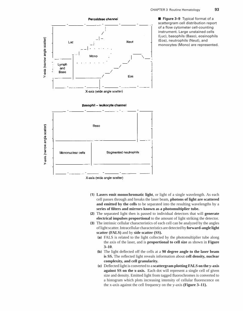
P1: OSO
LWBK192-03 LWBK192-Hubbard November 23, 2008 17:20
CHAPTER 3 Routine Hematology 93
■ Figure 3–9 Typical format of a
scattergram cell distribution report
of a flow cytometer cell-counting
instrument. Large unstained cells
(Luc), basophils (Baso), eosinophils
(Eos), neutrophils (Neut), and
monocytes (Mono) are represented.
(1) Lasers emit monochromatic light, or light of a single wavelength. As eachcell passes through and breaks the laser beam, photons of light are scatteredand emitted by the cells to be separated into the resulting wavelengths by aseries of filters and mirrors known as a photomultiplier tube.
(2) The separated light then is passed to individual detectors that will generateelectrical impulses proportional to the amount of light striking the detector.
(3) The intrinsic cellular characteristics of each cell can be analyzed by the anglesof light scatter. Intracellular characteristics are detected by forward-angle lightscatter (FALS) and by side scatter (SS).(a) FALS is related to the light collected by the photomultiplier tube along
the axis of the laser, and is proportional to cell size as shown in Figure3–10.
(b) The light deflected off the cells at a 90 degree angle to the laser beamis SS. The reflected light reveals information about cell density, nuclearcomplexity, and cell granularity.
(c) Deflected light is converted to a scattergram plotting FALS on the y-axisagainst SS on the x-axis. Each dot will represent a single cell of givensize and density. Emitted light from tagged fluorochromes is converted toa histogram which plots increasing intensity of cellular fluorescence onthe x-axis against the cell frequency on the y-axis (Figure 3–11).
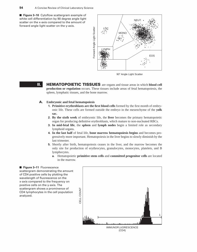
P1: OSO
LWBK192-03 LWBK192-Hubbard November 23, 2008 17:20
94 A Concise Review of Clinical Laboratory Science
90° Angle Light Scatter
Forw
ard
Angle
Lig
ht S
catter
NEUT.
LUC
MONO
LYMPH
EOS
■ Figure 3–10 Cytoflow scattergram example of
white cell differentiation by 90 degree angle light
scatter on the x-axis compared to the amount of
forward angle light scatter on the y-axis.
II. HEMATOPOIETIC TISSUES are organs and tissue areas in which blood cellproduction or regulation occurs. These tissues include areas of fetal hematopoiesis, thespleen, lymphatic tissues, and the bone marrow.
A. Embryonic and fetal hematopoiesis1. Primitive erythroblasts are the first blood cells formed by the first month of embry-
onic life. These cells are formed outside the embryo in the mesenchyme of the yolksac.
2. By the sixth week of embryonic life, the liver becomes the primary hematopoieticorgan for producing definitive erythroblasts, which mature to non-nucleated RBCs.
3. In mid-fetal life, the spleen and lymph nodes begin a limited role as secondarylymphoid organs.
4. In the last half of fetal life, bone marrow hematopoiesis begins and becomes pro-gressively more important. Hematopoiesis in the liver begins to slowly diminish by thelast trimester.
5. Shortly after birth, hematopoiesis ceases in the liver, and the marrow becomes theonly site for production of erythrocytes, granulocytes, monocytes, platelets, and Blymphocytes.a. Hematopoietic primitive stem cells and committed progenitor cells are located
in the marrow.
IMMUNOFLUORESCENCE(CD4)
CE
LL
CO
UN
T
1
■ Figure 3–11 Fluorescence
scattergram demonstrating the amount
of CD4 positive cells by plotting the
wavelength of fluorescence on the
x-axis compared to the frequency on
positive cells on the y-axis. The
scattergram shows a prominence of
CD4 lymphocytes in the cell population
analyzed.
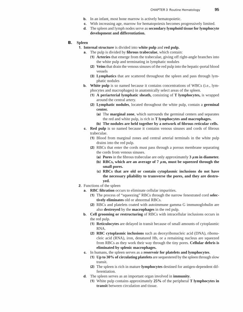
P1: OSO
LWBK192-03 LWBK192-Hubbard November 23, 2008 17:20
CHAPTER 3 Routine Hematology 95
b. In an infant, most bone marrow is actively hematopoietic.c. With increasing age, marrow for hematopoiesis becomes progressively limited.d. The spleen and lymph nodes serve as secondary lymphoid tissue for lymphocyte
development and differentiation.
B. Spleen1. Internal structure is divided into white pulp and red pulp.
a. The pulp is divided by fibrous trabeculae, which contain:(1) Arteries that emerge from the trabeculae, giving off right-angle branches into
the white pulp and terminating in lymphatic nodules(2) Veins that drain the venous sinuses of the red pulp into the hepatic-portal blood
vessels(3) Lymphatics that are scattered throughout the spleen and pass through lym-
phatic nodulesb. White pulp is so named because it contains concentrations of WBCs (i.e., lym-
phocytes and macrophages) in anatomically select areas of the spleen.(1) A periarterial lymphatic sheath, consisting of T lymphocytes, is wrapped
around the central artery.(2) Lymphatic nodules, located throughout the white pulp, contain a germinal
center.(a) The marginal zone, which surrounds the germinal centers and separates
the red and white pulp, is rich in T lymphocytes and macrophages.(b) The nodules are held together by a network of fibrous reticular cells.
c. Red pulp is so named because it contains venous sinuses and cords of fibroustrabeculae.(1) Blood from marginal zones and central arterial terminals in the white pulp
drains into the red pulp.(2) RBCs that enter the cords must pass through a porous membrane separating
the cords from venous sinuses.(a) Pores in the fibrous trabeculae are only approximately 3 μm in diameter.(b) RBCs, which are an average of 7 μm, must be squeezed through the
small pores.(c) RBCs that are old or contain cytoplasmic inclusions do not have
the necessary pliability to transverse the pores, and they are destro-yed.
2. Functions of the spleena. RBC filtration occurs to eliminate cellular impurities.
(1) The process of “squeezing” RBCs through the narrow fenestrated cord selec-tively eliminates old or abnormal RBCs.
(2) RBCs and platelets coated with autoimmune gamma G immunoglobulin arealso destroyed by the macrophages in the red pulp.
b. Cell grooming or restructuring of RBCs with intracellular inclusions occurs inthe red pulp.(1) Reticulocytes are delayed in transit because of small amounts of cytoplasmic
RNA.(2) RBC cytoplasmic inclusions such as deoxyribonucleic acid (DNA), ribonu-
cleic acid (RNA), iron, denatured Hb, or a remaining nucleus are squeezedfrom RBCs as they work their way through the tiny pores. Cellular debris iseliminated by splenic macrophages.
c. In humans, the spleen serves as a reservoir for platelets and lymphocytes.(1) Up to 30% of circulating platelets are sequestered by the spleen through slow
transit.(2) The spleen is rich in mature lymphocytes destined for antigen-dependent dif-
ferentiation.d. The spleen serves as an important organ involved in immunity.
(1) White pulp contains approximately 25% of the peripheral T lymphocytes intransit between circulation and tissue.
P1: OSO
LWBK192-03 LWBK192-Hubbard November 23, 2008 17:20
96 A Concise Review of Clinical Laboratory Science
(2) Approximately 10% to 15% of peripheral B lymphocytes are found in splenicnodules.
(3) The spleen provides a locale for immunity-induced lymphocyte differentiationand antibody production. It serves as an important organ for immunoglobulinM (IgM) production by B lymphocytes.
3. Asplenia (the absence of splenic function) can be caused by either surgical removal(e.g., splenectomy) or radiation overexposure. This condition can also occur in associ-ation with malabsorption syndromes. The hematologic results of asplenia include:a. Increased susceptibility to infectionb. Acute granulocytosisc. Acute thrombocytosis, with occasional giant plateletsd. Chronic and absolute lymphocytosis and monocytosise. Increased appearance of immature RBCs in the circulationf. Increased amount of circulating RBCs with cytoplasmic inclusions or abnormal
forms (e.g., Howell-Jolly bodies, target cells, and burr cells)4. Splenomegaly
a. Definitions(1) Splenomegaly describes an enlarged spleen.(2) Hypersplenism describes exaggerated inhibitory or destructive functions of
the spleen, which is usually accompanied by splenomegaly.b. Clinical presentation. Splenomegaly results in vascular congestion and portal
hypertension. RBCs, granulocytes, and as many as 90% of circulating plateletsmay be trapped, which results in anemia, leukocytopenia, and thrombocytopenia.A hypercellular marrow results in response to a chronic pancytopenia.
C. Lymphatic tissues1. Lymphopoiesis is anatomically divided between two areas.
a. Primary lymphatic tissues include the bone marrow for B lymphocytes, thethymus for T lymphocytes, and sites of active hematopoiesis in the fetus.
b. Secondary lymphatic tissues serve as reservoirs for already differentiated lym-phocytes. These tissues include the lymph nodes, spleen, and gut-associatedlymphatic tissue.
2. The thymus is a primary tissue for T-lymphocyte development.a. The cortex, or outer part of the organ, consists of several cell types.
(1) Small and medium primitive T cells (i.e., thymocytes) are the youngest cellsfound in the thymus.
(2) Epithelial cells serve as important effectors of T-cell differentiation.(3) The corticomedullary junction is rich in macrophages.
b. The medulla, or interior of the organ, is rich in maturing T lymphocytes, epithe-lial cells, and concentric swirls of squamous epithelial cells known as Hassall’scorpuscles. Mature immunocompetent T lymphocytes leave the medulla and enterthe circulation to migrate to secondary lymphoid tissues to await “specific” immunedifferentiation.
3. Lymph nodes, spread throughout the body in a “second” vascular system, comprisepart of the secondary lymphatic system.a. All lymph empties and is recirculated into the bloodstream via the right thoracic
duct and the left thoracic duct, which is the major lymphatic vessel.b. All nodes are separated from each other by thin-walled vessels known as lymph
ducts. The normal anatomic structure of each node includes a cortex, paracortex,and medulla.(1) The cortex contains lymphocyte follicles (nodules) with germinal centers.
These follicles are unique to lymph nodes and the spleen.(a) All follicles are arranged in a row beneath the surface capsular lining.(b) These follicles contain a concentration of macrophages and B lympho-
cytes.(c) The germinal centers are surrounded by a high concentration of T
lymphocytes.
P1: OSO
LWBK192-03 LWBK192-Hubbard November 23, 2008 17:20
CHAPTER 3 Routine Hematology 97
(2) The paracortex is defined as the area between the cortex and medulla. Thisarea of the node is rich in macrophages, as well as B and T lymphocytes intransit.
(3) The medulla, or inside of the node, is arranged in parallel cords of smalllymph and plasma cells.
4. Lymphopoiesis is defined as lymphocyte production or regulation in primary or sec-ondary lymphatic tissues.a. Lymphocyte production in primary tissues is continuous and independent of
antigen stimulation.(1) The production rate is in excess of demand.(2) Only a fraction of newly formed lymphocytes in the primary tissues survive to
gain access to secondary lymphoid organs.b. Lymphocyte proliferation in secondary tissues is antigenic stimulation-
dependent, resulting in the proliferation of specific clonal populations of lym-phocytes to carry out the immunologic response.
c. Of circulating lymphocytes, 75% to 85% are T lymphocytes, and 10% to 15%are B lymphocytes.
d. T lymphocytes are more actively motile and recirculate more than B lymphocytes.(1) T cells migrate among the blood (30 minutes), spleen (6 hours), and lymph
nodes (15–20 hours).(2) B lymphocytes do not freely circulate and may stay in a lymph node for as
long as 30 hours.
D. Bone marrow1. Structure and development
a. Fat-cell occupation of the bone marrow space begins by 4 years of age, atwhich time the growth of bone cavities has exceeded the body’s need, and theavailable space in bone cavities has grown faster than the needed circulation bloodmass.(1) Fat-cell growth occurs first at the diaphysis of long bones and slowly extends
to the center of the bone.(2) By 18 years of age, fatty replacement has limited active hematopoietic marrow
to the vertebrae, ribs, skull, sternum, proximal epiphyses of long bones, andthe iliac crest of the pelvis.
(3) Because of this available fatty bone marrow reserve, reactivation of ex-tramedullary organs rarely takes place.
b. Organization. The marrow is organized in a spoke-like pattern of venous sinusesand cords of hematopoietic tissue.(1) Venous sinuses are covered on the marrow side by endothelial cells and on
the sinus side by a basement membrane and reticular adventitial cells, whichput out projections or “nests” to support hematopoietic cells.
(2) Megakaryocytes lie within the cords close to the sinus wall. Strings of plateletspeel directly into the venous sinus.
(3) Erythroblasts lie close to venous sinuses in clusters or colonies.(a) Each cluster consists of a central macrophage (i.e., nurse cell) surrounded
by erythroblasts in various stages of maturation.(b) When mature, the reticulocyte squeezes through the basement mem-
brane and endothelial layer to be released into the venous sinus.c. Nerve supply. The marrow has an extensive supply of nerves, which may play an
important autoregulatory role to adjust blood flow to the rate of cellular maturationand proliferation.
2. Bone marrow functionsa. Minor. The marrow has a minor function in the antigen processing of cellular and
humoral immunity.b. Major. The major function of the marrow is the production and proliferation of
blood cells (hematopoiesis). Marrow hematopoiesis is divided into three majorcompartments or cell types (Figure 3–12).
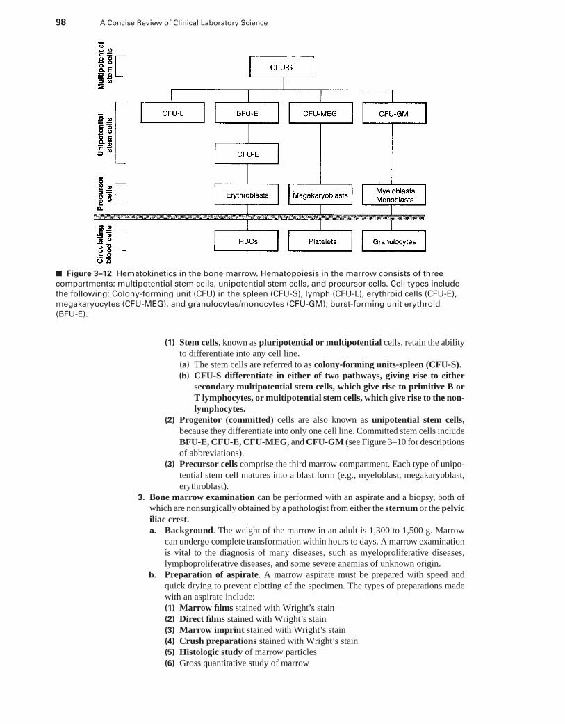
P1: OSO
LWBK192-03 LWBK192-Hubbard November 23, 2008 17:20
98 A Concise Review of Clinical Laboratory Science
■ Figure 3–12 Hematokinetics in the bone marrow. Hematopoiesis in the marrow consists of three
compartments: multipotential stem cells, unipotential stem cells, and precursor cells. Cell types include
the following: Colony-forming unit (CFU) in the spleen (CFU-S), lymph (CFU-L), erythroid cells (CFU-E),
megakaryocytes (CFU-MEG), and granulocytes/monocytes (CFU-GM); burst-forming unit erythroid
(BFU-E).
(1) Stem cells, known as pluripotential or multipotential cells, retain the abilityto differentiate into any cell line.(a) The stem cells are referred to as colony-forming units-spleen (CFU-S).(b) CFU-S differentiate in either of two pathways, giving rise to either
secondary multipotential stem cells, which give rise to primitive B orT lymphocytes, or multipotential stem cells, which give rise to the non-lymphocytes.
(2) Progenitor (committed) cells are also known as unipotential stem cells,because they differentiate into only one cell line. Committed stem cells includeBFU-E, CFU-E, CFU-MEG, and CFU-GM (see Figure 3–10 for descriptionsof abbreviations).
(3) Precursor cells comprise the third marrow compartment. Each type of unipo-tential stem cell matures into a blast form (e.g., myeloblast, megakaryoblast,erythroblast).
3. Bone marrow examination can be performed with an aspirate and a biopsy, both ofwhich are nonsurgically obtained by a pathologist from either the sternum or the pelviciliac crest.a. Background. The weight of the marrow in an adult is 1,300 to 1,500 g. Marrow
can undergo complete transformation within hours to days. A marrow examinationis vital to the diagnosis of many diseases, such as myeloproliferative diseases,lymphoproliferative diseases, and some severe anemias of unknown origin.
b. Preparation of aspirate. A marrow aspirate must be prepared with speed andquick drying to prevent clotting of the specimen. The types of preparations madewith an aspirate include:(1) Marrow films stained with Wright’s stain(2) Direct films stained with Wright’s stain(3) Marrow imprint stained with Wright’s stain(4) Crush preparations stained with Wright’s stain(5) Histologic study of marrow particles(6) Gross quantitative study of marrow
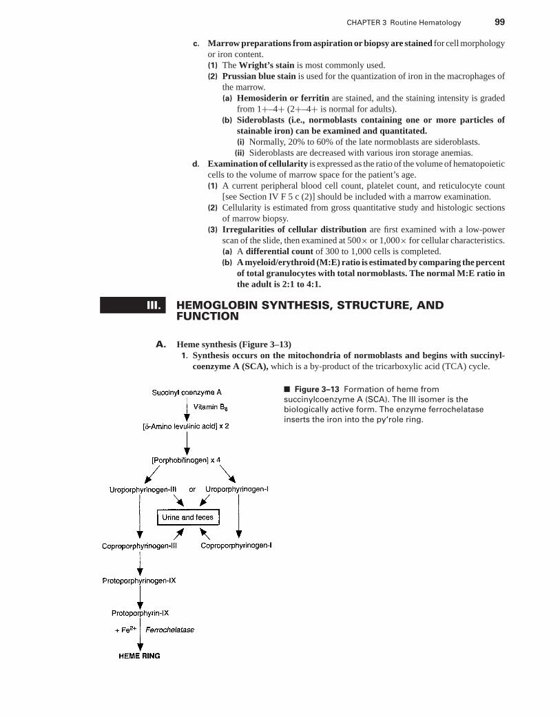
P1: OSO
LWBK192-03 LWBK192-Hubbard November 23, 2008 17:20
CHAPTER 3 Routine Hematology 99
c. Marrow preparations from aspiration or biopsy are stained for cell morphologyor iron content.(1) The Wright’s stain is most commonly used.(2) Prussian blue stain is used for the quantization of iron in the macrophages of
the marrow.(a) Hemosiderin or ferritin are stained, and the staining intensity is graded
from 1+–4+ (2+–4+ is normal for adults).(b) Sideroblasts (i.e., normoblasts containing one or more particles of
stainable iron) can be examined and quantitated.(i) Normally, 20% to 60% of the late normoblasts are sideroblasts.
(ii) Sideroblasts are decreased with various iron storage anemias.d. Examination of cellularity is expressed as the ratio of the volume of hematopoietic
cells to the volume of marrow space for the patient’s age.(1) A current peripheral blood cell count, platelet count, and reticulocyte count
[see Section IV F 5 c (2)] should be included with a marrow examination.(2) Cellularity is estimated from gross quantitative study and histologic sections
of marrow biopsy.(3) Irregularities of cellular distribution are first examined with a low-power
scan of the slide, then examined at 500× or 1,000× for cellular characteristics.(a) A differential count of 300 to 1,000 cells is completed.(b) A myeloid/erythroid (M:E) ratio is estimated by comparing the percent
of total granulocytes with total normoblasts. The normal M:E ratio inthe adult is 2:1 to 4:1.
III. HEMOGLOBIN SYNTHESIS, STRUCTURE, ANDFUNCTION
A. Heme synthesis (Figure 3–13)1. Synthesis occurs on the mitochondria of normoblasts and begins with succinyl-
coenzyme A (SCA), which is a by-product of the tricarboxylic acid (TCA) cycle.
■ Figure 3–13 Formation of heme from
succinylcoenzyme A (SCA). The III isomer is the
biologically active form. The enzyme ferrochelatase
inserts the iron into the py‘role ring.
P1: OSO
LWBK192-03 LWBK192-Hubbard November 23, 2008 17:20
100 A Concise Review of Clinical Laboratory Science
a. SCA combines with glycine to yield an unstable intermediate known as α-amino–β-ketoadipate.
b. The intermediate is decarboxylated to form delta (δ)-aminolevulinic acid (ALA).(1) This reaction occurs in mitochondria and requires pyridoxal phosphate (i.e.,
vitamin B6).(2) Trace amounts of ALA, which is normally found in urine, are increased in
certain abnormalities of heme synthesis (e.g., lead poisoning).c. Two molecules of ALA combine to form porphobilinogen (PBG).
(1) Normally, trace amounts of PBG can be measured in urine.(2) Increased amounts of PBG are excreted in acute intermittent porphyria and
are detected by a color reaction with Ehrlich’s aldehyde reagent.2. Four molecules of porphobilinogen combine to form uroporphyrinogen I or III.
a. The type III isomer form is converted by way of coproporphyrinogen III, andprotoporphyrinogen IX to protoporphyrin IX.
b. Iron is inserted into protoporphyrin by the mitochondrial enzyme, ferrochelatase,to complete the formation of the heme moiety.
c. In certain diseases, this pathway may be partially blocked.(1) Type I isomers of uroporphyrinogen and coproporphyrinogen are formed
and excreted in excess urinary amounts as uroporphyrin I and copropor-phyrin I.
(2) Protoporphyrin is normally found in mature RBCs, but concentrations areincreased in lead poisoning and iron deficiency anemia.
B. Globin chain synthesis1. Polypeptide chains are manufactured on ribosomes in the normoblast cytoplasm.2. Globin chains are assembled from two pairs of polypeptide chains (i.e., four chains per
hemoglobin molecule).3. Four primary chains (i.e., α, β, γ, δ) can be produced. Each type of globin chain is
different by only a few amino-acid substitutions.4. There are many Hb forms, depending on the combination of the two pairs of globin
chains.a. An embryonic form, α2, ε2, is detected early in fetal life.b. By 3 months of embryonic life, embryonic Hb is replaced by fetal Hb (Hb F).
(1) Hb F consists of two α chains and two γ chains (i.e., α2γ2).(2) Hb F is the major Hb in the fetus and newborn infant.(3) Hb F has a higher oxygen affinity than that of adult Hb.(4) β-chain production does not begin until the 20th week of prenatal life, so adult
Hb is approximately 10% between 20 and 35 weeks, and 15% to 40% at birth.(5) After birth, the production of Hb F slowly ceases, and by 6 months of age,
it constitutes <8% of the total Hb content.(a) By 1 year of age, infants have <2% Hb F.(b) Less than 1% Hb F is normally found in adults. Reactivation of Hb F
production may occur in pregnancy and in some disorders of erythro-poiesis.
c. Adult hemoglobin (HbA) is the major adult form, consisting of two α chains andtwo β chains (i.e., α2β2).
d. Hemoglobin A2 (HbA2) accounts for 1.5% to 3.5% of normal adult hemoglobin.(1) HbA2 consists of two α chains and two δ chains (i.e., α2δ2).(2) δ-Chain synthesis occurs only in normoblasts and is absent in reticulocytes.(3) The HbA2 form is increased in some β-thalassemias and in iron deficiency
anemia.e. Genetic control of globin-chain production is seen as gene separation.
(1) The production of α-chains is coded on chromosome 16.(2) The production of all other globin chains is coded on chromosome 11.
C. Structure and function of hemoglobin1. The main function of an RBC is to contain, transport, and protect hemoglobin
molecules.
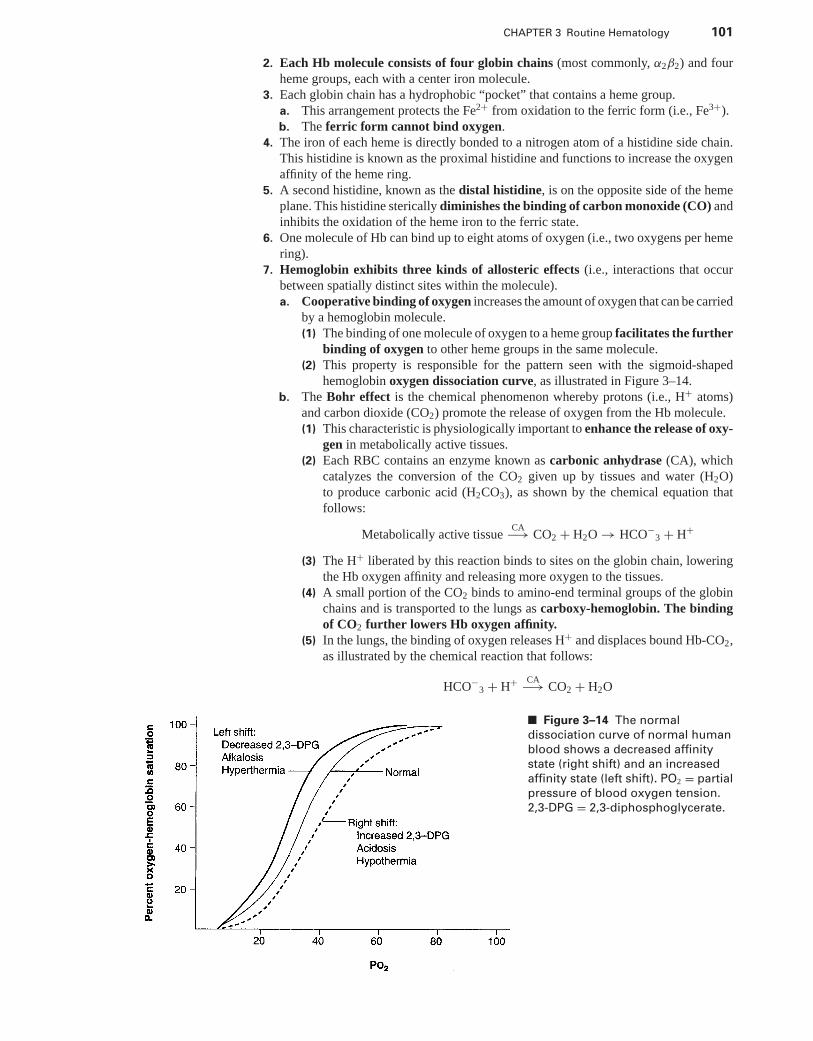
P1: OSO
LWBK192-03 LWBK192-Hubbard November 23, 2008 17:20
CHAPTER 3 Routine Hematology 101
2. Each Hb molecule consists of four globin chains (most commonly, α2β2) and fourheme groups, each with a center iron molecule.
3. Each globin chain has a hydrophobic “pocket” that contains a heme group.a. This arrangement protects the Fe2+ from oxidation to the ferric form (i.e., Fe3+).b. The ferric form cannot bind oxygen.
4. The iron of each heme is directly bonded to a nitrogen atom of a histidine side chain.This histidine is known as the proximal histidine and functions to increase the oxygenaffinity of the heme ring.
5. A second histidine, known as the distal histidine, is on the opposite side of the hemeplane. This histidine sterically diminishes the binding of carbon monoxide (CO) andinhibits the oxidation of the heme iron to the ferric state.
6. One molecule of Hb can bind up to eight atoms of oxygen (i.e., two oxygens per hemering).
7. Hemoglobin exhibits three kinds of allosteric effects (i.e., interactions that occurbetween spatially distinct sites within the molecule).a. Cooperative binding of oxygen increases the amount of oxygen that can be carried
by a hemoglobin molecule.(1) The binding of one molecule of oxygen to a heme group facilitates the further
binding of oxygen to other heme groups in the same molecule.(2) This property is responsible for the pattern seen with the sigmoid-shaped
hemoglobin oxygen dissociation curve, as illustrated in Figure 3–14.b. The Bohr effect is the chemical phenomenon whereby protons (i.e., H+ atoms)
and carbon dioxide (CO2) promote the release of oxygen from the Hb molecule.(1) This characteristic is physiologically important to enhance the release of oxy-
gen in metabolically active tissues.(2) Each RBC contains an enzyme known as carbonic anhydrase (CA), which
catalyzes the conversion of the CO2 given up by tissues and water (H2O)to produce carbonic acid (H2CO3), as shown by the chemical equation thatfollows:
Metabolically active tissueCA−→ CO2 + H2O → HCO−
3 + H+
(3) The H+ liberated by this reaction binds to sites on the globin chain, loweringthe Hb oxygen affinity and releasing more oxygen to the tissues.
(4) A small portion of the CO2 binds to amino-end terminal groups of the globinchains and is transported to the lungs as carboxy-hemoglobin. The bindingof CO2 further lowers Hb oxygen affinity.
(5) In the lungs, the binding of oxygen releases H+ and displaces bound Hb-CO2,as illustrated by the chemical reaction that follows:
HCO−3 + H+ CA−→ CO2 + H2O
■ Figure 3–14 The normal
dissociation curve of normal human
blood shows a decreased affinity
state (right shift) and an increased
affinity state (left shift). PO2 = partial
pressure of blood oxygen tension.
2,3-DPG = 2,3-diphosphoglycerate.
P1: OSO
LWBK192-03 LWBK192-Hubbard November 23, 2008 17:20
102 A Concise Review of Clinical Laboratory Science
c. The third allosteric effect demonstrated by Hb is the regulation of the oxygenaffinity of Hb by 2,3-diphosphoglycerate (2,3-DPG).(1) Only one molecule of 2,3-DPG can be bound per Hb molecule by cross-linking
between the two β chains.(2) 2,3-DPG binds more weakly to HbF than to HbA, which partially explains why
HbF has a higher oxygen affinity.d. Allosteric properties of Hb arise from interactions between the α and β chains.
Hemoglobin can exist in two allosteric forms.(1) The T (i.e., tense) form is the low oxygen affinity state of hemoglobin.
(a) The quaternary structure of Hb is stabilized by noncovalent electrostaticbonds between the different globin chains.
(b) Globin-chain bonds partially “close off” the heme pockets, making acces-sibility of oxygen to the heme iron more difficult.
(c) On oxygenation in the lungs of the first heme, the iron moves into theplane of the heme and pulls the proximal histidine.
(d) The resulting movement of the histidine breaks some of the noncovalentchain-chain bonds, opening up the reactive sites of the Hb molecule andshifting the equilibrium from the T form to a high-‘affinity state.
(e) The T form is stabilized by any one of the following:(i) Binding of 2,3-DPG
(ii) Binding of CO2
(iii) Binding of H+
(2) The R (i.e., reactive) form refers to the high oxygen affinity state, in whichthe iron in each heme ring is readily accessible to oxygen binding.
D. Oxygen transport1. Regulation. The amount of oxygen reaching the tissues can be regulated by either alter-
ing the number of circulating RBCs, which causes a change in the rate of erythropoiesis,or by altering the affinity of hemoglobin for oxygen.
2. In a normal steady state, the amount of oxygen and CO2 exchanged in the lungs isequal to the amount exchanged in the tissues.
3. Oxygen is carried in the blood in two forms.a. Approximately 3% of oxygen is dissolved in the plasma.b. The majority of oxygen is carried by Hb in the RBCs. Each gram of Hb has the
maximum capacity to bind 1.34 mL of oxygen.4. Carbon monoxide binds 210 times stronger than oxygen to a hemoglobin molecule.
a. If a person were breathing room air (i.e., 21% oxygen) contaminated with as littleas 0.1% CO, half of the Hb-binding sites would be filled with CO.
b. In addition to lowering Hb-oxygen saturation, CO results in a left shift of theoxygen dissociation curve, which reflects an increased Hb-oxygen affinitystate.
E. Carbon dioxide transport1. Approximately 5% of the total CO2 in arterial blood is physically dissolved in
plasma.2. Approximately 5% of the CO2, known as carbamino-CO2, is carried in blood bound to
amino groups of plasma proteins.3. Approximately 90% of the blood CO2 is converted to bicarbonate and H+ ions.
F. Abnormal hemoglobin variants1. Carboxyhemoglobin (HbCO) is a carbon monoxide (CO) variant of Hb found in the
blood at levels <1% of the total Hb in a normal individual.a. Pathophysiology
(1) Hb has a 200 times greater affinity for CO than O2.(2) Very small amounts in the atmosphere lead to asphyxiation.
(a) As little as 0.04% (v/v) of CO can result in a HbCO blood level of10%.
P1: OSO
LWBK192-03 LWBK192-Hubbard November 23, 2008 17:20
CHAPTER 3 Routine Hematology 103
(b) Exposure of CO up to a level of 0.1% (v/v) can increase HbCO bloodlevels to 50% to 70% resulting in:
(i) Unconsciousness(ii) Respiratory failure(iii) Death
b. Laboratory diagnosis(1) A blood level of 0.5% is typical in nonsmokers.(2) Blood levels of up to 5% are typical of chronic smokers.(3) Assays consist of screening and quantitative methods.
(a) Screening methods use a mixture of a patient hemolysate and 1Mol/L NAOH. Samples with more than 20% HbCO result in a lightred end product as compared to a brown end product for a normalsample.
(b) Quantitative methods consist of gas chromatography and spectrophotom-etry.
c. Treatment consists of removal of source and hyperbaric O2 therapy.2. Sulfhemoglobin is an Hb variant resulting from the oxidative degeneration of Hb by
the addition of a sulfur atom to each Hb molecule.a. Pathophysiology
(1) The O2 affinity of sulfhemoglobin is reduced to one hundredth the affinityof normal Hb.
(2) Is an acquired condition producing cyanosis at levels exceeding 3% to 4%Hb content.
(3) Results from exposure to certain sulfur-based drugs (i.e., sulfonamides), andchemicals.(a) Found to be elevated with severe constipation, and with a bacteremia
with Clostridium welchii.(b) Blood levels cannot be converted back to normal.
b. Laboratory diagnosis may be performed by analysis of a sample hemolysate fora increase in an absorption band at 620 nm.
c. Treatment consists of removal of cause.3. Methemoglobin (HbM) is Hb with the heme-iron in the ferric (Fe3+) valance.
a. Pathophysiology(1) Normal Hb is converted to 0.5% to 3.0% of HbM daily which is reduced by
RBC metabolic pathways [IV, D].(2) Mild clinical symptoms of HbM result from three physiological causes.
(a) Inherited methemoglobinemia can occur in two forms.(i) Is usually due to inheritance of a decrease in the NADH-
methemoglobin reductase enzyme.(ii) Various amino acid substitutions in the globin chain that directly
affect heme groups by shifting the iron to the ferric state sometimesoccur. There are five variants.
(b) Acquired methemoglobinemia can be caused by a variety of substances.(i) Antimalarial drugs
(ii) Sulfonamides(iii) Drugs of abuse(iv) Nitrate-rich water or foods
b. Laboratory diagnosis(1) Heinz bodies can be demonstrated on a peripheral blood preparation with
crystal violet staining.(2) Diaphorase screening tests with specific enzyme assays can quantitate levels
of HbM.(3) Quantitation with spectrophotometry.
(a) HbM levels above 1.5% of the total Hb demonstrate a characteristic ab-sorbance peak at 630 nm.
(b) KCN is added causing HbM to convert to cyanmethemoglobin whichdoes not absorb at 630 nm
P1: OSO
LWBK192-03 LWBK192-Hubbard November 23, 2008 17:20
104 A Concise Review of Clinical Laboratory Science
(c) Differences in absorbance before and after KCN addition are proportionalto HbM concentrations.
c. Treatment is reserved for patients with toxic levels of HbM >30%, which consistsof intravenous infusion of methylene blue.
4. Glycosylated hemoglobin (HbA1) is a minor component of adult Hb on chromatog-raphy analysis.a. Pathophysiology
(1) A carbohydrate component is added to the N-ternimus of the β-globin chainof Hb.
(2) HbA1c is Hb with a glucose irreversibly attached.(a) Synthesis is proportional to the time-averaged concentration of blood
glucose.(b) Older RBCs have a higher level of HbA1c than young RBCs.(c) Blood concentrations of glucose >400 mg/dL significantly increase levels
of HbA1c.b. Laboratory diagnosis
(1) Methods include variations of chromatography.(2) HbA1c levels used as an indicator of control of blood glucose levels in dia-
betes.(3) Average levels are 7.5% in diabetes and 3.5% in nondiabetic patients.
IV. ERYTHROCYTES AND ERYTHROPOIESIS
A. Erythroid maturation1. Production begins with the multipotential stem cell (CFU-S). The differentiation of
a stem cell, induced by certain microenvironmental influences, results in a committederythroid progenitor cell.
2. The committed unipotential cell compartment for erythropoiesis consists of twocompartments, as defined by their behavior in cell culture systems.a. BFU-E stem cells (see Figure 3–14) maintain an active cell cycle maintained by a
burst-promoting factor released by the local microenvironment.(1) BFU-E cells have a low concentration of erythropoietin (EPO) receptors
and respond only to high concentrations of EPO.(2) T lymphocytes are required for optimal BFU-E growth.
b. Differentiation of the BFU-E cell pool gives rise to the unipotential CFU-E stemcell pool (see Figure 3–14).(1) CFU-E cells have a high concentration of EPO membrane receptors; hence,
they respond to low EPO concentrations.(2) EPO stimulation transforms the CFU-E cell into the earliest recognizable ery-
throid precursor, the pronormoblast.3. Pronormoblasts (rubriblasts) comprise the first recognizable erythroid precursor
stage as well as the first hemoglobin-synthesizing cell. These blasts begin a con-trolled process of photoporphyrin production, globin-chain synthesis, iron uptake, andHb assembly.a. Size. Pronormoblasts measure approximately 20 μm in diameter (the largest of
erythroid precursors).b. Distinguishing morphologic and cytoplasmic characteristics are presented in
Table 3–2.c. This is an actively mitotic stage, and its division forms two basophilic nor-
moblasts.4. Basophilic normoblasts (prorubricytes) are slightly smaller in size than pronor-
moblasts.a. Distinguishing morphologic and cytoplasmic characteristics are given in Table
3–2.b. This is an actively mitotic stage, and its division gives rise to two polychromatic
erythroblasts.
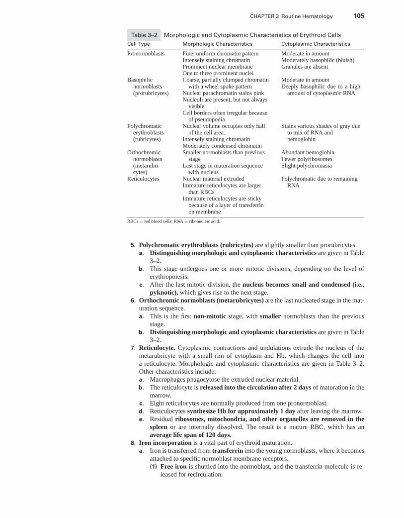
P1: OSO
LWBK192-03 LWBK192-Hubbard November 23, 2008 17:20
CHAPTER 3 Routine Hematology 105
Table 3–2 Morphologic and Cytoplasmic Characteristics of Erythroid Cells
Cell Type Morphologic Characteristics Cytoplasmic Characteristics
Pronormoblasts Fine, uniform chromatin patternIntensely staining chromatinProminent nuclear membraneOne to three prominent nuclei
Moderate in amountModerately basophilic (bluish)Granules are absent
Basophilicnormoblasts(prorubricytes)
Coarse, partially clumped chromatinwith a wheel spoke pattern
Nuclear parachromatin stains pinkNucleoli are present, but not always
visibleCell borders often irregular because
of pseudopodia
Moderate in amountDeeply basophilic due to a high
amount of cytoplasmic RNA
Polychromaticerythroblasts(rubricytes)
Nuclear volume occupies only halfof the cell area.
Intensely staining chromatinModerately condensed chromatin
Stains various shades of gray dueto mix of RNA andhemoglobin
Orthochromicnormoblasts(metarubri-cytes)
Smaller normoblasts than previousstage
Last stage in maturation sequencewith nucleus
Abundant hemoglobinFewer polyribosomesSlight polychromasia
Reticulocytes Nuclear material extrudedImmature reticulocytes are larger
than RBCsImmature reticulocytes are sticky
because of a layer of transferrinon membrane
Polychromatic due to remainingRNA
RBCs = red blood cells; RNA = ribonucleic acid.
5. Polychromatic erythroblasts (rubricytes) are slightly smaller than prorubricytes.a. Distinguishing morphologic and cytoplasmic characteristics are given in Table
3–2.b. This stage undergoes one or more mitotic divisions, depending on the level of
erythropoiesis.c. After the last mitotic division, the nucleus becomes small and condensed (i.e.,
pyknotic), which gives rise to the next stage.6. Orthochromic normoblasts (metarubricytes) are the last nucleated stage in the mat-
uration sequence.a. This is the first non-mitotic stage, with smaller normoblasts than the previous
stage.b. Distinguishing morphologic and cytoplasmic characteristics are given in Table
3–2.7. Reticulocyte. Cytoplasmic contractions and undulations extrude the nucleus of the
metarubricyte with a small rim of cytoplasm and Hb, which changes the cell intoa reticulocyte. Morphologic and cytoplasmic characteristics are given in Table 3–2.Other characteristics include:a. Macrophages phagocytose the extruded nuclear material.b. The reticulocyte is released into the circulation after 2 days of maturation in the
marrow.c. Eight reticulocytes are normally produced from one pronormoblast.d. Reticulocytes synthesize Hb for approximately 1 day after leaving the marrow.e. Residual ribosomes, mitochondria, and other organelles are removed in the
spleen or are internally dissolved. The result is a mature RBC, which has anaverage life span of 120 days.
8. Iron incorporation is a vital part of erythroid maturation.a. Iron is transferred from transferrin into the young normoblasts, where it becomes
attached to specific normoblast membrane receptors.(1) Free iron is shuttled into the normoblast, and the transferrin molecule is re-
leased for recirculation.
P1: OSO
LWBK192-03 LWBK192-Hubbard November 23, 2008 17:20
106 A Concise Review of Clinical Laboratory Science
(2) Intracellular iron is shuttled to the mitochondria for heme-ring production,or it is temporarily deposited in the cytoplasm as ferritin.
b. A secondary supply of iron is provided by a central macrophage through cell-to-cell delivery.
9. Normoblastic and megaloblastic maturation. Megaloblasts are abnormal erythroidcells. Megaloblastic development parallels all stages of erythrocyte maturation.a. Megaloblastosis is an abnormal maturation of erythroid precursors resulting
from a vitamin B12 or folic acid deficiency.b. Megaloblasts have an impaired ability to synthesize and replicate their own
nuclear DNA. Intermitotic and mitotic phases of mitosis are prolonged and outof synchrony.(1) As a result, nuclear maturation lags behind cytoplasmic maturation.(2) The nuclear chromatin pattern of megaloblasts is more open, and breaking up
of the nucleus is common (i.e., karyorrhexis).(3) This results in an increased frequency of circulating RBCs with Howell-Jolly
bodies, which are remnants of nuclear chromatin.
B. Regulation of erythrocyte production1. The number of circulating RBCs may be regulated by changing the rate of production
in the marrow or the rate of release from the marrow. This regulation is normallywell balanced, because the rate of RBC destruction does not significantly vary.
2. Impaired oxygen transport to the tissues and low intracellular oxygen tension triggersRBC production in the marrow. Conditions that stimulate erythropoiesis include:a. Anemiab. Cardiac or pulmonary disordersc. High altitudes
3. Oxygen tension in tissues is regulated, in part, by the oxygen affinity of Hb.a. Modulation. Hemoglobin-oxygen affinity is modulated by the concentration of
phosphates in the RBC.(1) The primary phosphate is 2,3-DPG.(2) 2,3-DPG combines with β chains of the reduced Hb molecule, which results
in reduced Hb affinity for oxygen binding.b. In areas of hypoxic tissue, as oxygen moves from Hb into the tissue, the amount
of reduced Hb decreases. This results in:(1) More 2,3-DPG being bound(2) Lower Hb oxygen affinity, which results is more oxygen being released to the
tissuesc. If tissue hypoxia persists, the depletion of 2,3-DPG leads to increased glycolysis
and production of more 2,3-DPG, which further lowers Hb oxygen affinity.4. Reduced intracellular oxygen tension leads to the production of EPO.
a. Background. EPO is a glycoprotein hormone that controls RBC production atthe level of the marrow.(1) Production. EPO is produced mainly by kidney glomeruli, when cells in the
glomerular tuft of the kidney experience a reduced oxygen tension.(2) Location. EPO is present in the plasma and urine of all mammals, with a
biologic half-life of 4 to 6 hours and a normal reference range of 22 to 54milli-immunochemical units per milliliter (mU/mL).
b. Three main EPO marrow effects include:(1) Stimulation of committed unipotential cells to proliferate and differentiate
into pronormoblasts(2) Shortening the generation time of each maturation stage(3) Promoting early release of reticulocytes into the blood
c. The end result of EPO stimulation is an increased number of marrow nor-moblasts.
5. Other regulators of erythropoiesisa. Plasma Hb may have a feedback stimulation of RBC production or result in
increased EPO production.
P1: OSO
LWBK192-03 LWBK192-Hubbard November 23, 2008 17:20
CHAPTER 3 Routine Hematology 107
b. Hemolytic anemias result in a higher reticulocyte count than other disorders thatstimulate RBC production.
c. Androgens have a synergistic effect on EPO release and are thought to be the causeof higher red cell values in men.
C. Erythrocyte structure1. General considerations. RBCs are biconcave disks with a mean diameter of 7 to 8
μm and a mean volume of 90 fL. The RBC has no nucleus or mitochondria andcannot synthesize protein but is packaged with enough limited metabolism to existfor 120 days.a. RBCs have more surface area than volume, which creates a soft, pliable cell.b. The life span of an RBC depends on the relationship between the RBC membrane
and metabolism. An RBC must have good self-healing ability. Cell damage orinjury can produce cell fragments rather than Hb leakage and cell lysis.
c. The main mission of an RBC is to carry oxygen and carbon dioxide to and fromall tissues.
d. With cell aging, enzymatic failure leads to the loss of pliability and splenic de-struction.
2. The RBC membrane is a lipid bilayer, two molecules thick, consisting of tightlypacked phospholipid molecules.a. Cholesterol is found in the bilayer in a 1:1 molar ratio with phospholipids.
(1) RBC membrane cholesterol is in rapid exchange with plasma unesterifiedcholesterol.
(2) Cholesterol content of the RBC membrane depends on the plasma concentra-tion of cholesterol, free bile acids, and the esterifying enzyme lecithin choles-terol acyltransferase (LCAT).
(3) Changes in RBC shape and survival can result from changes in plasma lipids.(a) Patients with hepatocellular disease or biliary obstruction have im-
paired LCAT activity.(b) This results in cholesterol overloading of the RBC membrane and
excess RBC membrane surface area.(c) Red cells appear as target cell forms and acanthocytes, which have a
reduced survival compared with normal RBCs.b. Membrane proteins are of two basic classes.
(1) Integral proteins provide anion channels through the RBC membrane.(a) These proteins are in contact with both sides of the membrane.(b) Oligosaccharide chains are attached to the external surface of transmem-
brane proteins, providing a negative charge to the RBC surface, whichprevents autoagglutination of RBCs.
(2) Peripheral proteins provide a structural network on the inner surface of themembrane, giving the cell its biconcave structure. Peripheral proteins consistof spectrin, components 4.1 and 5, and actin. Abnormalities of these pro-teins are responsible for cell-shape deformities and hemolytic anemias (e.g.,hereditary elliptocytosis, hereditary spherocytosis).
c. The outside layer of the RBC membrane contains numerous antigenic determi-nants, which provide a genetically determined “specific map” of the RBC surface.(1) More than 300 RBC antigens have been identified, comprising approxi-
mately 15 genetically distinct blood group systems.(2) RBC antigens are composed largely of the oligosaccharide groups of the inte-
gral proteins.(3) Almost all antigenic groups are intrinsic parts of the membrane and appear
during early cell development. The exception is the Lewis group, which issecondarily absorbed onto cell surfaces.
(4) Common RBC antigen systems include:(a) ABO(b) Secretor and Lewis(c) Ii
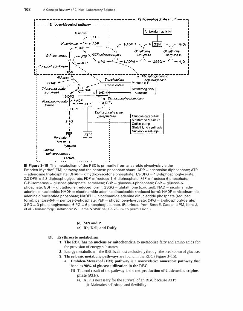
P1: OSO
LWBK192-03 LWBK192-Hubbard November 23, 2008 17:20
108 A Concise Review of Clinical Laboratory Science
■ Figure 3–15 The metabolism of the RBC is primarily from anaerobic glycolysis via the
Embden-Myerhof (EM) pathway and the pentose-phosphate shunt. ADP = adenosine diphosphate; ATP
= adenosine triphosphate; DHAP = dihydroxyacetone phosphate; 1,3-DPG = 1,3-diphosphoglycerate;
2,3-DPG = 2,3-diphosphoglycerate; FDP = fructose-1, 6-diphosphate; F6P = fructose-6-phosphate;
G-P-isomerase = glucose-phosphate isomerase; G3P = glucose-3-phosphate; G6P = glucose-6-
phosphate; GSH = glutathione (reduced form); GSSG = glutathione (oxidized); NAD = nicotinamide-
adenine dinucleotide; NADH = nicotinamide-adenine dinucleotide (reduced form); NADP = nicotinamide-
adenine dinucleotide phosphate; NADPH = nicotinamide-adenine dinucleotide phosphate (reduced
form); pentose-5-P = pentose-5-phosphate; PEP = phosphoenylpyruvate; 2-PG = 2-phosphoglycerate;
3-PG = 3-phosphoglycerate; 6-PG = 6-phosphogluconate. (Reprinted from Besa E, Catalano PM, Kant J,
et al. Hematology. Baltimore: Williams & Wilkins; 1992:98 with permission.)
(d) MN and P(e) Rh, Kell, and Duffy
D. Erythrocyte metabolism1. The RBC has no nucleus or mitochondria to metabolize fatty and amino acids for
the provision of energy substrates.2. Energy metabolism in the RBC is almost exclusively through the breakdown of glucose.3. Three basic metabolic pathways are found in the RBC (Figure 3–15).
a. Embden-Meyerhof (EM) pathway is a nonoxidative anaerobic pathway thathandles 90% of glucose utilization in the RBC.(1) The end result of the pathway is the net production of 2 adenosine triphos-
phate (ATP).(a) ATP is necessary for the survival of an RBC because ATP:
(i) Maintains cell shape and flexibility
P1: OSO
LWBK192-03 LWBK192-Hubbard November 23, 2008 17:20
CHAPTER 3 Routine Hematology 109
(ii) Energizes the metabolic pumps that control cellular sodium, potas-sium, and calcium flux
(iii) Preserves membrane lipids(b) Deficiencies in ATP, due to an inherited or acquired defect in glycolysis,
can reduce cell survival and result in hemolytic anemia.(2) The EM pathway also plays an essential role in maintaining pyridine nu-
cleotides in a reduced state [i.e., nicotinamide adenine dinucleotide, re-duced form (NADH)] to support the conversion of methemoglobin (HbM)to hemoglobin in the methemoglobin reductase pathway.(a) HbM results from the oxidation of iron (Fe) from ferrous ion (Fe2+) to
ferric ion (Fe3+).(b) HbM has a very low oxygen binding affinity.(c) The accumulation of HbM greatly reduces the oxygen-carrying capacity
of the RBC.(d) The methemoglobin reductase pathway counteracts the oxidized state by
reducing Fe3+ to Fe2+.(e) This pathway relies on the reducing capacity of nicotinamide adenine din-
ucleotide (NAD).(f) Persons homozygous for an abnormal methemoglobin reductase gene ac-
cumulate 20% to 40% Hb M in RBCs.(g) Persons with heterozygous enzyme deficiency maintain sufficient HbM
under normal conditions but are susceptible to hemolysis by oxidizingdrugs.
b. The Luebering-Rapaport pathway is necessary for the production of 2,3-DPG.(1) The amount of 2,3-DPG produced at any one time depends on the glycolysis
rate-limiting enzyme, phosphofructokinase.(2) 2,3-DPG production also depends on an adequate supply of inorganic phos-
phate.(3) 2,3-DPG binds to Hb and decreases the oxygen affinity of Hb, thereby re-
leasing more oxygen to the tissues.c. Hexose monophosphate shunt (i.e., pentose-phosphate shunt) couples oxidative
metabolism with nicotinamide-adenine dinucleotide phosphate (NADP) andglutathione reduction.(1) This pathway is functionally dependent on glucose-6-phosphate dehydro-
genase (G6PD).(2) When this pathway is functionally deficient, globin denaturation occurs, and
Hb precipitates out of solution to form inclusions (i.e., Heinz bodies) alongthe inner surface of the RBC membrane. This commonly occurs in patientswith X-linked G6PD deficiency.
E. Erythrocyte life cycle1. A constant red-cell mass in the circulation is ensured by the balance between delivery
of RBCs from the marrow to the blood and the removal of aged or abnormal RBCsfrom the circulation.
2. As RBCs age, catabolic changes occur that result from cellular enzyme depletion,which leads to a decrease in cell flexibility.a. Cell rigidity makes it more difficult for RBCs to get through small capillaries or
splenic sinusoids. The 2 to 3 μm-fenestrations of the splenic sinusoids removeaged or abnormal RBCs that have a higher degree of rigidity.
b. Splenic trapping results in cell lysis and monocyte/macrophage phagocytosis ofdebris.
3. Extravascular destruction is removal of RBCs by the spleen and liver (i.e., reticu-loendothelial system). This pathway is the most efficient method of cell removal andrecovery of essential components such as amino acids and iron.a. Intramacrophage RBC breakdown occurs following phagocytosis, when the
RBC is attacked by lysosomal enzymes. Hb is broken down by an enzyme knownas heme-oxygenase.
P1: OSO
LWBK192-03 LWBK192-Hubbard November 23, 2008 17:20
110 A Concise Review of Clinical Laboratory Science
b. Iron is released from the heme group, returned to plasma transferrin, and trans-ported back to the erythroid marrow. Small amounts of iron can be stored withinthe reticuloendothelial macrophage as ferritin or hemosiderin.
c. Amino acids from globin chains are redirected to the body’s amino acid pool.d. The photoporphyrin pyrrole ring is broken down at the α-methane bridge, and its
α-carbon is exhaled as CO.(1) The opened tetrapyrrole, bilirubin, is carried by plasma albumin in the un-
conjugated (i.e., the indirect bilirubin) form to the liver.(2) Bilirubin is conjugated in the liver to form bilirubin glucuronide (i.e., direct
bilirubin).e. Conjugated bilirubin is excreted from the liver into the small intestine via the bile
duct, where it is converted by bacterial flora to urobilinogen.(1) Most urobilinogen is excreted in the stool as urobilin.(2) Between 10% and 20% of urobilinogen is reabsorbed by the gut. The reab-
sorbed urobilinogen is either excreted in urine or returned to the gut via anenterohepatic cycle.
(3) With liver disease, the enterohepatic cycle is impaired, and an increased amountof urobilinogen is excreted in the urine.
4. Intravascular destruction of RBCs accounts for <10% of RBC loss, but it can in-crease in certain hemolytic diseases. Free hemoglobin is disposed of in the followingmanner:a. The free hemoglobin tetramer is unstable in plasma; therefore, tetramers are
quickly dissociated into α1β1 dimers.b. Hemoglobin dimers are quickly bound to plasma haptoglobin.
(1) Haptoglobin binding stabilizes the home-globin bond and prevents renal ex-cretion of Hb.
(2) The haptoglobin-hemoglobin complex is removed from the circulation by retic-uloendothelial macrophages and is processed intracellularly in the same manneras extracellular RBC destruction.
(3) There is a limited supply of plasma haptoglobin, so the number of haptoglobin-hemoglobin complexes that can be formed acutely is limited.(a) A sudden release of several grams of Hb intravascularly can exceed the
haptoglobin-binding capacity.(b) When the haptoglobin-hemoglobin complex is processed in the macro-
phage, the haptoglobin itself is also catabolized.(c) A decrease or absence of serum haptoglobin may be used to indicate in-
creased intravascular hemolysis.c. If haptoglobin is depleted, unbound Hb dimers are free to be filtered by the
renal glomerulus.(1) As much as 5 g/day can be reabsorbed by renal tubular epithelial cells and
converted to hemosiderin for storage.(2) If the amount of free Hb is high, the tubular uptake capacity can be exceeded.
Then, dimers are excreted in the urine as free Hb.(3) Large amounts of filtered Hb can be destructive to renal tubular cells.(4) A large amount of Hb excretion is accompanied by the excretion of hemosiderin
and iron loss.d. Part of the free Hb not bound to haptoglobin may be oxidized to methemoglobin.
(1) The heme rings dissociate from the globin chains and are bound to anothertransport protein, hemopexin.
(2) Heme-hemopexin complexes are cleared from the circulation by reticuloen-dothelial macrophages and catabolized.
(3) A small percent of the heme groups are bound to albumin as methemalbumin.
F. Measurements of RBC production and destruction1. Anemia occurs when delivery of RBCs to the circulation is decreased or when the re-
moval of RBCs from the blood is increased and cannot be compensated for by increasedmarrow production (see Chapter 4).
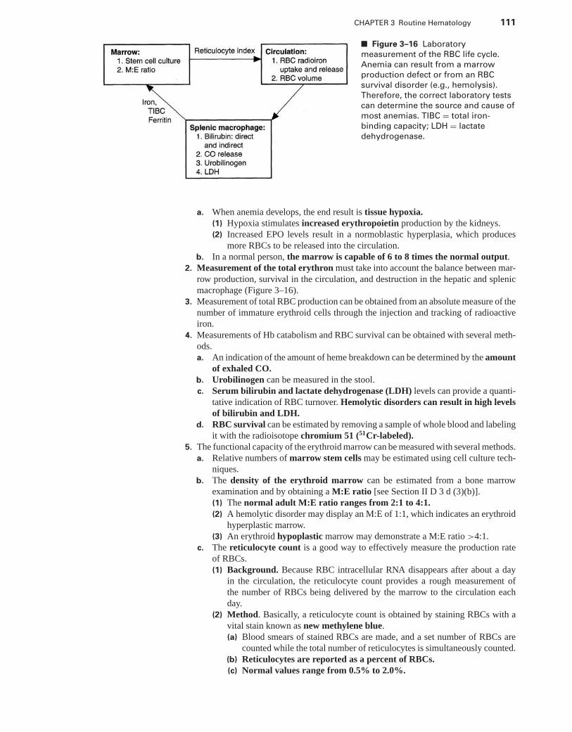
P1: OSO
LWBK192-03 LWBK192-Hubbard November 23, 2008 17:20
CHAPTER 3 Routine Hematology 111
■ Figure 3–16 Laboratory
measurement of the RBC life cycle.
Anemia can result from a marrow
production defect or from an RBC
survival disorder (e.g., hemolysis).
Therefore, the correct laboratory tests
can determine the source and cause of
most anemias. TIBC = total iron-
binding capacity; LDH = lactate
dehydrogenase.
a. When anemia develops, the end result is tissue hypoxia.(1) Hypoxia stimulates increased erythropoietin production by the kidneys.(2) Increased EPO levels result in a normoblastic hyperplasia, which produces
more RBCs to be released into the circulation.b. In a normal person, the marrow is capable of 6 to 8 times the normal output.
2. Measurement of the total erythron must take into account the balance between mar-row production, survival in the circulation, and destruction in the hepatic and splenicmacrophage (Figure 3–16).
3. Measurement of total RBC production can be obtained from an absolute measure of thenumber of immature erythroid cells through the injection and tracking of radioactiveiron.
4. Measurements of Hb catabolism and RBC survival can be obtained with several meth-ods.a. An indication of the amount of heme breakdown can be determined by the amount
of exhaled CO.b. Urobilinogen can be measured in the stool.c. Serum bilirubin and lactate dehydrogenase (LDH) levels can provide a quanti-
tative indication of RBC turnover. Hemolytic disorders can result in high levelsof bilirubin and LDH.
d. RBC survival can be estimated by removing a sample of whole blood and labelingit with the radioisotope chromium 51 (51Cr-labeled).
5. The functional capacity of the erythroid marrow can be measured with several methods.a. Relative numbers of marrow stem cells may be estimated using cell culture tech-
niques.b. The density of the erythroid marrow can be estimated from a bone marrow
examination and by obtaining a M:E ratio [see Section II D 3 d (3)(b)].(1) The normal adult M:E ratio ranges from 2:1 to 4:1.(2) A hemolytic disorder may display an M:E of 1:1, which indicates an erythroid
hyperplastic marrow.(3) An erythroid hypoplastic marrow may demonstrate a M:E ratio >4:1.
c. The reticulocyte count is a good way to effectively measure the production rateof RBCs.(1) Background. Because RBC intracellular RNA disappears after about a day
in the circulation, the reticulocyte count provides a rough measurement ofthe number of RBCs being delivered by the marrow to the circulation eachday.
(2) Method. Basically, a reticulocyte count is obtained by staining RBCs with avital stain known as new methylene blue.(a) Blood smears of stained RBCs are made, and a set number of RBCs are
counted while the total number of reticulocytes is simultaneously counted.(b) Reticulocytes are reported as a percent of RBCs.(c) Normal values range from 0.5% to 2.0%.
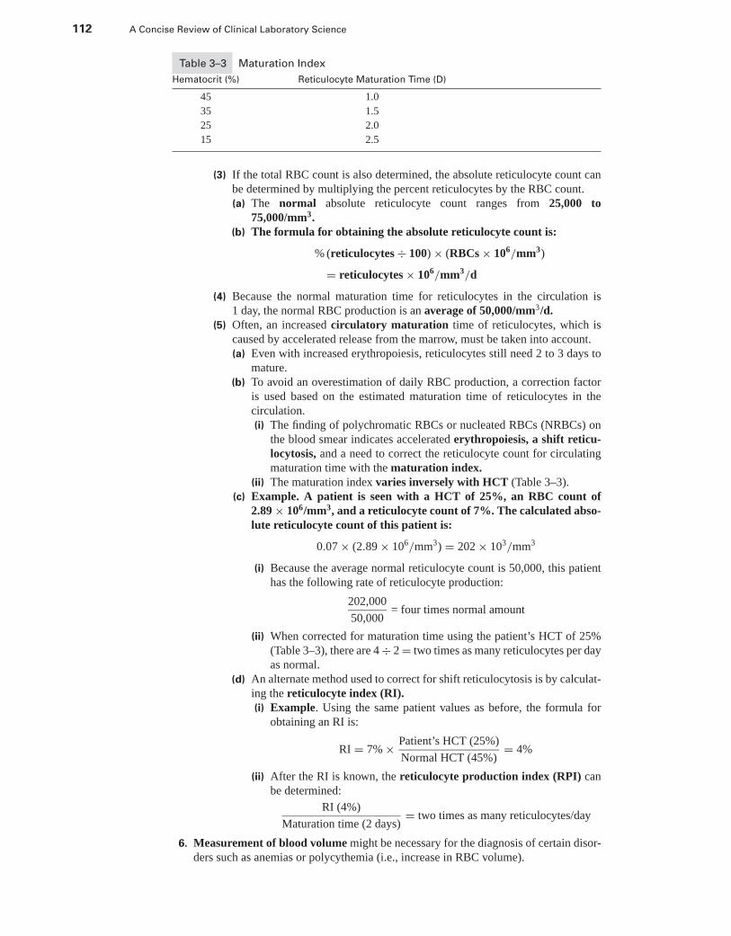
P1: OSO
LWBK192-03 LWBK192-Hubbard November 23, 2008 17:20
112 A Concise Review of Clinical Laboratory Science
Table 3–3 Maturation Index
Hematocrit (%) Reticulocyte Maturation Time (D)
45 1.0
35 1.5
25 2.0
15 2.5
(3) If the total RBC count is also determined, the absolute reticulocyte count canbe determined by multiplying the percent reticulocytes by the RBC count.(a) The normal absolute reticulocyte count ranges from 25,000 to
75,000/mm3.(b) The formula for obtaining the absolute reticulocyte count is:
% (reticulocytes ÷ 100) × (RBCs × 106/mm3)
= reticulocytes × 106/mm3/d
(4) Because the normal maturation time for reticulocytes in the circulation is1 day, the normal RBC production is an average of 50,000/mm3/d.
(5) Often, an increased circulatory maturation time of reticulocytes, which iscaused by accelerated release from the marrow, must be taken into account.(a) Even with increased erythropoiesis, reticulocytes still need 2 to 3 days to
mature.(b) To avoid an overestimation of daily RBC production, a correction factor
is used based on the estimated maturation time of reticulocytes in thecirculation.(i) The finding of polychromatic RBCs or nucleated RBCs (NRBCs) on
the blood smear indicates accelerated erythropoiesis, a shift reticu-locytosis, and a need to correct the reticulocyte count for circulatingmaturation time with the maturation index.
(ii) The maturation index varies inversely with HCT (Table 3–3).(c) Example. A patient is seen with a HCT of 25%, an RBC count of
2.89 × 10⁶/mm3, and a reticulocyte count of 7%. The calculated abso-lute reticulocyte count of this patient is:
0.07 × (2.89 × 106/mm3) = 202 × 103/mm3
(i) Because the average normal reticulocyte count is 50,000, this patienthas the following rate of reticulocyte production:
202,000
50,000= four times normal amount
(ii) When corrected for maturation time using the patient’s HCT of 25%(Table 3–3), there are 4 ÷ 2 = two times as many reticulocytes per dayas normal.
(d) An alternate method used to correct for shift reticulocytosis is by calculat-ing the reticulocyte index (RI).(i) Example. Using the same patient values as before, the formula for
obtaining an RI is:
RI = 7% × Patient’s HCT (25%)
Normal HCT (45%)= 4%
(ii) After the RI is known, the reticulocyte production index (RPI) canbe determined:
RI (4%)
Maturation time (2 days)= two times as many reticulocytes/day
6. Measurement of blood volume might be necessary for the diagnosis of certain disor-ders such as anemias or polycythemia (i.e., increase in RBC volume).
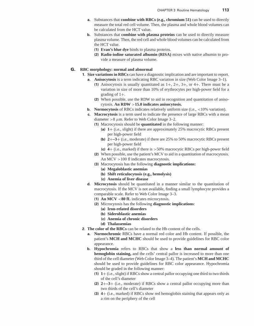
P1: OSO
LWBK192-03 LWBK192-Hubbard November 23, 2008 17:20
CHAPTER 3 Routine Hematology 113
a. Substances that combine with RBCs (e.g., chromium 51) can be used to directlymeasure the total red cell volume. Then, the plasma and whole blood volumes canbe calculated from the HCT value.
b. Substances that combine with plasma proteins can be used to directly measureplasma volume. Then, the red cell and whole blood volumes can be calculated fromthe HCT value.(1) Evan’s blue dye binds to plasma proteins.(2) Radio-iodine saturated albumin (RISA) mixes with native albumin to pro-
vide a measure of plasma volume.
G. RBC morphology: normal and abnormal1. Size variations in RBCs can have a diagnostic implication and are important to report.
a. Anisocytosis is a term indicating RBC variation in size (Web Color Image 3–1).(1) Anisocytosis is usually quantitated as 1+, 2+, 3+, or 4+. There must be a
variation in size of more than 10% of erythrocytes per high-power field for agrading of 1+.
(2) When possible, use the RDW to aid in recognition and quantitation of aniso-cytosis. An RDW >15.0 indicates anisocytosis.
b. Normocytosis of RBCs indicates relatively uniform size (i.e., <10% variation).c. Macrocytosis is a term used to indicate the presence of large RBCs with a mean
diameter >8 μm. Refer to Web Color Image 3–2.(1) Macrocytosis should be quantitated in the following manner:
(a) 1+ (i.e., slight) if there are approximately 25% macrocytic RBCs presentper high-power field
(b) 2+–3+ (i.e., moderate) if there are 25% to 50% macrocytic RBCs presentper high-power field
(c) 4+ (i.e., marked) if there is >50% macrocytic RBCs per high-power field(2) When possible, use the patient’s MCV to aid in a quantitation of macrocytosis.
An MCV >100 fl indicates macrocytosis.(3) Macrocytosis has the following diagnostic implications:
(a) Megaloblastic anemias(b) Shift reticulocytosis (e.g., hemolysis)(c) Anemia of liver disease
d. Microcytosis should be quantitated in a manner similar to the quantitation ofmacrocytosis. If the MCV is not available, finding a small lymphocyte provides acomparable scale. Refer to Web Color Image 3–3.(1) An MCV <80 fL indicates microcytosis.(2) Microcytosis has the following diagnostic implications:
(a) Iron-related disorders(b) Sideroblastic anemias(c) Anemia of chronic disorders(d) Thalassemias
2. The color of the RBCs can be related to the Hb content of the cells.a. Normochromic RBCs have a normal red color and Hb content. If possible, the
patient’s MCH and MCHC should be used to provide guidelines for RBC colorappearance.
b. Hypochromia refers to RBCs that show a less than normal amount ofhemoglobin staining, and the cells’ central pallor is increased to more than onethird of the cell diameter (Web Color Image 3–4). The patient’s MCH and MCHCshould be used to provide guidelines for RBC color appearance. Hypochromiashould be graded in the following manner:(1) 1+ (i.e., slight) if RBCs show a central pallor occupying one third to two thirds
of the cell’s diameter(2) 2+–3+ (i.e., moderate) if RBCs show a central pallor occupying more than
two thirds of the cell’s diameter(3) 4+ (i.e., marked) if RBCs show red hemoglobin staining that appears only as
a rim on the periphery of the cell
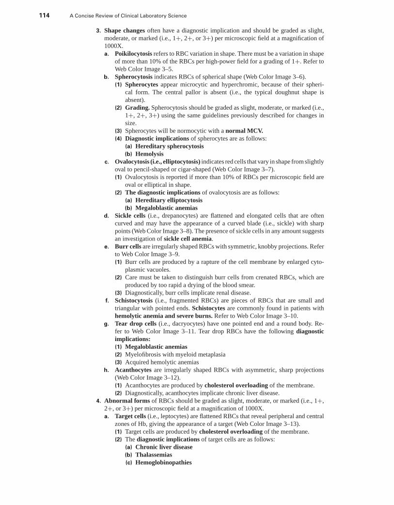
P1: OSO
LWBK192-03 LWBK192-Hubbard November 23, 2008 17:20
114 A Concise Review of Clinical Laboratory Science
3. Shape changes often have a diagnostic implication and should be graded as slight,moderate, or marked (i.e., 1+, 2+, or 3+) per microscopic field at a magnification of1000X.a. Poikilocytosis refers to RBC variation in shape. There must be a variation in shape
of more than 10% of the RBCs per high-power field for a grading of 1+. Refer toWeb Color Image 3–5.
b. Spherocytosis indicates RBCs of spherical shape (Web Color Image 3–6).(1) Spherocytes appear microcytic and hyperchromic, because of their spheri-
cal form. The central pallor is absent (i.e., the typical doughnut shape isabsent).
(2) Grading. Spherocytosis should be graded as slight, moderate, or marked (i.e.,1+, 2+, 3+) using the same guidelines previously described for changes insize.
(3) Spherocytes will be normocytic with a normal MCV.(4) Diagnostic implications of spherocytes are as follows:
(a) Hereditary spherocytosis(b) Hemolysis
c. Ovalocytosis (i.e., elliptocytosis) indicates red cells that vary in shape from slightlyoval to pencil-shaped or cigar-shaped (Web Color Image 3–7).(1) Ovalocytosis is reported if more than 10% of RBCs per microscopic field are
oval or elliptical in shape.(2) The diagnostic implications of ovalocytosis are as follows:
(a) Hereditary elliptocytosis(b) Megaloblastic anemias
d. Sickle cells (i.e., drepanocytes) are flattened and elongated cells that are oftencurved and may have the appearance of a curved blade (i.e., sickle) with sharppoints (Web Color Image 3–8). The presence of sickle cells in any amount suggestsan investigation of sickle cell anemia.
e. Burr cells are irregularly shaped RBCs with symmetric, knobby projections. Referto Web Color Image 3–9.(1) Burr cells are produced by a rapture of the cell membrane by enlarged cyto-
plasmic vacuoles.(2) Care must be taken to distinguish burr cells from crenated RBCs, which are
produced by too rapid a drying of the blood smear.(3) Diagnostically, burr cells implicate renal disease.
f. Schistocytosis (i.e., fragmented RBCs) are pieces of RBCs that are small andtriangular with pointed ends. Schistocytes are commonly found in patients withhemolytic anemia and severe burns. Refer to Web Color Image 3–10.
g. Tear drop cells (i.e., dacryocytes) have one pointed end and a round body. Re-fer to Web Color Image 3–11. Tear drop RBCs have the following diagnosticimplications:(1) Megaloblastic anemias(2) Myelofibrosis with myeloid metaplasia(3) Acquired hemolytic anemias
h. Acanthocytes are irregularly shaped RBCs with asymmetric, sharp projections(Web Color Image 3–12).(1) Acanthocytes are produced by cholesterol overloading of the membrane.(2) Diagnostically, acanthocytes implicate chronic liver disease.
4. Abnormal forms of RBCs should be graded as slight, moderate, or marked (i.e., 1+,2+, or 3+) per microscopic field at a magnification of 1000X.a. Target cells (i.e., leptocytes) are flattened RBCs that reveal peripheral and central
zones of Hb, giving the appearance of a target (Web Color Image 3–13).(1) Target cells are produced by cholesterol overloading of the membrane.(2) The diagnostic implications of target cells are as follows:
(a) Chronic liver disease(b) Thalassemias(c) Hemoglobinopathies
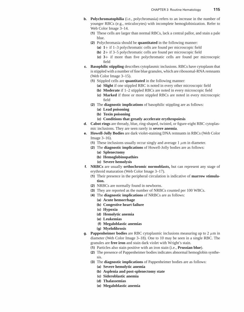
P1: OSO
LWBK192-03 LWBK192-Hubbard November 23, 2008 17:20
CHAPTER 3 Routine Hematology 115
b. Polychromatophilia (i.e., polychromasia) refers to an increase in the number ofyounger RBCs (e.g., reticulocytes) with incomplete hemoglobinization. Refer toWeb Color Image 3–14.(1) These cells are larger than normal RBCs, lack a central pallor, and stain a pale
blue.(2) Polychromasia should be quantitated in the following manner:
(a) 1+ if 1–3 polychromatic cells are found per microscopic field(b) 2+ if 3–5 polychromatic cells are found per microscopic field(c) 3+ if more than five polychromatic cells are found per microscopic
fieldc. Basophilic stippling describes cytoplasmic inclusions. RBCs have cytoplasm that
is stippled with a number of fine blue granules, which are ribosomal-RNA remnants(Web Color Image 3–15).(1) Stippled cells are quantitated in the following manner:
(a) Slight if one stippled RBC is noted in every other microscopic field(b) Moderate if 1–2 stippled RBCs are noted in every microscopic field(c) Marked if three or more stippled RBCs are noted in every microscopic
field(2) The diagnostic implications of basophilic stippling are as follows:
(a) Lead poisoning(b) Toxin poisoning(c) Conditions that greatly accelerate erythropoiesis
d. Cabot rings are thready, blue, ring-shaped, twisted, or figure-eight RBC cytoplas-mic inclusions. They are seen rarely in severe anemia.
e. Howell-Jolly Bodies are dark violet-staining DNA remnants in RBCs (Web ColorImage 3–16).(1) These inclusions usually occur singly and average 1 μm in diameter.(2) The diagnostic implications of Howell-Jolly bodies are as follows:
(a) Splenectomy(b) Hemoglobinopathies(c) Severe hemolysis
f. NRBCs are usually orthochromic normoblasts, but can represent any stage oferythroid maturation (Web Color Image 3–17).(1) Their presence in the peripheral circulation is indicative of marrow stimula-
tion.(2) NRBCs are normally found in newborns.(3) They are reported as the number of NRBCs counted per 100 WBCs.(4) The diagnostic implications of NRBCs are as follows:
(a) Acute hemorrhage(b) Congestive heart failure(c) Hypoxia(d) Hemolytic anemia(e) Leukemias(f) Megaloblastic anemias
(g) Myelofibrosisg. Pappenheimer bodies are RBC cytoplasmic inclusions measuring up to 2 μm in
diameter (Web Color Image 3–18). One to 10 may be seen in a single RBC. Thegranules are free iron and stain dark violet with Wright’s stain.(1) Particles also stain positive with an iron stain (i.e., Prussian blue).(2) The presence of Pappenheimer bodies indicates abnormal hemoglobin synthe-
sis.(3) The diagnostic implications of Pappenheimer bodies are as follows:
(a) Severe hemolytic anemia(b) Asplenia and post-splenectomy state(c) Sideroblastic anemia(d) Thalassemias(e) Megaloblastic anemia

P1: OSO
LWBK192-03 LWBK192-Hubbard November 23, 2008 17:20
116 A Concise Review of Clinical Laboratory Science
h. Rouleaux formation is reported if the RBCs become aligned in aggregates resem-bling stacks of coins. Rouleaux are often seen as an artifact in thick areas of theblood film. Refer to Web Color Image 3–19.(1) Rouleaux are quantitated in the following manner:
(a) Slight, if one to two RBC chains are found per thin microscopic field(b) Moderate, if three to four RBC chains are found per thin microscopic field(c) Marked, if five or more RBC chains are found per thin microscopic field
(2) The diagnostic implication of rouleaux is often associated with hyperproteine-mia disorders such as multiple myeloma.
Online Menu for This Chapter
Go to thePoint.lww.com/Hubbard2e for the following:
� Web Color Images 3.1 through 3.19� Study Questions and Answers
P1: OSO
LWBK192-04 LWBK192-Hubbard November 27, 2008 7:43
CHAPTER 4
Hematologic Disorders
JOEL HUBBARD, PhD, MT (ASCP)
I. RED BLOOD CELL INDICES AND THEIR USE IN THEDIAGNOSIS OF ANEMIA
A. Red blood cell (RBC) indices, known as corpuscular constants, are provided by themajority of automated cell counters. Indices can also be calculated individually from theRBC count, hemoglobin (Hb), and hematocrit (Hct) values.
B. These parameters are useful in the diagnosis, classification, and differentiation ofanemias, as seen in Table 4–1. They can also provide useful guidelines in accessing bloodsmear RBC morphology.
C. Individual corpuscular constants include the mean cell volume (MCV), mean corpuscularhemoglobin (MCH), and mean corpuscular hemoglobin concentration (MCHC).
1. MCV is defined as the mean or average size (in cubic microns) of the individualerythrocyte.a. The MCV may be calculated from the volume and number of erythrocytes in a
given quantity of blood (Box 4–1). The formula for calculation is as follows:
Hct % × 10
RBCs × 106/mm3= mcv fL
b. The normal MCV for an adult is 80 to 100 fL.(1) If the cells are larger than normal, the MCV is increased, and the condition is
called macrocytosis. If the condition is associated with an anemia, it is calleda macrocytic anemia.
(2) If the cells are smaller than normal, the MCV is decreased, and the condi-tion is called microcytosis (or microcytic anemia if associated with anemia).
2. MCH is the mean or average amount of Hb by weight per cell, expressed in micromi-crograms (μμg) or picograms (pg).a. Calculation. The MCH may be calculated from the Hb and the Hct values (Box
4–2). The formula for calculation is as follows:
Hb g/dL × 10
RBCs × 106/mm3= MCH (pg)
b. The normal MCH for the adult is 29 +/− 2 pg.(1) In macrocytic erythrocytes, the amount of Hb may be greater than normal. The
increase in Hb parallels the increase in cell size.(2) The RBC is never supersaturated with Hb. There is no such thing as a
hyperchromic RBC.
117
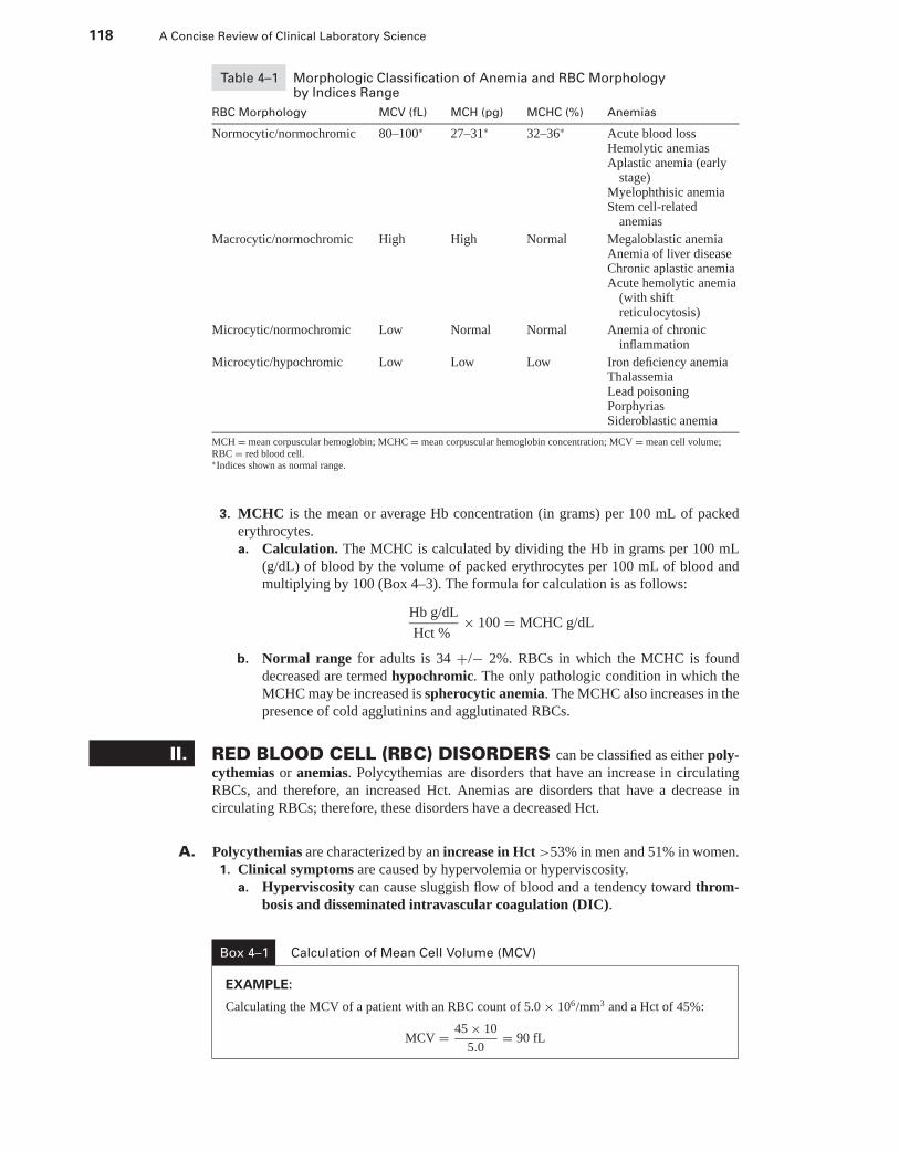
P1: OSO
LWBK192-04 LWBK192-Hubbard November 27, 2008 7:43
118 A Concise Review of Clinical Laboratory Science
Table 4–1 Morphologic Classification of Anemia and RBC Morphologyby Indices Range
RBC Morphology MCV (fL) MCH (pg) MCHC (%) Anemias
Normocytic/normochromic 80–100∗ 27–31∗ 32–36∗ Acute blood lossHemolytic anemiasAplastic anemia (early
stage)Myelophthisic anemiaStem cell-related
anemias
Macrocytic/normochromic High High Normal Megaloblastic anemiaAnemia of liver diseaseChronic aplastic anemiaAcute hemolytic anemia
(with shiftreticulocytosis)
Microcytic/normochromic Low Normal Normal Anemia of chronicinflammation
Microcytic/hypochromic Low Low Low Iron deficiency anemiaThalassemiaLead poisoningPorphyriasSideroblastic anemia
MCH = mean corpuscular hemoglobin; MCHC = mean corpuscular hemoglobin concentration; MCV = mean cell volume;RBC = red blood cell.∗Indices shown as normal range.
3. MCHC is the mean or average Hb concentration (in grams) per 100 mL of packederythrocytes.a. Calculation. The MCHC is calculated by dividing the Hb in grams per 100 mL
(g/dL) of blood by the volume of packed erythrocytes per 100 mL of blood andmultiplying by 100 (Box 4–3). The formula for calculation is as follows:
Hb g/dL
Hct %× 100 = MCHC g/dL
b. Normal range for adults is 34 +/− 2%. RBCs in which the MCHC is founddecreased are termed hypochromic. The only pathologic condition in which theMCHC may be increased is spherocytic anemia. The MCHC also increases in thepresence of cold agglutinins and agglutinated RBCs.
II. RED BLOOD CELL (RBC) DISORDERS can be classified as either poly-cythemias or anemias. Polycythemias are disorders that have an increase in circulatingRBCs, and therefore, an increased Hct. Anemias are disorders that have a decrease incirculating RBCs; therefore, these disorders have a decreased Hct.
A. Polycythemias are characterized by an increase in Hct >53% in men and 51% in women.1. Clinical symptoms are caused by hypervolemia or hyperviscosity.
a. Hyperviscosity can cause sluggish flow of blood and a tendency toward throm-bosis and disseminated intravascular coagulation (DIC).
Box 4–1 Calculation of Mean Cell Volume (MCV)
EXAMPLE:
Calculating the MCV of a patient with an RBC count of 5.0 × 10⁶/mm3 and a Hct of 45%:
MCV = 45 × 10
5.0= 90 fL
P1: OSO
LWBK192-04 LWBK192-Hubbard November 27, 2008 7:43
CHAPTER 4 Hematologic Disorders 119
Box 4–2 Calculation of Mean Corpuscular Hemoglobin (MCH)
EXAMPLE:
Calculating the MCH for a patient who has a Hb of 15 g/dL and an RBC count of 5.0 × 10⁶/mm3:
MCH = 15 × 10
5.0= 30 fL
(1) The decreased oxygen flow to tissues is compensated for by the higher Hct andincreased blood volume (i.e., hypervolemia), which increases vessel diameterand therefore results in increased tissue perfusion.
(2) This is not a benefit, however, because cardiac work is increased to deliverthe same amount of oxygen as normal.
b. Hypervolemia increases blood-vessel diameter and therefore results in increasedtissue perfusion. This can cause increased cardiac work, which is dangerous for anindividual who is predisposed to heart problems.
2. Types of polycythemia include relative and absolute. Absolute polycythemia must bedifferentiated from relative polycythemia before treatment can ensue.a. Relative polycythemia refers to a condition in which the total RBC mass is normal
but the Hct is elevated because the plasma volume is decreased.(1) Relative polycythemia can be caused by acute dehydration resulting from
severe diarrhea, burns, or chronic diuretic therapy.(2) Spurious (false) polycythemia is related to chronic smoking. Nicotine in-
duces a loss of plasma volume.b. Absolute polycythemia refers to an increase in the total RBC mass in the body. This
is a true erythrocytosis caused by marrow erythroid hyperplasia or, secondarily, byan increase in erythropoietin (EPO), as illustrated in Figure 4–1.
B. Anemias are characterized by a reduced Hb concentration of <12 g/dL in men and11 g/dL in women.
1. Clinical symptoms are caused by a decreased oxygen-carrying capacity of the bloodand tissue hypoxia.
2. The physiologic effects of anemia include the following:a. A decreased Hb oxygen-affinity state results in a right shift of the oxygen disso-
ciation curve.b. Decreased tissue perfusion in select areas of the body is caused by redistribution
of blood flow favoring the more oxygen-dependent tissues, such as the brain andmyocardium.
c. Increased cardiac output results in anemic patients to maintain adequate tissueoxygen tension.
d. Increased RBC production by the marrow results from an increase in EPOrelease from the kidneys because of a decrease in oxygen tension.(1) A compensatory increase in marrow erythropoietic cellularity occurs 4–5 days
following a decrease in Hb values.(2) Erythroid hyperplasia in the marrow results in increased reticulocyte
count and nucleated red blood cells (NRBCs) in the circulation.
Box 4–3 Calculation of Mean Corpuscular Hemoglobin Concentration
EXAMPLE:
Calculating the MCHC of a patient who has a Hb of 15 g/dL and a Hct of 45%:
MCHC = 15 × 100
45= 33 g/dL
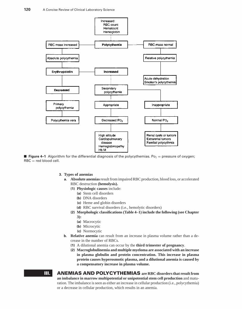
P1: OSO
LWBK192-04 LWBK192-Hubbard November 27, 2008 7:43
120 A Concise Review of Clinical Laboratory Science
■ Figure 4–1 Algorithm for the differential diagnosis of the polycythemias. Po2 = pressure of oxygen;
RBC = red blood cell.
3. Types of anemiasa. Absolute anemias result from impaired RBC production, blood loss, or accelerated
RBC destruction (hemolysis).(1) Physiologic causes include:
(a) Stem cell disorders(b) DNA disorders(c) Heme and globin disorders(d) RBC survival disorders (i.e., hemolytic disorders)
(2) Morphologic classifications (Table 4–1) include the following (see Chapter3):(a) Macrocytic(b) Microcytic(c) Normocytic
b. Relative anemia can result from an increase in plasma volume rather than a de-crease in the number of RBCs.(1) A dilutional anemia can occur by the third trimester of pregnancy.(2) Macroglobulinemia and multiple myeloma are associated with an increase
in plasma globulin and protein concentration. This increase in plasmaprotein causes hyperosmotic plasma, and a dilutional anemia is caused bya compensatory increase in plasma volume.
III. ANEMIAS AND POLYCYTHEMIAS are RBC disorders that result froman imbalance in marrow multipotential or unipotential stem cell production and matu-ration. The imbalance is seen as either an increase in cellular production (i.e., polycythemia)or a decrease in cellular production, which results in an anemia.
P1: OSO
LWBK192-04 LWBK192-Hubbard November 27, 2008 7:43
CHAPTER 4 Hematologic Disorders 121
A. Polycythemias (Figure 4–1)1. Primary absolute polycythemia is characterized by pancytosis or pancytopenia (i.e.,
all cell lines either increased or decreased). Initially, proliferation of erythroid, myeloid,and megakaryocytic bone marrow elements is uncontrolled. Polycythemia vera is aclonal disorder that results in failure of the multipotential stem cell.a. Laboratory characteristics of polycythemia vera include:
(1) Increased RBC count and Hct(2) Increased bone marrow erythroid iron stores, which trap and deplete tis-
sue iron stores(3) Decreased to absent EPO levels(4) Increased white blood cell (WBC) count, which is reflected by an increase
in levels of serum and urine muramidase and serum vitamin B12 andvitamin B12 binding proteins
(5) Thrombocytosisb. Patients who have polycythemia vera are commonly observed to have the clinical
characteristics of splenomegaly and spontaneous hemorrhaging due to thrombosisand hyperviscosity.
c. Treatments for polycythemia vera include the following:(1) Therapeutic phlebotomy is used to reduce the growing RBC volume. Thera-
peutic phlebotomy, however, further depletes tissue iron stores and can resultin an iron-deficient (i.e., microcytic/hypochromic) erythrocytosis.
(2) Splenectomy is used to relieve symptoms caused by splenomegaly andblood pooling.
(3) Chemotherapy is used to treat patients who have advanced disease to killrapidly growing malignant stem cells.
d. Polycythemia vera can evolve into myelofibrosis or a myeloid metaplasia fol-lowing chemotherapeutic treatment. Between 10% and 15% of patients progressto acute myelogenous leukemia.
2. Secondary absolute polycythemia has a normal WBC and platelet count and canbe classified as:a. Polycythemia caused by appropriate EPO productionb. Polycythemia caused by inappropriate EPO productionc. Familial polycythemia, which is seen in children who have functionally normal
Hb, Hb-oxygen saturation, and cardiopulmonary dysfunction(1) These children have high EPO levels unrelated to Hb concentration.(2) The uncontrolled EPO production is most likely caused by a defect in the
regulation of EPO production.(3) Another variation of this genetic disorder is an inherited deficiency of 2,3-
diphosphoglycerate phosphatase activities, which results in polycythemia.
B. Anemias (Figure 4–2) caused by a defect in stem cell erythropoiesis most commonlyresult from a depression of colony-forming units-erythroid (CFU-E) and burst-formingunits-erythroid (BFU-E) unipotential stem cell maturation and differentiation.
1. Anemia of chronic disorders is usually a mild form of anemia occurring secondaryto a chronic inflammatory disorder, such as an infection, rheumatoid arthritis (RA),or neoplastic diseases.a. RBC production by the marrow is normal but insufficient to compensate for a
decreased RBC survival.b. The marrow CFU-E is capable of responding to EPO, but EPO production is low.c. This anemia is partially caused by a defect in iron metabolism that results from
a block in the secondary iron storage system.(1) Monocytes and macrophages have a reduced ability to move stored intracellular
iron to the erythroid marrow cells.(2) This recycling block results in a low serum iron level and an increase in
the storage iron in tissue macrophages.(3) Systemic inflammation results in the release of interleukin-1 (IL-1) from
macrophages, which blocks macrophage iron release. This decreases thereutilization of recycled iron that is released from heme catabolism.
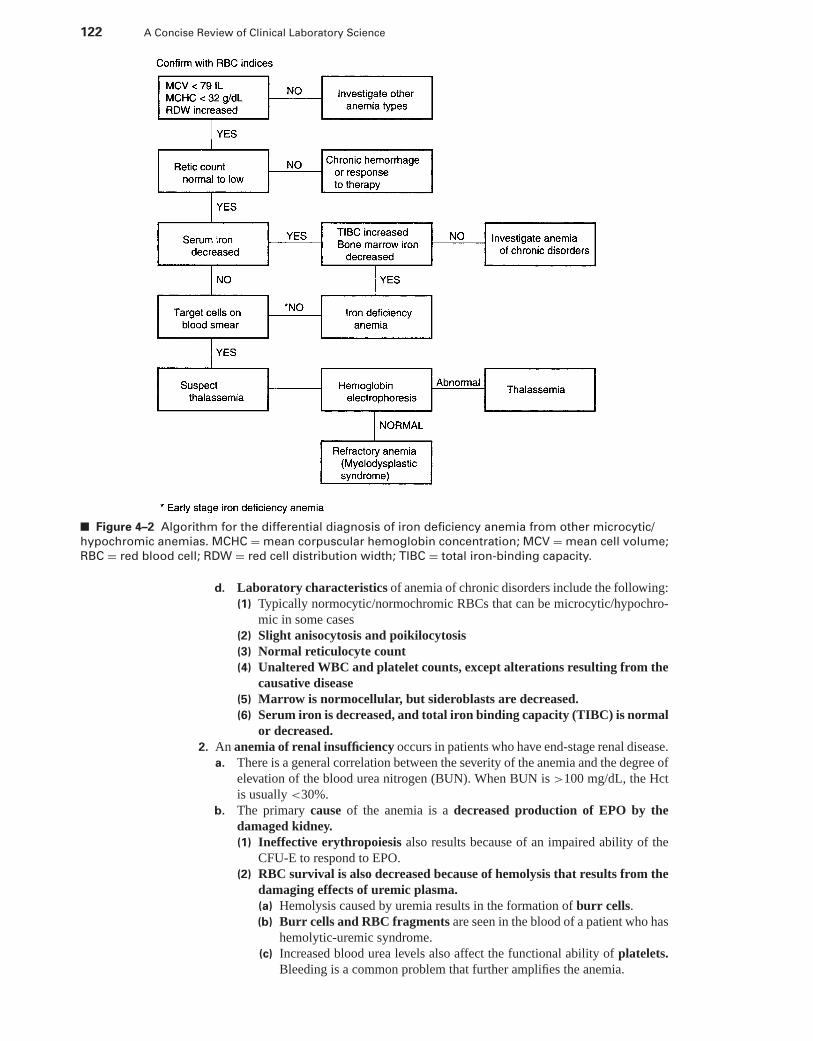
P1: OSO
LWBK192-04 LWBK192-Hubbard November 27, 2008 7:43
122 A Concise Review of Clinical Laboratory Science
■ Figure 4–2 Algorithm for the differential diagnosis of iron deficiency anemia from other microcytic/
hypochromic anemias. MCHC = mean corpuscular hemoglobin concentration; MCV = mean cell volume;
RBC = red blood cell; RDW = red cell distribution width; TIBC = total iron-binding capacity.
d. Laboratory characteristics of anemia of chronic disorders include the following:(1) Typically normocytic/normochromic RBCs that can be microcytic/hypochro-
mic in some cases(2) Slight anisocytosis and poikilocytosis(3) Normal reticulocyte count(4) Unaltered WBC and platelet counts, except alterations resulting from the
causative disease(5) Marrow is normocellular, but sideroblasts are decreased.(6) Serum iron is decreased, and total iron binding capacity (TIBC) is normal
or decreased.2. An anemia of renal insufficiency occurs in patients who have end-stage renal disease.
a. There is a general correlation between the severity of the anemia and the degree ofelevation of the blood urea nitrogen (BUN). When BUN is >100 mg/dL, the Hctis usually <30%.
b. The primary cause of the anemia is a decreased production of EPO by thedamaged kidney.(1) Ineffective erythropoiesis also results because of an impaired ability of the
CFU-E to respond to EPO.(2) RBC survival is also decreased because of hemolysis that results from the
damaging effects of uremic plasma.(a) Hemolysis caused by uremia results in the formation of burr cells.(b) Burr cells and RBC fragments are seen in the blood of a patient who has
hemolytic-uremic syndrome.(c) Increased blood urea levels also affect the functional ability of platelets.
Bleeding is a common problem that further amplifies the anemia.
P1: OSO
LWBK192-04 LWBK192-Hubbard November 27, 2008 7:43
CHAPTER 4 Hematologic Disorders 123
3. Anemia of liver disease is characterized by shortened RBC survival and inadequateRBC production that occurs secondary to a chronic liver disease.a. RBC morphology is usually macrocytic or normocytic with target cells or acan-
thocytes caused by increased surface membrane cholesterol.(1) These abnormal RBCs are removed at a higher rate by the spleen.(2) Their survival is reduced when compared with normal RBCs.
b. The patient often has an accompanying splenomegaly with cirrhosis, which furtherdecreases RBC survival.
c. The reticulocyte count is increased slightly, and theplatelet count is normal ordecreased.
d. A differential diagnosis must distinguish this anemia from other macrocytic ane-mias. Patients who have anemia of liver disease usually have a normal WBCcount and serum vitamin B12 and folate levels; patients who have megaloblasticanemias do not.
4. Anemias of endocrine disease can occur with hypothyroidism and androgen-deficientstates.a. Anemia of hypothyroidism involves a mild-to-moderate anemia with a normal
reticulocyte count.(1) Thyroid hormone regulates the cellular metabolic rate, and therefore, the tissue
oxygen requirement.(2) With a decrease in thyroid hormone (i.e., hypothyroidism), there is a
smaller tissue oxygen requirement, which is interpreted by the kidneysas a more than adequate oxygen tension. The net result is a decrease inthe production of EPO.
(3) This type of anemia often is a macrocytic/normochromic anemia but canalso be a normocytic/normochromic anemia.
(4) The anemia may be complicated by iron deficiency or folic acid or vita-min B12 deficiency, and the laboratory results may reflect these forms ofanemia.
b. A deficiency in testosterone secretion in men results in a decrease in RBC pro-duction and a decline of approximately 2 g/dL of Hb.
5. Myelophthisic anemia is an anemia associated with bone marrow infiltration andhyperproliferation by nonerythroid cells.a. A leukoerythroblastic reaction commonly accompanies the anemia.
(1) Normoblasts of varying degrees of maturation are typically found on the bloodsmear with a reticulocytosis.
(2) Increased leukocytes along with their immature forms are also found onthe peripheral blood smear.
(3) RBC morphology is normocytic/normochromic, but can be macrocytic.(4) Platelets are normal or decreased and often have abnormal forms.
b. The usual causes of myelophthisic anemia include:(1) Metastatic carcinoma(2) Multiple myeloma(3) Leukemia(4) Lymphoma(5) Lipidoses or storage disease
6. Aplastic anemia is a marrow disorder characterized by a reduction in the number orfunction of multipotential stem cells with a resulting pancytopenia. This disorder ismore commonly found in adults.a. The marrow is hypocellular with patchy areas of normal cellularity and increased
fat cell infiltration.b. The diagnosis of severe aplastic anemia is made in pancytopenic patients when at
least two of the following three peripheral blood values are found:(1) A WBC count lower than 500 cells/mm3
(2) A platelet count lower than 20,000/mm3
(3) A reticulocyte count lower than 1%c. Aplastic anemia can be acute and rapidly fatal or it can have a slow onset and a
chronic course.
P1: OSO
LWBK192-04 LWBK192-Hubbard November 27, 2008 7:43
124 A Concise Review of Clinical Laboratory Science
d. Most patients are initially seen with the following common clinical characteris-tics:(1) Bleeding as a result of thrombocytopenia(2) Increased susceptibility to infections due to leukopenia(3) All of the symptoms typical of anemia(4) The absence of splenomegaly(5) Iron overload from repeated transfusions
e. In 50% of patients, no specific agent can be correlated with the disease. Knowncauses of aplasia include:(1) Drugs (33%)(2) Chemicals or toxins (4%)(3) Infections and infectious hepatitis (4%)(4) Miscellaneous causes (59%)
f. The pathogenesis of aplastic anemia is thought to occur from either one or acombination of several of the following physiologic mechanisms:(1) Defective or insufficient multipotential colony-forming unit-spleen (CFU-S)
stem cell population(2) Altered microenvironment that is unable to provide for normal differen-
tiation and development of the stem cell compartment(3) Absent humoral and cellular stimulators for hematopoiesis(4) Excessive suppression of hematopoiesis by T lymphocytes or macro-
phages(5) Stem cells interacting among themselves with one clone inhibiting the
growth of another(6) Immunologic mechanisms that suppress hematopoiesis
g. Treatment focuses on symptoms and finding the cause of suppression. Acceptedcourses of treatment include:(1) Blood transfusions(2) Stimulation of residual marrow with androgens or adrenocorticoids(3) Controlling infections with antibiotics(4) Controlling bleeding problems(5) Bone marrow transplantation for acceptable candidates younger than
40 years(6) EPO therapy to stimulate residual bone marrow
h. Laboratory values for a patient who has aplastic anemia include the following:(1) RBC morphology is normocytic/normochromic but often is macrocytic with
a normal red cell distribution width (RDW)(2) Slight anisocytosis and poikilocytosis(3) Absence of circulating normoblasts in light of the degree of the anemia(4) An absolute leukopenia with a relative lymphocytosis(5) Thrombocytopenia(6) Elevated levels of leukocyte alkaline phosphatase (LAP)(7) Decreased marrow iron stores(8) High serum iron concentration(9) Very high plasma and urine EPO levels
i. Toxins. Aplastic anemia is often associated with toxic chemical or physical agents.(1) Toxic aplastic anemias are known to occur after exposure to mustard com-
pounds, benzene, chemotherapy drugs (e.g., busulfan, urethan), antimetabolitedrugs, or ionizing radiation.
(2) Hypersensitive aplastic anemia has been known to occur following treat-ment with certain classes of therapeutic drugs or chemicals that cause anautoimmune suppression of the bone marrow.(a) This effect has been noted with drugs such as antibiotics, anticonvulsants,
analgesics, antithyroid drugs, antihistamines, and insecticides.(b) Chloramphenicol is the most common cause of drug-induced aplasia. Up
to 50% of patients who receive this drug on a long-term basis will have aresulting mild pancytopenia that is reversible with drug withdrawal.
P1: OSO
LWBK192-04 LWBK192-Hubbard November 27, 2008 7:43
CHAPTER 4 Hematologic Disorders 125
j. Aplastic anemia can occur secondary to other diseases or conditions, such as:(1) Infection, most often occurring as a sequel to infectious hepatitis(2) Paroxysmal nocturnal hemoglobinuria (PNH)(3) Pregnancy (usually remits after delivery)
k. Aplastic anemia (Fanconi’s anemia) refers to individuals with a genetic predis-position to bone marrow failure.(1) Detection occurs between the ages of 1 and 8 years.(2) Often, several family members are affected.(3) There is a high incidence of developmental abnormalities, which include
some of the following symptoms:(a) Hyperpigmentation(b) Short stature(c) Hypogonadism(d) Malformation of the fingers and toes(e) Malformation of the organs(f) Abnormalities of the chromosome pattern of lymphocytes and marrow
cells7. Pure red cell aplasia is an unusual disease characterized by the selective depletion of
only the erythroid bone marrow tissue.a. The disorder is a unipotential (CFU-E) stem cell defect that is believed to be
related to an immunologic dysfunction.b. Transitory arrest of erythropoiesis may occur in the course of a hemolytic anemia
that is preceded by an infection.c. Congenital RBC aplasia (Blackfan-Diamond anemia) is a rare disorder diag-
nosed between the ages of 1 and 6 years.(1) CFU-E and colony-forming burst (CFU-B) stem cells are decreased.(2) Patients have a severe normocytic or slightly macrocytic anemia and a low
reticulocyte count.(3) Leukocytes and platelets are usually normal in numbers.(4) The marrow shows a reduction in all developing erythroid cells, except
pronormoblasts.(5) Hemoglobin F (HbF) is elevated as high as 5% to 25%.(6) The RBC fetal antigen is present.
d. Acquired pure red cell aplasia is rare but occurs more in adults than in children.(1) Typical laboratory findings include a decreased reticulocyte count and mar-
row erythroid precursor depletion.(2) Up to 50% of patients have an associated cancer of the thymus (i.e.,
thynoma).(3) Remission of the anemia after surgical removal of the thymus occurs in
25% of patients.(4) Acquired red cell aplasia is thought to occur through the production of
a cytotoxic autoantibody against erythroid precursors and a plasma in-hibitor of heme synthesis.
8. Refractory anemia is an ill-defined group of chronic anemias occurring in personsolder than 50 years.a. Laboratory features commonly include a normocytic or macrocytic anemia, de-
creased reticulocyte count, pancytopenia, and a hypercellular marrow witherythroid hypoplasia.
b. Refractory anemia is now classified with the myeloproliferative disorders as oneof five myelodysplastic syndromes.
c. The anemia can develop into an acute leukemia with the presence of blast cellsin the peripheral circulation.
C. Heme disorders represent a group of anemias that result from a defect in the synthesis ofthe heme ring in the mitochondria of developing normoblasts (Figure 4–3).
1. General considerationsa. Disorders of heme synthesis result in a microcytic/hypochromic anemia.
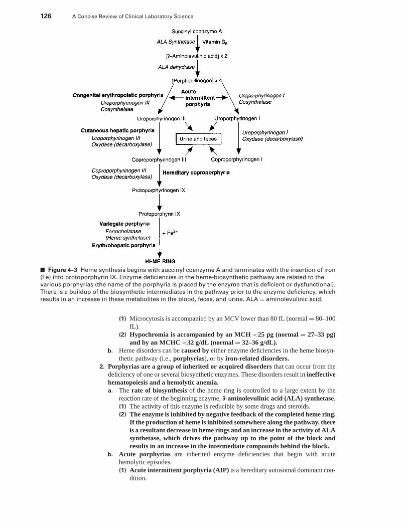
P1: OSO
LWBK192-04 LWBK192-Hubbard November 27, 2008 7:43
126 A Concise Review of Clinical Laboratory Science
■ Figure 4–3 Heme synthesis begins with succinyl coenzyme A and terminates with the insertion of iron
(Fe) into protoporphyrin IX. Enzyme deficiencies in the heme-biosynthetic pathway are related to the
various porphyrias (the name of the porphyria is placed by the enzyme that is deficient or dysfunctional).
There is a buildup of the biosynthetic intermediates in the pathway prior to the enzyme deficiency, which
results in an increase in these metabolites in the blood, feces, and urine. ALA = aminolevulinic acid.
(1) Microcytosis is accompanied by an MCV lower than 80 fL (normal = 80–100fL).
(2) Hypochromia is accompanied by an MCH <25 pg (normal = 27–33 pg)and by an MCHC <32 g/dL (normal = 32–36 g/dL).
b. Heme disorders can be caused by either enzyme deficiencies in the heme biosyn-thetic pathway (i.e., porphyrias), or by iron-related disorders.
2. Porphyrias are a group of inherited or acquired disorders that can occur from thedeficiency of one or several biosynthetic enzymes. These disorders result in ineffectivehematopoiesis and a hemolytic anemia.a. The rate of biosynthesis of the heme ring is controlled to a large extent by the
reaction rate of the beginning enzyme, δ-aminolevulinic acid (ALA) synthetase.(1) The activity of this enzyme is reducible by some drugs and steroids.(2) The enzyme is inhibited by negative feedback of the completed heme ring.
If the production of heme is inhibited somewhere along the pathway, thereis a resultant decrease in heme rings and an increase in the activity of ALAsynthetase, which drives the pathway up to the point of the block andresults in an increase in the intermediate compounds behind the block.
b. Acute porphyrias are inherited enzyme deficiencies that begin with acutehemolytic episodes.(1) Acute intermittent porphyria (AIP) is a hereditary autosomal dominant con-
dition.
P1: OSO
LWBK192-04 LWBK192-Hubbard November 27, 2008 7:43
CHAPTER 4 Hematologic Disorders 127
(a) Clinical symptoms include:(i) Abdominal pain with vomiting
(ii) Constipation(iii) Hypertension(iv) Peripheral neuritis(v) Behavioral changes and psychosis
(b) The common laboratory profile includes:(i) Slight elevations of bilirubin and alkaline phosphatase levels
(ii) Increased urine ALA and porphobilinogen (PBG)(iii) Leukocytosis
(c) Enzyme defects. AIP is caused by decreased levels of uroporphyrinogen Iand III synthetase, which results in increased production of ALA and PBG.
(2) Hereditary coproporphyria is a hereditary, autosomal dominant condi-tion.(a) Clinical symptoms. Patients are either asymptomatic, or they are initially
seen with mild neurologic, abdominal, or psychiatric symptoms. Patientscommonly have light-sensitive skin because of increased levels of PBG.
(b) The laboratory profile includes:(i) Increased coproporphyrinogen III in feces(ii) Intermittent increases of coproporphyrinogen, ALA, and PBG in
urine.(c) Enzyme defect. Patients have a deficiency in coproporphyrinogen decar-
boxylase activity.(3) Variegate porphyria is a hereditary autosomal dominant condition that
has a high incidence in the white population of South Africa.(a) Clinical symptoms. Symptoms are similar to AIP with the additional
symptom of cutaneous lesions, which are caused by highly photosensitiveskin.
(b) Laboratory profile. There are increased levels of urinary ALA and PBG,but there is also an increase in the levels of porphyrin precursors that occurfurther along the pathway.
(c) Enzyme defect. Patients who have this disorder have a deficiency in hemesynthetase (also known as ferrochelatase deficiency).
c. Chronic porphyrias are more commonly associated with solar photosensitivityof the skin and include either genetic or acquired conditions.(1) Congenital erythropoietic porphyria is rare and is inherited as an autosomal
recessive disorder.(a) Clinical symptoms of this disorder include:
(i) Redness of the skin(ii) Hemolytic anemia(iii) Severe skin photosensitivity(iv) Splenomegaly(v) Often fatal early in life
(b) Laboratory profile. Patients often excrete red pigmented urine because ofexcessive excretion of coproporphyrin I and uroporphyrin I.
(c) Enzyme defect. Patients with this disorder show a deficiency in uropor-phyrinogen III cosynthetase.
(2) Erythrohepatic protoporphyria is an inherited disorder so named becausethe enzyme defects are localized in both the hepatic and erythropoieticcells.(a) Clinical symptoms. This porphyria is a disease of early adulthood that
exhibits mild skin photosensitivity.(b) Laboratory profile. Patients who have this porphyria demonstrate an in-
creased level of RBC photoporphyrin and elevated levels of fecal copro-porphyrinogen and photoporphyrin.
(c) The enzyme defect causing this disorder is deficient activity of hemesynthetase, which leads to over-reactive ALA synthetase.
P1: OSO
LWBK192-04 LWBK192-Hubbard November 27, 2008 7:43
128 A Concise Review of Clinical Laboratory Science
(3) Cutaneous hepatic porphyria (i.e., Cutanea Tarda) is an acquired disorder.(a) Clinical symptoms. Patients are commonly first seen with skin lesions
because of solar photosensitivity. Patients who have this porphyria have amedical history associated with liver disease, alcoholism, estrogen therapy,or ingestion of hexachlorobenzene.
(b) Characteristic laboratory profile results include an elevated urine uropor-phyrin with the Type I isomers being excreted in greater amounts than theType III isomers.
(c) Enzyme defect. Patients diagnosed with this disorder have reduced activityof the enzyme uroporphyrinogen III decarboxylase.
(4) Lead intoxication is a commonly acquired porphyria that results from ablock in several of the heme biosynthetic enzymes.(a) Clinical symptoms of lead poisoning include:
(i) Abdominal pain(ii) Constipation(iii) Neuropathy(iv) Absence of skin photosensitivity, ferrochelatase, and copropor-
phyrinogen oxidase(b) The characteristic laboratory profile associated with lead toxicity in-
cludes:(i) Elevated urinary ALA and coproporphyrin levels
(ii) Increased RBC photoporphyrin(iii) Hypochromic RBCs with basophilic stippling due to increased
erythropoiesis(iv) Toxic granulation in neutrophils
(c) Enzyme defect. Lead inhibits several enzymes of the biosynthetic pathway.The most severely affected enzyme is ALA dehydrase.
3. Iron metabolism-associated disorders most commonly occur as a result of an irondeficiency but can result from a block of the enzyme that inserts iron (i.e., ferrochelatase)into the heme ring (Figure 4–3).a. Iron and its metabolism are vital to the body because a Hb molecule is nonfunc-
tioning without iron, and two thirds or more of the total body iron is in the RBCsand their precursors.(1) Each milliliter of RBCs contains 1 mg of iron.(2) Storage iron is present in macrophages or normoblasts as ferritin (i.e.,
Fe3+ plus apoferritin) or hemosiderin.(3) The majority of iron used in Hb synthesis is from the iron released and
recycled by Hb that is degraded in macrophages and transported to nor-moblasts by plasma transferrin.
(4) Only 0.9 to 1.3 mg of iron per day is lost from the body.(5) The small amount of iron lost from the body is maintained by dietary
absorption, which amounts to approximately 1 mg/day.(a) In men, dietary iron averages approximately 15 mg/day with only 6%
intestinal absorption.(b) In women, dietary iron averages approximately 11 mg/day with 12% in-
testinal absorption.(c) Dietary absorption is increased in an iron deficiency state, but only to a
maximum of 20%.(d) Dietary iron is digested in the ferric form (i.e., Fe3+) but is reduced to
the ferrous form (i.e., Fe2+) in the stomach by HCl and other reducingagents (e.g., food).
(e) The ferrous form is much more rapidly absorbed by the mucosal cells ofthe duodenum and upper jejunum than is the ferric form.
(f) Once in the mucosal cell, the ferrous form is oxidized again back to theferric form and coupled to the protein apoferritin to form ferritin.
(g) Iron enters the circulation bound to transferrin.(i) The normal serum iron concentration is 50 to 150 mg/dL.
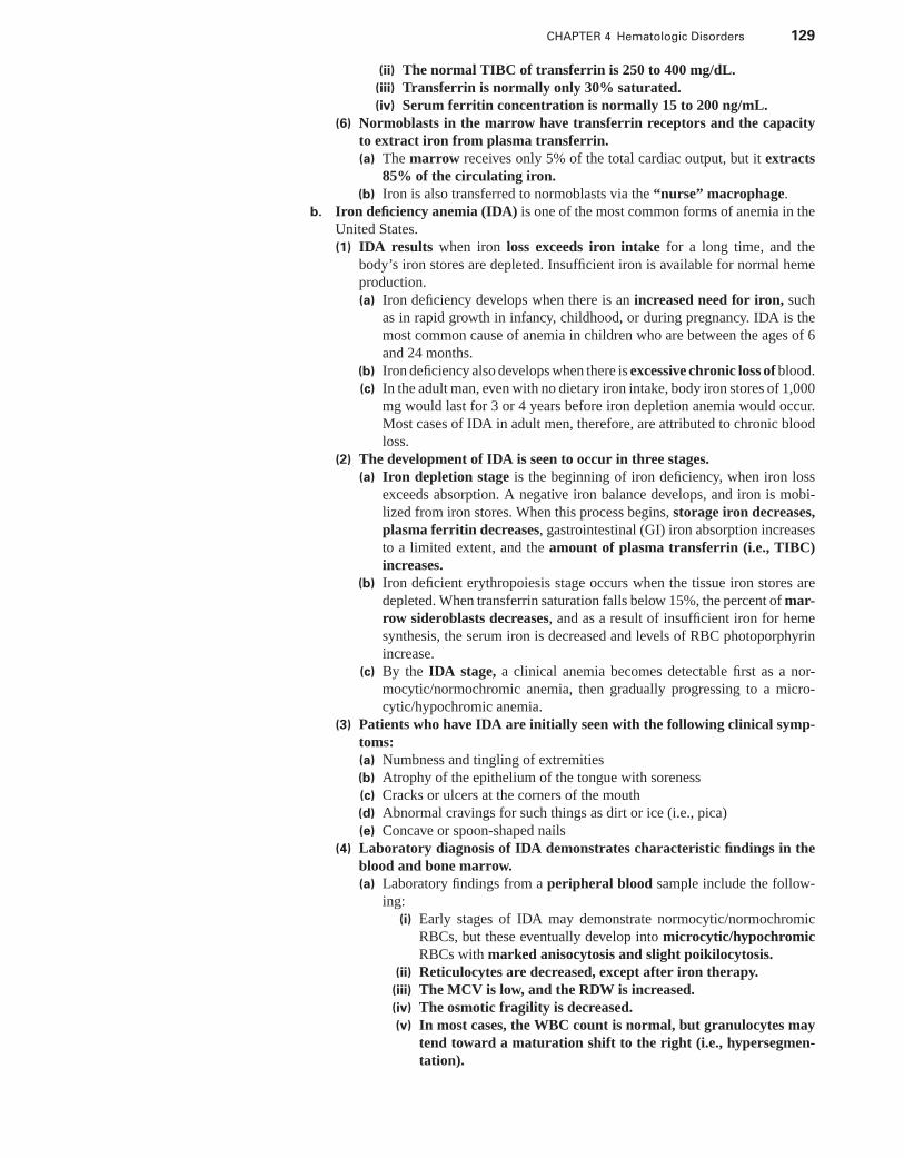
P1: OSO
LWBK192-04 LWBK192-Hubbard November 27, 2008 7:43
CHAPTER 4 Hematologic Disorders 129
(ii) The normal TIBC of transferrin is 250 to 400 mg/dL.(iii) Transferrin is normally only 30% saturated.(iv) Serum ferritin concentration is normally 15 to 200 ng/mL.
(6) Normoblasts in the marrow have transferrin receptors and the capacityto extract iron from plasma transferrin.(a) The marrow receives only 5% of the total cardiac output, but it extracts
85% of the circulating iron.(b) Iron is also transferred to normoblasts via the “nurse” macrophage.
b. Iron deficiency anemia (IDA) is one of the most common forms of anemia in theUnited States.(1) IDA results when iron loss exceeds iron intake for a long time, and the
body’s iron stores are depleted. Insufficient iron is available for normal hemeproduction.(a) Iron deficiency develops when there is an increased need for iron, such
as in rapid growth in infancy, childhood, or during pregnancy. IDA is themost common cause of anemia in children who are between the ages of 6and 24 months.
(b) Iron deficiency also develops when there is excessive chronic loss of blood.(c) In the adult man, even with no dietary iron intake, body iron stores of 1,000
mg would last for 3 or 4 years before iron depletion anemia would occur.Most cases of IDA in adult men, therefore, are attributed to chronic bloodloss.
(2) The development of IDA is seen to occur in three stages.(a) Iron depletion stage is the beginning of iron deficiency, when iron loss
exceeds absorption. A negative iron balance develops, and iron is mobi-lized from iron stores. When this process begins, storage iron decreases,plasma ferritin decreases, gastrointestinal (GI) iron absorption increasesto a limited extent, and the amount of plasma transferrin (i.e., TIBC)increases.
(b) Iron deficient erythropoiesis stage occurs when the tissue iron stores aredepleted. When transferrin saturation falls below 15%, the percent of mar-row sideroblasts decreases, and as a result of insufficient iron for hemesynthesis, the serum iron is decreased and levels of RBC photoporphyrinincrease.
(c) By the IDA stage, a clinical anemia becomes detectable first as a nor-mocytic/normochromic anemia, then gradually progressing to a micro-cytic/hypochromic anemia.
(3) Patients who have IDA are initially seen with the following clinical symp-toms:(a) Numbness and tingling of extremities(b) Atrophy of the epithelium of the tongue with soreness(c) Cracks or ulcers at the corners of the mouth(d) Abnormal cravings for such things as dirt or ice (i.e., pica)(e) Concave or spoon-shaped nails
(4) Laboratory diagnosis of IDA demonstrates characteristic findings in theblood and bone marrow.(a) Laboratory findings from a peripheral blood sample include the follow-
ing:(i) Early stages of IDA may demonstrate normocytic/normochromic
RBCs, but these eventually develop into microcytic/hypochromicRBCs with marked anisocytosis and slight poikilocytosis.
(ii) Reticulocytes are decreased, except after iron therapy.(iii) The MCV is low, and the RDW is increased.(iv) The osmotic fragility is decreased.(v) In most cases, the WBC count is normal, but granulocytes may
tend toward a maturation shift to the right (i.e., hypersegmen-tation).
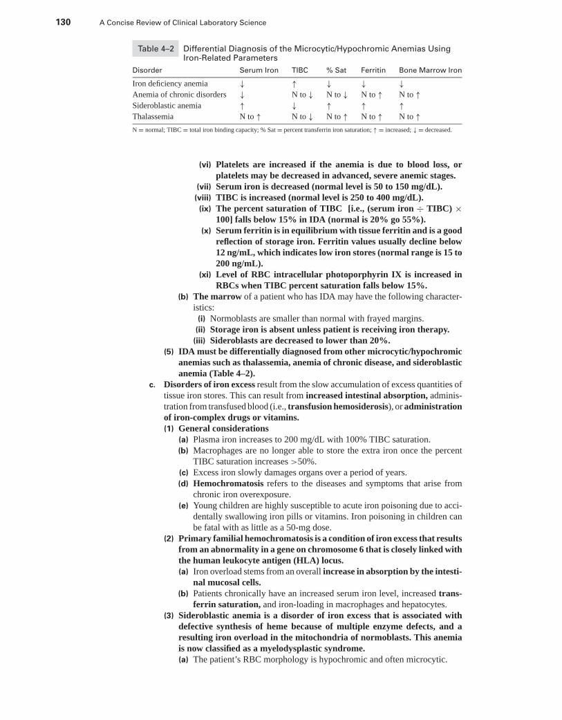
P1: OSO
LWBK192-04 LWBK192-Hubbard November 27, 2008 7:43
130 A Concise Review of Clinical Laboratory Science
Table 4–2 Differential Diagnosis of the Microcytic/Hypochromic Anemias UsingIron-Related Parameters
Disorder Serum Iron TIBC % Sat Ferritin Bone Marrow Iron
Iron deficiency anemia ↓ ↑ ↓ ↓ ↓Anemia of chronic disorders ↓ N to ↓ N to ↓ N to ↑ N to ↑Sideroblastic anemia ↑ ↓ ↑ ↑ ↑Thalassemia N to ↑ N to ↓ N to ↑ N to ↑ N to ↑N = normal; TIBC = total iron binding capacity; % Sat = percent transferrin iron saturation; ↑ = increased; ↓ = decreased.
(vi) Platelets are increased if the anemia is due to blood loss, orplatelets may be decreased in advanced, severe anemic stages.
(vii) Serum iron is decreased (normal level is 50 to 150 mg/dL).(viii) TIBC is increased (normal level is 250 to 400 mg/dL).
(ix) The percent saturation of TIBC [i.e., (serum iron ÷ TIBC) ×100] falls below 15% in IDA (normal is 20% go 55%).
(x) Serum ferritin is in equilibrium with tissue ferritin and is a goodreflection of storage iron. Ferritin values usually decline below12 ng/mL, which indicates low iron stores (normal range is 15 to200 ng/mL).
(xi) Level of RBC intracellular photoporphyrin IX is increased inRBCs when TIBC percent saturation falls below 15%.
(b) The marrow of a patient who has IDA may have the following character-istics:
(i) Normoblasts are smaller than normal with frayed margins.(ii) Storage iron is absent unless patient is receiving iron therapy.(iii) Sideroblasts are decreased to lower than 20%.
(5) IDA must be differentially diagnosed from other microcytic/hypochromicanemias such as thalassemia, anemia of chronic disease, and sideroblasticanemia (Table 4–2).
c. Disorders of iron excess result from the slow accumulation of excess quantities oftissue iron stores. This can result from increased intestinal absorption, adminis-tration from transfused blood (i.e., transfusion hemosiderosis), or administrationof iron-complex drugs or vitamins.(1) General considerations
(a) Plasma iron increases to 200 mg/dL with 100% TIBC saturation.(b) Macrophages are no longer able to store the extra iron once the percent
TIBC saturation increases >50%.(c) Excess iron slowly damages organs over a period of years.(d) Hemochromatosis refers to the diseases and symptoms that arise from
chronic iron overexposure.(e) Young children are highly susceptible to acute iron poisoning due to acci-
dentally swallowing iron pills or vitamins. Iron poisoning in children canbe fatal with as little as a 50-mg dose.
(2) Primary familial hemochromatosis is a condition of iron excess that resultsfrom an abnormality in a gene on chromosome 6 that is closely linked withthe human leukocyte antigen (HLA) locus.(a) Iron overload stems from an overall increase in absorption by the intesti-
nal mucosal cells.(b) Patients chronically have an increased serum iron level, increased trans-
ferrin saturation, and iron-loading in macrophages and hepatocytes.(3) Sideroblastic anemia is a disorder of iron excess that is associated with
defective synthesis of heme because of multiple enzyme defects, and aresulting iron overload in the mitochondria of normoblasts. This anemiais now classified as a myelodysplastic syndrome.(a) The patient’s RBC morphology is hypochromic and often microcytic.
P1: OSO
LWBK192-04 LWBK192-Hubbard November 27, 2008 7:43
CHAPTER 4 Hematologic Disorders 131
(b) Patients are commonly seen with increased serum iron levels, decreasedTIBC, and greatly increased transferrin saturation.
(c) The marrow shows a greatly increased storage iron by an increase insideroblasts >50% and an increase in siderotic granules per cell withgranules surrounding the nucleus (i.e., ring sideroblasts).
(d) Several variations of sideroblastic anemia have been noted:(i) A hereditary, X chromosome-linked type is found mostly in males
and does not show up until adolescence.(ii) Acquired idiopathic sideroblastic anemia can occur in either sex
and has its onset in late adulthood. Some of the RBCs may bemegaloblastic, and 10% of patients who have this type of anemiadevelop acute leukemia.
(iii) Pyridoxine-responsive (i.e., vitamin B6) sideroblastic anemia canbe treated with high amounts of vitamin B6 to maintain normalHb synthesis.
(iv) Drug-induced sideroblastic anemia has been known to occur inpatients chronically exposed to antituberculosis drugs (e.g., Isoni-azid), lead, chloramphenicol, or ethanol ingestion.
D. Globin chain disorders1. General considerations. Globin dysfunction can occur from genetic mutations giving
rise to either of the following two anemia classifications:a. Hemoglobinopathies represent the majority of abnormal Hb disorders. These Hb
disorders result from a single amino-acid substitution in one of the polypeptidechains (i.e., α, β, γ , δ).
b. Thalassemias are caused by an abnormal long or short polypeptide resultingfrom gene coding termination errors, frame-shift mutations, crossover in phase,deletion of codons, or fused hybrid chains. These gene coding abnormalities resultin the deficiency or absence of one of the types of globin chains.
2. Hemoglobinopathiesa. Nomenclature
(1) The abnormal Hb disorder is most commonly represented by its letter abbrevi-ation. For example, sickle cell anemia is denoted as Hb S, methemoglobinemiais written as Hb M.
(2) Hb variants can also be written to describe their amino acid substitution (e.g.,Hb S = α2β2 6 val).
b. Sickle-cell disease (Hb SS) is a genetically homozygous condition that results ina serious chronic hemolytic anemia.(1) The anemia shows up at childhood and is often fatal by 30–40 years of
age.(2) Hb S is found almost exclusively in the black population. Between 0.1%
and 0.2% of American blacks are affected by this disorder.(3) Genetic alteration results in the glutamic amino acid in position 6 of the
β-chain being replaced by a valine amino acid. This molecular alterationchanges the Hb molecule’s overall charge and electrophoretic mobility.
(4) Hb S is freely soluble in its fully oxygenated form.(a) In its deoxygenated form, polymerization of the abnormal Hb
molecules occurs, which leads to the formation of intracellular crystalsthat deform the RBC to the sickle shape.
(b) With homozygous Hb SS disease, sickling occurs even at physiologic oxy-gen tensions.
(5) The rigidity of the RBC in the sickle shape is responsible for the trappingand hemolysis of the RBCs. This results in intravascular hemolysis, and themarrow becomes hyperplastic early in childhood, expanding the marrowspace.
(6) Clinical complications accompanying Hb SS disease are serious, resultingin systemic organ damage.
P1: OSO
LWBK192-04 LWBK192-Hubbard November 27, 2008 7:43
132 A Concise Review of Clinical Laboratory Science
(a) Early in childhood, bilateral painful swelling of the hands and feet occurdue to RBC sickling and capillary plugging (i.e., hand-foot syndrome).
(b) Splenic complications occur because of splenic blood sequestration andpooling of blood, which causes rapid splenomegaly and systemic hy-povolemia. Patients can develop functional asplenia, which leads to animpaired immune system and renders the patient more susceptible toinfections.
(c) Anoxic damage to the kidneys results from vascular plugging. Patients can-not produce a concentrated urine; they commonly are seen with hematuria(i.e., RBCs in the urine).
(d) Vaso-occlusive crises due to capillary occluding by sickle cells and loss ofcirculation to tissues intermittently affects patients with joint and abdom-inal pain.
(e) Aplastic crises can occur in the marrow because of systemic intravascularhemolysis.
(f) Leg ulcers are common because of capillary blocking and ischemic tissue.(7) It is common for patients who have homozygous Hb SS to have the follow-
ing hematologic profile:(a) Normocytic/normochromic anemia(b) Marked polychromasia on the blood smear(c) Normoblasts found on the blood smear(d) Numerous target cells and Howell-Jolly bodies because of asplenia(e) Sickled RBCs found on the blood smear(f) Decreased osmotic fragility test(g) Neutrophilia and thrombocytosis
(8) The marrow aspirate shows normoblastic hyperplasia and increased ironstorage.
(9) Common laboratory screening tests for Hb S include the following:(a) Sickling of RBCs containing Hb S can be induced on a microscope slide
with sodium metabisulfite.(i) Metabisulfite is a reducing substance that enhances deoxygenation and
sickling.(ii) This test cannot differentiate homozygous Hb SS and heterozygous
sickle cell trait (Hb AS).(b) Solubility (i.e., turbidity) tests screen for Hb S with dithionite or toluene.
(i) The RBCs are first lysed, and Hb S is reduced by dithionite ortoluene.
(ii) Reduced Hb S is insoluble in concentrated inorganic buffers, andthe polymers of Hb S produce turbidity. The amount of turbidityis proportional to the amount of Hb S in the RBCs.
(10) A definitive diagnosis of Hb SS can be made with a hemoglobin elec-trophoresis at alkaline pH. Table 4–3 shows the electrophoresis resultsseen in homozygous Hb S. (Note: Abnormal hemoglobins Hb D and HbG produce the same electrophoresis mobility, but the sickle-cell screeningtests are negative.)
c. Hb AS is a heterozygous β-chain defect and the most common hemoglobinopathyin the United States. The genetic trait is present in 9% of American blacks andnormally has no clinical signs or symptoms.(1) With physiologic acidosis, hypoxia of high altitudes, respiratory tract infection,
anesthesia, or congestive heart failure, sickling occurs with the same symptomsas seen in the homozygous form.
(2) Hb AS protects persons from the lethal effects of Falciparum malaria.(3) The patient’s RBC count and morphology are normal, except for a few
target cells.(4) All sickle cell tests are positive.(5) Heterozygous Hb AS produces the Hb electrophoresis results at an alkaline
pH (Table 4–3).
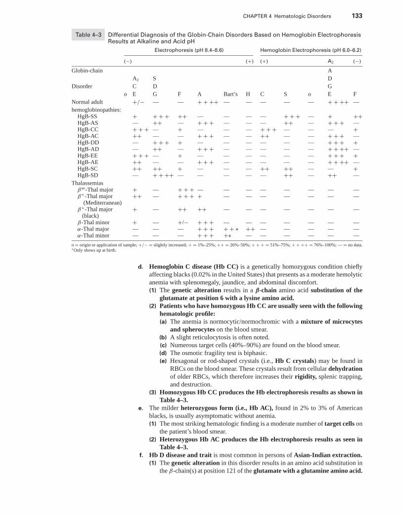
P1: OSO
LWBK192-04 LWBK192-Hubbard November 27, 2008 7:43
CHAPTER 4 Hematologic Disorders 133
Table 4–3 Differential Diagnosis of the Globin-Chain Disorders Based on Hemoglobin ElectrophoresisResults at Alkaline and Acid pH
Electrophoresis (pH 8.4–8.6) Hemoglobin Electrophoresis (pH 6.0–6.2)
(−) (+) (+) A2 (−)
Globin-chain A
A2 S D
Disorder C D G
o E G F A Bart’s H C S o E F
Normal adult +/− — — + + ++ — — — — — + + ++ —
hemoglobinopathies:HgB-SS + + + + ++ — — — — + + + — + ++HgB-AS — ++ — + + + — — — ++ — + + + —HgB-CC + + + — + — — — + + + — — — +HgB-AC ++ — — + + + — — ++ — — + + + —HgB-DD — + + + + — — — — — — + + + +HgB-AD — ++ — + + + — — — — — + + ++ —HgB-EE + + + — + — — — — — — + + + +HgB-AE ++ — — + + + — — — — — + + ++ —HgB-SC ++ ++ + — — — ++ ++ — — +HgB-SD — + + ++ — — — — — ++ — ++ —
Thalassemiasβao-Thal major + — + + + — — — — — — — —β+-Thal major ++ — + + + + — — — — — — —
(Mediterranean)β+-Thal major
(black)+ — ++ ++ — — — — — — —
β-Thal minor + — +/− + + + — — — — — — —α-Thal major — — — + + + + + ∗ ++ — — — — —α-Thal minor — — — + + + +∗ — — — — — —
o = origin or application of sample; +/− = slightly increased; + = 1%–25%; ++ = 26%–50%; + + + = 51%–75%; + + ++ = 76%–100%; — = no data.∗Only shows up at birth.
d. Hemoglobin C disease (Hb CC) is a genetically homozygous condition chieflyaffecting blacks (0.02% in the United States) that presents as a moderate hemolyticanemia with splenomegaly, jaundice, and abdominal discomfort.(1) The genetic alteration results in a β-chain amino acid substitution of the
glutamate at position 6 with a lysine amino acid.(2) Patients who have homozygous Hb CC are usually seen with the following
hematologic profile:(a) The anemia is normocytic/normochromic with a mixture of microcytes
and spherocytes on the blood smear.(b) A slight reticulocytosis is often noted.(c) Numerous target cells (40%–90%) are found on the blood smear.(d) The osmotic fragility test is biphasic.(e) Hexagonal or rod-shaped crystals (i.e., Hb C crystals) may be found in
RBCs on the blood smear. These crystals result from cellular dehydrationof older RBCs, which therefore increases their rigidity, splenic trapping,and destruction.
(3) Homozygous Hb CC produces the Hb electrophoresis results as shown inTable 4–3.
e. The milder heterozygous form (i.e., Hb AC), found in 2% to 3% of Americanblacks, is usually asymptomatic without anemia.(1) The most striking hematologic finding is a moderate number of target cells on
the patient’s blood smear.(2) Heterozygous Hb AC produces the Hb electrophoresis results as seen in
Table 4–3.f. Hb D disease and trait is most common in persons of Asian-Indian extraction.
(1) The genetic alteration in this disorder results in an amino acid substitution inthe β-chain(s) at position 121 of the glutamate with a glutamine amino acid.
P1: OSO
LWBK192-04 LWBK192-Hubbard November 27, 2008 7:43
134 A Concise Review of Clinical Laboratory Science
(2) The heterozygous trait is asymptomatic with a normal blood profile.(3) Hb D migrates on the electrophoretic gel with the Hb S fraction, but the
sickle tests are negative. Hb D produces the Hb electrophoresis results asseen in Table 4–3.
g. Hb E disease and trait is found mainly in individuals of Asian origin.(1) Homozygous patients have a mild anemia with moderate microcytosis and
target cells on their blood smear.(2) Hb E produces the Hb electrophoresis results shown in Table 4–3.
h. Doubly heterozygous β-hemoglobinopathies are disorders in which the individ-ual inherits a different abnormal β-chain gene from each parent.(1) Hb SC disease has an incidence similar to the homozygous Hb SS disease that
occurs in American blacks.(a) The severity of the disease is intermediate between sickle cell trait and
sickle-cell disease.(b) Onset is in childhood, but physical difficulties do not occur until the teenage
years.(c) Basically, the symptoms are the same as Hb SS, but splenomegaly is more
common in patients with Hb SC.(d) Patients are commonly seen with the following hematologic profile:
(i) Moderate-to-mild normocytic/normochromic anemia(ii) Moderate-to-severe anisocytosis and poikilocytosis(iii) Target cells can comprise up to 85% of the RBCs(iv) Sickle cells as well as Hb CC crystals on the blood smear(v) Positive sickle cell tests
(e) Hb SC produces the Hb electrophoresis pattern shown in Table 4–3.(2) Hb SD disease simulates but is less severe than Hb SS anemia. This disorder
cannot be distinguished between Hb SC or sickle cell trait.i. In some heterozygous hemoglobinopathies, the amino acid substitutions occur
in the heme pocket where they either increase the stability of the Hb M or alterthe affinity of the heme ring for oxygen.(1) Some abnormal hemoglobins are associated with cyanosis and Hb M.
(a) The reduction to the ferrous heme and binding of oxygen is prevented byan amino acid substitution near the heme ring.
(b) With α-chain Hb M, cyanosis is present from birth. However, with β-chainHb M, cyanosis does not occur until β-chain production reaches adultlevels.
(2) Some hemoglobinopathies result in increased oxygen-affinity and poly-cythemia.(a) Because of the high oxygen affinity, the tissue is hypoxic at any given
oxygen tension. The result is increased EPO and hence, polycythe-mia.
(b) Because the amino acid substitution is inside the molecule, the abnormalityis indistinguishable from Hb A on Hb electrophoresis.
(c) Example: Hemoglobin Chesapeake is an α-chain abnormality that resultsin a mild polycythemia.
(3) Some hemoglobins are associated with decreased oxygen affinity andcyanosis. An example of such an abnormal Hb is hemoglobin Kansas.
j. Unstable hemoglobins have an amino acid substitution at a place in the α or β
chain that affects the formation of the bonds between chains. The majority of thesehemoglobins are β-chain substitutions.(1) Hb precipitates out in the circulating RBC and attaches to the RBC membrane.
(a) Heinz bodies are Hb intracellular precipitates.(b) Intracellular precipitates render the cells inflexible and shorten RBC sur-
vival.(2) The majority of Heinz bodies are removed from the RBCs by the spleen.(3) Jaundice and splenomegaly are common, and patients may have a darkly
pigmented urine.
P1: OSO
LWBK192-04 LWBK192-Hubbard November 27, 2008 7:43
CHAPTER 4 Hematologic Disorders 135
(4) Following splenectomy, Heinz bodies are numerous in the circulatingRBCs.
(5) Unstable hemoglobins produce the following Hb electrophoresis results atan alkaline pH:(a) Approximately 25% of patients have a normal electrophoretic pattern(b) Hb A2; increased in β-chain variants(c) Hb F; increased to 10% to 15%
(6) Unstable hemoglobins can also be detected in the laboratory with the heatinstability test and the isopropanol precipitation test.
3. Thalassemiasa. Background
(1) Thalassemias are disorders of Hb synthesis that occur mainly in persons ofMediterranean, African, and Asian ancestry.
(2) The dysfunction is a diminished rate of synthesis of certain globin chains,but the chain formed in most cases is of normal amino acid sequence.
(3) Total lack of α- or β-globin production is known as thalassemia major.Production at a decreased rate is known as thalassemia minor.
(4) α-Thalassemia is associated with a decrease in the production of α-chains.β-Thalassemia is associated with a decrease in the production in β-chains.
b. Globin-chain molecular defects and nomenclature includes the following:(1) β◦-Thalassemia denotes a condition in which β-chain synthesis is absent.
The gene is not deleted, but the messenger ribonucleic acid (mRNA) isabsent or nonfunctioning.
(2) β+-Thalassemia is a condition in which β-chain synthesis is only reduced.Such disorders result from defects in transcription and processing ofmRNA.
(3) δβ-Thalassemia is caused by gene deletions involving both the δ and β
genes.(4) γδβ-Thalassemia is caused by a long deletion including the γ-genes and
δ-genes that stops short of the β-gene, but also reduces the output of β-chains.
(5) Lepore thalassemia shows normal α-genes. Abnormal δβ-genes caused byδβ-fusion of genes results from gene crossover.
(6) α-Thalassemias are generally caused by gene deletions of various lengthsand can also be αγ or α+.
(7) Hb constant spring is caused by an abnormal termination codon in anα-gene. This results in an elongated α-chain with 31 extra amino acids andslowed or reduced α-globin synthesis.
c. Homozygous β-Thalassemia (thalassemia major) results from either a decreaseor an absence in β-chain production by both gene alleles.(1) With a decrease in β-chain production, γ -chain production is high, which
results in increased Hb F.(2) There is an excess of α-chains due to a lack of matching β-chains.
(a) Unstable tetramers of α-chains (i.e., α4) precipitate out of solution inthe normoblast or RBC.
(b) These α-chain precipitates adhere to the inner membrane and damagethe cell, which results in ineffective erythropoiesis and a severe hemolyticanemia.
(3) Clinical symptoms of β-Thalassemia major include:(a) Jaundice and splenomegaly early in childhood(b) Prominent frontal bones (i.e., cheek, jaws)(c) Chronic marrow hyperplasia resulting in a thinned cortex of the long bones(d) Stunted growth and delayed puberty(e) Hemochromatosis from regular transfusions(f) Cardiac failure (i.e., major cause of death) due to myocardial siderosis by
30 years of age
P1: OSO
LWBK192-04 LWBK192-Hubbard November 27, 2008 7:43
136 A Concise Review of Clinical Laboratory Science
(4) Patients who have homozygous β-Thalassemia commonly present with thefollowing hematologic profile:(a) Unlike most hemolytic anemias, β-Thalassemia is morphologically a
microcytic/hypochromic anemia caused by a defect in either the rateof Hb synthesis or in the lack of synthesis.
(b) Peripheral blood smears show extreme poikilocytosis and anisocytosis inRBCs, as well as such morphologic abnormalities as target cells, ovalo-cytes, Howell-Jolly bodies, normoblasts, siderocytes, and Cabot’s rings.
(c) The reticulocyte count is less elevated than what would be expected forthe degree of anemia because of the destruction of erythroid precursors inthe marrow.
(d) The osmotic fragility test is decreased.(e) Serum iron is increased.(f) The indirect bilirubin level is increased.(g) The MCV is decreased with an increased RDW.
(5) Marrow aspirate reveals normoblastic hyperplasia, increased storage iron,and sideroblasts.
(6) Homozygous β-Thalassemia can exist in three genetic forms, each produc-ing a different electrophoresis pattern (Table 4–3).(a) β+-Thalassemia represents a partial decrease in β-chain production. Per-
sons of Mediterranean descent with this form of thalassemia have severeclinical symptoms. They demonstrate the electrophoretic pattern as seenin Table 4–3.
(b) β+-Thalassemia is a less clinically severe disorder that occurs in personsof African descent. These patients show the electrophoretic pattern as seenin Table 4–3.
d. Heterozygous β-Thalassemia (i.e., thalassemia minor) results from the absenceor decrease in β-chain production at one gene allele.(1) Patients may be seen with the following clinical symptoms:
(a) The degree of anemia may vary from a severe microcytic/hypochromicanemia to normal clinical findings.
(b) A moderate-to-severe anemia is more common in individuals of Mediter-ranean descent.
(c) Patients who have more severe disease have a slight hemolytic jaundiceand splenomegaly.
(2) Patients who have heterozygous β-Thalassemia are commonly seen withthe following hematologic profile:(a) Characteristically, the RBC count is increased, and the Hb and Hct are
reduced.(b) The RBC indices are commonly below normal, and the RDW is increased.(c) RBC morphology on the peripheral blood smear shows moderate poikilo-
cytosis, target cells, and basophilic stippling.(d) Osmotic fragility is decreased.(e) Serum iron levels are normal to high.
(3) Marrow often demonstrates the same characteristics as homozygous β-Thalassemia.
(4) Thalassemia minor produces the Hb electrophoresis results shown in Table4–3.
(5) δ-β-Thalassemias produce clinical symptoms similar to β-Thalassemias.(a) Homozygotes have intermediate thalassemia symptoms, and their elec-
trophoretic pattern shows an absence of Hb A and Hb A2.(b) Heterozygotes clinically have thalassemia minor symptoms and demon-
strate 5% to 20% Hb F and normal Hb A2 on their electrophoretic pattern.(6) Hb Lepore syndromes produce an abnormal Hb that has a normal α-chain
combined with a fused δβ-chain.(a) Because the δβ-chain is synthesized at a slower rate, it results in a micro-
cytic/hypochromic anemia and resembles a thalassemia.
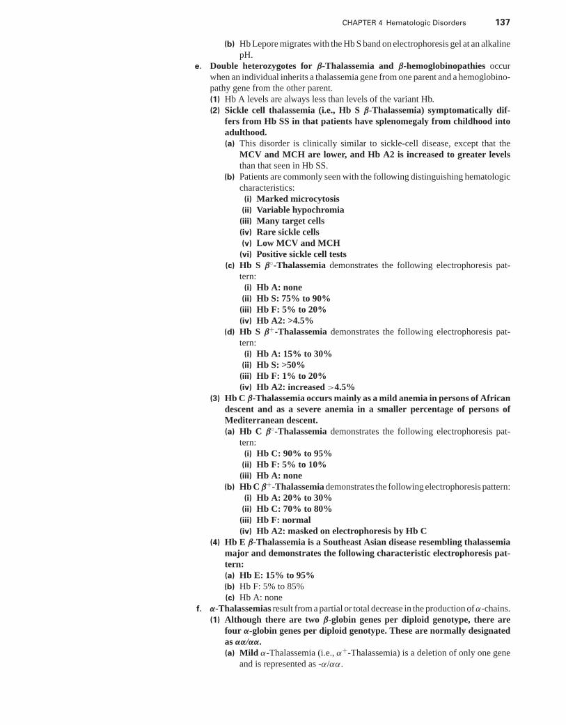
P1: OSO
LWBK192-04 LWBK192-Hubbard November 27, 2008 7:43
CHAPTER 4 Hematologic Disorders 137
(b) Hb Lepore migrates with the Hb S band on electrophoresis gel at an alkalinepH.
e. Double heterozygotes for β-Thalassemia and β-hemoglobinopathies occurwhen an individual inherits a thalassemia gene from one parent and a hemoglobino-pathy gene from the other parent.(1) Hb A levels are always less than levels of the variant Hb.(2) Sickle cell thalassemia (i.e., Hb S β-Thalassemia) symptomatically dif-
fers from Hb SS in that patients have splenomegaly from childhood intoadulthood.(a) This disorder is clinically similar to sickle-cell disease, except that the
MCV and MCH are lower, and Hb A2 is increased to greater levelsthan that seen in Hb SS.
(b) Patients are commonly seen with the following distinguishing hematologiccharacteristics:
(i) Marked microcytosis(ii) Variable hypochromia(iii) Many target cells(iv) Rare sickle cells(v) Low MCV and MCH(vi) Positive sickle cell tests
(c) Hb S β◦-Thalassemia demonstrates the following electrophoresis pat-tern:(i) Hb A: none
(ii) Hb S: 75% to 90%(iii) Hb F: 5% to 20%(iv) Hb A2: >4.5%
(d) Hb S β+-Thalassemia demonstrates the following electrophoresis pat-tern:(i) Hb A: 15% to 30%
(ii) Hb S: >50%(iii) Hb F: 1% to 20%(iv) Hb A2: increased >4.5%
(3) Hb C β-Thalassemia occurs mainly as a mild anemia in persons of Africandescent and as a severe anemia in a smaller percentage of persons ofMediterranean descent.(a) Hb C β◦-Thalassemia demonstrates the following electrophoresis pat-
tern:(i) Hb C: 90% to 95%
(ii) Hb F: 5% to 10%(iii) Hb A: none
(b) Hb Cβ+-Thalassemia demonstrates the following electrophoresis pattern:(i) Hb A: 20% to 30%
(ii) Hb C: 70% to 80%(iii) Hb F: normal(iv) Hb A2: masked on electrophoresis by Hb C
(4) Hb E β-Thalassemia is a Southeast Asian disease resembling thalassemiamajor and demonstrates the following characteristic electrophoresis pat-tern:(a) Hb E: 15% to 95%(b) Hb F: 5% to 85%(c) Hb A: none
f. α-Thalassemias result from a partial or total decrease in the production of α-chains.(1) Although there are two β-globin genes per diploid genotype, there are
four α-globin genes per diploid genotype. These are normally designatedas αα/αα.(a) Mild α-Thalassemia (i.e., α+-Thalassemia) is a deletion of only one gene
and is represented as -α/αα.
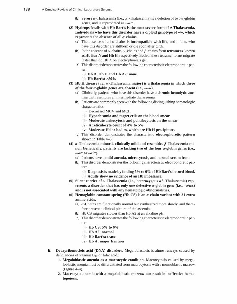
P1: OSO
LWBK192-04 LWBK192-Hubbard November 27, 2008 7:43
138 A Concise Review of Clinical Laboratory Science
(b) Severe α-Thalassemia (i.e., α◦-Thalassemia) is a deletion of two α-globingenes, and is represented as –/αα.
(2) Hydrops fetalis with Hb Bart’s is the most severe form of α-Thalassemia.Individuals who have this disorder have a diploid genotype of –/–, whichrepresents the absence of all α-chains.(a) The absence of all α-chains is incompatible with life, and infants who
have this disorder are stillborn or die soon after birth.(b) In the absence of α-chains, γ -chains and β-chains form tetramers known
as Hb Bart’s and Hb H, respectively. Both of these tetramer forms migratefaster than do Hb A on electrophoresis gel.
(c) This disorder demonstrates the following characteristic electrophoretic pat-tern:(i) Hb A, Hb F, and Hb A2: none(ii) Hb Bart’s: >80%
(3) Hb H disease (i.e., α-Thalassemia major) is a thalassemia in which threeof the four α-globin genes are absent (i.e., –/–α).(a) Clinically, patients who have this disorder have a chronic hemolytic ane-
mia that resembles an intermediate thalassemia.(b) Patients are commonly seen with the following distinguishing hematologic
characteristics:(i) Decreased MCV and MCH
(ii) Hypochromia and target cells on the blood smear(iii) Moderate anisocytosis and poikilocytosis on the smear(iv) A reticulocyte count of 4% to 5%(v) Moderate Heinz bodies, which are Hb H precipitates
(c) This disorder demonstrates the characteristic electrophoretic patternshown in Table 4–3.
(4) α-Thalassemia minor is clinically mild and resembles β-Thalassemia mi-nor. Genetically, patients are lacking two of the four α-globin genes (i.e.,–/αα or –α/α).(a) Patients have a mild anemia, microcytosis, and normal serum iron.(b) This disorder demonstrates the following characteristic electrophoretic pat-
tern:(i) Diagnosis is made by finding 5% to 6% of Hb Bart’s in cord blood.(ii) Adults show no evidence of an Hb imbalance.
(5) Silent carrier of α-Thalassemia (i.e., heterozygous α+-Thalassemia) rep-resents a disorder that has only one defective α-globin gene (i.e., –α/αα)and is not associated with any hematologic abnormalities.
(6) Hemoglobin constant spring (Hb CS) is an α-chain variant with 31 extraamino acids.(a) α-Chains are functionally normal but synthesized more slowly, and there-
fore present a clinical picture of thalassemia.(b) Hb CS migrates slower than Hb A2 at an alkaline pH.(c) This disorder demonstrates the following characteristic electrophoretic pat-
tern:(i) Hb CS: 5% to 6%
(ii) Hb A2: normal(iii) Hb Bart’s: trace(iv) Hb A: major fraction
E. Deoxyribonucleic acid (DNA) disorders. Megaloblastosis is almost always caused bydeficiencies of vitamin B12 or folic acid.1. Megaloblastic anemia as a macrocytic condition. Macrocytosis caused by mega-
loblastic anemia must be differentiated from macrocytosis with a normoblastic marrow(Figure 4–4).
2. Macrocytic anemia with a megaloblastic marrow can result in ineffective hema-topoiesis.
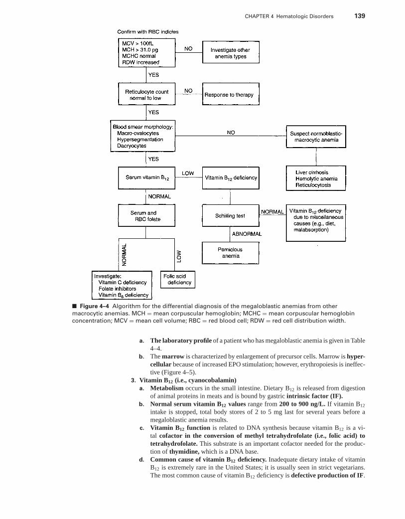
P1: OSO
LWBK192-04 LWBK192-Hubbard November 27, 2008 7:43
CHAPTER 4 Hematologic Disorders 139
■ Figure 4–4 Algorithm for the differential diagnosis of the megaloblastic anemias from other
macrocytic anemias. MCH = mean corpuscular hemoglobin; MCHC = mean corpuscular hemoglobin
concentration; MCV = mean cell volume; RBC = red blood cell; RDW = red cell distribution width.
a. The laboratory profile of a patient who has megaloblastic anemia is given in Table4–4.
b. The marrow is characterized by enlargement of precursor cells. Marrow is hyper-cellular because of increased EPO stimulation; however, erythropoiesis is ineffec-tive (Figure 4–5).
3. Vitamin B12 (i.e., cyanocobalamin)a. Metabolism occurs in the small intestine. Dietary B12 is released from digestion
of animal proteins in meats and is bound by gastric intrinsic factor (IF).b. Normal serum vitamin B12 values range from 200 to 900 ng/L. If vitamin B12
intake is stopped, total body stores of 2 to 5 mg last for several years before amegaloblastic anemia results.
c. Vitamin B12 function is related to DNA synthesis because vitamin B12 is a vi-tal cofactor in the conversion of methyl tetrahydrofolate (i.e., folic acid) totetrahydrofolate. This substrate is an important cofactor needed for the produc-tion of thymidine, which is a DNA base.
d. Common cause of vitamin B12 deficiency. Inadequate dietary intake of vitaminB12 is extremely rare in the United States; it is usually seen in strict vegetarians.The most common cause of vitamin B12 deficiency is defective production of IF.
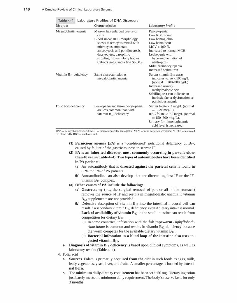
P1: OSO
LWBK192-04 LWBK192-Hubbard November 27, 2008 7:43
140 A Concise Review of Clinical Laboratory Science
Table 4–4 Laboratory Profiles of DNA Disorders
Disorder Characteristics Laboratory Profile
Megaloblastic anemia Marrow has enlarged precursorcells
Blood smear RBC morphologyshows macrocytes mixed withmicrocytes, moderateanisocytosis and poikilocytosis,dacryocytes, basophilicstippling, Howell-Jolly bodies,Cabot’s rings, and a few NRBCs
PancytopeniaLow RBC countLow hemoglobinLow hematocritMCV >100 fLIncreased to normal MCHLeukopenia with
hypersegmentation ofneutrophils
Mild thrombocytopeniaIncreased serum iron
Vitamin B12 deficiency Same characteristics asmegaloblastic anemia
Serum vitamin B12 assayindicates value <100 ng/L(normal = 200–900 ng/L)
Increased urinarymethylmalonic acid
Schilling test can indicate anintrinsic factor dysfunction orpernicious anemia
Folic acid deficiency Leukopenia and thrombocytopeniaare less common than withvitamin B12 deficiency
Serum folate <3 mcg/L (normal= 5–21 mcg/L)
RBC folate <150 mcg/L (normal= 150–600 mcg/L).
Urinary formimonoglutamicacid level is increased
DNA = deoxyribonucleic acid; MCH = mean corpuscular hemoglobin; MCV = mean corpuscular volume; NRBCs = nucleatedred blood cells; RBC = red blood cell.
(1) Pernicious anemia (PA) is a “conditioned” nutritional deficiency of B12,caused by failure of the gastric mucosa to secrete IF.
(2) PA is an inherited disorder, most commonly occurring in persons olderthan 40 years (Table 4–4). Two types of autoantibodies have been identifiedin PA patients:(a) An autoantibody that is directed against the parietal cells is found in
85% to 95% of PA patients.(b) Autoantibodies can also develop that are directed against IF or the IF-
vitamin B12 complex.(3) Other causes of PA include the following:
(a) Gastrectomy (i.e., the surgical removal of part or all of the stomach)removes the source of IF and results in megaloblastic anemia if vitaminB12 supplements are not provided.
(b) Defective absorption of vitamin B12 into the intestinal mucosal cell canresult in a secondary vitamin B12 deficiency, even if dietary intake is normal.Lack of availability of vitamin B12 in the small intestine can result fromcompetition for dietary B12.(i) In some countries, infestation with the fish tapeworm Diphylloboth-
rium latum is common and results in vitamin B12 deficiency becausethe worm competes for the available dietary vitamin B12.
(ii) Bacterial infestation in a blind loop of the intestine also uses in-gested vitamin B12.
e. Diagnosis of vitamin B12 deficiency is based upon clinical symptoms, as well aslaboratory results (Table 4–4).
4. Folic acida. Sources. Folate is primarily acquired from the diet in such foods as eggs, milk,
leafy vegetables, yeast, liver, and fruits. A smaller percentage is formed by intesti-nal flora.
b. The minimum daily dietary requirement has been set at 50 mg. Dietary ingestionjust barely meets the minimum daily requirement. The body’s reserve lasts for only3 months.
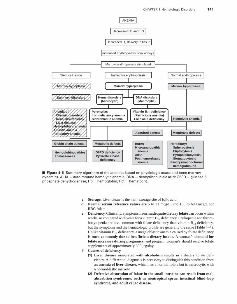
P1: OSO
LWBK192-04 LWBK192-Hubbard December 23, 2008 12:1
CHAPTER 4 Hematologic Disorders 141
ANEMIA
Decreased Hb and Hct
Decreased O2 delivery to tissue
Increased erythropoietin from kidneys
Stem cell lesion
Stem cell disorders
Marrow hypoplasia
Marrow erythropoiesis stimulated
Ineffective erythropoiesis Normal erythropoiesis
Marrow hyperplasia
Heme disorders
(Microcytic)
Porphyrias
Iron deficiency anemia
Sideroblastic anemia
Anemia of:
Chronic disorders
Renal insufficiency
Liver disease
Myelophthisic anemia
Aplastic anemia
Refractory anemia
Globin chain defects Metabolic defects
G6PD deficiency
Pyruvate kinase
deficiency
Burns
Microangiopathic
anemia
AIHA
Posthemorrhagic
anemia
Hereditary:
Spherocytosis
Eliptocytosis
Pyropoikilocytosis
Stomatocytosis
Paroxysmal nocturnal
hemoglobinuria
Acquired defects Membrane defects
Hemolytic anemia
Marrow hyperplasia
Hemoglobinopathies
Thalassemias
Vitamin B12 deficiency
(Pernicious anemia)
Folic acid deficiency
DNA disorders
(Macrocytic)
■ Figure 4–5 Summary algorithm of the anemias based on physiologic cause and bone marrow
dynamics. AIHA = autoimmune hemolytic anemia; DNA = deoxyribonucleic acid; G6PD = glucose-6-
phosphate dehydrogenase; Hb = hemoglobin; Hct = hematocrit.
c. Storage. Liver tissue is the main storage site of folic acid.d. Normal serum reference values are 5 to 21 mcg/L, and 150 to 600 mcg/L for
RBC folate.e. Deficiency. Clinically, symptoms from inadequate dietary folate can occur within
weeks, as compared with years for a vitamin B12 deficiency. Leukopenia and throm-bocytopenia are less common with folate deficiency than vitamin B12 deficiency,but the symptoms and the hematologic profile are generally the same (Table 4–4).Unlike vitamin B12 deficiency, a megaloblastic anemia caused by folate deficiencyis most commonly due to insufficient dietary intake. A woman’s demand forfolate increases during pregnancy, and pregnant woman’s should receive folatesupplements of approximately 500 μg/day.
f. Causes of deficiency(1) Liver disease associated with alcoholism results in a dietary folate defi-
ciency. A differential diagnosis is necessary to distinguish this condition froman anemia of liver disease, which has a normal folate but is macrocytic witha normoblastic marrow.
(2) Defective absorption of folate in the small intestine can result from mal-absorbtion syndromes, such as nontropical sprue, intestinal blind-loopsyndrome, and adult celiac disease.
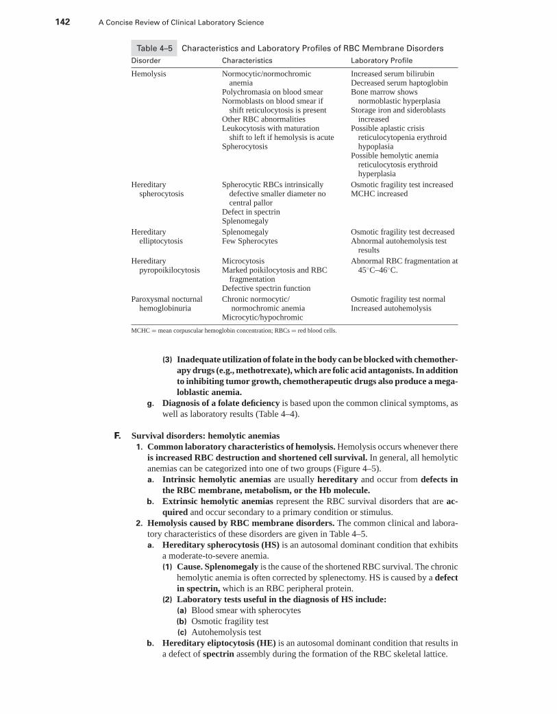
P1: OSO
LWBK192-04 LWBK192-Hubbard November 27, 2008 7:43
142 A Concise Review of Clinical Laboratory Science
Table 4–5 Characteristics and Laboratory Profiles of RBC Membrane Disorders
Disorder Characteristics Laboratory Profile
Hemolysis Normocytic/normochromicanemia
Polychromasia on blood smearNormoblasts on blood smear if
shift reticulocytosis is presentOther RBC abnormalitiesLeukocytosis with maturation
shift to left if hemolysis is acuteSpherocytosis
Increased serum bilirubinDecreased serum haptoglobinBone marrow shows
normoblastic hyperplasiaStorage iron and sideroblasts
increasedPossible aplastic crisis
reticulocytopenia erythroidhypoplasia
Possible hemolytic anemiareticulocytosis erythroidhyperplasia
Hereditaryspherocytosis
Spherocytic RBCs intrinsicallydefective smaller diameter nocentral pallor
Defect in spectrinSplenomegaly
Osmotic fragility test increasedMCHC increased
Hereditaryelliptocytosis
SplenomegalyFew Spherocytes
Osmotic fragility test decreasedAbnormal autohemolysis test
results
Hereditarypyropoikilocytosis
MicrocytosisMarked poikilocytosis and RBC
fragmentationDefective spectrin function
Abnormal RBC fragmentation at45◦C–46◦C.
Paroxysmal nocturnalhemoglobinuria
Chronic normocytic/normochromic anemia
Microcytic/hypochromic
Osmotic fragility test normalIncreased autohemolysis
MCHC = mean corpuscular hemoglobin concentration; RBCs = red blood cells.
(3) Inadequate utilization of folate in the body can be blocked with chemother-apy drugs (e.g., methotrexate), which are folic acid antagonists. In additionto inhibiting tumor growth, chemotherapeutic drugs also produce a mega-loblastic anemia.
g. Diagnosis of a folate deficiency is based upon the common clinical symptoms, aswell as laboratory results (Table 4–4).
F. Survival disorders: hemolytic anemias1. Common laboratory characteristics of hemolysis. Hemolysis occurs whenever there
is increased RBC destruction and shortened cell survival. In general, all hemolyticanemias can be categorized into one of two groups (Figure 4–5).a. Intrinsic hemolytic anemias are usually hereditary and occur from defects in
the RBC membrane, metabolism, or the Hb molecule.b. Extrinsic hemolytic anemias represent the RBC survival disorders that are ac-
quired and occur secondary to a primary condition or stimulus.2. Hemolysis caused by RBC membrane disorders. The common clinical and labora-
tory characteristics of these disorders are given in Table 4–5.a. Hereditary spherocytosis (HS) is an autosomal dominant condition that exhibits
a moderate-to-severe anemia.(1) Cause. Splenomegaly is the cause of the shortened RBC survival. The chronic
hemolytic anemia is often corrected by splenectomy. HS is caused by a defectin spectrin, which is an RBC peripheral protein.
(2) Laboratory tests useful in the diagnosis of HS include:(a) Blood smear with spherocytes(b) Osmotic fragility test(c) Autohemolysis test
b. Hereditary eliptocytosis (HE) is an autosomal dominant condition that results ina defect of spectrin assembly during the formation of the RBC skeletal lattice.
P1: OSO
LWBK192-04 LWBK192-Hubbard November 27, 2008 7:43
CHAPTER 4 Hematologic Disorders 143
(1) Elliptocytes are abundant and often represent more than 25% of the RBCpopulation.
(2) Of all patients who have HE, 90% are nonanemic. The other 10% ofpatients have a mild-to-moderate hemolytic anemia.
c. Hereditary pyropoikilocytosis (HPP) is an autosomal recessive, severe hemolyticanemia that occurs rarely in blacks. HPP is also thought to involve defectivespectrin function.
d. Hereditary stomatocytosis results in a mild-to-moderate hemolytic anemia.(1) Stomatocytes are RBCs that, on an air-dried blood film, have a slit-shaped
central pallor rather than a circular central pallor.(2) Stomatocyte forms are not as flexible as the normal biconcave disk-shaped
RBC, and they have a shortened survival time.e. Paroxysmal nocturnal hemoglobinuria (PNH) occurs more frequently in young
adults as an acquired intrinsic defect of the RBC membrane. The defectrenders the RBCs hypersensitive to complement C3 binding, and therefore,hemolysis.(1) PNH is characterized by chronic intravascular hemolysis with or without
hemoglobinuria.(2) Hemosiderinuria is also present. If excessive, it can lead to a serious iron
loss.(3) Two or three different populations of RBCs with different degrees of com-
plement sensitivity are present.(4) Platelets and neutrophils may also be hypersensitive to complement, and
therefore low in count.(5) Laboratory tests useful in the diagnosis of PNH include the following:
(a) The sucrose hemolysis test (i.e., sugar-water test) is based on the principlethat sucrose provides a medium of low ionic strength and promotes thebinding of complement to RBCs. Patients who have PNH demonstratea higher degree of hemolysis with this procedure.
(b) The acidified serum test (i.e., Ham test) is based on the principle that acid-ified serum activates complement by the alternate pathway. Patients whohave PNH demonstrate a higher degree of hemolysis with this procedure.
3. Hemolysis caused by RBC metabolic disorders.a. Glucose-6-phosphate dehydrogenase (G6PD) deficiencies are inherited as a sex-
linked trait. Anemia caused by a G6PD deficiency is found in all races, but thehighest incidence is among African and Mediterranean cultures.(1) Two genetic isoenzyme variations of G6PD have been identified. These
subtypes are Type A and Type B, depending on their electrophoreticmobility.(a) Type A. Approximately 10% of blacks who have Type A isoenzyme have an
inherited deficiency that results in only 10% to 15% G6PD enzyme activity.These individuals have clinically mild conditions but can be affected byoxidants to produce a hemolytic crisis.
(b) Type B. Persons of Mediterranean descent who have a G6PD deficiencymore commonly have a Type B deficiency. Patients who have a Type B.deficiency have an enzyme activity of only 1%. These patients are moresusceptible to severe oxidant hemolysis than are Type A individuals.
(c) Favism. A subgroup of G6PD-deficient patients have severe life-threatening hemolysis that occurs within hours after eating fava beans.
(2) Cause. Nicotinamide-adenine dinucleotide phosphate reduction (NADPH)in the RBC is linked to glutathione (GSH) reduction. GSH is a vital reducerof oxidants, such as hydrogen peroxide. This metabolic pathway preservesvital enzymes and Hb. A deficiency of G6PD limits the regeneration ofNADPH, and therefore GSH. Lack of GSH renders the RBC vulnerableto the oxidative degeneration of Hb.(a) G6PD is normally highest in young RBCs and decreases as the cell ages;
therefore, the older RBCs are preferentially destroyed.
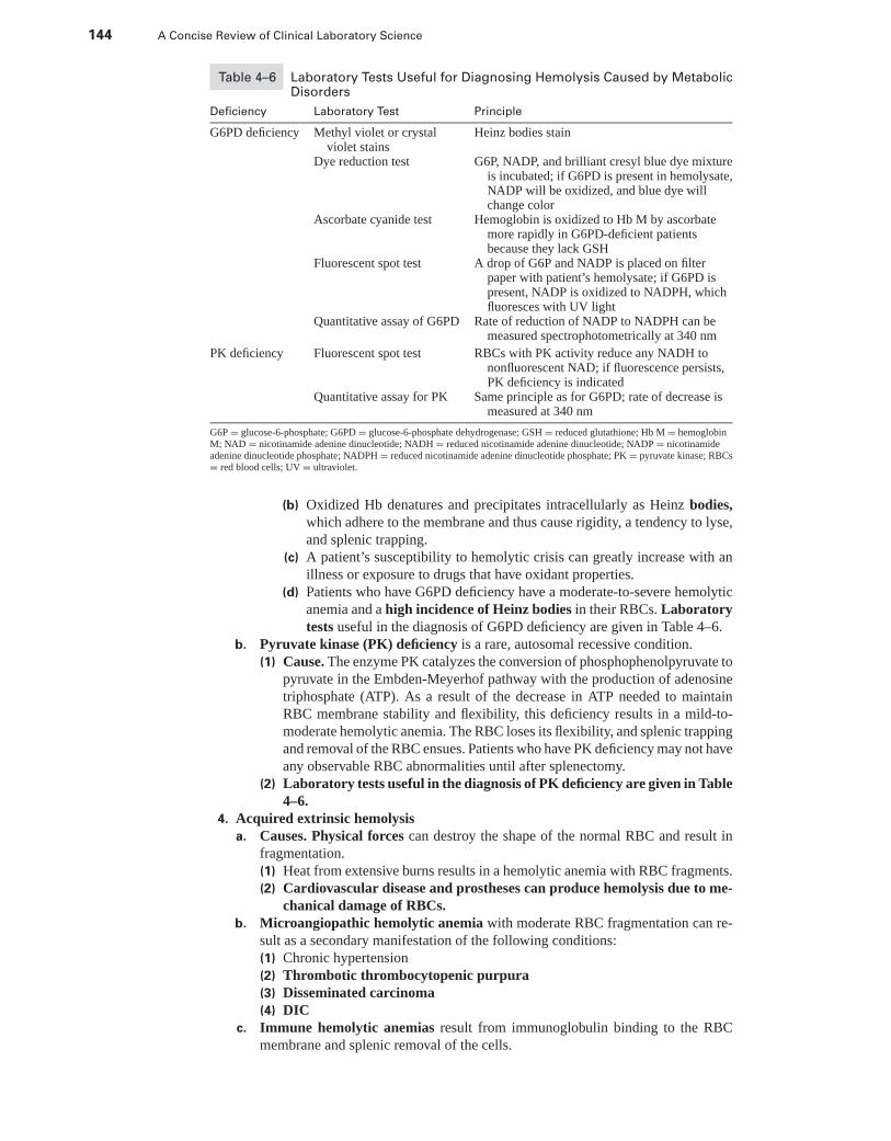
P1: OSO
LWBK192-04 LWBK192-Hubbard November 27, 2008 7:43
144 A Concise Review of Clinical Laboratory Science
Table 4–6 Laboratory Tests Useful for Diagnosing Hemolysis Caused by MetabolicDisorders
Deficiency Laboratory Test Principle
G6PD deficiency Methyl violet or crystalviolet stains
Heinz bodies stain
Dye reduction test G6P, NADP, and brilliant cresyl blue dye mixtureis incubated; if G6PD is present in hemolysate,NADP will be oxidized, and blue dye willchange color
Ascorbate cyanide test Hemoglobin is oxidized to Hb M by ascorbatemore rapidly in G6PD-deficient patientsbecause they lack GSH
Fluorescent spot test A drop of G6P and NADP is placed on filterpaper with patient’s hemolysate; if G6PD ispresent, NADP is oxidized to NADPH, whichfluoresces with UV light
Quantitative assay of G6PD Rate of reduction of NADP to NADPH can bemeasured spectrophotometrically at 340 nm
PK deficiency Fluorescent spot test RBCs with PK activity reduce any NADH tononfluorescent NAD; if fluorescence persists,PK deficiency is indicated
Quantitative assay for PK Same principle as for G6PD; rate of decrease ismeasured at 340 nm
G6P = glucose-6-phosphate; G6PD = glucose-6-phosphate dehydrogenase; GSH = reduced glutathione; Hb M = hemoglobinM; NAD = nicotinamide adenine dinucleotide; NADH = reduced nicotinamide adenine dinucleotide; NADP = nicotinamideadenine dinucleotide phosphate; NADPH = reduced nicotinamide adenine dinucleotide phosphate; PK = pyruvate kinase; RBCs= red blood cells; UV = ultraviolet.
(b) Oxidized Hb denatures and precipitates intracellularly as Heinz bodies,which adhere to the membrane and thus cause rigidity, a tendency to lyse,and splenic trapping.
(c) A patient’s susceptibility to hemolytic crisis can greatly increase with anillness or exposure to drugs that have oxidant properties.
(d) Patients who have G6PD deficiency have a moderate-to-severe hemolyticanemia and a high incidence of Heinz bodies in their RBCs. Laboratorytests useful in the diagnosis of G6PD deficiency are given in Table 4–6.
b. Pyruvate kinase (PK) deficiency is a rare, autosomal recessive condition.(1) Cause. The enzyme PK catalyzes the conversion of phosphophenolpyruvate to
pyruvate in the Embden-Meyerhof pathway with the production of adenosinetriphosphate (ATP). As a result of the decrease in ATP needed to maintainRBC membrane stability and flexibility, this deficiency results in a mild-to-moderate hemolytic anemia. The RBC loses its flexibility, and splenic trappingand removal of the RBC ensues. Patients who have PK deficiency may not haveany observable RBC abnormalities until after splenectomy.
(2) Laboratory tests useful in the diagnosis of PK deficiency are given in Table4–6.
4. Acquired extrinsic hemolysisa. Causes. Physical forces can destroy the shape of the normal RBC and result in
fragmentation.(1) Heat from extensive burns results in a hemolytic anemia with RBC fragments.(2) Cardiovascular disease and prostheses can produce hemolysis due to me-
chanical damage of RBCs.b. Microangiopathic hemolytic anemia with moderate RBC fragmentation can re-
sult as a secondary manifestation of the following conditions:(1) Chronic hypertension(2) Thrombotic thrombocytopenic purpura(3) Disseminated carcinoma(4) DIC
c. Immune hemolytic anemias result from immunoglobulin binding to the RBCmembrane and splenic removal of the cells.
P1: OSO
LWBK192-04 LWBK192-Hubbard November 27, 2008 7:43
CHAPTER 4 Hematologic Disorders 145
(1) Autoimmune hemolytic anemia (AIHA) presents a hematologic picture of ahemolytic anemia with a positive direct antiglobulin test (i.e., Coombs’ test).(a) AIHA can be associated with warm antibodies. These immunoglobulins
are usually immunoglobulin G (IgG) autoantibodies directed againstRh antigens that demonstrate maximum RBC binding at 37◦C.
(b) AIHA associated with cold antibodies are most often immunoglobulinM (IgM) autoantibodies that react most strongly at or below 25◦C.(i) Cold hemagglutinin disease involves complement-fixing autoanti-
bodies against the I antigen.(ii) Paroxysmal cold hemoglobinuria is most often caused by a
complement-fixing IgG autoantibody directed against the P anti-gen.
(2) Isoimmune hemolytic anemia is caused by an immune response to a foreignantigen. An example is hemolytic disease of the newborn (HDN), which iscaused by Rh or ABO incompatibility between mother and fetus.
(3) Drug-induced immune hemolytic anemias are caused by the adsorptionof drug-stimulated immune complexes to RBC membranes.(a) The drug-induced antibody fixes complement that binds to the RBC mem-
branes, which results in lysis.(b) Drugs such as penicillin or cephalosporin can act as haptens, inducing an
immune response when they attach to the RBC membrane.5. Anemia of blood loss
a. Acute posthemorrhagic anemia, caused by a sudden blood loss, first results inearly hypovolemia without any signs of anemia.(1) After approximately 24 hours, blood volume is returned to normal to maintain
blood pressure. Fluid from tissues move into the circulation, increasing theplasma volume. When a systemic fluid shift occurs, anemia becomes evident.
(2) Hematologic changes seen with acute blood loss include the follow-ing:(a) The earliest change is a brief thrombocytopenia, which soon returns to
normal.(b) A moderate leukocytosis with a shift to the left occurs for the first few
days after hemorrhage.(c) Hb and Hct do not decline until plasma volume is increased by inward
movement of fluid.(d) After 2 to 3 days, normocytic-normochromic anemia occurs with minimum
anisocytosis and poikilocytosis.(e) Increased erythropoietin release results in a reticulocytosis after 3 to
5 days and peaks at approximately 10 days after hemorrhage. With retic-ulocytosis, the anemia may be macrocytic with polychromasia caused byshift reticulocytosis.
b. Chronic posthemorrhagic anemia is caused by slow, sustained blood loss (e.g.,GI bleeding).(1) The reticulocyte count may be normal or only slightly increased.(2) The anemia that develops is one of iron deficiency and presents as a
microcytic-hypochromic anemia.(3) The WBC count is normal or slightly decreased.(4) Thrombocytosis is common until iron deficiency is severe.
IV. LEUKOCYTE DISORDERS
A. Blood film white cell morphology and differential1. Staining the blood film
a. Blood dyes are of two general types: methylene blue and eosin. Cell structures arestained by either one or a combination of both dyes.(1) Methylene blue is known as a basic stain.
P1: OSO
LWBK192-04 LWBK192-Hubbard November 27, 2008 7:43
146 A Concise Review of Clinical Laboratory Science
(a) WBC nuclei, cytoplasmic RNA, and platelets are stained by basic stains.(b) Staining of cell structures with methylene blue is known as basophilic
staining.(2) Eosin is known as an acid stain. Hb and other cytoplasmic cellular or-
ganelles are stained by eosin. Cell structures that take up acid stains areknown as acidophilic or eosinophilic.
b. Wright’s stain is most commonly used for routine blood film staining. It containsboth basic dyes and acid stains.(1) A good staining reaction is pH dependent (i.e., 6.4–6.7). A properly stained
blood smear should demonstrate the following characteristics:(a) RBCs should be pink or a light red.(b) Color should be uniform.(c) Nuclei of WBCs should stain blue to dark purple.(d) Platelets should be clearly visible.
(2) Excessive basophilia results from over staining with the methylene blueportion of Wright’s stain. The RBCs appear light blue. Basophilia canresult from the following errors in staining technique:(a) Blood film too thick(b) Prolonged staining time(c) Inadequate washing(d) Excessive alkalinity of stain or buffer
(3) Excessive acidophilia results from over staining with the eosin componentof Wright’s stain. RBCs will stain bright red or orange, and the WBCnuclear material will appear pale blue. Acidophilia can result from thefollowing errors in technique:(a) Insufficient staining time(b) Prolonged washing time(c) Excessive acidity of the stain or buffer
B. Phagocyte function and kinetics1. Leukocyte maturation (Tables 4–7 and 4–8). The morphology of myeloid (i.e., gran-
ulocyte) maturation is important to know for differentiating and visually identifyingcells of each maturation stage.a. Myeloblasts are the earliest recognizable stage of myeloid maturation.b. Promyelocytes are the second stage following one or more mitotic divisions of
myeloblasts.c. The myelocyte stage of maturation begins with the appearance of a new type of
granulocyte-specific granule. These granules (secondary granules) give the ma-ture granulocyte its characteristic appearance and functional destiny. Therefore,the maturation of the myelocyte, which is based on granule type, gives rise toneutrophils, eosinophils, or basophils.(1) Metamyelocytes emerge after several mitotic divisions of the myelocytes.(2) The band neutrophil stage is reached after the round metamyelocyte nu-
cleus flattens.(3) The final stage of a segmented neutrophil reaches full maturity once the
band nucleus pinches off into segments.2. Function of granulocytes and monocytes
a. Phagocytic function involves the following general steps:(1) Increase in cell numbers at the infected site(2) Cell attachment to the foreign or dead material(3) Engulfment of foreign or dead material(4) Dissolving of foreign or dead material(5) Disposition of catabolic components
b. Granulocytes are the first line of defense against microbial organisms.(1) Neutrophil migration is directed by infections or inflamed tissue.
(a) Tissue injury results in the release of vasoactive and chemotactic factors.(i) Vasoactive factors, such as prostaglandins and leukotrienes, in-
crease capillary permeability and induce local migration.
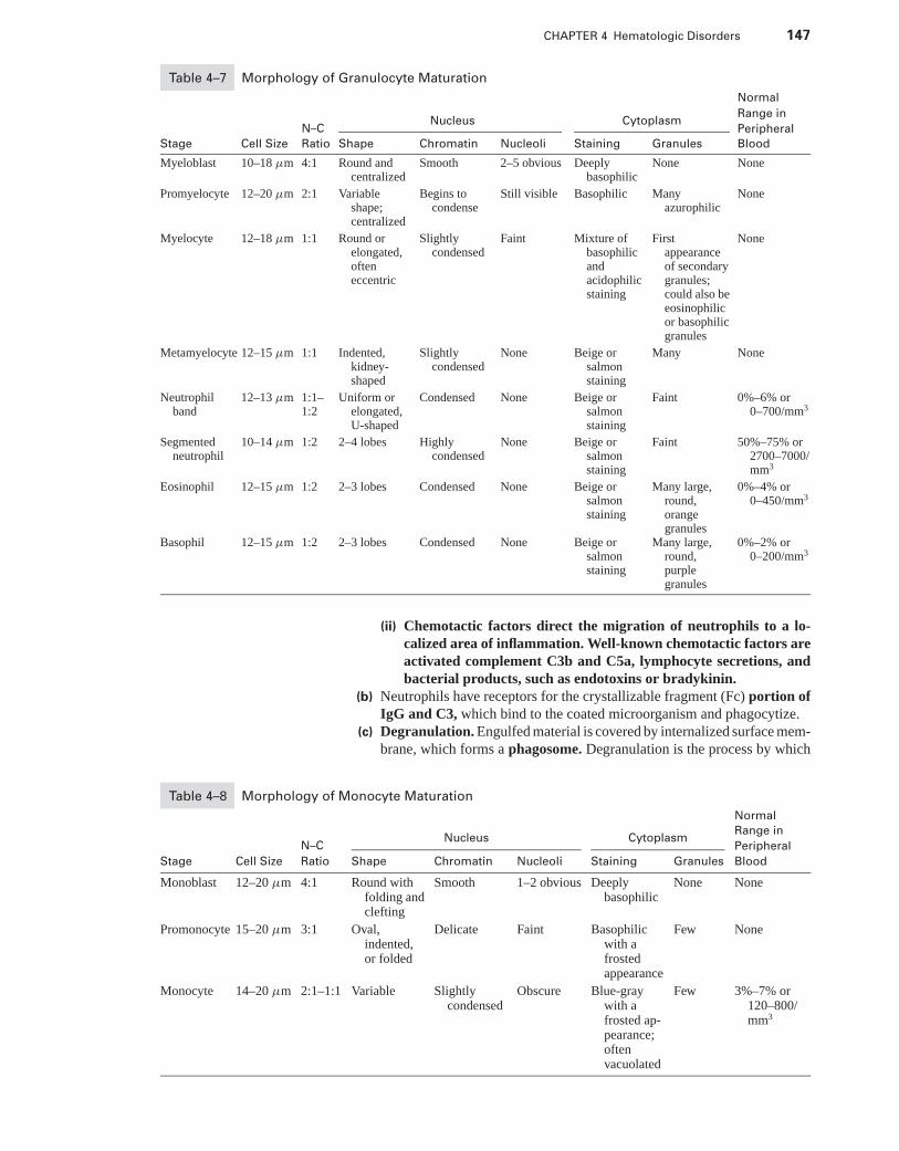
P1: OSO
LWBK192-04 LWBK192-Hubbard November 27, 2008 7:43
CHAPTER 4 Hematologic Disorders 147
Table 4–7 Morphology of Granulocyte Maturation
Normal
Range in
N–CNucleus Cytoplasm
Peripheral
Stage Cell Size Ratio Shape Chromatin Nucleoli Staining Granules Blood
Myeloblast 10–18 μm 4:1 Round andcentralized
Smooth 2–5 obvious Deeplybasophilic
None None
Promyelocyte 12–20 μm 2:1 Variableshape;centralized
Begins tocondense
Still visible Basophilic Manyazurophilic
None
Myelocyte 12–18 μm 1:1 Round orelongated,ofteneccentric
Slightlycondensed
Faint Mixture ofbasophilicandacidophilicstaining
Firstappearanceof secondarygranules;could also beeosinophilicor basophilicgranules
None
Metamyelocyte 12–15 μm 1:1 Indented,kidney-shaped
Slightlycondensed
None Beige orsalmonstaining
Many None
Neutrophilband
12–13 μm 1:1–1:2
Uniform orelongated,U-shaped
Condensed None Beige orsalmonstaining
Faint 0%–6% or0–700/mm3
Segmentedneutrophil
10–14 μm 1:2 2–4 lobes Highlycondensed
None Beige orsalmonstaining
Faint 50%–75% or2700–7000/mm3
Eosinophil 12–15 μm 1:2 2–3 lobes Condensed None Beige orsalmonstaining
Many large,round,orangegranules
0%–4% or0–450/mm3
Basophil 12–15 μm 1:2 2–3 lobes Condensed None Beige orsalmonstaining
Many large,round,purplegranules
0%–2% or0–200/mm3
(ii) Chemotactic factors direct the migration of neutrophils to a lo-calized area of inflammation. Well-known chemotactic factors areactivated complement C3b and C5a, lymphocyte secretions, andbacterial products, such as endotoxins or bradykinin.
(b) Neutrophils have receptors for the crystallizable fragment (Fc) portion ofIgG and C3, which bind to the coated microorganism and phagocytize.
(c) Degranulation. Engulfed material is covered by internalized surface mem-brane, which forms a phagosome. Degranulation is the process by which
Table 4–8 Morphology of Monocyte Maturation
Normal
Range in
N–CNucleus Cytoplasm
Peripheral
Stage Cell Size Ratio Shape Chromatin Nucleoli Staining Granules Blood
Monoblast 12–20 μm 4:1 Round withfolding andclefting
Smooth 1–2 obvious Deeplybasophilic
None None
Promonocyte 15–20 μm 3:1 Oval,indented,or folded
Delicate Faint Basophilicwith afrostedappearance
Few None
Monocyte 14–20 μm 2:1–1:1 Variable Slightlycondensed
Obscure Blue-graywith afrosted ap-pearance;oftenvacuolated
Few 3%–7% or120–800/mm3
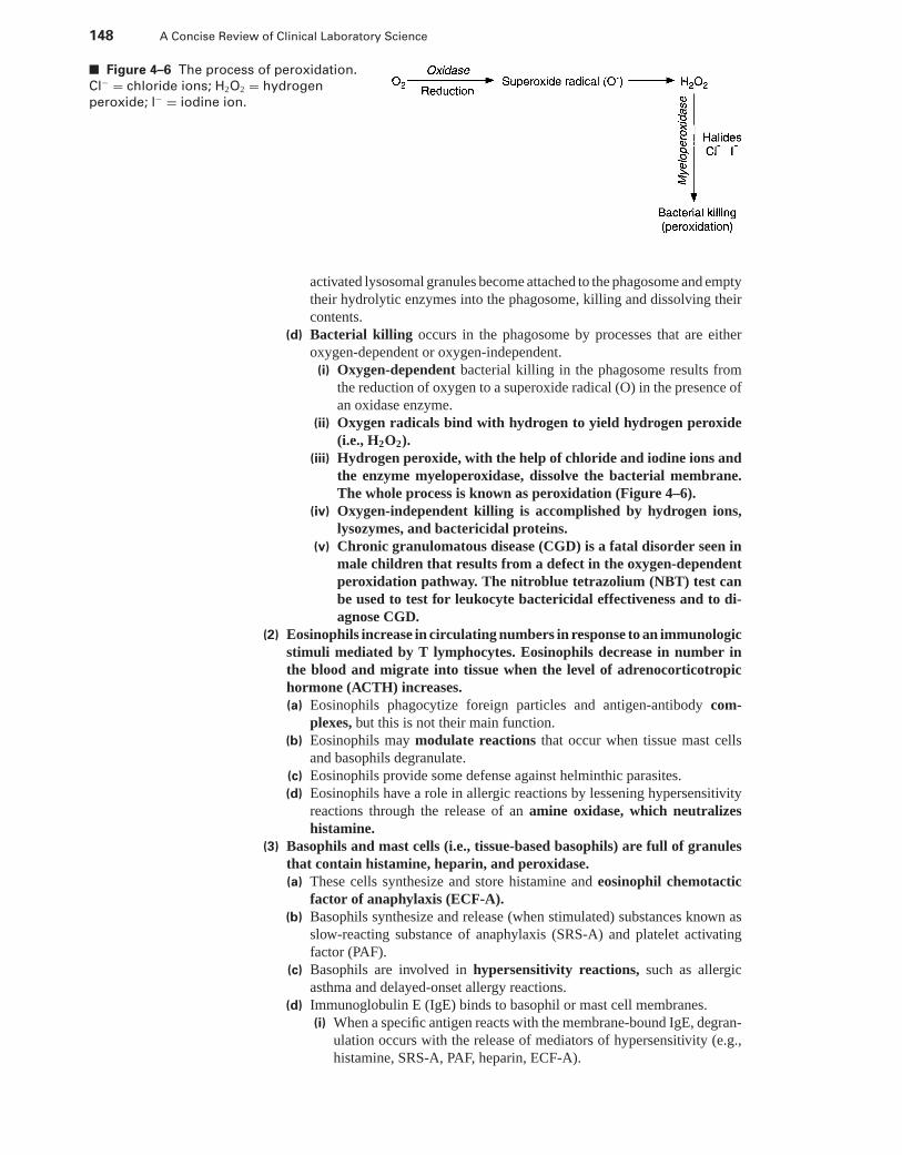
P1: OSO
LWBK192-04 LWBK192-Hubbard November 27, 2008 7:43
148 A Concise Review of Clinical Laboratory Science
■ Figure 4–6 The process of peroxidation.
Cl− = chloride ions; H2O2 = hydrogen
peroxide; l− = iodine ion.
activated lysosomal granules become attached to the phagosome and emptytheir hydrolytic enzymes into the phagosome, killing and dissolving theircontents.
(d) Bacterial killing occurs in the phagosome by processes that are eitheroxygen-dependent or oxygen-independent.
(i) Oxygen-dependent bacterial killing in the phagosome results fromthe reduction of oxygen to a superoxide radical (O) in the presence ofan oxidase enzyme.
(ii) Oxygen radicals bind with hydrogen to yield hydrogen peroxide(i.e., H2O2).
(iii) Hydrogen peroxide, with the help of chloride and iodine ions andthe enzyme myeloperoxidase, dissolve the bacterial membrane.The whole process is known as peroxidation (Figure 4–6).
(iv) Oxygen-independent killing is accomplished by hydrogen ions,lysozymes, and bactericidal proteins.
(v) Chronic granulomatous disease (CGD) is a fatal disorder seen inmale children that results from a defect in the oxygen-dependentperoxidation pathway. The nitroblue tetrazolium (NBT) test canbe used to test for leukocyte bactericidal effectiveness and to di-agnose CGD.
(2) Eosinophils increase in circulating numbers in response to an immunologicstimuli mediated by T lymphocytes. Eosinophils decrease in number inthe blood and migrate into tissue when the level of adrenocorticotropichormone (ACTH) increases.(a) Eosinophils phagocytize foreign particles and antigen-antibody com-
plexes, but this is not their main function.(b) Eosinophils may modulate reactions that occur when tissue mast cells
and basophils degranulate.(c) Eosinophils provide some defense against helminthic parasites.(d) Eosinophils have a role in allergic reactions by lessening hypersensitivity
reactions through the release of an amine oxidase, which neutralizeshistamine.
(3) Basophils and mast cells (i.e., tissue-based basophils) are full of granulesthat contain histamine, heparin, and peroxidase.(a) These cells synthesize and store histamine and eosinophil chemotactic
factor of anaphylaxis (ECF-A).(b) Basophils synthesize and release (when stimulated) substances known as
slow-reacting substance of anaphylaxis (SRS-A) and platelet activatingfactor (PAF).
(c) Basophils are involved in hypersensitivity reactions, such as allergicasthma and delayed-onset allergy reactions.
(d) Immunoglobulin E (IgE) binds to basophil or mast cell membranes.(i) When a specific antigen reacts with the membrane-bound IgE, degran-
ulation occurs with the release of mediators of hypersensitivity (e.g.,histamine, SRS-A, PAF, heparin, ECF-A).
P1: OSO
LWBK192-04 LWBK192-Hubbard November 27, 2008 7:43
CHAPTER 4 Hematologic Disorders 149
(ii) ECF-A leads to an accumulation of eosinophils at the site of in-flammation, which acts to counteract and balance the mediatorsof inflammation.
(e) Basophils are also involved in delayed hypersensitivity reactions, such ascontact allergies.
c. Monocytes and macrophages(1) The monocyte migrates into the tissue, where it is transformed into a
macrophage (i.e., histiocyte).(2) Some macrophages are motile and respond to chemotactic factors and
factors from activated lymphocytes. Other macrophages are tissue-based,such as Kupffer’s cells or alveolar macrophages.
(3) Monocytes become immobilized by migration inhibiting factor (MIF),which is released from activated lymphocytes.
(4) Monocytes and macrophages become activated to phagocytize by comple-ment, prostaglandins, or previously phagocytized material.
(5) Functions. Monocytes and macrophages are highly versatile cells that per-form many vital functions, including:(a) A defense against microorganisms such as mycobacterium, fungi, bacteria,
protozoa, and viruses(b) A role in the antigen-induced blast transformation of lymphocytes
(i) Monocytes secrete IL-1, which activates helper T cells.(ii) Monocytes physically present and deliver the antigen to the specific
membrane surface receptor sites.(c) A major role in the daily destruction of aged blood cells, denatured plasma
proteins, and lipids(d) Heme oxidase activity, which enables some tissue macrophages to break
down RBC Hb and recycle it [see Section III C 3 a (3)].3. Granulopoiesis is controlled by glycoproteins isolated from lymphocytes and mono-
cytes. These glycoproteins are known as interleukins (IL) and monokines, re-spectively. Because most research in this area was done on bone marrow cul-tures, these growth stimulators are also known as colony stimulating factors(CSF).a. Multi-CSF (i.e., IL-3) production is stimulated by endotoxin released from in-
fection.(1) Source. IL-3 is secreted by marrow fibroblasts, T lymphocytes, macrophages,
and monocytes.(2) Function. IL-3 stimulates regeneration, maturation, and differentiation of
multipotential and unipotential stem cells.b. Granulocyte/monocyte colony stimulating factor (GM-CSF) is important for
myeloid maturation in the marrow.(1) Source. GM-CSF is secreted by T lymphocytes, marrow fibroblasts, marrow
endothelial cells, and monocytes.(2) Function. GM-CSF stimulates neutrophil, eosinophil, and monocyte
growth.c. Granulocyte colony stimulating factor (G-CSF) is a more specific granulocyte
growth factor.(1) Source. G-CSF is secreted by monocytes, marrow fibroblasts, and endothelial
cells.(2) Function. G-CSF stimulates the growth of neutrophils and enhances the
functional response of neutrophils.d. Monocyte/macrophage colony stimulating factor (M-CSF), also known as CSF-
1, is the primary monocytic growth factor.(1) Source. M-CSF is secreted by mature monocytes, marrow fibroblasts, and
marrow endothelial cells.(2) Function. M-CSF stimulates macrophages and the release of G-CSF from
monocytes. This monocytic growth factor also stimulates the release oftumor necrosis factor, interferon, and IL-1 from macrophages.
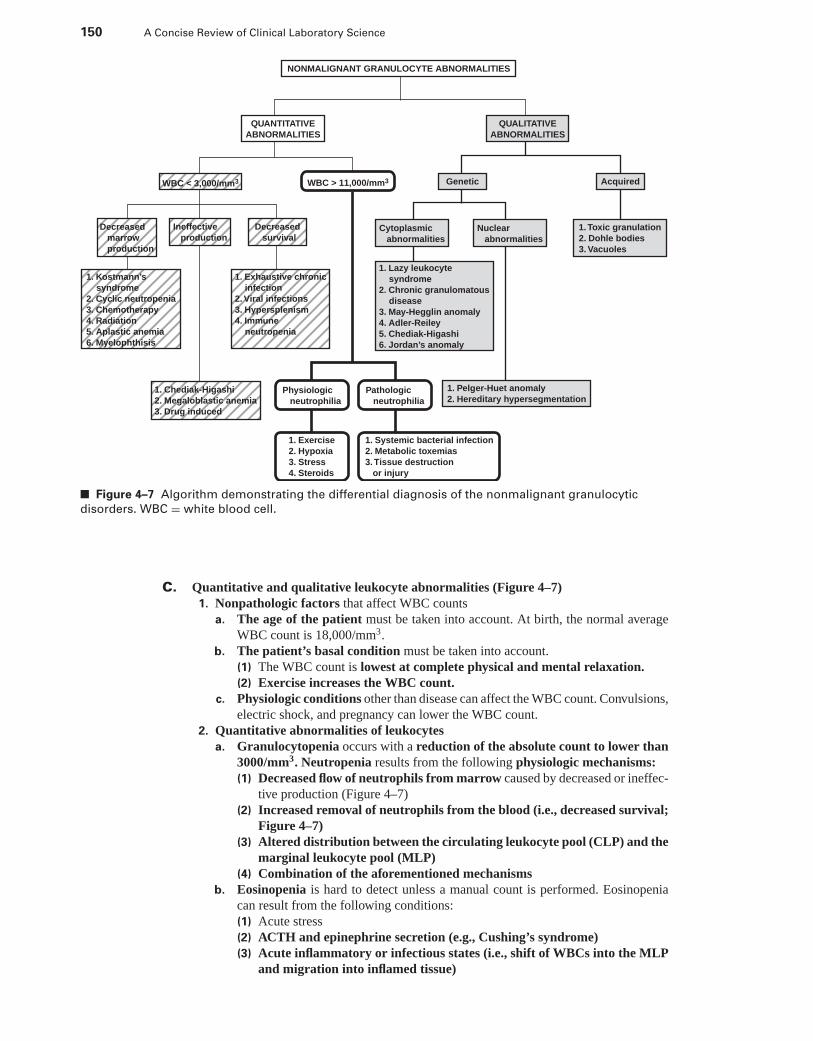
P1: OSO
LWBK192-04 LWBK192-Hubbard December 23, 2008 12:1
150 A Concise Review of Clinical Laboratory Science
NONMALIGNANT GRANULOCYTE ABNORMALITIES
QUANTITATIVE
ABNORMALITIES
QUALITATIVE
ABNORMALITIES
Genetic
Cytoplasmic
abnormalities
Nuclear
abnormalities
1. Toxic granulation
2. Dohle bodies
3. Vacuoles
WBC < 3,000/mm3
Decreased
marrow
production
1. Kostmann’s
syndrome
2. Cyclic neutropenia
3. Chemotherapy
4. Radiation
5. Aplastic anemia
6. Myelophthisis
1. Exhaustive chronic
infection
2. Viral infections
3. Hypersplenism
4. Immune
neutropenia
1. Chediak-Higashi
2. Megaloblastic anemia
3. Drug induced
Ineffective
production
Decreased
survival
WBC > 11,000/mm3 Acquired
Physiologic
neutrophilia
1. Exercise
2. Hypoxia
3. Stress
4. Steroids
1. Systemic bacterial infection
2. Metabolic toxemias
3. Tissue destruction
or injury
Pathologic
neutrophilia
1. Pelger-Huet anomaly
2. Hereditary hypersegmentation
1. Lazy leukocyte
syndrome
2. Chronic granulomatous
disease
3. May-Hegglin anomaly
4. Adler-Reiley
5. Chediak-Higashi
6. Jordan’s anomaly
■ Figure 4–7 Algorithm demonstrating the differential diagnosis of the nonmalignant granulocytic
disorders. WBC = white blood cell.
C. Quantitative and qualitative leukocyte abnormalities (Figure 4–7)1. Nonpathologic factors that affect WBC counts
a. The age of the patient must be taken into account. At birth, the normal averageWBC count is 18,000/mm3.
b. The patient’s basal condition must be taken into account.(1) The WBC count is lowest at complete physical and mental relaxation.(2) Exercise increases the WBC count.
c. Physiologic conditions other than disease can affect the WBC count. Convulsions,electric shock, and pregnancy can lower the WBC count.
2. Quantitative abnormalities of leukocytesa. Granulocytopenia occurs with a reduction of the absolute count to lower than
3000/mm3. Neutropenia results from the following physiologic mechanisms:(1) Decreased flow of neutrophils from marrow caused by decreased or ineffec-
tive production (Figure 4–7)(2) Increased removal of neutrophils from the blood (i.e., decreased survival;
Figure 4–7)(3) Altered distribution between the circulating leukocyte pool (CLP) and the
marginal leukocyte pool (MLP)(4) Combination of the aforementioned mechanisms
b. Eosinopenia is hard to detect unless a manual count is performed. Eosinopeniacan result from the following conditions:(1) Acute stress(2) ACTH and epinephrine secretion (e.g., Cushing’s syndrome)(3) Acute inflammatory or infectious states (i.e., shift of WBCs into the MLP
and migration into inflamed tissue)
P1: OSO
LWBK192-04 LWBK192-Hubbard November 27, 2008 7:43
CHAPTER 4 Hematologic Disorders 151
c. Granulocytosis refers to an absolute increase in the concentration of circulatingleukocytes higher than normal for a certain age (i.e., >11,000/mm3 in adults;Figure 4–7).(1) Physiologic neutrophilia
(a) Granulocytosis results from the following physiologic mechanisms:(i) Increased rate of inflow of cells into the circulation from the marrow
(ii) Shift in cells from the MLP into the CLP(iii) A decrease in the rate of outflow of cells from the blood
(b) A leukemoid reaction is a nonleukemic leukocytosis with a WBC count>30,000/mm3.
(i) This condition is characterized by the lack of myeloblasts in the pe-ripheral blood.
(ii) A maturation shift to the left occurs with increased band formsand few metamyelocytes or myelocytes in the peripheral blood.
(iii) The LAP level will be increased, which is a distinguishing factoras compared with a leukemic granulocytosis [see Section V D 5 e(1) (c)].
(iv) Leukemoid reactions are seen most commonly in children whohave infections (e.g., pneumonia, meningitis, tuberculosis) andalso in cases of severe hemolysis or metastatic carcinoma.
(c) Physiologic leukocytosis is produced by factors that do not involve tissuedamage.(i) Excessive exercise, hypoxia, or stress can cause a shift from the MLP
into the CLP, which appears as an increased peripheral WBC count.(ii) Increased outflow of cells from the marrow into the blood increases
both the CLP and the MLP.(2) Pathologic neutrophilia results from disease, and is usually in response to
tissue damage from a chronic infection.(a) Three physiologic phases of shifting WBC counts can be observed in acute
and chronic infections.(i) Phase I in early infection results in the acute migration of cells from
the MLP into tissue releasing chemotactic factor. Because the CLP andthe MLP are in equilibrium, a transient neutropenia results. Myeloidgrowth factors are also released to stimulate the marrow to increaseproduction.
(ii) Phase II is related to established chronic and severe infections whenthe marrow supply slowly rises to the increased demand and in-creases the flow of cells into the circulation. This results in a neu-trophilia, a shift to the left, and a decrease in eosinophils.
(iii) Phase III is related either to recovery from the infection or to mar-row exhaustion when the infection begins to subside, and the feverbreaks. The total number of granulocytes decreases, and the totalnumber of monocytes increases (i.e., secondary line of defense). Ifrecovery does not occur, the marrow becomes depleted, and pro-duction cannot keep up with granulocyte loss. A granulocytopeniaresults.
(b) Miscellaneous disorders and conditions associated with neutrophilia in-clude the following:
(i) Toxic factors that produce neutrophilia can be classified as eithermetabolic diseases (e.g., uremia, gout, diabetic acidosis) or as drugsand chemicals (e.g., lead, mercury, digitalis, epinephrine, ACTH).
(ii) Tissue destruction or necrosis involving a large amount of tissuecan cause neutrophilia (e.g., myocardial infarction, burns, surgery,crush injuries, fractures).
(iii) Hemorrhage, if significant, can result in neutrophilia.(iv) Acute massive hemolysis can increase the CLP.(v) Hematologic disorders such as myeloproliferative disorders are
associated with an absolute neutrophilia.

P1: OSO
LWBK192-04 LWBK192-Hubbard November 27, 2008 7:43
152 A Concise Review of Clinical Laboratory Science
(3) Eosinophilia is clinically defined as an absolute eosinophil count>400/mm3 in the peripheral blood. Disorders and conditions that demon-strate eosinophilia include the following:(a) Allergic diseases(b) Skin disorders(c) Parasitic infections (e.g., Trichinosis)(d) Infectious diseases (e.g., scarlet fever with skin rash)(e) Pulmonary eosinophilias (e.g., Loffler’s syndrome)(f) Blood diseases such as chronic myelogenous leukemia (CML) and other
myeloproliferative disorders(g) Splenectomy(h) Various drugs (e.g., pilocarpine, digitalis)
(4) Basophilia is defined as an absolute basophil count >40/mm3 in the pe-ripheral blood. Basophilias have causes similar to eosinophilias.
(5) Monocytosis is defined as an absolute monocyte count >800/mm3. Mono-cytosis is commonly observed during the recovery phase of infections andin various myeloproliferative diseases.
3. Persons who have qualitative leukocyte abnormalities demonstrate a decreased re-sistance to infection, despite normal leukocyte counts.a. Lazy leukocyte syndrome is a rare inherited condition seen in children. Granu-
locytes do not respond to chemotactic factors; therefore, they fail to accumulateat the inflamed tissue.
b. CGD is a qualitative disorder in which the granulocytes are capable of phagocytosisand degranulation [IV B 2 b (1) (c)], but are incapable of the subsequent bacterialkilling process.(1) Lysozymes have lost their bactericidal properties due to a decrease in the
production of H2O2.(2) Granulocytes in this disorder, when stained with NBT, demonstrate a weak
staining of <10% of the blood granulocytes.(3) The disorder is clinically characterized by frequent infections, enlarged
spleen and liver, lymphadenitis, and granulomas (i.e., aggregates ofmononuclear cells) in many organs.
c. Congenital and acquired qualitative disorders of neutrophils can affect eitherthe cytoplasm or the nucleus.(1) Qualitative disorders affecting the neutrophil cytoplasm include the follow-
ing:(a) Toxic granulation appears as tiny, dark blue-to-purple cytoplasmic gran-
ules in the metamyelocyte, band, or segmented neutrophil stages.(i) These granules are peroxidase positive.(ii) They are most commonly found in severe infections or other toxic
conditions.(iii) Toxic granules are actually azurophil granules (i.e., secondary
granules) that have retained their basophilic staining qualities be-cause of lack of maturation or skipped maturation divisions duringaccelerated granulopoiesis.
(iv) Cytoplasmic vacuoles are also signs of toxic change.(b) Dohle’s inclusion bodies are small, oval inclusions in the peripheral cy-
toplasm of neutrophils that stain pale blue with Wright’s stain.(i) Dohle’s bodies are remnants of RNA and free ribosomes or rough
endoplasmic reticulum.(ii) These cytoplasmic inclusions stain positive with the periodic acid-
Schiff (PAS) cytochemical stain.(iii) The appearance of Dohle’s bodies is often associated with scar-
let fever, and these inclusion bodies are occasionally seen in pa-tients who have burns; infectious diseases (i.e., often with toxicgranulation); aplastic anemia; and following exposure to toxicchemicals.
P1: OSO
LWBK192-04 LWBK192-Hubbard November 27, 2008 7:43
CHAPTER 4 Hematologic Disorders 153
(c) May-Hegglin anomaly is a rare, autosomal dominant, qualitative leuko-cyte abnormality.(i) The granulocytes of patients who have this disorder demonstrate pale
blue inclusions that are larger and more prominent than the Dohle’sbodies found with infections.
(ii) Compared with Dohle’s bodies occurring secondary to chronicinfection, the cytoplasmic inclusions in May-Hegglin anomaly arePAS negative.
(iii) Cytoplasmic inclusions result from structural RNA alterations.(iv) Inclusions have been found in eosinophils, basophils, and mono-
cytes, as well as neutrophils.(v) Patients are seen to have a mild thrombocytopenia, giant platelets,
and a slight bleeding tendency.(d) Alder-Reilly anomaly is a genetic abnormality that results in dense
azurophilic granulation in all types of leukocytes.(i) Cytoplasmic granulation is not transient or related to an infectious
disease, as it is with toxic granulation.(ii) Granulation results from an abnormal deposition and storage of
mucopolysaccharides.(e) Chediak-Steinbrinck-Higashi anomaly is an autosomal recessive disorder
that results in qualitative abnormalities in all types of leukocytes.(i) Large, coarse, irregular lysosome granules are found in the cyto-
plasm of granulocytes and monocytes.(ii) Patients also demonstrate abnormal pigmentation, neuropathies,
photophobia, and recurrent infections.(iii) Leukocytes of both the blood and bone marrow are affected.
(f) Jordan’s anomaly is a genetic qualitative disorder in which abundantsudanophilic inclusions (i.e., lipid) are found in the cytoplasm of granu-locytes, lymphocytes, and plasma cells in the blood and marrow.
(2) Qualitative disorders affecting the neutrophil nucleus include the follow-ing:(a) Pelger-Huet anomaly is an autosomal dominant condition in which there
is a failure of normal segmentation of neutrophil nuclei.(i) Most nuclei are band-shaped, have two segments, or are peanut-shaped.(ii) The nuclear chromatin of these cells is coarse and clumped beyond
the degree of clumping found in normal immature band forms.(b) Hereditary hypersegmentation of neutrophils is a disorder in which the
majority of the patient’s neutrophils have four or more lobes. This disor-der must be differentiated from the hypersegmentation of megaloblasticanemia.
d. Monocyte-macrophage qualitative abnormalities are rare, autosomal recessivelipid-storage diseases.(1) Common characteristics
(a) There is a deficiency in one of the catabolic enzymes involved in the break-down of sphingolipids.
(b) Because macrophages play an important role in the catabolism of lipid-rich membranes, they can accumulate undegraded lipid products in theircytoplasm. This leads to the production of lipid-heavy and blocked foamymacrophages, which stimulate further macrophage production and resultin an expansion of the monocyte-macrophage system.
(2) Gaucher’s disease is a genetic disorder that results in the accumulation ofglycosphingolipids in the cytoplasm of monocytes and macrophages.(a) There is a deficiency of β-glucosidase, which is the enzyme that splits
glucose from sphingolipids.(b) Macrophages and monocytes in this disease are PAS positive because of ac-
cumulated cytoplasmic carbohydrate (i.e., monocytes are normally weaklypositive to negative for PAS).
P1: OSO
LWBK192-04 LWBK192-Hubbard November 27, 2008 7:43
154 A Concise Review of Clinical Laboratory Science
(c) Macrophage cytoplasm has a pale onion-skin appearance (i.e., Gaucher’scells).
(3) Niemann-Pick disease is a similar disorder in which there is a deficiency inthe enzyme that cleaves phosphoryl choline from sphingolipids. Macrophagesand monocytes have a “foam-cell” appearance but are PAS negative.
D. Leukemias and cytochemical stains1. Common characteristics of leukemias
a. Incidence. Leukemia is a relatively common disorder with approximately sevennew cases per 100,000 population reported annually in the United States.
b. Definition. Leukemia can be defined as a generalized abnormal neoplastic prolif-eration or accumulation of hemopoietic cells in the bone marrow, organs, and theperipheral blood.
c. Clinical classifications(1) Acute leukemias are refractory to remission, usually fatal within 3 months,
and demonstrate a bone marrow packed with primitive cells (i.e., blasts) ofthe cell type involved with little differentiation.
(2) Subacute leukemias have a longer patient survival of 3 to 12 months, andusually have a clinical picture of an acute leukemia.
(3) Chronic leukemias are defined as having a survival of >1 year if no re-mission occurs. Blasts are elevated in marrow and blood, but are usually<5%. Maturation within a cell line still occurs.
d. Cytologic classifications are myeloid or lymphoid.e. Common laboratory results include the following:
(1) Anemia (i.e., present in more than 90% of leukemia patients)(2) Leukocytosis
(a) Almost 30% of leukemic patients demonstrate normal or decreased WBCcounts (i.e., aleukemic leukemia).
(b) Approximately 20% of leukemic patients have WBC counts higher than100,000/mm3.
(c) The remainder of leukemic patients have elevated counts lower than100,000/mm3.
(3) Thrombocytopenia (present in 80% of leukemic patients)(4) Basophilia in the peripheral blood and bone marrow(5) Immature precursor or blast cells in the peripheral blood and marrow(6) NRBCs in the peripheral blood(7) Decrease in the LAP level(8) Nonhematologic clues such as an elevation in serum uric acid, lactate
dehydrogenase, and vitamin B12 levels or B12-binding capacity (i.e., dueto increased amounts of transcobalamin I produced by neutrophils)
2. Cytochemical stains are useful diagnostic tools (Table 4–9). The cytochemical stainshelp to differentiate the leukemic blasts of acute myelogenous leukemia (AML)from leukemias of lymphoid origin. It is difficult to distinguish between leukemiaswith Wright’s stain.a. The general principle of most cytochemical stains is to incubate cells on a blood
smear with a substrate that reacts with an intracellular marker (e.g., lipid, lysozyme,glycogen, enzyme). In enzymatic procedures, the reaction product is coupled witha diazonium salt (i.e., dye) to produce a visible reaction product.
b. Sudan black B and peroxidase (i.e., myeloperoxidase) cytochemical stains sharea common staining pattern.(1) Sudan black B stains phospholipids and sterols (i.e., lipids) in cytoplasmic
lysosome and mitochondrial membranes.(a) Azurophilic and specific neutrophil granules are stained.(b) Cytoplasmic granules stain faintly in myeloblast cells and strongly in ma-
ture neutrophils. As the cell matures from the promyelocyte stage to themature segmented neutrophil, each stage is generally more positive.
(c) Eosinophilic granules stain positive.
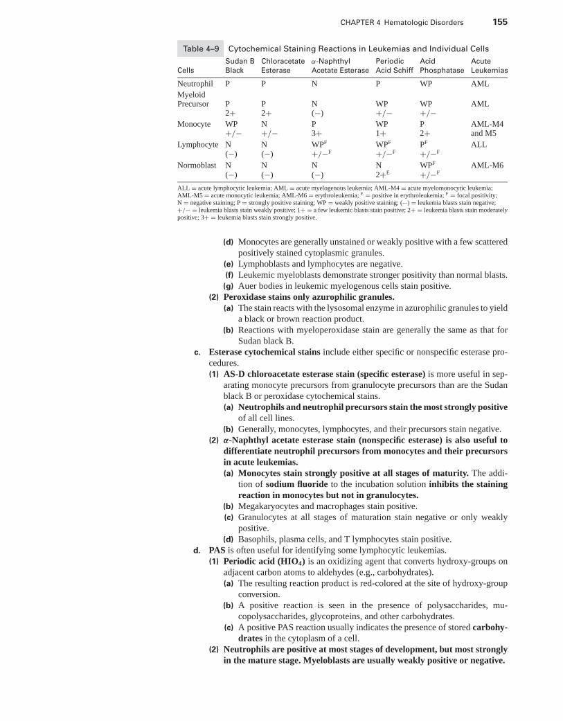
P1: OSO
LWBK192-04 LWBK192-Hubbard November 27, 2008 7:43
CHAPTER 4 Hematologic Disorders 155
Table 4–9 Cytochemical Staining Reactions in Leukemias and Individual Cells
Sudan B Chloracetate α-Naphthyl Periodic Acid Acute
Cells Black Esterase Acetate Esterase Acid Schiff Phosphatase Leukemias
Neutrophil P P N P WP AML
MyeloidPrecursor P P N WP WP AML
2+ 2+ (−) +/− +/−Monocyte WP N P WP P AML-M4
+/− +/− 3+ 1+ 2+ and M5
Lymphocyte N N WPF WPF PF ALL(−) (−) +/−F +/−F +/−F
Normoblast N N N N WPF AML-M6(−) (−) (−) 2+E +/−F
ALL = acute lymphocytic leukemia; AML = acute myelogenous leukemia; AML-M4 = acute myelomonocytic leukemia;AML-M5 = acute monocytic leukemia; AML-M6 = erythroleukemia; E = positive in erythroleukemia; F = focal positivity;N = negative staining; P = strongly positive staining; WP = weakly positive staining; (−) = leukemia blasts stain negative;+/− = leukemia blasts stain weakly positive; 1+ = a few leukemic blasts stain positive; 2+ = leukemia blasts stain moderatelypositive; 3+ = leukemia blasts stain strongly positive.
(d) Monocytes are generally unstained or weakly positive with a few scatteredpositively stained cytoplasmic granules.
(e) Lymphoblasts and lymphocytes are negative.(f) Leukemic myeloblasts demonstrate stronger positivity than normal blasts.
(g) Auer bodies in leukemic myelogenous cells stain positive.(2) Peroxidase stains only azurophilic granules.
(a) The stain reacts with the lysosomal enzyme in azurophilic granules to yielda black or brown reaction product.
(b) Reactions with myeloperoxidase stain are generally the same as that forSudan black B.
c. Esterase cytochemical stains include either specific or nonspecific esterase pro-cedures.(1) AS-D chloroacetate esterase stain (specific esterase) is more useful in sep-
arating monocyte precursors from granulocyte precursors than are the Sudanblack B or peroxidase cytochemical stains.(a) Neutrophils and neutrophil precursors stain the most strongly positive
of all cell lines.(b) Generally, monocytes, lymphocytes, and their precursors stain negative.
(2) α-Naphthyl acetate esterase stain (nonspecific esterase) is also useful todifferentiate neutrophil precursors from monocytes and their precursorsin acute leukemias.(a) Monocytes stain strongly positive at all stages of maturity. The addi-
tion of sodium fluoride to the incubation solution inhibits the stainingreaction in monocytes but not in granulocytes.
(b) Megakaryocytes and macrophages stain positive.(c) Granulocytes at all stages of maturation stain negative or only weakly
positive.(d) Basophils, plasma cells, and T lymphocytes stain positive.
d. PAS is often useful for identifying some lymphocytic leukemias.(1) Periodic acid (HIO4) is an oxidizing agent that converts hydroxy-groups on
adjacent carbon atoms to aldehydes (e.g., carbohydrates).(a) The resulting reaction product is red-colored at the site of hydroxy-group
conversion.(b) A positive reaction is seen in the presence of polysaccharides, mu-
copolysaccharides, glycoproteins, and other carbohydrates.(c) A positive PAS reaction usually indicates the presence of stored carbohy-
drates in the cytoplasm of a cell.(2) Neutrophils are positive at most stages of development, but most strongly
in the mature stage. Myeloblasts are usually weakly positive or negative.
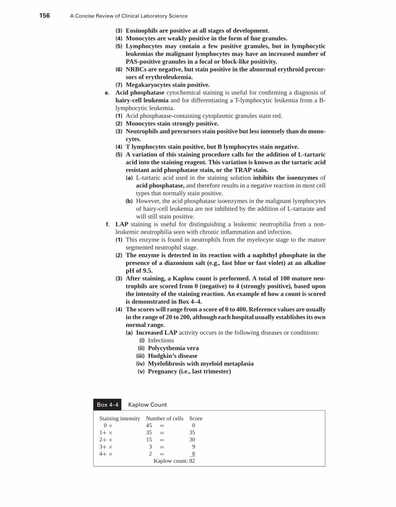
P1: OSO
LWBK192-04 LWBK192-Hubbard November 27, 2008 7:43
156 A Concise Review of Clinical Laboratory Science
(3) Eosinophils are positive at all stages of development.(4) Monocytes are weakly positive in the form of fine granules.(5) Lymphocytes may contain a few positive granules, but in lymphocytic
leukemias the malignant lymphocytes may have an increased number ofPAS-positive granules in a focal or block-like positivity.
(6) NRBCs are negative, but stain positive in the abnormal erythroid precur-sors of erythroleukemia.
(7) Megakaryocytes stain positive.e. Acid phosphatase cytochemical staining is useful for confirming a diagnosis of
hairy-cell leukemia and for differentiating a T-lymphocytic leukemia from a B-lymphocytic leukemia.(1) Acid phosphatase-containing cytoplasmic granules stain red.(2) Monocytes stain strongly positive.(3) Neutrophils and precursors stain positive but less intensely than do mono-
cytes.(4) T lymphocytes stain positive, but B lymphocytes stain negative.(5) A variation of this staining procedure calls for the addition of L-tartaric
acid into the staining reagent. This variation is known as the tartaric acidresistant acid phosphatase stain, or the TRAP stain.(a) L-tartaric acid used in the staining solution inhibits the isoenzymes of
acid phosphatase, and therefore results in a negative reaction in most celltypes that normally stain positive.
(b) However, the acid phosphatase isoenzymes in the malignant lymphocytesof hairy-cell leukemia are not inhibited by the addition of L-tartarate andwill still stain positive.
f. LAP staining is useful for distinguishing a leukemic neutrophilia from a non-leukemic neutrophilia seen with chronic inflammation and infection.(1) This enzyme is found in neutrophils from the myelocyte stage to the mature
segmented neutrophil stage.(2) The enzyme is detected in its reaction with a naphthyl phosphate in the
presence of a diazonium salt (e.g., fast blue or fast violet) at an alkalinepH of 9.5.
(3) After staining, a Kaplow count is performed. A total of 100 mature neu-trophils are scored from 0 (negative) to 4 (strongly positive), based uponthe intensity of the staining reaction. An example of how a count is scoredis demonstrated in Box 4–4.
(4) The scores will range from a score of 0 to 400. Reference values are usuallyin the range of 20 to 200, although each hospital usually establishes its ownnormal range.(a) Increased LAP activity occurs in the following diseases or conditions:
(i) Infections(ii) Polycythemia vera(iii) Hodgkin’s disease(iv) Myelofibrosis with myeloid metaplasia(v) Pregnancy (i.e., last trimester)
Box 4–4 Kaplow Count
Staining intensity Number of cells Score0 × 45 = 0
1+ × 35 = 35
2+ × 15 = 30
3+ × 3 = 9
4+ × 2 = 8
Kaplow count: 82
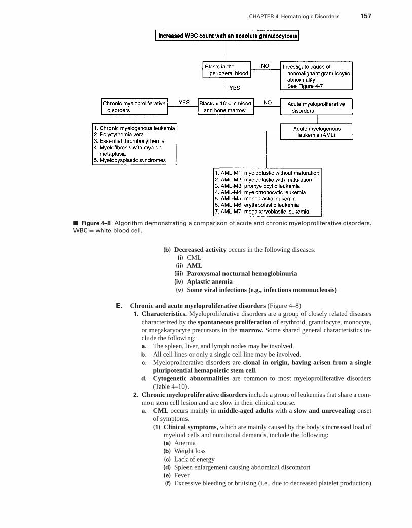
P1: OSO
LWBK192-04 LWBK192-Hubbard November 27, 2008 7:43
CHAPTER 4 Hematologic Disorders 157
■ Figure 4–8 Algorithm demonstrating a comparison of acute and chronic myeloproliferative disorders.
WBC = white blood cell.
(b) Decreased activity occurs in the following diseases:(i) CML
(ii) AML(iii) Paroxysmal nocturnal hemoglobinuria(iv) Aplastic anemia(v) Some viral infections (e.g., infections mononucleosis)
E. Chronic and acute myeloproliferative disorders (Figure 4–8)1. Characteristics. Myeloproliferative disorders are a group of closely related diseases
characterized by the spontaneous proliferation of erythroid, granulocyte, monocyte,or megakaryocyte precursors in the marrow. Some shared general characteristics in-clude the following:a. The spleen, liver, and lymph nodes may be involved.b. All cell lines or only a single cell line may be involved.c. Myeloproliferative disorders are clonal in origin, having arisen from a single
pluripotential hemapoietic stem cell.d. Cytogenetic abnormalities are common to most myeloproliferative disorders
(Table 4–10).2. Chronic myeloproliferative disorders include a group of leukemias that share a com-
mon stem cell lesion and are slow in their clinical course.a. CML occurs mainly in middle-aged adults with a slow and unrevealing onset
of symptoms.(1) Clinical symptoms, which are mainly caused by the body’s increased load of
myeloid cells and nutritional demands, include the following:(a) Anemia(b) Weight loss(c) Lack of energy(d) Spleen enlargement causing abdominal discomfort(e) Fever(f) Excessive bleeding or bruising (i.e., due to decreased platelet production)
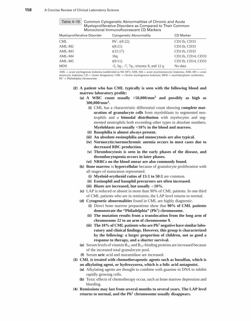
P1: OSO
LWBK192-04 LWBK192-Hubbard November 27, 2008 7:43
158 A Concise Review of Clinical Laboratory Science
Table 4–10 Common Cytogenetic Abnormalities of Chronic and AcuteMyeloproliferative Disorders as Compared to Their CommonMonoclonal Immunofluorescent CD Markers
Myeloproliferative Disorder Cytogenetic Abnormality CD Marker
CML Ph1, t(9:22) CD11b, CD33
AML-M2 t(8:21) CD11b, CD33
AML-M3 t(15:17) CD11b, CD33
AML-M4 16q CD11b, CD14, CD33
AML-M5 t(9:11) CD11b, CD14, CD33
MDS -5, 5q-, -7, 7q-, trisomy 8, and 12 q No data
AML = acute myelogenous leukemia (subdivided as M1-M7); AML-M4 = acute myelomonocytic leukemia; AML-M5 = acutemonocytic leukemia; CD = cluster designation; CML = chronic myelogenous leukemia; MDS = myelodysplastic syndromes;Ph1 = Philadelphia chromosome.
(2) A patient who has CML typically is seen with the following blood andmarrow laboratory profile:(a) A WBC count usually >50,000/mm3 and possibly as high as
300,000/mm3.(i) CML has a characteristic differential count showing complete mat-
uration of granulocyte cells from myeloblasts to segmented neu-trophils and a bimodal distribution with myelocytes and seg-mented neutrophils both exceeding other types in absolute numbers.Myeloblasts are usually <10% in the blood and marrow.
(ii) Basophilia is almost always present.(iii) An absolute eosinophilia and monocytosis are also typical.(iv) Normocytic/normochromic anemia occurs in most cases due to
decreased RBC production.(v) Thrombocytosis is seen in the early phases of the disease, and
thrombocytopenia occurs in later phases.(vi) NRBCs on the blood smear are also commonly found.
(b) Bone marrow is hypercellular because of granulocyte proliferation withall stages of maturation represented.
(i) Myeloid-erythroid ratios of 15:1 to 50:1 are common.(ii) Eosinophil and basophil precursors are often increased.(iii) Blasts are increased, but usually <10%.
(c) LAP is reduced or absent in more than 90% of CML patients. In one thirdof CML patients who are in remission, the LAP level returns to normal.
(d) Cytogenetic abnormalities found in CML are highly diagnostic.(i) Direct bone marrow preparations show that 90% of CML patients
demonstrate the “Philadelphia” (Ph1) chromosome.(ii) The mutation results from a translocation from the long arm of
chromosome 22 to an arm of chromosome 9.(iii) The 10% of CML patients who are Ph1 negative have similar labo-
ratory and clinical findings. However, this group is characterizedby the following: a larger proportion of children, not as good aresponse to therapy, and a shorter survival.
(e) Serum levels of vitamin B12 and B12-binding proteins are increased becauseof the increased total granulocyte pool.
(f) Serum uric acid and muramidase are increased.(3) CML is treated with chemotherapeutic agents such as busulfan, which is
an alkylating agent, or hydroxyurea, which is a folic acid antagonist.(a) Alkylating agents are thought to combine with guanine in DNA to inhibit
rapidly growing cells.(b) Toxic effects of chemotherapy occur, such as bone marrow depression and
bleeding.(4) Remissions may last from several months to several years. The LAP level
returns to normal, and the Ph1 chromosome usually disappears.
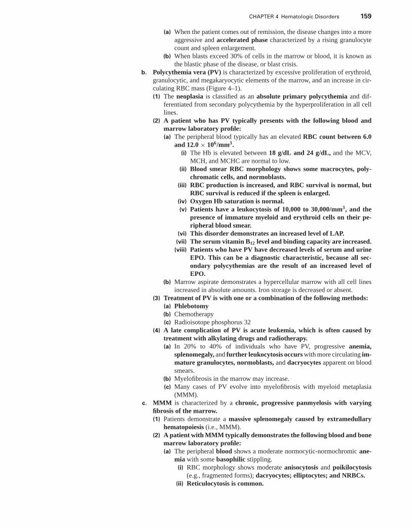
P1: OSO
LWBK192-04 LWBK192-Hubbard November 27, 2008 7:43
CHAPTER 4 Hematologic Disorders 159
(a) When the patient comes out of remission, the disease changes into a moreaggressive and accelerated phase characterized by a rising granulocytecount and spleen enlargement.
(b) When blasts exceed 30% of cells in the marrow or blood, it is known asthe blastic phase of the disease, or blast crisis.
b. Polycythemia vera (PV) is characterized by excessive proliferation of erythroid,granulocytic, and megakaryocytic elements of the marrow, and an increase in cir-culating RBC mass (Figure 4–1).(1) The neoplasia is classified as an absolute primary polycythemia and dif-
ferentiated from secondary polycythemia by the hyperproliferation in all celllines.
(2) A patient who has PV typically presents with the following blood andmarrow laboratory profile:(a) The peripheral blood typically has an elevated RBC count between 6.0
and 12.0 × 10⁶/mm3.(i) The Hb is elevated between 18 g/dL and 24 g/dL, and the MCV,
MCH, and MCHC are normal to low.(ii) Blood smear RBC morphology shows some macrocytes, poly-
chromatic cells, and normoblasts.(iii) RBC production is increased, and RBC survival is normal, but
RBC survival is reduced if the spleen is enlarged.(iv) Oxygen Hb saturation is normal.(v) Patients have a leukocytosis of 10,000 to 30,000/mm3, and the
presence of immature myeloid and erythroid cells on their pe-ripheral blood smear.
(vi) This disorder demonstrates an increased level of LAP.(vii) The serum vitamin B12 level and binding capacity are increased.(viii) Patients who have PV have decreased levels of serum and urine
EPO. This can be a diagnostic characteristic, because all sec-ondary polycythemias are the result of an increased level ofEPO.
(b) Marrow aspirate demonstrates a hypercellular marrow with all cell linesincreased in absolute amounts. Iron storage is decreased or absent.
(3) Treatment of PV is with one or a combination of the following methods:(a) Phlebotomy(b) Chemotherapy(c) Radioisotope phosphorus 32
(4) A late complication of PV is acute leukemia, which is often caused bytreatment with alkylating drugs and radiotherapy.(a) In 20% to 40% of individuals who have PV, progressive anemia,
splenomegaly, and further leukocytosis occurs with more circulating im-mature granulocytes, normoblasts, and dacryocytes apparent on bloodsmears.
(b) Myelofibrosis in the marrow may increase.(c) Many cases of PV evolve into myelofibrosis with myeloid metaplasia
(MMM).c. MMM is characterized by a chronic, progressive panmyelosis with varying
fibrosis of the marrow.(1) Patients demonstrate a massive splenomegaly caused by extramedullary
hematopoiesis (i.e., MMM).(2) A patient with MMM typically demonstrates the following blood and bone
marrow laboratory profile:(a) The peripheral blood shows a moderate normocytic-normochromic ane-
mia with some basophilic stippling.(i) RBC morphology shows moderate anisocytosis and poikilocytosis
(e.g., fragmented forms); dacryocytes; elliptocytes; and NRBCs.(ii) Reticulocytosis is common.
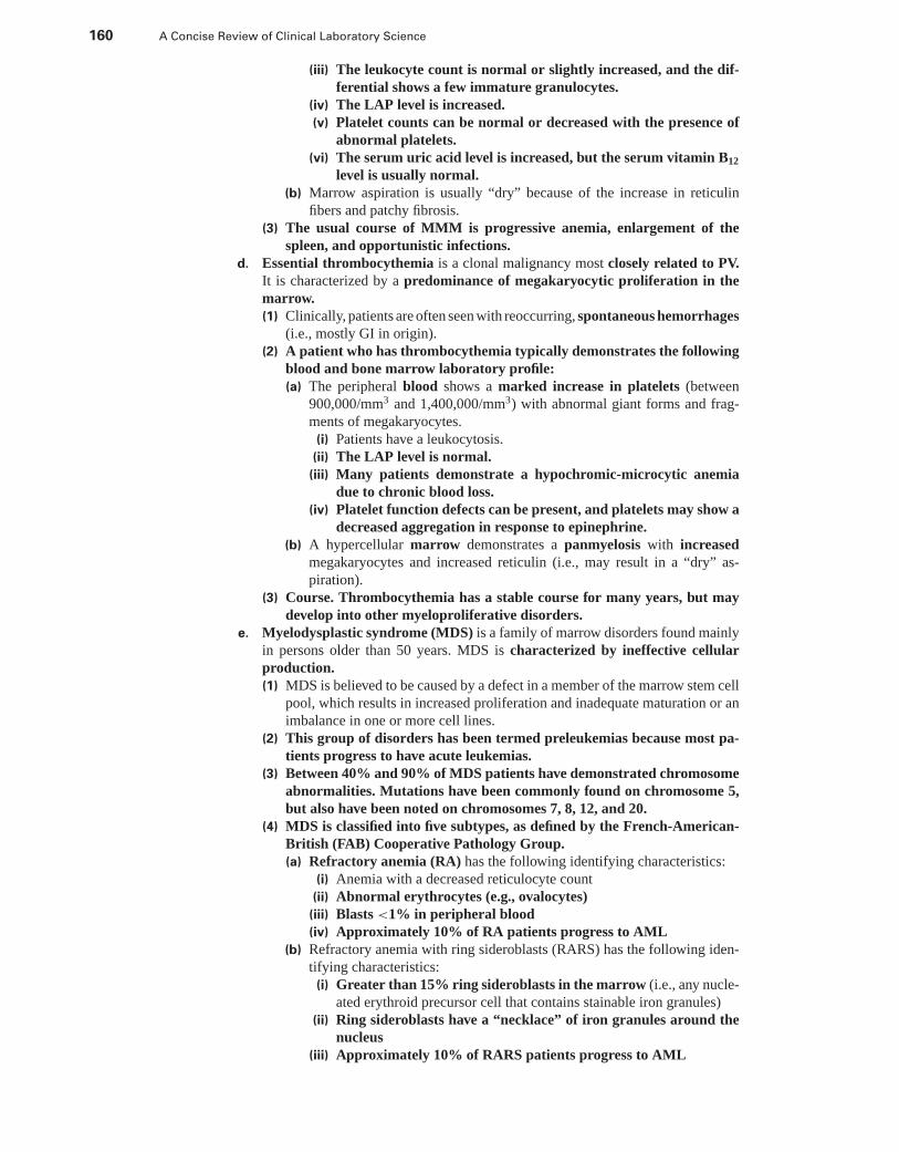
P1: OSO
LWBK192-04 LWBK192-Hubbard November 27, 2008 7:43
160 A Concise Review of Clinical Laboratory Science
(iii) The leukocyte count is normal or slightly increased, and the dif-ferential shows a few immature granulocytes.
(iv) The LAP level is increased.(v) Platelet counts can be normal or decreased with the presence of
abnormal platelets.(vi) The serum uric acid level is increased, but the serum vitamin B12
level is usually normal.(b) Marrow aspiration is usually “dry” because of the increase in reticulin
fibers and patchy fibrosis.(3) The usual course of MMM is progressive anemia, enlargement of the
spleen, and opportunistic infections.d. Essential thrombocythemia is a clonal malignancy most closely related to PV.
It is characterized by a predominance of megakaryocytic proliferation in themarrow.(1) Clinically, patients are often seen with reoccurring, spontaneous hemorrhages
(i.e., mostly GI in origin).(2) A patient who has thrombocythemia typically demonstrates the following
blood and bone marrow laboratory profile:(a) The peripheral blood shows a marked increase in platelets (between
900,000/mm3 and 1,400,000/mm3) with abnormal giant forms and frag-ments of megakaryocytes.(i) Patients have a leukocytosis.
(ii) The LAP level is normal.(iii) Many patients demonstrate a hypochromic-microcytic anemia
due to chronic blood loss.(iv) Platelet function defects can be present, and platelets may show a
decreased aggregation in response to epinephrine.(b) A hypercellular marrow demonstrates a panmyelosis with increased
megakaryocytes and increased reticulin (i.e., may result in a “dry” as-piration).
(3) Course. Thrombocythemia has a stable course for many years, but maydevelop into other myeloproliferative disorders.
e. Myelodysplastic syndrome (MDS) is a family of marrow disorders found mainlyin persons older than 50 years. MDS is characterized by ineffective cellularproduction.(1) MDS is believed to be caused by a defect in a member of the marrow stem cell
pool, which results in increased proliferation and inadequate maturation or animbalance in one or more cell lines.
(2) This group of disorders has been termed preleukemias because most pa-tients progress to have acute leukemias.
(3) Between 40% and 90% of MDS patients have demonstrated chromosomeabnormalities. Mutations have been commonly found on chromosome 5,but also have been noted on chromosomes 7, 8, 12, and 20.
(4) MDS is classified into five subtypes, as defined by the French-American-British (FAB) Cooperative Pathology Group.(a) Refractory anemia (RA) has the following identifying characteristics:
(i) Anemia with a decreased reticulocyte count(ii) Abnormal erythrocytes (e.g., ovalocytes)(iii) Blasts <1% in peripheral blood(iv) Approximately 10% of RA patients progress to AML
(b) Refractory anemia with ring sideroblasts (RARS) has the following iden-tifying characteristics:
(i) Greater than 15% ring sideroblasts in the marrow (i.e., any nucle-ated erythroid precursor cell that contains stainable iron granules)
(ii) Ring sideroblasts have a “necklace” of iron granules around thenucleus
(iii) Approximately 10% of RARS patients progress to AML
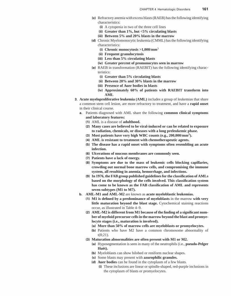
P1: OSO
LWBK192-04 LWBK192-Hubbard November 27, 2008 7:43
CHAPTER 4 Hematologic Disorders 161
(c) Refractory anemia with excess blasts (RAEB) has the following identifyingcharacteristics:
(i) A cytopenia in two of the three cell lines(ii) Greater than 1%, but <5% circulating blasts(iii) Between 5% and 20% blasts in the marrow
(d) Chronic Myelomonocytic leukemia (CMML) has the following identifyingcharacteristics:
(i) Chronic monocytosis >1,000/mm3
(ii) Frequent granulocytosis(iii) Less than 5% circulating blasts(iv) Greater percent of promonocytes seen in marrow
(e) RAEB in transformation (RAEBIT) has the following identifying charac-teristics:
(i) Greater than 5% circulating blasts(ii) Between 20% and 30% blasts in the marrow(iii) Presence of Auer bodies in blasts(iv) Approximately 60% of patients with RAEBIT transform into
AML3. Acute myeloproliferative leukemia (AML) includes a group of leukemias that share
a common stem cell lesion, are more refractory to treatment, and have a rapid onsetin their clinical course.a. Patients diagnosed with AML share the following common clinical symptoms
and laboratory features:(1) AML is a disease of adulthood.(2) Many cases are believed to be viral-induced or can be related to exposure
to radiation, chemicals, or diseases with a long preleukemic phase.(3) Most patients have very high WBC counts (e.g., 200,000/mm3).(4) AML is resistant to treatment with chemotherapeutic agents.(5) The disease has a rapid onset with symptoms often resembling an acute
infection.(6) Ulcerations of mucous membranes are commonly seen.(7) Patients have a lack of energy.(8) Symptoms are due to the mass of leukemic cells blocking capillaries,
crowding out normal bone marrow cells, and compromising the immunesystem, all resulting in anemia, hemorrhage, and infections.
(9) In 1976, the FAB group published guidelines for the classification of AMLsbased on the morphology of the cells involved. This classification systemhas come to be known as the FAB classification of AML and representsseven subtypes (M1 to M7).
b. AML-M1 and AML-M2 are known as acute myeloblastic leukemias.(1) M1 is defined by a predominance of myeloblasts in the marrow with very
little maturation beyond the blast stage. Cytochemical staining reactionsoccur, as illustrated in Table 4–9.
(2) AML-M2 is different from M1 because of the finding of a significant num-ber of myeloid precursor cells in the marrow beyond the blast and promye-locyte stages (i.e., maturation is involved).(a) More than 50% of marrow cells are myeloblasts or promyelocytes.(b) Patients who have M2 have a common chromosome abnormality of
t(8;21).(3) Maturation abnormalities are often present with M1 or M2.
(a) Hyposegmentation is seen in many of the neutrophils (i.e., pseudo-PelgerHuet).
(b) Myeloblasts can show bilobed or reniform nuclear shapes.(c) Some blasts may present with azurophilic granules.(d) Auer bodies can be found in the cytoplasm of a few blasts.
(i) These inclusions are linear or spindle-shaped, red-purple inclusions inthe cytoplasm of blasts or promyelocytes.
P1: OSO
LWBK192-04 LWBK192-Hubbard November 27, 2008 7:43
162 A Concise Review of Clinical Laboratory Science
(ii) They are thought to be giant lysosomes, and they stain positive forSudan black B, peroxidase, AS-D chloroacetate esterase, and acidphosphatase.
c. AML-M3 is known as Acute Promyelocytic Leukemia (APL). Cytochemicalstaining reactions are summarized in Table 4–9.(1) Promyelocytes instead of myeloblasts are the major abnormal cell found in the
marrow and blood.(2) These promyeloctes have an abundance of azurophilic granules.(3) Auer bodies are found in most patients who have M3.(4) Two variants of APL are found based on the staining intensity of promye-
locyte granularity.(a) Hypergranular promyelocytes are the most common variety of M3.
(i) Characteristically, promyelocytes may have bundles of cytoplasmicAuer bodies and intensely staining azurophilic granules.
(ii) The nucleus of these promyelocytes may vary in size and shape(i.e., kidney-shaped or bilobed).
(iii) Patients who have hypergranular M3 often have bad bleedingproblems. DIC is easily initiated by procoagulant material re-leased from granules of abnormal hypergranular promyelocytes.
(iv) The WBC count is not usually as greatly elevated as that seen withother AMLs.
(b) Hypogranular promyelocytes (M3V) may be easily confused withmyeloblastic leukemia (i.e., M1 or M2) because promyelocytes are faintlygranulated and may appear as myeloblasts.(i) In approximately 60% of patients who have M3V, a t(15;17) chro-
mosome translocation is demonstrated.(ii) Patients who have this variant usually demonstrate very high WBC
counts (up to 200,000/mm3).d. AML-M4, acute myelomonocytic leukemia (AMML), demonstrates a predom-
inance of both monocyte and granulocyte precursor cells in the marrow andperipheral blood.(1) This leukemia is defined as a malignancy in which more than 20% promono-
cytes and monocytes are found in marrow and peripheral blood with the pres-ence of more than 20% myeloblasts and abnormal granulocyte precursor cells.
(2) Chromosome abnormalities are seen as a 16q mutation.e. AML-M5 is a relative pure monocytic leukemia.
(1) This leukemia is defined as a malignancy in which more than 20% promono-cytes and monocytes, and <20% myeloblasts and granulocytic precursors arefound in the marrow or peripheral blood.
(2) Two subtypes of M5 are found.(a) Differentiated monoblastic leukemia (M5a) has monocytes and promono-
cytes as the majority in the marrow and blood. The blood has a higherpercentage of monocytes than the marrow, and the most common type ofcell is the promonocyte.
(b) Poorly differentiated (M5b) is characterized by a predominance of largeblasts in the bone marrow and blood.
(3) Chromosome abnormalities are seen in M5 as a t(9;11).(4) Promonocytes and leukemic monoblasts in AML-M5 demonstrate the fol-
lowing morphologic characteristics:(a) Delicate reticular chromatin(b) One to two nucleoli(c) Folded and indented nuclei(d) Moderate nucleus:cytoplasm (N:C) ratio (i.e., 1:1 to 2:1)(e) Phagocytosis of RBCs or cellular debris(f) Auer bodies(g) Abundance of basophilic-staining cytoplasm with pseudopodia and possi-
bly rare azurophilic granules
P1: OSO
LWBK192-04 LWBK192-Hubbard November 27, 2008 7:43
CHAPTER 4 Hematologic Disorders 163
(5) AML-M5 may have lymph node enlargement.(6) Cytochemical staining reactions for M4 and M5 can be found in Table 4–9.(7) Patients who have monocytic leukemias characteristically have very high
levels of muramidase in the serum or urine.f. AML-M6 or erythroleukemia was formerly known as Di Guglielmo syndrome.
(1) This type of AML results from an abnormal proliferation of both erythroidand granulocytic precursors with a predominance of erythroid precursorsin the marrow representing 50% or greater of the marrow precursor cellpopulation.
(2) Myeloid-erythroid ratios decrease to between 1:2 and 1:4.(3) Erythremic myelosis is a variant of M6 that has a more rapid course and
an arrest in the maturation of the erythrocytic precursors. Mainly, onlythe erythroblast stage of maturation is seen in the erythroid marrow.
(4) Often, M6 terminates in AML-M1.(5) Erythroid precursors are abnormal in appearance, predominate in the
marrow and blood, and demonstrate the following cellular morphologiccharacteristics:(a) Blasts irregular in outline, often with pseudopodia(b) Medium N:C ratio(c) Bizarre large nucleoli and nuclear shape(d) Numerous multinucleated giant forms(e) Cytoplasmic vacuolation
(6) Myeloblasts are increased in the chronic form of erythroleukemia, andAuer bodies may be present.
(7) Abnormal megakaryocytes are present with giant forms and nuclear frag-mentation. Atypical platelets may also be found in the blood.
(8) The cytochemistry results of M6 show a strong positivity to PAS. Erythroidprecursors are normally PAS negative.
(9) Patients with AML-M6 have a poor prognosis.g. AML-M7 or Acute Megakaryoblastic Leukemia (i.e., acute myelofibrosis) is
the newest FAB classification of acute nonlymphocytic leukemias.(1) This leukemia demonstrates a predominance of megakaryoblasts and
megakaryocytes in the marrow with an increase in reticulum fibrosis.(2) M7 is the rarest subtype.
h. Prognosis. As a group, AMLs generally have a terminal prognosis of approximately3 to 6 months, and complete remission is achieved in 40%–60% of patients.
4. Rare forms of leukemiaa. Acute stem cell leukemia is a leukemia in which the predominant hyperprolifer-
ating cells are blast forms that cannot be classified using cytochemical or immuno-logic techniques.
b. Chloroma is a rare form of AML in which green-pigmented tumors of myeloblastsform in the tissues surrounding the bones.
c. Myeloblastic sarcoma is a form of AML in which localized tumors of myeloblastsare found in tissues. This type differs from chloroma only in the absence of thegreen pigment found in the tumor.
d. Eosinophilic leukemia can occur, although rarely, as a variant of AML or CML.(1) Types. There are acute and chronic forms.(2) Laboratory profile. Immature eosinophils infiltrate the tissues in the body.
Other immature myeloid precursor cells are usually involved, and theirlevels are increased in the blood and marrow.
(3) Differential diagnosis. This disorder must be distinguished from hypere-osinophilic syndrome seen with parasitic infection.
(4) Diagnosis. The identification of immature forms in the blood and an in-crease of eosinophils in the marrow of >5% help diagnose eosinophilicleukemia.
e. Basophilic leukemia can be a variant of CML in the accelerated phase. This variantis associated with an increase in blood histamine levels.
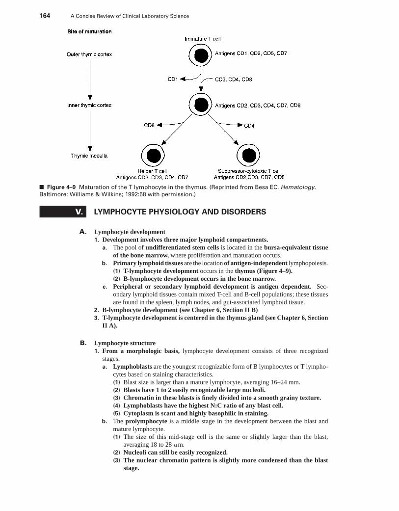
P1: OSO
LWBK192-04 LWBK192-Hubbard November 27, 2008 7:43
164 A Concise Review of Clinical Laboratory Science
■ Figure 4–9 Maturation of the T lymphocyte in the thymus. (Reprinted from Besa EC. Hematology.
Baltimore: Williams & Wilkins; 1992:58 with permission.)
V. LYMPHOCYTE PHYSIOLOGY AND DISORDERS
A. Lymphocyte development1. Development involves three major lymphoid compartments.
a. The pool of undifferentiated stem cells is located in the bursa-equivalent tissueof the bone marrow, where proliferation and maturation occurs.
b. Primary lymphoid tissues are the location of antigen-independent lymphopoiesis.(1) T-lymphocyte development occurs in the thymus (Figure 4–9).(2) B-lymphocyte development occurs in the bone marrow.
c. Peripheral or secondary lymphoid development is antigen dependent. Sec-ondary lymphoid tissues contain mixed T-cell and B-cell populations; these tissuesare found in the spleen, lymph nodes, and gut-associated lymphoid tissue.
2. B-lymphocyte development (see Chapter 6, Section II B)3. T-lymphocyte development is centered in the thymus gland (see Chapter 6, Section
II A).
B. Lymphocyte structure1. From a morphologic basis, lymphocyte development consists of three recognized
stages.a. Lymphoblasts are the youngest recognizable form of B lymphocytes or T lympho-
cytes based on staining characteristics.(1) Blast size is larger than a mature lymphocyte, averaging 16–24 mm.(2) Blasts have 1 to 2 easily recognizable large nucleoli.(3) Chromatin in these blasts is finely divided into a smooth grainy texture.(4) Lymphoblasts have the highest N:C ratio of any blast cell.(5) Cytoplasm is scant and highly basophilic in staining.
b. The prolymphocyte is a middle stage in the development between the blast andmature lymphocyte.(1) The size of this mid-stage cell is the same or slightly larger than the blast,
averaging 18 to 28 μm.(2) Nucleoli can still be easily recognized.(3) The nuclear chromatin pattern is slightly more condensed than the blast
stage.
P1: OSO
LWBK192-04 LWBK192-Hubbard November 27, 2008 7:43
CHAPTER 4 Hematologic Disorders 165
(4) Prolyphocytes have a high-to-moderate N:C ratio.(5) The cytoplasm is more abundant than the blast but still deeply basophilic.
c. Mature lymphocytes in the peripheral blood represent non-dividing B lymphocytesor T lymphocytes between the primary and secondary development.(1) Size. Small lymphocytes are just slightly larger than an RBC (i.e., 8–12 μm).(2) Organelles. These cells have a sharply defined, round nucleus that contains
heavy concentrations of dense chromatin appearing blocked or “smudgy.”The nucleus stains deep blue to purple and is sometimes indented at one side.The cytoplasm stains a pale blue. Normally, lymphocytes do not demon-strate specific cytoplasmic granules.
(3) In a small lymphocyte, the nucleus occupies most of the cell area, and theremay be just a thin perinuclear zone of cytoplasm.
(4) Large lymphocytes may be found in the peripheral blood, especially inchildren. These lymphocytes demonstrate the following morphologic char-acteristics:(a) 12 to 15 μm in diameter(b) Abundant cytoplasm(c) Nuclei less densely staining(d) Irregular borders and shape(e) May appear as a small monocyte(f) Few bluish purple cytoplasmic granules
(5) Lymphocytes average 25% to 45% of all circulating WBCs with a totalabsolute count range of 1,000 to 4,800/mm3.
(6) T and B lymphocytes cannot be distinguished from each other morpholog-ically.
d. Plasma cells originate from B lymphocytes. They are cells designed for thesynthesis of immunoglobulin.(1) Plasmablasts, or immunoblasts, have a large blast-like nucleus with nucleoli,
but have other plasma cell characteristics.(2) Plasma cells are identified by their eccentrically placed nucleus and the
following morphologic characteristics:(a) The nucleus is round with a small indentation on the cytoplasm side.(b) Nuclear chromatin is distributed more regularly in a pattern resembling the
spokes of a wheel.(c) The cytoplasm, except for one small area, stains deeply bluish green.(d) The unstained portion of cytoplasm corresponds to the indented portion of
the nucleus. A large golgi apparatus exists here because of the large amountof protein synthesis.
2. Lymphocytes demonstrate a heterogeneity of functional types.a. B lymphocytes function as precursors for plasma cells. They also synthesize and
release immunoglobulin, which is easily detectable on their surface membranes.(1) Approximately 10% to 15% of circulating blood lymphocytes are B cells.(2) Diversity
(a) Individual B cells are limited to the one type of antibody they can synthesizeand release.
(b) IgM or immunoglobulin D (IgD) are found on most circulating B cells.(c) B lymphocytes with surface IgM or IgG are found mainly in organized
secondary lymphoid tissue.(d) Immunoglobulin A (IgA)- and IgE-bearing B cells are predominately at sites
of external Ig secretion (e.g., the GI and respiratory tracts, saliva).(3) B-lymphocyte surface receptors include the following identifying markers:
(a) Fc receptors (i.e., CD7) that recognize the Fc portion of immunoglobulins(b) C3 receptors (i.e., CD21) for complement fixation(c) Specific receptor for the Epstein-Barr virus antigen(d) Mouse RBC receptor
b. T lymphocytes are responsible for reactions of cellular immunity and modulationof humoral immunity (Figure 4–9).
P1: OSO
LWBK192-04 LWBK192-Hubbard November 27, 2008 7:43
166 A Concise Review of Clinical Laboratory Science
(1) T cells interact with macrophages for the proper delivery of antigens to B lym-phocytes.
(2) T cell-mediated immunity is antigen directed; therefore, T cells must havesurface receptors to recognize antigens.(a) T lymphocytes display a specific receptor for sheep RBCs (i.e., CD2),
forming E-rosettes.(b) Fc receptors are present but not abundant.(c) T cell-specific antigens CD1 to CD8 are also abundant.
c. Null lymphocytes are lymphocytes that cannot be classified as either T or B typeson the basis of surface properties. These cells constitute approximately 10% of thelymphocyte population.(1) Null cells possibly represent undifferentiated stem cells, immature T or B cells,
or those lymphocytes that have lost their recognizable surface receptors.(2) L cells are lymphocytes that do not proliferate in response to antigens but
are capable of enhancing the responses of T lymphocytes in the presence ofmonocytes.(a) These lymphocytes bear surface IgG and Fc receptors.(b) L cells do not develop into antibody-producing cells.
(3) Large granular lymphocytes (LGLs) mediate antibody-dependent cytotox-icity.(a) LGLs include natural killer (NK) and killer (K) cells.(b) NK cells mediate cytotoxic reactions without prior sensitization in defensive
mechanisms against tumors and virally transformed cells.
C. Function and regulation of the immune response1. Monocyte-macrophages secrete IL-1, which activates the helper-inducer T lympho-
cytes. IL-1 also has pyogenic effects on the central nervous system (CNS) in raisingbody temperature.
2. Helper-inducer T lymphocyte lymphokines, which are secreted in an immune reaction(see Chapter 6, Section II A), consist of the following substances:a. Once activated by IL-1, the helper-inducer T lymphocyte secretes IL-2, which
stimulates other helper-inducer T lymphocytes to multiply.b. B-cell growth factor (BCGF) is released to stimulate B-cell proliferation.c. B-cell differentiation factor (BCDF) is released to halt replication of the im-
munospecific B cells and stimulates antibody production.d. γ-interferon is released to stimulate B-cell antibody production, activate killer T
lymphocytes, and localize macrophages at the site of infection.3. B lymphocytes (see Chapter 6, Section II B) produce and secrete an antigen-specific
antibody.
D. Lymphocyte pathophysiology1. The normal circulating concentration of lymphocytes is 1,500 to 4,000/mm3.
a. An absolute circulating lymphocyte count below 1,500/mm3 is considered clini-cally to be a lymphocytopenia.
b. The reduction in circulating lymphocytes affects mostly T cells, because they rep-resent the greatest percentage of circulating lymphocytes.
2. Nonmalignant disorders of lymphocytopenia often include a hypogammaglobuline-mia (Figure 4–10). A lymphocytopenia can be found in the following disorders orconditions:a. A blockage of the lymphatic thoracic duct results in a lymphocytopenia.b. Radiation overexposure is highly toxic to lymphopoiesis and results in a reduction
of lymphocytes.c. Acute stress results in a reduction of lymphocytes in the circulating pool.d. Therapy with corticoids shifts distribution of lymphocytes from the blood into
the extravascular spaces. Cell lysis and an inhibition of cell proliferation are minordrug-induced mechanisms of lymphopenia.
e. Chemotherapeutic alkylating drugs interfere with lymphocyte proliferation.f. Acquired immunodeficiency syndrome (AIDS) is a virally induced lymphocy-
topenia.
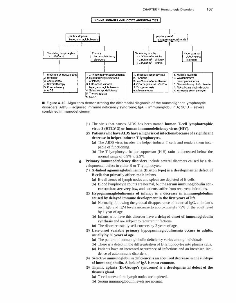
P1: OSO
LWBK192-04 LWBK192-Hubbard November 27, 2008 7:43
CHAPTER 4 Hematologic Disorders 167
■ Figure 4–10 Algorithm demonstrating the differential diagnosis of the nonmalignant lymphocytic
disorders. AIDS = acquired immune deficiency syndrome; IgA = immunoglobulin A; SCID = severe
combined immunodeficiency.
(1) The virus that causes AIDS has been named human T-cell lymphotrophicvirus-3 (HTLV-3) or human immunodeficiency virus (HIV).
(2) Patients who have AIDS have a high risk of infections because of a significantdecrease in helper-inducer T lymphocytes.(a) The AIDS virus invades the helper-inducer T cells and renders them inca-
pable of functioning.(b) The T lymphocyte helper-suppressor (H-S) ratio is decreased below the
normal range of 0.9% to 2.9%.g. Primary immunodeficiency disorders include several disorders caused by a de-
velopmental defect in either B or T lymphocytes.(1) X-linked agammaglobulinemia (Bruton type) is a developmental defect of
B cells that primarily affects male infants.(a) B-cell zones of lymph nodes and spleen are depleted of B cells.(b) Blood lymphocyte counts are normal, but the serum immunoglobulin con-
centrations are very low, and patients suffer from recurrent infections.(2) Hypogammaglobulinemia of infancy is a decrease in immunoglobulins
caused by delayed immune development in the first years of life.(a) Normally, following the gradual disappearance of maternal IgG, an infant’s
own IgG and IgM levels increase to approximately 75% of the adult levelby 1 year of age.
(b) Infants who have this disorder have a delayed onset of immunoglobulinsynthesis and are subject to recurrent infections.
(c) The disorder usually self-corrects by 2 years of age.(3) Late-onset variable primary hypogammaglobulinemia occurs in adults,
usually by 30 years of age.(a) The pattern of immunoglobulin deficiency varies among individuals.(b) There is a defect in the differentiation of B lymphocytes into plasma cells.(c) Patients have an increased occurrence of infections and an increased inci-
dence of autoimmune disorders.(4) Selective immunoglobulin deficiency is an acquired decrease in one subtype
of immunoglobulin. A lack of IgA is most common.(5) Thymic aplasia (Di-George’s syndrome) is a developmental defect of the
thymus gland.(a) T-cell zones of the lymph nodes are depleted.(b) Serum immunoglobulin levels are normal.
P1: OSO
LWBK192-04 LWBK192-Hubbard November 27, 2008 7:43
168 A Concise Review of Clinical Laboratory Science
(6) Severe combined immune deficiency is a defect in the common stem cellthat leads to a deficiency of both T and B lymphocytes.
3. Lymphocytosis is clinically defined as an absolute circulating increase in the lymphocytecount >4,000/mm3 in adults, 7,000/mm3 in children, and 9,000/mm3 in infants.Disorders that demonstrate lymphocytosis include the following characteristics:a. Infectious lymphocytosis occurs mainly in children.
(1) The disorder is believed to be caused by viruses (e.g., coxsackievirus A and B6,echoviruses, adenovirus).
(2) Symptoms include vomiting, fever, and abdominal discomfort.(3) Laboratory findings
(a) A leukocytosis of 20,000 to 50,000/mm3 is one of the earliest findings.(b) The blood differential shows 60% to 95% normal lymphocytes.(c) Atypical lymphocytes are not seen with this disorder.(d) Eosinophilia is common.(e) The lymphocytosis usually lasts only 3 to 5 weeks and, in most cases, is acute
and self-correcting. Rarely, some patients demonstrate a chronic course.(f) Serologically, patients have a negative test for infectious mononucleosis
(IM).(g) Bone marrow of these patients is normal.
b. Pertussis (i.e., whooping cough) is a childhood inflammatory reaction of the res-piratory system.(1) Cause. The etiologic agent is the bacterium Bordetella pertussis.(2) Laboratory findings. Patients demonstrate a significant lymphocytosis as
high as 30,000/mm3. Lymphocytosis is highest in the first 3 weeks of thedisease, then decreases after 4 weeks. Lymphocytosis is caused by therelease of lymphocytosis-promoting factor (LPF) from B. pertussis. LPFinduces an acute release of lymphocytes from lymph nodes and also in-hibits the migration of lymphocytes from the circulation into the lympha-tics.
(3) Morphologic characteristics. The lymphocytes in this disorder are smalland mature.
c. IM (see Chapter 6, Section XIII B)(1) Serious complications of IM may require hospitalization.
(a) AIHA is reported in 1% to 3% of patients, which is related to the develop-ment of an autoimmune anti-i.
(b) Mild thrombocytopenia (i.e., approximately 5,000 to 100,000/mm3) is re-ported in 50% of patients who have IM.
(c) Liver involvement can occur, which leads to mild jaundice and hepatitis.(d) Splenomegaly can also be a dangerous complication.
(2) Recognizing hematologic features of IM can result in a rapid diagnosis.(a) Leukocytosis between 12,000/mm3 and 25,000/mm3 persists for the first 3
weeks of infection with a differential showing 60% to 90% lymphocytes.(b) A neutrophilia occurs during the first week of infection with a left shift,
metamyelocytes, toxic granulation, and Dohle’s bodies.(c) Abundant atypical lymphocytes are found on the peripheral blood smear.
These are active or transformed lymphocytes involved in an immune reac-tion. Atypical lymphocytes are not exclusive to IM and are also found inthe following disorders:
(i) Cytomegalovirus infections(ii) Toxoplasmosis(iii) Infectious hepatitis(iv) Viral pneumonia and mumps
d. Cytomegalovirus infection (CMV) demonstrates clinical and laboratory symp-toms that are in some ways identical to those of IM.(1) Patients receiving massive blood transfusions are in a high-risk category for
contracting CMV.(2) Presentation. Patients have a leukocytosis involving a lymphocytosis in
which 20% or more of lymphocytes may be reactive.
P1: OSO
LWBK192-04 LWBK192-Hubbard November 27, 2008 7:43
CHAPTER 4 Hematologic Disorders 169
(3) Serologic tests. Patients are negative for heterophil antibodies and Epstein-Barr virus (EBV) antibodies.
(4) Diagnosis is made by the demonstration of CMV antibodies by complementfixation or hemagglutination techniques.
e. Toxoplasmosis also produces clinical and laboratory symptoms similar to those ofIM.(1) Presentation. Patients have an absolute lymphocytosis and atypical lympho-
cytes on the peripheral blood smear.(2) Serologic tests for EBV and heterophil antibodies are negative.(3) Diagnosis is made by the demonstration of toxoplasmosis antibodies by the
Sabin-Feldman dye test, fluorescent antibody, or hemagglutination.f. Miscellaneous causes of lymphocytosis
(1) Syphilis(2) Smallpox(3) Para-aminosalicylic acid hypersensitivity(4) Phenytoin (i.e., Dilantin) and Mesentoin hypersensitivity
4. Hypergammaglobulinemias are related disorders in which the levels of one or moreserum immunoglobulins are increased above normal levels. Hypergamma-globulinemiasmay or may not be accompanied by an absolute lymphocytosis.a. Multiple myeloma (MM) is a monoclonal gammopathy (i.e., an abnormality of
only one B-cell clone) in which only one type of gamma-globulin is increased.(1) MM involves a neoplastic proliferation of plasma cells primarily in the bone
marrow. Plasma cell proliferation may be either nodular or diffuse.(2) Clinical symptoms are found primarily in persons older than 40 years.
(a) Bone pain is the most common symptom, and MM patients are often firstseen with bone fractures.
(b) Tumor growth in the marrow increases bone destruction, and the serumcalcium level is high.
(c) Patients have a high susceptibility to infection.(d) Renal insufficiency is also a common symptom with MM.
(3) The MM laboratory profile includes the following results:(a) Normocytic/normochromic anemia(b) Normoblasts on the peripheral blood smear(c) Rouleaux formation and increased erythrocyte sedimentation rate (ESR)
(i.e., due to increase in serum globulins)(d) Possible shift to the left with metamyelocytes(e) Increased serum calcium level(f) Circulating plasma cells
(g) An increase in one of the serum gamma-globulins as demonstrated by proteinelectrophoresis
(4) Bone marrow examination shows the presence of plasma cells varying from1% to 90%.
(5) Serum immunoglobulins are increased in a variety of electrophoresis pat-terns among different patients. Serum protein electrophoresis usually showsa homogeneous band in the gamma or beta region of the electrophoretictracing known as an “M-spot.”(a) Most patients with MM are hypergammaglobulin producers, but in 25% of
patients, only the light chains of the globulin are produced (i.e., Bence Jonesproteins) by the abnormal plasma cells. A serum hypogammaglobulinemiais found in these patients, because light chains are cleared by the kidney intothe urine.
(b) Half of reported MM patients show an increase in IgG only.(c) A monoclonal increase in IgA is found in approximately 20% of MM pa-
tients.(d) IgD is increased in approximately 1% of patients.(e) IgE is rarely found to demonstrate a monoclonal increase.(f) Proteinuria with Bence Jones proteins can also be demonstrated in more
than 50% of MM patients.
P1: OSO
LWBK192-04 LWBK192-Hubbard November 27, 2008 7:43
170 A Concise Review of Clinical Laboratory Science
(6) Course. MM usually has a chronic course with the median survival approx-imately 3 years.(a) In 5% of MM patients, AMML develops as a secondary manifestation of
the malignancy.(b) In some patients, plasmacytosis may be found in the blood and bone marrow,
and the disease is then classified as plasma cell leukemia.b. Waldenstrom’s macroglobulinemia (WM) is a hypergammaglobulinemia variant
of chronic lymphocytic leukemia, in which there is a greater degree of maturationof the B lymphocytes into plasma cells.(1) WM is characterized by a generalized proliferation of B cells and plasma
cells and an increase of monoclonal IgM in the serum that amounts to atleast 15% of the total serum protein.
(2) Clinical symptoms of WM are primarily found in individuals older than40 years. Symptoms are caused by the cellular proliferation and increasedblood viscosity caused by the increased IgM. Patients who have WM arecommonly seen with the following symptoms:(a) Neurologic abnormalities(b) Renal insufficiency(c) Heart failure(d) Clotting abnormalities (e.g., DIC)
(3) The laboratory profile includes the following results:(a) IgM in excess of 1.0 g/dL(b) Normocytic-normochromic anemia (i.e., occasionally hemolytic with a pos-
itive direct Coombs’ test)(c) Thrombocytopenia caused by IgM platelet clumping or pancytopenia
caused by marrow infiltration(d) Lymphocytosis(e) Marked rouleaux and increased ESR(f) Bence Jones proteinuria in 10% of patients
(4) The bone marrow often cannot be aspirated easily. A biopsy demonstratesan increase in small normal lymphocytes and plasma cells.
c. Heavy-chain diseases are disorders related to the production and excretion of theimmunoglobulin heavy chains without the light chains.(1) Gamma heavy-chain disease (γ-HCD) resembles a malignant lymphoma
rather than a myeloma.(a) The patient has a blood picture of anemia, leukopenia, thrombocytopenia,
atypical lymphocytes, and plasma cells.(b) A broad serum protein “spike” is found in the beta-gamma region of the
electrophoretic pattern, accompanied by serum hypogammaglobulinemia.(2) Alpha heavy-chain disease (α-HCD) is more common than γ-HCD and oc-
curs in a younger age group.(a) Common symptoms include intestinal involvement with mal- absorption,
diarrhea, and a massive lymphoplasmacytic infiltration in the intestinal mu-cosa.
(b) The bone marrow and lymph nodes are not usually involved.(c) Protein electrophoresis is normal, but small amounts of α chain may be
detected in serum and urine on immunoelectrophoresis.(3) Mu heavy-chain disease (μ-HCD) can be diagnosed only by a serum immu-
noelectrophoresis that demonstrates an increase in μ-chains.5. Lymphoproliferative diseases
a. These disorders represent a group of clonal disorders originating from cells of thelymphoreticular system (Table 4–11).(1) When neoplastic cells involve mainly the bone marrow and blood, the disorder
is known as a leukemia.(2) When the disease is limited mainly to lymph nodes or organs, the disease is
known as a lymphoma.(3) Occasionally, a lymphoma develops into leukemia.
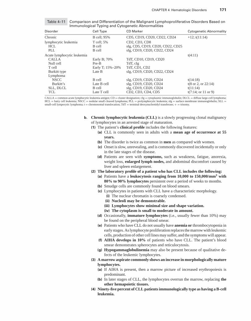
P1: OSO
LWBK192-04 LWBK192-Hubbard November 27, 2008 7:43
CHAPTER 4 Hematologic Disorders 171
Table 4–11 Comparison and Differentiation of the Malignant Lymphoproliferative Disorders Based onImmunological Typing and Cytogenetic Abnormalities
Disorder Cell Type CD Marker Cytogenetic Abnormality
Chronic B cell; 95% CD5, CD19, CD20, CD22, CD24 +12; t(11:14)
lymphocytic leukemia T cell; 5% CD2, CD3, CD8HCL B cell sIg, CD5, CD19, CD20, CD22, CD25PLL B cell sIg, CD19, CD20, CD22, CD24
Acute lymphocytic leukemia t(4:11)CALLA Early B; 70% TdT, CD10, CD19, CD20Null cell Pre-B TdT, cIgT cell Early T; 15%–20% TdT, CD1, CD2Burkitt type Late B sIg, CD19, CD20, CD22, CD24Lymphoma
NSCC B cell sIg, CD19, CD20, CD24 t(14:18)Burkitt’s Late B cell sIg, CD19, CD20, CD24 t(8 or 2, or 22:14)
SLL, DLCL B cell sIg, CD19, CD20, CD24 t(11:14)TCL Late T cell CD2, CD3, CD4, CD5 t(7:14; or 11 or 9)
CALLA = common acute lymphocytic leukemia antigen; CD = cluster designation; cIg = cytoplasmic immunoglobulin; DLCL = diffuse large-cell lymphoma;HCL = hairy cell leukemia; NSCC = nodular small cleaved lymphoma; PLL = prolymphocytic leukemia; sIg = surface membrane immunoglobulin; SLL =small-cell lympocytic lymphoma; t = chromosomal translocation; TdT = terminal deoxynucleotidyl transferase; + = trisomy.
b. Chronic lymphocytic leukemia (CLL) is a slowly progressing clonal malignancyof lymphocytes in an arrested stage of maturation.(1) The patient’s clinical profile includes the following features:
(a) CLL is commonly seen in adults with a mean age of occurrence at 55years.
(b) The disorder is twice as common in men as compared with women.(c) Onset is slow, unrevealing, and is commonly discovered incidentally or only
in the late stages of the disease.(d) Patients are seen with symptoms, such as weakness, fatigue, anorexia,
weight loss, enlarged lymph nodes, and abdominal discomfort caused byliver and spleen enlargement.
(2) The laboratory profile of a patient who has CLL includes the following:(a) Patients have a leukocytosis ranging from 10,000 to 150,000/mm3 with
80% to 90% lymphocytes persistent over a period of weeks to months.(b) Smudge cells are commonly found on blood smears.(c) Lymphocytes in patients with CLL have a characteristic morphology.
(i) The nuclear chromatin is coarsely condensed.(ii) Nucleoli may be demonstrable.(iii) Lymphocytes show minimal size and shape variation.(iv) The cytoplasm is small to moderate in amount.
(d) Occasionally, immature lymphocytes (i.e., usually fewer than 10%) maybe found on the peripheral blood smear.
(e) Patients who have CLL do not usually have anemia or thrombocytopenia inearly stages. As lymphocyte proliferation replaces the marrow with leukemiccells, production of other cell lines may suffer, and the symptoms will appear.
(f) AIHA develops in 10% of patients who have CLL. The patient’s bloodsmear demonstrates spherocytes and reticulocytosis.
(g) Hypogammaglobulinemia may also be present because of qualitative de-fects of the leukemic lymphocytes.
(3) A marrow aspirate commonly shows an increase in morphologically maturelymphocytes.(a) If AIHA is present, then a marrow picture of increased erythropoiesis is
predominant.(b) In later stages of CLL, the lymphocytes overrun the marrow, replacing the
other hemapoietic tissues.(4) Ninety-five percent of CLL patients immunologically type as having a B-cell
leukemia.
P1: OSO
LWBK192-04 LWBK192-Hubbard November 27, 2008 7:43
172 A Concise Review of Clinical Laboratory Science
(a) Immunologic cell-marker assays for lymphocytic membrane receptors,surface antigens, and enzymes are useful in differentiating between lympho-cytic leukemias. Cluster designation markers for B lymphocytes includethe following:(i) CD5: positive in B-lymphocyte CLLs and some lymphomas
(ii) CD22: positive for late B cells and hairy-cell leukemia(iii) CD24: positive in all stages of B-lymphocyte development (i.e.,
Pan-B)(b) Surface IgM can be commonly detected but is weak.(c) In most cases of B-cell leukemia, the ability of the lymphocytes to form spon-
taneous rosettes with mouse RBCs can be demonstrated (i.e., M rosettes).(5) Five percent of CLL patients have a T-cell leukemia.
(a) Leukemic T lymphocytes may have a cleft-or clover-shaped nucleus (i.e.,Sezary cells).
(b) Skin involvement and lymphocyte infiltration occur in 60% of T-cell CLLpatients.
(c) Immunologic markers for T-cell leukemia include the following:(i) CD2: E-rosette marker (i.e., sheep RBCs)
(ii) CD3: positive for T-lymphocyte CLL, T-cell prolymphocyticleukemia, and infectious mononucleosis
(iii) CD8: positive for T-lymphocyte CLL(6) Staging systems have been devised to help categorize patients into prognos-
tic groups.(a) Criteria for staging include lymphadenopathy, anemia, hepatospleno-
megaly, and thrombocytopenia.(b) The absence of these symptoms indicates a good prognosis with a long
survival (i.e., 20 years).(c) With these symptoms, the patient’s survival can be up to 5 or 6 years with
treatment.(7) Treatment of CLL includes one or a combination of the following agents:
(a) Chemotherapy with alkylating drugs(b) Glucocorticoid administration(c) Radiotherapy
(8) Treatment can relieve symptoms and improve life expectancy and bloodcounts. However, hypogammaglobulinemia is often not corrected with treat-ment, and patients can succumb to infection.
c. Several clinical variations of CLL can be found.(1) Hairy-cell leukemia (leukemic reticuloendotheliosis) is a rare form of CLL
found four times more in men than women. The mean age of occurrence is50 years.(a) The onset of the disease is slow and is characterized by proliferation of
abnormal lymphocytes in the secondary lymphoid organs.(b) Splenomegaly is a common physical finding.(c) The laboratory profile includes the following:
(i) Pancytopenia, or a depression of two cell lines, is common.(ii) The peripheral blood smear may have variable numbers of “hairy-
cell” lymphocytes. These lymphocytes are medium-sized cells witha round, oval, notched, or dumbbell-shaped nucleus. The cytoplasmof these lymphocytes is moderate in amount with numerous hair-like projections and frayed borders.
(iii) Hairy-cell lymphocytes contain an acid phosphatase resistant toinhibition by L-tartarate, resulting in a positive TRAP stain.
(iv) Lymphocytes in hairy-cell leukemia are usually B cells that demon-strate strong surface membrane immunoglobulin (sIg) and a pos-itive reaction for common B-cell markers, such as CD19; CD20;CD22; CD24; and CD25 (i.e., IL2 receptor), which is unique topatients who have hairy-cell leukemia.
(d) The median survival of patients is 5 to 6 years.
P1: OSO
LWBK192-04 LWBK192-Hubbard November 27, 2008 7:43
CHAPTER 4 Hematologic Disorders 173
(2) Lymphosarcoma cell leukemia is a lymphocytic lymphoma that has trans-formed into a leukemic phase with the invasion of the marrow and blood ofleukemic lymphocytes.(a) The clinical course is more aggressive than in common B-cell CLL.(b) Morphologic differences. Leukemic lymphocytes are B cells, but are mor-
phologically different than those cells seen in regular B-cell CLL.(i) Nuclear chromatin is smoother with an oval or notched nucleus.
(ii) Distinct nucleoli are easily seen in malignant lymphocytes.(iii) A lower N:C ratio is found in these lymphocytes when compared
with the N:C ratio in regular B-cell CLL.(iv) Lymphocytes demonstrate a strong expression of sIg.
(3) Prolymphocytic leukemia (PLL) is a variation of CLL in which there isa high number of morphologically immature larger lymphocytes, whichappear as prolymphocytes.(a) This variation is typically seen in older men (i.e., older than 50 years).(b) Is characterized by a lymphocytosis up to 350,000/mm3.(c) Lymphocytes are the B-cell type.(d) Prolymphocytes can be distinguished by their smooth nuclear chromatin
and large nucleoli.(e) Patients commonly have a massive splenomegaly, but their lymph nodes
are not enlarged.(f) The prognosis is usually subacute and more resistant to treatment than is
common CLL. Patients have a mean survival of 1 year.(g) PLL prolymphocytes commonly type positive with the membrane markers
CD19, CD20, CD22, CD24, and strong expression of sIg.d. Acute lymphocytic leukemia (ALL) is a rapidly progressing clonal malignancy of
early immature lymphocytes in an arrested stage of maturation.(1) The clinical profile of ALL includes the following features:
(a) ALL is mainly a disease of childhood with a peak incidence at 4 years.The second peak of incidence (i.e., bimodal distribution) is in young adultsbetween 20 and 40 years.
(b) Onset of leukemia is sudden with symptoms of anemia, bleeding, fever,and fatigue.
(c) The spleen, liver, and lymph nodes are commonly enlarged.(2) The laboratory profile of ALL includes the following results:
(a) A normocytic-normochromic anemia is common.(b) Normoblasts are found on the peripheral blood smear.(c) The leukocyte count shows three patterns. The WBC count is occasionally
very high (i.e., >100,000/mm3); often is slightly elevated; but most often isnormal or decreased.
(d) The predominant cell is the lymphoblast or immature lymphocyte.(e) The serum level of uric acid is often elevated.(f) A small percent of ALL patients are positive for the Ph1 chromosome.
(g) Immunodeficiency or hypogammaglobulinemia is not as common as inCLL.
(3) A marrow aspirate commonly shows diffuse infiltration of lymphoblasts.(4) Acute lymphocytic leukemias are classified by the FAB group as L1, L2,
and L3 (Table 4–12).(a) Approximately 70% of children with ALL are Type L1, and 27% are
Type L2. The remaining 3% are Type L3.(b) The L2 type of ALL is the most common type of adult ALL.(c) L3 represents the B-cell Burkitt’s lymphoma type of ALL with vacuolated
lymphoblasts.(5) The diagnosis of ALL cannot be made with complete certainty until cyto-
chemical staining procedures have been performed to distinguish the lym-phoblasts and lymphocytes from positively reacting cells of AML.(a) ALL blasts are negative for Sudan black B, peroxidase, and naphthyl
AS-D chloroacetate esterase.
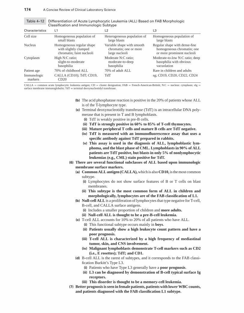
P1: OSO
LWBK192-04 LWBK192-Hubbard November 27, 2008 7:43
174 A Concise Review of Clinical Laboratory Science
Table 4–12 Differentiation of Acute Lymphocytic Leukemia (ALL) Based on FAB MorphologicClassification and Immunologic Subtype
Characteristics L1 L2 L3
Cell size Homogeneous population ofsmall blasts
Heterogeneous population oflarge blasts
Homogeneous population oflarge blasts
Nucleus Homogeneous regular shapewith slightly clumpedchromatin; faint nucleoli
Variable shape with smoothchromatin; one or morelarge nucleoli
Regular shape with dense-finehomogeneous chromatin; oneor more prominent nucleoli
Cytoplasm High N:C ratio;slight-to-moderatebasophilia
Moderate N:C ratio;moderate-to-deepbasophilia
Moderate-to-low N:C ratio; deepbasophilia with obviousvacuolation
Patient age 70% of childhood ALL 70% of adult ALL Rare in children and adults
Immunologicmarkers
CALLA (CD10); TdT; CD19,CD20
TdT sg; CD19, CD20, CD22, CD24
CALLA = common acute lymphocytic leukemia antigen; CD = cluster designation; FAB = French-American-British; N:C = nucleus: cytoplasm; sIg =surface membrane immunoglobulin; TdT = terminal deoxynucleotidyl transferase.
(b) The acid phosphatase reaction is positive in the 20% of patients whose ALLis of the T-lymphocyte type.
(c) Terminal deoxynucleotidly transferase (TdT) is an intracellular DNA poly-merase that is present in T and B lymphoblasts.
(i) TdT is weakly positive in pre-B cells.(ii) TdT is strongly positive in 60% to 85% of T-cell thymocytes.(iii) Mature peripheral T cells and mature B cells are TdT negative.(iv) TdT is measured with an immunofluorescence assay that uses a
specific antibody against TdT prepared in rabbits.(v) This assay is used in the diagnosis of ALL, lymphoblastic lym-
phoma, and the blast phase of CML. Lymphoblasts in 90% of ALLpatients are TdT positive, but blasts in only 5% of nonlymphocyticleukemias (e.g., CML) stain positive for TdT.
(6) There are several functional subclasses of ALL based upon immunologicmembrane surface markers.(a) Common ALL antigen (CALLA), which is also CD10, is the most common
subtype.(i) Lymphocytes do not show surface features of B or T cells on blast
membranes.(ii) This subtype is the most common form of ALL in children and
morphologically, lymphocytes are of the FAB classification of L1.(b) Null-cell ALL is a proliferation of lymphocytes that type negative for T-cell,
B-cell, and CALLA surface antigens.(i) Includes a smaller proportion of children and more adults.(ii) Null-cell ALL is thought to be a pre-B-cell leukemia.
(c) T-cell ALL accounts for 10% to 20% of all patients who have ALL.(i) This functional subtype occurs mainly in boys.
(ii) Patients usually show a high leukocyte count pattern and have apoor prognosis.
(iii) T-cell ALL is characterized by a high frequency of mediastinaltumor, skin, and CNS involvement.
(iv) Malignant lymphoblasts demonstrate T-cell markers such as CD2(i.e., E rosettes); TdT; and CD1.
(d) B-cell ALL is the rarest of subtypes, and it corresponds to the FAB classi-fication Burkitt’s Type L3.
(i) Patients who have Type L3 generally have a poor prognosis.(ii) L3 can be diagnosed by demonstration of B-cell typical surface Ig
receptors.(iii) This disorder is thought to be a memory-cell leukemia.
(7) Better prognosis is seen in female patients, patients with lower WBC counts,and patients diagnosed with the FAB classification L1 subtype.
P1: OSO
LWBK192-04 LWBK192-Hubbard November 27, 2008 7:43
CHAPTER 4 Hematologic Disorders 175
(8) Treatment is now able to produce complete remission in 95% of childrenand in 50% to 70% of adults with ALL. Approximately 50% of children inremission show signs of being completely cured.
e. Lymphomas are a group of lymphoproliferative disorders in which there is neo-plastic proliferation of an arrested stage of secondary lymphocyte maturation thatusually begins in and involves mainly the lymph nodes. As the disease progresses,many other organs and tissues (e.g., spleen, liver, skin, marrow) can become in-vaded by malignant lymph. All lymphomas are categorized into either Hodgkin’slymphoma or non-Hodgkin’s lymphoma.(1) The general laboratory profile of a patient who has lymphoma includes the
following results:(a) The WBC count may vary, depending on the progression of the malignancy.
(i) Neutrophilia is seen only when lymph nodes are involved, but neutrope-nia persists when the bone marrow is involved.
(ii) Most frequent are WBC counts from 12,000 to 25,000/mm3 withlymphophilia and monocytosis.
(iii) Eosinophilia is found in 20% of patients who have lymphoma.(iv) Platelet count depends upon the extent of marrow involvement.
(b) Severe Normochromic/normocytic anemia in 50% of patients.(c) The LAP level is elevated in the active phases of the disease.(d) Reed-Sternberg cells, which are the hallmark of Hodgkin’s lymphoma,
may be found in the lymph nodes and marrow of a patient who has thisdisease.(i) These cells can be identified as giant binucleated or multi-nucleated cells
with acidophilic nuclei.(ii) This cell type is thought to originate from the monocyte-macrophage
cell line.(e) Bone marrow biopsy frequently reveals granulocytic hyperplasia with a
shift to the left, slight monocytosis, and eosinophilia. Reed-Sternberg cellsmay be present, depending on the clinical subtype.
(2) The Hodgkin’s type of lymphoma has an increased frequency of occurrencein persons between the ages of 15 and 30 years and after 50 years.(a) The clinical subclassification and diagnosis of Hodgkin’s lymphoma is made
histologically from a lymph node biopsy.(i) Lymphocytic-predominant Hodgkin’s lymphoma is most frequent in
young men and localizes to the cervical lymph nodes. Lymph-nodebiopsy reveals numerous lymphocytes, no fibrosis or necrosis, and fewReed-Sternberg cells.
(ii) Nodular-sclerosis Hodgkin’s lymphoma is a common variety mostoften discovered as a mediastinal mass in young women. Lymph-node biopsy is characterized by broad bands of collagen separatingnodules of lymphoid tissue and the presence of lacunar cells, whichare atypical histiocytes. Few Reed-Sternberg cells are found.
(iii) The mixed type of Hodgkin’s lymphoma is characterized on lymph-node biopsy by a variety of all cell types (e.g., lymphocytes, plasmacells, eosinophils, histiocytes, and numerous Reed-Sternberg cells).Necrosis and fibrosis may also be present.
(iv) The lymphocyte depletion type of Hodgkin’s lymphoma is the raresttype. Lymph-node biopsy reveals diffuse fibrosis and a lack of lym-phoid growth. This type is accompanied by pancytopenia and lym-phocytopenia, because there is a more frequent involvement of bonemarrow.
(b) Clinical staging of Hodgkin’s lymphoma is based on the extent of lymphnode and systemic involvement.
(i) Stage I of the disease is limited to lymph nodes in one or two anatomicregions on one side of the diaphragm.
(ii) Stage II of the disease involves more than two regions of lymphnode involvement on one side of the diaphragm.
P1: OSO
LWBK192-04 LWBK192-Hubbard November 27, 2008 7:43
176 A Concise Review of Clinical Laboratory Science
(iii) Stage III of the disease involves two or more regions of node in-volvement on both sides of the diaphragm.
(iv) Stage IV of the disease involves bone marrow and other organs, inaddition to lymph nodes. Cell-mediated immunity is defective bythis stage.
(c) Treatment. Without treatment, few patients survive as long as 10 years. Withmodern treatment methods, 85% of patients diagnosed in Stages I and II canbe cured. The cure rate is 50% for patients diagnosed in Stages III or IV.Treatment consists of long-term chemotherapy and localized radiotherapyfor infected lymph nodes.
(3) Non-Hodgkin’s lymphomas are classified as a group of clonal prolifera-tions of lymphocytes that have undergone malignant transformation at anarrested stage of their differentiation. Non-Hodgkin’s lymphomas involvemostly B lymphocytes and are reported mostly in middle-age and olderpatients.(a) The Rappaport classification of non-Hodgkin’s lymphomas is based upon
nodular and diffuse arrangement of malignant cells in a lymph-node biopsy,as well as on size characteristics of the predominating lymphocytes.
(b) Many subtypes of non-Hodgkin’s lymphoma are closely related to a defectin immune regulation.
(c) Some subtypes are believed to be virally related.(i) Burkitt’s lymphoma, which is associated with EBV, has a strong affin-
ity for B cells; this affinity causes B-cell proliferation. When the cytolytickiller T lymphocytes’ response is deficient, an unchecked B-cell prolif-eration can result in lymphoma.
(ii) Cutaneous T-cell lymphoma (i.e., Mycosis Fungoides) is an HTLV-1-promoted helper-inducer T-cell malignancy. This lymphoma is as-sociated with a lymphocytosis of Sezary cells.
(d) Most patients who have non-Hodgkin’s lymphomas have associated chro-mosome abnormalities (Table 4–11).(i) Nodular small cleaved-cell lymphoma is associated with a translo-
cation between the genes of Ig heavy chains found on chromosome14 and an arm of chromosome 18.
(ii) Burkitt’s lymphoma is associated with a translocation between chro-mosomes 8 and 2 or 22 and 14.
(iii) Small lymphocytic lymphoma and diffuse large-cell lymphoma areassociated with a translocation between chromosomes 11 and 14.
(iv) Cutaneous T-cell lymphoma is associated with a translocation be-tween chromosomes 7 and 14 or 11 and 9.
Online Menu for This Chapter
Go to thePoint.lww.com/Hubbard2e for the following:
� Study Questions and Answers

P1: OSO
LWBK192-05 LWBK192-Hubbard November 27, 2008 7:45
CHAPTER 5
Immunology andSerology
WADE REDMAN, MBA, MT (ASCP) DLM AND JOEL HUBBARD,PhD, MT (ASCP)
I. INTRODUCTION
A. Antigens and haptens. An antigen is a substance that stimulates antibody formation andhas the ability to bind to an antibody. A hapten is a low-molecular-weight, nonantigenicsubstance that, when combined with an antigen, changes the antigenic specificity of thatantigen.
B. An antibody is a glycoprotein substance (immunoglobulin) that is produced by B lympho-cytes in response to an antigen.1. Antibodies may be monoclonal or polyclonal. Monoclonal antibodies are derived
from a single B-cell clone and are produced as a single class of immunoglobulin withspecificity unique to the antigenic stimulus.
2. Polyclonal antibodies are produced as different classes of immunoglobulins by manyB-cell clones in response to an antigen.
3. Antibodies produced in response to antigens from another species are called heteroan-tibodies or xenoantibodies.
4. Alloantibodies are formed in response to antigens from individuals of the samespecies.
5. Autoantibodies are produced by the body’s immune system against “self” antigens.They function by facilitating phagocytosis and microbial killing and by neutralizingtoxic substances. They also combine with antigens on cell surfaces, which results ineither intravascular or extravascular destruction of the target cells.
C. Antigen-antibody reactions1. An epitope is the part of an antigen that reacts specifically with an antibody or T-cell
receptor (Figure 5–1).2. Agglutination is the clumping of particulate antigens by antibodies specific for the
antigens.3. Affinity is the tendency that an epitope has for combining with the antigen-binding site
on an antibody molecule.4. Avidity is the strength of the bond between the antigen and the antibody.5. Sensitivity is the smallest amount of antigen or antibody that can be detected.6. Specificity is the ability of an antibody to bind to an antigen with complementary
determinants and not to an antigen with dissimilar determinants.
177
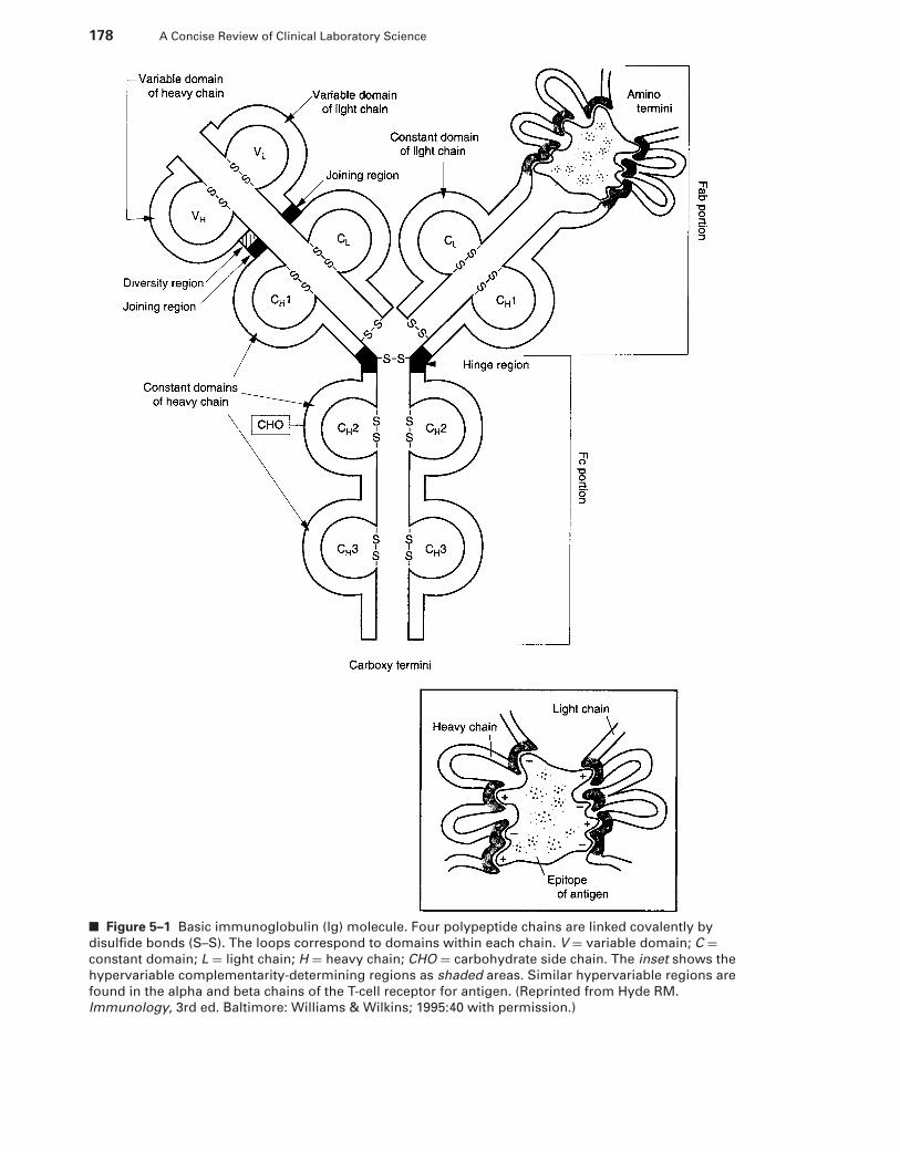
P1: OSO
LWBK192-05 LWBK192-Hubbard November 27, 2008 7:45
178 A Concise Review of Clinical Laboratory Science
■ Figure 5–1 Basic immunoglobulin (lg) molecule. Four polypeptide chains are linked covalently by
disulfide bonds (S–S). The loops correspond to domains within each chain. V = variable domain; C =constant domain; L = light chain; H = heavy chain; CHO = carbohydrate side chain. The inset shows the
hypervariable complementarity-determining regions as shaded areas. Similar hypervariable regions are
found in the alpha and beta chains of the T-cell receptor for antigen. (Reprinted from Hyde RM.
Immunology, 3rd ed. Baltimore: Williams & Wilkins; 1995:40 with permission.)
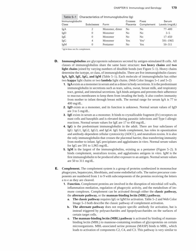
P1: OSO
LWBK192-05 LWBK192-Hubbard November 27, 2008 7:45
CHAPTER 5 Immunology and Serology 179
Table 5–1 Characteristics of Immunoglobulins (Ig)
Immunoglobulin Crosses Fixes Serum
Class Subclasses Form Placenta Complement Levels (mg/dL)
IgA 2 Monomer, dimer No No 77–400
IgD 0 Monomer No No 3–5
IgE 0 Monomer No No 17–450
IgG 4 Monomer Yes Yes∗ 591–1965
IgM 0 Pentamer No Yes 50–311
∗IgG4 does not fix complement.
D. Immunoglobulins are glycoprotein substances secreted by antigen-stimulated B cells. Allclasses of immunoglobulins share the same basic structure: two heavy chains and twolight chains joined by varying numbers of disulfide bonds (see Figure 5–1). Heavy chainsdetermine the isotype, or class, of immunoglobulin. There are five immunoglobulin classes:IgA, IgD, IgE, IgG, and IgM (Table 5–1). Each molecule of immunoglobulin has eithertwo kappa light chains or two lambda light chains. (Web Color Images 5–1 and 5–2)
1. IgA exists as a monomer in serum and as a dimer in body secretions. It is the predominantimmunoglobulin in secretions such as tears, saliva, sweat, breast milk, and respiratorytract, genital, and intestinal secretions. IgA binds antigens and prevents their adherenceto mucous membranes to keep them from invading the body. It also confers immunityfrom mother to infant through breast milk. The normal range for serum IgA is 77 to400 mg/dL.
2. IgD exists as a monomer, and its function is unknown. Normal serum values of IgDare 3 to 5 mg/dL.
3. IgE exists in serum as a monomer. It binds to crystallizable fragment (Fc) receptors onmast cells and basophils and is elevated during parasitic infections and Type I allergicreactions. Normal serum values for IgE are 17 to 450 mg/dL.
4. IgG is the predominant immunoglobulin in the adult. There are four subclasses ofIgG: IgG1, IgG2, IgG3, and IgG4. IgG binds complement, has roles in opsonizationand antibody-dependent cellular cytotoxicity (ADCC), and neutralizes toxins. It is alsothe only immunoglobulin that crosses the placental barrier, thus transferring immunityfrom mother to infant. IgG precipitates and agglutinates in vitro. Normal serum valuesfor IgG are 591 to 1,965 mg/dL.
5. IgM is the largest of the immunoglobulins, existing as a pentamer (Figure 5–2). Itbinds complement, neutralizes toxins, and agglutinates antigens in vitro. IgM is thefirst immunoglobulin to be produced after exposure to an antigen. Normal serum valuesare 50 to 311 mg/dL.
E. Complement. The complement system is a group of proteins synthesized in mononuclearphagocytes, hepatocytes, fibroblasts, and some endothelial cells. The native precursor com-ponents are numbered from 1 to 9 with subcomponents of the proteins receiving the lettersa to e as they are cleaved.
1. Function. Complement proteins are involved in the disruption of microbial cell walls,inflammation mediation, regulation of phagocytic activity, and the metabolism of im-mune complexes. Complement can be activated through either the classic pathway,the alternate pathway, or the mannan-binding lectin (MBL) pathway.a. The classic pathway requires IgG or IgM for activation. Table 5–2 and Web Color
Image 5–3 both describe the classic pathway of complement activation.b. The alternate pathway does not require specific antibody for activation, but is
instead triggered by polysaccharides and lipopolysaccharides on the surfaces ofcertain target cells.
c. The mannan-binding lectin (MBL) pathway is activated by binding of mannan-binding lectin (MBL) to mannose-containing residues of glycoproteins on certainmicroorganisms. MBL-associated serine protease (MASP) binds to MBL, whichleads to activation of components C2, C4, and C3. This pathway is very similar to
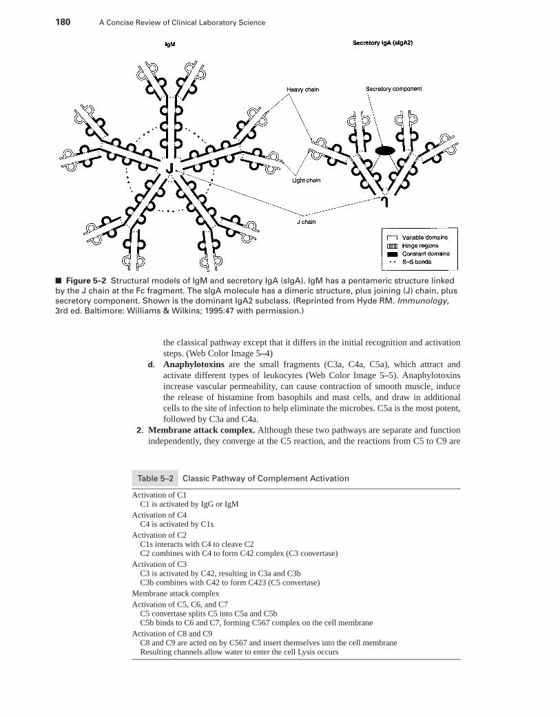
P1: OSO
LWBK192-05 LWBK192-Hubbard November 27, 2008 7:45
180 A Concise Review of Clinical Laboratory Science
■ Figure 5–2 Structural models of IgM and secretory IgA (slgA). IgM has a pentameric structure linked
by the J chain at the Fc fragment. The slgA molecule has a dimeric structure, plus joining (J) chain, plus
secretory component. Shown is the dominant IgA2 subclass. (Reprinted from Hyde RM. Immunology,
3rd ed. Baltimore: Williams & Wilkins; 1995:47 with permission.)
the classical pathway except that it differs in the initial recognition and activationsteps. (Web Color Image 5–4)
d. Anaphylotoxins are the small fragments (C3a, C4a, C5a), which attract andactivate different types of leukocytes (Web Color Image 5–5). Anaphylotoxinsincrease vascular permeability, can cause contraction of smooth muscle, inducethe release of histamine from basophils and mast cells, and draw in additionalcells to the site of infection to help eliminate the microbes. C5a is the most potent,followed by C3a and C4a.
2. Membrane attack complex. Although these two pathways are separate and functionindependently, they converge at the C5 reaction, and the reactions from C5 to C9 are
Table 5–2 Classic Pathway of Complement Activation
Activation of C1C1 is activated by IgG or IgM
Activation of C4C4 is activated by C1s
Activation of C2C1s interacts with C4 to cleave C2C2 combines with C4 to form C42 complex (C3 convertase)
Activation of C3C3 is activated by C42, resulting in C3a and C3bC3b combines with C42 to form C423 (C5 convertase)
Membrane attack complex
Activation of C5, C6, and C7C5 convertase splits C5 into C5a and C5bC5b binds to C6 and C7, forming C567 complex on the cell membrane
Activation of C8 and C9C8 and C9 are acted on by C567 and insert themselves into the cell membraneResulting channels allow water to enter the cell Lysis occurs
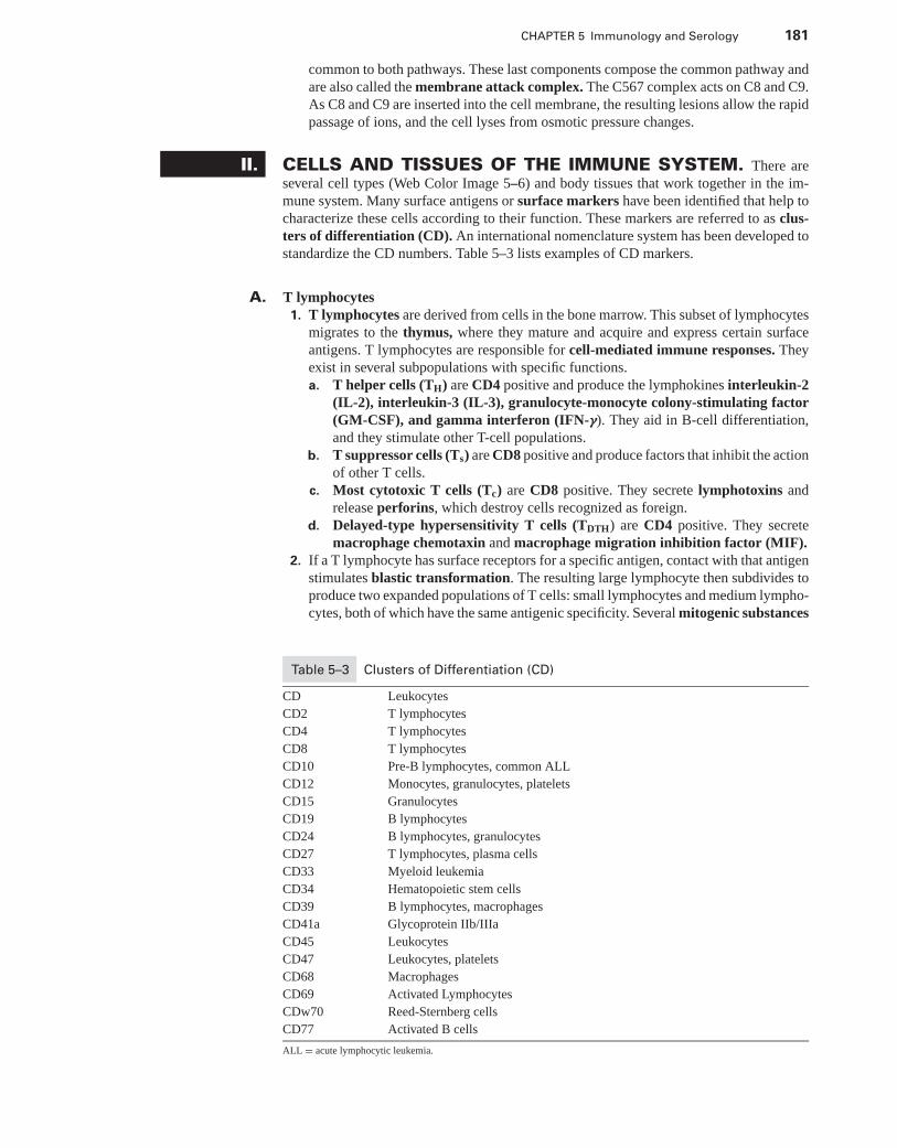
P1: OSO
LWBK192-05 LWBK192-Hubbard November 27, 2008 7:45
CHAPTER 5 Immunology and Serology 181
common to both pathways. These last components compose the common pathway andare also called the membrane attack complex. The C567 complex acts on C8 and C9.As C8 and C9 are inserted into the cell membrane, the resulting lesions allow the rapidpassage of ions, and the cell lyses from osmotic pressure changes.
II. CELLS AND TISSUES OF THE IMMUNE SYSTEM. There areseveral cell types (Web Color Image 5–6) and body tissues that work together in the im-mune system. Many surface antigens or surface markers have been identified that help tocharacterize these cells according to their function. These markers are referred to as clus-ters of differentiation (CD). An international nomenclature system has been developed tostandardize the CD numbers. Table 5–3 lists examples of CD markers.
A. T lymphocytes1. T lymphocytes are derived from cells in the bone marrow. This subset of lymphocytes
migrates to the thymus, where they mature and acquire and express certain surfaceantigens. T lymphocytes are responsible for cell-mediated immune responses. Theyexist in several subpopulations with specific functions.a. T helper cells (TH) are CD4 positive and produce the lymphokines interleukin-2
(IL-2), interleukin-3 (IL-3), granulocyte-monocyte colony-stimulating factor(GM-CSF), and gamma interferon (IFN-γ). They aid in B-cell differentiation,and they stimulate other T-cell populations.
b. T suppressor cells (Ts) are CD8 positive and produce factors that inhibit the actionof other T cells.
c. Most cytotoxic T cells (Tc) are CD8 positive. They secrete lymphotoxins andrelease perforins, which destroy cells recognized as foreign.
d. Delayed-type hypersensitivity T cells (TDTH) are CD4 positive. They secretemacrophage chemotaxin and macrophage migration inhibition factor (MIF).
2. If a T lymphocyte has surface receptors for a specific antigen, contact with that antigenstimulates blastic transformation. The resulting large lymphocyte then subdivides toproduce two expanded populations of T cells: small lymphocytes and medium lympho-cytes, both of which have the same antigenic specificity. Several mitogenic substances
Table 5–3 Clusters of Differentiation (CD)
CD Leukocytes
CD2 T lymphocytes
CD4 T lymphocytes
CD8 T lymphocytes
CD10 Pre-B lymphocytes, common ALL
CD12 Monocytes, granulocytes, platelets
CD15 Granulocytes
CD19 B lymphocytes
CD24 B lymphocytes, granulocytes
CD27 T lymphocytes, plasma cells
CD33 Myeloid leukemia
CD34 Hematopoietic stem cells
CD39 B lymphocytes, macrophages
CD41a Glycoprotein IIb/IIIa
CD45 Leukocytes
CD47 Leukocytes, platelets
CD68 Macrophages
CD69 Activated Lymphocytes
CDw70 Reed-Sternberg cells
CD77 Activated B cells
ALL = acute lymphocytic leukemia.
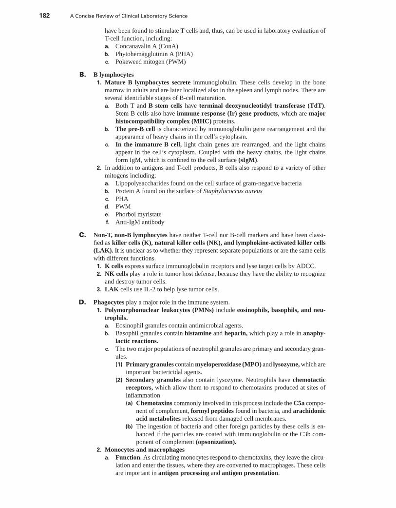
P1: OSO
LWBK192-05 LWBK192-Hubbard November 27, 2008 7:45
182 A Concise Review of Clinical Laboratory Science
have been found to stimulate T cells and, thus, can be used in laboratory evaluation ofT-cell function, including:a. Concanavalin A (ConA)b. Phytohemagglutinin A (PHA)c. Pokeweed mitogen (PWM)
B. B lymphocytes1. Mature B lymphocytes secrete immunoglobulin. These cells develop in the bone
marrow in adults and are later localized also in the spleen and lymph nodes. There areseveral identifiable stages of B-cell maturation.a. Both T and B stem cells have terminal deoxynucleotidyl transferase (TdT).
Stem B cells also have immune response (Ir) gene products, which are majorhistocompatibility complex (MHC) proteins.
b. The pre-B cell is characterized by immunoglobulin gene rearrangement and theappearance of heavy chains in the cell’s cytoplasm.
c. In the immature B cell, light chain genes are rearranged, and the light chainsappear in the cell’s cytoplasm. Coupled with the heavy chains, the light chainsform IgM, which is confined to the cell surface (sIgM).
2. In addition to antigens and T-cell products, B cells also respond to a variety of othermitogens including:a. Lipopolysaccharides found on the cell surface of gram-negative bacteriab. Protein A found on the surface of Staphylococcus aureusc. PHAd. PWMe. Phorbol myristatef. Anti-IgM antibody
C. Non-T, non-B lymphocytes have neither T-cell nor B-cell markers and have been classi-fied as killer cells (K), natural killer cells (NK), and lymphokine-activated killer cells(LAK). It is unclear as to whether they represent separate populations or are the same cellswith different functions.
1. K cells express surface immunoglobulin receptors and lyse target cells by ADCC.2. NK cells play a role in tumor host defense, because they have the ability to recognize
and destroy tumor cells.3. LAK cells use IL-2 to help lyse tumor cells.
D. Phagocytes play a major role in the immune system.1. Polymorphonuclear leukocytes (PMNs) include eosinophils, basophils, and neu-
trophils.a. Eosinophil granules contain antimicrobial agents.b. Basophil granules contain histamine and heparin, which play a role in anaphy-
lactic reactions.c. The two major populations of neutrophil granules are primary and secondary gran-
ules.(1) Primary granules contain myeloperoxidase (MPO) and lysozyme, which are
important bactericidal agents.(2) Secondary granules also contain lysozyme. Neutrophils have chemotactic
receptors, which allow them to respond to chemotaxins produced at sites ofinflammation.(a) Chemotaxins commonly involved in this process include the C5a compo-
nent of complement, formyl peptides found in bacteria, and arachidonicacid metabolites released from damaged cell membranes.
(b) The ingestion of bacteria and other foreign particles by these cells is en-hanced if the particles are coated with immunoglobulin or the C3b com-ponent of complement (opsonization).
2. Monocytes and macrophagesa. Function. As circulating monocytes respond to chemotaxins, they leave the circu-
lation and enter the tissues, where they are converted to macrophages. These cellsare important in antigen processing and antigen presentation.
P1: OSO
LWBK192-05 LWBK192-Hubbard November 27, 2008 7:45
CHAPTER 5 Immunology and Serology 183
b. Surface receptors. Macrophages have several different surface receptors, includ-ing the integrin Mac 1-CR3, which binds the C3b component of complement;Fc receptors, which bind immunoglobulin; CR1, which binds C3b and C4b; andClass II MHC receptors.
c. Secreted products. Macrophages secrete products that assist in the ir.(1) Interleukin 1 (IL-1) stimulates T-cell growth.(2) Interleukin 6 (IL-6) stimulates B cells.(3) Tumor necrosis factor (TNFα) has antitumor and antibacterial activity and
stimulates production of IL-1 and interferon.(4) Endothelial leukocyte adhesion molecule (ELAM-1) plays a role in dia-
pedesis (migration of cells into tissues from the circulation).
E. Other cells involved in antigen presentation are:1. Dendritic cells2. Langerhans cells3. B lymphocytes
F. Lymphoid tissues are found throughout the entire body.1. Distribution
a. Spleenb. Thymusc. Thoracic ductd. Lymph nodese. Bone marrowf. Peyer’s patches
g. Tonsilsh. Appendix
2. Concentration. Although both T and B lymphocytes can be found in all lymphoidtissues, the highest concentration of T cells is found in the thymus. The highest con-centration of B cells is found in the bone marrow.
III. IMMUNITY may be natural or acquired.
A. Natural immunity is present at birth and provides protection against disease and aidsin recovery from disease. However, it also provides the basis for organ rejection aftertransplantation. Factors involved in natural immunity include:1. Physical barriers, such as skin and mucous membranes2. Genetically controlled susceptibility and nonsusceptibility to certain diseases3. Inflammation, which involves a vascular response and a cellular response by phago-
cytic cells4. Acute-phase plasma proteins, such as C-reactive protein, haptoglobin, and fibrinogen,
which are produced in response to injury and aid in wound healing
B. Acquired or specific immunity results when immunologic memory and antibody specificto a foreign antigen develop in response to the antigen. Acquired immunity may be active(through immunization or disease) or passive (through transplacental transfer). This typeof immunity involves both cell-mediated and humoral immune responses.
IV. IMMUNE RESPONSE (IR). The IR may be roughly divided into two com-ponents, cell-mediated and humoral. Although most Ir contain elements of both, certainantigens elicit a strong humoral response with little cell-mediated involvement, and otherantigens elicit primarily a cell-mediated response.
A. Humoral immunity involves immunoglobulin (antibody) production by B lymphocytes.Complement can also be considered a humoral component because it can be activated byimmunoglobulin. The humoral response occurs in three phases:
P1: OSO
LWBK192-05 LWBK192-Hubbard November 27, 2008 7:45
184 A Concise Review of Clinical Laboratory Science
1. Antigen elimination. This phase is accomplished by phagocytosis. Most injected anti-gen is removed within minutes, but complete removal may take months or years.
2. The primary response. After exposure to an antigen, there is a latent period of ap-proximately 5 to 15 days before antibody appears in the serum. The antibody titerincreases, plateaus, then decreases. IgM is the first immunoglobulin to appear. Al-though a small amount of IgG is made later, the majority of immunoglobulin producedduring a primary response is IgM.
3. The secondary response. A second or any subsequent exposure to the same antigenelicits a secondary response. This time, there is a rapid antibody response, usuallywithin 2 to 4 days after antigen exposure. IgG is the predominant immunoglobulin.The circulating antibody titer is much higher and lasts longer than that seen in theprimary response.
B. Cell-mediated immunity is especially important in viral and fungal infections and in in-fections caused by acid-fast bacilli (e.g., tuberculosis, Hansen’s disease).
1. Macrophages, Tc, and NK cells play a role in cell-mediated immunity (see SectionsII A 1 c, II C 2, and II D 2).
2. ADCC. Cells with cytolytic activity and Fc receptors, especially NK cells, are able todirectly lyse antibody-coated (usually IgG) target cells.
3. Cytokines are protein messengers produced by cells. Many play a role in cell-mediatedimmunity. (Web Color Image 5–7)a. Lymphokines are produced primarily by activated T lymphocytes and include:
(1) IL-2(2) IL-3(3) IL-4(4) GM-CSF(5) B-cell growth factor 2(6) Macrophage activating factor(7) MIF(8) IFN-γ
b. Monokines are produced by monocytes and include:(1) IL-1(2) TNFα
V. MAJOR HISTOCOMPATIBILITY COMPLEX
A. In transplantation studies, MHC gene products were identified as responsible for graftrejection. Likewise, studies on responses to antigens, demonstrated that MHC gene productswere found to control immune responses. It was determined that antigen-specific T cellsrecognize portions of protein antigens that are bound noncovalently to MHC gene products.
B. The Major histocompatibility complex is found on human chromosome 6.1. Have a role in intercellular recognition and discrimination of self/nonself.2. Have a role in development of humoral and cell-mediated immune responses.3. One’s set of MHC genes influences the repertoire of antigens to which that person’s TH
and TC cells can respond, therefore, one’s MHC plays a role in susceptibility to diseaseand autoimmunity.
C. Organization of MHC is summarized in Table 5–4.1. Class I MHC are glycoproteins found on most nucleated cells. Class I molecules
present antigenic peptides to the TC.2. Class II MHC are glycoproteins found on antigen presenting cells (APC).3. Class III MHC consists of secreted proteins with immune function.4. Inheritance
a. MHC alleles are inherited as two sets. One set from each parent.b. One set is the haplotype.
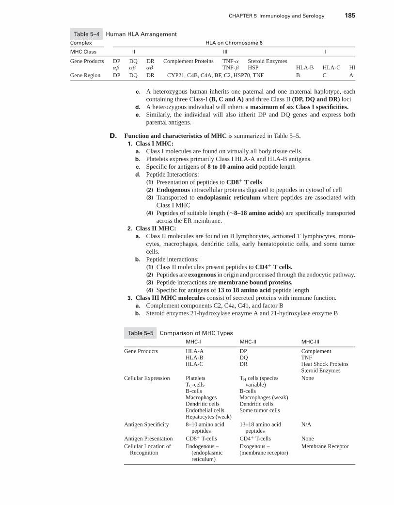
P1: OSO
LWBK192-05 LWBK192-Hubbard November 27, 2008 7:45
CHAPTER 5 Immunology and Serology 185
Table 5–4 Human HLA Arrangement
Complex HLA on Chromosome 6
MHC Class II III I
Gene Products DP DQ DR Complement Proteins TNF-α Steroid Enzymesαβ αβ αβ TNF-β HSP HLA-B HLA-C HI
Gene Region DP DQ DR CYP21, C4B, C4A, BF, C2, HSP70, TNF B C A
c. A heterozygous human inherits one paternal and one maternal haplotype, eachcontaining three Class-I (B, C and A) and three Class II (DP, DQ and DR) loci
d. A heterozygous individual will inherit a maximum of six Class I specificities.e. Similarly, the individual will also inherit DP and DQ genes and express both
parental antigens.
D. Function and characteristics of MHC is summarized in Table 5–5.1. Class I MHC:
a. Class I molecules are found on virtually all body tissue cells.b. Platelets express primarily Class I HLA-A and HLA-B antigens.c. Specific for antigens of 8 to 10 amino acid peptide lengthd. Peptide Interactions:
(1) Presentation of peptides to CD8+ T cells(2) Endogenous intracellular proteins digested to peptides in cytosol of cell(3) Transported to endoplasmic reticulum where peptides are associated with
Class I MHC(4) Peptides of suitable length (∼8–18 amino acids) are specifically transported
across the ER membrane.2. Class II MHC:
a. Class II molecules are found on B lymphocytes, activated T lymphocytes, mono-cytes, macrophages, dendritic cells, early hematopoietic cells, and some tumorcells.
b. Peptide interactions:(1) Class II molecules present peptides to CD4+ T cells.(2) Peptides are exogenous in origin and processed through the endocytic pathway.(3) Peptide interactions are membrane bound proteins.(4) Specific for antigens of 13 to 18 amino acid peptide length
3. Class III MHC molecules consist of secreted proteins with immune function.a. Complement components C2, C4a, C4b, and factor Bb. Steroid enzymes 21-hydroxylase enzyme A and 21-hydroxylase enzyme B
Table 5–5 Comparison of MHC Types
MHC-I MHC-II MHC-III
Gene Products HLA-AHLA-BHLA-C
DPDQDR
ComplementTNFHeat Shock ProteinsSteroid Enzymes
Cellular Expression PlateletsTC-cellsB-cellsMacrophagesDendritic cellsEndothelial cellsHepatocytes (weak)
TH cells (speciesvariable)
B-cellsMacrophages (weak)Dendritic cellsSome tumor cells
None
Antigen Specificity 8–10 amino acidpeptides
13–18 amino acidpeptides
N/A
Antigen Presentation CD8+ T-cells CD4+ T-cells None
Cellular Location ofRecognition
Endogenous –(endoplasmicreticulum)
Exogenous –(membrane receptor)
Membrane Receptor
P1: OSO
LWBK192-05 LWBK192-Hubbard November 27, 2008 7:45
186 A Concise Review of Clinical Laboratory Science
c. Inflammatory proteins TNF-α and TNF-β.d. Heat shock proteins such as HSP70.e. These are not related structurally to the MHC-I and MHC-II molecules.f. They have no role in antigen presentation.
g. They do play a role in the immune response.
E. Diversity of the human MHC locus1. Class I MHC–known alleles; 60A × 110B × 40C = 2.6 × 10⁵
2. Class II MHC–known alleles; 4DP × 6DQ × 488 DR = 1.2 × 10⁴
3. Total possible combination; (2.6 × 10⁵)(1.2 × 10⁴) = 3.12 × 10⁹
F. Clinical significance of HLA1. Organ transplant & bone marrow transplant will have an increased survival rate of
the patient and a decreased risk of graft vs. host disease if HLA and MHC matchingbetween donor and recipient is done.
2. Platelet transfusion matching between donor and recipient is useful to patients whoare retractile to random donor platelets
3. HLA compatibility exerts the strongest influence on long-term kidney survival aftera transplant.
4. HLA identical donors for bone marrow transplantation to reduce the frequency of graftvs. host disease.
5. Paternity testing for exclusion or nonexclusion.6. Disease association
a. Association between HLA phenotype and some diseasesb. HLA-B27 is found in >90% of patients with ankylosing spondylitis, but in only
∼10% normal individuals.
G. HLA Testing1. A heterozygous human inherits one paternal and one maternal haplotype, each
containing three Class I (B, C and A) and three Class II (DP, DQ and DR) loci.2. A heterozygous individual will inherit a maximum of six Class I specificities.3. Similarly, the individual will also inherit DP and DQ genes and express both parental
antigens.4. The Class II MHC molecule consists of two chains (α and β), with some antigenic
determinants (specificities) on each chain.a. DR α- and β-chains can associate in ether cis (both from the same parent) or
trans (one from each parent) combinations, an individual can have additional DRspecificities.
b. Also, there is more than one functional DR β-chain gene. Hence, many DR speci-ficities can be found in any one individual.
5. A microcytotoxicity test demonstrates that if the antigen is present on the lymphocytes,addition of complement will cause them to become porous and unable to exclude theadded dye.a. White blood cells from the potential donors and recipient are distributed into a
series of wells on a microtiter plate, and then antibodies specific for various ClassI and Class II MHC alleles are added to different wells.
b. After incubation, complement is added to the wells and cytotoxicity is assed bythe uptake or exclusion of various dyes by the cells.
c. If the WBC expresses the MHC allele for which a particular monoclonal antibodyis specific, then the cells will lysed upon addition of complement, and these deadcells will take up a dye such as trypan blue.
d. The reaction of donor and recipient cells with a single antibody directed against anHLA-A antigen.
6. Mixed-lymphocyte reaction (MLR) can be used to quantify the degree of class IIMHC compatibility between potential donors and a recipient.a. Lymphocytes from potential donor that have been x-irradiated or treated with mit-
omycin C serve as the stimulator cells, and lymphocytes from the recipient serveas responder cells.
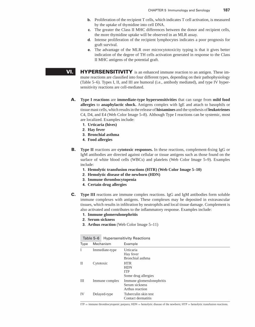
P1: OSO
LWBK192-05 LWBK192-Hubbard November 27, 2008 7:45
CHAPTER 5 Immunology and Serology 187
b. Proliferation of the recipient T cells, which indicates T cell activation, is measuredby the uptake of thymidine into cell DNA.
c. The greater the Class II MHC differences between the donor and recipient cells,the more thymidine uptake will be observed in an MLR assay.
d. Intense proliferation of the recipient lymphocytes indicates a poor prognosis forgraft survival.
e. The advantage of the MLR over microcytotoxicity typing is that it gives betterindication of the degree of TH cells activation generated in response to the ClassII MHC antigens of the potential graft.
VI. HYPERSENSITIVITY is an enhanced immune reaction to an antigen. These im-mune reactions are classified into four different types, depending on their pathophysiology(Table 5–6). Types I, II, and III are humoral (i.e., antibody mediated), and type IV hyper-sensitivity reactions are cell-mediated.
A. Type I reactions are immediate-type hypersensitivities that can range from mild foodallergies to anaphylactic shock. Antigens complex with IgE and attach to basophils ortissue mast cells, which results in the release of histamines and the synthesis of leukotrienesC4, D4, and E4 (Web Color Image 5–8). Although Type I reactions can be systemic, mostare localized. Examples include:
1. Urticaria (hives)2. Hay fever3. Bronchial asthma4. Food allergies
B. Type II reactions are cytotoxic responses. In these reactions, complement-fixing IgG orIgM antibodies are directed against cellular or tissue antigens such as those found on thesurface of white blood cells (WBCs) and platelets (Web Color Image 5–9). Examplesinclude:
1. Hemolytic transfusion reactions (HTR) (Web Color Image 5–10)2. Hemolytic disease of the newborn (HDN)3. Immune thrombocytopenia4. Certain drug allergies
C. Type III reactions are immune complex reactions. IgG and IgM antibodies form solubleimmune complexes with antigens. These complexes may be deposited in extravasculartissues, which results in infiltration by neutrophils and local tissue damage. Complement isalso activated and contributes to the inflammatory response. Examples include:
1. Immune glomerulonephritis2. Serum sickness3. Arthus reaction (Web Color Image 5–11)
Table 5–6 Hypersensitivity Reactions
Type Mechanism Example
I Immediate-type UrticariaHay feverBronchial asthma
II Cytotoxic HTRHDNITPSome drug allergies
III Immune complex Immune glomerulonephritisSerum sicknessArthus reaction
IV Delayed-type Tuberculin skin testContact dermatitis
ITP = immune thrombocytopenic purpura; HDN = hemolytic disease of the newborn; HTP = hemolytic transfusion reactions.
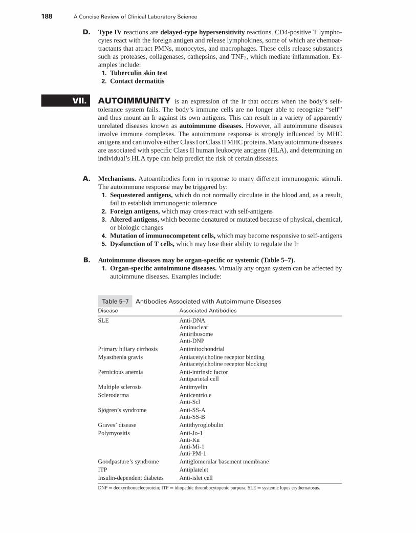
P1: OSO
LWBK192-05 LWBK192-Hubbard November 27, 2008 7:45
188 A Concise Review of Clinical Laboratory Science
D. Type IV reactions are delayed-type hypersensitivity reactions. CD4-positive T lympho-cytes react with the foreign antigen and release lymphokines, some of which are chemoat-tractants that attract PMNs, monocytes, and macrophages. These cells release substancessuch as proteases, collagenases, cathepsins, and TNF?, which mediate inflammation. Ex-amples include:1. Tuberculin skin test2. Contact dermatitis
VII. AUTOIMMUNITY is an expression of the Ir that occurs when the body’s self-tolerance system fails. The body’s immune cells are no longer able to recognize “self”and thus mount an Ir against its own antigens. This can result in a variety of apparentlyunrelated diseases known as autoimmune diseases. However, all autoimmune diseasesinvolve immune complexes. The autoimmune response is strongly influenced by MHCantigens and can involve either Class I or Class II MHC proteins. Many autoimmune diseasesare associated with specific Class II human leukocyte antigens (HLA), and determining anindividual’s HLA type can help predict the risk of certain diseases.
A. Mechanisms. Autoantibodies form in response to many different immunogenic stimuli.The autoimmune response may be triggered by:
1. Sequestered antigens, which do not normally circulate in the blood and, as a result,fail to establish immunogenic tolerance
2. Foreign antigens, which may cross-react with self-antigens3. Altered antigens, which become denatured or mutated because of physical, chemical,
or biologic changes4. Mutation of immunocompetent cells, which may become responsive to self-antigens5. Dysfunction of T cells, which may lose their ability to regulate the Ir
B. Autoimmune diseases may be organ-specific or systemic (Table 5–7).1. Organ-specific autoimmune diseases. Virtually any organ system can be affected by
autoimmune diseases. Examples include:
Table 5–7 Antibodies Associated with Autoimmune Diseases
Disease Associated Antibodies
SLE Anti-DNAAntinuclearAntiribosomeAnti-DNP
Primary biliary cirrhosis Antimitochondrial
Myasthenia gravis Antiacetylcholine receptor bindingAntiacetylcholine receptor blocking
Pernicious anemia Anti-intrinsic factorAntiparietal cell
Multiple sclerosis Antimyelin
Scleroderma AnticentrioleAnti-Scl
Sjogren’s syndrome Anti-SS-AAnti-SS-B
Graves’ disease Antithyroglobulin
Polymyositis Anti-Jo-1Anti-KuAnti-Mi-1Anti-PM-1
Goodpasture’s syndrome Antiglomerular basement membrane
ITP Antiplatelet
Insulin-dependent diabetes Anti-islet cell
DNP = deoxyribonucleoprotein; ITP = idiopathic thrombocytopenic purpura; SLE = systemic lupus erythematosus.
P1: OSO
LWBK192-05 LWBK192-Hubbard November 27, 2008 7:45
CHAPTER 5 Immunology and Serology 189
a. Hematologic disorders(1) Paroxysmal cold hemoglobinuria(2) Warm autoimmune hemolytic anemias (AIHAs)(3) Immune thrombocytopenia purpura (ITP)
b. Endocrine disorders(1) Graves’ diseases(2) Hashimoto’s thyroiditis(3) Insulin-dependent diabetes mellitus
c. Neuromuscular disorders(1) Myasthenia gravis(2) Multiple sclerosis
d. Renal disorders(1) Goodpasture’s syndrome(2) Tubulo-interstitial nephritis
e. Gastrointestinal disorders(1) Pernicious anemia(2) Primary biliary cirrhosis
2. Systemic diseases are characterized by widespread involvement. Autoantibodies arenot confined to any one specific organ. Examples include:a. Rheumatoid arthritis (RA)
(1) General considerations. RA is characterized by the presence of abnormalcirculating IgM autoantibodies known collectively as rheumatoid factor (RF).These antibodies react specifically with the Fc portion of IgG molecules. RFmay also be seen in patients with chronic hepatitis, systemic lupus erythe-matosus (SLE), syphilis, hypogammaglobulinemia, or hypergammaglobuli-nemia.
(2) Testing. There are a variety of tests for RF, including:(a) Latex fixation(b) Latex agglutination (commonly used test; considered positive for RF if the
titer is at least 20)(c) Sheep red cell agglutination(d) Quantitation of IgM RF
b. Polymyositis(1) Description. Polymyositis is characterized by inflammation and degeneration
of skeletal muscle.(2) Autoantibodies. Antinuclear antibodies (ANA), RF, and anti-Jo-1 antibodies
are present.c. SLE
(1) Description. SLE is a systemic rheumatic disorder that is characterized by thepresence of circulating immune complexes.(a) Epidemiology. It is most commonly seen in women and persons of African
descent.(b) Symptoms. More than half of SLE patients develop a characteristics rash
or other skin abnormalities during the course of the disease. Other com-mon symptoms include myocarditis, lymphadenopathy, glomerulonephri-tis, and serositis.
(c) Causes. SLE may be idiopathic or drug-induced. Drugs, such as pro-cainamide, phenytoin, methyldopa, penicillin, and sulfonamides, maycause a lupus-like syndrome. ANA are present and may persist for monthsafter the drug has been discontinued.
(2) Pathophysiology. Antibodies to native or altered self-antigens are producedand form circulating immune complexes with the antigens. Typically, the anti-gens involved are nuclear antigens, especially deoxyribonucleic acid (DNA).These complexes are deposited in various organs and cause inflammation,which eventually results in tissue injury.
(3) ANA are autoantibodies that react with the body’s nuclear proteins, DNA, orhistones. In general, they are neither organ-specific nor species-specific, and
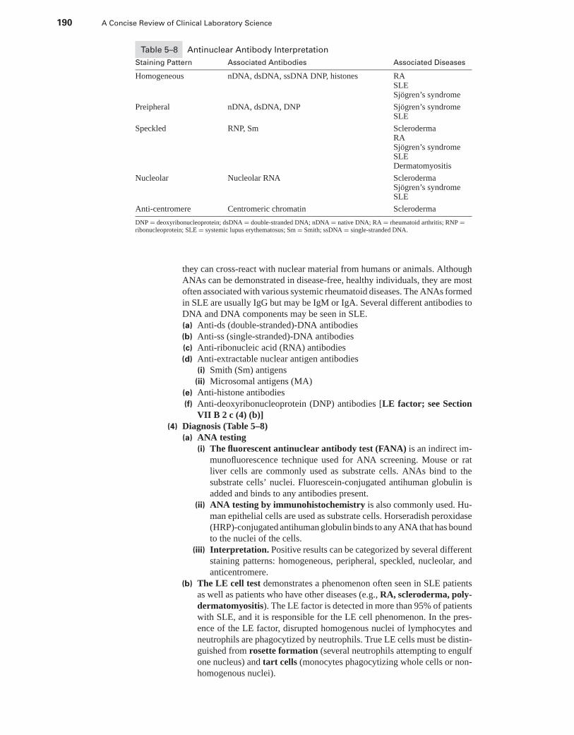
P1: OSO
LWBK192-05 LWBK192-Hubbard November 27, 2008 7:45
190 A Concise Review of Clinical Laboratory Science
Table 5–8 Antinuclear Antibody Interpretation
Staining Pattern Associated Antibodies Associated Diseases
Homogeneous nDNA, dsDNA, ssDNA DNP, histones RASLESjogren’s syndrome
Preipheral nDNA, dsDNA, DNP Sjogren’s syndromeSLE
Speckled RNP, Sm SclerodermaRASjogren’s syndromeSLEDermatomyositis
Nucleolar Nucleolar RNA SclerodermaSjogren’s syndromeSLE
Anti-centromere Centromeric chromatin Scleroderma
DNP = deoxyribonucleoprotein; dsDNA = double-stranded DNA; nDNA = native DNA; RA = rheumatoid arthritis; RNP =ribonucleoprotein; SLE = systemic lupus erythematosus; Sm = Smith; ssDNA = single-stranded DNA.
they can cross-react with nuclear material from humans or animals. AlthoughANAs can be demonstrated in disease-free, healthy individuals, they are mostoften associated with various systemic rheumatoid diseases. The ANAs formedin SLE are usually IgG but may be IgM or IgA. Several different antibodies toDNA and DNA components may be seen in SLE.(a) Anti-ds (double-stranded)-DNA antibodies(b) Anti-ss (single-stranded)-DNA antibodies(c) Anti-ribonucleic acid (RNA) antibodies(d) Anti-extractable nuclear antigen antibodies
(i) Smith (Sm) antigens(ii) Microsomal antigens (MA)
(e) Anti-histone antibodies(f) Anti-deoxyribonucleoprotein (DNP) antibodies [LE factor; see Section
VII B 2 c (4) (b)](4) Diagnosis (Table 5–8)
(a) ANA testing(i) The fluorescent antinuclear antibody test (FANA) is an indirect im-
munofluorescence technique used for ANA screening. Mouse or ratliver cells are commonly used as substrate cells. ANAs bind to thesubstrate cells’ nuclei. Fluorescein-conjugated antihuman globulin isadded and binds to any antibodies present.
(ii) ANA testing by immunohistochemistry is also commonly used. Hu-man epithelial cells are used as substrate cells. Horseradish peroxidase(HRP)-conjugated antihuman globulin binds to any ANA that has boundto the nuclei of the cells.
(iii) Interpretation. Positive results can be categorized by several differentstaining patterns: homogeneous, peripheral, speckled, nucleolar, andanticentromere.
(b) The LE cell test demonstrates a phenomenon often seen in SLE patientsas well as patients who have other diseases (e.g., RA, scleroderma, poly-dermatomyositis). The LE factor is detected in more than 95% of patientswith SLE, and it is responsible for the LE cell phenomenon. In the pres-ence of the LE factor, disrupted homogenous nuclei of lymphocytes andneutrophils are phagocytized by neutrophils. True LE cells must be distin-guished from rosette formation (several neutrophils attempting to engulfone nucleus) and tart cells (monocytes phagocytizing whole cells or non-homogenous nuclei).
P1: OSO
LWBK192-05 LWBK192-Hubbard November 27, 2008 7:45
CHAPTER 5 Immunology and Serology 191
VIII. IMMUNODEFICIENCIES. Deficiencies of the immune system include disor-ders of phagocytic cells, B lymphocytes, a combination of T and B lymphocytes, and thecomplement system.
A. Phagocytic cell deficiencies1. Phagocytic cell deficiencies result in a decreased ability to phagocytize and kill bacteria.
a. Chronic granulomatous disease (CGD) is a genetic disease characterized byineffective killing of bacteria by neutrophils.(1) Cause. CGD is caused by a defect in cytochrome b, which results in decreased
hydrogen peroxide production. Hydrogen peroxide is necessary for produc-ing the toxic superoxides that are critical in bacterial killing.
(2) Diagnosis. The nitroblue tetrazolium (NBT) reductase test is used to detectimpaired neutrophil phagocytosis. The neutrophils of CGD patients fail toreduce the NBT dye.
(3) Symptoms. Patients with CGD suffer from recurrent infections caused bycatalase-positive bacteria and yeast and fungi.
(4) Treatment includes the use of GM-CSF or G-CSF and IFN-γ .b. MPO deficiency is inherited as an autosomal recessive trait and is one of the most
common inherited disorders. The MPO in the primary granules of neutrophils isdecreased or absent, and although phagocytosis takes place normally, bacte-rial killing is inefficient. Fungal killing is more seriously impaired than bacterialkilling. Although otherwise healthy patients with MPO deficiency do not have anincreased frequency of infection, diabetic patients who have this disorder may havean increase in Candida sp infections.
c. Glucose-6-phosphate dehydrogenase (G6PD) deficiency is another inherited dis-order in which the aerobic system of neutrophils is impaired. This deficiency resultsin a substantial decrease in the amount of hydrogen peroxide produced duringphagocytosis, and thus decreased bacterial killing efficiency. Patients with G6PDdeficiency experience recurrent bacterial infections.
d. CR3 (iC3b receptor) deficiency is a rare, autosomal recessive trait character-ized by a decrease or absence of specific complement component receptorson neutrophils, monocytes, and lymphocytes. These receptors are responsible foradherence-related functions. Abnormalities result in defective margination anddiapedesis of neutrophils, impaired chemotaxis, and ineffective phagocyto-sis. T lymphocytes adhere poorly to target cells. Clinically, there is an increasedfrequency of bacterial infections, a decreased inflammatory response, and neu-trophilia.
e. Specific granule deficiency is inherited as an autosomal recessive trait. Neu-trophils fail to develop specific granules during myelopoiesis, and as a result,patients who have this disorder experience severe, recurrent bacterial infections.
f. Chediak-Higashi syndrome is an inherited disorder that is characterized by theabnormal fusion of primary granules in neutrophils. During phagocytosis, de-granulation is impaired, and little or no MPO is released into the phagosome.Patients who have Chediak-Higashi syndrome have recurrent bacterial infectionsand are also characterized by albinism and extreme photosensitivity.
g. Lazy leukocyte syndromes include:(1) Job syndrome, also known as hyperimmunoglobulin E, is characterized by
poor chemotaxis and recurrent skin infections and abscesses.(2) Tuftsin deficiency. Tuftsin is a chemotaxin that also improves phagocyte
motility, engulfment, and oxidative metabolism. Affected persons experiencerecurrent bacterial infections.
(3) Actin dysfunction. A deficiency of the cytoskeletal protein actin can re-sult in decreased cell motility and chemotaxis. Patients experience recurrentbacterial infections.
B. B-lymphocyte immunodeficiencies may be inherited or acquired and account for morethan half of all immunodeficiencies. Affectedness varies widely, depending on the class
P1: OSO
LWBK192-05 LWBK192-Hubbard November 27, 2008 7:45
192 A Concise Review of Clinical Laboratory Science
of immunoglobulin that is deficient. A deficiency of a minor immunoglobulin, such as IgD,causes little if any increase in the incidence of bacterial infections. However, because 75%to 85% of total immunoglobulin is IgG, an individual deficient in IgG would be significantlyaffected.
1. Bruton’s agammaglobulinemia is a sex-linked disorder that primarily affects men.It is usually recognized early in life when antibodies fail to develop. Pre-B cells maybe found in the bone marrow, but they do not mature. Few mature B cells are found inthe peripheral blood. Gamma globulin levels are markedly decreased. This disordermay be treated with gamma globulin preparations.
2. Common variable hypogammaglobulinemia is an acquired disorder in which oneor two immunoglobulin classes are deficient. Total immunoglobulin levels may benormal, because a decrease in one immunoglobulin is often compensated by an increasein the production of another. Selective IgA deficiency is one of the most common ofthese deficiencies. Typically, only those patients whose disease includes IgG deficiencysuffer from increased bacterial infections.
3. Neonatal hypogammaglobulinemia is caused by the normal immaturity of theneonate’s immune system. It corrects itself between the ages of 6 and 12 monthsas the infant’s immune system matures.
C. T-lymphocyte immunodeficiencies without an accompanying loss of B-cell function arerare, composing only 7% of all immunodeficiencies. These disorders may be acquired orinherited.
1. DiGeorge syndrome results when the thymus gland develops abnormally during em-bryogenesis. Abnormalities of other endoderm-derived tissues are also seen. T lympho-cytes are usually decreased, but may be normal. Most patients have a high CD4-CD8ratio. Although antibody responses may be normal, cell-mediated immune responsesare impaired.
2. Nezelof syndrome is an autosomal recessive disorder. Patients are athymic and areespecially susceptible to viral and fungal infections, which can be fatal in these patients.
D. Combined B- and T-lymphocyte immunodeficiencies are the most serious of the immun-odeficiencies, because both cell-mediated and humoral immune responses are affected.
1. Bare-lymphocyte syndromes are characterized by defects in Class I MHC antigenexpression, Class II MHC antigen expression, or a combination of both. CD4-positiveT lymphocytes are decreased in number, and B- and T-cell activation is reduced.
2. Severe combined immunodeficiency disease may be inherited as autosomal recessiveor X-linked traits. All are characterized by markedly decreased numbers of both T andB lymphocytes.
3. Acquired immunodeficiency syndrome (AIDS) is caused by the human immunod-eficiency virus 1 (HIV-1) or the human immunodeficiency virus 2 (HIV-2). TheCD4-positive T lymphocytes are the primary target cells. Approximately 5% of Blymphocytes are also infected.a. Transmission of the HIV virus is primarily through sexual contact with infected
persons and parenteral routes (e.g., transfusion of infected blood and blood prod-ucts). Other body fluids may also transmit the virus.
b. There are three stages of clinical manifestation of HIV infection.(1) The primary stage may last from many months to years. Infected persons may
experience an initial flulike illness, and then remain asymptomatic or exhibitonly chronic lymphadenopathy for many years.
(2) The intermediate stage is also known as AIDS-related complex (ARC).There are physical symptoms but no opportunistic infections.
(3) The final stage, AIDS, is usually seen within 2 to 10 years after initial in-fection with the HIV virus. Opportunistic infections are common and includePneumocystis carinii pneumonia, candidiasis, and histoplasmosis.
c. Effects on the immune system primarily involve CD4-positive T lymphocytes.(1) The CD4:CD8 ratio is reduced from normal (2:1 to 0.5:1).(2) CD4-positive T-cell counts are decreased.
P1: OSO
LWBK192-05 LWBK192-Hubbard November 27, 2008 7:45
CHAPTER 5 Immunology and Serology 193
(3) CD4-positive T-cell function is impaired.(4) B-cell activation is abnormal.(5) Macrophage function is impaired.(6) NK cell function is impaired.
d. Laboratory detection of HIV infection. A number of tests have been developedto detect the presence of HIV antigens, antibodies, DNA, and RNA.(1) Tests for antigens. The presence of HIV antigen indicates active infection.
(a) The HIV isolation technique can detect antigens before antibodies de-velop and can be used to monitor antiviral treatment. The patient’s mono-cytes are stimulated and grown in culture. The culture supernatant can betested for the presence of antigens.
(b) A modified enzyme-linked immunosorbent assay (ELISA) can be per-formed on plasma, serum, cerebrospinal fluid (CSF), and culture fluids todetect the presence of HIV antigens.
(c) Immunofluorescence assay (IFA) can be used to detect HIV antigens.(2) Tests for antibodies. The presence of HIV antibodies indicates prior exposure
to the HIV antigen. Antibody development follows a predictable course fol-lowing exposure. The p24 core protein is the major structural protein of theHIV-1 virus. IgM antibody against the p24 core protein usually developswithin 6 to 8 weeks after infection. Within weeks, IgG antibodies against p24appear, as do antibodies against envelope gene products (i.e., gp160, gp120,gp41) and polymerase gene products (i.e., p31, p51, p66).(a) The ELISA is the most commonly performed screening test for HIV an-
tibodies. Both false-positive and false-negative results may occur. Repeat-edly reactive results necessitate confirmatory testing.
(b) Western blot assays are the most commonly used confirmatory test forHIV antibodies. Protein from disrupted virus is separated onto polyacry-lamide gels by electrophoresis. False-positive reactions may occur.
(c) Other tests for HIV antibodies include:(i) IFA
(ii) Radioimmunoassay (RIA)(iii) Radioimmunoprecipitation assay(iv) Slide agglutination tests
(3) Tests for DNA and RNA(a) In situ hybridization and filter hybridization can be used to detect viral
RNA. Cells from peripheral blood and lymph nodes can be used.(b) Southern blot hybridization can be used to detect viral DNA in infected
cells.(c) Polymerase chain reaction (PCR) techniques can be used to amplify
both RNA and DNA, which provides extremely sensitive systems for RNAand DNA detection.
IX. TECHNIQUES IN IMMUNOLOGY AND SEROLOGY
A. Agglutination assays demonstrate the presence of antigen-antibody reactions by the visibleaggregation of antigen-antibody complexes. These tests are simple to perform and are oftenthe most sensitive test method.
1. Flocculation tests. Antibody is detected when soluble antigen interacts with antibodyand a precipitate is formed. Antigen is bound to reagent particles; visible agglutinationresults when the particles bind to antibody.
2. Latex agglutination. Antibody is bound to latex beads; visible agglutination occurswhen antigen binds to the latex-bound antibody.
3. Direct bacterial agglutination. Antibodies bind to the surface antigens of bacteria insuspension, which results in visible agglutination.
4. Hemagglutination tests are used to detect antibodies to red blood cell (RBC) antigens.In passive or indirect hemagglutination tests, soluble antigens are adsorbed onto the
P1: OSO
LWBK192-05 LWBK192-Hubbard November 27, 2008 7:45
194 A Concise Review of Clinical Laboratory Science
surface of RBCs. These antigens then bind to any corresponding antibody present, andthe RBCs agglutinate.
5. Agglutination inhibition assays are very sensitive and can detect small amounts ofantigen. In hemagglutination inhibition (HAI) assays, antigen and antibody are boundto RBCs. If antibody is present in the test sample, agglutination does not occur becausethere is already antibody bound to the antigen. Agglutination is a negative result. Inother assays, antigen and antibody are bound to latex particles.
B. Precipitation assays. When both are present in proper proportions, antigens and antibodiesinteract and form visible precipitates. The largest amount of precipitation is seen whenantigens and antibodies are present in optimal proportions; this is known as the equivalencezone. False-negative reactions can occur when either antigen or antibody is present in excess.(Web Color Images 5–12 and 5–13)
1. Double immunodiffusion (Ouchterlony method). Antigens and antibodies are al-lowed to diffuse in a semisolid medium such as agar or agarose. When the antigensand antibodies meet, a precipitate forms. Three basic reaction patterns can result fromthis interaction (Web Color Image 5–14).a. Identity. A single smooth arc of precipitation forms between the antigens and
antibodies, which indicates that the antibodies are precipitating identical antigenspecificities.
b. Nonidentity. Two separate lines of precipitation cross each other, which in-dicates that the antigens and antibodies are unrelated and do not precipitatetogether.
c. Partial identity. The two precipitating lines meet, forming a spur. This indicatesthat the antigens share some common epitopes, but that one of the antigens has aunique epitope.
2. Radial immunodiffusion (RID) is a single diffusion method that can be used to quan-titate immunoglobulins and other serum proteins. Samples are introduced into wellscut in agarose containing antiserum and allowed to diffuse, usually overnight. The di-ameters of the resulting precipitation rings correspond to the amount of antigen present(Web Color Image 5–15).
3. Electroimmunodiffusion combines the speed of electrophoresis with the sensitivityof immunodiffusion.a. Countercurrent immunoelectrophoresis (CIE). This technique is essentially
a double diffusion technique in which voltage is applied to move the antigensand antibodies together. A precipitin band forms when a zone of equivalence isreached.
b. Rocket electrophoresis. This technique applies an electrical charge to a RID assay,which results in a rocket-shaped line of precipitation. The height of the rocket isproportional to the antigen concentration.
C. Electrophoresis1. Immunoelectrophoresis (IEP) is often used to diagnose monoclonal gammopathies.
After the serum or urine specimen is electrophoresed in a gel medium, a trough iscut in the agar parallel to the line of separated proteins. Monoclonal or polyclonalantisera are loaded into the trough, and the gels are incubated to allow the antigensand antibodies to diffuse toward each other. Precipitation lines become visible whena zone of equivalence is reached. Typically, control serum is run above the trough,and the patient sample is run below the trough for easy comparison (Web ColorImage 5–16).
2. Immunofixation electrophoresis (IFE). After serum, urine, or CSF samples areelectrophoresed in an agarose gel, cellulose acetate impregnated with antiserum isplaced on the gel. The antiserum from the cellulose acetate diffuses into the gel, andantigen-antibody precipitates form. The cellulose acetate is stained to visualize theprecipitation bands. IFE and IEP are often used together to work up monoclonal gam-mopathies.
P1: OSO
LWBK192-05 LWBK192-Hubbard November 27, 2008 7:45
CHAPTER 5 Immunology and Serology 195
D. Labeling immunoassays can be qualitative or quantitative and can occur in the solubleor solid phase. Radioactive, enzyme, chemiluminescent, or fluorescent labels can beused.
1. RIA is a rapid and sensitive method that can be used to detect small amounts of antigenor antibody. However, exposure to radioisotopes can damage DNA and lead to radiationsickness, an increased incidence of neoplasms, or death. These potential hazards are adisadvantage to the RIA method (Web Color Image 5–17).
2. Radioallergosorbent test (RAST) is an RIA method specifically designed to measureantigen-specific IgE.
3. Radioimmunosorbent test (RIST) is a competitive binding technique used to quan-titate IgE.
4. ELISA is similar in principle to RIA and has the same sensitivity, but uses an enzymelabel instead of a radioactive label. HRP and alkaline phosphatase (ALP) are the mostcommonly used enzymes. ELISAs can be used to detect extremely small amounts ofantigen or antibody (Web Color Image 5–18).
5. IFA are often used to identify antigens in tissue sections or air-dried smears of pe-ripheral blood, bone marrow aspirates, touch preparations, or fine needle aspirate sam-ples.a. Labeling. Fluorescein isothiocyanate is the most commonly used fluorescent la-
bel. Labeling may be direct (using a labeled antibody) or indirect (using a labeledsecondary antibody). After staining, the slides are washed and dried and immedi-ately examined using a fluorescence microscope. Fluorescence quenches quicklywhen exposed to light, which is the major disadvantage of immunofluorescencemethods (Web Color Image 5–19).
b. Immunocytochemical techniques are modifications of the direct and indirect im-munofluorescence methods; enzyme labels such as HRP and ALP are used insteadof fluorescent labels. These reaction products do not fade, so slides may be storedto provide permanent records.
E. Nephelometry (turbidimetry) photometrically measures the turbidity of solutions createdby particles in suspension. It is accurate, rapid, and precise and is often used to quanti-tate immunoglobulins, complement components, and immune complexes. This methodcan also be used to measure antigen concentration. The light source in nephelometryinstruments produces a wave-length of 840 nm.
F. Neutralization tests. Neutralizing antibodies can destroy the infectivity of viruses, whichprovides the basis for assays that can determine the amount of viral antibody present. Thesetechniques are often used to detect antibodies against herpes simplex virus Types 1 and 2(HSV-1 and HSV-2) and echovirus.
G. Cellular assays. A wide variety of techniques exists for assessing the function of the cellsof the immune system.
1. Functional assays. These tests are rarely performed except in large medical centersand reference laboratories. Some tests are even considered obsolete. Functional assaysinclude the following.a. ADCC assays use bacteria-infected tumor cells to assess the killing ability of NK
cells.b. The Boyden chamber uses chemotactic substances to assess the chemotactic re-
sponse of neutrophils.c. Cell-mediated monocytolysis uses tumor cells to assess the killing functions of
monocytes.d. Latex bead ingestion assesses phagocytic activity.e. Lymphocyte transformation tests assess the ability of lymphocytes to response
to mitogens or specific antigens.f. Lympholysis tests assess the ability of Tc to lyse labeled target cells.
g. Microcytotoxicity studies are used to detect HLA antigens and antibodies.
P1: OSO
LWBK192-05 LWBK192-Hubbard November 27, 2008 7:45
196 A Concise Review of Clinical Laboratory Science
h. Migration inhibition techniques determine the ability of lymphocytes to producechemotactic factors in response to granulocytes and monocytes.
i. Mixed-lymphocyte cultures are used to assess the human leukocyte antigen D(HLA-D) compatibility of donor and recipient lymphocytes.
j. NBT reductase tests are used to assess the intracellular killing ability of neu-trophils.
k. Phagocytosis assays mix bacteria with neutrophils to assess the cells’ phagocyticability.
2. Flow cytometry can be used to identify subpopulations of cells such as reticulocytes,granulocytes, T-cell subsets, B lymphocytes, and others. Fluorescent dyes, such asfluorescein, acridine orange, and phycoerythrin, are bound specifically to the cellmarker of interest. Cells in suspension pass singly through a laminar-flow saline sheath.As the stained cells pass through a laser beam (usually argon or krypton), the dye isactivated, and the cell fluoresces. The fluorescence is detected and collected by sensorsplaced at 90◦ relative to the source beam, and the information is processed by a computer(Web Color Image 5–20).
X. SYPHILIS SEROLOGY
A. Human syphilis is caused by the spirochete Treponema pallidum. Antibodies against tre-ponemal antigens and nontreponemal cardiolipin antigens (Wasser-mann antigens) developand elicit a cell-mediated and humoral Ir, which results in the formation of immune com-plexes.
B. Disease. Sexual contact with infected persons is the most common form of transmissionof human syphilis. Transmission through blood or blood-product transfusion can occurbut is rare now because of effective pretransfusion testing. In addition, syphilis can bepassed from an infected pregnant woman to her fetus. There are four clinical stages ofdisease.
1. Primary (early) syphilis. Inflammatory lesions (chancres) appear 2 to 8 weeksafter infection and last for 1 to 5 weeks. Serum tests for syphilis are positivein 90% of patients after 3 weeks. The antibodies that develop are predominantlyIgM.
2. Secondary syphilis usually occurs 6 to 8 weeks after chancres first appear. This stageis characterized by a generalized rash, and secondary lesions may develop in the eyes,joints, or central nervous system (CNS). These lesions are highly contagious, but healspontaneously within 2 to 6 weeks. Serologic tests are positive in secondary syphilis.Antibodies are mostly IgG.
3. The latent stage of syphilis is contagious and is generally considered to begin afterthe second year of infection. There are no clinical symptoms, although serologic testsare still positive. After 4 years, syphilis is not usually contagious; however, the diseasemay still be transmitted from mother to fetus.
4. Tertiary syphilis is characterized by granulomatous lesions known as gummata. Theselesions may develop in skin, mucous membranes, joints, muscles, and bones, causinglittle or no clinical problems. Approximately 80% of patients experience CNS involve-ment, which can result in paralysis or dementia. Approximately 10% of patients developcardiovascular problems, which can result in aortic aneurysm.
C. Congenital syphilis. Syphilis can be transmitted to a fetus after the 18th week of gestation.Treatment of the infected mother before the 18th week will prevent infection; treatmentafter the 18th week will cure it.
D. Treatment. Syphilis is easily and effectively treated. Penicillin is the drug of choice, al-though tetracycline or erythromycin can also be used. Treatment may or may not result inserologic tests becoming nonreactive, depending on the stage of the disease at the time oftreatment.
P1: OSO
LWBK192-05 LWBK192-Hubbard November 27, 2008 7:45
CHAPTER 5 Immunology and Serology 197
1. Primary stage. A seropositive patient in the primary stage of disease usually becomesnonreactive approximately 6 months after treatment.
2. Secondary stage. If treatment occurs during the secondary stage, the patient usuallybecomes nonreactive within 12 to 18 months after treatment. Patients treated 10 yearsor more after infection may always remain seropositive.
E. Tests for syphilis are based on the detection of nontreponemal antibodies or treponemalantibodies.
1. Nontreponemal antibody detection. Reagin antibodies are formed after exposure toTreponema pallidum and react with lipoidal antigens used in screening tests for syphilis.The most commonly performed regain tests are:a. The Venereal Disease Research Laboratory (VDRL) slide test. The VDRL is
a qualitative agglutination test using heat-inactivated patient serum. CSF can alsobe used. The test can be modified and used as a quantitative test.
b. The rapid plasma reagin (RPR) test. The RPR is an agglutination test. In addi-tion to lipoidal antigens and cholesterol, the antigen reagent contains charcoal tofacilitate macroscopic interpretation of results. Unheated serum is the specimen ofchoice, although plasma may be used.
c. Results. VDRL and RPR test reactions are graded as nonreactive (NR), weaklyreactive (WR), or reactive (R). False-positive and false-negative reactions mayoccur.(1) False-positive reactions may be caused by:
(a) SLE(b) RA(c) Infectious mononucleosis(d) Pregnancy(e) Old age
(2) False-negative reactions may be caused by:(a) Technical errors(b) Low antibody titers(c) Prozone phenomenon
2. Treponemal antibody detection. The serum of patients who have syphilis also containsan antibody distinct from the antireagin antibody. Tests for these antibodies are used asconfirmatory tests when reactive results are obtained using screening methods. Thesetests are also used to determine antibody titers. The most commonly used treponemaltests are:a. The fluorescent treponemal antibody absorption test (FTA-ABS). The FTA-
ABS test detects treponemal antibodies by using a killed suspension of T. pallidumas an antigen and a fluorescein-conjugated antihuman globulin reagent.
b. The microhemagglutination assay for T. pallidum (MHA-TP) uses RBCs coatedwith treponemal antigens to detect antibodies.
XI. ACUTE PHASE PROTEINS are a group of plasma proteins whose levelsincrease significantly and independently during the acute phase of an inflammatory process.They are primarily produced in the liver by the parenchymal cells.
A. α1-antitrypsin is a serine protease inhibitor that inhibits the action of specific proteasesthat cause lung damage and pulmonary inflammation. Heterozygous deficiencies result inincreased risk of liver disease, glomerulonephritis, and connective tissue diseases. Homozy-gous deficiencies result in premature emphysema and liver disease.
B. α2-macroglobulin is a protease inhibitor that plays a role in the coagulation, fibrinolytic,and complement components of hemostasis.
C. Ceruloplasmin is a major copper-transport protein. An absence or deficiency of cerulo-plasmin is associated with Wilson’s disease.
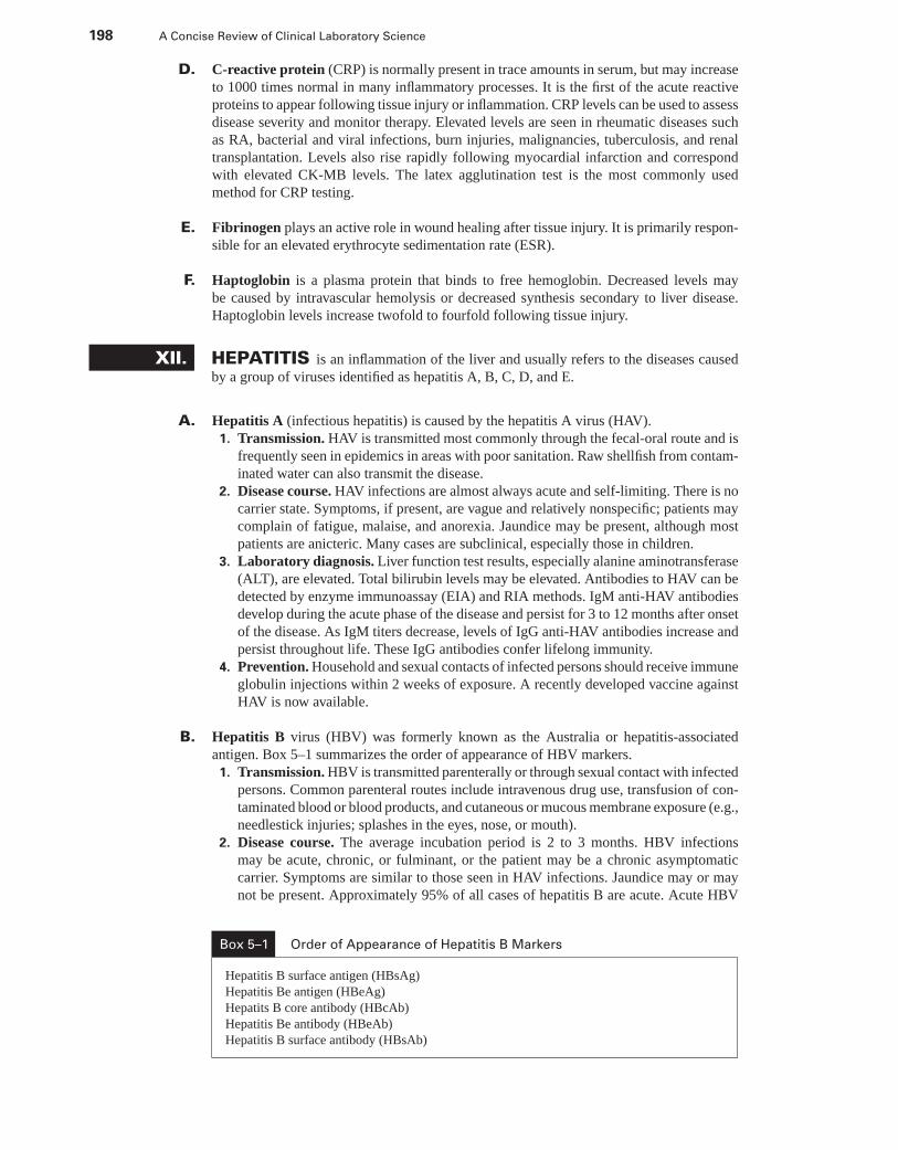
P1: OSO
LWBK192-05 LWBK192-Hubbard November 27, 2008 7:45
198 A Concise Review of Clinical Laboratory Science
D. C-reactive protein (CRP) is normally present in trace amounts in serum, but may increaseto 1000 times normal in many inflammatory processes. It is the first of the acute reactiveproteins to appear following tissue injury or inflammation. CRP levels can be used to assessdisease severity and monitor therapy. Elevated levels are seen in rheumatic diseases suchas RA, bacterial and viral infections, burn injuries, malignancies, tuberculosis, and renaltransplantation. Levels also rise rapidly following myocardial infarction and correspondwith elevated CK-MB levels. The latex agglutination test is the most commonly usedmethod for CRP testing.
E. Fibrinogen plays an active role in wound healing after tissue injury. It is primarily respon-sible for an elevated erythrocyte sedimentation rate (ESR).
F. Haptoglobin is a plasma protein that binds to free hemoglobin. Decreased levels maybe caused by intravascular hemolysis or decreased synthesis secondary to liver disease.Haptoglobin levels increase twofold to fourfold following tissue injury.
XII. HEPATITIS is an inflammation of the liver and usually refers to the diseases causedby a group of viruses identified as hepatitis A, B, C, D, and E.
A. Hepatitis A (infectious hepatitis) is caused by the hepatitis A virus (HAV).1. Transmission. HAV is transmitted most commonly through the fecal-oral route and is
frequently seen in epidemics in areas with poor sanitation. Raw shellfish from contam-inated water can also transmit the disease.
2. Disease course. HAV infections are almost always acute and self-limiting. There is nocarrier state. Symptoms, if present, are vague and relatively nonspecific; patients maycomplain of fatigue, malaise, and anorexia. Jaundice may be present, although mostpatients are anicteric. Many cases are subclinical, especially those in children.
3. Laboratory diagnosis. Liver function test results, especially alanine aminotransferase(ALT), are elevated. Total bilirubin levels may be elevated. Antibodies to HAV can bedetected by enzyme immunoassay (EIA) and RIA methods. IgM anti-HAV antibodiesdevelop during the acute phase of the disease and persist for 3 to 12 months after onsetof the disease. As IgM titers decrease, levels of IgG anti-HAV antibodies increase andpersist throughout life. These IgG antibodies confer lifelong immunity.
4. Prevention. Household and sexual contacts of infected persons should receive immuneglobulin injections within 2 weeks of exposure. A recently developed vaccine againstHAV is now available.
B. Hepatitis B virus (HBV) was formerly known as the Australia or hepatitis-associatedantigen. Box 5–1 summarizes the order of appearance of HBV markers.
1. Transmission. HBV is transmitted parenterally or through sexual contact with infectedpersons. Common parenteral routes include intravenous drug use, transfusion of con-taminated blood or blood products, and cutaneous or mucous membrane exposure (e.g.,needlestick injuries; splashes in the eyes, nose, or mouth).
2. Disease course. The average incubation period is 2 to 3 months. HBV infectionsmay be acute, chronic, or fulminant, or the patient may be a chronic asymptomaticcarrier. Symptoms are similar to those seen in HAV infections. Jaundice may or maynot be present. Approximately 95% of all cases of hepatitis B are acute. Acute HBV
Box 5–1 Order of Appearance of Hepatitis B Markers
Hepatitis B surface antigen (HBsAg)
Hepatitis Be antigen (HBeAg)
Hepatits B core antibody (HBcAb)
Hepatitis Be antibody (HBeAb)
Hepatitis B surface antibody (HBsAb)
P1: OSO
LWBK192-05 LWBK192-Hubbard November 27, 2008 7:45
CHAPTER 5 Immunology and Serology 199
infections can be complicated by circulating HBV antigen-antibody complexes, whichcan cause polyarteritis, arthritis, glomerulonephritis, pancreatitis, or cryoglobulinemia.Chronic hepatitis B can progress to cirrhosis, which may later progress to hepatocellularcarcinoma.
3. Laboratory diagnosis. Elevated ALT levels peak approximately the same time symp-toms appear. Serologic tests not only identify HBV as the cause of the disease, but canalso be used to pinpoint the stage of the disease.a. Hepatitis B surface antigens (HBsAg) appear 1 week to 2 months after exposure,
and approximately 2 weeks to 2 months before the onset of symptoms. HBsAgdisappears during the convalescent phase of acute disease and is an indicator ofacute infection or chronic infection with unresolved antigenemia.
b. Hepatitis B surface antibodies (HBsAb) appear as the antigens disappear. Thepresence of these antibodies indicates recovery and lifelong immunity.
c. Hepatitis B core antibodies (HBcAb) appear shortly after the surface antigensappear. At this time, the ALT levels begin to rise. HBcAb persist throughout lifeand are a marker for previous infection.
d. Hepatitis Be antigen appears before the onset of clinical disease, after the ap-pearance of the surface antigens, and disappears within approximately 2 weeks.The presence of the Be antigen indicates active viral replication, and HBV is mostinfectious when the Be antigen is detectable.
e. Antibodies to hepatitis Be antigen appear shortly after the antigen disappears.Their presence in patients with acute hepatitis B suggests that the infection is re-solving. In most cases of chronic hepatitis B infection, the presence of hepatitisBe antibodies indicates that the infection is resolving or that there is no compli-cating liver disease. Hepatitis Be antibodies may be seen in chronic asymptomaticcarriers.
4. Prevention. Avoidance of high-risk behavior (e.g., intravenous drug abuse and sexualcontact with infected persons) is a major factor in preventing HBV infection. A vaccineagainst HBV has been available since 1982. In a health care setting, HBV vaccinationand the use of universal precautions can greatly reduce the risk of occupationallyacquired HBV. The HBV vaccine also protects against HDV infection, because HDVinfection only occurs with HBV infection.
C. Hepatitis C virus (HCV) has recently been identified as the causative agent in the majorityof cases of what was previously known as non-A, non-B hepatitis (see Web Case Study5–1).
1. Transmission. Like HBV, HCV is transmitted most commonly by parenteral routes orthrough sexual contact with infected persons. The majority of post-transfusion non-A,non-B hepatitis cases are caused by HCV.
2. Disease course. The average incubation period is 7 to 8 weeks. HCV infections maybe acute or chronic. Symptoms are similar to those seen in HAV and HBV infections.Approximately 50% of HCV-infected patients are chronic carriers. Approximately20% of these patients develop cirrhosis, and approximately 20% of those patientseventually develop hepatocellular carcinoma. HCV is also associated with immune-complex glomerulonephritis.
3. Laboratory diagnosis. Elevated ALT levels are associated with HCV infection. EIAand RIA assays have been developed to detect antibodies to the HCV antigens, buta high number of false-positive results are seen with these methods. Identifying in-fected persons is also difficult because antibody tests are positive in only 70% to85% of patients with post-transfusion HCV and in only 50% of patients with dis-ease from other causes. Additionally, it can take up to 12 months for an infectedperson to seroconvert and test positive for HCV antibodies. Serum or liver tissuecan be analyzed for HCV RNA by PCR, but this method largely remains a researchtool.
4. Prevention. As with HBV, avoiding high-risk behavior (e.g., intravenous drug abuseand sexual contact with infected persons) is important in preventing HCV infections.No vaccine for HCV currently exists.
P1: OSO
LWBK192-05 LWBK192-Hubbard November 27, 2008 7:45
200 A Concise Review of Clinical Laboratory Science
D. Hepatitis D is associated exclusively with HBV infections, either as a co-infection or as asuperinfection. It is seen most commonly in intravenous drug users and hemophiliacs. Novaccine is available. The only means of preventing hepatitis D infection is to prevent HBVinfection.
E. Hepatitis E causes sporadic and epidemic hepatitis in developing countries such as India,Pakistan, Africa, and Mexico. The disease clinically resembles HAV infections and istransmitted through the fecal-oral route. There is no chronic infection, and the hepatitis Evirus is not associated with hepatocellular carcinoma.
XIII. STREPTOCOCCAL SEROLOGY. Streptococcus pyogenes is a gram-positivecoccus responsible for a number of human infections, some of which can have serioussequelae. The M protein is the major virulence factor for S. pyogenes; more than 60 Mserotypes have been identified.
A. Bacterial toxins. Two hemolysins are produced by virtually all strains of S. pyogenes.1. Streptolysin O (SLO) is an oxygen-labile enzyme that causes hemolysis by binding
to cholesterol in the RBC membrane. It is antigenic, and the presence of antibodies toSLO is an indicator of recent streptococcal infection.
2. Streptolysin S is a nonantigenic, oxygen-stable enzyme. It causes hemolysis by dis-rupting the selective permeability of the RBC membrane.
B. Infections and sequelae1. Skin infections caused by S. pyogenes include:
a. Cellulitisb. Impetigoc. Erysipelas
2. Upper respiratory tract infections caused by S. pyogenes are characterized by fever,sore throat, and pharyngeal edema.
3. Scarlet fever is caused by a strain of S. pyogenes that produces an erythrogenic toxin,which results in a characteristic rash. Fever and sore throat are also present.
4. Rheumatic fever (RF) is a complication seen following upper respiratory tract in-fections. All M serotypes that cause pharyngitis have been implicated in RF. RFresults in damage to heart valves, and patients with rheumatic heart disease havean increased risk of developing endocarditis and other cardiac problems in lateryears.
5. Post-streptococcal glomerulonephritis may occur after pharyngitis or skin infections.Only a few M serotypes cause this type of glomerulonephritis. Patients have an increasedrisk of developing renal failure later.
6. The mechanisms by which these sequelae occur are not fully understood. Antibodiesto streptococcal cell membranes cross-react with myosin in cardiac muscle cells, whichresults in cell damage. Antigen-antibody complexes form at the glomerular basementmembrane and attract inflammatory cells that cause renal tissue damage.
C. Laboratory diagnosis. During an infection with S. pyogenes, the SLO produced elicits anIr, and specific antibodies are formed. These antibodies neutralize the hemolytic activityof the SLO; this neutralization provides the basis for the most commonly used test in thedetection of streptococcal infections.
1. Antistreptolysin O (ASO) titer. The ASO titer begins to increase approximately 7days after infection and peaks after 4 to 6 weeks.a. Principle. SLO is added to serial dilutions of patient serum, along with group O
RBCs as indicator cells. If the patient serum contains antibodies against SLO, theantibodies will complex with the corresponding antigens. These complexes blockthe hemolytic activity of the antigen, and no hemolysis occurs. The ASO titer isreported as the reciprocal of the highest dilution that shows no hemolysis and isexpressed in Todd units.
P1: OSO
LWBK192-05 LWBK192-Hubbard November 27, 2008 7:45
CHAPTER 5 Immunology and Serology 201
b. Normal values. ASO values vary widely among healthy individuals, making itdifficult to establish normal values. Most healthy adults have ASO titers of lessthan 166 Todd units, with the usual titer decreasing after 50 years of age. Althoughan elevated titer is generally regarded as evidence of recent streptococcal infection,a 30% rise in titer above a previous level is of greater significance than a singletiter. The titer can remain elevated for weeks or months following acute disease.Approximately 80% to 85% of patients with RF have increased ASO titers.
2. Anti-DNAse B (AD-B). Streptococci produce the enzyme deoxyribonuclease B(DNAse B). The anti-DN-B test is a neutralization test that can demonstrate recentstreptococcal infection. Anti-DN-B neutralizes the activity of DNAse B. Anti-DN-Blevels are increased in the 15% to 20% of RF patients who do not have elevated ASOtiters.
3. Other tests. Several rapid tests are now available to detect streptococcal antigens. Theadvantage is their speed over other test methods. However, a significant number offalse-negative results occur.
XIV. EPSTEIN-BARR VIRUS (EBV) SEROLOGY
A. Description. EBV is the causative agent of Burkitt’s lymphoma, nasopharyngeal carcinoma,and most commonly, infectious mononucleosis (IM). The virus is ubiquitous; 80% to 90%of healthy adults have EBV antibodies. EBV infects B lymphocytes.
B. Infectious mononucleosis (IM) is an acute, self-limiting disease typically seen in youngadults. The disease is characterized by fever, sore throat, cervical lymphadenopathy,splenomegaly, and mild hepatitis. The WBC count is elevated, and reactive lymphocytes areseen in the peripheral blood. There is a relative and absolute lymphocytosis. The averageincubation period is approximately 2 to 8 weeks (see Web Case Study 5–2).
C. Antigens and antibodies1. Viral capsid antigen (VCA) is found in the cytoplasm of EBV-infected lymphocytes.
IgM antibodies against VCA are detectable early in the infections, but disappear within2 to 4 months. IgG antibodies against VCA develop within 1 week after infection andcan persist for life.
2. Early antigen-diffuse (EA-D) and early antigen-restricted (EA-R) antigens are foundin the cytoplasm of infected B lymphocytes. EA-D is also found in the nucleus. IgGantibodies to EA-D can be indicators of active disease. IgG antibodies to EA-R aresometimes seen in young children who have active IM infection, but not in infectedyoung adults.
3. Epstein-Barr nuclear antigen (EBNA) is found in the nuclei of all infected cells. IgGantibodies to EBNA develop slowly but can remain detectable throughout life.
4. Heterophile antibodies are stimulated by one antigen and will react with unrelatedantigens from different mammalian species. The heterophile antibodies of IM are IgMantibodies and are seen in 50% to 70% of patients with IM. They persist for 4 to8 weeks after infection.
D. Testing1. The Monospot test is based on the principle that horse RBCs are agglutinated by the
heterophile antibodies of IM. Horse RBCs contain both Forssman and IM antigens;patient serum must be differentially absorbed to distinguish the antibodies. The patientserum is absorbed with guinea pig kidney, which will absorb only heterophile anti-bodies of the Forssman type, and beef erythrocyte stroma, which will absorb only theheterophile antibodies of IM. The Monospot test is positive if the horse RBCs are ag-glutinated by the patient serum absorbed with guinea pig kidney, and not agglutinatedby the patient serum absorbed with beef erythrocyte stroma.
2. The Davidsohn differential test can distinguish heterophile sheep cell agglutinins inhuman serum caused by IM, serum sickness, and Forssman antigen.
P1: OSO
LWBK192-05 LWBK192-Hubbard November 27, 2008 7:45
202 A Concise Review of Clinical Laboratory Science
3. The Paul-Bunnell test can detect only the presence or absence of heterophile antibod-ies. It cannot determine the specificity of the antibodies.
XV. RUBELLA SEROLOGY
A. The rubella virus is the causative agent of acquired rubella, which is also known as Germanmeasles or 3-day measles. It is highly contagious, although its incidence has decreaseddramatically because of vaccination. Rubella is characterized by rash, fever, and lym-phadenopathy. Both IgG and IgM antibodies develop during acute infection. IgM antibodiesdisappear within a few weeks, but IgG antibodies persist and confer lifelong immunity.
B. Congenital rubella syndrome is caused by infection during pregnancy and can result ina wide spectrum of birth defects. In utero infection can also result in fetal death. The fetusis most likely to develop anomalies if the mother becomes infected during the first monthof pregnancy.
C. Testing. Because IgM antibodies disappear quickly after infection, their presence isindicative of a current or very recent infection. However, because IgG antibodies appearearly in the course of the disease and persist, their presence does not necessarily indicatecurrent or recent infection. Because IgM does not cross the placental barrier, demonstrationof IgM rubella antibodies in a neonate is diagnostic of congenital rubella syndrome.Pregnant women are often tested for rubella early in their pregnancy. Several methods ofdetecting rubella antibodies are commonly used:
1. HAI is the most widely used test for the detection and quantitation of rubellaantibodies. It does not distinguish between IgG and IgM antibodies and is thereforemost useful as an indicator of immune status. A titer of at least 8 is considered to beindicative of prior infection.
2. Other tests include:a. Passive latex agglutination, which is faster and more convenient than the HAIb. Quantitative methods, such as EIA and IFA
XVI. FEBRILE DISEASE SEROLOGY
A. Febrile diseases are a group of microbial infections characterized by fever and the produc-tion of antibodies known as febrile agglutinins. These diseases include:
1. Brucellosis, which is caused by the bacteria Brucella abortus2. Paratyphoid fever, which is caused by Salmonella paratyphi3. Rocky Mountain spotted fever, which is caused by rickettsiae4. Tularemia, which is caused by Francisella tularensis5. Q fever, which is caused by rickettsiae
B. Tests for febrile diseases include:1. Widal’s test, which can detect antibodies in typhoid fever, tularemia, and brucellosis2. The Weil-Felix test, which is an agglutination test based on the cross-reactivity of
rickettsial antibodies with antibodies to the somatic “O” antigens of the OX-19 andOX-2 strains of Proteus vulgaris and the OX-K strain of Proteus mirabilisa. The Weil-Felix test is useful for identifying several rickettsial diseases, such as
murine typhus and Q fever.b. Titers of 160 are usually considered significant.
XVII. BORRELIA BURDORFERI SEROLOGY
A. Organism–Borrelia Burgdoferi is a bacterium discovered by Dr. Willy Burgdorfer in 1982who isolated the spirochetes from mid-guts of Ixodes ticks.
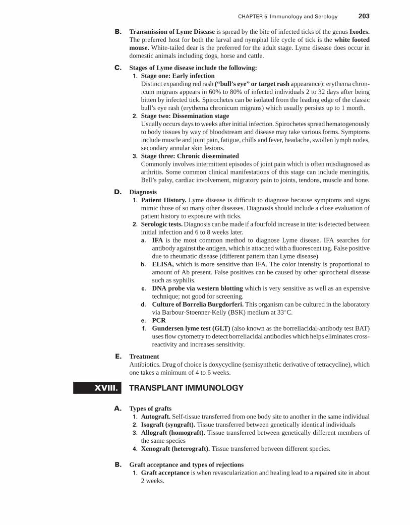
P1: OSO
LWBK192-05 LWBK192-Hubbard November 27, 2008 7:45
CHAPTER 5 Immunology and Serology 203
B. Transmission of Lyme Disease is spread by the bite of infected ticks of the genus Ixodes.The preferred host for both the larval and nymphal life cycle of tick is the white footedmouse. White-tailed dear is the preferred for the adult stage. Lyme disease does occur indomestic animals including dogs, horse and cattle.
C. Stages of Lyme disease include the following:1. Stage one: Early infection
Distinct expanding red rash (“bull’s eye” or target rash appearance): erythema chron-icum migrans appears in 60% to 80% of infected individuals 2 to 32 days after beingbitten by infected tick. Spirochetes can be isolated from the leading edge of the classicbull’s eye rash (erythema chronicum migrans) which usually persists up to 1 month.
2. Stage two: Dissemination stageUsually occurs days to weeks after initial infection. Spirochetes spread hematogenouslyto body tissues by way of bloodstream and disease may take various forms. Symptomsinclude muscle and joint pain, fatigue, chills and fever, headache, swollen lymph nodes,secondary annular skin lesions.
3. Stage three: Chronic disseminatedCommonly involves intermittent episodes of joint pain which is often misdiagnosed asarthritis. Some common clinical manifestations of this stage can include meningitis,Bell’s palsy, cardiac involvement, migratory pain to joints, tendons, muscle and bone.
D. Diagnosis1. Patient History. Lyme disease is difficult to diagnose because symptoms and signs
mimic those of so many other diseases. Diagnosis should include a close evaluation ofpatient history to exposure with ticks.
2. Serologic tests. Diagnosis can be made if a fourfold increase in titer is detected betweeninitial infection and 6 to 8 weeks later.a. IFA is the most common method to diagnose Lyme disease. IFA searches for
antibody against the antigen, which is attached with a fluorescent tag. False positivedue to rheumatic disease (different pattern than Lyme disease)
b. ELISA, which is more sensitive than IFA. The color intensity is proportional toamount of Ab present. False positives can be caused by other spirochetal diseasesuch as syphilis.
c. DNA probe via western blotting which is very sensitive as well as an expensivetechnique; not good for screening.
d. Culture of Borrelia Burgdorferi. This organism can be cultured in the laboratoryvia Barbour-Stoenner-Kelly (BSK) medium at 33◦C.
e. PCRf. Gundersen lyme test (GLT) (also known as the borreliacidal-antibody test BAT)
uses flow cytometry to detect borreliacidal antibodies which helps eliminates cross-reactivity and increases sensitivity.
E. TreatmentAntibiotics. Drug of choice is doxycycline (semisynthetic derivative of tetracycline), whichone takes a minimum of 4 to 6 weeks.
XVIII. TRANSPLANT IMMUNOLOGY
A. Types of grafts1. Autograft. Self-tissue transferred from one body site to another in the same individual2. Isograft (syngraft). Tissue transferred between genetically identical individuals3. Allograft (homograft). Tissue transferred between genetically different members of
the same species4. Xenograft (heterograft). Tissue transferred between different species.
B. Graft acceptance and types of rejections1. Graft acceptance is when revascularization and healing lead to a repaired site in about
2 weeks.
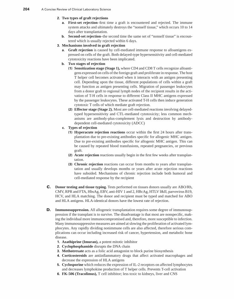
P1: OSO
LWBK192-05 LWBK192-Hubbard November 27, 2008 7:45
204 A Concise Review of Clinical Laboratory Science
2. Two types of graft rejectionsa. First-set rejection–first time a graft is encountered and rejected. The immune
system attacks and ultimately destroys the “nonself tissue,” which occurs 10 to 14days after transplantation.
b. Second-set rejection–the second time the same set of “nonself tissue” is encoun-tered which is usually rejected within 6 days.
3. Mechanisms involved in graft rejectiona. Graft rejection is caused by cell-mediated immune response to alloantigens ex-
pressed on cells of the graft. Both delayed-type hypersensitivity and cell-mediatedcytotoxicity reactions have been implicated.
b. Two stages of rejection(1) Sensitization stage (Stage 1), where CD4 and CD8 T cells recognize alloanti-
gens expressed on cells of the foreign graft and proliferate in response. The hostT helper cell becomes activated when it interacts with an antigen presentingcell. Depending upon the tissue, different populations of cells within a graftmay function as antigen presenting cells. Migration of passenger leukocytesfrom a donor graft to regional lymph nodes of the recipient results in the acti-vation of T-H cells in response to different Class II MHC antigens expressedby the passenger leukocytes. These activated T-H cells then induce generationcytotoxic T cells of which mediate graft rejection.
(2) Effector stage (Stage 2). Most are cell-mediated reactions involving delayed-typed hypersensitivity and CTL-mediated cytotoxicity; less common mech-anisms are antibody-plus-complement lysis and destruction by antibody-dependent cell-mediated cytotoxicity (ADCC)
c. Types of rejection(1) Hyperacute rejection reactions occur within the first 24 hours after trans-
plantation due to pre-existing antibodies specific for allogenic MHC antigen.Due to pre-existing antibodies specific for allogenic MHC antigen. This canbe caused by repeated blood transfusions, repeated pregnancies, or previousgraft.
(2) Acute rejection reactions usually begin in the first few weeks after transplan-tation.
(3) Chronic rejection reactions can occur from months to years after transplan-tation and usually develops months or years after acute rejection reactionshave subsided. Mechanisms of chronic rejection include both humoral andcell-mediated response by the recipient
C. Donor testing and tissue typing. Tests performed on tissues donors usually are ABO/Rh,CMV, RPR and FTA, HbsAg, EBV, anti-HIV 1 and 2, HBcAg, HTLV I&II, parvovirus B19,HCV, and HLA matching. The donor and recipient must be typed and matched for ABOand HLA antigens. HLA-identical donors have the lowest rate of rejection.
D. Immunosuppression. All allogeneic transplantation requires some degree of immunosup-pression if the transplant is to survive. The disadvantage is that most are nonspecific, mak-ing the individual more immunocompromised and, therefore, more susceptible to infection.Many immunosuppressive measures are aimed at slowing the proliferation of activated lym-phocytes. Any rapidly dividing nonimmune cells are also affected, therefore serious com-plications can occur including increased risk of cancer, hypertension, and metabolic bonedisease.1. Azathiprine (Imuran), a potent mitotic inhibitor2. Cyclophosphamide disrupts the DNA chain3. Methotrexate acts as a folic acid antagonist to block purine biosynthesis4. Corticosteroids are antiinflammatory drugs that affect activated macrophages and
decrease the expression of HLA antigens5. Cyclosporine which reduces the expression of IL-2 receptors on affected lymphocytes
and decreases lymphokine production of T helper cells. Prevents T-cell activation6. FK-506 (Tracolimus), T cell inhibitor; less toxic to kidneys, liver and CNS
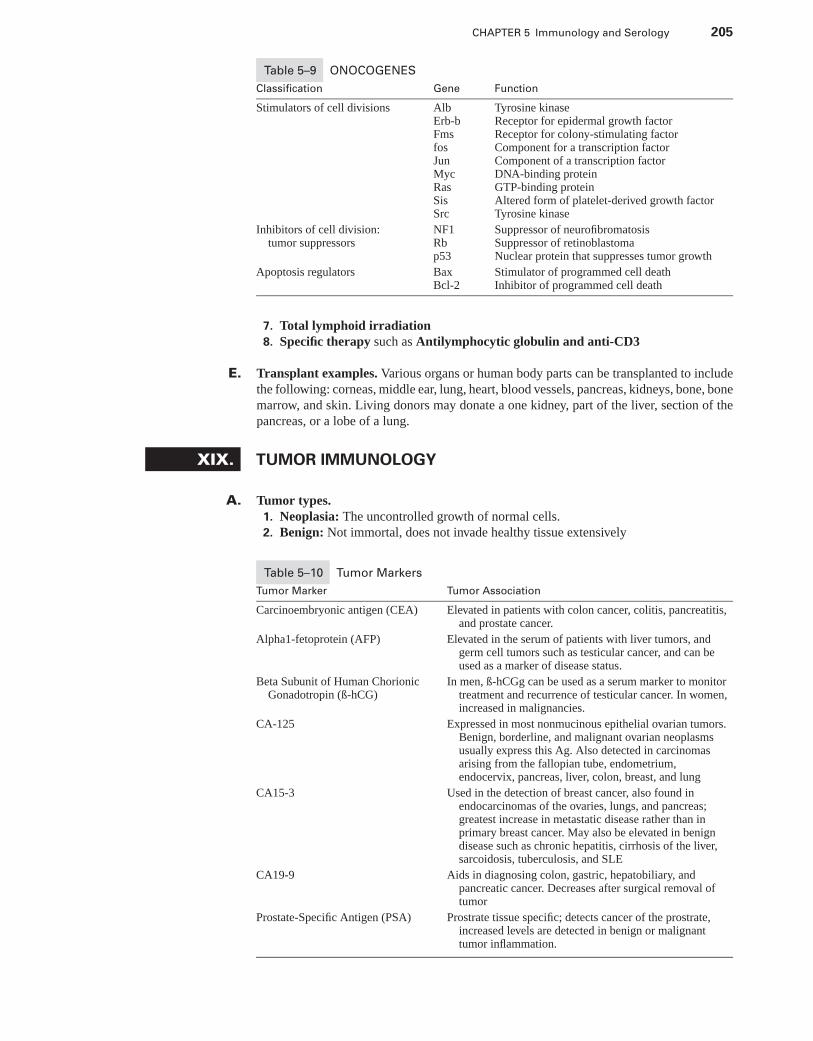
P1: OSO
LWBK192-05 LWBK192-Hubbard November 27, 2008 7:45
CHAPTER 5 Immunology and Serology 205
Table 5–9 ONOCOGENES
Classification Gene Function
Stimulators of cell divisions Alb Tyrosine kinaseErb-b Receptor for epidermal growth factorFms Receptor for colony-stimulating factorfos Component for a transcription factorJun Component of a transcription factorMyc DNA-binding proteinRas GTP-binding proteinSis Altered form of platelet-derived growth factorSrc Tyrosine kinase
Inhibitors of cell division: NF1 Suppressor of neurofibromatosistumor suppressors Rb Suppressor of retinoblastoma
p53 Nuclear protein that suppresses tumor growth
Apoptosis regulators Bax Stimulator of programmed cell deathBcl-2 Inhibitor of programmed cell death
7. Total lymphoid irradiation8. Specific therapy such as Antilymphocytic globulin and anti-CD3
E. Transplant examples. Various organs or human body parts can be transplanted to includethe following: corneas, middle ear, lung, heart, blood vessels, pancreas, kidneys, bone, bonemarrow, and skin. Living donors may donate a one kidney, part of the liver, section of thepancreas, or a lobe of a lung.
XIX. TUMOR IMMUNOLOGY
A. Tumor types.1. Neoplasia: The uncontrolled growth of normal cells.2. Benign: Not immortal, does not invade healthy tissue extensively
Table 5–10 Tumor Markers
Tumor Marker Tumor Association
Carcinoembryonic antigen (CEA) Elevated in patients with colon cancer, colitis, pancreatitis,and prostate cancer.
Alpha1-fetoprotein (AFP) Elevated in the serum of patients with liver tumors, andgerm cell tumors such as testicular cancer, and can beused as a marker of disease status.
Beta Subunit of Human ChorionicGonadotropin (ß-hCG)
In men, ß-hCGg can be used as a serum marker to monitortreatment and recurrence of testicular cancer. In women,increased in malignancies.
CA-125 Expressed in most nonmucinous epithelial ovarian tumors.Benign, borderline, and malignant ovarian neoplasmsusually express this Ag. Also detected in carcinomasarising from the fallopian tube, endometrium,endocervix, pancreas, liver, colon, breast, and lung
CA15-3 Used in the detection of breast cancer, also found inendocarcinomas of the ovaries, lungs, and pancreas;greatest increase in metastatic disease rather than inprimary breast cancer. May also be elevated in benigndisease such as chronic hepatitis, cirrhosis of the liver,sarcoidosis, tuberculosis, and SLE
CA19-9 Aids in diagnosing colon, gastric, hepatobiliary, andpancreatic cancer. Decreases after surgical removal oftumor
Prostate-Specific Antigen (PSA) Prostrate tissue specific; detects cancer of the prostrate,increased levels are detected in benign or malignanttumor inflammation.
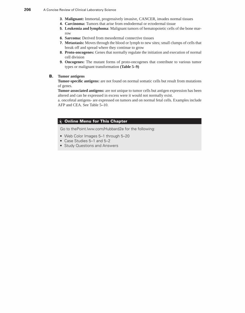
P1: OSO
LWBK192-05 LWBK192-Hubbard November 27, 2008 7:45
206 A Concise Review of Clinical Laboratory Science
3. Malignant: Immortal, progressively invasive, CANCER, invades normal tissues4. Carcinoma: Tumors that arise from endodermal or ectodermal tissue5. Leukemia and lymphoma: Malignant tumors of hematopoietic cells of the bone mar-
row6. Sarcoma: Derived from mesodermal connective tissues7. Metastasis: Moves through the blood or lymph to new sites; small clumps of cells that
break off and spread where they continue to grow8. Proto-oncogenes: Genes that normally regulate the initiation and execution of normal
cell division9. Oncogenes: The mutant forms of proto-oncogenes that contribute to various tumor
types or malignant transformation (Table 5–9)
B. Tumor antigensTumor-specific antigens: are not found on normal somatic cells but result from mutationsof genes.Tumor-associated antigens: are not unique to tumor cells but antigen expression has beenaltered and can be expressed in excess were it would not normally exist.a. oncofetal antigens- are expressed on tumors and on normal fetal cells. Examples includeAFP and CEA. See Table 5–10.
Online Menu for This Chapter
Go to thePoint.lww.com/Hubbard2e for the following:
� Web Color Images 5–1 through 5–20� Case Studies 5–1 and 5–2� Study Questions and Answers
P1: OSO
LWBK192-06 LWBK192-Hubbard November 21, 2008 13:30
CHAPTER 6
Immunohematology
WADE REDMAN, MBA, MT (ASCP) DLM
I. INTRODUCTION
A. Definitions1. Antigens (Ag) are substances that are recognized as foreign by the body and, therefore,
elicit immune responses. Blood group antigens exist on the surface of red bloodcells (RBCs). Only a relatively small number of blood group antigens are consideredclinically significant, but more than 600 have been described.
2. Antibodies (Ab) are immunoglobulins (Ig) that are developed in response to the pres-ence of antigens.a. Heteroantibodies (xenoantibodies) are antibodies produced in response to anti-
gens from another species.b. Alloantibodies are formed in response to antigens from individuals of the same
species and are the type of antibodies involved in transfusion reactions.c. Autoantibodies are made in response to the body’s own antigens.
B. Immunoglobulins are produced and secreted by activated B lymphocytes. All sharethe same basic structure of two heavy chains and two light chains held together bydisulfide bonds. There are five classes—IgG, IgA, IgM, IgE, and IgD. Only IgG, IgM,and (rarely) IgA antibodies are produced against RBC antigens. (See Web Color Image6–1.)
1. IgA antibodies against RBC antigens usually occur with IgG and IgM antibodies havingthe same specificity. They do not cross the placental barrier and do not fix complement.IgA antibodies can cause agglutination in saline.
2. IgG antibodies account for the majority of the clinically significant antibodies directedagainst blood group antigens. There are four subclasses of IgG: IgG1, IgG2, IgG3,and IgG4.a. Most IgG antibodies contain all four subclasses, but some are predominantly or
exclusively composed of a single subclass.b. The subclasses display different biologic properties.
(1) All bind to the crystallizable fragment (Fc) receptors on macrophages; all cancross the placental barrier; and all but IgG4 are able to bind complement throughthe classic pathway.
(2) IgG1 and IgG3 bind complement much more efficiently than does IgG2.(3) IgG1 comprises 65% to 70% of the total IgG found in serum.
3. IgM antibodies agglutinate very strongly in saline, so they are considered completeantibodies. IgM exists in serum as a pentamer and cannot cross the placental barrier.It strongly fixes complement through the classic pathway.
207
P1: OSO
LWBK192-06 LWBK192-Hubbard November 21, 2008 13:30
208 A Concise Review of Clinical Laboratory Science
C. Antigen-antibody reactions. Antigen-antibody reactions important to immunohematologyinvolve the agglutination of erythrocytes by antibodies.
1. Hemagglutination is agglutination of the RBCs. It occurs in two stages.a. Sensitization occurs when the antigen-binding sites of the antibodies become
closely associated with the antigenic determinants of the RBC membrane. Theantibodies and binding sites are held together loosely by noncovalent bonds.
b. Visible agglutination occurs when several RBCs are physically joined together bythe antigen-antibody union. This stage depends on many factors, such as the pH,temperature, and ionic strength of the suspension medium.(1) The use of proteolytic enzymes such as papain or ficin can enhance cell-to-
cell contact.(2) RBCs sensitized by incomplete antibodies (antibodies that will not react in
saline) agglutinate when antiserum against human IgG is added.2. The amount of available antigen and antibody also affects hemagglutination.
a. Prozone occurs when antibody molecules are in excess of available antigenic sites,resulting in false-negative reactions.
b. Equivalence—optimal proportions of antigen and antibody present—allowshemagglutination to occur.
c. Antigen excess (postzone) also results in false-negative reactions.
D. Complement proteins play a number of biologic roles, but the most important role inimmunohematology is their ability to lyse the cell membranes of antibody-coated RBCs.Because complement components are unstable and heat liable, it is important for serumspecimens to be fresh for blood bank testing. The native precursor components are numberedfrom 1 to 9 with subcomponents of the proteins receiving the letters from a to e as theyare cleaved. Complement is activated through two pathways: the classic pathway and thealternate pathway. Although these two pathways are independent, they converge at the C5reaction, and the reactions from C5 to C9 are common to both pathways. (See Web ColorImage 6–2.)
1. The classic pathway is activated by both IgG and IgM antibodies when the C1 com-ponent binds to the Fc portion of the antibody molecule. IgM and IgG3 antibodies aremost efficient at complement activation.
2. The alternate pathway does not require specific antibody for activation, but is insteadtriggered by polysaccharides and lipopolysaccharides on the surfaces of certain targetcells.
3. The reactions that take place from C5 to C9 are termed the membrane attack complexand result in lesions on the RBC surface. These lesions allow the rapid passage of ions,and the cell lyses from osmotic pressure changes.
II. BLOOD GROUP SYSTEMS
A. The ABO blood group system is the most important system in transfusion and transplan-tation therapy.
1. Antigen inheritance. ABO genes are inherited in a codominant manner followingsimple Mendelian genetics laws. Several other genetic loci interact with the ABOlocus: H, Se, Le, I, and P. ABO group inheritance is controlled by the A, B, and Hgenes. These genes code not for the antigens themselves but for the production ofglycotransferases, which are involved in the formation of their respective antigenicdeterminants. (Web Color Image 6–3.)a. H gene: L-fucoseb. A gene: N-acetylgalactosaminec. B gene: D-galactose
2. Antigen developmenta. H antigens are precursors for the A and B antigens. A, B, and H antigens may
be found in other body secretions, including saliva, urine, tears, amniotic fluid,milk, bile, exudates, and digestive fluids. The presence of H substances in body
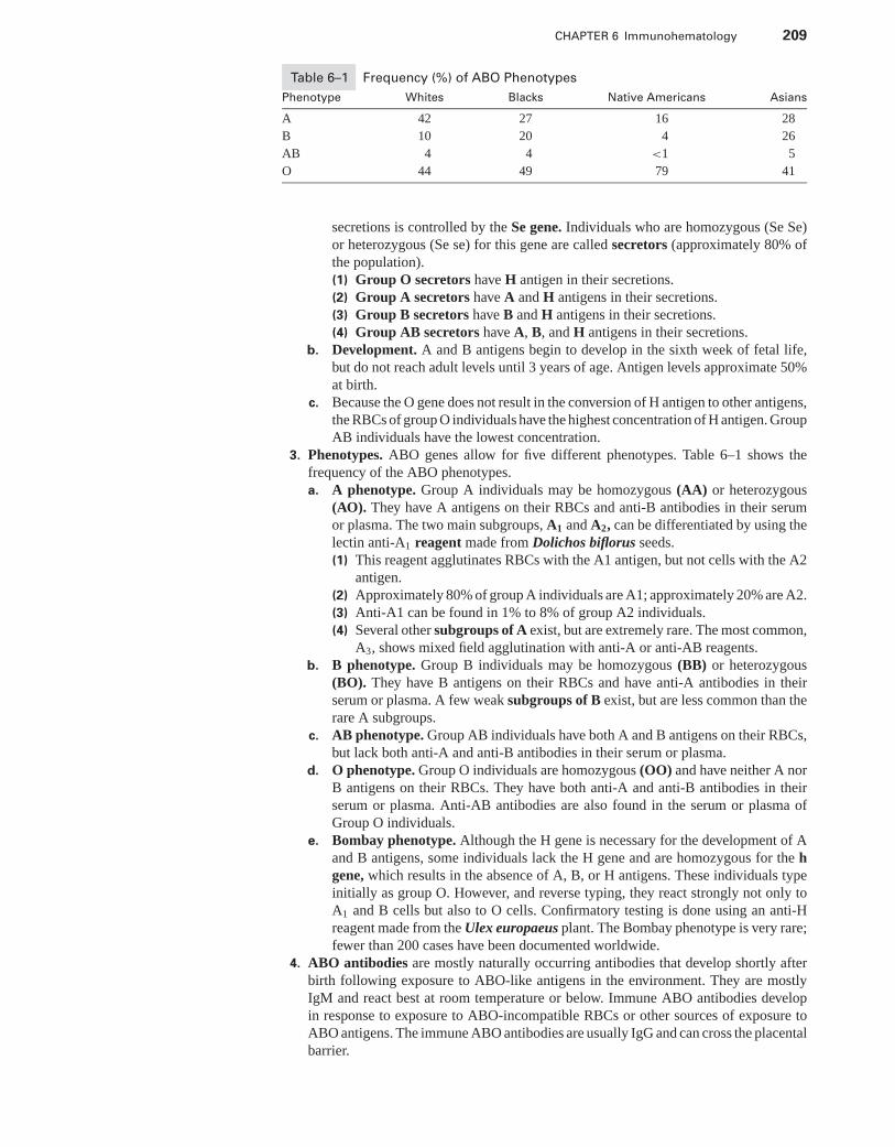
P1: OSO
LWBK192-06 LWBK192-Hubbard November 21, 2008 13:30
CHAPTER 6 Immunohematology 209
Table 6–1 Frequency (%) of ABO Phenotypes
Phenotype Whites Blacks Native Americans Asians
A 42 27 16 28
B 10 20 4 26
AB 4 4 <1 5
O 44 49 79 41
secretions is controlled by the Se gene. Individuals who are homozygous (Se Se)or heterozygous (Se se) for this gene are called secretors (approximately 80% ofthe population).(1) Group O secretors have H antigen in their secretions.(2) Group A secretors have A and H antigens in their secretions.(3) Group B secretors have B and H antigens in their secretions.(4) Group AB secretors have A, B, and H antigens in their secretions.
b. Development. A and B antigens begin to develop in the sixth week of fetal life,but do not reach adult levels until 3 years of age. Antigen levels approximate 50%at birth.
c. Because the O gene does not result in the conversion of H antigen to other antigens,the RBCs of group O individuals have the highest concentration of H antigen. GroupAB individuals have the lowest concentration.
3. Phenotypes. ABO genes allow for five different phenotypes. Table 6–1 shows thefrequency of the ABO phenotypes.a. A phenotype. Group A individuals may be homozygous (AA) or heterozygous
(AO). They have A antigens on their RBCs and anti-B antibodies in their serumor plasma. The two main subgroups, A1 and A2, can be differentiated by using thelectin anti-A1 reagent made from Dolichos biflorus seeds.(1) This reagent agglutinates RBCs with the A1 antigen, but not cells with the A2
antigen.(2) Approximately 80% of group A individuals are A1; approximately 20% are A2.(3) Anti-A1 can be found in 1% to 8% of group A2 individuals.(4) Several other subgroups of A exist, but are extremely rare. The most common,
A3, shows mixed field agglutination with anti-A or anti-AB reagents.b. B phenotype. Group B individuals may be homozygous (BB) or heterozygous
(BO). They have B antigens on their RBCs and have anti-A antibodies in theirserum or plasma. A few weak subgroups of B exist, but are less common than therare A subgroups.
c. AB phenotype. Group AB individuals have both A and B antigens on their RBCs,but lack both anti-A and anti-B antibodies in their serum or plasma.
d. O phenotype. Group O individuals are homozygous (OO) and have neither A norB antigens on their RBCs. They have both anti-A and anti-B antibodies in theirserum or plasma. Anti-AB antibodies are also found in the serum or plasma ofGroup O individuals.
e. Bombay phenotype. Although the H gene is necessary for the development of Aand B antigens, some individuals lack the H gene and are homozygous for the hgene, which results in the absence of A, B, or H antigens. These individuals typeinitially as group O. However, and reverse typing, they react strongly not only toA1 and B cells but also to O cells. Confirmatory testing is done using an anti-Hreagent made from the Ulex europaeus plant. The Bombay phenotype is very rare;fewer than 200 cases have been documented worldwide.
4. ABO antibodies are mostly naturally occurring antibodies that develop shortly afterbirth following exposure to ABO-like antigens in the environment. They are mostlyIgM and react best at room temperature or below. Immune ABO antibodies developin response to exposure to ABO-incompatible RBCs or other sources of exposure toABO antigens. The immune ABO antibodies are usually IgG and can cross the placentalbarrier.
P1: OSO
LWBK192-06 LWBK192-Hubbard November 21, 2008 13:30
210 A Concise Review of Clinical Laboratory Science
5. Testinga. Forward grouping analyzes patient cells for the presence of ABO antigens. Testing
is performed at room temperature by adding anti-A and anti-B reagents to thepatient’s RBCs. Cell suspensions used in blood banking procedures are usually 3%to 5%. All group O donors should also be tested with anti-AB reagent. Agglutinationis a positive reaction.
b. Reverse grouping analyzes patient serum or plasma for the presence of anti-Aor anti-B antibodies. In adults, anti-A or anti-B is present in the serum whenthe corresponding antigen is not present on the RBCs. Because neonates havenot developed these types of antibodies, they are not candidates for reverse ABOgrouping. Testing is performed at room temperature by adding the patient’s serumor plasma to suspensions of known A1 and B RBCs. Agglutination is a positivereaction.
c. Discrepancies may occur in ABO testing for a variety of reasons, including:(1) Subgroups of A or B. Individuals with subgroups of A or B forward type as
group O, but reverse type as group A or B. This discrepancy can be resolvedby adsorbing and eluting anti-A or anti-B from the RBCs.
(2) Polyagglutination can be caused by Tn activation or acquired B phe-nomenon. Tn activation occurs when a somatic mutation exposes a hiddenRBC antigen. These discrepancies can be resolved by determining if the cellsare Tn activated or have the acquired B antigen.
(3) Interfering substances in the serum or plasma. False-positive agglutinationcan result from such substances as Wharton’s jelly or increased serum proteins.The patient will forward type as AB. This discrepancy can be resolved bythoroughly washing the patient’s RBCs and repeating the test.
(4) IgM alloantibodies. Some IgM antibodies such as anti-Lea, anti-P, anti-M,and anti-N can cause false-positive results. Reverse-grouping reagent cellsmay have the antigens against which the alloantibodies are formed. Patientsmay forward type as A or B, but reverse type as O. This discrepancy can beresolved by identifying the alloantibody and retesting with reagent cells thatlack the antigen.
(5) Strong autoantibodies. Autoantibodies, such as anti-I, may cause a patient toforward type as A or B, but reverse type as O. This discrepancy can be resolvedby autoabsorbing the serum and using it to repeat the testing.
(6) Rouleaux caused by increased serum proteins (e.g., myeloma proteins) cancause a patient to forward type as AB and reverse type as O. This discrepancycan be resolved by performing a saline replacement to repeat the testing.
(7) The age of the patient can cause discrepancies in ABO typing. Neonates do nothave ABO antibodies and cannot be reverse typed, which makes it impossibleto confirm the forward typing. Elderly patients may have weakened levels ofABO antibodies.
(8) Disease. Certain diseases associated with hypogammaglobulinemia or agam-maglobulinemia can result in patients reverse typing as AB.
B. The Rh blood group system is one of the most complex systems, because nearly 50different Rh antigens have been identified.1. Nomenclature. Four different nomenclature systems have been developed to identify
the Rh antigens and to describe phenotypes. Table 6–2 and Table 6–3 compare thenomenclature and frequency of the most common Rh phenotypes.a. Fisher-Race. Rh antigens are inherited as three closely linked sets of alleles with
little or no crossing over between loci. The five major Rh antigens are defined asD, C, E, c, and e.
b. Wiener. Rh antigens are inherited as products of single genes at single loci. Thefive major Rh antigens are defined as Rh0, rh′, rh′′, hr′, and hr′′, which correspondto D, C, E, c, and e, respectively.
c. Rosenfield. Rh antigens are assigned numbers to correspond to antigens alreadydesignated by other nomenclature. Newer antigens are numbered based on the
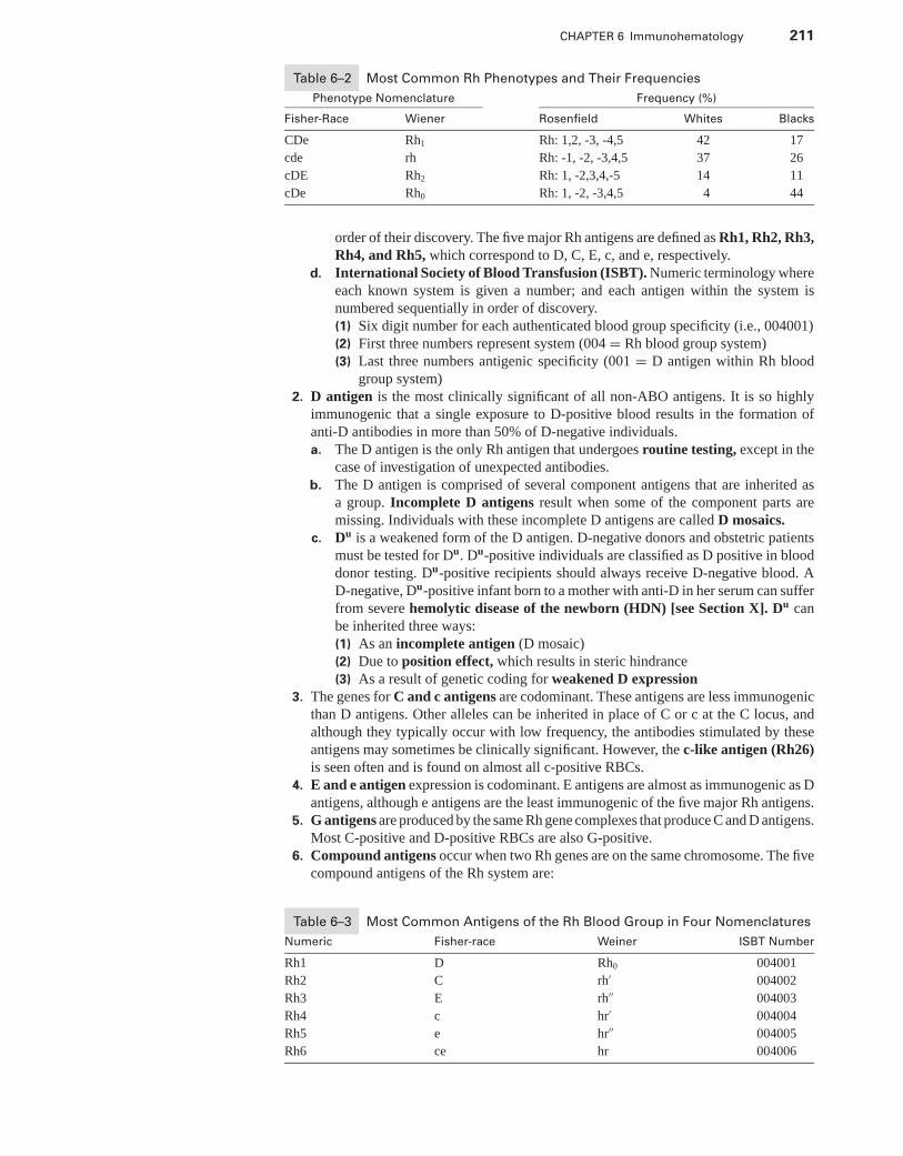
P1: OSO
LWBK192-06 LWBK192-Hubbard November 21, 2008 13:30
CHAPTER 6 Immunohematology 211
Table 6–2 Most Common Rh Phenotypes and Their Frequencies
Phenotype Nomenclature Frequency (%)
Fisher-Race Wiener Rosenfield Whites Blacks
CDe Rh1 Rh: 1,2, -3, -4,5 42 17
cde rh Rh: -1, -2, -3,4,5 37 26
cDE Rh2 Rh: 1, -2,3,4,-5 14 11
cDe Rh0 Rh: 1, -2, -3,4,5 4 44
order of their discovery. The five major Rh antigens are defined as Rh1, Rh2, Rh3,Rh4, and Rh5, which correspond to D, C, E, c, and e, respectively.
d. International Society of Blood Transfusion (ISBT). Numeric terminology whereeach known system is given a number; and each antigen within the system isnumbered sequentially in order of discovery.(1) Six digit number for each authenticated blood group specificity (i.e., 004001)(2) First three numbers represent system (004 = Rh blood group system)(3) Last three numbers antigenic specificity (001 = D antigen within Rh blood
group system)2. D antigen is the most clinically significant of all non-ABO antigens. It is so highly
immunogenic that a single exposure to D-positive blood results in the formation ofanti-D antibodies in more than 50% of D-negative individuals.a. The D antigen is the only Rh antigen that undergoes routine testing, except in the
case of investigation of unexpected antibodies.b. The D antigen is comprised of several component antigens that are inherited as
a group. Incomplete D antigens result when some of the component parts aremissing. Individuals with these incomplete D antigens are called D mosaics.
c. Du is a weakened form of the D antigen. D-negative donors and obstetric patientsmust be tested for Du. Du-positive individuals are classified as D positive in blooddonor testing. Du-positive recipients should always receive D-negative blood. AD-negative, Du-positive infant born to a mother with anti-D in her serum can sufferfrom severe hemolytic disease of the newborn (HDN) [see Section X]. Du canbe inherited three ways:(1) As an incomplete antigen (D mosaic)(2) Due to position effect, which results in steric hindrance(3) As a result of genetic coding for weakened D expression
3. The genes for C and c antigens are codominant. These antigens are less immunogenicthan D antigens. Other alleles can be inherited in place of C or c at the C locus, andalthough they typically occur with low frequency, the antibodies stimulated by theseantigens may sometimes be clinically significant. However, the c-like antigen (Rh26)is seen often and is found on almost all c-positive RBCs.
4. E and e antigen expression is codominant. E antigens are almost as immunogenic as Dantigens, although e antigens are the least immunogenic of the five major Rh antigens.
5. G antigens are produced by the same Rh gene complexes that produce C and D antigens.Most C-positive and D-positive RBCs are also G-positive.
6. Compound antigens occur when two Rh genes are on the same chromosome. The fivecompound antigens of the Rh system are:
Table 6–3 Most Common Antigens of the Rh Blood Group in Four Nomenclatures
Numeric Fisher-race Weiner ISBT Number
Rh1 D Rh0 004001
Rh2 C rh′ 004002
Rh3 E rh′′ 004003
Rh4 c hr′ 004004
Rh5 e hr′′ 004005
Rh6 ce hr 004006
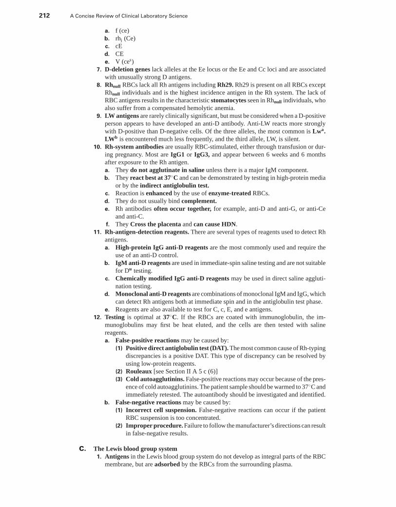
P1: OSO
LWBK192-06 LWBK192-Hubbard November 21, 2008 13:30
212 A Concise Review of Clinical Laboratory Science
a. f (ce)b. rhj (Ce)c. cEd. CEe. V (ces)
7. D-deletion genes lack alleles at the Ee locus or the Ee and Cc loci and are associatedwith unusually strong D antigens.
8. Rhnull RBCs lack all Rh antigens including Rh29. Rh29 is present on all RBCs exceptRhnull individuals and is the highest incidence antigen in the Rh system. The lack ofRBC antigens results in the characteristic stomatocytes seen in Rhnull individuals, whoalso suffer from a compensated hemolytic anemia.
9. LW antigens are rarely clinically significant, but must be considered when a D-positiveperson appears to have developed an anti-D antibody. Anti-LW reacts more stronglywith D-positive than D-negative cells. Of the three alleles, the most common is Lwa.LWb is encountered much less frequently, and the third allele, LW, is silent.
10. Rh-system antibodies are usually RBC-stimulated, either through transfusion or dur-ing pregnancy. Most are IgG1 or IgG3, and appear between 6 weeks and 6 monthsafter exposure to the Rh antigen.a. They do not agglutinate in saline unless there is a major IgM component.b. They react best at 37◦C and can be demonstrated by testing in high-protein media
or by the indirect antiglobulin test.c. Reaction is enhanced by the use of enzyme-treated RBCs.d. They do not usually bind complement.e. Rh antibodies often occur together, for example, anti-D and anti-G, or anti-Ce
and anti-C.f. They Cross the placenta and can cause HDN.
11. Rh-antigen-detection reagents. There are several types of reagents used to detect Rhantigens.a. High-protein IgG anti-D reagents are the most commonly used and require the
use of an anti-D control.b. IgM anti-D reagents are used in immediate-spin saline testing and are not suitable
for Du testing.c. Chemically modified IgG anti-D reagents may be used in direct saline aggluti-
nation testing.d. Monoclonal anti-D reagents are combinations of monoclonal IgM and IgG, which
can detect Rh antigens both at immediate spin and in the antiglobulin test phase.e. Reagents are also available to test for C, c, E, and e antigens.
12. Testing is optimal at 37◦C. If the RBCs are coated with immunoglobulin, the im-munoglobulins may first be heat eluted, and the cells are then tested with salinereagents.a. False-positive reactions may be caused by:
(1) Positive direct antiglobulin test (DAT). The most common cause of Rh-typingdiscrepancies is a positive DAT. This type of discrepancy can be resolved byusing low-protein reagents.
(2) Rouleaux [see Section II A 5 c (6)](3) Cold autoagglutinins. False-positive reactions may occur because of the pres-
ence of cold autoagglutinins. The patient sample should be warmed to 37◦C andimmediately retested. The autoantibody should be investigated and identified.
b. False-negative reactions may be caused by:(1) Incorrect cell suspension. False-negative reactions can occur if the patient
RBC suspension is too concentrated.(2) Improper procedure. Failure to follow the manufacturer’s directions can result
in false-negative results.
C. The Lewis blood group system1. Antigens in the Lewis blood group system do not develop as integral parts of the RBC
membrane, but are adsorbed by the RBCs from the surrounding plasma.
P1: OSO
LWBK192-06 LWBK192-Hubbard November 21, 2008 13:30
CHAPTER 6 Immunohematology 213
a. Expression. The amount of antigen present is variable and is partially dependenton the individual’s ABO phenotype and age, because Lewis antigens develop grad-ually. Lewis antigen expression is affected by H, Se, and Le genes. Lewis antigenexpression may decrease dramatically during pregnancy.
b. The most common phenotypes are:(1) Le (a+b−)(2) Le (a−b+)(3) Le (a+b+)(4) Le (a−b−)
c. Most neonates type as Le (a−b−) regardless of which Lewis genes they haveinherited.
2. Lewis antibodiesa. Anti-Lea antibodies are usually IgM, but may also be IgG in total or in part.
(1) Most of these antibodies react best at room temperature, but may react at37◦C.
(2) Although most Lea antibodies are considered clinically insignificant, they canbind complement and are, therefore, capable of triggering in vitro hemoly-sis. Although Lea antibodies are not associated with HDN, the antibodies thatreact at 37◦C or that cause in vitro hemolysis may be associated with hemolytictransfusion reactions (HTR).
(3) Anti-Lea activity is enhanced by enzyme treatment.b. Anti-Leb antibodies are usually IgM and react best at room temperature. They
bind complement poorly. Anti-Leb activity is enhanced by enzyme treatment.Leb antibodies are not associated with HDN and rarely cause HTR.
c. Lewis antibodies may appear transiently during pregnancy in Le (a−b−)women, but disappear after delivery.
d. Lewis antigens neutralize Lewis antibodies. Because both anti-Lea and anti-Leb
react with most cells on routine RBC panels, Lewis substance can be used toneutralize the antibodies and allow the detection of any other antibodies present.
D. The MNS blood group system1. Antigens
a. The MNS antigens are determined by the MN and Ss loci; MN is associated withglycophorin A; Ss is associated with glycophorin B.
b. There are five principal antigens in the MNS system: M, N, S, s, and U. RBCs withthe S or s antigen also have the U antigen.
c. MNS antigens are important markers in paternity studies.2. Antibodies
a. Anti-M antibodies are relatively common. They are usually naturally occurringand may be both IgM and IgG. Anti-M antibodies do not bind complement andreact optimally at room temperature or below. They are only rarely associatedwith HDN or HTR.
b. Anti-N antibodies are rare. They are weak, naturally occurring IgM antibodiesthat react best at room temperature or below. They are not usually associatedwith HDN or HTR.
c. Anti-S, anti-s, and anti-U antibodies are rare. These IgG antibodies usuallydevelop following RBC stimulation, and all have been associated with severe HDNand HTR. Although S, s, and U antibodies are usually reactive in the antiglobulinphase of testing, some saline reactive antibodies have been reported.
3. Testinga. In addition to human, rabbit, and monoclonal serum-typing reagents, lectin reagents
are also used to test for M and N antigens. Examples include Vicia graminia andseveral Bauhinia species for anti-N, and several Iberis species for anti-M.
b. Many M, N, and S antibodies demonstrate a dosage effect; that is, they react morestrongly with homozygous than heterozygous cells.
c. M and N antigens are destroyed by enzyme treatment, whereas S, s, and U arenot as easily destroyed or have no effect.
P1: OSO
LWBK192-06 LWBK192-Hubbard November 21, 2008 13:30
214 A Concise Review of Clinical Laboratory Science
E. The P blood group system1. Antigens. P blood group antigens are structurally related to ABO antigens and exist as
glycoproteins and glycolipids. There are five phenotypes:a. P1 is one of the two most common phenotypes. Approximately 75% of the popu-
lation possesses the P1 phenotype. P1 individuals have both P and P1 antigens. TheP antigen is well developed at birth; the P1 antigen is poorly developed at birth.P expression in adults is widely variable.
b. P2 is the other most common phenotype. Individuals with this phenotype have onlyP antigen on their RBCs.
c. Pk1 phenotype is very rare. Individuals with this phenotype have both P1 and Pk
antigens on their RBCs.d. Pk
2 phenotype is also very rare. Individuals with this phenotype have both the P2
and Pk antigens on their RBCs.e. The p phenotype is extremely rare. The RBCs of individuals with this phenotype
are negative for P, P1, and Pk antigens.2. Antibodies
a. Anti-P1 antibodies are naturally occurring, cold-reacting, IgM anti-bodies oftenseen in individuals with the P2 phenotype. They rarely react at higher than roomtemperature, but they do bind complement. Anti-P1 antibodies do not causeHDN and are rarely associated with HTR.
b. Anti-P antibodies are produced by individuals with Pk1 or Pk
2 phenotypes and cantrigger severe HTR. Autoanti-P (the Donath-Landsteiner antibody) is a cold-reacting antibody associated with paroxysmal cold hemoglobinuria (PCH).
c. Anti-PP, Pk, and anti-p antibodies occur only rarely.
F. The I blood group system1. Antigens. The I blood group antigens are structurally related to the ABO antigens and
are found on RBC membranes as well as in plasma, milk, and amniotic fluid. Both Iand i antigens are found on all RBCs. I antigens are poorly developed at birth. As theI antigenic strength increases, i antigen strength decreases. Most adults have strong Iantigen expression and weak i antigen expression.
2. Antibodiesa. Anti-I antibodies are naturally occurring, cold-reacting IgM anti-bodies. They
fail to react with cord RBCs. Some anti-I antibodies react in a broader temperaturerange and can cause cold-agglutinin disease (CAD). CAD may be idiopathic ormay be associated with diseases, such as Mycoplasma pneumoniae infections.Anti-I antibodies are not associated with HDN.
b. Anti-i antibodies are rare. Like anti-I, they are cold-reacting antibodies. Theymay be seen in cases of infectious mononucleosis and may cause an associatedhemolytic anemia that disappears as the infection resolves.
3. Testing for anti-I or anti-I antibodies is done at 4◦C using group O RBCs or cord RBCs.
G. The Duffy blood group system. The Duffy system (FY) has four alleles that are responsiblefor the major antigens and resulting phenotypes: Fya, Fyb, Fy, and Fyx.1. Antigens. Fya and Fyb antigens are produced by codominant alleles. Fyx is a weakened
form of Fyb, and the Fy allele produces no gene product. There are four phenotypes:a. Fy (a+b−)b. Fy (a−b+)c. Fy (a+b+)d. Fy (a−b−) (There is a high incidence of this phenotype among blacks. The RBCs
of individuals with this phenotype are resistant to infection by Plasmodium vivax.)2. Antibodies. The most common FY antibodies are Fya and Fyb, which are both com-
monly encountered in blood banks. Other Duffy antibodies are very rare. Anti-Fya
and anti-Fyb antibodies are usually IgG, often bind complement, and generally reactonly at the antiglobulin phase of testing. Both are destroyed by proteolytic enzymetreatment and heating to 56◦C. Anti-Fya antibodies are seen more frequently than Fyb
antibodies, and both occur more commonly in combination with other RBC antibodies
P1: OSO
LWBK192-06 LWBK192-Hubbard November 21, 2008 13:30
CHAPTER 6 Immunohematology 215
than alone. Although both can cause delayed HTR, only Fya has occasionally beenimplicated in HDN.
H. The Kell blood group system1. Antigens. The Kell (K) system is comprised of 21 high- and low-incidence antigens,
the most significant of which are K and k. The K0 or Knull phenotype lack all Kellantigens. An antigen associated with the Kell system, Kx, is located on the X chro-mosome. RBCs that lack the Kx antigen also have greatly weakened expression of theother Kell system antigens. These RBCs are morphologically acanthocytes, have de-creased survival, and are less permeable to water. This syndrome, known as MacLeodsyndrome, is also characterized by splenomegaly, reticulocytosis, and occasional as-sociation with chronic granulomatous disease (CGD). Kell antigens are destroyedor inactivated by sulfhydryl reagents.
2. Antibodies. The two most important antibodies of the Kell system are anti-K andanti-k (Cellano).a. The K antigen is second only to the D antigen in immunogenicity, and the resulting
antibody is relatively common in transfusion practice. Finding K-negative donorunits is rarely a problem, however, because more than 90% of the population isK-negative.
b. Anti-K antibodies are usually IgG antibodies that react best at 37◦C and mayoccasionally bind complement. They can cause both HDN and HTR.
c. Anti-k antibodies are rare but can cause both HDN and HTR.d. Antibodies to K0 (Knull) antigens are called anti-Ku or anti-KEL5 antibodies
and are considered clinically significant. K0 patients should be transfused withK0 cells. In testing, K0 cells can be made by treating normal RBCs with 2-aminoethylisothiouronium (AET) bromide or dithiothreitol (DTT) plus cysteine-activated papain (ZZAP).
I. The Kidd blood group system1. Antigens
a. There are two major antigens in the Kidd (JK) system, Jka and Jkb, which allowsfor four phenotypes:(1) Jk (a+b−)(2) Jk (a+b+)(3) Jk (a−b+)(4) Jk (a−b−)
b. A third antigen, Jk3, is present on both Jka- and Jkb-positive RBCs. Both Jka andJkb may exhibit dosage effect; reactivity may be enhanced by enzyme treatment.
2. Antibodies. Although Jka and Jkb antigens are poorly immunogenic, the resultingantibodies can cause severe HTR, and both are especially noted for causing delayedreactions. Both can occasionally cause mild HDN. Most Jka and Jkb antibodies areIgG1 or IgG3 and bind complement very efficiently. Both react best at 37◦C in theantiglobulin phase of testing, but saline-reactive antibodies may be seen. Jka and Jkb
antibodies can be difficult to detect, and enhancement techniques, such as enzymes,low ionic strength saline (LISS), or polyethylene glycol (PEG), can help identify them.
J. The Lutheran blood group system1. Antigens
a. The two most common antigens in the Lutheran (Lu) system are Lua and Lub,which allows for four phenotypes:(1) Lu (a+b−); rare(2) Lu (a+b+); rare(3) Lu (a−b+); common(4) Lu (a−b−); extremely rare
b. The genes encoding for the Lu antigens are linked to the Se (secretor) genes.2. Antibodies. Both the anti-Lua and the anti-Lub antibodies are rarely seen. Lua an-
tibodies react best at 37◦C and give a mixed field agglutination reaction. Lub
P1: OSO
LWBK192-06 LWBK192-Hubbard November 21, 2008 13:30
216 A Concise Review of Clinical Laboratory Science
antibodies react best at 37◦C in the antiglobulin phase of testing. Lub antibodiescan cause mild HTR, but cause mild HDN only rarely. Lua antibodies are often nat-urally occurring, whereas Lub antibodies usually develop after RBC stimulation bytransfusion or pregnancy.
K. Other blood group systems1. The Cartwright (Yt) system has two antigens, Yta and Ytb. The majority of individuals
are Yt (a+). Antibodies are rare. If they develop, they are IgG antibodies that reactbest in the antiglobulin phase of testing and are not clinically significant.
2. The Colton (Co) system has three antigens, Coa, Cob, and Coab. Antibodies to Coa
and Cob are rare, but can cause HTR. Neither cause HDN.3. The Diego (Di) system has two antigens, Dia and Dib. Antibodies to both antigens are
rare, although they have been reported as causes of rare cases of HDN. The Dia antigenis useful as a racial marker, because it is seen almost exclusively in individuals ofMongolian extraction, such as North, Central, and South American Indians, Japanese,and Chinese.
4. The Dombrock (Do) system has two antigens, Doa and Dob. Antibodies are rare.Anti-Doa antibodies can cause HTR, but neither cause HDN.
5. The Scianna (SC) system has three antigens, SC1, SC2, and SC3. Antibodies to SC1and SC2 are rare IgG antibodies that can be clinically significant.
6. The Xg system has only one antigen, Xga, and two resulting phenotypes: Xg (a+) andXg (a−). The Xga antigen is X linked and is more common in women. The antigen isdestroyed by enzyme treatment. Xga antibodies are usually IgG antibodies that bindcomplement but do not cause HDN or HTR.
7. The Cromer system is composed of eight high-incidence antigens (Cra, Tca, TCab, Dra,Esa, WESb, UMC, and IFC) and three low-incidence antigens (Tcb, Tcc, and WESa).These antigens are located on the decay-accelerating factor on the RBC membrane.Patients with paroxysmal nocturnal hemoglobinuria have weakly expressed or absentCromer-related antigens.
L. Miscellaneous blood group antigens. Several other RBC antigens have been identified,but have not been classified as part of any blood group system.
1. Bg antigens. There are three Bg antigens: Bga, Bgb, and Bgc. Bg antigens are related tohuman leukocyte antigens (HLA) on RBCs. Antibodies to Bg antigens are not clinicallysignificant.
2. Sda antigens are high-incidence antigens found in several tissues and body fluids.Antibodies to these antigens are not clinically significant. Mixed-field agglutinationis characteristically seen with Sda antibodies.
3. High-titer low-avidity (HTLA) antigens occur with high frequency, but the resultingantibodies are very weak and have little or no clinical significance. They do not causeHDN or HTR. There are several HTLA antigens:a. Chido (Cha) and Rodgers (Rga) genes are linked to HLA genes, and the anti-
gens are associated with the C4d component of complement. Both antigens aredenatured by proteolytic enzymes.
b. Cost-Sterling (CSa) and York (Yka) antigensc. Knops (Kna) and McCoy (McCa) antigens
4. Independent high-frequency antigens rarely produce antibodies.a. Gregory (Gya) and Holley (Hya) antibodies are IgG antibodies that can cause
accelerated red cell destruction. They do not cause HDN.b. The Gerbich collection of antigens includes five antigens. Antibodies are ex-
tremely rare but may cause HDN or accelerated RBC destruction.c. Vel antigens. Antibodies to Vel antigens may cause HTR, but not HDN. They are
usually IgG and can bind complement.5. Independent low-frequency antigens very rarely cause antibody formation. Such
antibodies are not clinically significant. Table 6–4 outlines the frequency of the mostcommon blood group antigens.
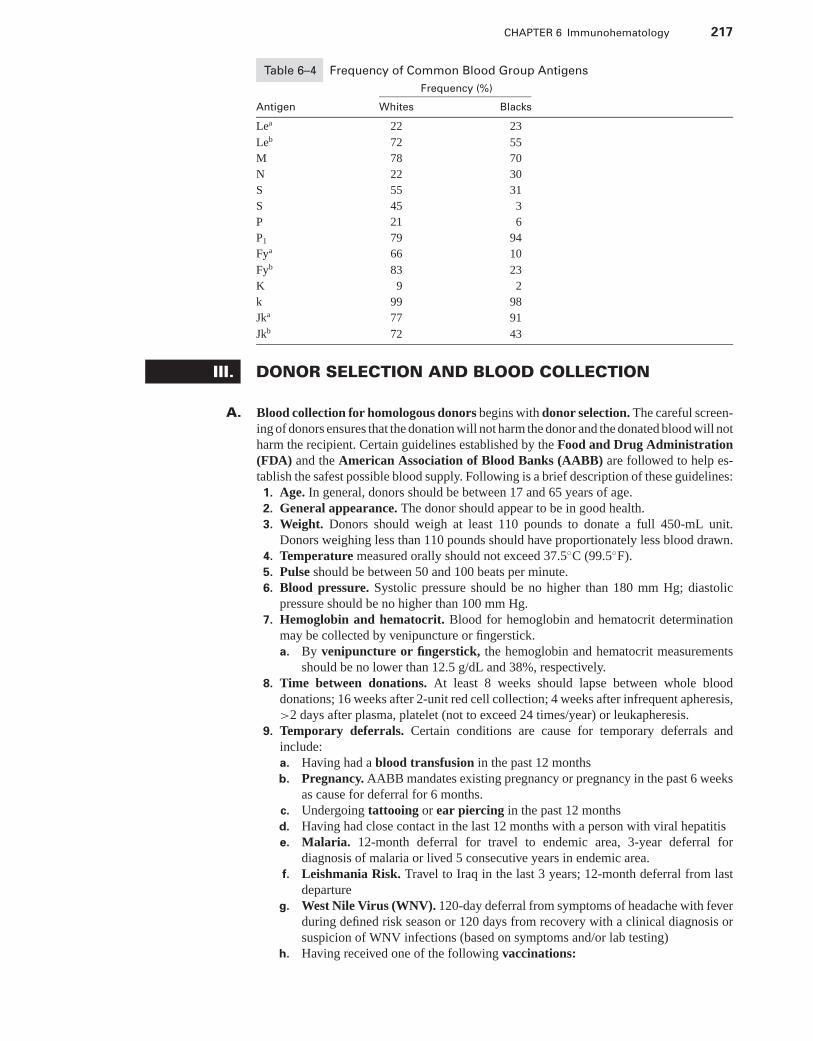
P1: OSO
LWBK192-06 LWBK192-Hubbard November 21, 2008 13:30
CHAPTER 6 Immunohematology 217
Table 6–4 Frequency of Common Blood Group Antigens
Frequency (%)
Antigen Whites Blacks
Lea 22 23
Leb 72 55
M 78 70
N 22 30
S 55 31
S 45 3
P 21 6
P1 79 94
Fya 66 10
Fyb 83 23
K 9 2
k 99 98
Jka 77 91
Jkb 72 43
III. DONOR SELECTION AND BLOOD COLLECTION
A. Blood collection for homologous donors begins with donor selection. The careful screen-ing of donors ensures that the donation will not harm the donor and the donated blood will notharm the recipient. Certain guidelines established by the Food and Drug Administration(FDA) and the American Association of Blood Banks (AABB) are followed to help es-tablish the safest possible blood supply. Following is a brief description of these guidelines:
1. Age. In general, donors should be between 17 and 65 years of age.2. General appearance. The donor should appear to be in good health.3. Weight. Donors should weigh at least 110 pounds to donate a full 450-mL unit.
Donors weighing less than 110 pounds should have proportionately less blood drawn.4. Temperature measured orally should not exceed 37.5◦C (99.5◦F).5. Pulse should be between 50 and 100 beats per minute.6. Blood pressure. Systolic pressure should be no higher than 180 mm Hg; diastolic
pressure should be no higher than 100 mm Hg.7. Hemoglobin and hematocrit. Blood for hemoglobin and hematocrit determination
may be collected by venipuncture or fingerstick.a. By venipuncture or fingerstick, the hemoglobin and hematocrit measurements
should be no lower than 12.5 g/dL and 38%, respectively.8. Time between donations. At least 8 weeks should lapse between whole blood
donations; 16 weeks after 2-unit red cell collection; 4 weeks after infrequent apheresis,>2 days after plasma, platelet (not to exceed 24 times/year) or leukapheresis.
9. Temporary deferrals. Certain conditions are cause for temporary deferrals andinclude:a. Having had a blood transfusion in the past 12 monthsb. Pregnancy. AABB mandates existing pregnancy or pregnancy in the past 6 weeks
as cause for deferral for 6 months.c. Undergoing tattooing or ear piercing in the past 12 monthsd. Having had close contact in the last 12 months with a person with viral hepatitise. Malaria. 12-month deferral for travel to endemic area, 3-year deferral for
diagnosis of malaria or lived 5 consecutive years in endemic area.f. Leishmania Risk. Travel to Iraq in the last 3 years; 12-month deferral from last
departureg. West Nile Virus (WNV). 120-day deferral from symptoms of headache with fever
during defined risk season or 120 days from recovery with a clinical diagnosis orsuspicion of WNV infections (based on symptoms and/or lab testing)
h. Having received one of the following vaccinations:
P1: OSO
LWBK192-06 LWBK192-Hubbard November 21, 2008 13:30
218 A Concise Review of Clinical Laboratory Science
(1) Smallpox, measles, mumps, yellow fever, or oral poliomyelitis vaccine in thepast 2 weeks
(2) Rubella in the past 4 weeks(3) Rabies in the past 12 months(4) Hepatitis B immunoglobulin in the past 12 months
i. Taking one of the following medications per AABB: http://www.aabb.org/Documents/Donate Blood/Donor History Questionnaire/udhqmeddef05.doc(1) If you have taken or are taking Proscar, Avodart, Propecia, Accutane,
Soriatane, or Tegison, these medications can cause birth defects. Yourdonated blood could contain high enough levels to damage the unborn baby iftransfused to a pregnant woman. After the medication has been cleared fromyour blood, you may donate again. Following the last dose, the deferral periodis 1 month Proscar, Propecia and Accutane, 6 months for Avodart and 3 yearsfor Soriatane. Tegison is a permanent deferral.
(2) Growth hormone from human pituitary glands was prescribed for childrenwith delayed or impaired growth. The hormone was obtained from humanpituitary glands, which are found in the brain. Some people who took thishormone developed a rare nervous system condition called Creutzfeldt-Jakobdisease (CJD, for short). The deferral is permanent.
(3) Insulin from cows (bovine, or beef, insulin) is an injected material usedto treat diabetes. If this insulin was imported into the United States fromcountries in which “Mad Cow Disease” has been found, it could containmaterial from infected cattle. There is concern that “Mad Cow Disease” istransmitted by transfusion. The deferral is indefinite.
(4) Hepatitis B immune globulin (HBIG) is an injected material used to preventinfection following an exposure to hepatitis B. HBIG does not prevent hepatitisB infection in every case, therefore, persons who have received HBIG mustwait 12 months to donate blood to be sure they were not infected becausehepatitis B can be transmitted through transfusion to a patient.
(5) Unlicensed vaccine is usually associated with a research protocol and theeffect on blood transmission is unknown. Deferral is 1 year unless otherwiseindicated by the medical director.
10. Permanent deferrals are caused by the following conditions listed by the AABB:a. Anyone who has ever used intravenous drugs (illegal IV drugs)b. Men who have had sexual contact with other men since 1977c. Anyone who has ever received clotting factor concentratesd. Anyone with a positive test for HIV (AIDS virus)e. Men and women who have engaged in sex for money or drugs since 1977f. Anyone who has had hepatitis since his or her 11th birthday
g. Anyone who has had babesiosis or Chagas diseaseh. Anyone who has taken Tegison for psoriasisi. Anyone who has risk factors for Creutzfeldt-Jakob disease (CJD) or who has an
immediate family member with CJDj. Anyone who has risk factors for variant CJDk. Anyone who spent 3 months or more in the United Kingdom from 1980 through
1996l. Anyone who has spent 5 years in Europe from 1980 to the present.
11. Other questions concerning a donor’s medical history may reveal underlying medicalproblems that could be cause for deferral. Any case in question should be referredto the donor center’s medical director. Most prescription and over-the-countermedications are not cause for deferral, although any questions should be referred tothe medical director.
B. Apheresis donors. The same selection criteria that apply to whole blood homologousdonors apply to apheresis donors. Additionally:
1. Plasmapheresis donors must have a serum total protein of at least 6.0 g/dL. A serumprotein electrophoresis should be performed in these individuals every 4 months.There should also be at least 48 hours between donations.
P1: OSO
LWBK192-06 LWBK192-Hubbard November 21, 2008 13:30
CHAPTER 6 Immunohematology 219
2. Plateletpheresis donors should have a platelet count of at least 150 × 109/L. Aspiriningestion within 3 days of donation is cause for temporary deferral. There should beat least 8 weeks between donations.
3. Leukapheresis donors must have an absolute granulocyte count of at least 4 × 10⁹/L.4. Therapeutic apheresis may be performed for certain conditions, such as Good-
pasture’s syndrome, thrombotic thrombocytopenic purpura, myeloid leukemia withhyperleukocytosis, and acute complications of sickle-cell disease.
C. Criteria for autologous donors are not as well defined or regulated as are those forhomologous donors. Some general guidelines for autologous donor suitability have beenestablished by the AABB, but the facility medical director should make final decisions.In general, donors should have a hemoglobin of at least 11.0 g/dL and should not havesigns or symptoms of active infection. Blood should not be donated within 72 hours ofscheduled surgery or transfusion.
D. Donor reactions. Although rare, some blood donors experience donation-related reactions,which range from mild to severe.
1. Symptoms include:a. Pallorb. Sweatingc. Dizzinessd. Nausea and vomitinge. Twitching and muscle spasmsf. Loss of consciousness
2. True convulsions are extremely rare.3. Phlebotomy should be discontinued immediately and the donor should be treated
symptomatically.
E. Anticoagulants and preservatives. Several anticoagulants are currently approved by theFDA for storage of blood and blood components. FDA approval for anticoagulants andadditive solutions requires that 70% of transfused RBCs must be present and viable 24hours after transfusion.
1. The purposes of anticoagulants and preservative solutions are:a. Preventing physical changes to maintain the viability and function of the blood
constituentsb. Preventing bacterial contaminationc. Minimizing cell lysis
2. Citrate-phosphate-dextrose (CPD) can be used to store blood for 21 days whenstored between 1◦C and 6◦C. The citrate functions as an anticoagulant by bindingcalcium. The phosphate helps to increase adenosine triphosphate (ATP) production,and the dextrose provides energy for the RBCs.
3. The adenine in citrate-phosphate-dextrose-adenine (CPDA-1) provides a substratefor ATP production. The shelf life of blood collected in CPDA-1 is 35 days.
4. Although not used for routine blood collection, heparin may be used as an anticoag-ulant. Because heparin has no preservative qualities, blood collected in heparin mustbe transfused within 24 to 48 hours, preferably within 24 hours.
5. Stored RBCs may be rejuvenated up to 2 to 3 days after their expiration date bythe addition of a solution containing pyruvate, inosine, glucose, and phosphate.They may be either washed and transfused within 24 hours or glycerolized andfrozen.
IV. DONOR PROCESSING
A. Homologous donor processing. The FDA and AABB require that certain tests be per-formed on all blood units collected for homologous transfusion. All reagents used fordonor blood processing must be FDA approved, and accurate and thorough record keeping iscritical.
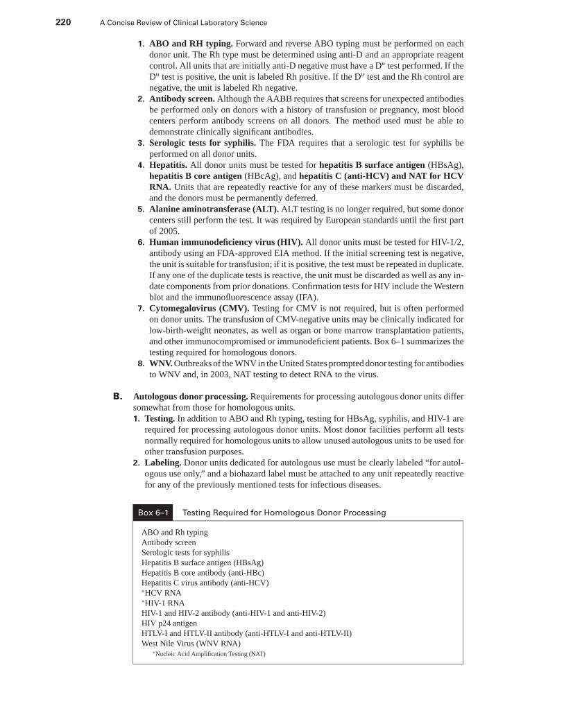
P1: OSO
LWBK192-06 LWBK192-Hubbard November 21, 2008 13:30
220 A Concise Review of Clinical Laboratory Science
1. ABO and RH typing. Forward and reverse ABO typing must be performed on eachdonor unit. The Rh type must be determined using anti-D and an appropriate reagentcontrol. All units that are initially anti-D negative must have a Du test performed. If theDu test is positive, the unit is labeled Rh positive. If the Du test and the Rh control arenegative, the unit is labeled Rh negative.
2. Antibody screen. Although the AABB requires that screens for unexpected antibodiesbe performed only on donors with a history of transfusion or pregnancy, most bloodcenters perform antibody screens on all donors. The method used must be able todemonstrate clinically significant antibodies.
3. Serologic tests for syphilis. The FDA requires that a serologic test for syphilis beperformed on all donor units.
4. Hepatitis. All donor units must be tested for hepatitis B surface antigen (HBsAg),hepatitis B core antigen (HBcAg), and hepatitis C (anti-HCV) and NAT for HCVRNA. Units that are repeatedly reactive for any of these markers must be discarded,and the donors must be permanently deferred.
5. Alanine aminotransferase (ALT). ALT testing is no longer required, but some donorcenters still perform the test. It was required by European standards until the first partof 2005.
6. Human immunodeficiency virus (HIV). All donor units must be tested for HIV-1/2,antibody using an FDA-approved EIA method. If the initial screening test is negative,the unit is suitable for transfusion; if it is positive, the test must be repeated in duplicate.If any one of the duplicate tests is reactive, the unit must be discarded as well as any in-date components from prior donations. Confirmation tests for HIV include the Westernblot and the immunofluorescence assay (IFA).
7. Cytomegalovirus (CMV). Testing for CMV is not required, but is often performedon donor units. The transfusion of CMV-negative units may be clinically indicated forlow-birth-weight neonates, as well as organ or bone marrow transplantation patients,and other immunocompromised or immunodeficient patients. Box 6–1 summarizes thetesting required for homologous donors.
8. WNV. Outbreaks of the WNV in the United States prompted donor testing for antibodiesto WNV and, in 2003, NAT testing to detect RNA to the virus.
B. Autologous donor processing. Requirements for processing autologous donor units differsomewhat from those for homologous units.1. Testing. In addition to ABO and Rh typing, testing for HBsAg, syphilis, and HIV-1 are
required for processing autologous donor units. Most donor facilities perform all testsnormally required for homologous units to allow unused autologous units to be used forother transfusion purposes.
2. Labeling. Donor units dedicated for autologous use must be clearly labeled “for autol-ogous use only,” and a biohazard label must be attached to any unit repeatedly reactivefor any of the previously mentioned tests for infectious diseases.
Box 6–1 Testing Required for Homologous Donor Processing
ABO and Rh typing
Antibody screen
Serologic tests for syphilis
Hepatitis B surface antigen (HBsAg)
Hepatitis B core antibody (anti-HBc)
Hepatitis C virus antibody (anti-HCV)∗HCV RNA∗HIV-1 RNA
HIV-1 and HIV-2 antibody (anti-HIV-1 and anti-HIV-2)
HIV p24 antigen
HTLV-I and HTLV-II antibody (anti-HTLV-I and anti-HTLV-II)
West Nile Virus (WNV RNA)∗Nucleic Acid Amplification Testing (NAT)
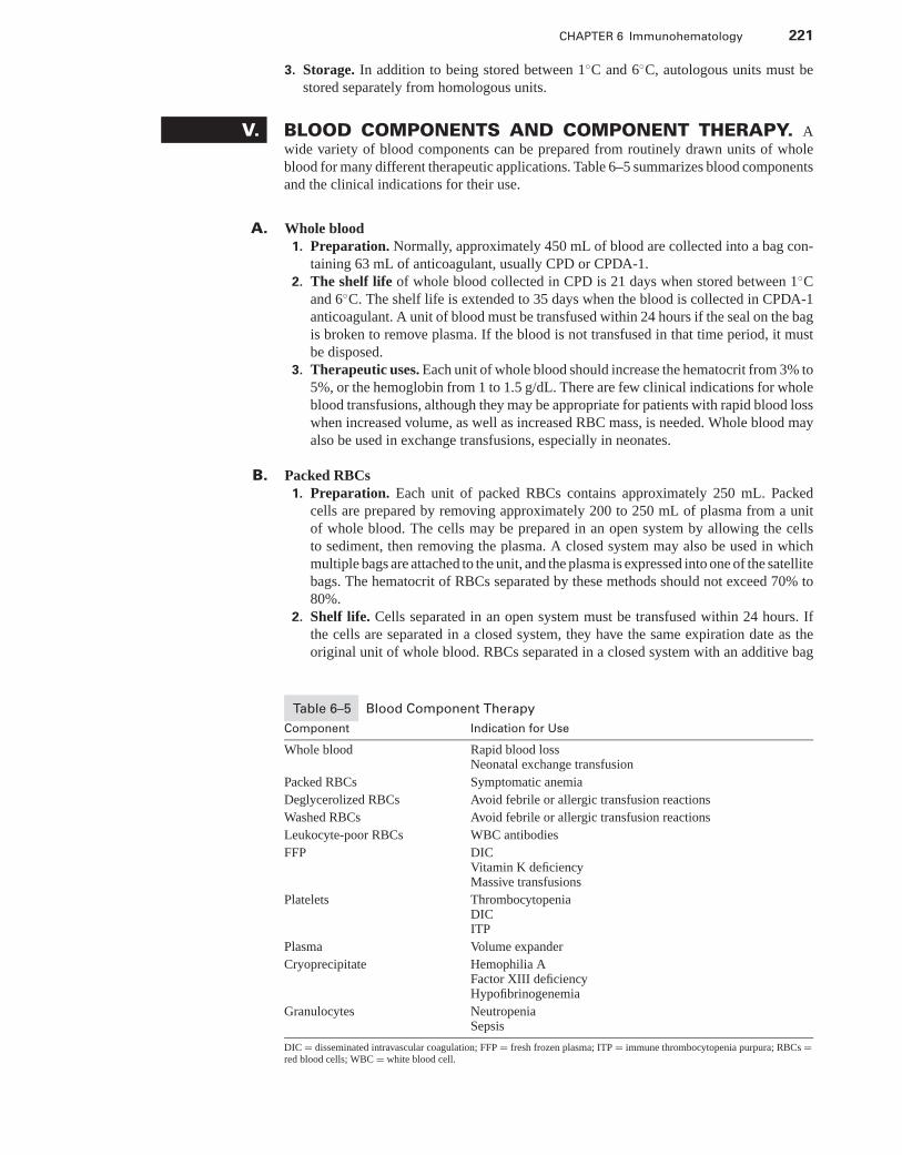
P1: OSO
LWBK192-06 LWBK192-Hubbard November 21, 2008 13:30
CHAPTER 6 Immunohematology 221
3. Storage. In addition to being stored between 1◦C and 6◦C, autologous units must bestored separately from homologous units.
V. BLOOD COMPONENTS AND COMPONENT THERAPY. Awide variety of blood components can be prepared from routinely drawn units of wholeblood for many different therapeutic applications. Table 6–5 summarizes blood componentsand the clinical indications for their use.
A. Whole blood1. Preparation. Normally, approximately 450 mL of blood are collected into a bag con-
taining 63 mL of anticoagulant, usually CPD or CPDA-1.2. The shelf life of whole blood collected in CPD is 21 days when stored between 1◦C
and 6◦C. The shelf life is extended to 35 days when the blood is collected in CPDA-1anticoagulant. A unit of blood must be transfused within 24 hours if the seal on the bagis broken to remove plasma. If the blood is not transfused in that time period, it mustbe disposed.
3. Therapeutic uses. Each unit of whole blood should increase the hematocrit from 3% to5%, or the hemoglobin from 1 to 1.5 g/dL. There are few clinical indications for wholeblood transfusions, although they may be appropriate for patients with rapid blood losswhen increased volume, as well as increased RBC mass, is needed. Whole blood mayalso be used in exchange transfusions, especially in neonates.
B. Packed RBCs1. Preparation. Each unit of packed RBCs contains approximately 250 mL. Packed
cells are prepared by removing approximately 200 to 250 mL of plasma from a unitof whole blood. The cells may be prepared in an open system by allowing the cellsto sediment, then removing the plasma. A closed system may also be used in whichmultiple bags are attached to the unit, and the plasma is expressed into one of the satellitebags. The hematocrit of RBCs separated by these methods should not exceed 70% to80%.
2. Shelf life. Cells separated in an open system must be transfused within 24 hours. Ifthe cells are separated in a closed system, they have the same expiration date as theoriginal unit of whole blood. RBCs separated in a closed system with an additive bag
Table 6–5 Blood Component Therapy
Component Indication for Use
Whole blood Rapid blood lossNeonatal exchange transfusion
Packed RBCs Symptomatic anemia
Deglycerolized RBCs Avoid febrile or allergic transfusion reactions
Washed RBCs Avoid febrile or allergic transfusion reactions
Leukocyte-poor RBCs WBC antibodies
FFP DICVitamin K deficiencyMassive transfusions
Platelets ThrombocytopeniaDICITP
Plasma Volume expander
Cryoprecipitate Hemophilia AFactor XIII deficiencyHypofibrinogenemia
Granulocytes NeutropeniaSepsis
DIC = disseminated intravascular coagulation; FFP = fresh frozen plasma; ITP = immune thrombocytopenia purpura; RBCs =red blood cells; WBC = white blood cell.
P1: OSO
LWBK192-06 LWBK192-Hubbard November 21, 2008 13:30
222 A Concise Review of Clinical Laboratory Science
can have a second preservative solution added that will extend the shelf life to 42 days.They should be stored between 1◦C and 6◦C.
3. Therapeutic uses. The increase in hemoglobin and hematocrit in response to a unit ofpacked RBCs is the same as for whole blood. Packed RBCs are used to increase theRBC mass in patients who have symptomatic anemia.
C. Deglycerolized frozen RBCs1. Preparation. RBCs to be frozen are collected in CPD, CPDA-1, or other additive
systems and normally should be frozen within 6 hours. The cells are warmed and mixedwith high molar concentrations of glycerol, then frozen at −65◦C. Frozen units must bestored for up to 10 years. The cells must be deglycerolized before they can be transfused.Deglycerolization begins with thawing the cells at 37◦C, then washing multiple timesin a gradient concentration of saline, beginning with hypertonic concentrations andending with an isotonic saline solution containing glucose. One unit of deglycerolizedRBCs contains approximately 180 mL of cells.
2. Shelf life. Deglycerolized RBCs are stored between 1◦C and 6◦C and must be transfusedwithin 2 hours of deglycerolization.
3. Therapeutic uses. The increase in hemoglobin and hematocrit in response to a unitis the same as for whole blood or packed cells. Freezing cells allows for long-termstorage of rare donor units or autologous units. Transfusing deglycerolized RBCs alsominimizes febrile or allergic reactions.
D. Washed RBCs1. Preparation. Plasma is removed from whole blood after centrifugation, and the re-
maining RBCs are washed three times with 0.9% saline.2. Shelf life. Washed RBCs have a shelf life of 24 hours after the original unit is opened,
and they should be stored between 1◦C and 6◦C.3. Therapeutic uses. One unit of washed RBCs increases the hemoglobin and hemat-
ocrit by the same amount as do unwashed cells. Washing RBCs removes most of theleukocytes and plasma from a unit of blood, which greatly reduces the risk of febrileor allergic (anaphylactic) reactions in susceptible patients. Washed RBCs are used forthe rare patient who has anti-IgA antibodies because of IgA deficiency.
E. Leukocyte-poor RBCs1. Preparation. Leukocyte-poor RBC preparations have at least 70% of the original white
blood cells (WBCs) removed, and at least 70% of the original RBCs are left. There areseveral different methods of obtaining leukocyte-poor RBCs, including centrifugation,filtration, and washing.
2. Shelf life. If a closed preparation system is used, the shelf life is the same as theoriginal unit of blood. The shelf life is reduced to 24 hours if an open system is used.Leukocyte-poor RBCs should be stored between 1◦C and 6◦C.
3. Therapeutic uses. In addition to increasing RBC mass, leukocyte-poor RBCs alsominimize febrile transfusion reactions in patients who have leukocyte antibodies aswell as reducing CMV transmission.
F. Fresh frozen plasma (FFP)1. Preparation. Plasma is separated from whole blood and frozen within 6 hours of
collection. Plasma can be removed from whole blood using a double bag collectionsystem to preserve a closed system. The plasma is immediately frozen at or below−18◦C.
2. Shelf life. After freezing, the plasma should be stored at or below −18◦C. FFP has ashelf life of 1 year after collection of the original unit of blood. It should be thawed at37◦C and transfused within 24 hours of thawing. Thawed FFP should be stored between1◦C and 6◦C if it is not transfused immediately.
3. Therapeutic uses. Because FFP contains all of the coagulation factors, it can beused to treat patients who have liver failure, vitamin K deficiency, and dissemi-nated intravascular coagulation (DIC), or to patients who have received massive trans-fusions.
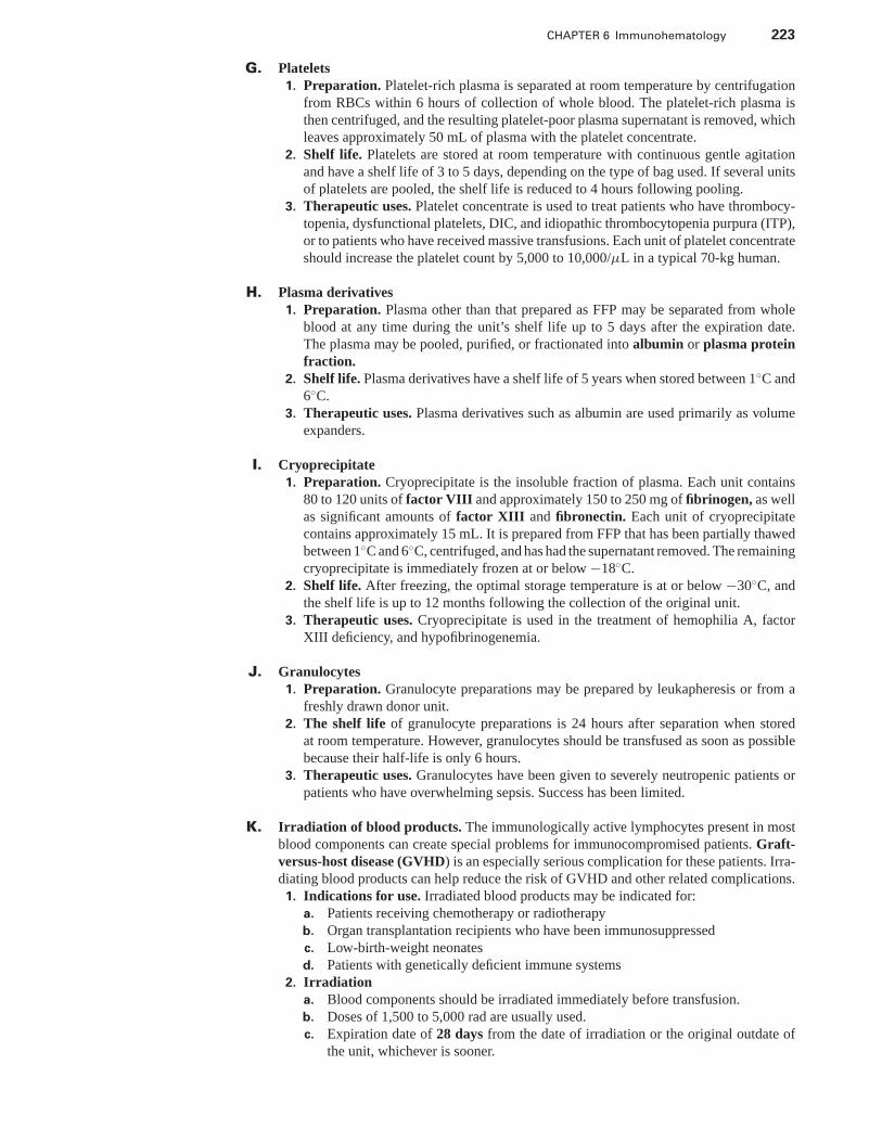
P1: OSO
LWBK192-06 LWBK192-Hubbard November 21, 2008 13:30
CHAPTER 6 Immunohematology 223
G. Platelets1. Preparation. Platelet-rich plasma is separated at room temperature by centrifugation
from RBCs within 6 hours of collection of whole blood. The platelet-rich plasma isthen centrifuged, and the resulting platelet-poor plasma supernatant is removed, whichleaves approximately 50 mL of plasma with the platelet concentrate.
2. Shelf life. Platelets are stored at room temperature with continuous gentle agitationand have a shelf life of 3 to 5 days, depending on the type of bag used. If several unitsof platelets are pooled, the shelf life is reduced to 4 hours following pooling.
3. Therapeutic uses. Platelet concentrate is used to treat patients who have thrombocy-topenia, dysfunctional platelets, DIC, and idiopathic thrombocytopenia purpura (ITP),or to patients who have received massive transfusions. Each unit of platelet concentrateshould increase the platelet count by 5,000 to 10,000/μL in a typical 70-kg human.
H. Plasma derivatives1. Preparation. Plasma other than that prepared as FFP may be separated from whole
blood at any time during the unit’s shelf life up to 5 days after the expiration date.The plasma may be pooled, purified, or fractionated into albumin or plasma proteinfraction.
2. Shelf life. Plasma derivatives have a shelf life of 5 years when stored between 1◦C and6◦C.
3. Therapeutic uses. Plasma derivatives such as albumin are used primarily as volumeexpanders.
I. Cryoprecipitate1. Preparation. Cryoprecipitate is the insoluble fraction of plasma. Each unit contains
80 to 120 units of factor VIII and approximately 150 to 250 mg of fibrinogen, as wellas significant amounts of factor XIII and fibronectin. Each unit of cryoprecipitatecontains approximately 15 mL. It is prepared from FFP that has been partially thawedbetween 1◦C and 6◦C, centrifuged, and has had the supernatant removed. The remainingcryoprecipitate is immediately frozen at or below −18◦C.
2. Shelf life. After freezing, the optimal storage temperature is at or below −30◦C, andthe shelf life is up to 12 months following the collection of the original unit.
3. Therapeutic uses. Cryoprecipitate is used in the treatment of hemophilia A, factorXIII deficiency, and hypofibrinogenemia.
J. Granulocytes1. Preparation. Granulocyte preparations may be prepared by leukapheresis or from a
freshly drawn donor unit.2. The shelf life of granulocyte preparations is 24 hours after separation when stored
at room temperature. However, granulocytes should be transfused as soon as possiblebecause their half-life is only 6 hours.
3. Therapeutic uses. Granulocytes have been given to severely neutropenic patients orpatients who have overwhelming sepsis. Success has been limited.
K. Irradiation of blood products. The immunologically active lymphocytes present in mostblood components can create special problems for immunocompromised patients. Graft-versus-host disease (GVHD) is an especially serious complication for these patients. Irra-diating blood products can help reduce the risk of GVHD and other related complications.
1. Indications for use. Irradiated blood products may be indicated for:a. Patients receiving chemotherapy or radiotherapyb. Organ transplantation recipients who have been immunosuppressedc. Low-birth-weight neonatesd. Patients with genetically deficient immune systems
2. Irradiationa. Blood components should be irradiated immediately before transfusion.b. Doses of 1,500 to 5,000 rad are usually used.c. Expiration date of 28 days from the date of irradiation or the original outdate of
the unit, whichever is sooner.
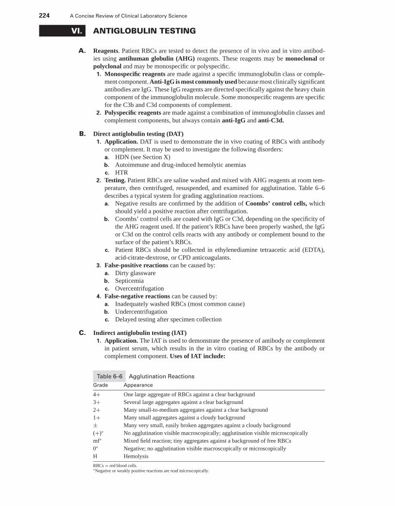
P1: OSO
LWBK192-06 LWBK192-Hubbard November 21, 2008 13:30
224 A Concise Review of Clinical Laboratory Science
VI. ANTIGLOBULIN TESTING
A. Reagents. Patient RBCs are tested to detect the presence of in vivo and in vitro antibod-ies using antihuman globulin (AHG) reagents. These reagents may be monoclonal orpolyclonal and may be monospecific or polyspecific.
1. Monospecific reagents are made against a specific immunoglobulin class or comple-ment component. Anti-IgG is most commonly used because most clinically significantantibodies are IgG. These IgG reagents are directed specifically against the heavy chaincomponent of the immunoglobulin molecule. Some monospecific reagents are specificfor the C3b and C3d components of complement.
2. Polyspecific reagents are made against a combination of immunoglobulin classes andcomplement components, but always contain anti-IgG and anti-C3d.
B. Direct antiglobulin testing (DAT)1. Application. DAT is used to demonstrate the in vivo coating of RBCs with antibody
or complement. It may be used to investigate the following disorders:a. HDN (see Section X)b. Autoimmune and drug-induced hemolytic anemiasc. HTR
2. Testing. Patient RBCs are saline washed and mixed with AHG reagents at room tem-perature, then centrifuged, resuspended, and examined for agglutination. Table 6–6describes a typical system for grading agglutination reactions.a. Negative results are confirmed by the addition of Coombs’ control cells, which
should yield a positive reaction after centrifugation.b. Coombs’ control cells are coated with IgG or C3d, depending on the specificity of
the AHG reagent used. If the patient’s RBCs have been properly washed, the IgGor C3d on the control cells reacts with any antibody or complement bound to thesurface of the patient’s RBCs.
c. Patient RBCs should be collected in ethylenediamine tetraacetic acid (EDTA),acid-citrate-dextrose, or CPD anticoagulants.
3. False-positive reactions can be caused by:a. Dirty glasswareb. Septicemiac. Overcentrifugation
4. False-negative reactions can be caused by:a. Inadequately washed RBCs (most common cause)b. Undercentrifugationc. Delayed testing after specimen collection
C. Indirect antiglobulin testing (IAT)1. Application. The IAT is used to demonstrate the presence of antibody or complement
in patient serum, which results in the in vitro coating of RBCs by the antibody orcomplement component. Uses of IAT include:
Table 6–6 Agglutination Reactions
Grade Appearance
4+ One large aggregate of RBCs against a clear background
3+ Several large aggregates against a clear background
2+ Many small-to-medium aggregates against a clear background
1+ Many small aggregates against a cloudy background
± Many very small, easily broken aggregates against a cloudy background
(+)∗ No agglutination visible macroscopically; agglutination visible microscopically
mf∗ Mixed field reaction; tiny aggregates against a background of free RBCs
0∗ Negative; no agglutination visible macroscopically or microscopically
H Hemolysis
RBCs = red blood cells.∗Negative or weakly positive reactions are read microscopically.
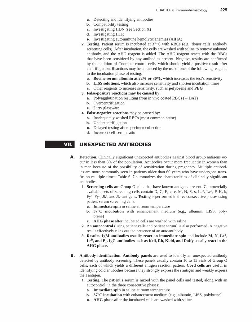
P1: OSO
LWBK192-06 LWBK192-Hubbard November 21, 2008 13:30
CHAPTER 6 Immunohematology 225
a. Detecting and identifying antibodiesb. Compatibility testingc. Investigating HDN (see Section X)d. Investigating HTRe. Investigating autoimmune hemolytic anemias (AIHA)
2. Testing. Patient serum is incubated at 37◦C with RBCs (e.g., donor cells, antibodyscreening cells). After incubation, the cells are washed with saline to remove unboundantibody, and the AHG reagent is added. The AHG reagent reacts with the RBCsthat have been sensitized by any antibodies present. Negative results are confirmedby the addition of Coombs’ control cells, which should yield a positive result aftercentrifugation. Reactions may be enhanced by the use of one of the following reagentsto the incubation phase of testing:a. Bovine serum albumin at 22% or 30%, which increases the test’s sensitivityb. LISS solutions, which also increase sensitivity and shorten incubation timesc. Other reagents to increase sensitivity, such as polybrene and PEG
3. False-positive reactions may be caused by:a. Polyagglutination resulting from in vivo coated RBCs (+ DAT)b. Overcentrifugationc. Dirty glassware
4. False-negative reactions may be caused by:a. Inadequately washed RBCs (most common cause)b. Undercentrifugationc. Delayed testing after specimen collectiond. Incorrect cell-serum ratio
VII. UNEXPECTED ANTIBODIES
A. Detection. Clinically significant unexpected antibodies against blood group antigens oc-cur in less than 3% of the population. Antibodies occur more frequently in women thanin men because of the possibility of sensitization during pregnancy. Multiple antibod-ies are more commonly seen in patients older than 60 years who have undergone trans-fusion multiple times. Table 6–7 summarizes the characteristics of clinically significantantibodies.
1. Screening cells are Group O cells that have known antigens present. Commerciallyavailable sets of screening cells contain D, C, E, c, e, M, N, S, s, Lea, Leb, P, K, k,Fya, Fyb, Jka, and Jkb antigens. Testing is performed in three consecutive phases usingpatient serum screening cells:a. Immediate spin in saline at room temperatureb. 37◦C incubation with enhancement medium (e.g., albumin, LISS, poly-
brene)c. AHG phase after incubated cells are washed with saline
2. An autocontrol (using patient cells and patient serum) is also performed. A negativeresult effectively rules out the presence of an autoantibody.
3. Results. IgM antibodies usually react on immediate spin and include M, N, Lea,Leb, and P1. IgG antibodies such as Kell, Rh, Kidd, and Duffy usually react in theAHG phase.
B. Antibody identification. Antibody panels are used to identify an unexpected antibodydetected by antibody screening. These panels usually contain 10 to 15 vials of Group Ocells, each of which yields a different antigen reaction pattern. Cord cells are useful inidentifying cold antibodies because they strongly express the i antigen and weakly expressthe I antigen.
1. Testing. The patient’s serum is mixed with the panel cells and tested, along with anautocontrol, in the three consecutive phases:a. Immediate spin in saline at room temperatureb. 37◦C incubation with enhancement medium (e.g., albumin, LISS, polybrene)c. AHG phase after the incubated cells are washed with saline
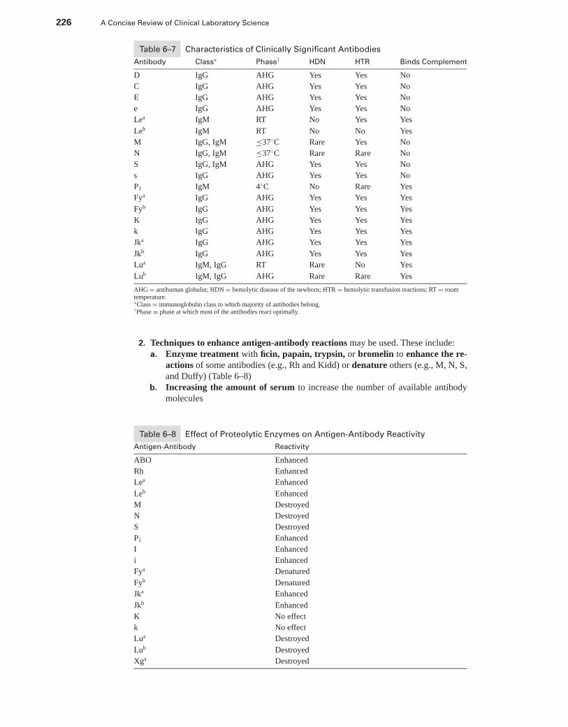
P1: OSO
LWBK192-06 LWBK192-Hubbard November 21, 2008 13:30
226 A Concise Review of Clinical Laboratory Science
Table 6–7 Characteristics of Clinically Significant Antibodies
Antibody Class∗ Phase† HDN HTR Binds Complement
D IgG AHG Yes Yes No
C IgG AHG Yes Yes No
E IgG AHG Yes Yes No
e IgG AHG Yes Yes No
Lea IgM RT No Yes Yes
Leb IgM RT No No Yes
M IgG, IgM ≤37◦C Rare Yes No
N IgG, IgM ≤37◦C Rare Rare No
S IgG, IgM AHG Yes Yes No
s IgG AHG Yes Yes No
P1 IgM 4◦C No Rare Yes
Fya IgG AHG Yes Yes Yes
Fyb IgG AHG Yes Yes Yes
K IgG AHG Yes Yes Yes
k IgG AHG Yes Yes Yes
Jka IgG AHG Yes Yes Yes
Jkb IgG AHG Yes Yes Yes
Lua IgM, IgG RT Rare No Yes
Lub IgM, IgG AHG Rare Rare Yes
AHG = antihuman globulin; HDN = hemolytic disease of the newborn; HTR = hemolytic transfusion reactions; RT = roomtemperature.∗Class = immunoglobulin class to which majority of antibodies belong.†Phase = phase at which most of the antibodies react optimally.
2. Techniques to enhance antigen-antibody reactions may be used. These include:a. Enzyme treatment with ficin, papain, trypsin, or bromelin to enhance the re-
actions of some antibodies (e.g., Rh and Kidd) or denature others (e.g., M, N, S,and Duffy) (Table 6–8)
b. Increasing the amount of serum to increase the number of available antibodymolecules
Table 6–8 Effect of Proteolytic Enzymes on Antigen-Antibody Reactivity
Antigen-Antibody Reactivity
ABO Enhanced
Rh Enhanced
Lea Enhanced
Leb Enhanced
M Destroyed
N Destroyed
S Destroyed
P1 Enhanced
I Enhanced
i Enhanced
Fya Denatured
Fyb Denatured
Jka Enhanced
Jkb Enhanced
K No effect
k No effect
Lua Destroyed
Lub Destroyed
Xga Destroyed
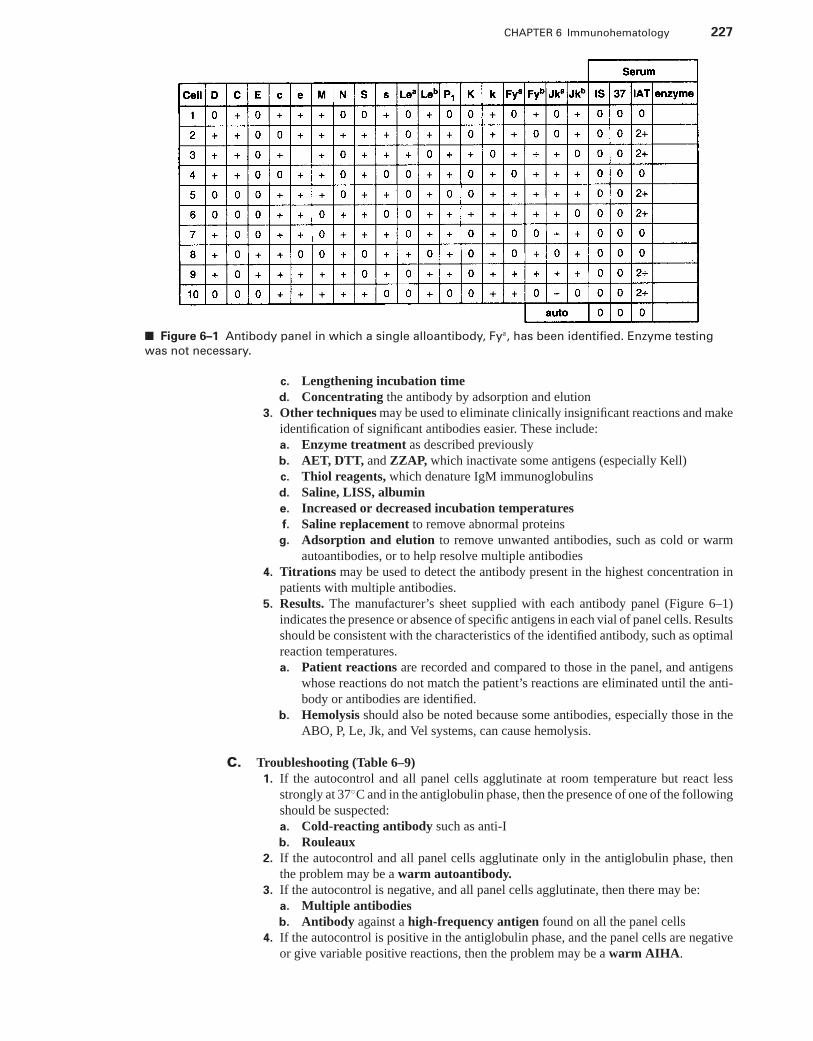
P1: OSO
LWBK192-06 LWBK192-Hubbard November 21, 2008 13:30
CHAPTER 6 Immunohematology 227
■ Figure 6–1 Antibody panel in which a single alloantibody, Fya, has been identified. Enzyme testing
was not necessary.
c. Lengthening incubation timed. Concentrating the antibody by adsorption and elution
3. Other techniques may be used to eliminate clinically insignificant reactions and makeidentification of significant antibodies easier. These include:a. Enzyme treatment as described previouslyb. AET, DTT, and ZZAP, which inactivate some antigens (especially Kell)c. Thiol reagents, which denature IgM immunoglobulinsd. Saline, LISS, albumine. Increased or decreased incubation temperaturesf. Saline replacement to remove abnormal proteins
g. Adsorption and elution to remove unwanted antibodies, such as cold or warmautoantibodies, or to help resolve multiple antibodies
4. Titrations may be used to detect the antibody present in the highest concentration inpatients with multiple antibodies.
5. Results. The manufacturer’s sheet supplied with each antibody panel (Figure 6–1)indicates the presence or absence of specific antigens in each vial of panel cells. Resultsshould be consistent with the characteristics of the identified antibody, such as optimalreaction temperatures.a. Patient reactions are recorded and compared to those in the panel, and antigens
whose reactions do not match the patient’s reactions are eliminated until the anti-body or antibodies are identified.
b. Hemolysis should also be noted because some antibodies, especially those in theABO, P, Le, Jk, and Vel systems, can cause hemolysis.
C. Troubleshooting (Table 6–9)1. If the autocontrol and all panel cells agglutinate at room temperature but react less
strongly at 37◦C and in the antiglobulin phase, then the presence of one of the followingshould be suspected:a. Cold-reacting antibody such as anti-Ib. Rouleaux
2. If the autocontrol and all panel cells agglutinate only in the antiglobulin phase, thenthe problem may be a warm autoantibody.
3. If the autocontrol is negative, and all panel cells agglutinate, then there may be:a. Multiple antibodiesb. Antibody against a high-frequency antigen found on all the panel cells
4. If the autocontrol is positive in the antiglobulin phase, and the panel cells are negativeor give variable positive reactions, then the problem may be a warm AIHA.
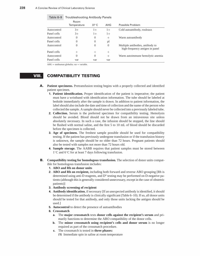
P1: OSO
LWBK192-06 LWBK192-Hubbard November 21, 2008 13:30
228 A Concise Review of Clinical Laboratory Science
Table 6–9 Troubleshooting Antibody Panels
Room
Temperature 37◦C AHG Possible Problem
Autocontrol 3+ 1+ 1+ Cold autoantibody, rouleaux
Panel cells 3+ 1+ 1+Autocontrol 0 0 + Warm autoantibody
Panel cells 0 0 pl
Autocontrol 0 0 0 Multiple antibodies, antibody tohigh-frequency antigen in panel
Panel cells + + +Autocontrol 0 0 + Warm autoimmune hemolytic anemia
Panel cells var var var
AHG = antihuman globulin; var = variable.
VIII. COMPATIBILITY TESTING
A. Patient specimens. Pretransfusion testing begins with a properly collected and identifiedpatient specimen.
1. Patient identification. Proper identification of the patient is imperative; the patientmust have a wristband with identification information. The tube should be labeled atbedside immediately after the sample is drawn. In addition to patient information, thelabel should also include the date and time of collection and the name of the person whocollected the sample. A sample should never be collected into a previously labeled tube.
2. Collection. Serum is the preferred specimen for compatibility testing. Hemolysisshould be avoided. Blood should not be drawn from an intravenous site unlessabsolutely necessary. In such a case, the infusion should be stopped, the line shouldbe flushed with normal saline, and the first 5 to 10 mL of blood should be discardedbefore the specimen is collected.
3. Age of specimen. The freshest sample possible should be used for compatibilitytesting. If the patient has previously undergone transfusion or if the transfusion historyis unknown, the sample should be no older than 72 hours. Pregnant patients shouldalso be tested with samples not more than 72 hours old.
4. Sample storage. The AABB requires that patient samples must be stored between1◦C and 6◦C for at least 7 days following transfusion.
B. Compatibility testing for homologous transfusion. The selection of donor units compat-ible for homologous transfusion includes:
1. ABO and Rh on donor units2. ABO and Rh on recipient, including both forward and reverse ABO grouping [Rh is
determined using anti-D reagents, and Du testing may be performed on D-negative pa-tients (although this is generally considered unnecessary, except in the case of obstetricpatients)]
3. Antibody screening of recipient4. Antibody identification, if necessary [If an unexpected antibody is identified, it should
be determined if the antibody is clinically significant (Table 6–10). If so, all donor unitsshould be tested for that antibody, and only those units lacking the antigen should beused.]
5. Autocontrol to detect the presence of autoantibodies6. Crossmatch
a. The major crossmatch tests donor cells against the recipient’s serum and pri-marily functions to determine the ABO compatibility of the donor cells.
b. The minor crossmatch using recipient’s cells and donor serum is no longerrequired as part of the crossmatch procedure.
c. The crossmatch is tested in three phases:(1) Immediate spin in saline at room temperature
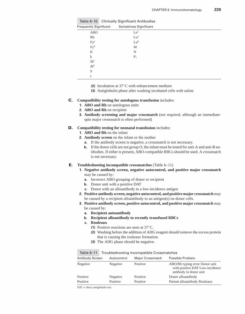
P1: OSO
LWBK192-06 LWBK192-Hubbard November 21, 2008 13:30
CHAPTER 6 Immunohematology 229
Table 6–10 Clinically Significant Antibodies
Frequently Significant Sometimes Significant
ABO Lea
Rh Lua
Fya Lub
Fyb M
K N
k P1
Jka
Jkb
S
s
(2) Incubation at 37◦C with enhancement medium(3) Antiglobulin phase after washing incubated cells with saline
C. Compatibility testing for autologous transfusion includes:1. ABO and Rh on autologous units2. ABO and Rh on recipient3. Antibody screening and major crossmatch (not required, although an immediate-
spin major crossmatch is often performed)
D. Compatibility testing for neonatal transfusion includes:1. ABO and Rh on the infant2. Antibody screen on the infant or the mother
a. If the antibody screen is negative, a crossmatch is not necessary.b. If the donor cells are not group O, the infant must be tested for anti-A and anti-B an-
tibodies. If either is present, ABO-compatible RBCs should be used. A crossmatchis not necessary.
E. Troubleshooting incompatible crossmatches (Table 6–11)1. Negative antibody screen, negative autocontrol, and positive major crossmatch
may be caused by:a. Incorrect ABO grouping of donor or recipientb. Donor unit with a positive DATc. Donor with an alloantibody to a low-incidence antigen
2. Positive antibody screen, negative autocontrol, and positive major crossmatch maybe caused by a recipient alloantibody to an antigen(s) on donor cells.
3. Positive antibody screen, positive autocontrol, and positive major crossmatch maybe caused by:a. Recipient autoantibodyb. Recipient alloantibody to recently transfused RBCsc. Rouleaux
(1) Positive reactions are seen at 37◦C.(2) Washing before the addition of AHG reagent should remove the excess protein
that is causing the rouleaux formation.(3) The AHG phase should be negative.
Table 6–11 Troubleshooting Incompatible Crossmatches
Antibody Screen Autocontrol Major Crossmatch Possible Problem
Negative Negative Positive ABO/Rh typing error Donor unitwith positive DAT Low-incidenceantibody in donor unit
Positive Negative Positive Donor alloantibody
Positive Positive Positive Patient alloantibody Rouleaux
DAT = direct antiglobulin test.
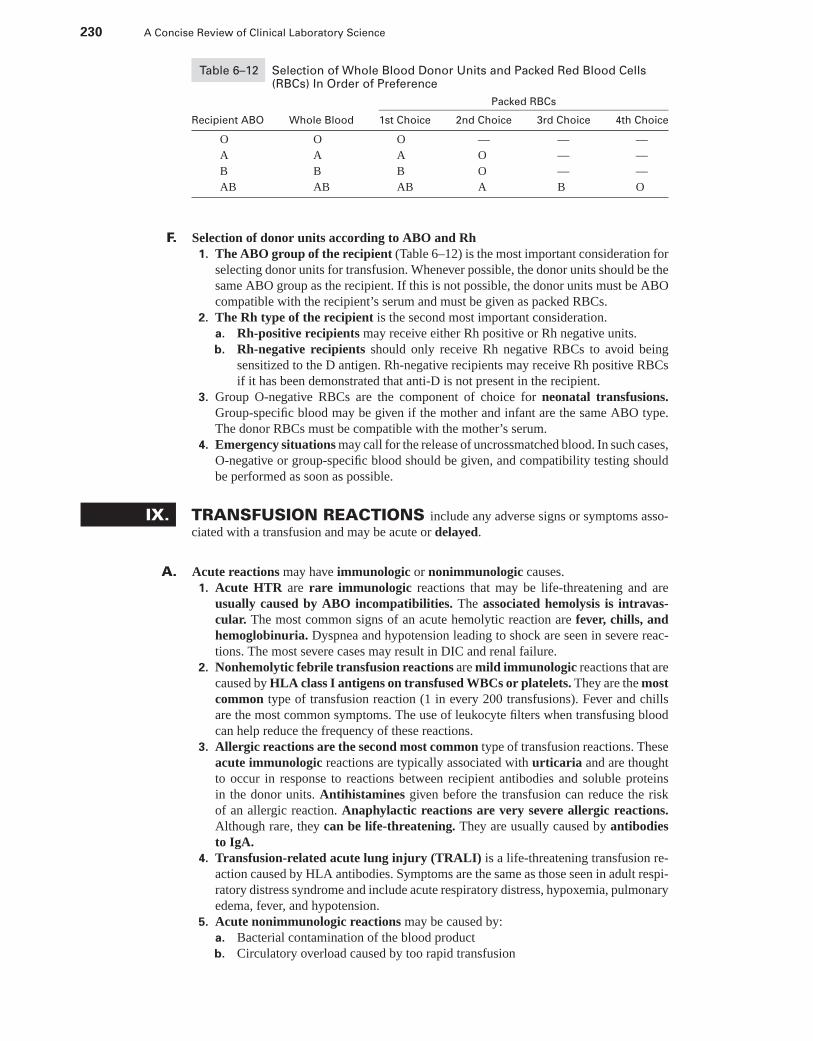
P1: OSO
LWBK192-06 LWBK192-Hubbard November 21, 2008 13:30
230 A Concise Review of Clinical Laboratory Science
Table 6–12 Selection of Whole Blood Donor Units and Packed Red Blood Cells(RBCs) In Order of Preference
Packed RBCs
Recipient ABO Whole Blood 1st Choice 2nd Choice 3rd Choice 4th Choice
O O O — — —
A A A O — —
B B B O — —
AB AB AB A B O
F. Selection of donor units according to ABO and Rh1. The ABO group of the recipient (Table 6–12) is the most important consideration for
selecting donor units for transfusion. Whenever possible, the donor units should be thesame ABO group as the recipient. If this is not possible, the donor units must be ABOcompatible with the recipient’s serum and must be given as packed RBCs.
2. The Rh type of the recipient is the second most important consideration.a. Rh-positive recipients may receive either Rh positive or Rh negative units.b. Rh-negative recipients should only receive Rh negative RBCs to avoid being
sensitized to the D antigen. Rh-negative recipients may receive Rh positive RBCsif it has been demonstrated that anti-D is not present in the recipient.
3. Group O-negative RBCs are the component of choice for neonatal transfusions.Group-specific blood may be given if the mother and infant are the same ABO type.The donor RBCs must be compatible with the mother’s serum.
4. Emergency situations may call for the release of uncrossmatched blood. In such cases,O-negative or group-specific blood should be given, and compatibility testing shouldbe performed as soon as possible.
IX. TRANSFUSION REACTIONS include any adverse signs or symptoms asso-ciated with a transfusion and may be acute or delayed.
A. Acute reactions may have immunologic or nonimmunologic causes.1. Acute HTR are rare immunologic reactions that may be life-threatening and are
usually caused by ABO incompatibilities. The associated hemolysis is intravas-cular. The most common signs of an acute hemolytic reaction are fever, chills, andhemoglobinuria. Dyspnea and hypotension leading to shock are seen in severe reac-tions. The most severe cases may result in DIC and renal failure.
2. Nonhemolytic febrile transfusion reactions are mild immunologic reactions that arecaused by HLA class I antigens on transfused WBCs or platelets. They are the mostcommon type of transfusion reaction (1 in every 200 transfusions). Fever and chillsare the most common symptoms. The use of leukocyte filters when transfusing bloodcan help reduce the frequency of these reactions.
3. Allergic reactions are the second most common type of transfusion reactions. Theseacute immunologic reactions are typically associated with urticaria and are thoughtto occur in response to reactions between recipient antibodies and soluble proteinsin the donor units. Antihistamines given before the transfusion can reduce the riskof an allergic reaction. Anaphylactic reactions are very severe allergic reactions.Although rare, they can be life-threatening. They are usually caused by antibodiesto IgA.
4. Transfusion-related acute lung injury (TRALI) is a life-threatening transfusion re-action caused by HLA antibodies. Symptoms are the same as those seen in adult respi-ratory distress syndrome and include acute respiratory distress, hypoxemia, pulmonaryedema, fever, and hypotension.
5. Acute nonimmunologic reactions may be caused by:a. Bacterial contamination of the blood productb. Circulatory overload caused by too rapid transfusion
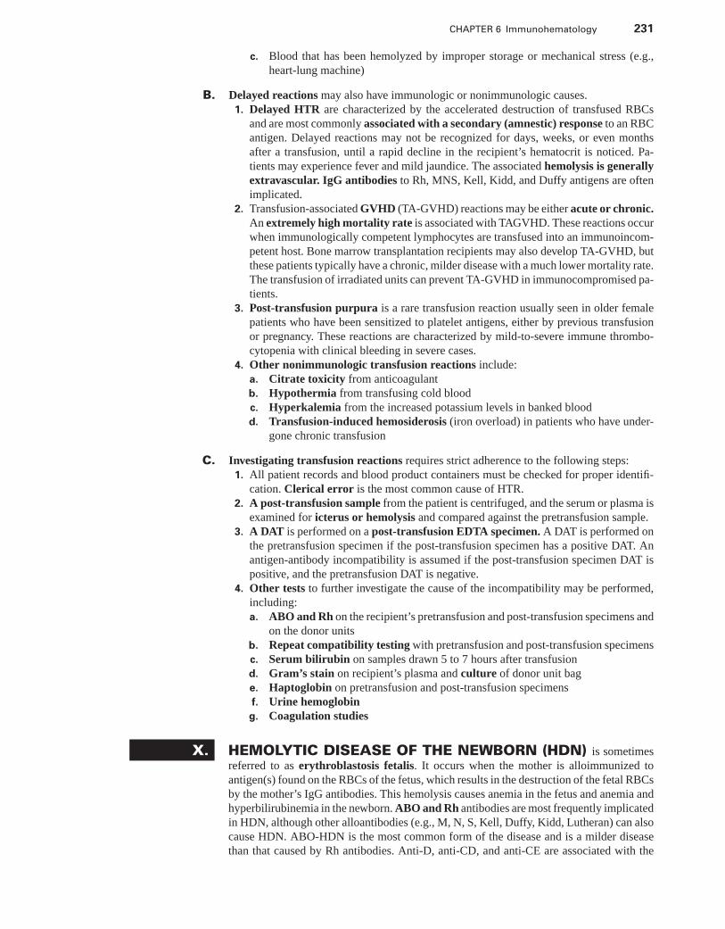
P1: OSO
LWBK192-06 LWBK192-Hubbard November 21, 2008 13:30
CHAPTER 6 Immunohematology 231
c. Blood that has been hemolyzed by improper storage or mechanical stress (e.g.,heart-lung machine)
B. Delayed reactions may also have immunologic or nonimmunologic causes.1. Delayed HTR are characterized by the accelerated destruction of transfused RBCs
and are most commonly associated with a secondary (amnestic) response to an RBCantigen. Delayed reactions may not be recognized for days, weeks, or even monthsafter a transfusion, until a rapid decline in the recipient’s hematocrit is noticed. Pa-tients may experience fever and mild jaundice. The associated hemolysis is generallyextravascular. IgG antibodies to Rh, MNS, Kell, Kidd, and Duffy antigens are oftenimplicated.
2. Transfusion-associated GVHD (TA-GVHD) reactions may be either acute or chronic.An extremely high mortality rate is associated with TAGVHD. These reactions occurwhen immunologically competent lymphocytes are transfused into an immunoincom-petent host. Bone marrow transplantation recipients may also develop TA-GVHD, butthese patients typically have a chronic, milder disease with a much lower mortality rate.The transfusion of irradiated units can prevent TA-GVHD in immunocompromised pa-tients.
3. Post-transfusion purpura is a rare transfusion reaction usually seen in older femalepatients who have been sensitized to platelet antigens, either by previous transfusionor pregnancy. These reactions are characterized by mild-to-severe immune thrombo-cytopenia with clinical bleeding in severe cases.
4. Other nonimmunologic transfusion reactions include:a. Citrate toxicity from anticoagulantb. Hypothermia from transfusing cold bloodc. Hyperkalemia from the increased potassium levels in banked bloodd. Transfusion-induced hemosiderosis (iron overload) in patients who have under-
gone chronic transfusion
C. Investigating transfusion reactions requires strict adherence to the following steps:1. All patient records and blood product containers must be checked for proper identifi-
cation. Clerical error is the most common cause of HTR.2. A post-transfusion sample from the patient is centrifuged, and the serum or plasma is
examined for icterus or hemolysis and compared against the pretransfusion sample.3. A DAT is performed on a post-transfusion EDTA specimen. A DAT is performed on
the pretransfusion specimen if the post-transfusion specimen has a positive DAT. Anantigen-antibody incompatibility is assumed if the post-transfusion specimen DAT ispositive, and the pretransfusion DAT is negative.
4. Other tests to further investigate the cause of the incompatibility may be performed,including:a. ABO and Rh on the recipient’s pretransfusion and post-transfusion specimens and
on the donor unitsb. Repeat compatibility testing with pretransfusion and post-transfusion specimensc. Serum bilirubin on samples drawn 5 to 7 hours after transfusiond. Gram’s stain on recipient’s plasma and culture of donor unit bage. Haptoglobin on pretransfusion and post-transfusion specimensf. Urine hemoglobin
g. Coagulation studies
X. HEMOLYTIC DISEASE OF THE NEWBORN (HDN) is sometimesreferred to as erythroblastosis fetalis. It occurs when the mother is alloimmunized toantigen(s) found on the RBCs of the fetus, which results in the destruction of the fetal RBCsby the mother’s IgG antibodies. This hemolysis causes anemia in the fetus and anemia andhyperbilirubinemia in the newborn. ABO and Rh antibodies are most frequently implicatedin HDN, although other alloantibodies (e.g., M, N, S, Kell, Duffy, Kidd, Lutheran) can alsocause HDN. ABO-HDN is the most common form of the disease and is a milder diseasethan that caused by Rh antibodies. Anti-D, anti-CD, and anti-CE are associated with the
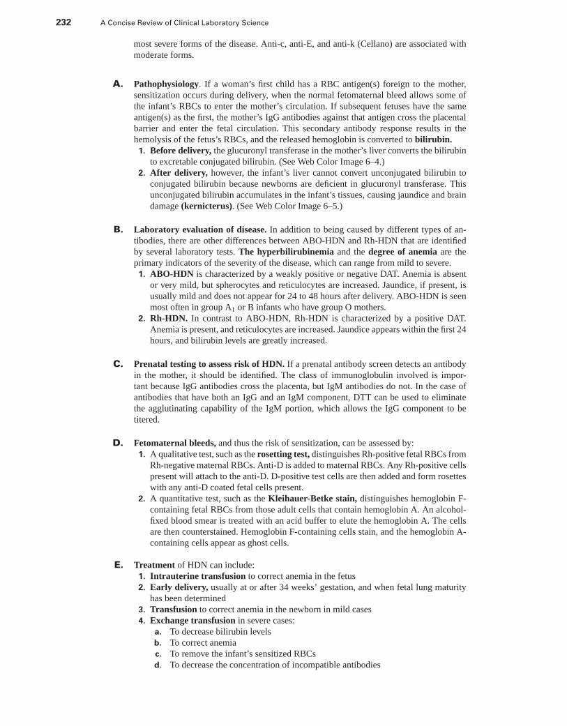
P1: OSO
LWBK192-06 LWBK192-Hubbard November 21, 2008 13:30
232 A Concise Review of Clinical Laboratory Science
most severe forms of the disease. Anti-c, anti-E, and anti-k (Cellano) are associated withmoderate forms.
A. Pathophysiology. If a woman’s first child has a RBC antigen(s) foreign to the mother,sensitization occurs during delivery, when the normal fetomaternal bleed allows some ofthe infant’s RBCs to enter the mother’s circulation. If subsequent fetuses have the sameantigen(s) as the first, the mother’s IgG antibodies against that antigen cross the placentalbarrier and enter the fetal circulation. This secondary antibody response results in thehemolysis of the fetus’s RBCs, and the released hemoglobin is converted to bilirubin.
1. Before delivery, the glucuronyl transferase in the mother’s liver converts the bilirubinto excretable conjugated bilirubin. (See Web Color Image 6–4.)
2. After delivery, however, the infant’s liver cannot convert unconjugated bilirubin toconjugated bilirubin because newborns are deficient in glucuronyl transferase. Thisunconjugated bilirubin accumulates in the infant’s tissues, causing jaundice and braindamage (kernicterus). (See Web Color Image 6–5.)
B. Laboratory evaluation of disease. In addition to being caused by different types of an-tibodies, there are other differences between ABO-HDN and Rh-HDN that are identifiedby several laboratory tests. The hyperbilirubinemia and the degree of anemia are theprimary indicators of the severity of the disease, which can range from mild to severe.
1. ABO-HDN is characterized by a weakly positive or negative DAT. Anemia is absentor very mild, but spherocytes and reticulocytes are increased. Jaundice, if present, isusually mild and does not appear for 24 to 48 hours after delivery. ABO-HDN is seenmost often in group A1 or B infants who have group O mothers.
2. Rh-HDN. In contrast to ABO-HDN, Rh-HDN is characterized by a positive DAT.Anemia is present, and reticulocytes are increased. Jaundice appears within the first 24hours, and bilirubin levels are greatly increased.
C. Prenatal testing to assess risk of HDN. If a prenatal antibody screen detects an antibodyin the mother, it should be identified. The class of immunoglobulin involved is impor-tant because IgG antibodies cross the placenta, but IgM antibodies do not. In the case ofantibodies that have both an IgG and an IgM component, DTT can be used to eliminatethe agglutinating capability of the IgM portion, which allows the IgG component to betitered.
D. Fetomaternal bleeds, and thus the risk of sensitization, can be assessed by:1. A qualitative test, such as the rosetting test, distinguishes Rh-positive fetal RBCs from
Rh-negative maternal RBCs. Anti-D is added to maternal RBCs. Any Rh-positive cellspresent will attach to the anti-D. D-positive test cells are then added and form rosetteswith any anti-D coated fetal cells present.
2. A quantitative test, such as the Kleihauer-Betke stain, distinguishes hemoglobin F-containing fetal RBCs from those adult cells that contain hemoglobin A. An alcohol-fixed blood smear is treated with an acid buffer to elute the hemoglobin A. The cellsare then counterstained. Hemoglobin F-containing cells stain, and the hemoglobin A-containing cells appear as ghost cells.
E. Treatment of HDN can include:1. Intrauterine transfusion to correct anemia in the fetus2. Early delivery, usually at or after 34 weeks’ gestation, and when fetal lung maturity
has been determined3. Transfusion to correct anemia in the newborn in mild cases4. Exchange transfusion in severe cases:
a. To decrease bilirubin levelsb. To correct anemiac. To remove the infant’s sensitized RBCsd. To decrease the concentration of incompatible antibodies
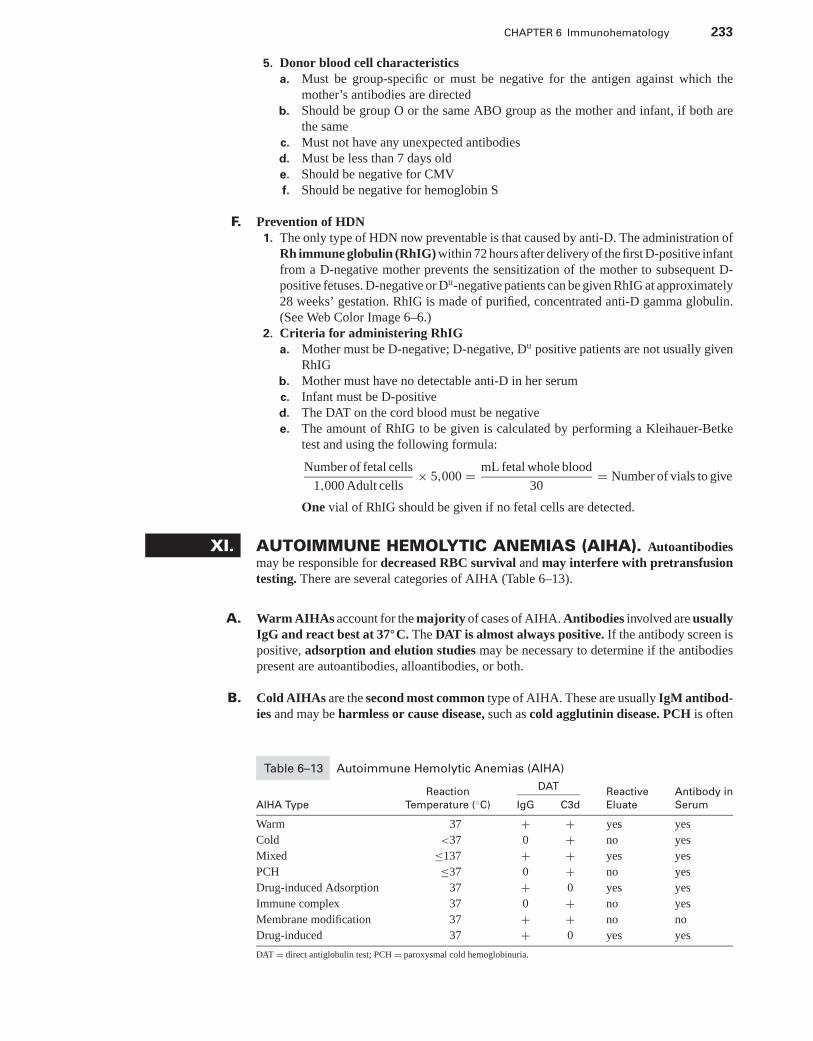
P1: OSO
LWBK192-06 LWBK192-Hubbard November 21, 2008 13:30
CHAPTER 6 Immunohematology 233
5. Donor blood cell characteristicsa. Must be group-specific or must be negative for the antigen against which the
mother’s antibodies are directedb. Should be group O or the same ABO group as the mother and infant, if both are
the samec. Must not have any unexpected antibodiesd. Must be less than 7 days olde. Should be negative for CMVf. Should be negative for hemoglobin S
F. Prevention of HDN1. The only type of HDN now preventable is that caused by anti-D. The administration of
Rh immune globulin (RhIG) within 72 hours after delivery of the first D-positive infantfrom a D-negative mother prevents the sensitization of the mother to subsequent D-positive fetuses. D-negative or Du-negative patients can be given RhIG at approximately28 weeks’ gestation. RhIG is made of purified, concentrated anti-D gamma globulin.(See Web Color Image 6–6.)
2. Criteria for administering RhIGa. Mother must be D-negative; D-negative, Du positive patients are not usually given
RhIGb. Mother must have no detectable anti-D in her serumc. Infant must be D-positived. The DAT on the cord blood must be negativee. The amount of RhIG to be given is calculated by performing a Kleihauer-Betke
test and using the following formula:
Number of fetal cells
1,000 Adult cells× 5,000 = mL fetal whole blood
30= Number of vials to give
One vial of RhIG should be given if no fetal cells are detected.
XI. AUTOIMMUNE HEMOLYTIC ANEMIAS (AIHA). Autoantibodiesmay be responsible for decreased RBC survival and may interfere with pretransfusiontesting. There are several categories of AIHA (Table 6–13).
A. Warm AIHAs account for the majority of cases of AIHA. Antibodies involved are usuallyIgG and react best at 37◦C. The DAT is almost always positive. If the antibody screen ispositive, adsorption and elution studies may be necessary to determine if the antibodiespresent are autoantibodies, alloantibodies, or both.
B. Cold AIHAs are the second most common type of AIHA. These are usually IgM antibod-ies and may be harmless or cause disease, such as cold agglutinin disease. PCH is often
Table 6–13 Autoimmune Hemolytic Anemias (AIHA)
DATReaction Reactive Antibody in
AIHA Type Temperature (◦C) IgG C3d Eluate Serum
Warm 37 + + yes yes
Cold <37 0 + no yes
Mixed ≤137 + + yes yes
PCH ≤37 0 + no yes
Drug-induced Adsorption 37 + 0 yes yes
Immune complex 37 0 + no yes
Membrane modification 37 + + no no
Drug-induced 37 + 0 yes yes
DAT = direct antiglobulin test; PCH = paroxysmal cold hemoglobinuria.
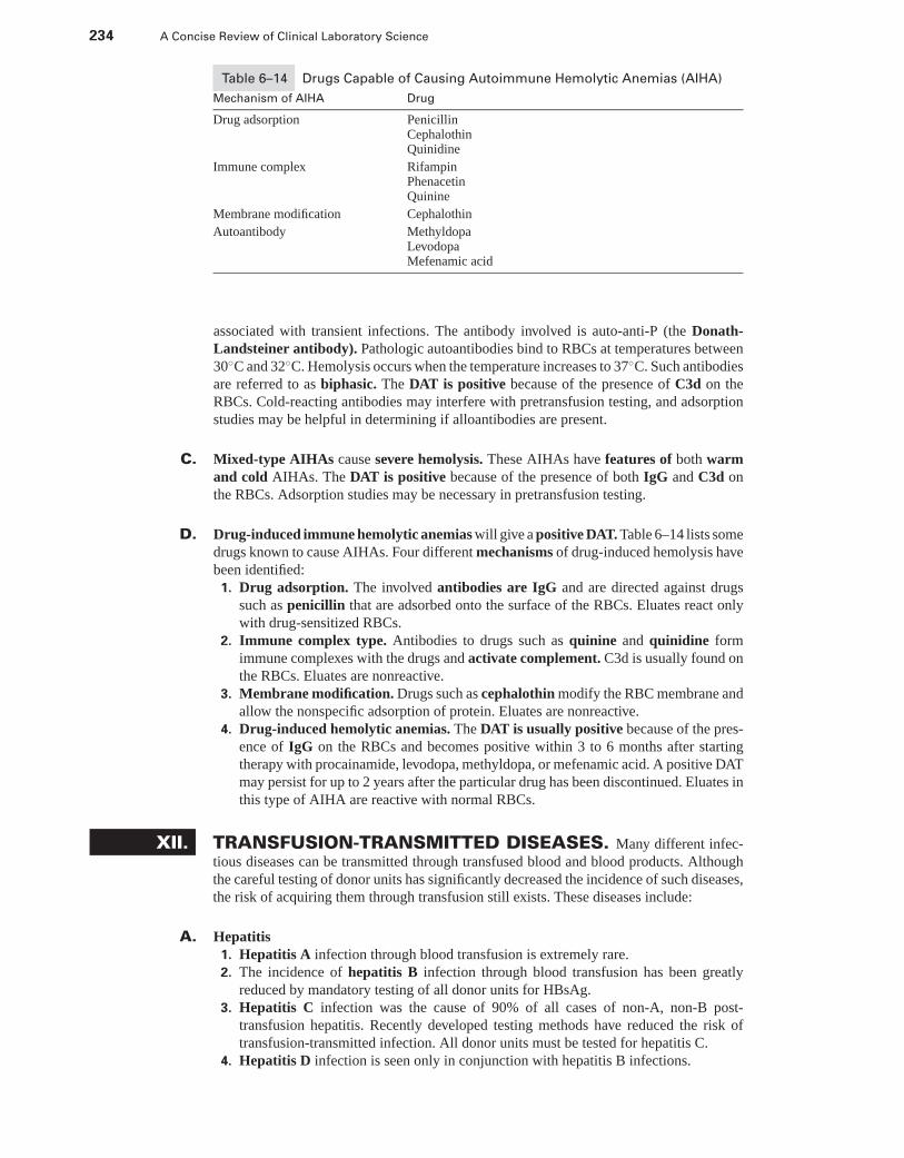
P1: OSO
LWBK192-06 LWBK192-Hubbard November 21, 2008 13:30
234 A Concise Review of Clinical Laboratory Science
Table 6–14 Drugs Capable of Causing Autoimmune Hemolytic Anemias (AIHA)
Mechanism of AIHA Drug
Drug adsorption PenicillinCephalothinQuinidine
Immune complex RifampinPhenacetinQuinine
Membrane modification Cephalothin
Autoantibody MethyldopaLevodopaMefenamic acid
associated with transient infections. The antibody involved is auto-anti-P (the Donath-Landsteiner antibody). Pathologic autoantibodies bind to RBCs at temperatures between30◦C and 32◦C. Hemolysis occurs when the temperature increases to 37◦C. Such antibodiesare referred to as biphasic. The DAT is positive because of the presence of C3d on theRBCs. Cold-reacting antibodies may interfere with pretransfusion testing, and adsorptionstudies may be helpful in determining if alloantibodies are present.
C. Mixed-type AIHAs cause severe hemolysis. These AIHAs have features of both warmand cold AIHAs. The DAT is positive because of the presence of both IgG and C3d onthe RBCs. Adsorption studies may be necessary in pretransfusion testing.
D. Drug-induced immune hemolytic anemias will give a positive DAT. Table 6–14 lists somedrugs known to cause AIHAs. Four different mechanisms of drug-induced hemolysis havebeen identified:
1. Drug adsorption. The involved antibodies are IgG and are directed against drugssuch as penicillin that are adsorbed onto the surface of the RBCs. Eluates react onlywith drug-sensitized RBCs.
2. Immune complex type. Antibodies to drugs such as quinine and quinidine formimmune complexes with the drugs and activate complement. C3d is usually found onthe RBCs. Eluates are nonreactive.
3. Membrane modification. Drugs such as cephalothin modify the RBC membrane andallow the nonspecific adsorption of protein. Eluates are nonreactive.
4. Drug-induced hemolytic anemias. The DAT is usually positive because of the pres-ence of IgG on the RBCs and becomes positive within 3 to 6 months after startingtherapy with procainamide, levodopa, methyldopa, or mefenamic acid. A positive DATmay persist for up to 2 years after the particular drug has been discontinued. Eluates inthis type of AIHA are reactive with normal RBCs.
XII. TRANSFUSION-TRANSMITTED DISEASES. Many different infec-tious diseases can be transmitted through transfused blood and blood products. Althoughthe careful testing of donor units has significantly decreased the incidence of such diseases,the risk of acquiring them through transfusion still exists. These diseases include:
A. Hepatitis1. Hepatitis A infection through blood transfusion is extremely rare.2. The incidence of hepatitis B infection through blood transfusion has been greatly
reduced by mandatory testing of all donor units for HBsAg.3. Hepatitis C infection was the cause of 90% of all cases of non-A, non-B post-
transfusion hepatitis. Recently developed testing methods have reduced the risk oftransfusion-transmitted infection. All donor units must be tested for hepatitis C.
4. Hepatitis D infection is seen only in conjunction with hepatitis B infections.
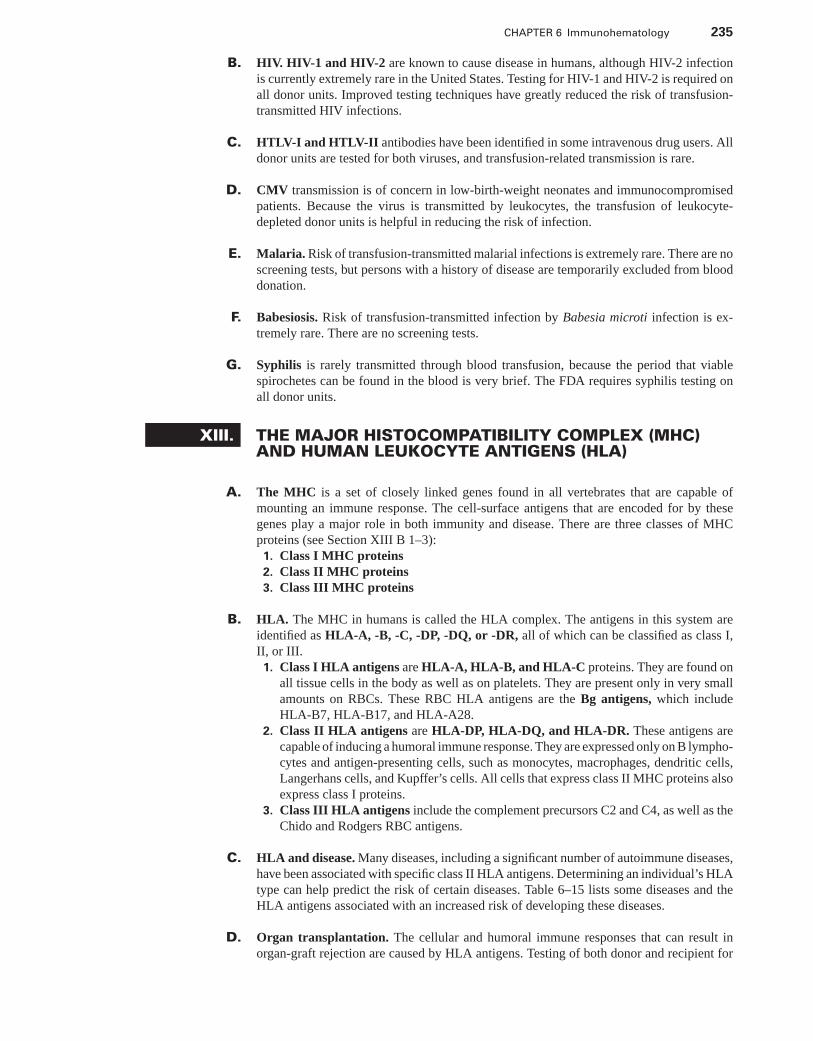
P1: OSO
LWBK192-06 LWBK192-Hubbard November 21, 2008 13:30
CHAPTER 6 Immunohematology 235
B. HIV. HIV-1 and HIV-2 are known to cause disease in humans, although HIV-2 infectionis currently extremely rare in the United States. Testing for HIV-1 and HIV-2 is required onall donor units. Improved testing techniques have greatly reduced the risk of transfusion-transmitted HIV infections.
C. HTLV-I and HTLV-II antibodies have been identified in some intravenous drug users. Alldonor units are tested for both viruses, and transfusion-related transmission is rare.
D. CMV transmission is of concern in low-birth-weight neonates and immunocompromisedpatients. Because the virus is transmitted by leukocytes, the transfusion of leukocyte-depleted donor units is helpful in reducing the risk of infection.
E. Malaria. Risk of transfusion-transmitted malarial infections is extremely rare. There are noscreening tests, but persons with a history of disease are temporarily excluded from blooddonation.
F. Babesiosis. Risk of transfusion-transmitted infection by Babesia microti infection is ex-tremely rare. There are no screening tests.
G. Syphilis is rarely transmitted through blood transfusion, because the period that viablespirochetes can be found in the blood is very brief. The FDA requires syphilis testing onall donor units.
XIII. THE MAJOR HISTOCOMPATIBILITY COMPLEX (MHC)AND HUMAN LEUKOCYTE ANTIGENS (HLA)
A. The MHC is a set of closely linked genes found in all vertebrates that are capable ofmounting an immune response. The cell-surface antigens that are encoded for by thesegenes play a major role in both immunity and disease. There are three classes of MHCproteins (see Section XIII B 1–3):
1. Class I MHC proteins2. Class II MHC proteins3. Class III MHC proteins
B. HLA. The MHC in humans is called the HLA complex. The antigens in this system areidentified as HLA-A, -B, -C, -DP, -DQ, or -DR, all of which can be classified as class I,II, or III.
1. Class I HLA antigens are HLA-A, HLA-B, and HLA-C proteins. They are found onall tissue cells in the body as well as on platelets. They are present only in very smallamounts on RBCs. These RBC HLA antigens are the Bg antigens, which includeHLA-B7, HLA-B17, and HLA-A28.
2. Class II HLA antigens are HLA-DP, HLA-DQ, and HLA-DR. These antigens arecapable of inducing a humoral immune response. They are expressed only on B lympho-cytes and antigen-presenting cells, such as monocytes, macrophages, dendritic cells,Langerhans cells, and Kupffer’s cells. All cells that express class II MHC proteins alsoexpress class I proteins.
3. Class III HLA antigens include the complement precursors C2 and C4, as well as theChido and Rodgers RBC antigens.
C. HLA and disease. Many diseases, including a significant number of autoimmune diseases,have been associated with specific class II HLA antigens. Determining an individual’s HLAtype can help predict the risk of certain diseases. Table 6–15 lists some diseases and theHLA antigens associated with an increased risk of developing these diseases.
D. Organ transplantation. The cellular and humoral immune responses that can result inorgan-graft rejection are caused by HLA antigens. Testing of both donor and recipient for
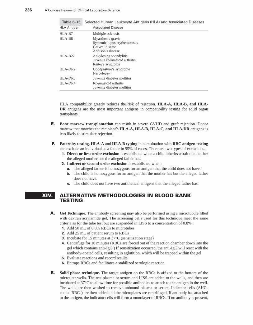
P1: OSO
LWBK192-06 LWBK192-Hubbard November 21, 2008 13:30
236 A Concise Review of Clinical Laboratory Science
Table 6–15 Selected Human Leukocyte Antigens (HLA) and Associated Diseases
HLA Antigen Associated Disease
HLA-B7 Multiple sclerosis
HLA-B8 Myasthenia gravisSystemic lupus erythematosusGraves’ diseaseAddison’s disease
HLA-B27 Ankylosing spondylitisJuvenile rheumatoid arthritisReiter’s syndrome
HLA-DR2 Goodpasture’s syndromeNarcolepsy
HLA-DR3 Juvenile diabetes mellitus
HLA-DR4 Rheumatoid arthritisJuvenile diabetes mellitus
HLA compatibility greatly reduces the risk of rejection. HLA-A, HLA-B, and HLA-DR antigens are the most important antigens in compatibility testing for solid organtransplants.
E. Bone marrow transplantation can result in severe GVHD and graft rejection. Donormarrow that matches the recipient’s HLA-A, HLA-B, HLA-C, and HLA-DR antigens isless likely to stimulate rejection.
F. Paternity testing. HLA-A and HLA-B typing in combination with RBC antigen testingcan exclude an individual as a father in 95% of cases. There are two types of exclusions.
1. Direct or first-order exclusion is established when a child inherits a trait that neitherthe alleged mother nor the alleged father has.
2. Indirect or second-order exclusion is established when:a. The alleged father is homozygous for an antigen that the child does not have.b. The child is homozygous for an antigen that the mother has but the alleged father
does not have.c. The child does not have two antithetical antigens that the alleged father has.
XIV. ALTERNATIVE METHODOLOGIES IN BLOOD BANKTESTING
A. Gel Technique. The antibody screening may also be performed using a microtubule filledwith dextran acrylamide gel. The screening cells used for this technique meet the samecriteria as for the tube test but are suspended in LISS to a concentration of 0.8%.
1. Add 50 mL of 0.8% RBCs to microtubes2. Add 25 mL of patient serum to RBCs3. Incubate for 15 minutes at 37◦C (sensitization stage)4. Centrifuge for 10 minutes (RBCs are forced out of the reaction chamber down into the
gel which contains anti-IgG.) If sensitization occurred, the anti-IgG will react with theantibody-coated cells, resulting in aglutition, which will be trapped within the gel
5. Evaluate reactions and record results.6. Entraps RBCs and facilitates a stabilized serologic reaction
B. Solid phase technique. The target antigen on the RBCs is affixed to the bottom of themicrotiter wells. The test plasma or serum and LISS are added to the wells, and then areincubated at 37◦C to allow time for possible antibodies to attach to the antigen in the well.The wells are then washed to remove unbound plasma or serum. Indicator cells (AHG-coated RBCs) are then added and the microplates are centrifuged. If antibody has attachedto the antigen, the indicator cells will form a monolayer of RBCs. If no antibody is present,

P1: OSO
LWBK192-06 LWBK192-Hubbard November 21, 2008 13:30
CHAPTER 6 Immunohematology 237
nothing is attached to the antigen, and the indicator cells form a clearly delineated buttonat the center of the microplate well following certification.
Online Menu for This Chapter
Go to thePoint.lww.com/Hubbard2e for the following:
� Web Color Images 6–1 through 6–7� Web Case Studies 6–1 through 6–4� Study Questions and Answers
P1: OSO
LWBK192-07 LWBK192-Hubbard November 27, 2008 7:48
CHAPTER 7
Clinical Microbiology
LYNNE HAMILTON, PhD, MT (ASCP) AND HAL LARSEN, MT(ASCP), CLS (NCA), PhD
I. BACTERIA
A. Prokaryotic organisms1. Bacteria are unicellular organisms that lack a true nucleus and nuclear membrane. The
lack of mitochondria, endoplasmic reticulum, and Golgi bodies also differentiates theprokaryotic bacteria from eukaryotes.
2. The bacterial genome is a single, closed, circular chromosome of double-strandedDNA called the nucleoid. Plasmids are small circular molecules of extrachromosomalcircular DNA. Antibiotic resistance is often encoded by genes located on plasmids.Chromosomal or plasmid gene exchange via transformation, transduction, or conjuga-tion are means of genetic recombination.
3. Reproduction of bacteria is achieved by binary fission, a means of asexual reproduc-tion. Prokaryotes do not reproduce sexually.
4. Energy generation of bacteria is cytoplasmic membrane-associated via the electrontransport chain.
5. Bacteria usually range in size from 0.2 to 2 um in diameter and 1 to 6 um in length. Thefour basic morphological types are cocci (spherical-shaped cells), bacilli (rod-shapedcells), spirilla (spiral-shaped cells), and vibrios (comma-shaped cells (Figure 7–1).
B. Growth and nutrition1. Prokaryotic bacteria have three major nutritional needs for growth.
a. A source of carbon is needed for the synthesis of cellular constituents.b. A source of nitrogen is necessary for the synthesis of protein.c. Energy (ATP) is needed for cellular functions.
2. The optimum pH for the growth of most bacteria is 7.0 to 7.5. Acidophiles grow atan acidic pH and alkaliphiles grow optimally at an alkaline pH.
3. A wide range of growth temperatures facilitate the growth of bacteria. The optimumgrowth temperature for most human bacterial pathogens is 35◦C to 37◦C, the tem-perature of the human body.
4. Bacteria may be classified based on the atmospheric requirement for the growth.a. Obligate aerobes require oxygen for growth.b. Aerotolerant aerobes can grow in the presence of oxygen, but grow best in an
anaerobic environment.c. Facultative anaerobes grow in both aerobic and anaerobic environments.d. Obligate anaerobes cannot grow in the presence of oxygen.e. Capnophilic bacteria require concentrations of 5% to 10% CO2 for optimal growth.f. Microaerophilic bacteria grow optimally in a reduced level of oxygen.
238
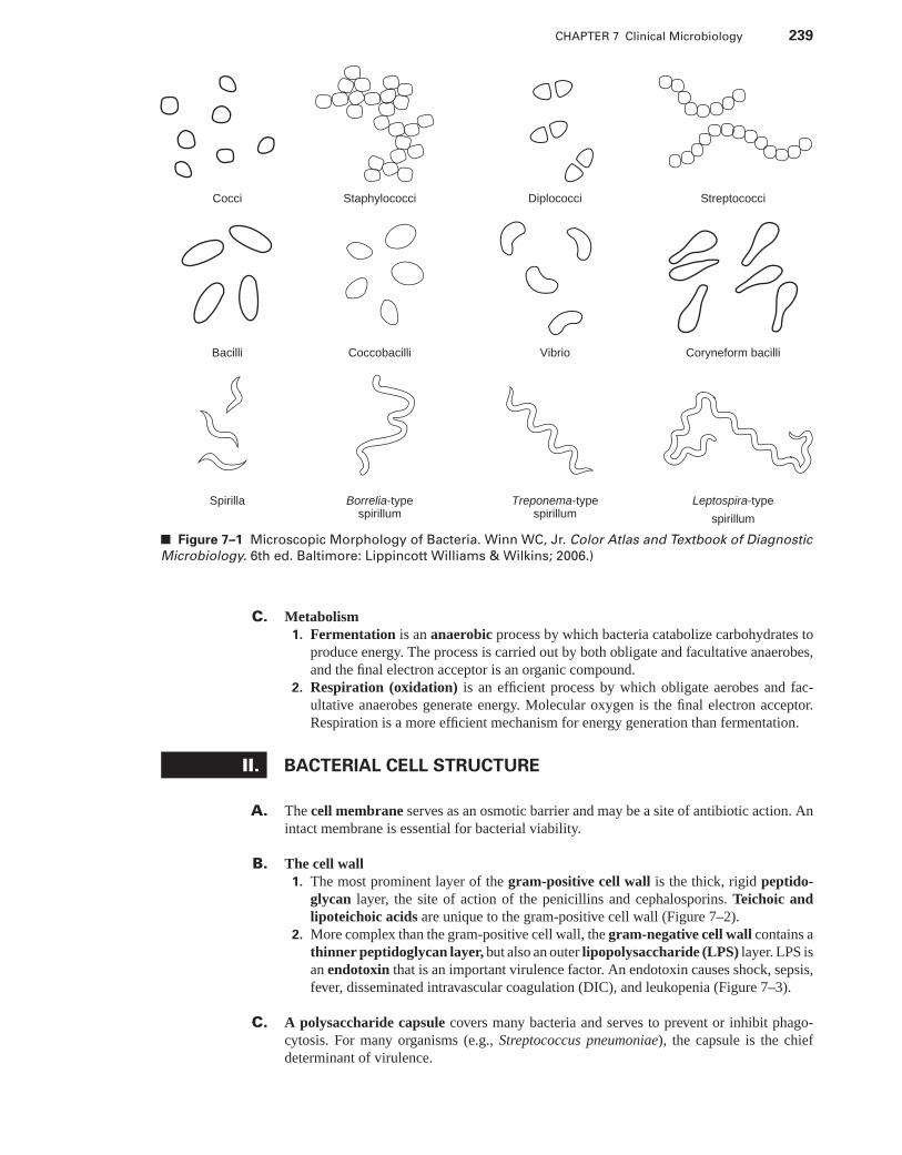
P1: OSO
LWBK192-07 LWBK192-Hubbard November 27, 2008 7:48
CHAPTER 7 Clinical Microbiology 239
Cocci Staphylococci StreptococciDiplococci
Bacilli Coccobacilli Coryneform bacilliVibrio
Spirilla Borrelia-typespirillum
Leptospira-type
spirillum
Treponema-typespirillum
■ Figure 7–1 Microscopic Morphology of Bacteria. Winn WC, Jr. Color Atlas and Textbook of Diagnostic
Microbiology. 6th ed. Baltimore: Lippincott Williams & Wilkins; 2006.)
C. Metabolism1. Fermentation is an anaerobic process by which bacteria catabolize carbohydrates to
produce energy. The process is carried out by both obligate and facultative anaerobes,and the final electron acceptor is an organic compound.
2. Respiration (oxidation) is an efficient process by which obligate aerobes and fac-ultative anaerobes generate energy. Molecular oxygen is the final electron acceptor.Respiration is a more efficient mechanism for energy generation than fermentation.
II. BACTERIAL CELL STRUCTURE
A. The cell membrane serves as an osmotic barrier and may be a site of antibiotic action. Anintact membrane is essential for bacterial viability.
B. The cell wall1. The most prominent layer of the gram-positive cell wall is the thick, rigid peptido-
glycan layer, the site of action of the penicillins and cephalosporins. Teichoic andlipoteichoic acids are unique to the gram-positive cell wall (Figure 7–2).
2. More complex than the gram-positive cell wall, the gram-negative cell wall contains athinner peptidoglycan layer, but also an outer lipopolysaccharide (LPS) layer. LPS isan endotoxin that is an important virulence factor. An endotoxin causes shock, sepsis,fever, disseminated intravascular coagulation (DIC), and leukopenia (Figure 7–3).
C. A polysaccharide capsule covers many bacteria and serves to prevent or inhibit phago-cytosis. For many organisms (e.g., Streptococcus pneumoniae), the capsule is the chiefdeterminant of virulence.
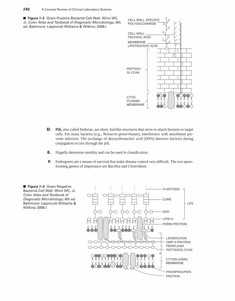
P1: OSO
LWBK192-07 LWBK192-Hubbard November 27, 2008 7:48
240 A Concise Review of Clinical Laboratory Science
CELL WALL SPECIFICPOLYSACCHARIDE
CELL WALL TEICHOIC ACID
MEMBRANELIPOTEICHOIC ACID
PEPTIDO-GLYCAN
CYTO-PLASMICMEMBRANE
■ Figure 7–2 Gram-Positive Bacterial Cell Wall. Winn WC,
Jr. Color Atlas and Textbook of Diagnostic Microbiology. 6th
ed. Baltimore: Lippincott Williams & Wilkins; 2006.)
D. Pili, also called fimbriae, are short, hairlike structures that serve to attach bacteria to targetcells. For many bacteria (e.g., Neisseria gonorrhoeae), interference with attachment pre-vents infection. The exchange of deoxyribonucleic acid (DNA) between bacteria duringconjugation occurs through the pili.
E. Flagella determine motility and can be used in classification.
F. Endospores are a means of survival that make disease control very difficult. The two spore-forming genera of importance are Bacillus and Clostridium.
O-ANTIGEN
CORELPS
KDO
LIPID-A
PEPTIDOGLYCANPERIPLASM
LIPOPROTEIN
PORIN PROTEIN
PHOSPHOLIPIDS
OMP A PROTEIN
CYTOPLASMICMEMBRANE
PROTEIN
■ Figure 7–3 Gram-Negative
Bacterial Cell Wall. Winn WC, Jr.
Color Atlas and Textbook of
Diagnostic Microbiology. 6th ed.
Baltimore: Lippincott Williams &
Wilkins; 2006.)
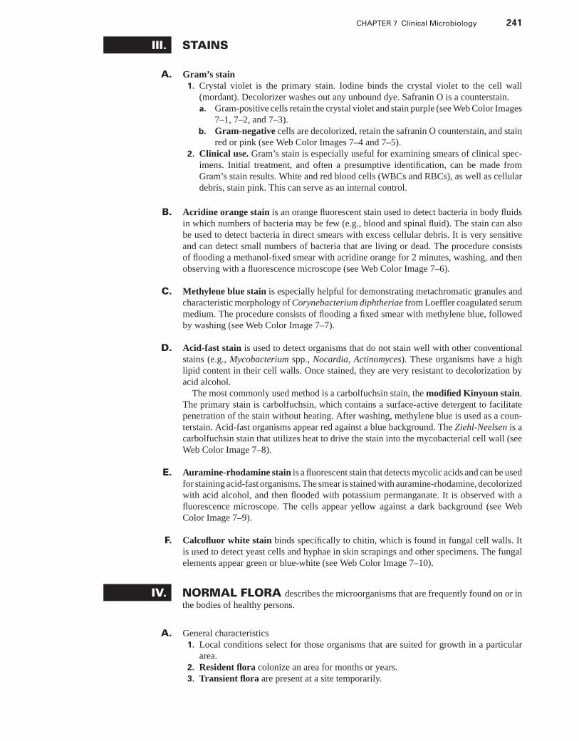
P1: OSO
LWBK192-07 LWBK192-Hubbard November 27, 2008 7:48
CHAPTER 7 Clinical Microbiology 241
III. STAINS
A. Gram’s stain1. Crystal violet is the primary stain. Iodine binds the crystal violet to the cell wall
(mordant). Decolorizer washes out any unbound dye. Safranin O is a counterstain.a. Gram-positive cells retain the crystal violet and stain purple (see Web Color Images
7–1, 7–2, and 7–3).b. Gram-negative cells are decolorized, retain the safranin O counterstain, and stain
red or pink (see Web Color Images 7–4 and 7–5).2. Clinical use. Gram’s stain is especially useful for examining smears of clinical spec-
imens. Initial treatment, and often a presumptive identification, can be made fromGram’s stain results. White and red blood cells (WBCs and RBCs), as well as cellulardebris, stain pink. This can serve as an internal control.
B. Acridine orange stain is an orange fluorescent stain used to detect bacteria in body fluidsin which numbers of bacteria may be few (e.g., blood and spinal fluid). The stain can alsobe used to detect bacteria in direct smears with excess cellular debris. It is very sensitiveand can detect small numbers of bacteria that are living or dead. The procedure consistsof flooding a methanol-fixed smear with acridine orange for 2 minutes, washing, and thenobserving with a fluorescence microscope (see Web Color Image 7–6).
C. Methylene blue stain is especially helpful for demonstrating metachromatic granules andcharacteristic morphology of Corynebacterium diphtheriae from Loeffler coagulated serummedium. The procedure consists of flooding a fixed smear with methylene blue, followedby washing (see Web Color Image 7–7).
D. Acid-fast stain is used to detect organisms that do not stain well with other conventionalstains (e.g., Mycobacterium spp., Nocardia, Actinomyces). These organisms have a highlipid content in their cell walls. Once stained, they are very resistant to decolorization byacid alcohol.
The most commonly used method is a carbolfuchsin stain, the modified Kinyoun stain.The primary stain is carbolfuchsin, which contains a surface-active detergent to facilitatepenetration of the stain without heating. After washing, methylene blue is used as a coun-terstain. Acid-fast organisms appear red against a blue background. The Ziehl-Neelsen is acarbolfuchsin stain that utilizes heat to drive the stain into the mycobacterial cell wall (seeWeb Color Image 7–8).
E. Auramine-rhodamine stain is a fluorescent stain that detects mycolic acids and can be usedfor staining acid-fast organisms. The smear is stained with auramine-rhodamine, decolorizedwith acid alcohol, and then flooded with potassium permanganate. It is observed with afluorescence microscope. The cells appear yellow against a dark background (see WebColor Image 7–9).
F. Calcofluor white stain binds specifically to chitin, which is found in fungal cell walls. Itis used to detect yeast cells and hyphae in skin scrapings and other specimens. The fungalelements appear green or blue-white (see Web Color Image 7–10).
IV. NORMAL FLORA describes the microorganisms that are frequently found on or inthe bodies of healthy persons.
A. General characteristics1. Local conditions select for those organisms that are suited for growth in a particular
area.2. Resident flora colonize an area for months or years.3. Transient flora are present at a site temporarily.
P1: OSO
LWBK192-07 LWBK192-Hubbard November 27, 2008 7:48
242 A Concise Review of Clinical Laboratory Science
4. Organisms that live at the expense of the host are parasites.5. Organisms that benefit the host are symbionts.6. Commensals have a neutral effect on the host.7. A carrier harbors the organism without manifesting symptoms, but is capable of trans-
mitting infection (carrier state).8. Opportunists are organisms that do not normally cause infection, but can do so if the
condition of the host changes (e.g., immunosuppression).
B. Normal flora of the skin1. The skin contains a wide variety of microorganisms that are not eliminated by washing
or superficial antisepsis.a. Propionibacterium acnes colonizes the sebaceous glands.b. Micrococcus, Staphylococcus spp., and diphtheroids are common.
2. Intact skin is an effective barrier to microbial invasion.
C. Normal flora of the mouth. The mouth contains large numbers of bacteria, most com-monly Streptococcus spp. (especially viridans species), coagulase-negative Staphylo-coccus, Peptostreptococcus, and other anaerobes.
D. Normal flora of the respiratory tract1. The respiratory tract beyond the oropharynx is normally sterile.2. The ciliary action of epithelial cells and mucus movement remove invading organisms.3. The nose and nasopharynx contain Staphylococcus aureus, S. epidermidis, and Strep-
tococcus spp. The following may be present transiently during community outbreaksof infection: Streptococcus pneumoniae, Haemophilus influenzae, and Neisseriameningitidis.
4. The normal flora in the oropharynx mirrors that of the mouth.
E. Normal flora of the gastrointestinal tract1. Most microorganisms are destroyed in the stomach due to the acidic pH. The survivors
multiply in the colon.2. More than 90% of the microbial population is comprised of anaerobes.3. Alteration of the normal flora by antibiotics may allow a superinfection by Clostridium
difficile (necrotizing enterocolitis), Candida albicans, or S. aureus.4. The most commonly found organisms in the gastrointestinal tract are: Bacteroides spp.,
Clostridium spp., Eubacterium spp., anaerobic streptococci, Enterococcus spp., andEnterobacteriaceae.
F. Normal flora of the genitourinary tract1. The outermost segment of the urethra is colonized by skin organisms.2. The vagina is colonized with Lactobacillus, anaerobic gram-negative rod-shaped
bacteria, and gram-positive cocci.
V. PATHOGENESIS OF INFECTION
A. Host resistance1. Innate immunity, or natural immunity, is a nonspecific mechanism of resistance to
disease.a. Physical barriers such as the skin, mucous membranes, and cilia help prevent the
invasion of pathogenic bacteria.b. The cleansing action of fluids in the eyes, respiratory, digestive, urinary, and
genital tracts are also effective host resistance mechanisms.c. Antimicrobial substances such as secretory IgA, lysozyme, B-lysins, and inter-
feron are produced in the human host.d. Indigenous microbial flora compete with invading bacteria for colonization sites,
as well as produce inhibitory bacteriocins.
P1: OSO
LWBK192-07 LWBK192-Hubbard November 27, 2008 7:48
CHAPTER 7 Clinical Microbiology 243
e. Phagocytosis, inflammation, and complement activation are also important non-specific defense mechanisms of the innate immunity.
2. Acquired or specific immunity develops in response to a specific foreign antigen (e.g.,invading bacterium). Acquired immunity may be active (a response to immunizationor disease) or passive (through placental transfer or infusions of serum or plasma).a. The humoral response involves antibody production by B lymphocytes.b. Cell-medicated immunity involves cells, such as macrophages, cytokines, and
T lymphocytes. The cellular response provides immunity against intracellularpathogens, including viruses, fungi, and mycobacteria.
B. Criteria for a successful pathogen1. The degree of pathogenicity of a microorganism is directly related to its virulence. A
virulent strain is characterized by its ability to evade or overcome host defenses andcause disease. Microbial virulence factors include adhesions, capsular polysaccharides,and production of extracellular toxins and enzymes that may cause damage to thehost.
2. An appropriate portal of entry is necessary for successful transmission of disease. Forexample, respiratory pathogens enter the respiratory tract via inhalation of aerosolizedrespiratory secretions. Gastrointestinal pathogens enter the GI tract via ingestion ofcontaminated food and water.
3. A sufficient infective dose is needed for a microorganism to be able to cause disease inits host. Some organisms can establish an infection with a relatively low infective dose(e.g., Shigella), whereas others require a much higher infective dose to cause infection(e.g., Salmonella).
4. A successful pathogen must be able to overcome the host resistance mechanisms,including the immune responses, and cause disease in a susceptible host.
5. For continued transmission of disease, pathogenic microorganisms must be able toleave the host via an appropriate portal of exit. For most pathogens, it is the same asthe portal of entry.
C. Transmission1. Airborne transmission involves the respiratory spread of infectious disease via
aerosolized respiratory secretions or contact with contaminated inanimate objects orfingers and hands.
2. Ingestion of contaminated food and water is the usual means for transmission ofgastrointestinal infections. Infection can also occur via the fecal-oral route.
3. Sexual transmission is the route of infection for venereal diseases. Some diseasescan be also transmitted via skin-to-skin contact or via direct transfer of saliva (e.g.,kissing).
4. Animal and human bites can result in serious wound infections, usually caused bymouth flora. Rabies is an example of a viral infection transmitted from an animal bite.Opportunistic pathogens, including environmental bacteria, can enter the human viacuts and trauma wounds.
5. Arthropod vectors, such as ticks, fleas, and mites, are responsible for the transmissionof diseases such as malaria, Lyme disease, plague, Rocky Mountain spotted fever, andhemorrhagic fevers. The infectious agent multiplies in the arthropod and is transmittedwhen the arthropod feeds off a human host.
6. Zoonoses are diseases of animals that are transmitted to humans. Transmission is usu-ally via contact with infected animals, animal secretions, or animal products. Zoonosescan also be transmitted via animal bites or arthropod vectors.
VI. COLLECTION AND HANDLING OF CLINICAL SPECIMENS
A. Collection. A properly collected specimen is absolutely crucial to quality diagnostic infor-mation and patient care.
1. Safety
P1: OSO
LWBK192-07 LWBK192-Hubbard November 27, 2008 7:48
244 A Concise Review of Clinical Laboratory Science
a. Universal precautions are followed throughout the collection and handling pro-cess. Persons collecting or handling specimens should wear gloves and a laboratorycoat. Eye protection should also be worn if splashing is a potential hazard.
b. Accidents or injuries must be reported immediately.2. General guidelines
a. The specimen should be from the infection site and not contaminated by the sur-rounding area (e.g., culture within a wound and not the surface or the surroundingskin).
b. Whenever possible, the specimen should be collected before antimicrobials areadministered.
c. Appropriate collection devices and containers should be used and must be sterile.Aseptic technique is required.
d. The specimen container should be labeled with the patient’s identification, the dateand time of collection, and the source of specimen.
3. Collection from various body sitesa. Throat. The tongue should be depressed before swabbing between the tonsillar
pillars and behind the uvula. The cheek, tongue, and teeth should not be touched.b. Nasopharynx. A flexible wire nasopharyngeal swab should be gently inserted
through the nose into the posterior nasopharynx, rotated, and then removed.c. Sputum. Whenever possible, the patient should gargle with water (not mouthwash)
immediately before sampling. Early morning specimens are best. Expectoratedspecimens from a deep cough should be collected into a sterile specimen cup.
d. Stool should be collected in a clean, wide-mouthed container with a tight-fitting lid.If the specimen cannot be plated within 1 hour of collection, it should be mixed witha transport medium (e.g., buffered glycerol saline, Cary-Blair transport medium).The change in pH and temperature over time is detrimental to Shigella spp. Stoolspecimens should never be taken from the toilet and should not be contaminatedwith urine. Commercial systems with preservatives are available for collection ofspecimens for both bacterial culture and ova and parasite examination.
e. Urine. Midstream clean-catch is the most common collection method. Propercleansing of the urethral area is important, especially in women. The first fewmilliliters, which flush out the urethra, are discarded. The specimen should be col-lected into a sterile specimen cup and transported immediately to the laboratory orrefrigerated, because contaminants grow readily at room temperature. Cultures ofcatheterized urine specimens usually have less contaminating bacterial flora.
f. Blood. Two to three cultures should be collected at random times during a 24-hourperiod. Collecting more than three sets of cultures in a 24-hour period does not sig-nificantly increase the probability of detecting bacteremia. Skin is disinfected with70% alcohol, followed by iodine. The disinfectant is allowed to dry. The puncturesite should not be palpated after disinfection. Ideally, 20 to 30 mL of blood perculture is collected from an adult (1–5 mL from infants and small children). Iodineshould be cleaned from the puncture site with alcohol following the venipuncture.
g. Cerebrospinal fluid should be collected aseptically by a physician. This specimenshould be processed immediately and not exposed to heat or refrigeration.
h. Abscess aspirates or exudates, as well as synovial, pericardial, and chest fluidshould be collected by a physician with a needle and syringe. The use of swabsmay inhibit growth of anaerobes or increase the likelihood of contamination withindigenous bacteria flora from adjacent tissues (e.g., mucous membranes or su-perficial skin surfaces). Care should be taken not to inject an air bubble into thesyringe.
i. Genital tract(1) Men. Exudates may be expressed from the urethral orifice or a small-diameter
swab may be inserted 3 to 4 cm into the urethra. The specimen should be platedimmediately on the appropriate media and not allowed to dry or be exposed tocold temperatures. A direct Gram’s stain smear should be prepared.
(2) Women. Cervical specimens are obtained by a physician with the aid of aspeculum. Lubricants, which may be lethal to Neisseria gonorrhoeae, should
P1: OSO
LWBK192-07 LWBK192-Hubbard November 27, 2008 7:48
CHAPTER 7 Clinical Microbiology 245
not be used on the speculum. The cervical mucus plug is removed, and asterile swab is inserted into the cervix, rotated, and allowed to remain for a fewseconds. The specimen should be plated immediately to the appropriate media,and swabs should not be refrigerated, as refrigeration may be lethal to genitalpathogens. A smear for Gram’s stain should be prepared from the specimen.
B. Handling1. Transport all specimens to the laboratory promptly. Anaerobic specimens must be
transported in an anaerobic transport system. If transport cannot occur immediately,most specimens can be held at 2◦C to 8◦C. Exceptions to this include specimens thatlikely contain temperature-sensitive organisms (e.g., Neisseria), blood culture bottles,and cerebrospinal fluid (CSF). Generally, swabs are the least desirable collection andtransport method. However, organisms can be successfully cultured if the swab ishandled and transported properly (i.e., not allowed to dry out). Swabs are inappropriatefor culturing anaerobes, although in some clinical settings, culturettes are often used.If swabs are used for the culture of anaerobes, is essential that an anaerobic culturettebe used. Use of aerobic culturettes for the culture of anaerobes is criteria for specimenrejection.
2. Processinga. Media selection. Some general principles apply to the use of primary plating
media. In most cases, the concern is that the primary media will grow and isolateall or a majority of the possible organisms from a clinical specimen. In those casesin which certain organisms are excluded, the decision of what media is used is basedon time, cost, and probability of isolation information. In many cases, the choice ofprimary media is an individual laboratory choice. Selective and differential mediamay be used in addition to all-purpose and enriched agars. These specializedmedia are used for selective recovery and preliminary differentiation of specificbacteria. It is especially important that the microbiologist understands the rangeand purpose of each primary isolation medium, as well as the various reactions ofthe organisms, since individual organism groups may react differently on specificmedia.(1) Most isolation protocols call for the use of blood agar (with 5% sheep RBC).(2) Chocolate agar is used for fastidious isolates.(3) Specialized media [e.g., mannitol salt agar, bismuth sulfite agar, Campylobac-
ter agar, thiosulfate-citrate-bile salts-sucrose (TCBS) agar] are used when spe-cific organisms are suspected.
(4) Substitutions may be made with acceptable results (e.g., MacConkey agar inplace of Eosin Methylene Blue agar).
(5) Prereduced anaerobically sterilized (PRAS) culture media is recommendedfor the culture of anaerobes (see anaerobe section).
b. Incubation(1) The normal incubation temperature for bacterial cultures is 35◦C to 37◦C. Cul-
ture plates may be incubated in ambient air, but incubation in an capnophilicatmosphere of 5% to 10% CO2 is recommended to enhance the growth offastidious bacterial isolates. Anaerobic cultures should be incubated anaero-bically at 35◦C to 37◦C.
(2) Anaerobic bags, jars, or an anaerobic chamber are appropriate for incubationof anaerobic cultures.
(3) Recommended incubation of stool cultures for isolation of Campylobacterjejuni is in a microaerophilic, capnophilic atmosphere at 42◦C to 45◦C.
c. Specimen rejection criteria. Rejection criteria should be part of the written pol-icy of every clinical laboratory. These criteria should be clearly listed and madeavailable to anyone who might submit specimens for culture. Processing and cul-ture of inappropriate specimens leads to increased costs and misinformation. Inthe event a specimen is rejected, the person submitting the request should becontacted and informed. In some cases, the difficulty of collection makes cul-turing necessary, although the results are not optimal. The following situations or
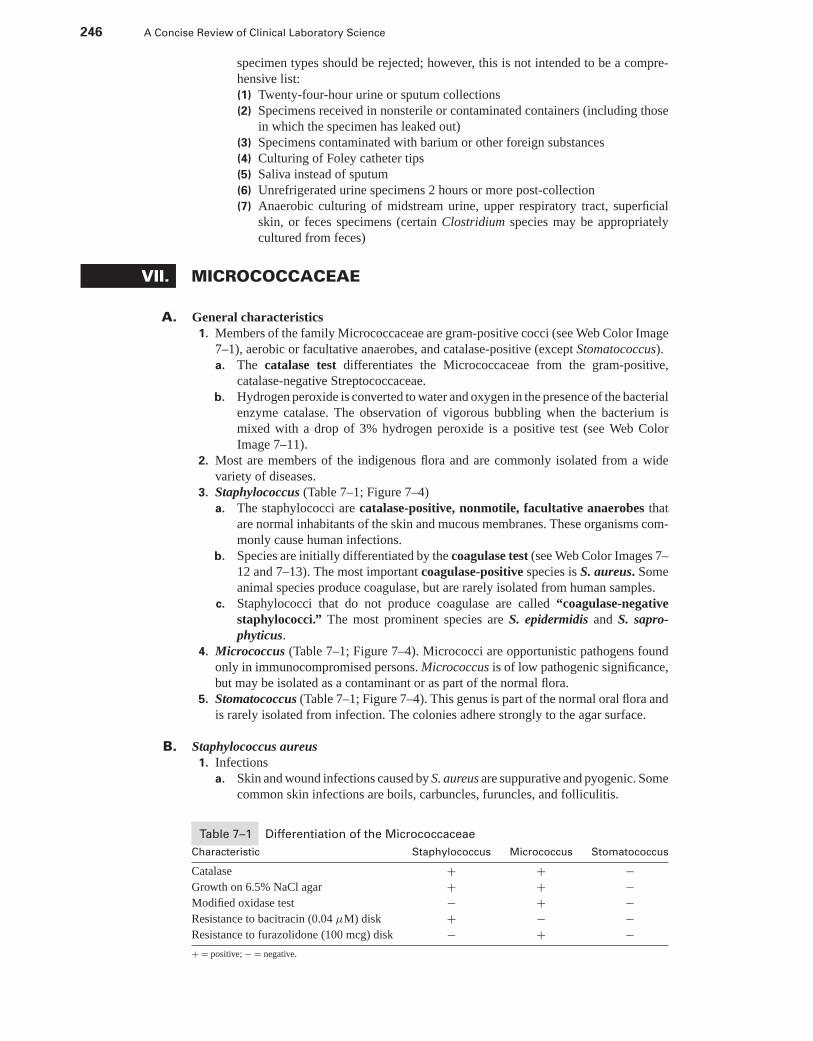
P1: OSO
LWBK192-07 LWBK192-Hubbard November 27, 2008 7:48
246 A Concise Review of Clinical Laboratory Science
specimen types should be rejected; however, this is not intended to be a compre-hensive list:(1) Twenty-four-hour urine or sputum collections(2) Specimens received in nonsterile or contaminated containers (including those
in which the specimen has leaked out)(3) Specimens contaminated with barium or other foreign substances(4) Culturing of Foley catheter tips(5) Saliva instead of sputum(6) Unrefrigerated urine specimens 2 hours or more post-collection(7) Anaerobic culturing of midstream urine, upper respiratory tract, superficial
skin, or feces specimens (certain Clostridium species may be appropriatelycultured from feces)
VII. MICROCOCCACEAE
A. General characteristics1. Members of the family Micrococcaceae are gram-positive cocci (see Web Color Image
7–1), aerobic or facultative anaerobes, and catalase-positive (except Stomatococcus).a. The catalase test differentiates the Micrococcaceae from the gram-positive,
catalase-negative Streptococcaceae.b. Hydrogen peroxide is converted to water and oxygen in the presence of the bacterial
enzyme catalase. The observation of vigorous bubbling when the bacterium ismixed with a drop of 3% hydrogen peroxide is a positive test (see Web ColorImage 7–11).
2. Most are members of the indigenous flora and are commonly isolated from a widevariety of diseases.
3. Staphylococcus (Table 7–1; Figure 7–4)a. The staphylococci are catalase-positive, nonmotile, facultative anaerobes that
are normal inhabitants of the skin and mucous membranes. These organisms com-monly cause human infections.
b. Species are initially differentiated by the coagulase test (see Web Color Images 7–12 and 7–13). The most important coagulase-positive species is S. aureus. Someanimal species produce coagulase, but are rarely isolated from human samples.
c. Staphylococci that do not produce coagulase are called “coagulase-negativestaphylococci.” The most prominent species are S. epidermidis and S. sapro-phyticus.
4. Micrococcus (Table 7–1; Figure 7–4). Micrococci are opportunistic pathogens foundonly in immunocompromised persons. Micrococcus is of low pathogenic significance,but may be isolated as a contaminant or as part of the normal flora.
5. Stomatococcus (Table 7–1; Figure 7–4). This genus is part of the normal oral flora andis rarely isolated from infection. The colonies adhere strongly to the agar surface.
B. Staphylococcus aureus1. Infections
a. Skin and wound infections caused by S. aureus are suppurative and pyogenic. Somecommon skin infections are boils, carbuncles, furuncles, and folliculitis.
Table 7–1 Differentiation of the Micrococcaceae
Characteristic Staphylococcus Micrococcus Stomatococcus
Catalase + + −Growth on 6.5% NaCl agar + + −Modified oxidase test − + −Resistance to bacitracin (0.04 μM) disk + − −Resistance to furazolidone (100 mcg) disk − + −+ = positive; − = negative.
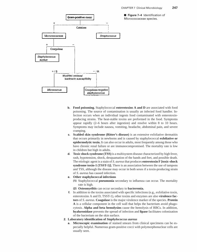
P1: OSO
LWBK192-07 LWBK192-Hubbard November 27, 2008 7:48
CHAPTER 7 Clinical Microbiology 247
■ Figure 7–4 Identification of
Micrococcaceae species.
b. Food poisoning. Staphylococcal enterotoxins A and D are associated with foodpoisoning. The source of contamination is usually an infected food handler. In-fection occurs when an individual ingests food contaminated with enterotoxin-producing strains. The heat-stable toxins are preformed in the food. Symptomsappear rapidly (2–6 hours after ingestion) and resolve within 8 to 10 hours.Symptoms may include nausea, vomiting, headache, abdominal pain, and severecramping.
c. Scalded skin syndrome (Ritter’s disease) is an extensive exfoliative dermatitisthat occurs primarily in newborns and is caused by staphylococcal exfoliative orepidermolytic toxin. It can also occur in adults, most frequently among those whohave chronic renal failure or are immunocompromised. The mortality rate is lowin children but high in adults.
d. Toxic shock syndrome (TSS) is a multisystem disease characterized by high fever,rash, hypotension, shock, desquamation of the hands and feet, and possible death.The etiologic agent is a stain of S. aureus that produces enterotoxin F [toxic-shocksyndrome toxin-1 (TSST-1)]. There is an association between the use of tamponsand TSS, although the disease may occur in both sexes if a toxin-producing strainof S. aureus has caused infection.
e. Other staphylococcal infections(1) Staphylococcal pneumonia secondary to influenza can occur. The mortality
rate is high.(2) Osteomyelitis can occur secondary to bacteremia.
f. In addition to the toxins associated with specific infections (e.g., exfoliative toxin,enterotoxins A and D, TSST-1), other toxins and enzymes are also virulence fac-tors of S. aureus. Coagulase is the major virulence marker of the species. ProteinA is a cellular component in the cell wall that helps the bacterium avoid phago-cytosis. Alpha and beta hemolysins cause the hemolysis of RBCs. In addition,hyaluronidase prevents the spread of infection and lipase facilitates colonizationof the bacterium on the skin surface.
2. Laboratory identification of Staphylococcus aureusa. Microscopic examination of stained smears from clinical specimens can be es-
pecially helpful. Numerous gram-positive cocci with polymorphonuclear cells areusually seen.
P1: OSO
LWBK192-07 LWBK192-Hubbard November 27, 2008 7:48
248 A Concise Review of Clinical Laboratory Science
b. S. aureus grows readily on common laboratory media. On sheep blood agar,colonies appear as round, smooth and white or pigmented (yellow-orange). Theyare usually β-hemolytic. (See Web Color Image 7–14.)
c. S. aureus is most often identified by the coagulase test. Isolates may show cell-bound (clumping factor) or free (extracellular) coagulase. Cell-bound coagulase isidentified by mixing the suspected organism with a drop of rabbit plasma on a glassslide. (See Web Color Image 7–12.) If clumping occurs, the isolate demonstratescell-bound coagulase and is identified as S. aureus. Isolates that do not clump aretested for free coagulase by the tube method (see Web Color Image 7–13), in whichthe organism is mixed with 0.5 mL of rabbit plasma, and following incubation at37◦C for 4 hours, is observed for clot formation.
d. Selective media that can be used to isolate S. aureus from heavily contaminatedspecimens or when it is the only isolate of concern are mannitol salt agar (MSA),phenylethyl alcohol (PEA) blood agar, and Columbia-naladixic acid Agar(CNA).(1) MSA provides mannitol as a fermentable carbohydrate source as well as 6.5%
sodium chloride (NaCl). Generally, only Staphylococcus species grow on thismedium because of the high salt content. S. aureus ferments the mannitol toproduce acid, which turns the pink agar yellow. The colonies are identified bya yellow halo. This test is presumptive because some strains of S. epidermidisand few other species can also ferment mannitol.
(2) PEA and CNA agars both inhibit the growth of gram-negative organisms,whereas the gram-positive bacteria, including Staphylococcus species, growwell. Five-percent sheep blood is incorporated in the medium for additionalenrichment and detection of hemolytic reactions.
e. Rapid methods use plasma-coated latex particles. The plasma detects clump-ing factor and causes agglutination of the particles. Protein A in the cell wallof S. aureus (with IgG) may also be detected with rapid methods. Other speciesthat produce clumping factor produce positive reactions, but are tube-coagulase-negative.
C. Coagulase-negative staphylococci1. General characteristics. The coagulase-negative staphylococci are found as normal
flora in humans and animals. The incidence of infection by these organisms has in-creased. They are often hospital-acquired (nosocomial). Predisposing factors includecatheterization, prosthetic device implants, and immunosuppressive therapy. The mostcommon species isolated from clinical infections are S. epidermidis and S. saprophyti-cus. S. saprophyticus has been associated with UTIs in young, sexually active women.Other species of coagulase-negative staphylococci are not isolated frequently. Threespecies that can cause a wide range of infections, but do so only occasionally, areS. haemolyticus, S. lugdunensis, and S. schleiferi. The latter two produce clumpingfactor and may yield a positive slide coagulase test.
2. Laboratory identification of coagulase-negative staphylococcia. On sheep blood agar, colonies are usually round, smooth, and white without hemol-
ysis. (See Web Color Image 7–15.)b. The most common isolates are S. epidermidis and S. saprophyticus.c. Urine isolates that are coagulase-negative are further tested to presumptively iden-
tify S. saprophyticus. This is done by testing for novobiocin susceptibility using a5-mg novobiocin disk. S. saprophyticus is resistant to novobiocin, whereas othercoagulase-negative staphylococci are susceptible.
d. Species identification of the coagulase-negative staphylococci requires differen-tiation using many biochemical tests. Various commercial identification systemsexist and may be used if appropriate.
D. Micrococcus species1. General characteristics. Micrococcus species are environmental organisms, as well
as normal skin flora. They may also be normal flora in the respiratory tract or other sites
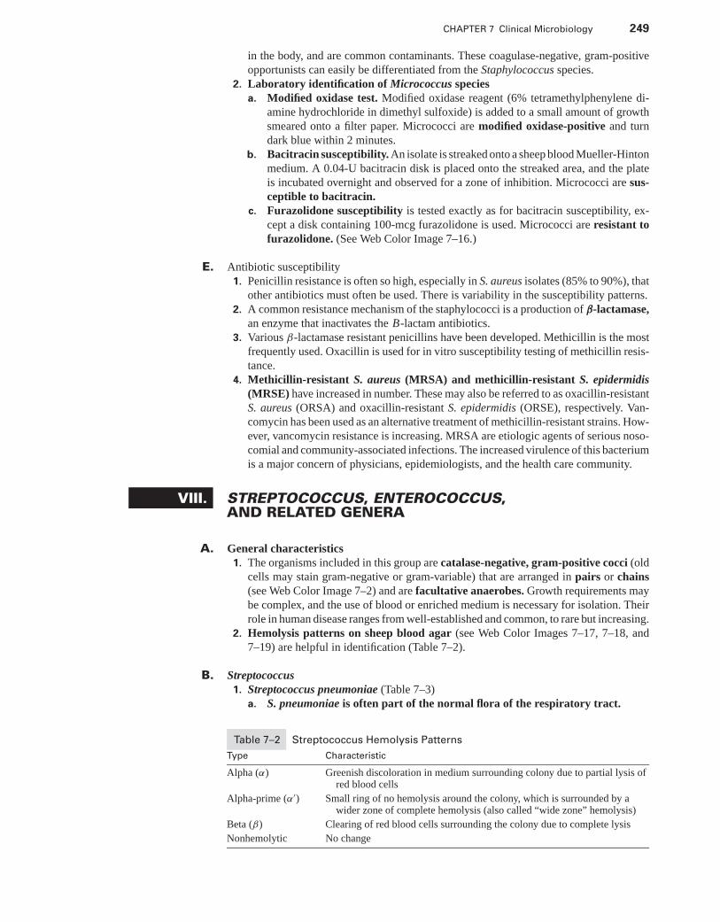
P1: OSO
LWBK192-07 LWBK192-Hubbard November 27, 2008 7:48
CHAPTER 7 Clinical Microbiology 249
in the body, and are common contaminants. These coagulase-negative, gram-positiveopportunists can easily be differentiated from the Staphylococcus species.
2. Laboratory identification of Micrococcus speciesa. Modified oxidase test. Modified oxidase reagent (6% tetramethylphenylene di-
amine hydrochloride in dimethyl sulfoxide) is added to a small amount of growthsmeared onto a filter paper. Micrococci are modified oxidase-positive and turndark blue within 2 minutes.
b. Bacitracin susceptibility. An isolate is streaked onto a sheep blood Mueller-Hintonmedium. A 0.04-U bacitracin disk is placed onto the streaked area, and the plateis incubated overnight and observed for a zone of inhibition. Micrococci are sus-ceptible to bacitracin.
c. Furazolidone susceptibility is tested exactly as for bacitracin susceptibility, ex-cept a disk containing 100-mcg furazolidone is used. Micrococci are resistant tofurazolidone. (See Web Color Image 7–16.)
E. Antibiotic susceptibility1. Penicillin resistance is often so high, especially in S. aureus isolates (85% to 90%), that
other antibiotics must often be used. There is variability in the susceptibility patterns.2. A common resistance mechanism of the staphylococci is a production of β-lactamase,
an enzyme that inactivates the B-lactam antibiotics.3. Various β-lactamase resistant penicillins have been developed. Methicillin is the most
frequently used. Oxacillin is used for in vitro susceptibility testing of methicillin resis-tance.
4. Methicillin-resistant S. aureus (MRSA) and methicillin-resistant S. epidermidis(MRSE) have increased in number. These may also be referred to as oxacillin-resistantS. aureus (ORSA) and oxacillin-resistant S. epidermidis (ORSE), respectively. Van-comycin has been used as an alternative treatment of methicillin-resistant strains. How-ever, vancomycin resistance is increasing. MRSA are etiologic agents of serious noso-comial and community-associated infections. The increased virulence of this bacteriumis a major concern of physicians, epidemiologists, and the health care community.
VIII. STREPTOCOCCUS, ENTEROCOCCUS,AND RELATED GENERA
A. General characteristics1. The organisms included in this group are catalase-negative, gram-positive cocci (old
cells may stain gram-negative or gram-variable) that are arranged in pairs or chains(see Web Color Image 7–2) and are facultative anaerobes. Growth requirements maybe complex, and the use of blood or enriched medium is necessary for isolation. Theirrole in human disease ranges from well-established and common, to rare but increasing.
2. Hemolysis patterns on sheep blood agar (see Web Color Images 7–17, 7–18, and7–19) are helpful in identification (Table 7–2).
B. Streptococcus1. Streptococcus pneumoniae (Table 7–3)
a. S. pneumoniae is often part of the normal flora of the respiratory tract.
Table 7–2 Streptococcus Hemolysis Patterns
Type Characteristic
Alpha (α) Greenish discoloration in medium surrounding colony due to partial lysis ofred blood cells
Alpha-prime (α′) Small ring of no hemolysis around the colony, which is surrounded by awider zone of complete hemolysis (also called “wide zone” hemolysis)
Beta (β) Clearing of red blood cells surrounding the colony due to complete lysis
Nonhemolytic No change
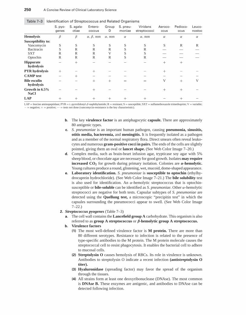
P1: OSO
LWBK192-07 LWBK192-Hubbard November 27, 2008 7:48
250 A Concise Review of Clinical Laboratory Science
Table 7–3 Identification of Streptococcus and Related Organisms
S. pyo- S. agala- Entero- Group S. pneu- Viridans Aeroco- Pedioco- Leuco-
genes ctiae coccus D moniae streptococci ccus ccus nostoc
Hemolysis β β α, β, non α, non α α, non α α α
Susceptibility to:Vancomycin S S S S S S S R RBacitracin S R R R S R — — —SXT R R R V S S — — —Optochin R R R R S R — — —
Hippuratehydrolysis
− + − − − − + +
PYR hydrolysis + − + − − − + − −CAMP test − + − − − − − − −Bile esculin
hydrolysis− − + + = = V + V
Growth in 6.5%NaCl
− − + − − − + + −
LAP + + + + + + − + −LAP = leucine aminopeptidase; PYR = l-pyrrolidonyl-β-naphthylamide; R = resistant; S = susceptible; SXT = sulfamethoxazole trimethoprim; V = variable;− = negative; + = positive; — = tests not done (vancomycin-resistance is the key characteristic).
b. The key virulence factor is an antiphagocytic capsule. There are approximately80 antigenic types.
c. S. pneumoniae is an important human pathogen, causing pneumonia, sinusitis,otitis media, bacteremia, and meningitis. It is frequently isolated as a pathogenand as a member of the normal respiratory flora. Direct smears often reveal leuko-cytes and numerous gram-positive cocci in pairs. The ends of the cells are slightlypointed, giving them an oval or lancet shape. (See Web Color Image 7–20.)
d. Complex media, such as brain-heart infusion agar, trypticase soy agar with 5%sheep blood, or chocolate agar are necessary for good growth. Isolates may requireincreased CO2 for growth during primary isolation. Colonies are α-hemolytic.Young cultures produce a round, glistening, wet, mucoid, dome-shaped appearance.
e. Laboratory identification. S. pneumoniae is susceptible to optochin (ethylhy-drocuprein hydrochloride). (See Web Color Image 7–21.) The bile solubility testis also used for identification. An α-hemolytic streptococcus that is optochin-susceptible or bile-soluble can be identified as S. pneumoniae. Other α-hemolyticstreptococci are negative for both tests. Capsular subtypes of S. pneumoniae aredetected using the Quellung test, a microscopic “precipitin test” in which thecapsules surrounding the pneumococci appear to swell. (See Web Color Image7–22.)
2. Streptococcus pyogenes (Table 7–3)a. The cell wall contains the Lancefield group A carbohydrate. This organism is also
referred to as group A streptococcus or β-hemolytic group A streptococcus.b. Virulence factors
(1) The most well-defined virulence factor is M protein. There are more than80 different serotypes. Resistance to infection is related to the presence oftype-specific antibodies to the M protein. The M protein molecule causes thestreptococcal cell to resist phagocytosis. It enables the bacterial cell to adhereto mucosal cells.
(2) Streptolysin O causes hemolysis of RBCs. Its role in virulence is unknown.Antibodies to streptolysin O indicate a recent infection (antistreptolysin Otiter).
(3) Hyaluronidase (spreading factor) may favor the spread of the organismthrough the tissues.
(4) All strains form at least one deoxyribonuclease (DNAse). The most commonis DNAse B. These enzymes are antigenic, and antibodies to DNAse can bedetected following infection.
P1: OSO
LWBK192-07 LWBK192-Hubbard November 27, 2008 7:48
CHAPTER 7 Clinical Microbiology 251
(5) Some strains of S. pyogenes cause a red spreading rash referred to as scarletfever. This condition is caused by erythrogenic toxin.
(6) Protein F is a fibronectin-binding protein that facilitates adhesion to epithelialcells.
(7) Streptokinase causes the lysis of fibrin clots.c. Infections
(1) Pharyngitis is one of the most common S. pyogenes infections. “Strepthroat” is most frequently seen in children between the ages of 5 and15 years. Diagnosis relies on a throat culture or a positive quick “strep”test, in which group A antigens are detected from a throat swab in a mat-ter of minutes. A throat culture is recommended if the antigen-detecting test isnegative.
(2) Skin infections include impetigo, necrotizing fasciitis, and pyoderma.(3) Scarlet fever is a red rash that appears on the upper chest and spreads to the
trunk and extremities following infection with S. pyogenes.(4) Rheumatic fever and glomerulonephritis may result from infection at other
sites in the body. Damage appears to result from cross-reactivity of the strep-tococcal antigens with host tissue antigens.
(5) Streptococcal TSS (toxic shock syndrome) is similar to that caused by Staphy-lococcus aureus.
d. Laboratory identification. Colonies of S. pyogenes on blood agar are small, trans-parent, and smooth, and they show β hemolysis. Gram’s stain reveals gram-positivecocci with some short chains. The bacterium is susceptible to bacitracin or TaxoA (see Web Color Image 7–23) and resistant to SXT. In addition, S. pyogeneshydrolyzes l-pyrrolidonyl-β-naphthylamide (PYR). A positive test is develop-ment of a red color after the addition of dimethylaminocinnamaldehyde reagent toan inoculated PYR disk (see Web Color Image 7–24).
3. Streptococcus agalactiae (Table 7–3)a. The cell wall contains the Lancefield group B carbohydrate. This organism is
also referred to as group B streptococcus. It may be found as normal flora in thegenitourinary tract.
b. Virulence factors(1) The capsule is the most important virulence factor.(2) Other factors (e.g., DNAse, hyaluronidase) have not been shown to be factors
in infection.c. Infections
(1) Neonatal sepsis (usually manifest as pneumonia or meningitis) occurs soonafter birth. The most important factor in infection is the presence of group Bstreptococcus in the vagina of the mother.
(2) Postpartum fever and sepsis may occur after birth and may manifest as en-dometritis or a wound infection.
d. Laboratory identification. Group B streptococci grow on blood agar as grayishwhite mucoid colonies surrounded by a small zone of β hemolysis. They are gram-positive cocci that form short chains in clinical specimens and long chains in culture.Group B streptococci are CAMP test-positive, demonstrating an arrowhead-shaped area of synergistic hemolysis when streaked perpendicular to a β-hemolyticS. aureus (see Web Color Image 7–25). Group B streptococci are also hippuratehydrolysis-positive, resistant to SXT, and PYR-negative.
4. Groups C and Ga. There are three hemolytic species in Lancefield group C that are occasionally
isolated from clinical specimens: S. equi, S. zooepidemicus, and S. equisim-ilis. The major species found in group G is S. canis. It occasionally causes in-fection, and is part of the normal skin flora. Minute colony types of group Gare part of the S. milleri group, with S. anginosus being the most prominentspecies.
b. These groups produce a variety of infections similar to those caused by groups Aand B. Group C can cause pharyngitis.
P1: OSO
LWBK192-07 LWBK192-Hubbard November 27, 2008 7:48
252 A Concise Review of Clinical Laboratory Science
c. Laboratory identification. Groups C and G can be identified by extensive bio-chemical tests. However, serologic tests to identify the group carbohydrate in thecell wall of the isolate (e.g., agglutination) are best.
5. Group D (Table 7–3)a. The group D streptococci include S. bovis and S. equinus. They may be found as
normal intestinal flora.b. The group D streptococci may be etiologic agents of bacterial endocarditis, UTIs,
and other infections, such as abscesses and wound infections. An association hasbeen made between bacteremia due to S. bovis and the presence of gastrointestinaltumors. Isolation of S. bovis from a blood culture may be the first indication thatthe patient has an occult tumor.
c. Laboratory Identification. Hemolysis is usually absent, or α hemolysis is present.Key reactions of group D streptococci include a positive bile esculin test (formationof a black precipitate due to the hydrolysis of esculin) with no growth in 6.5%NaCl broth. Group D can be separated from Enterococcus by the l-pyrrolidonyl-β-naphthylamide (PYR) test because it is negative and Enterococcus is positive.(The enterococci also grow in 6.5% NaCl broth.) Serotyping should be done toidentify an isolate such as S. bovis, because it cannot be distinguished from someof the viridans group by biochemical tests alone.
6. Enterococcus (Table 7–3)a. This genus is found in the intestinal tract. The species found in this genus include E.
faecalis, which is the most common isolate, E. faecium, E. avium, and E. durans.These enterococcal species share a number of characteristics with the group Dstreptococci, including the group D antigen. They show resistance to several ofthe commonly used antibiotics, so differentiation from Group D Streptococcus andsusceptibility testing is important.
b. The infections caused are similar to those caused by the group D streptococci. Themost common is a urinary tract infection.
c. Laboratory Identification. It is not difficult to differentiate between Enterococcusand group D—streptococci. In addition to being positive for bile esculin (blackprecipitate), Enterococcus species grow in 6.5% NaCl broth (see Web ColorImage 7–26), are PYR-positive (see Web Color Image 7–24), and SXT-resistant.
d. Enterococci may be screened for high-level aminoglycoside resistance becauseaminoglycosides are usually used in combination with ampicillin or penicillin foreffective treatment of enterococcal infections. Resistant strains cannot be used forsynergistic treatment. Gentamicin and streptomycin resistance can be detected withbroth or agar dilution and disk diffusion tests.
e. The emergence of vancomycin-resistant Enterococcus (VRE), encoded by thevanA gene, is a major concern of physicians, microbiologists, and hospital infectioncontrol personnel. E. faecium is the most common species, followed by E. faecalis.Most microbiology laboratories screen for VR colonization using vancomycin-containing agar. Susceptibility testing is performed only on clinically significantisolates.
7. Viridans streptococci (Table 7–3)a. The viridans group includes those α-hemolytic streptococci that lack Lancefield
group antigens and do not meet the criteria for S. pneumoniae. They are part of thenormal flora of the oropharynx and intestine.
b. The most common infection caused by these organisms is subacute bacterialendocarditis.
c. The viridans streptococci are fastidious and some strains require increased CO2
for growth. Identification of the viridans streptococci to the species level is adifficult task. Part of the reason for this is that there is not widespread agreementon a classification scheme. Species of viridans streptococci include S. mutans,S. salivarius, S. sanguis, S. mitis, and S. milleri (not β hemolytic).
8. Nutritionally variant streptococci (NVS)a. The NVS subgroup of viridans streptococci are nutritionally deficient and have
been isolated from patients who have endocarditis and otitis media. This
P1: OSO
LWBK192-07 LWBK192-Hubbard November 27, 2008 7:48
CHAPTER 7 Clinical Microbiology 253
subgroup is also known as pyridoxal (vitamin B6)-dependent, thiol-dependent,or symbiotic streptococci. Pyridoxal is not present in most liquid and solid bac-teriologic media, so bacteriologic media must be supplemented with pyridoxal(vitamin B6) to support the growth of NVS. The NVS colonies are small, mea-suring 0.2 to 0.5 mm in diameter. When gram stained, the morphology can varyfrom classic gram-positive streptococci to gram-negative or gram-variable pleo-morphic forms. As the optimal concentrations or required nutrients decrease, thecells become pleomorphic, even showing globular and filamentous forms.
b. NVS satellite around or grow adjacent to staphylococcal isolates. The staphylococciprovide the growth requirements needed to facilitate the growth of the NVS.
c. A clue to the presence of NVS is a positive Gram’s stain, but negative cultures.9. Treatment of streptococcal and enterococcal infections. Most species of Streptococ-
cus are susceptible to penicillin. S. agalactiae is less susceptible than group A and mayrequire a combination of ampicillin and an aminoglycoside. Group D is susceptibleto penicillin, whereas Enterococcus is usually resistant. Enterococcus is often treatedwith a penicillin-aminoglycoside combination (synergy). Some isolates are resistantto this combination therapy. Although most pneumococcal isolates are susceptible topenicillin, some strains have shown resistance. Resistant streptococcal strains are oftentreated with erythromycin. Linezolid is often used for treatment of infections causedby vancomycin-resistant enterococci (VRE).
C. Streptococcus-like organisms (Table 7–3)1. Aerococcus is very similar to Enterococcus on blood agar. The gram-positive coccus
is susceptible to vancomycin and can be isolated from tissue samples of endocarditisand other varied infections.
2. Leuconostoc is very similar to viridans streptococci on blood agar. It is found inthe general environment. A Gram’s stain shows gram-positive coccobacilli in pairsand short chains. Leuconostoc has been found in patients who have meningitis andendocarditis. It is intrinsically resistant to vancomycin.
3. Pediococcus is also found in the general environment. A Gram’s stain shows gram-positive cocci in pairs, tetrads, and clusters. Pediococcus is a rare isolate in patientswho have septicemia. The bacterium is intrinsically resistant to vancomycin.
D. Laboratory identification of Streptococci1. Hemolysis on blood agar is an important characteristic (Table 7–2). (See Web Color
Images 7–17, 7–18, and 7–19.)2. Bile solubility measures autolysis of bacteria under the influence of a bile salt (sodium
deoxycholate). S. pneumoniae is bile soluble.3. Optochin (ethylhydrocuprein hydrochloride) susceptibility is determined by a zone
of inhibition (>14 mm with a 5 mcg optochin disk) after growing the organism onblood agar with a filter paper disk containing optochin. Results correlate with bilesolubility; that is, optochin-susceptible isolates are bile soluble. S. pneumoniae isoptochin susceptible. (See Web Color Image 7–21.)
4. Bacitracin (Taxo A) susceptibility is a characteristic of S. pyogenes. The test is per-formed by placing a filter paper disk containing bacitracin on an inoculated bloodagar plate, and measuring the zone of inhibition following incubation. (See Web ColorImage 7–23.)
5. Group A and B streptococci are resistant to sulfamethoxazole-trimethoprim (SXT).This resistance can be measured with a filter paper disk or by incorporating SXT intoblood agar. The latter technique allows for selective isolation. Enterococcus speciesare also SXT-resistant.
6. Group B streptococci hydrolyze hippurate. The glycine liberated can be detected bytriketohydrindene hydrate (Ninhydrin), which imparts a purple color.
7. The Christie, Atkins, and Munch-Petersen (CAMP) test presumptively identifiesgroup B streptococcus by measuring the enhanced hemolytic activity of staphylococ-cal β-lysin by S. agalactiae. Group B streptococci, plated perpendicular to S. aureus,demonstrate a characteristic arrow-shaped hemolysis pattern. (See Web Color Image7–25.)
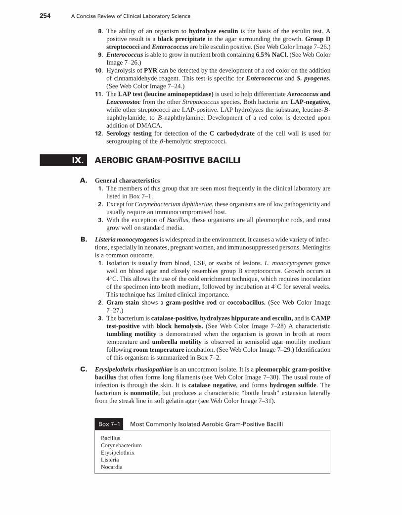
P1: OSO
LWBK192-07 LWBK192-Hubbard November 27, 2008 7:48
254 A Concise Review of Clinical Laboratory Science
8. The ability of an organism to hydrolyze esculin is the basis of the esculin test. Apositive result is a black precipitate in the agar surrounding the growth. Group Dstreptococci and Enterococcus are bile esculin positive. (See Web Color Image 7–26.)
9. Enterococcus is able to grow in nutrient broth containing 6.5% NaCl. (See Web ColorImage 7–26.)
10. Hydrolysis of PYR can be detected by the development of a red color on the additionof cinnamaldehyde reagent. This test is specific for Enterococcus and S. pyogenes.(See Web Color Image 7–24.)
11. The LAP test (leucine aminopeptidase) is used to help differentiate Aerococcus andLeuconostoc from the other Streptococcus species. Both bacteria are LAP-negative,while other streptococci are LAP-positive. LAP hydrolyzes the substrate, leucine-B-naphthylamide, to B-naphthylamine. Development of a red color is detected uponaddition of DMACA.
12. Serology testing for detection of the C carbodydrate of the cell wall is used forserogrouping of the β-hemolytic streptococci.
IX. AEROBIC GRAM-POSITIVE BACILLI
A. General characteristics1. The members of this group that are seen most frequently in the clinical laboratory are
listed in Box 7–1.2. Except for Corynebacterium diphtheriae, these organisms are of low pathogenicity and
usually require an immunocompromised host.3. With the exception of Bacillus, these organisms are all pleomorphic rods, and most
grow well on standard media.
B. Listeria monocytogenes is widespread in the environment. It causes a wide variety of infec-tions, especially in neonates, pregnant women, and immunosuppressed persons. Meningitisis a common outcome.
1. Isolation is usually from blood, CSF, or swabs of lesions. L. monocytogenes growswell on blood agar and closely resembles group B streptococcus. Growth occurs at4◦C. This allows the use of the cold enrichment technique, which requires inoculationof the specimen into broth medium, followed by incubation at 4◦C for several weeks.This technique has limited clinical importance.
2. Gram stain shows a gram-positive rod or coccobacillus. (See Web Color Image7–27.)
3. The bacterium is catalase-positive, hydrolyzes hippurate and esculin, and is CAMPtest-positive with block hemolysis. (See Web Color Image 7–28) A characteristictumbling motility is demonstrated when the organism is grown in broth at roomtemperature and umbrella motility is observed in semisolid agar motility mediumfollowing room temperature incubation. (See Web Color Image 7–29.) Identificationof this organism is summarized in Box 7–2.
C. Erysipelothrix rhusiopathiae is an uncommon isolate. It is a pleomorphic gram-positivebacillus that often forms long filaments (see Web Color Image 7–30). The usual route ofinfection is through the skin. It is catalase negative, and forms hydrogen sulfide. Thebacterium is nonmotile, but produces a characteristic “bottle brush” extension laterallyfrom the streak line in soft gelatin agar (see Web Color Image 7–31).
Box 7–1 Most Commonly Isolated Aerobic Gram-Positive Bacilli
Bacillus
Corynebacterium
Erysipelothrix
Listeria
Nocardia
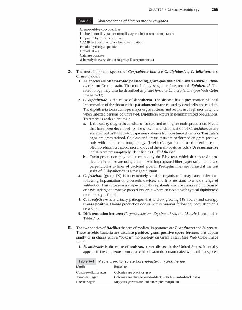
P1: OSO
LWBK192-07 LWBK192-Hubbard November 27, 2008 7:48
CHAPTER 7 Clinical Microbiology 255
Box 7–2 Characteristics of Listeria monocytogenes
Gram-positive coccobacillus
Umbrella motility pattern (motility agar tube) at room temperature
Hippurate hydrolysis positive
CAMP test positive–block hemolysis pattern
Esculin hydrolysis positive
Growth at 4◦C
Catalase positive
β hemolytic (very similar to group B streptococcus)
D. The most important species of Corynebacterium are C. diphtheriae, C. jeikeium, andC. urealyticum.
1. All species are pleomorphic, pallisading, gram-positive bacilli and resemble C. diph-theriae on Gram’s stain. The morphology was, therefore, termed diphtheroid. Themorphology may also be described as picket fence or Chinese letters (see Web ColorImage 7–32).
2. C. diphtheriae is the cause of diphtheria. The disease has a presentation of localinflammation of the throat with a pseudomembrane caused by dead cells and exudate.The diphtheria toxin damages major organ systems and results in a high mortality ratewhen infected persons go untreated. Diphtheria occurs in nonimmunized populations.Treatment is with an antitoxin.a. Laboratory diagnosis consists of culture and testing for toxin production. Media
that have been developed for the growth and identification of C. diphtheriae aresummarized in Table 7–4. Suspicious colonies from cystine-tellurite or Tinsdale’sagar are gram stained. Catalase and urease tests are performed on gram-positiverods with diphtheroid morphology. (Loeffler’s agar can be used to enhance thepleomorphic microscopic morphology of the gram-positive rods.). Urease negativeisolates are presumptively identified as C. diphtheriae.
b. Toxin production may be determined by the Elek test, which detects toxin pro-duction by an isolate using an antitoxin-impregnated filter paper strip that is laidperpendicular to lines of bacterial growth. Precipitin lines are formed if the teststain of C. diphtheriae is a toxigenic strain.
3. C. jeikeium (group JK) is an extremely virulent organism. It may cause infectionsfollowing implantation of prosthetic devices, and it is resistant to a wide range ofantibiotics. This organism is suspected in those patients who are immunocompromisedor have undergone invasive procedures or in whom an isolate with typical diphtheroidmorphology is found.
4. C. urealyticum is a urinary pathogen that is slow growing (48 hours) and stronglyurease positive. Urease production occurs within minutes following inoculation on aurea slant.
5. Differentiation between Corynebacterium, Erysipelothrix, and Listeria is outlined inTable 7–5.
E. The two species of Bacillus that are of medical importance are B. anthracis and B. cereus.These aerobic bacteria are catalase-positive, gram-positive spore formers that appearsingly or in chains with a “boxcar” morphology on Gram’s stain (see Web Color Image7–33).
1. B. anthracis is the cause of anthrax, a rare disease in the United States. It usuallyappears in the cutaneous form as a result of wounds contaminated with anthrax spores.
Table 7–4 Media Used to Isolate Corynebacterium diphtheriae
Media Reaction
Cystine-tellurite agar Colonies are black or gray
Tinsdale’s agar Colonies are dark brown-to-black with brown-to-black halos
Loeffler agar Supports growth and enhances pleomorphism
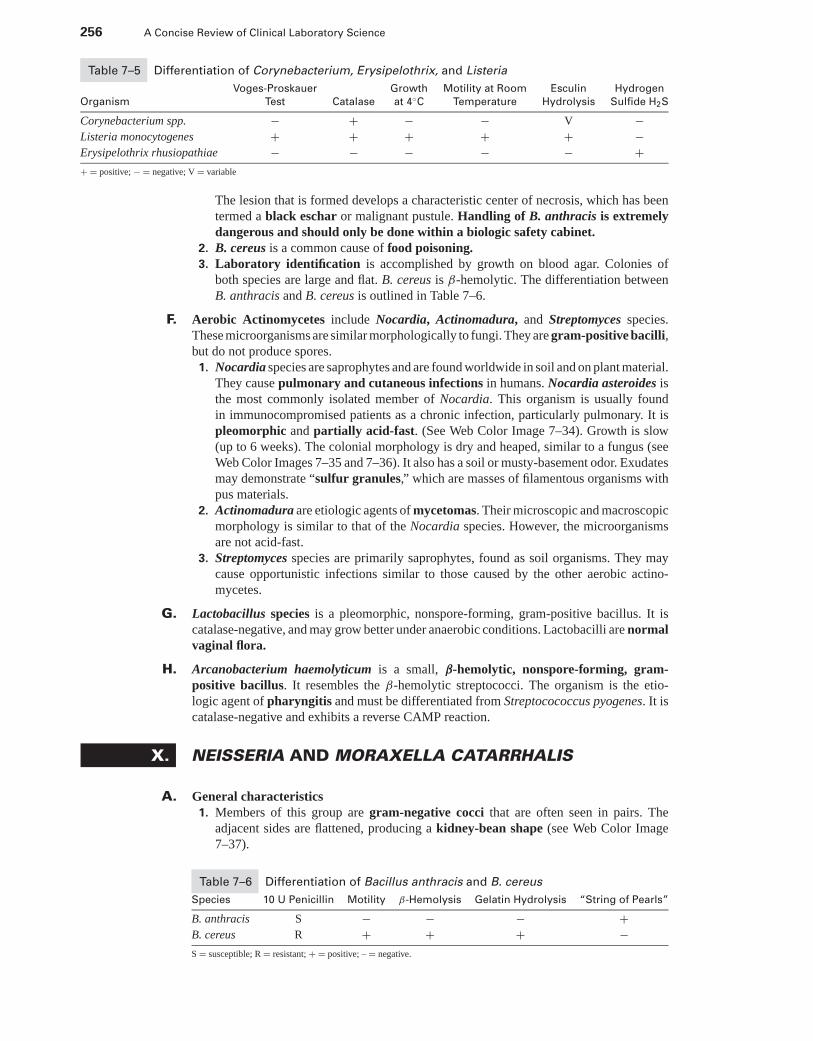
P1: OSO
LWBK192-07 LWBK192-Hubbard November 27, 2008 7:48
256 A Concise Review of Clinical Laboratory Science
Table 7–5 Differentiation of Corynebacterium, Erysipelothrix, and Listeria
Voges-Proskauer Growth Motility at Room Esculin Hydrogen
Organism Test Catalase at 4◦C Temperature Hydrolysis Sulfide H2S
Corynebacterium spp. − + − − V −Listeria monocytogenes + + + + + −Erysipelothrix rhusiopathiae − − − − − ++ = positive; − = negative; V = variable
The lesion that is formed develops a characteristic center of necrosis, which has beentermed a black eschar or malignant pustule. Handling of B. anthracis is extremelydangerous and should only be done within a biologic safety cabinet.
2. B. cereus is a common cause of food poisoning.3. Laboratory identification is accomplished by growth on blood agar. Colonies of
both species are large and flat. B. cereus is β-hemolytic. The differentiation betweenB. anthracis and B. cereus is outlined in Table 7–6.
F. Aerobic Actinomycetes include Nocardia, Actinomadura, and Streptomyces species.These microorganisms are similar morphologically to fungi. They are gram-positive bacilli,but do not produce spores.
1. Nocardia species are saprophytes and are found worldwide in soil and on plant material.They cause pulmonary and cutaneous infections in humans. Nocardia asteroides isthe most commonly isolated member of Nocardia. This organism is usually foundin immunocompromised patients as a chronic infection, particularly pulmonary. It ispleomorphic and partially acid-fast. (See Web Color Image 7–34). Growth is slow(up to 6 weeks). The colonial morphology is dry and heaped, similar to a fungus (seeWeb Color Images 7–35 and 7–36). It also has a soil or musty-basement odor. Exudatesmay demonstrate “sulfur granules,” which are masses of filamentous organisms withpus materials.
2. Actinomadura are etiologic agents of mycetomas. Their microscopic and macroscopicmorphology is similar to that of the Nocardia species. However, the microorganismsare not acid-fast.
3. Streptomyces species are primarily saprophytes, found as soil organisms. They maycause opportunistic infections similar to those caused by the other aerobic actino-mycetes.
G. Lactobacillus species is a pleomorphic, nonspore-forming, gram-positive bacillus. It iscatalase-negative, and may grow better under anaerobic conditions. Lactobacilli are normalvaginal flora.
H. Arcanobacterium haemolyticum is a small, β-hemolytic, nonspore-forming, gram-positive bacillus. It resembles the β-hemolytic streptococci. The organism is the etio-logic agent of pharyngitis and must be differentiated from Streptocococcus pyogenes. It iscatalase-negative and exhibits a reverse CAMP reaction.
X. NEISSERIA AND MORAXELLA CATARRHALIS
A. General characteristics1. Members of this group are gram-negative cocci that are often seen in pairs. The
adjacent sides are flattened, producing a kidney-bean shape (see Web Color Image7–37).
Table 7–6 Differentiation of Bacillus anthracis and B. cereus
Species 10 U Penicillin Motility β-Hemolysis Gelatin Hydrolysis “String of Pearls”
B. anthracis S − − − +B. cereus R + + + −S = susceptible; R = resistant; + = positive; – = negative.
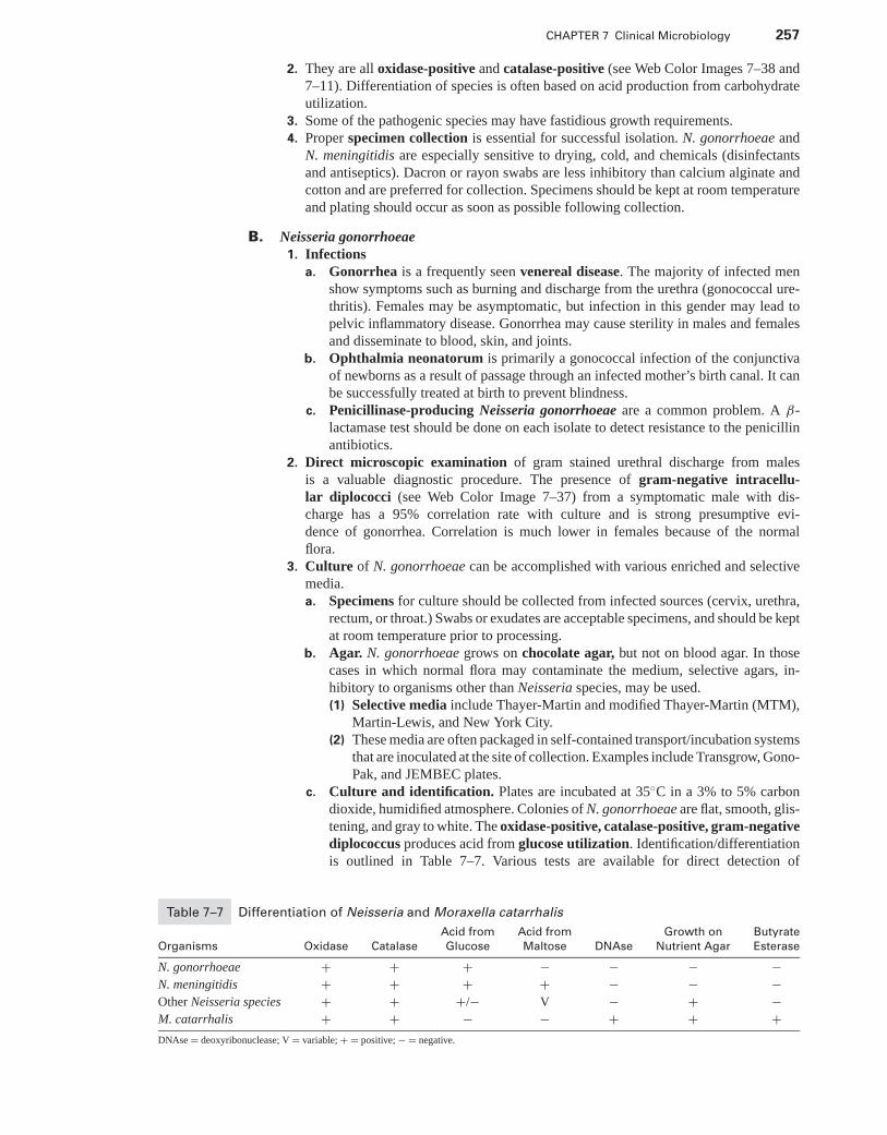
P1: OSO
LWBK192-07 LWBK192-Hubbard November 27, 2008 7:48
CHAPTER 7 Clinical Microbiology 257
2. They are all oxidase-positive and catalase-positive (see Web Color Images 7–38 and7–11). Differentiation of species is often based on acid production from carbohydrateutilization.
3. Some of the pathogenic species may have fastidious growth requirements.4. Proper specimen collection is essential for successful isolation. N. gonorrhoeae and
N. meningitidis are especially sensitive to drying, cold, and chemicals (disinfectantsand antiseptics). Dacron or rayon swabs are less inhibitory than calcium alginate andcotton and are preferred for collection. Specimens should be kept at room temperatureand plating should occur as soon as possible following collection.
B. Neisseria gonorrhoeae1. Infections
a. Gonorrhea is a frequently seen venereal disease. The majority of infected menshow symptoms such as burning and discharge from the urethra (gonococcal ure-thritis). Females may be asymptomatic, but infection in this gender may lead topelvic inflammatory disease. Gonorrhea may cause sterility in males and femalesand disseminate to blood, skin, and joints.
b. Ophthalmia neonatorum is primarily a gonococcal infection of the conjunctivaof newborns as a result of passage through an infected mother’s birth canal. It canbe successfully treated at birth to prevent blindness.
c. Penicillinase-producing Neisseria gonorrhoeae are a common problem. A β-lactamase test should be done on each isolate to detect resistance to the penicillinantibiotics.
2. Direct microscopic examination of gram stained urethral discharge from malesis a valuable diagnostic procedure. The presence of gram-negative intracellu-lar diplococci (see Web Color Image 7–37) from a symptomatic male with dis-charge has a 95% correlation rate with culture and is strong presumptive evi-dence of gonorrhea. Correlation is much lower in females because of the normalflora.
3. Culture of N. gonorrhoeae can be accomplished with various enriched and selectivemedia.a. Specimens for culture should be collected from infected sources (cervix, urethra,
rectum, or throat.) Swabs or exudates are acceptable specimens, and should be keptat room temperature prior to processing.
b. Agar. N. gonorrhoeae grows on chocolate agar, but not on blood agar. In thosecases in which normal flora may contaminate the medium, selective agars, in-hibitory to organisms other than Neisseria species, may be used.(1) Selective media include Thayer-Martin and modified Thayer-Martin (MTM),
Martin-Lewis, and New York City.(2) These media are often packaged in self-contained transport/incubation systems
that are inoculated at the site of collection. Examples include Transgrow, Gono-Pak, and JEMBEC plates.
c. Culture and identification. Plates are incubated at 35◦C in a 3% to 5% carbondioxide, humidified atmosphere. Colonies of N. gonorrhoeae are flat, smooth, glis-tening, and gray to white. The oxidase-positive, catalase-positive, gram-negativediplococcus produces acid from glucose utilization. Identification/differentiationis outlined in Table 7–7. Various tests are available for direct detection of
Table 7–7 Differentiation of Neisseria and Moraxella catarrhalis
Acid from Acid from Growth on Butyrate
Organisms Oxidase Catalase Glucose Maltose DNAse Nutrient Agar Esterase
N. gonorrhoeae + + + − − − −N. meningitidis + + + + − − −Other Neisseria species + + +/− V − + −M. catarrhalis + + − − + + +DNAse = deoxyribonuclease; V = variable; + = positive; − = negative.
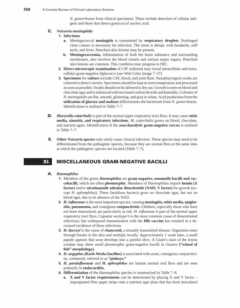
P1: OSO
LWBK192-07 LWBK192-Hubbard November 27, 2008 7:48
258 A Concise Review of Clinical Laboratory Science
N. gonorrhoeae from clinical specimens. These include detection of cellular anti-gens and those that detect gonococcal nucleic acid.
C. Neisseria meningitidis1. Infections
a. Meningococcal meningitis is transmitted by respiratory droplets. Prolongedclose contact is necessary for infection. The onset is abrupt, with headache, stiffneck, and fever. Petechial skin lesions may be present.
b. Meningococcemia, inflammation of both the brain substance and surroundingmembranes, also involves the blood vessels and various major organs. Petechialskin lesions are common. This condition may progress to DIC.
2. Direct microscopic examination of CSF sediment may reveal intracellular and extra-cellular gram-negative diplococci (see Web Color Image 7–37).
3. Specimens for culture include CSF, blood, and joint fluid. Nasopharyngeal swabs arecultured to detect carriers. Specimens should be kept at room temperature and processedas soon as possible. Swabs should not be allowed to dry out. Growth is seen on blood andchocolate agar and is enhanced with increased carbon dioxide and humidity. Colonies ofN. meningitidis are flat, smooth, glistening, and gray to white. Acid production from theutilization of glucose and maltose differentiates the bacterium from N. gonorrhoeae.Identification is outlined in Table 7–7.
D. Moraxella catarrhalis is part of the normal upper respiratory tract flora. It may cause otitismedia, sinusitis, and respiratory infections. M. catarrhalis grows on blood, chocolate,and nutrient agars. Identification of the asaccharolytic gram-negative coccus is outlinedin Table 7–7.
E. Other Neisseria species only rarely cause clinical infection. These species may need to bedifferentiated from the pathogenic species, because they are normal flora at the same sitesat which the pathogenic species are located (Table 7–7).
XI. MISCELLANEOUS GRAM-NEGATIVE BACILLI
A. Haemophilus1. Members of the genus Haemophilus are gram-negative, nonmotile bacilli and coc-
cobacilli, which are often pleomorphic. Members of Haemophilus require hemin (Xfactor) and/or nicotinamide adenine dinucleotide (NAD; V factor) for growth (ex-cept H. aphrophilus). These fastidious bacteria grow on chocolate agar, but not onblood agar, due to its absence of the NAD.
2. H. influenzae is the most important species, causing meningitis, otitis media, epiglot-titis, pneumonia, and contagious conjunctivitis. Children, especially those who havenot been immunized, are particularly at risk. H. influenzae is part of the normal upperrespiratory tract flora. Capsular serotype b is the most common cause of disseminatedinfections, but widespread immunization with the Hib vaccine has resulted in a de-creased incidence of these infections.
3. H. ducreyi is the cause of chancroid, a sexually transmitted disease. Organisms enterthrough breaks in the skin and multiply locally. Approximately 1 week later, a smallpapule appears that soon develops into a painful ulcer. A Gram’s stain of the lesionexudate may show small pleomorphic gram-negative bacilli in clusters (“school offish” morphology).
4. H. aegyptius (Koch-Weeks bacillus) is associated with acute, contagious conjunctivi-tis, commonly referred to as “pinkeye.”
5. H. parainfluenzae and H. aphrophilus are human normal oral flora and are seenprimarily in endocarditis.
6. Differentiation of the Haemophilus species is summarized in Table 7–8.a. X and V factor requirements can be determined by placing X and V factor –
impregnated filter paper strips onto a nutrient agar plate that has been inoculated
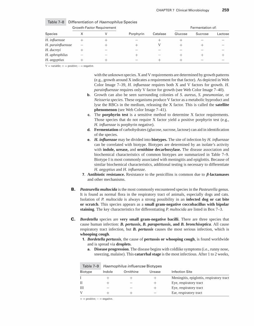
P1: OSO
LWBK192-07 LWBK192-Hubbard November 27, 2008 7:48
CHAPTER 7 Clinical Microbiology 259
Table 7–8 Differentiation of Haemophilus Species
Growth-Factor Requirement Fermentation of:
Species X V Porphyrin Catalase Glucose Sucrose Lactose
H. influenzae + + − + + − −H. parainfluenzae − + + V + + −H. ducreyi + − − − − − −H. aphrophilus − − + − + + +H. aegyptius + + − + + − −V = variable; + = positive; – = negative.
with the unknown species. X and V requirements are determined by growth patterns(e.g., growth around X indicates a requirement for that factor). As depicted in WebColor Image 7–39, H. influenzae requires both X and V factors for growth. H.parainfluenzae requires only V factor for growth (see Web Color Image 7–40).
b. Growth can also be seen surrounding colonies of S. aureus, S. pneumoniae, orNeisseria species. These organisms produce V factor as a metabolic byproduct andlyse the RBCs in the medium, releasing the X factor. This is called the satellitephenomenon (see Web Color Image 7–41).
c. The porphyrin test is a sensitive method to determine X factor requirements.Those species that do not require X factor yield a positive porphyrin test (e.g.,H. influenzae is porphyrin negative).
d. Fermentation of carbohydrates (glucose, sucrose, lactose) can aid in identificationof the species.
e. H. influenzae may be divided into biotypes. The site of infection by H. influenzaecan be correlated with biotype. Biotypes are determined by an isolate’s activitywith indole, urease, and ornithine decarboxylase. The disease association andbiochemical characteristics of common biotypes are summarized in Table 7–9.Biotype I is most commonly associated with meningitis and epiglottis. Because ofsimilar biochemical characteristics, additional testing is necessary to differentiateH. aegyptius and H. influenzae.
7. Antibiotic resistance. Resistance to the penicillins is common due to β-lactamasesand other mechanisms.
B. Pasteurella multocida is the most commonly encountered species in the Pasteurella genus.It is found as normal flora in the respiratory tract of animals, especially dogs and cats.Isolation of P. multocida is always a strong possibility in an infected dog or cat biteor scratch. This species appears as a small gram-negative coccobacillus with bipolarstaining. The key characteristics for differentiating P. multocida are listed in Box 7–3.
C. Bordetella species are very small gram-negative bacilli. There are three species thatcause human infection: B. pertussis, B. parapertussis, and B. bronchiseptica. All causerespiratory tract infection, but B. pertussis causes the most serious infection, which iswhooping cough.
1. Bordetella pertussis, the cause of pertussis or whooping cough, is found worldwideand is spread via droplets.a. Disease progression. The disease begins with coldlike symptoms (i.e., runny nose,
sneezing, malaise). This catarrhal stage is the most infectious. After 1 to 2 weeks,
Table 7–9 Haemophilus influenzae Biotypes
Biotype Indole Ornithine Urease Infection Site
I + + + Meningitis, epiglottis, respiratory tract
II + − + Eye, respiratory tract
III − − + Eye, respiratory tract
V + + − Ear, respiratory tract
+ = positive; − = negative.
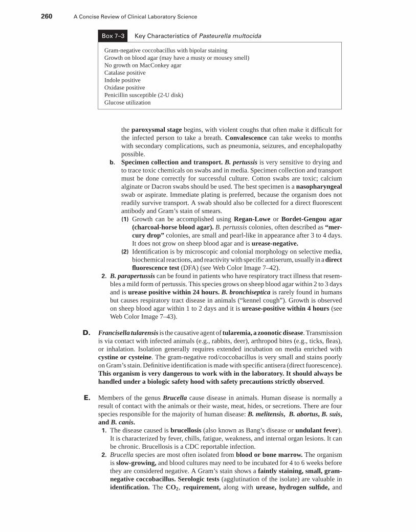
P1: OSO
LWBK192-07 LWBK192-Hubbard November 27, 2008 7:48
260 A Concise Review of Clinical Laboratory Science
Box 7–3 Key Characteristics of Pasteurella multocida
Gram-negative coccobacillus with bipolar staining
Growth on blood agar (may have a musty or mousey smell)
No growth on MacConkey agar
Catalase positive
Indole positive
Oxidase positive
Penicillin susceptible (2-U disk)
Glucose utilization
the paroxysmal stage begins, with violent coughs that often make it difficult forthe infected person to take a breath. Convalescence can take weeks to monthswith secondary complications, such as pneumonia, seizures, and encephalopathypossible.
b. Specimen collection and transport. B. pertussis is very sensitive to drying andto trace toxic chemicals on swabs and in media. Specimen collection and transportmust be done correctly for successful culture. Cotton swabs are toxic; calciumalginate or Dacron swabs should be used. The best specimen is a nasopharyngealswab or aspirate. Immediate plating is preferred, because the organism does notreadily survive transport. A swab should also be collected for a direct fluorescentantibody and Gram’s stain of smears.(1) Growth can be accomplished using Regan-Lowe or Bordet-Gengou agar
(charcoal-horse blood agar). B. pertussis colonies, often described as “mer-cury drop” colonies, are small and pearl-like in appearance after 3 to 4 days.It does not grow on sheep blood agar and is urease-negative.
(2) Identification is by microscopic and colonial morphology on selective media,biochemical reactions, and reactivity with specific antiserum, usually in a directfluorescence test (DFA) (see Web Color Image 7–42).
2. B. parapertussis can be found in patients who have respiratory tract illness that resem-bles a mild form of pertussis. This species grows on sheep blood agar within 2 to 3 daysand is urease positive within 24 hours. B. bronchiseptica is rarely found in humansbut causes respiratory tract disease in animals (“kennel cough”). Growth is observedon sheep blood agar within 1 to 2 days and it is urease-positive within 4 hours (seeWeb Color Image 7–43).
D. Francisella tularensis is the causative agent of tularemia, a zoonotic disease. Transmissionis via contact with infected animals (e.g., rabbits, deer), arthropod bites (e.g., ticks, fleas),or inhalation. Isolation generally requires extended incubation on media enriched withcystine or cysteine. The gram-negative rod/coccobacillus is very small and stains poorlyon Gram’s stain. Definitive identification is made with specific antisera (direct fuorescence).This organism is very dangerous to work with in the laboratory. It should always behandled under a biologic safety hood with safety precautions strictly observed.
E. Members of the genus Brucella cause disease in animals. Human disease is normally aresult of contact with the animals or their waste, meat, hides, or secretions. There are fourspecies responsible for the majority of human disease: B. melitensis, B. abortus, B. suis,and B. canis.
1. The disease caused is brucellosis (also known as Bang’s disease or undulant fever).It is characterized by fever, chills, fatigue, weakness, and internal organ lesions. It canbe chronic. Brucellosis is a CDC reportable infection.
2. Brucella species are most often isolated from blood or bone marrow. The organismis slow-growing, and blood cultures may need to be incubated for 4 to 6 weeks beforethey are considered negative. A Gram’s stain shows a faintly staining, small, gram-negative coccobacillus. Serologic tests (agglutination of the isolate) are valuable inidentification. The CO2, requirement, along with urease, hydrogen sulfide, and
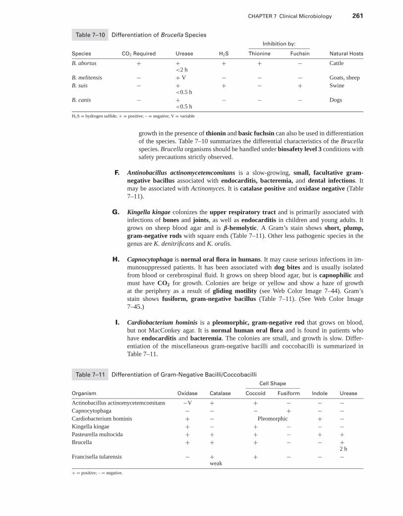
P1: OSO
LWBK192-07 LWBK192-Hubbard November 27, 2008 7:48
CHAPTER 7 Clinical Microbiology 261
Table 7–10 Differentiation of Brucella Species
Inhibition by:
Species CO2 Required Urease H2S Thionine Fuchsin Natural Hosts
B. abortus + +<2 h
+ + − Cattle
B. melitensis − + V − − − Goats, sheep
B. suis − +<0.5 h
+ − + Swine
B. canis − +<0.5 h
− − − Dogs
H2S = hydrogen sulfide; + = positive; – = negative; V = variable
growth in the presence of thionin and basic fuchsin can also be used in differentiationof the species. Table 7–10 summarizes the differential characteristics of the Brucellaspecies. Brucella organisms should be handled under biosafety level 3 conditions withsafety precautions strictly observed.
F. Antinobacillus actinomycetemcomitans is a slow-growing, small, facultative gram-negative bacillus associated with endocarditis, bacteremia, and dental infections. Itmay be associated with Actinomyces. It is catalase positive and oxidase negative (Table7–11).
G. Kingella kingae colonizes the upper respiratory tract and is primarily associated withinfections of bones and joints, as well as endocarditis in children and young adults. Itgrows on sheep blood agar and is β-hemolytic. A Gram’s stain shows short, plump,gram-negative rods with square ends (Table 7–11). Other less pathogenic species in thegenus are K. denitrificans and K. oralis.
H. Capnocytophaga is normal oral flora in humans. It may cause serious infections in im-munosuppressed patients. It has been associated with dog bites and is usually isolatedfrom blood or cerebrospinal fluid. It grows on sheep blood agar, but is capnophilic andmust have CO2 for growth. Colonies are beige or yellow and show a haze of growthat the periphery as a result of gliding motility (see Web Color Image 7–44). Gram’sstain shows fusiform, gram-negative bacillus (Table 7–11). (See Web Color Image7–45.)
I. Cardiobacterium hominis is a pleomorphic, gram-negative rod that grows on blood,but not MacConkey agar. It is normal human oral flora and is found in patients whohave endocarditis and bacteremia. The colonies are small, and growth is slow. Differ-entiation of the miscellaneous gram-negative bacilli and coccobacilli is summarized inTable 7–11.
Table 7–11 Differentiation of Gram-Negative Bacilli/Coccobacilli
Cell Shape
Organism Oxidase Catalase Coccoid Fusiform Indole Urease
Actinobacillus actinomycetemcomitans −V + + − − −Capnocytophaga − − − + − −Cardiobacterium hominis + − Pleomorphic + −Kingella kingae + − + − − −Pasteurella multocida + + + − + +Brucella + + + − − +
2 h
Francisella tularensis − +weak
+ − − −
+ = positive; – = negative.
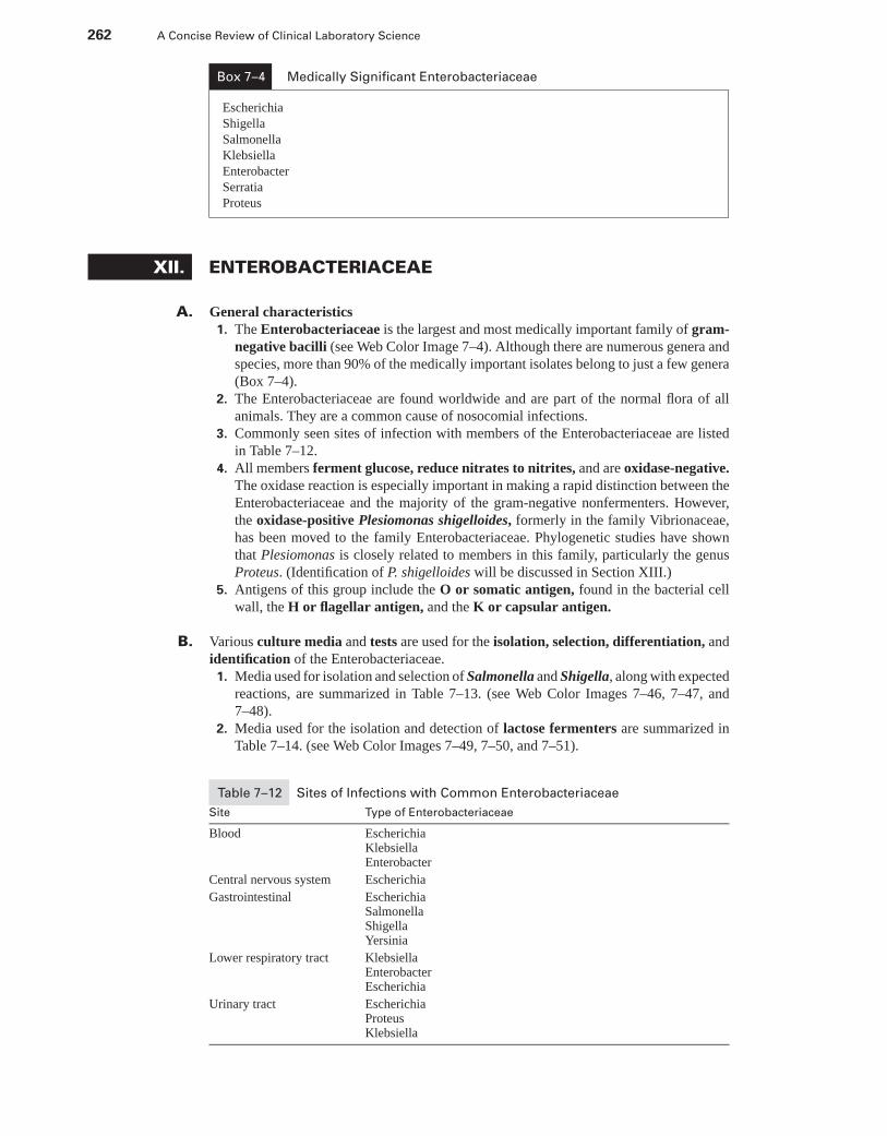
P1: OSO
LWBK192-07 LWBK192-Hubbard November 27, 2008 7:48
262 A Concise Review of Clinical Laboratory Science
Box 7–4 Medically Significant Enterobacteriaceae
Escherichia
Shigella
Salmonella
Klebsiella
Enterobacter
Serratia
Proteus
XII. ENTEROBACTERIACEAE
A. General characteristics1. The Enterobacteriaceae is the largest and most medically important family of gram-
negative bacilli (see Web Color Image 7–4). Although there are numerous genera andspecies, more than 90% of the medically important isolates belong to just a few genera(Box 7–4).
2. The Enterobacteriaceae are found worldwide and are part of the normal flora of allanimals. They are a common cause of nosocomial infections.
3. Commonly seen sites of infection with members of the Enterobacteriaceae are listedin Table 7–12.
4. All members ferment glucose, reduce nitrates to nitrites, and are oxidase-negative.The oxidase reaction is especially important in making a rapid distinction between theEnterobacteriaceae and the majority of the gram-negative nonfermenters. However,the oxidase-positive Plesiomonas shigelloides, formerly in the family Vibrionaceae,has been moved to the family Enterobacteriaceae. Phylogenetic studies have shownthat Plesiomonas is closely related to members in this family, particularly the genusProteus. (Identification of P. shigelloides will be discussed in Section XIII.)
5. Antigens of this group include the O or somatic antigen, found in the bacterial cellwall, the H or flagellar antigen, and the K or capsular antigen.
B. Various culture media and tests are used for the isolation, selection, differentiation, andidentification of the Enterobacteriaceae.1. Media used for isolation and selection of Salmonella and Shigella, along with expected
reactions, are summarized in Table 7–13. (see Web Color Images 7–46, 7–47, and7–48).
2. Media used for the isolation and detection of lactose fermenters are summarized inTable 7–14. (see Web Color Images 7–49, 7–50, and 7–51).
Table 7–12 Sites of Infections with Common Enterobacteriaceae
Site Type of Enterobacteriaceae
Blood EscherichiaKlebsiellaEnterobacter
Central nervous system Escherichia
Gastrointestinal EscherichiaSalmonellaShigellaYersinia
Lower respiratory tract KlebsiellaEnterobacterEscherichia
Urinary tract EscherichiaProteusKlebsiella
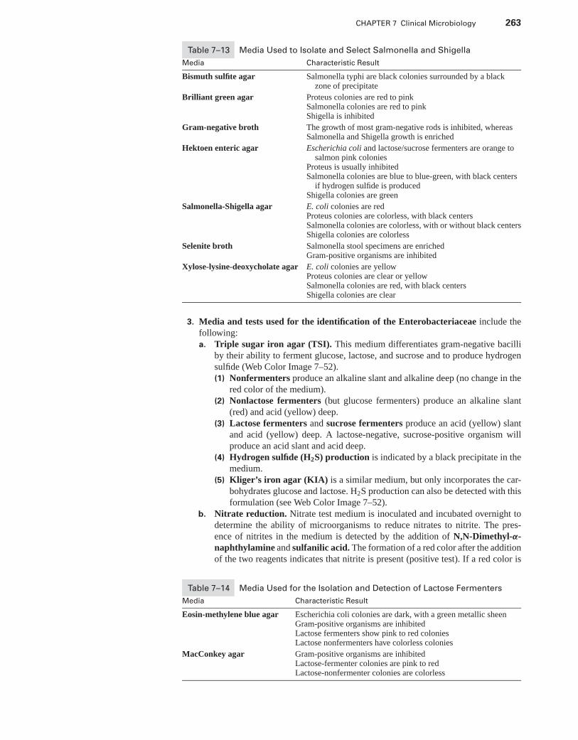
P1: OSO
LWBK192-07 LWBK192-Hubbard November 27, 2008 7:48
CHAPTER 7 Clinical Microbiology 263
Table 7–13 Media Used to Isolate and Select Salmonella and Shigella
Media Characteristic Result
Bismuth sulfite agar Salmonella typhi are black colonies surrounded by a blackzone of precipitate
Brilliant green agar Proteus colonies are red to pinkSalmonella colonies are red to pinkShigella is inhibited
Gram-negative broth The growth of most gram-negative rods is inhibited, whereasSalmonella and Shigella growth is enriched
Hektoen enteric agar Escherichia coli and lactose/sucrose fermenters are orange tosalmon pink colonies
Proteus is usually inhibitedSalmonella colonies are blue to blue-green, with black centers
if hydrogen sulfide is producedShigella colonies are green
Salmonella-Shigella agar E. coli colonies are redProteus colonies are colorless, with black centersSalmonella colonies are colorless, with or without black centersShigella colonies are colorless
Selenite broth Salmonella stool specimens are enrichedGram-positive organisms are inhibited
Xylose-lysine-deoxycholate agar E. coli colonies are yellowProteus colonies are clear or yellowSalmonella colonies are red, with black centersShigella colonies are clear
3. Media and tests used for the identification of the Enterobacteriaceae include thefollowing:a. Triple sugar iron agar (TSI). This medium differentiates gram-negative bacilli
by their ability to ferment glucose, lactose, and sucrose and to produce hydrogensulfide (Web Color Image 7–52).(1) Nonfermenters produce an alkaline slant and alkaline deep (no change in the
red color of the medium).(2) Nonlactose fermenters (but glucose fermenters) produce an alkaline slant
(red) and acid (yellow) deep.(3) Lactose fermenters and sucrose fermenters produce an acid (yellow) slant
and acid (yellow) deep. A lactose-negative, sucrose-positive organism willproduce an acid slant and acid deep.
(4) Hydrogen sulfide (H2S) production is indicated by a black precipitate in themedium.
(5) Kliger’s iron agar (KIA) is a similar medium, but only incorporates the car-bohydrates glucose and lactose. H2S production can also be detected with thisformulation (see Web Color Image 7–52).
b. Nitrate reduction. Nitrate test medium is inoculated and incubated overnight todetermine the ability of microorganisms to reduce nitrates to nitrite. The pres-ence of nitrites in the medium is detected by the addition of N,N-Dimethyl-α-naphthylamine and sulfanilic acid. The formation of a red color after the additionof the two reagents indicates that nitrite is present (positive test). If a red color is
Table 7–14 Media Used for the Isolation and Detection of Lactose Fermenters
Media Characteristic Result
Eosin-methylene blue agar Escherichia coli colonies are dark, with a green metallic sheenGram-positive organisms are inhibitedLactose fermenters show pink to red coloniesLactose nonfermenters have colorless colonies
MacConkey agar Gram-positive organisms are inhibitedLactose-fermenter colonies are pink to redLactose-nonfermenter colonies are colorless
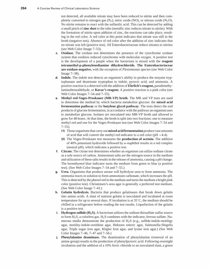
P1: OSO
LWBK192-07 LWBK192-Hubbard November 27, 2008 7:48
264 A Concise Review of Clinical Laboratory Science
not detected, all available nitrate may have been reduced to nitrite and then com-pletely converted to nitrogen gas (N2), nitric oxide (NO), or nitrous oxide (N2O).No nitrite remains to react with the sulfanilic acid. This can be detected by addinga small pinch of zinc dust to the tube (metallic zinc reduces nitrate to nitrite). Withthe formation of nitrite upon addition of zinc, the reactions can take place, result-ing in the red color. A red color at this point indicates that nitrate was still in thebroth (negative test). Absence of red color after the addition of zinc indicates thatno nitrate was left (positive test). All Enterobacteriaceae reduce nitrates to nitrites(see Web Color Image 7–53).
c. Oxidase. The oxidase test determines the presence of the cytochrome oxidasesystem that oxidizes reduced cytochrome with molecular oxygen. A positive testis the development of a purple when the bacterium is mixed with the reagenttetramethyl-p-phenylenediamine dihydrochloride. The Enterobacteriaceaeare oxidase-negative, with the exception of Plesiomonas species (see Web ColorImage 7–38).
d. Indole. The indole test detects an organism’s ability to produce the enzyme tryp-tophanase and deaminate tryptophan to indole, pyruvic acid, and ammonia. Apositive reaction is a detected with the addition of Ehrlich’s reagent, paradimethy-laminobenzaldehyde, or Kovac’s reagent. A positive reaction is a pink color (seeWeb Color Images 7–54 and 7–55).
e. Methyl red-Voges-Proskauer (MR-VP) broth. The MR and VP tests are usedto determine the method by which bacteria metabolize glucose: the mixed acidfermentation pathway or the butylene glycol pathway. The tests detect the endproducts of glucose fermentation, in accordance with the pathway an organism usesto metabolize glucose. Isolates are inoculated into MR-VP broth and allowed togrow for 48 hours. At that time, the broth is split into two fractions: one to measuremethyl red and one for the Voges-Proskauer test (see Web Color Images 7–54 and7–55).(1) Those organisms that carry out mixed acid fermentation produce vast amounts
of acid that will convert the methyl red indicator to a red color (pH <4.4).(2) The Voges-Proskauer test measures the production of acetoin. The addition
of 40% potassium hydroxide followed by α-naphthol results in a red complex(neutral pH), which indicates a positive test.
f. Citrate. The citrate test determines whether an organism can utilize sodium citrateas a sole source of carbon. Ammonium salts are the nitrogen source in the mediumand utilization of these salts results in the release of ammonia, causing a pH change.The bromthymol blue indicator turns the medium from green to blue (a positivetest). (See Web Color Images 7–54 and 7–55.)
g. Urea. Organisms that produce urease will hydrolyze urea to form ammonia. Theammonia reacts in solution to form ammonium carbonate, which increases the pH.This is detected by the phenol red in the medium and turns the medium a bright pinkcolor (positive test). Christensen’s urea agar is generally a preferred test medium.(See Web Color Image 7–43.)
h. Gelatin hydrolysis. Bacteria that produce gelatinases that break down gelatininto amino acids. A slant of nutrient gelatin is inoculated and incubated at roomtemperature for up to several days. If incubation is at 35◦C, the medium should bechilled in a refrigerator before reading the test results. Liquefaction of the gelatinis a positive test.
i. Hydrogen sulfide (H2S). A bacterium utilizes the sodium thiosulfate sulfur sourceto form H2S, a colorless gas. H2S combines with the indicator, ferrous sulfate. Nu-merous media demonstrate the production of H2S (e.g., sulfide-indole-motiligyagar, motility-indole-ornithine agar, Hektoen enteric agar, Salmonella-Shigellaagar, Triple sugar iron agar, Kligler Iron agar, and lysine iron agar.) (See WebColor Images 7–46, 7–47 and 7–56.)
j. Phenylalanine deaminase. The deamination of phenylalanine (removal of anamine group) results in the production of phenylpyruvic acid. Following overnightincubation and the addition of a 10% ferric chloride to an inoculated slant, a green
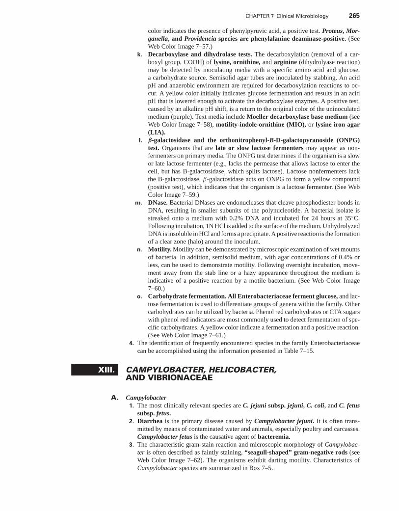
P1: OSO
LWBK192-07 LWBK192-Hubbard November 27, 2008 7:48
CHAPTER 7 Clinical Microbiology 265
color indicates the presence of phenylpyruvic acid, a positive test. Proteus, Mor-ganella, and Providencia species are phenylalanine deaminase-positive. (SeeWeb Color Image 7–57.)
k. Decarboxylase and dihydrolase tests. The decarboxylation (removal of a car-boxyl group, COOH) of lysine, ornithine, and arginine (dihydrolyase reaction)may be detected by inoculating media with a specific amino acid and glucose,a carbohydrate source. Semisolid agar tubes are inoculated by stabbing. An acidpH and anaerobic environment are required for decarboxylation reactions to oc-cur. A yellow color initially indicates glucose fermentation and results in an acidpH that is lowered enough to activate the decarboxylase enzymes. A positive test,caused by an alkaline pH shift, is a return to the original color of the uninoculatedmedium (purple). Text media include Moeller decarboxylase base medium (seeWeb Color Image 7–58), motility-indole-ornithine (MIO), or lysine iron agar(LIA).
l. β-galactosidase and the orthonitrophenyl-B-D-galactopyranoside (ONPG)test. Organisms that are late or slow lactose fermenters may appear as non-fermenters on primary media. The ONPG test determines if the organism is a slowor late lactose fermenter (e.g., lacks the permease that allows lactose to enter thecell, but has B-galactosidase, which splits lactose). Lactose nonfermenters lackthe B-galactosidase. β-galactosidase acts on ONPG to form a yellow compound(positive test), which indicates that the organism is a lactose fermenter. (See WebColor Image 7–59.)
m. DNase. Bacterial DNases are endonucleases that cleave phosphodiester bonds inDNA, resulting in smaller subunits of the polynucleotide. A bacterial isolate isstreaked onto a medium with 0.2% DNA and incubated for 24 hours at 35◦C.Following incubation, 1N HCl is added to the surface of the medium. UnhydrolyzedDNA is insoluble in HCl and forms a precipitate. A positive reaction is the formationof a clear zone (halo) around the inoculum.
n. Motility. Motility can be demonstrated by microscopic examination of wet mountsof bacteria. In addition, semisolid medium, with agar concentrations of 0.4% orless, can be used to demonstrate motility. Following overnight incubation, move-ment away from the stab line or a hazy appearance throughout the medium isindicative of a positive reaction by a motile bacterium. (See Web Color Image7–60.)
o. Carbohydrate fermentation. All Enterobacteriaceae ferment glucose, and lac-tose fermentation is used to differentiate groups of genera within the family. Othercarbohydrates can be utilized by bacteria. Phenol red carbohydrates or CTA sugarswith phenol red indicators are most commonly used to detect fermentation of spe-cific carbohydrates. A yellow color indicate a fermentation and a positive reaction.(See Web Color Image 7–61.)
4. The identification of frequently encountered species in the family Enterobacteriaceaecan be accomplished using the information presented in Table 7–15.
XIII. CAMPYLOBACTER, HELICOBACTER,AND VIBRIONACEAE
A. Campylobacter1. The most clinically relevant species are C. jejuni subsp. jejuni, C. coli, and C. fetus
subsp. fetus.2. Diarrhea is the primary disease caused by Campylobacter jejuni. It is often trans-
mitted by means of contaminated water and animals, especially poultry and carcasses.Campylobacter fetus is the causative agent of bacteremia.
3. The characteristic gram-stain reaction and microscopic morphology of Campylobac-ter is often described as faintly staining, “seagull-shaped” gram-negative rods (seeWeb Color Image 7–62). The organisms exhibit darting motility. Characteristics ofCampylobacter species are summarized in Box 7–5.
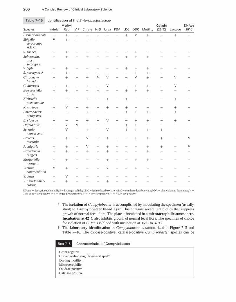
P1: OSO
LWBK192-07 LWBK192-Hubbard November 27, 2008 7:48
266 A Concise Review of Clinical Laboratory Science
Table 7–15 Identification of the Enterobacteriaceae
Methyl Gelatin DNAse
Species Indole Red V-P Citrate H2S Urea PDA LDC ODC Motility (22◦C) Lactose (25◦C)
Escherichia coli + + − − − − − + V + − + −Shigella
serogroupsA,B,C
V + − − − − − − − − − − −
S. sonnei − + − − − − − − + − − − −Salmonella,
mostserotypes
− + − + + − − + + + − − −
S. typhi − + − − + − − + − + − − −S. paratyphi A − + − − − − − − + + − − −Citrobacter
freundii− + − + V V − − V + − V −
C. diversus + + − + − V − − + + − V −Edwardsiella
tarda+ + − − + − − + + + − − −
Klebsiellapneumoniae
− − + + − + − + − − − + −
K. oxytoca + V + + − + − + − − − + −Enterobacter
aerogenes− − + + − − − + + + − + −
E. cloacae − − + + − V − − + + − + −Hafnia alvei − V V − − − − + + − − − −Serratia
marcescens− V + + − V − + + + + − +
Proteusmirabilis
− + − V + + + − + + + − V
P. vulgaris + + − V + + + − − + + − V
Providenciarettgeri
+ + − + − + + − − + − − −
Morganellamorganii
+ + − − − + + − + + − − −
Yersiniaenterocolitica
V + − − − V − − + − − − −
Y. pestis − V − − − − − − − − − − −Y. pseudotuber-
culosis− + − − − + − − − − − − −
DNAse = deoxyribonuclease; H2S = hydrogen sulfide; LDC = lysine decarboxylase; ODC = ornithine decarboxylase; PDA = phenylalanine deaminase; V =10% to 89% are positive; V-P = Voges-Proskauer test; + = ≥ 90% are positive; − = ≤10% are positive.
4. The isolation of Campylobacter is accomplished by inoculating the specimen (usuallystool) to Campylobacter blood agar. This contains several antibiotics that suppressgrowth of normal fecal flora. The plate is incubated in a microaerophilic atmosphere.Incubation at 42◦C also inhibits growth of normal fecal flora. The specimen of choicefor isolation of C. fetus is blood with incubation at 35◦C to 37◦C.
5. The laboratory identification of Campylobacter is summarized in Figure 7–5 andTable 7–16. The oxidase-positive, catalase-positive Campylobacter species can be
Box 7–5 Characteristics of Campylobacter
Gram negative
Curved rods–“seagull-wing-shaped”
Darting motility
Microaerophilic
Oxidase positive
Catalase positive
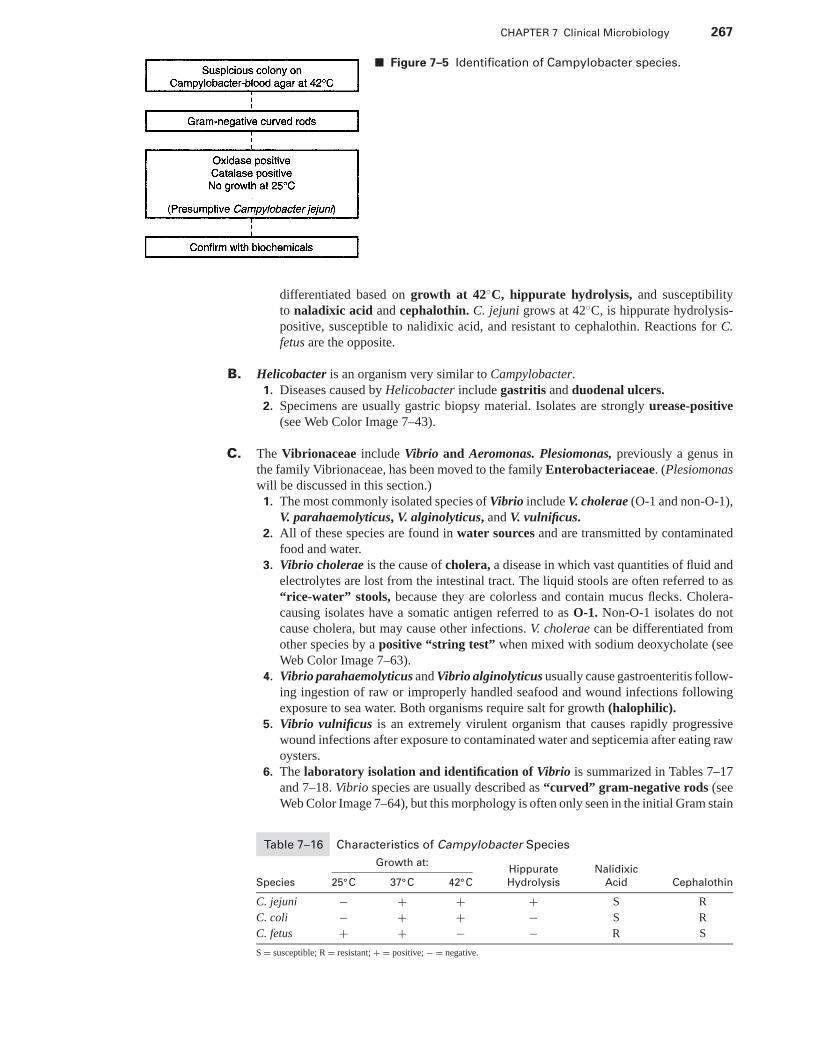
P1: OSO
LWBK192-07 LWBK192-Hubbard November 27, 2008 7:48
CHAPTER 7 Clinical Microbiology 267
■ Figure 7–5 Identification of Campylobacter species.
differentiated based on growth at 42◦C, hippurate hydrolysis, and susceptibilityto naladixic acid and cephalothin. C. jejuni grows at 42◦C, is hippurate hydrolysis-positive, susceptible to nalidixic acid, and resistant to cephalothin. Reactions for C.fetus are the opposite.
B. Helicobacter is an organism very similar to Campylobacter.1. Diseases caused by Helicobacter include gastritis and duodenal ulcers.2. Specimens are usually gastric biopsy material. Isolates are strongly urease-positive
(see Web Color Image 7–43).
C. The Vibrionaceae include Vibrio and Aeromonas. Plesiomonas, previously a genus inthe family Vibrionaceae, has been moved to the family Enterobacteriaceae. (Plesiomonaswill be discussed in this section.)1. The most commonly isolated species of Vibrio include V. cholerae (O-1 and non-O-1),
V. parahaemolyticus, V. alginolyticus, and V. vulnificus.2. All of these species are found in water sources and are transmitted by contaminated
food and water.3. Vibrio cholerae is the cause of cholera, a disease in which vast quantities of fluid and
electrolytes are lost from the intestinal tract. The liquid stools are often referred to as“rice-water” stools, because they are colorless and contain mucus flecks. Cholera-causing isolates have a somatic antigen referred to as O-1. Non-O-1 isolates do notcause cholera, but may cause other infections. V. cholerae can be differentiated fromother species by a positive “string test” when mixed with sodium deoxycholate (seeWeb Color Image 7–63).
4. Vibrio parahaemolyticus and Vibrio alginolyticus usually cause gastroenteritis follow-ing ingestion of raw or improperly handled seafood and wound infections followingexposure to sea water. Both organisms require salt for growth (halophilic).
5. Vibrio vulnificus is an extremely virulent organism that causes rapidly progressivewound infections after exposure to contaminated water and septicemia after eating rawoysters.
6. The laboratory isolation and identification of Vibrio is summarized in Tables 7–17and 7–18. Vibrio species are usually described as “curved” gram-negative rods (seeWeb Color Image 7–64), but this morphology is often only seen in the initial Gram stain
Table 7–16 Characteristics of Campylobacter Species
Growth at:Hippurate Nalidixic
Species 25◦C 37◦C 42◦C Hydrolysis Acid Cephalothin
C. jejuni − + + + S R
C. coli − + + − S R
C. fetus + + − − R S
S = susceptible; R = resistant; + = positive; − = negative.
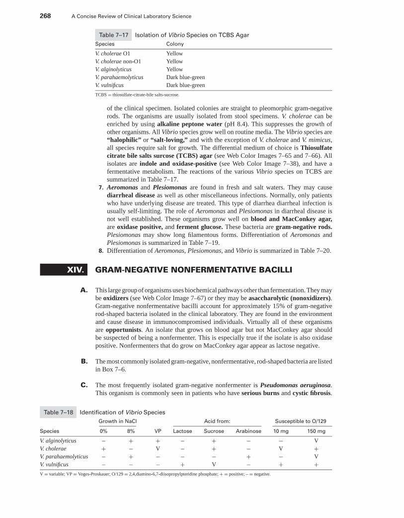
P1: OSO
LWBK192-07 LWBK192-Hubbard November 27, 2008 7:48
268 A Concise Review of Clinical Laboratory Science
Table 7–17 Isolation of Vibrio Species on TCBS Agar
Species Colony
V. cholerae O1 Yellow
V. cholerae non-O1 Yellow
V. alginolyticus Yellow
V. parahaemolyticus Dark blue-green
V. vulnificus Dark blue-green
TCBS = thiosulfate-citrate-bile salts-sucrose.
of the clinical specimen. Isolated colonies are straight to pleomorphic gram-negativerods. The organisms are usually isolated from stool specimens. V. cholerae can beenriched by using alkaline peptone water (pH 8.4). This suppresses the growth ofother organisms. All Vibrio species grow well on routine media. The Vibrio species are“halophilic” or “salt-loving,” and with the exception of V. cholerae and V. mimicus,all species require salt for growth. The differential medium of choice is Thiosulfatecitrate bile salts surcose (TCBS) agar (see Web Color Images 7–65 and 7–66). Allisolates are indole and oxidase-positive (see Web Color Image 7–38), and have afermentative metabolism. The reactions of the various Vibrio species on TCBS aresummarized in Table 7–17.
7. Aeromonas and Plesiomonas are found in fresh and salt waters. They may causediarrheal disease as well as other miscellaneous infections. Normally, only patientswho have underlying disease are treated. This type of diarrhea diarrheal infection isusually self-limiting. The role of Aeromonas and Plesiomonas in diarrheal disease isnot well established. These organisms grow well on blood and MacConkey agar,are oxidase positive, and ferment glucose. These bacteria are gram-negative rods.Plesiomonas may show long filamentous forms. Differentiation of Aeromonas andPlesiomonas is summarized in Table 7–19.
8. Differentiation of Aeromonas, Plesiomonas, and Vibrio is summarized in Table 7–20.
XIV. GRAM-NEGATIVE NONFERMENTATIVE BACILLI
A. This large group of organisms uses biochemical pathways other than fermentation. They maybe oxidizers (see Web Color Image 7–67) or they may be asaccharolytic (nonoxidizers).Gram-negative nonfermentative bacilli account for approximately 15% of gram-negativerod-shaped bacteria isolated in the clinical laboratory. They are found in the environmentand cause disease in immunocompromised individuals. Virtually all of these organismsare opportunists. An isolate that grows on blood agar but not MacConkey agar shouldbe suspected of being a nonfermenter. This is especially true if the isolate is also oxidasepositive. Nonfermenters that do grow on MacConkey agar appear as lactose negative.
B. The most commonly isolated gram-negative, nonfermentative, rod-shaped bacteria are listedin Box 7–6.
C. The most frequently isolated gram-negative nonfermenter is Pseudomonas aeruginosa.This organism is commonly seen in patients who have serious burns and cystic fibrosis.
Table 7–18 Identification of Vibrio Species
Growth in NaCl Acid from: Susceptible to O/129
Species 0% 8% VP Lactose Sucrose Arabinose 10 mg 150 mg
V. alginolyticus − + + − + − − V
V. cholerae + − V − + − V +V. parahaemolyticus − + − − − + − V
V. vulnificus − − − + V − + +V = variable; VP = Voges-Proskauer; O/129 = 2,4,diamino-6,7-diisopropylpteridine phosphate; + = positive; – = negative.
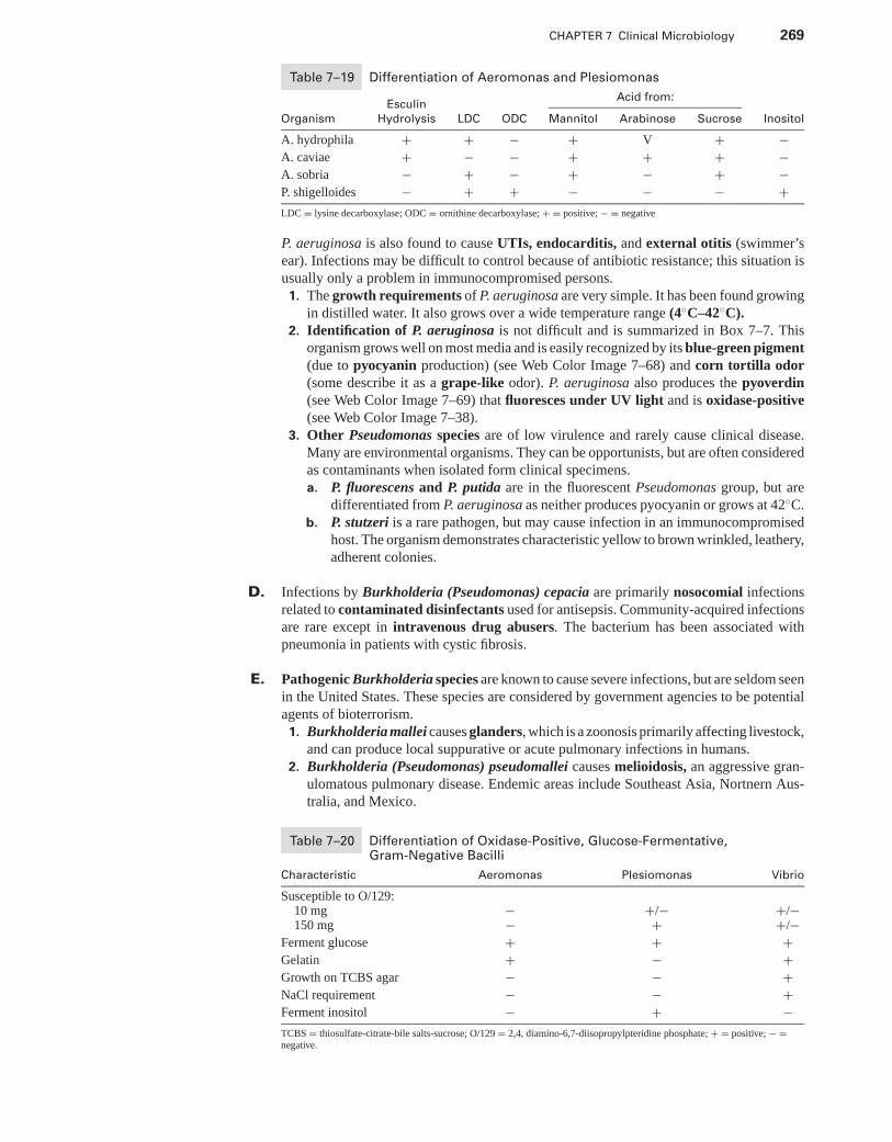
P1: OSO
LWBK192-07 LWBK192-Hubbard November 27, 2008 7:48
CHAPTER 7 Clinical Microbiology 269
Table 7–19 Differentiation of Aeromonas and Plesiomonas
Acid from:Esculin
Organism Hydrolysis LDC ODC Mannitol Arabinose Sucrose Inositol
A. hydrophila + + − + V + −A. caviae + − − + + + −A. sobria − + − + − + −P. shigelloides − + + − − − +LDC = lysine decarboxylase; ODC = ornithine decarboxylase; + = positive; − = negative
P. aeruginosa is also found to cause UTIs, endocarditis, and external otitis (swimmer’sear). Infections may be difficult to control because of antibiotic resistance; this situation isusually only a problem in immunocompromised persons.1. The growth requirements of P. aeruginosa are very simple. It has been found growing
in distilled water. It also grows over a wide temperature range (4◦C–42◦C).2. Identification of P. aeruginosa is not difficult and is summarized in Box 7–7. This
organism grows well on most media and is easily recognized by its blue-green pigment(due to pyocyanin production) (see Web Color Image 7–68) and corn tortilla odor(some describe it as a grape-like odor). P. aeruginosa also produces the pyoverdin(see Web Color Image 7–69) that fluoresces under UV light and is oxidase-positive(see Web Color Image 7–38).
3. Other Pseudomonas species are of low virulence and rarely cause clinical disease.Many are environmental organisms. They can be opportunists, but are often consideredas contaminants when isolated form clinical specimens.a. P. fluorescens and P. putida are in the fluorescent Pseudomonas group, but are
differentiated from P. aeruginosa as neither produces pyocyanin or grows at 42◦C.b. P. stutzeri is a rare pathogen, but may cause infection in an immunocompromised
host. The organism demonstrates characteristic yellow to brown wrinkled, leathery,adherent colonies.
D. Infections by Burkholderia (Pseudomonas) cepacia are primarily nosocomial infectionsrelated to contaminated disinfectants used for antisepsis. Community-acquired infectionsare rare except in intravenous drug abusers. The bacterium has been associated withpneumonia in patients with cystic fibrosis.
E. Pathogenic Burkholderia species are known to cause severe infections, but are seldom seenin the United States. These species are considered by government agencies to be potentialagents of bioterrorism.1. Burkholderia mallei causes glanders, which is a zoonosis primarily affecting livestock,
and can produce local suppurative or acute pulmonary infections in humans.2. Burkholderia (Pseudomonas) pseudomallei causes melioidosis, an aggressive gran-
ulomatous pulmonary disease. Endemic areas include Southeast Asia, Nortnern Aus-tralia, and Mexico.
Table 7–20 Differentiation of Oxidase-Positive, Glucose-Fermentative,Gram-Negative Bacilli
Characteristic Aeromonas Plesiomonas Vibrio
Susceptible to O/129:10 mg − +/− +/−150 mg − + +/−
Ferment glucose + + +Gelatin + − +Growth on TCBS agar − − +NaCl requirement − − +Ferment inositol − + −TCBS = thiosulfate-citrate-bile salts-sucrose; O/129 = 2,4, diamino-6,7-diisopropylpteridine phosphate; + = positive; − =negative.
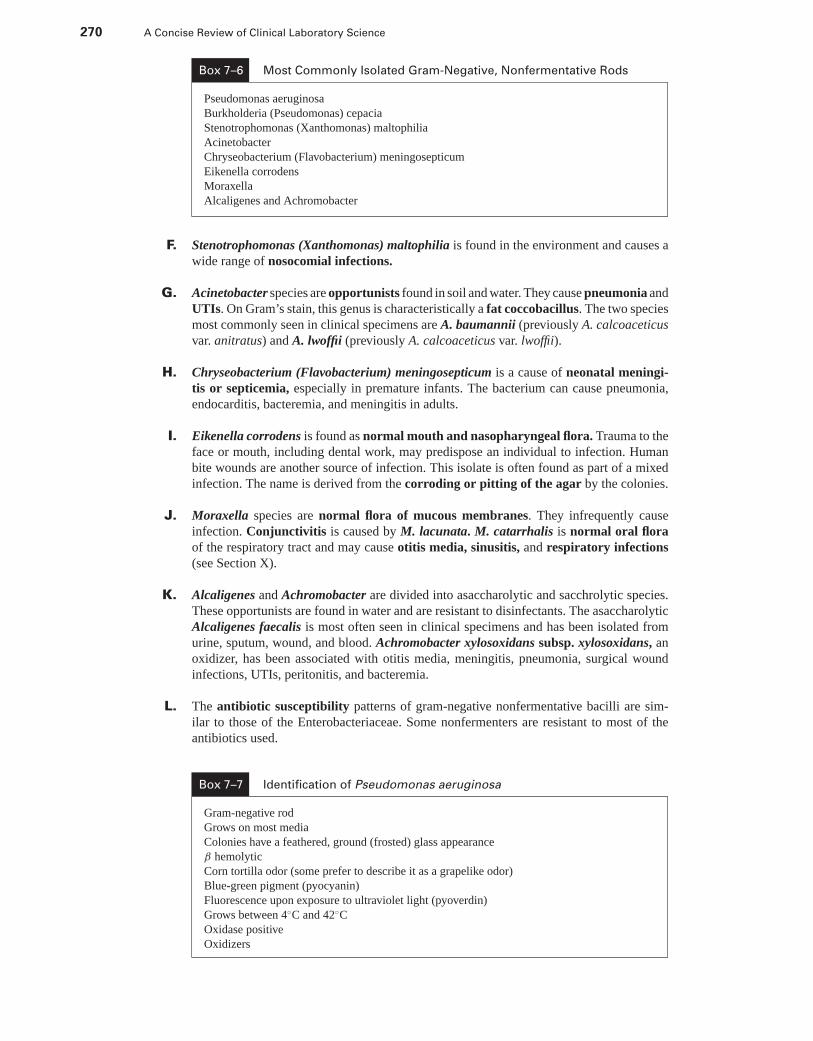
P1: OSO
LWBK192-07 LWBK192-Hubbard November 27, 2008 7:48
270 A Concise Review of Clinical Laboratory Science
Box 7–6 Most Commonly Isolated Gram-Negative, Nonfermentative Rods
Pseudomonas aeruginosa
Burkholderia (Pseudomonas) cepacia
Stenotrophomonas (Xanthomonas) maltophilia
Acinetobacter
Chryseobacterium (Flavobacterium) meningosepticum
Eikenella corrodens
Moraxella
Alcaligenes and Achromobacter
F. Stenotrophomonas (Xanthomonas) maltophilia is found in the environment and causes awide range of nosocomial infections.
G. Acinetobacter species are opportunists found in soil and water. They cause pneumonia andUTIs. On Gram’s stain, this genus is characteristically a fat coccobacillus. The two speciesmost commonly seen in clinical specimens are A. baumannii (previously A. calcoaceticusvar. anitratus) and A. lwoffii (previously A. calcoaceticus var. lwoffii).
H. Chryseobacterium (Flavobacterium) meningosepticum is a cause of neonatal meningi-tis or septicemia, especially in premature infants. The bacterium can cause pneumonia,endocarditis, bacteremia, and meningitis in adults.
I. Eikenella corrodens is found as normal mouth and nasopharyngeal flora. Trauma to theface or mouth, including dental work, may predispose an individual to infection. Humanbite wounds are another source of infection. This isolate is often found as part of a mixedinfection. The name is derived from the corroding or pitting of the agar by the colonies.
J. Moraxella species are normal flora of mucous membranes. They infrequently causeinfection. Conjunctivitis is caused by M. lacunata. M. catarrhalis is normal oral floraof the respiratory tract and may cause otitis media, sinusitis, and respiratory infections(see Section X).
K. Alcaligenes and Achromobacter are divided into asaccharolytic and sacchrolytic species.These opportunists are found in water and are resistant to disinfectants. The asaccharolyticAlcaligenes faecalis is most often seen in clinical specimens and has been isolated fromurine, sputum, wound, and blood. Achromobacter xylosoxidans subsp. xylosoxidans, anoxidizer, has been associated with otitis media, meningitis, pneumonia, surgical woundinfections, UTIs, peritonitis, and bacteremia.
L. The antibiotic susceptibility patterns of gram-negative nonfermentative bacilli are sim-ilar to those of the Enterobacteriaceae. Some nonfermenters are resistant to most of theantibiotics used.
Box 7–7 Identification of Pseudomonas aeruginosa
Gram-negative rod
Grows on most media
Colonies have a feathered, ground (frosted) glass appearance
β hemolytic
Corn tortilla odor (some prefer to describe it as a grapelike odor)
Blue-green pigment (pyocyanin)
Fluorescence upon exposure to ultraviolet light (pyoverdin)
Grows between 4◦C and 42◦C
Oxidase positive
Oxidizers
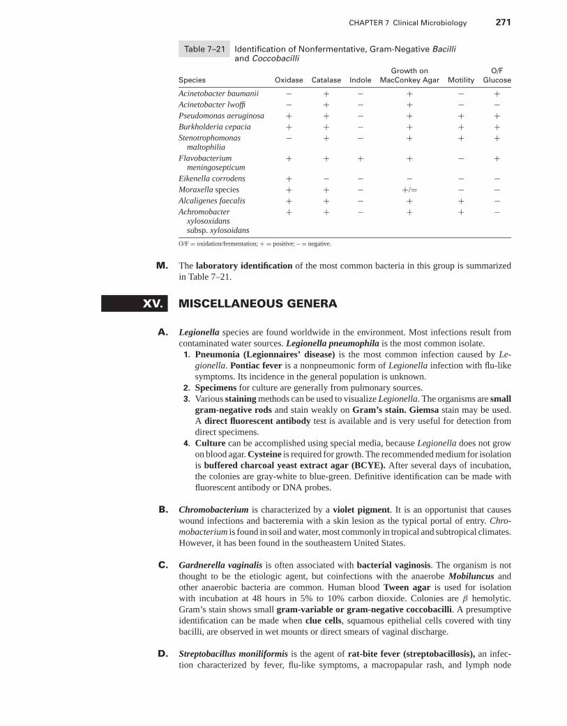
P1: OSO
LWBK192-07 LWBK192-Hubbard November 27, 2008 7:48
CHAPTER 7 Clinical Microbiology 271
Table 7–21 Identification of Nonfermentative, Gram-Negative Bacilliand Coccobacilli
Growth on O/F
Species Oxidase Catalase Indole MacConkey Agar Motility Glucose
Acinetobacter baumanii − + − + − +Acinetobacter lwoffi − + − + − −Pseudomonas aeruginosa + + − + + +Burkholderia cepacia + + − + + +Stenotrophomonas
maltophilia− + − + + +
Flavobacteriummeningosepticum
+ + + + − +
Eikenella corrodens + − − − − −Moraxella species + + − +/= − −Alcaligenes faecalis + + − + + −Achromobacter
xylosoxidanssubsp. xylosoidans
+ + − + + −
O/F = oxidation/fermentation; + = positive; – = negative.
M. The laboratory identification of the most common bacteria in this group is summarizedin Table 7–21.
XV. MISCELLANEOUS GENERA
A. Legionella species are found worldwide in the environment. Most infections result fromcontaminated water sources. Legionella pneumophila is the most common isolate.
1. Pneumonia (Legionnaires’ disease) is the most common infection caused by Le-gionella. Pontiac fever is a nonpneumonic form of Legionella infection with flu-likesymptoms. Its incidence in the general population is unknown.
2. Specimens for culture are generally from pulmonary sources.3. Various staining methods can be used to visualize Legionella. The organisms are small
gram-negative rods and stain weakly on Gram’s stain. Giemsa stain may be used.A direct fluorescent antibody test is available and is very useful for detection fromdirect specimens.
4. Culture can be accomplished using special media, because Legionella does not growon blood agar. Cysteine is required for growth. The recommended medium for isolationis buffered charcoal yeast extract agar (BCYE). After several days of incubation,the colonies are gray-white to blue-green. Definitive identification can be made withfluorescent antibody or DNA probes.
B. Chromobacterium is characterized by a violet pigment. It is an opportunist that causeswound infections and bacteremia with a skin lesion as the typical portal of entry. Chro-mobacterium is found in soil and water, most commonly in tropical and subtropical climates.However, it has been found in the southeastern United States.
C. Gardnerella vaginalis is often associated with bacterial vaginosis. The organism is notthought to be the etiologic agent, but coinfections with the anaerobe Mobiluncus andother anaerobic bacteria are common. Human blood Tween agar is used for isolationwith incubation at 48 hours in 5% to 10% carbon dioxide. Colonies are β hemolytic.Gram’s stain shows small gram-variable or gram-negative coccobacilli. A presumptiveidentification can be made when clue cells, squamous epithelial cells covered with tinybacilli, are observed in wet mounts or direct smears of vaginal discharge.
D. Streptobacillus moniliformis is the agent of rat-bite fever (streptobacillosis), an infec-tion characterized by fever, flu-like symptoms, a macropapular rash, and lymph node
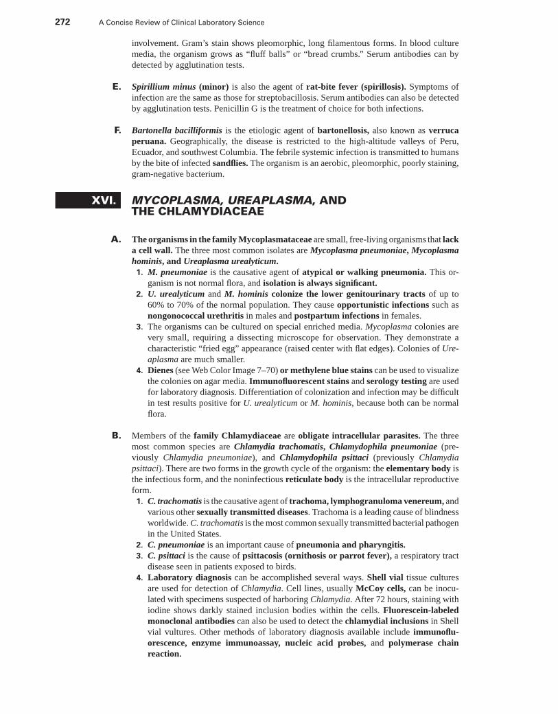
P1: OSO
LWBK192-07 LWBK192-Hubbard November 27, 2008 7:48
272 A Concise Review of Clinical Laboratory Science
involvement. Gram’s stain shows pleomorphic, long filamentous forms. In blood culturemedia, the organism grows as “fluff balls” or “bread crumbs.” Serum antibodies can bydetected by agglutination tests.
E. Spirillium minus (minor) is also the agent of rat-bite fever (spirillosis). Symptoms ofinfection are the same as those for streptobacillosis. Serum antibodies can also be detectedby agglutination tests. Penicillin G is the treatment of choice for both infections.
F. Bartonella bacilliformis is the etiologic agent of bartonellosis, also known as verrucaperuana. Geographically, the disease is restricted to the high-altitude valleys of Peru,Ecuador, and southwest Columbia. The febrile systemic infection is transmitted to humansby the bite of infected sandflies. The organism is an aerobic, pleomorphic, poorly staining,gram-negative bacterium.
XVI. MYCOPLASMA, UREAPLASMA, ANDTHE CHLAMYDIACEAE
A. The organisms in the family Mycoplasmataceae are small, free-living organisms that lacka cell wall. The three most common isolates are Mycoplasma pneumoniae, Mycoplasmahominis, and Ureaplasma urealyticum.
1. M. pneumoniae is the causative agent of atypical or walking pneumonia. This or-ganism is not normal flora, and isolation is always significant.
2. U. urealyticum and M. hominis colonize the lower genitourinary tracts of up to60% to 70% of the normal population. They cause opportunistic infections such asnongonococcal urethritis in males and postpartum infections in females.
3. The organisms can be cultured on special enriched media. Mycoplasma colonies arevery small, requiring a dissecting microscope for observation. They demonstrate acharacteristic “fried egg” appearance (raised center with flat edges). Colonies of Ure-aplasma are much smaller.
4. Dienes (see Web Color Image 7–70) or methylene blue stains can be used to visualizethe colonies on agar media. Immunofluorescent stains and serology testing are usedfor laboratory diagnosis. Differentiation of colonization and infection may be difficultin test results positive for U. urealyticum or M. hominis, because both can be normalflora.
B. Members of the family Chlamydiaceae are obligate intracellular parasites. The threemost common species are Chlamydia trachomatis, Chlamydophila pneumoniae (pre-viously Chlamydia pneumoniae), and Chlamydophila psittaci (previously Chlamydiapsittaci). There are two forms in the growth cycle of the organism: the elementary body isthe infectious form, and the noninfectious reticulate body is the intracellular reproductiveform.1. C. trachomatis is the causative agent of trachoma, lymphogranuloma venereum, and
various other sexually transmitted diseases. Trachoma is a leading cause of blindnessworldwide. C. trachomatis is the most common sexually transmitted bacterial pathogenin the United States.
2. C. pneumoniae is an important cause of pneumonia and pharyngitis.3. C. psittaci is the cause of psittacosis (ornithosis or parrot fever), a respiratory tract
disease seen in patients exposed to birds.4. Laboratory diagnosis can be accomplished several ways. Shell vial tissue cultures
are used for detection of Chlamydia. Cell lines, usually McCoy cells, can be inocu-lated with specimens suspected of harboring Chlamydia. After 72 hours, staining withiodine shows darkly stained inclusion bodies within the cells. Fluorescein-labeledmonoclonal antibodies can also be used to detect the chlamydial inclusions in Shellvial vultures. Other methods of laboratory diagnosis available include immunoflu-orescence, enzyme immunoassay, nucleic acid probes, and polymerase chainreaction.
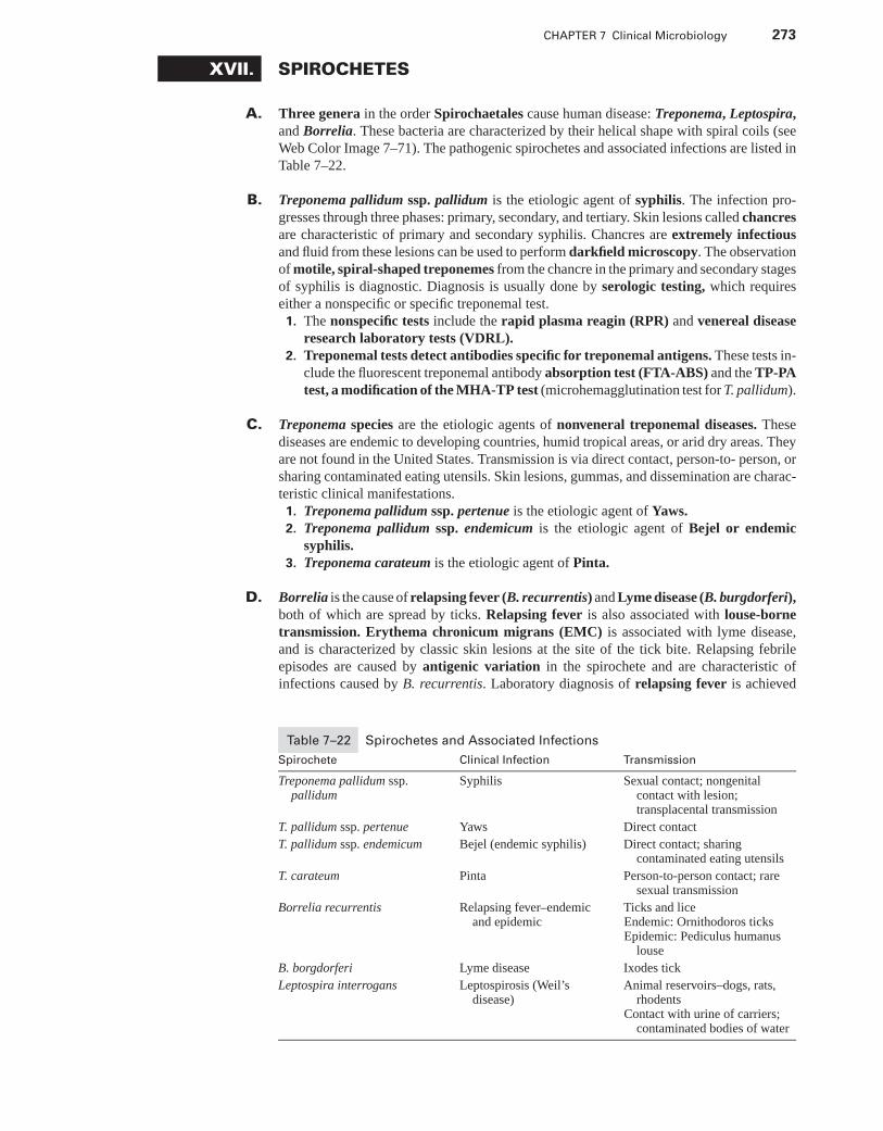
P1: OSO
LWBK192-07 LWBK192-Hubbard November 27, 2008 7:48
CHAPTER 7 Clinical Microbiology 273
XVII. SPIROCHETES
A. Three genera in the order Spirochaetales cause human disease: Treponema, Leptospira,and Borrelia. These bacteria are characterized by their helical shape with spiral coils (seeWeb Color Image 7–71). The pathogenic spirochetes and associated infections are listed inTable 7–22.
B. Treponema pallidum ssp. pallidum is the etiologic agent of syphilis. The infection pro-gresses through three phases: primary, secondary, and tertiary. Skin lesions called chancresare characteristic of primary and secondary syphilis. Chancres are extremely infectiousand fluid from these lesions can be used to perform darkfield microscopy. The observationof motile, spiral-shaped treponemes from the chancre in the primary and secondary stagesof syphilis is diagnostic. Diagnosis is usually done by serologic testing, which requireseither a nonspecific or specific treponemal test.
1. The nonspecific tests include the rapid plasma reagin (RPR) and venereal diseaseresearch laboratory tests (VDRL).
2. Treponemal tests detect antibodies specific for treponemal antigens. These tests in-clude the fluorescent treponemal antibody absorption test (FTA-ABS) and the TP-PAtest, a modification of the MHA-TP test (microhemagglutination test for T. pallidum).
C. Treponema species are the etiologic agents of nonveneral treponemal diseases. Thesediseases are endemic to developing countries, humid tropical areas, or arid dry areas. Theyare not found in the United States. Transmission is via direct contact, person-to- person, orsharing contaminated eating utensils. Skin lesions, gummas, and dissemination are charac-teristic clinical manifestations.
1. Treponema pallidum ssp. pertenue is the etiologic agent of Yaws.2. Treponema pallidum ssp. endemicum is the etiologic agent of Bejel or endemic
syphilis.3. Treponema carateum is the etiologic agent of Pinta.
D. Borrelia is the cause of relapsing fever (B. recurrentis) and Lyme disease (B. burgdorferi),both of which are spread by ticks. Relapsing fever is also associated with louse-bornetransmission. Erythema chronicum migrans (EMC) is associated with lyme disease,and is characterized by classic skin lesions at the site of the tick bite. Relapsing febrileepisodes are caused by antigenic variation in the spirochete and are characteristic ofinfections caused by B. recurrentis. Laboratory diagnosis of relapsing fever is achieved
Table 7–22 Spirochetes and Associated Infections
Spirochete Clinical Infection Transmission
Treponema pallidum ssp.pallidum
Syphilis Sexual contact; nongenitalcontact with lesion;transplacental transmission
T. pallidum ssp. pertenue Yaws Direct contact
T. pallidum ssp. endemicum Bejel (endemic syphilis) Direct contact; sharingcontaminated eating utensils
T. carateum Pinta Person-to-person contact; raresexual transmission
Borrelia recurrentis Relapsing fever–endemicand epidemic
Ticks and liceEndemic: Ornithodoros ticksEpidemic: Pediculus humanus
louse
B. borgdorferi Lyme disease Ixodes tick
Leptospira interrogans Leptospirosis (Weil’sdisease)
Animal reservoirs–dogs, rats,rhodents
Contact with urine of carriers;contaminated bodies of water
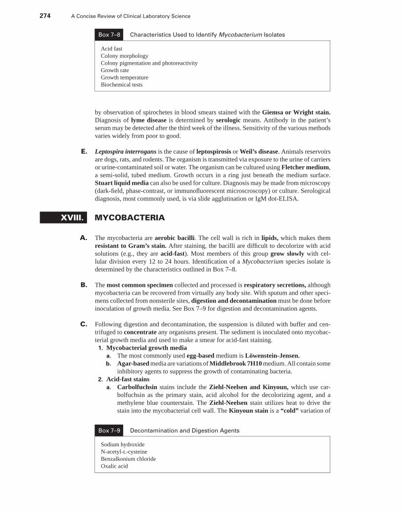
P1: OSO
LWBK192-07 LWBK192-Hubbard November 27, 2008 7:48
274 A Concise Review of Clinical Laboratory Science
Box 7–8 Characteristics Used to Identify Mycobacterium Isolates
Acid fast
Colony morphology
Colony pigmentation and photoreactivity
Growth rate
Growth temperature
Biochemical tests
by observation of spirochetes in blood smears stained with the Giemsa or Wright stain.Diagnosis of lyme disease is determined by serologic means. Antibody in the patient’sserum may be detected after the third week of the illness. Sensitivity of the various methodsvaries widely from poor to good.
E. Leptospira interrogans is the cause of leptospirosis or Weil’s disease. Animals reservoirsare dogs, rats, and rodents. The organism is transmitted via exposure to the urine of carriersor urine-contaminated soil or water. The organism can be cultured using Fletcher medium,a semi-solid, tubed medium. Growth occurs in a ring just beneath the medium surface.Stuart liquid media can also be used for culture. Diagnosis may be made from microscopy(dark-field, phase-contrast, or immunofluorescent microscroscopy) or culture. Serologicaldiagnosis, most commonly used, is via slide agglutination or IgM dot-ELISA.
XVIII. MYCOBACTERIA
A. The mycobacteria are aerobic bacilli. The cell wall is rich in lipids, which makes themresistant to Gram’s stain. After staining, the bacilli are difficult to decolorize with acidsolutions (e.g., they are acid-fast). Most members of this group grow slowly with cel-lular division every 12 to 24 hours. Identification of a Mycobacterium species isolate isdetermined by the characteristics outlined in Box 7–8.
B. The most common specimen collected and processed is respiratory secretions, althoughmycobacteria can be recovered from virtually any body site. With sputum and other speci-mens collected from nonsterile sites, digestion and decontamination must be done beforeinoculation of growth media. See Box 7–9 for digestion and decontamination agents.
C. Following digestion and decontamination, the suspension is diluted with buffer and cen-trifuged to concentrate any organisms present. The sediment is inoculated onto mycobac-terial growth media and used to make a smear for acid-fast staining.1. Mycobacterial growth media
a. The most commonly used egg-based medium is Lowenstein-Jensen.b. Agar-based media are variations of Middlebrook 7H10 medium. All contain some
inhibitory agents to suppress the growth of contaminating bacteria.2. Acid-fast stains
a. Carbolfuchsin stains include the Ziehl-Neelsen and Kinyoun, which use car-bolfuchsin as the primary stain, acid alcohol for the decolorizing agent, and amethylene blue counterstain. The Ziehl-Neelsen stain utilizes heat to drive thestain into the mycobacterial cell wall. The Kinyoun stain is a “cold” variation of
Box 7–9 Decontamination and Digestion Agents
Sodium hydroxide
N-acetyl-l-cysteine
Benzalkonium chloride
Oxalic acid
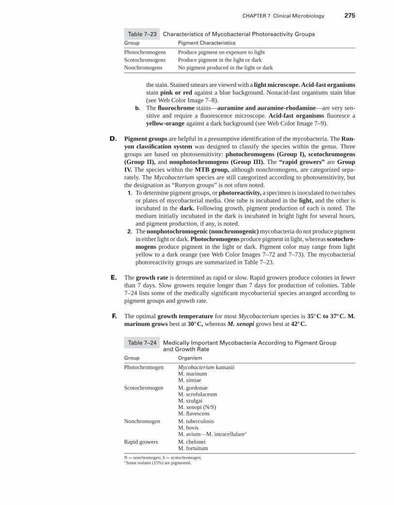
P1: OSO
LWBK192-07 LWBK192-Hubbard November 27, 2008 7:48
CHAPTER 7 Clinical Microbiology 275
Table 7–23 Characteristics of Mycobacterial Photoreactivity Groups
Group Pigment Characteristics
Photochromogens Produce pigment on exposure to light
Scotochromogens Produce pigment in the light or dark
Nonchromogens No pigment produced in the light or dark
the stain. Stained smears are viewed with a light microscope. Acid-fast organismsstain pink or red against a blue background. Nonacid-fast organisms stain blue(see Web Color Image 7–8).
b. The fluorochrome stains—auramine and auramine-rhodamine—are very sen-sitive and require a fluorescence microscope. Acid-fast organisms fluoresce ayellow-orange against a dark background (see Web Color Image 7–9).
D. Pigment groups are helpful in a presumptive identification of the mycobacteria. The Run-yon classification system was designed to classify the species within the genus. Threegroups are based on photosensitivity: photochromogens (Group I), scotochromogens(Group II), and nonphotochromogens (Group III). The “rapid growers” are GroupIV. The species within the MTB group, although nonchromogens, are categorized sepa-rately. The Mycobacterium species are still categorized according to photosensitivity, butthe designation as “Runyon groups” is not often noted.
1. To determine pigment groups, or photoreactivity, a specimen is inoculated to two tubesor plates of mycobacterial media. One tube is incubated in the light, and the other isincubated in the dark. Following growth, pigment production of each is noted. Themedium initially incubated in the dark is incubated in bright light for several hours,and pigment production, if any, is noted.
2. The nonphotochromogenic (nonchromogenic) mycobacteria do not produce pigmentin either light or dark. Photochromogens produce pigment in light, whereas scotochro-mogens produce pigment in the light or dark. Pigment color may range from lightyellow to a dark orange (see Web Color Images 7–72 and 7–73). The mycobacterialphotoreactivity groups are summarized in Table 7–23.
E. The growth rate is determined as rapid or slow. Rapid growers produce colonies in fewerthan 7 days. Slow growers require longer than 7 days for production of colonies. Table7–24 lists some of the medically significant mycobacterial species arranged according topigment groups and growth rate.
F. The optimal growth temperature for most Mycobacterium species is 35◦C to 37◦C. M.marinum grows best at 30◦C, whereas M. xenopi grows best at 42◦C.
Table 7–24 Medically Important Mycobacteria According to Pigment Groupand Growth Rate
Group Organism
Photochromogen Mycobacterium kansasiiM. marinumM. simiae
Scotochromogen M. gordonaeM. scrofulaceumM. szulgaiM. xenopi (N/S)M. flavescens
Nonchromogen M. tuberculosisM. bovisM. avium—M. intracellulare∗
Rapid growers M. cheloneiM. fortuitum
N = nonchromogen; S = scotochromogen;∗Some isolates (15%) are pigmented.
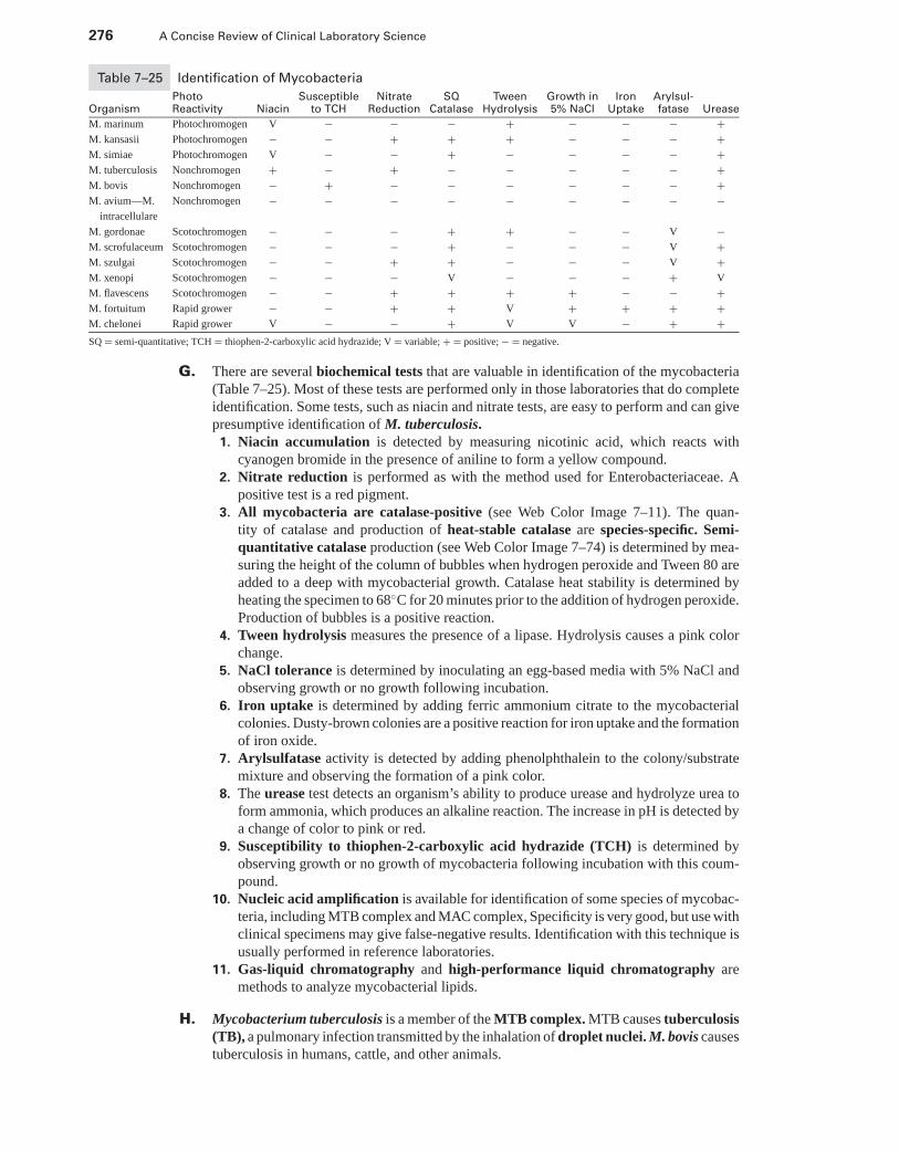
P1: OSO
LWBK192-07 LWBK192-Hubbard November 27, 2008 7:48
276 A Concise Review of Clinical Laboratory Science
Table 7–25 Identification of Mycobacteria
Photo Susceptible Nitrate SQ Tween Growth in Iron Arylsul-Organism Reactivity Niacin to TCH Reduction Catalase Hydrolysis 5% NaCl Uptake fatase Urease
M. marinum Photochromogen V − − − + − − − +M. kansasii Photochromogen − − + + + − − − +M. simiae Photochromogen V − − + − − − − +M. tuberculosis Nonchromogen + − + − − − − − +M. bovis Nonchromogen − + − − − − − − +M. avium—M. Nonchromogen − − − − − − − − −
intracellulare
M. gordonae Scotochromogen − − − + + − − V −M. scrofulaceum Scotochromogen − − − + − − − V +M. szulgai Scotochromogen − − + + − − − V +M. xenopi Scotochromogen − − − V − − − + V
M. flavescens Scotochromogen − − + + + + − − +M. fortuitum Rapid grower − − + + V + + + +M. chelonei Rapid grower V − − + V V − + +SQ = semi-quantitative; TCH = thiophen-2-carboxylic acid hydrazide; V = variable; + = positive; − = negative.
G. There are several biochemical tests that are valuable in identification of the mycobacteria(Table 7–25). Most of these tests are performed only in those laboratories that do completeidentification. Some tests, such as niacin and nitrate tests, are easy to perform and can givepresumptive identification of M. tuberculosis.
1. Niacin accumulation is detected by measuring nicotinic acid, which reacts withcyanogen bromide in the presence of aniline to form a yellow compound.
2. Nitrate reduction is performed as with the method used for Enterobacteriaceae. Apositive test is a red pigment.
3. All mycobacteria are catalase-positive (see Web Color Image 7–11). The quan-tity of catalase and production of heat-stable catalase are species-specific. Semi-quantitative catalase production (see Web Color Image 7–74) is determined by mea-suring the height of the column of bubbles when hydrogen peroxide and Tween 80 areadded to a deep with mycobacterial growth. Catalase heat stability is determined byheating the specimen to 68◦C for 20 minutes prior to the addition of hydrogen peroxide.Production of bubbles is a positive reaction.
4. Tween hydrolysis measures the presence of a lipase. Hydrolysis causes a pink colorchange.
5. NaCl tolerance is determined by inoculating an egg-based media with 5% NaCl andobserving growth or no growth following incubation.
6. Iron uptake is determined by adding ferric ammonium citrate to the mycobacterialcolonies. Dusty-brown colonies are a positive reaction for iron uptake and the formationof iron oxide.
7. Arylsulfatase activity is detected by adding phenolphthalein to the colony/substratemixture and observing the formation of a pink color.
8. The urease test detects an organism’s ability to produce urease and hydrolyze urea toform ammonia, which produces an alkaline reaction. The increase in pH is detected bya change of color to pink or red.
9. Susceptibility to thiophen-2-carboxylic acid hydrazide (TCH) is determined byobserving growth or no growth of mycobacteria following incubation with this coum-pound.
10. Nucleic acid amplification is available for identification of some species of mycobac-teria, including MTB complex and MAC complex, Specificity is very good, but use withclinical specimens may give false-negative results. Identification with this technique isusually performed in reference laboratories.
11. Gas-liquid chromatography and high-performance liquid chromatography aremethods to analyze mycobacterial lipids.
H. Mycobacterium tuberculosis is a member of the MTB complex. MTB causes tuberculosis(TB), a pulmonary infection transmitted by the inhalation of droplet nuclei. M. bovis causestuberculosis in humans, cattle, and other animals.
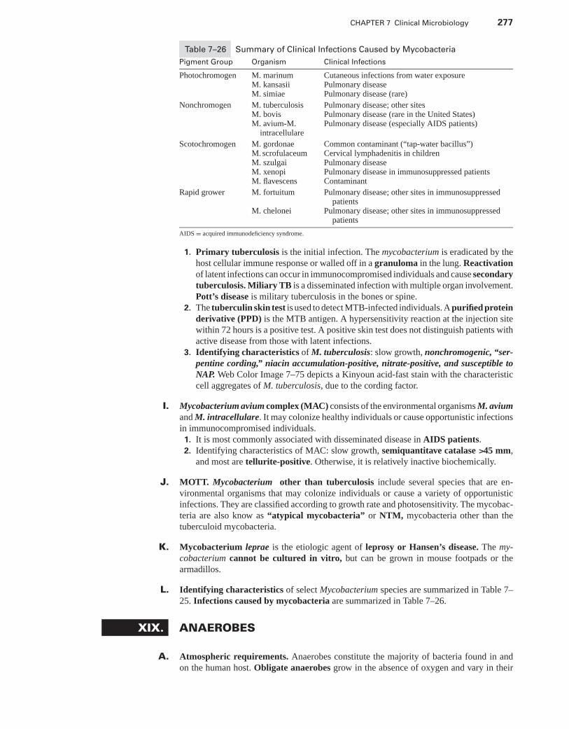
P1: OSO
LWBK192-07 LWBK192-Hubbard November 27, 2008 7:48
CHAPTER 7 Clinical Microbiology 277
Table 7–26 Summary of Clinical Infections Caused by Mycobacteria
Pigment Group Organism Clinical Infections
Photochromogen M. marinum Cutaneous infections from water exposureM. kansasii Pulmonary diseaseM. simiae Pulmonary disease (rare)
Nonchromogen M. tuberculosis Pulmonary disease; other sitesM. bovis Pulmonary disease (rare in the United States)M. avium-M.
intracellularePulmonary disease (especially AIDS patients)
Scotochromogen M. gordonae Common contaminant (“tap-water bacillus”)M. scrofulaceum Cervical lymphadenitis in childrenM. szulgai Pulmonary diseaseM. xenopi Pulmonary disease in immunosuppressed patientsM. flavescens Contaminant
Rapid grower M. fortuitum Pulmonary disease; other sites in immunosuppressedpatients
M. chelonei Pulmonary disease; other sites in immunosuppressedpatients
AIDS = acquired immunodeficiency syndrome.
1. Primary tuberculosis is the initial infection. The mycobacterium is eradicated by thehost cellular immune response or walled off in a granuloma in the lung. Reactivationof latent infections can occur in immunocompromised individuals and cause secondarytuberculosis. Miliary TB is a disseminated infection with multiple organ involvement.Pott’s disease is military tuberculosis in the bones or spine.
2. The tuberculin skin test is used to detect MTB-infected individuals. A purified proteinderivative (PPD) is the MTB antigen. A hypersensitivity reaction at the injection sitewithin 72 hours is a positive test. A positive skin test does not distinguish patients withactive disease from those with latent infections.
3. Identifying characteristics of M. tuberculosis: slow growth, nonchromogenic, “ser-pentine cording,” niacin accumulation-positive, nitrate-positive, and susceptible toNAP. Web Color Image 7–75 depicts a Kinyoun acid-fast stain with the characteristiccell aggregates of M. tuberculosis, due to the cording factor.
I. Mycobacterium avium complex (MAC) consists of the environmental organisms M. aviumand M. intracellulare. It may colonize healthy individuals or cause opportunistic infectionsin immunocompromised individuals.
1. It is most commonly associated with disseminated disease in AIDS patients.2. Identifying characteristics of MAC: slow growth, semiquantitave catalase >45 mm,
and most are tellurite-positive. Otherwise, it is relatively inactive biochemically.
J. MOTT. Mycobacterium other than tuberculosis include several species that are en-vironmental organisms that may colonize individuals or cause a variety of opportunisticinfections. They are classified according to growth rate and photosensitivity. The mycobac-teria are also know as “atypical mycobacteria” or NTM, mycobacteria other than thetuberculoid mycobacteria.
K. Mycobacterium leprae is the etiologic agent of leprosy or Hansen’s disease. The my-cobacterium cannot be cultured in vitro, but can be grown in mouse footpads or thearmadillos.
L. Identifying characteristics of select Mycobacterium species are summarized in Table 7–25. Infections caused by mycobacteria are summarized in Table 7–26.
XIX. ANAEROBES
A. Atmospheric requirements. Anaerobes constitute the majority of bacteria found in andon the human host. Obligate anaerobes grow in the absence of oxygen and vary in their
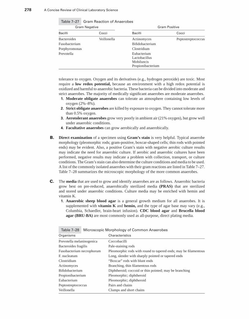
P1: OSO
LWBK192-07 LWBK192-Hubbard November 27, 2008 7:48
278 A Concise Review of Clinical Laboratory Science
Table 7–27 Gram Reaction of Anaerobes
Gram Negative Gram Positive
Bacilli Cocci Bacilli Cocci
Bacteroides Veillonella Actinomyces Peptostreptococcus
Fusobacterium Bifidobacterium
Porphyromonas Clostridium
Prevotella EubacteriumLactobacillusMobiluncisPropionibacterium
tolerance to oxygen. Oxygen and its derivatives (e.g., hydrogen peroxide) are toxic. Mostrequire a low redox potential, because an environment with a high redox potential isoxidized and harmful to anaerobic bacteria. These bacteria can be divided into moderate andstrict anaerobes. The majority of medically significant anaerobes are moderate anaerobes.1. Moderate obligate anaerobes can tolerate an atmosphere containing low levels of
oxygen (2%–8%).2. Strict obligate anaerobes are killed by exposure to oxygen. They cannot tolerate more
than 0.5% oxygen.3. Aerotolerant anaerobes grow very poorly in ambient air (21% oxygen), but grow well
under anaerobic conditions.4. Facultative anaerobes can grow aerobically and anaerobically.
B. Direct examination of a specimen using Gram’s stain is very helpful. Typical anaerobemorphology (pleomorphic rods; gram-positive, boxcar-shaped cells; thin rods with pointedends) may be evident. Also, a positive Gram’s stain with negative aerobic culture resultsmay indicate the need for anaerobic culture. If aerobic and anaerobic cultures have beenperformed, negative results may indicate a problem with collection, transport, or cultureconditions. The Gram’s stain can also determine the culture conditions and media to be used.A list of the commonly isolated anaerobes with their gram reactions are listed in Table 7–27.Table 7–28 summarizes the microscopic morphology of the more common anaerobes.
C. The media that are used to grow and identify anaerobes are as follows. Anaerobic bacteriagrow best on pre-reduced, anaerobically sterilized media (PRAS) that are sterilizedand stored under anaerobic conditions. Culture media may be enriched with hemin andvitamin K.
1. Anaerobic sheep blood agar is a general growth medium for all anaerobes. It issupplemented with vitamin K and hemin, and the type of agar base may vary (e.g.,Columbia, Schaedler, brain-heart infusion). CDC blood agar and Brucella bloodagar (BRU-BA) are most commonly used as all-purpose, direct plating media.
Table 7–28 Microscopic Morphology of Common Anaerobes
Organisms Characteristics
Prevotella melaninogenica Coccobacilli
Bacteroides fragilis Pale-staining rods
Fusobacterium necrophorum Pleomorphic rods with round to tapered ends; may be filamentous
F. nucleatum Long, slender with sharply pointed or tapered ends
Clostridium “Boxcar” rods with blunt ends
Actinomyces Branching, thin filamentous rods
Bifidobacterium Diphtheroid; coccoid or thin pointed; may be branching
Propionibacterium Pleomorphic; diphtheroid
Eubacterium Pleomorphic; diphtheroid
Peptostreptococcus Pairs and chains
Veillonella Clumps and short chains
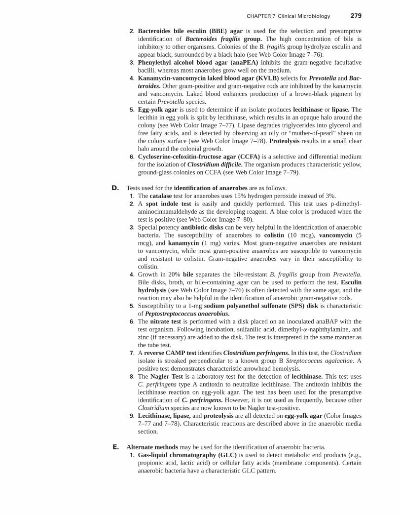
P1: OSO
LWBK192-07 LWBK192-Hubbard November 27, 2008 7:48
CHAPTER 7 Clinical Microbiology 279
2. Bacteroides bile esculin (BBE) agar is used for the selection and presumptiveidentification of Bacteroides fragilis group. The high concentration of bile isinhibitory to other organisms. Colonies of the B. fragilis group hydrolyze esculin andappear black, surrounded by a black halo (see Web Color Image 7–76).
3. Phenylethyl alcohol blood agar (anaPEA) inhibits the gram-negative facultativebacilli, whereas most anaerobes grow well on the medium.
4. Kanamycin-vancomycin laked blood agar (KVLB) selects for Prevotella and Bac-teroides. Other gram-positive and gram-negative rods are inhibited by the kanamycinand vancomycin. Laked blood enhances production of a brown-black pigment bycertain Prevotella species.
5. Egg-yolk agar is used to determine if an isolate produces lecithinase or lipase. Thelecithin in egg yolk is split by lecithinase, which results in an opaque halo around thecolony (see Web Color Image 7–77). Lipase degrades triglycerides into glycerol andfree fatty acids, and is detected by observing an oily or “mother-of-pearl” sheen onthe colony surface (see Web Color Image 7–78). Proteolysis results in a small clearhalo around the colonial growth.
6. Cycloserine-cefoxitin-fructose agar (CCFA) is a selective and differential mediumfor the isolation of Clostridium difficile. The organism produces characteristic yellow,ground-glass colonies on CCFA (see Web Color Image 7–79).
D. Tests used for the identification of anaerobes are as follows.1. The catalase test for anaerobes uses 15% hydrogen peroxide instead of 3%.2. A spot indole test is easily and quickly performed. This test uses p-dimethyl-
aminocinnamaldehyde as the developing reagent. A blue color is produced when thetest is positive (see Web Color Image 7–80).
3. Special potency antibiotic disks can be very helpful in the identification of anaerobicbacteria. The susceptibility of anaerobes to colistin (10 mcg), vancomycin (5mcg), and kanamycin (1 mg) varies. Most gram-negative anaerobes are resistantto vancomycin, while most gram-positive anaerobes are susceptible to vancomycinand resistant to colistin. Gram-negative anaerobes vary in their susceptibility tocolistin.
4. Growth in 20% bile separates the bile-resistant B. fragilis group from Prevotella.Bile disks, broth, or bile-containing agar can be used to perform the test. Esculinhydrolysis (see Web Color Image 7–76) is often detected with the same agar, and thereaction may also be helpful in the identification of anaerobic gram-negative rods.
5. Susceptibility to a 1-mg sodium polyanethol sulfonate (SPS) disk is characteristicof Peptostreptococcus anaerobius.
6. The nitrate test is performed with a disk placed on an inoculated anaBAP with thetest organism. Following incubation, sulfanilic acid, dimethyl-α-naphthylamine, andzinc (if necessary) are added to the disk. The test is interpreted in the same manner asthe tube test.
7. A reverse CAMP test identifies Clostridium perfringens. In this test, the Clostridiumisolate is streaked perpendicular to a known group B Streptococcus agalactiae. Apositive test demonstrates characteristic arrowhead hemolysis.
8. The Nagler Test is a laboratory test for the detection of lecithinase. This test usesC. perfringens type A antitoxin to neutralize lecithinase. The antitoxin inhibits thelecithinase reaction on egg-yolk agar. The test has been used for the presumptiveidentification of C. perfringens. However, it is not used as frequently, because otherClostridium species are now known to be Nagler test-positive.
9. Lecithinase, lipase, and proteolysis are all detected on egg-yolk agar (Color Images7–77 and 7–78). Characteristic reactions are described above in the anaerobic mediasection.
E. Alternate methods may be used for the identification of anaerobic bacteria.1. Gas-liquid chromatography (GLC) is used to detect metabolic end products (e.g.,
propionic acid, lactic acid) or cellular fatty acids (membrane components). Certainanaerobic bacteria have a characteristic GLC pattern.
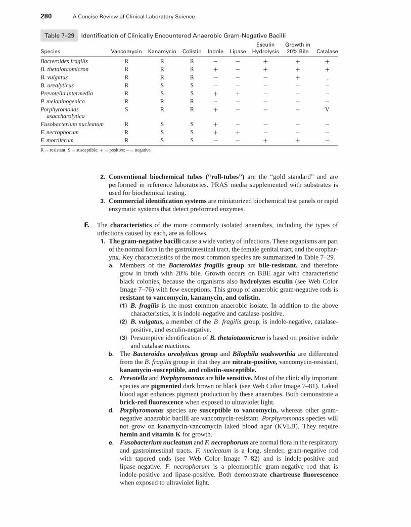
P1: OSO
LWBK192-07 LWBK192-Hubbard November 27, 2008 7:48
280 A Concise Review of Clinical Laboratory Science
Table 7–29 Identification of Clinically Encountered Anaerobic Gram-Negative Bacilli
Esculin Growth in
Species Vancomycin Kanamycin Colistin Indole Lipase Hydrolysis 20% Bile Catalase
Bacteroides fragilis R R R − − + + +B. thetaiotaomicron R R R + − + + +B. vulgatus R R R − − − +B. urealyticus R S S − − − − −Prevotella intermedia R S S + + − − −P. melaninogenica R R R − − − − −Porphyromonas
asaccharolyticaS R R + − − − V
Fusobacterium nucleatum R S S + − − − −F. necrophorum R S S + + − − −F. mortiferum R S S − − + + −R = resistant; S = susceptible; + = positive; – = negative.
2. Conventional biochemical tubes (“roll-tubes”) are the “gold standard” and areperformed in reference laboratories. PRAS media supplemented with substrates isused for biochemical testing.
3. Commercial identification systems are miniaturized biochemical test panels or rapidenzymatic systems that detect preformed enzymes.
F. The characteristics of the more commonly isolated anaerobes, including the types ofinfections caused by each, are as follows.1. The gram-negative bacilli cause a wide variety of infections. These organisms are part
of the normal flora in the gastrointestinal tract, the female genital tract, and the orophar-ynx. Key characteristics of the most common species are summarized in Table 7–29.a. Members of the Bacteroides fragilis group are bile-resistant, and therefore
grow in broth with 20% bile. Growth occurs on BBE agar with characteristicblack colonies, because the organisms also hydrolyzes esculin (see Web ColorImage 7–76) with few exceptions. This group of anaerobic gram-negative rods isresistant to vancomycin, kanamycin, and colistin.(1) B. fragilis is the most common anaerobic isolate. In addition to the above
characteristics, it is indole-negative and catalase-positive.(2) B. vulgatus, a member of the B. fragilis group, is indole-negative, catalase-
positive, and esculin-negative.(3) Presumptive identification of B. thetaiotaomicron is based on positive indole
and catalase reactions.b. The Bacteroides ureolyticus group and Bilophila wadsworthia are differented
from the B. fragilis group in that they are nitrate-positive, vancomycin-resistant,kanamycin-susceptible, and colistin-susceptible.
c. Prevotella and Porphyromonas are bile sensitive. Most of the clinically importantspecies are pigmented dark brown or black (see Web Color Image 7–81). Lakedblood agar enhances pigment production by these anaerobes. Both demonstrate abrick-red fluorescence when exposed to ultraviolet light.
d. Porphyromonas species are susceptible to vancomycin, whereas other gram-negative anaerobic bacilli are vancomycin-resistant. Porphyromonas species willnot grow on kanamycin-vancomycin laked blood agar (KVLB). They requirehemin and vitamin K for growth.
e. Fusobacterium nucleatum and F. necrophorum are normal flora in the respiratoryand gastrointestinal tracts. F. nucleatum is a long, slender, gram-negative rodwith tapered ends (see Web Color Image 7–82) and is indole-positive andlipase-negative. F. necrophorum is a pleomorphic gram-negative rod that isindole-positive and lipase-positive. Both demonstrate chartreuse fluorescencewhen exposed to ultraviolet light.
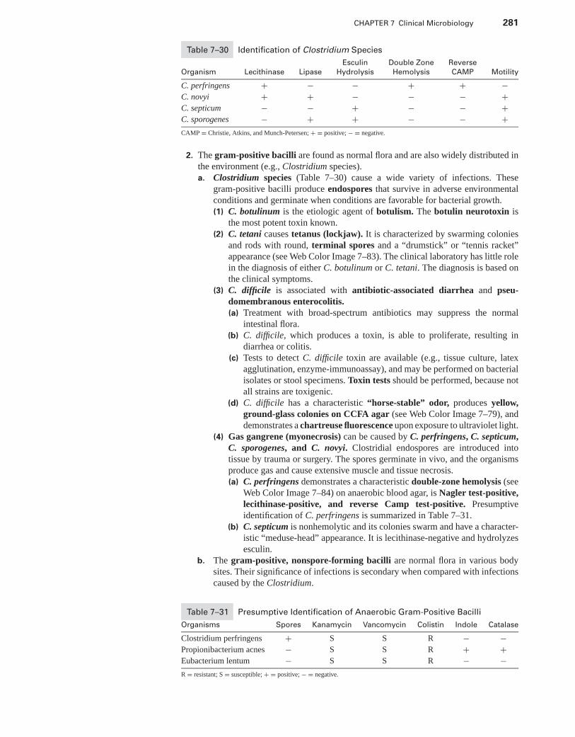
P1: OSO
LWBK192-07 LWBK192-Hubbard November 27, 2008 7:48
CHAPTER 7 Clinical Microbiology 281
Table 7–30 Identification of Clostridium Species
Esculin Double Zone Reverse
Organism Lecithinase Lipase Hydrolysis Hemolysis CAMP Motility
C. perfringens + − − + + −C. novyi + + − − − +C. septicum − − + − − +C. sporogenes − + + − − +CAMP = Christie, Atkins, and Munch-Petersen; + = positive; − = negative.
2. The gram-positive bacilli are found as normal flora and are also widely distributed inthe environment (e.g., Clostridium species).a. Clostridium species (Table 7–30) cause a wide variety of infections. These
gram-positive bacilli produce endospores that survive in adverse environmentalconditions and germinate when conditions are favorable for bacterial growth.(1) C. botulinum is the etiologic agent of botulism. The botulin neurotoxin is
the most potent toxin known.(2) C. tetani causes tetanus (lockjaw). It is characterized by swarming colonies
and rods with round, terminal spores and a “drumstick” or “tennis racket”appearance (see Web Color Image 7–83). The clinical laboratory has little rolein the diagnosis of either C. botulinum or C. tetani. The diagnosis is based onthe clinical symptoms.
(3) C. difficile is associated with antibiotic-associated diarrhea and pseu-domembranous enterocolitis.(a) Treatment with broad-spectrum antibiotics may suppress the normal
intestinal flora.(b) C. difficile, which produces a toxin, is able to proliferate, resulting in
diarrhea or colitis.(c) Tests to detect C. difficile toxin are available (e.g., tissue culture, latex
agglutination, enzyme-immunoassay), and may be performed on bacterialisolates or stool specimens. Toxin tests should be performed, because notall strains are toxigenic.
(d) C. difficile has a characteristic “horse-stable” odor, produces yellow,ground-glass colonies on CCFA agar (see Web Color Image 7–79), anddemonstrates a chartreuse fluorescence upon exposure to ultraviolet light.
(4) Gas gangrene (myonecrosis) can be caused by C. perfringens, C. septicum,C. sporogenes, and C. novyi. Clostridial endospores are introduced intotissue by trauma or surgery. The spores germinate in vivo, and the organismsproduce gas and cause extensive muscle and tissue necrosis.(a) C. perfringens demonstrates a characteristic double-zone hemolysis (see
Web Color Image 7–84) on anaerobic blood agar, is Nagler test-positive,lecithinase-positive, and reverse Camp test-positive. Presumptiveidentification of C. perfringens is summarized in Table 7–31.
(b) C. septicum is nonhemolytic and its colonies swarm and have a character-istic “meduse-head” appearance. It is lecithinase-negative and hydrolyzesesculin.
b. The gram-positive, nonspore-forming bacilli are normal flora in various bodysites. Their significance of infections is secondary when compared with infectionscaused by the Clostridium.
Table 7–31 Presumptive Identification of Anaerobic Gram-Positive Bacilli
Organisms Spores Kanamycin Vancomycin Colistin Indole Catalase
Clostridium perfringens + S S R − −Propionibacterium acnes − S S R + +Eubacterium lentum − S S R − −R = resistant; S = susceptible; + = positive; − = negative.
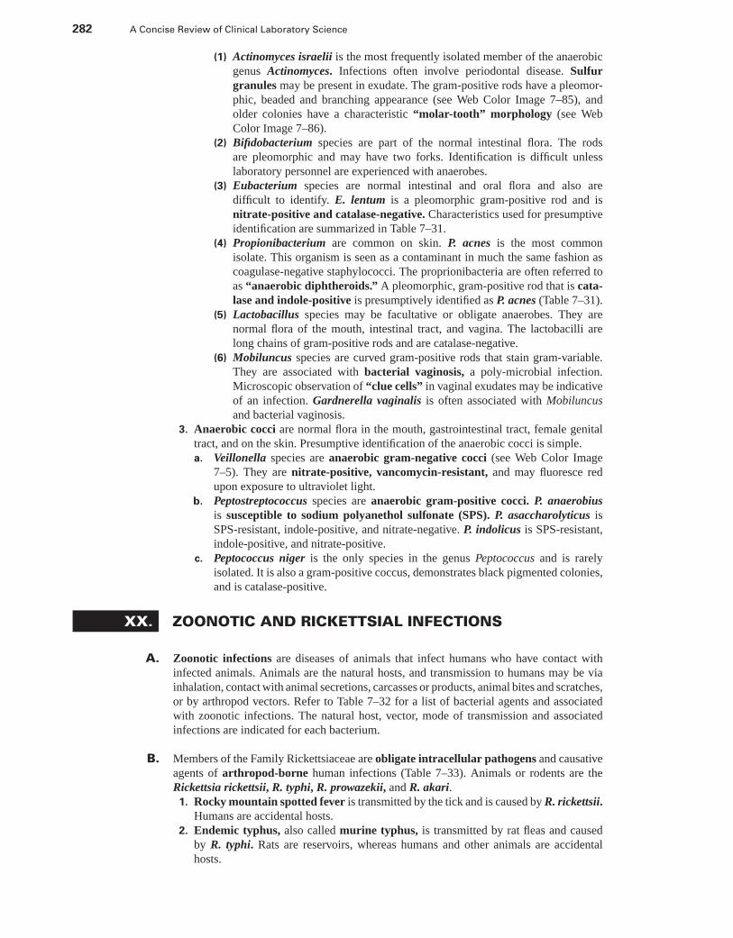
P1: OSO
LWBK192-07 LWBK192-Hubbard November 27, 2008 7:48
282 A Concise Review of Clinical Laboratory Science
(1) Actinomyces israelii is the most frequently isolated member of the anaerobicgenus Actinomyces. Infections often involve periodontal disease. Sulfurgranules may be present in exudate. The gram-positive rods have a pleomor-phic, beaded and branching appearance (see Web Color Image 7–85), andolder colonies have a characteristic “molar-tooth” morphology (see WebColor Image 7–86).
(2) Bifidobacterium species are part of the normal intestinal flora. The rodsare pleomorphic and may have two forks. Identification is difficult unlesslaboratory personnel are experienced with anaerobes.
(3) Eubacterium species are normal intestinal and oral flora and also aredifficult to identify. E. lentum is a pleomorphic gram-positive rod and isnitrate-positive and catalase-negative. Characteristics used for presumptiveidentification are summarized in Table 7–31.
(4) Propionibacterium are common on skin. P. acnes is the most commonisolate. This organism is seen as a contaminant in much the same fashion ascoagulase-negative staphylococci. The proprionibacteria are often referred toas “anaerobic diphtheroids.” A pleomorphic, gram-positive rod that is cata-lase and indole-positive is presumptively identified as P. acnes (Table 7–31).
(5) Lactobacillus species may be facultative or obligate anaerobes. They arenormal flora of the mouth, intestinal tract, and vagina. The lactobacilli arelong chains of gram-positive rods and are catalase-negative.
(6) Mobiluncus species are curved gram-positive rods that stain gram-variable.They are associated with bacterial vaginosis, a poly-microbial infection.Microscopic observation of “clue cells” in vaginal exudates may be indicativeof an infection. Gardnerella vaginalis is often associated with Mobiluncusand bacterial vaginosis.
3. Anaerobic cocci are normal flora in the mouth, gastrointestinal tract, female genitaltract, and on the skin. Presumptive identification of the anaerobic cocci is simple.a. Veillonella species are anaerobic gram-negative cocci (see Web Color Image
7–5). They are nitrate-positive, vancomycin-resistant, and may fluoresce redupon exposure to ultraviolet light.
b. Peptostreptococcus species are anaerobic gram-positive cocci. P. anaerobiusis susceptible to sodium polyanethol sulfonate (SPS). P. asaccharolyticus isSPS-resistant, indole-positive, and nitrate-negative. P. indolicus is SPS-resistant,indole-positive, and nitrate-positive.
c. Peptococcus niger is the only species in the genus Peptococcus and is rarelyisolated. It is also a gram-positive coccus, demonstrates black pigmented colonies,and is catalase-positive.
XX. ZOONOTIC AND RICKETTSIAL INFECTIONS
A. Zoonotic infections are diseases of animals that infect humans who have contact withinfected animals. Animals are the natural hosts, and transmission to humans may be viainhalation, contact with animal secretions, carcasses or products, animal bites and scratches,or by arthropod vectors. Refer to Table 7–32 for a list of bacterial agents and associatedwith zoonotic infections. The natural host, vector, mode of transmission and associatedinfections are indicated for each bacterium.
B. Members of the Family Rickettsiaceae are obligate intracellular pathogens and causativeagents of arthropod-borne human infections (Table 7–33). Animals or rodents are theRickettsia rickettsii, R. typhi, R. prowazekii, and R. akari.
1. Rocky mountain spotted fever is transmitted by the tick and is caused by R. rickettsii.Humans are accidental hosts.
2. Endemic typhus, also called murine typhus, is transmitted by rat fleas and causedby R. typhi. Rats are reservoirs, whereas humans and other animals are accidentalhosts.
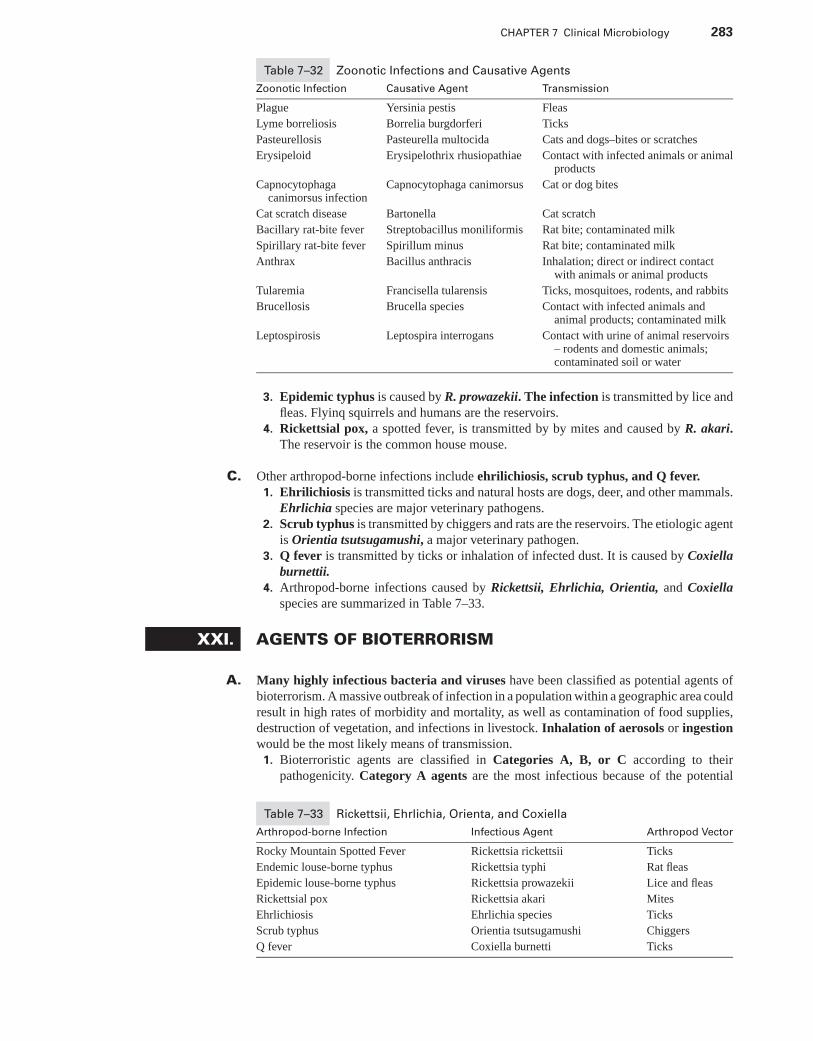
P1: OSO
LWBK192-07 LWBK192-Hubbard November 27, 2008 7:48
CHAPTER 7 Clinical Microbiology 283
Table 7–32 Zoonotic Infections and Causative Agents
Zoonotic Infection Causative Agent Transmission
Plague Yersinia pestis Fleas
Lyme borreliosis Borrelia burgdorferi Ticks
Pasteurellosis Pasteurella multocida Cats and dogs–bites or scratches
Erysipeloid Erysipelothrix rhusiopathiae Contact with infected animals or animalproducts
Capnocytophagacanimorsus infection
Capnocytophaga canimorsus Cat or dog bites
Cat scratch disease Bartonella Cat scratch
Bacillary rat-bite fever Streptobacillus moniliformis Rat bite; contaminated milk
Spirillary rat-bite fever Spirillum minus Rat bite; contaminated milk
Anthrax Bacillus anthracis Inhalation; direct or indirect contactwith animals or animal products
Tularemia Francisella tularensis Ticks, mosquitoes, rodents, and rabbits
Brucellosis Brucella species Contact with infected animals andanimal products; contaminated milk
Leptospirosis Leptospira interrogans Contact with urine of animal reservoirs– rodents and domestic animals;contaminated soil or water
3. Epidemic typhus is caused by R. prowazekii. The infection is transmitted by lice andfleas. Flyinq squirrels and humans are the reservoirs.
4. Rickettsial pox, a spotted fever, is transmitted by by mites and caused by R. akari.The reservoir is the common house mouse.
C. Other arthropod-borne infections include ehrilichiosis, scrub typhus, and Q fever.1. Ehrilichiosis is transmitted ticks and natural hosts are dogs, deer, and other mammals.
Ehrlichia species are major veterinary pathogens.2. Scrub typhus is transmitted by chiggers and rats are the reservoirs. The etiologic agent
is Orientia tsutsugamushi, a major veterinary pathogen.3. Q fever is transmitted by ticks or inhalation of infected dust. It is caused by Coxiella
burnettii.4. Arthropod-borne infections caused by Rickettsii, Ehrlichia, Orientia, and Coxiella
species are summarized in Table 7–33.
XXI. AGENTS OF BIOTERRORISM
A. Many highly infectious bacteria and viruses have been classified as potential agents ofbioterrorism. A massive outbreak of infection in a population within a geographic area couldresult in high rates of morbidity and mortality, as well as contamination of food supplies,destruction of vegetation, and infections in livestock. Inhalation of aerosols or ingestionwould be the most likely means of transmission.
1. Bioterroristic agents are classified in Categories A, B, or C according to theirpathogenicity. Category A agents are the most infectious because of the potential
Table 7–33 Rickettsii, Ehrlichia, Orienta, and Coxiella
Arthropod-borne Infection Infectious Agent Arthropod Vector
Rocky Mountain Spotted Fever Rickettsia rickettsii Ticks
Endemic louse-borne typhus Rickettsia typhi Rat fleas
Epidemic louse-borne typhus Rickettsia prowazekii Lice and fleas
Rickettsial pox Rickettsia akari Mites
Ehrlichiosis Ehrlichia species Ticks
Scrub typhus Orientia tsutsugamushi Chiggers
Q fever Coxiella burnetti Ticks
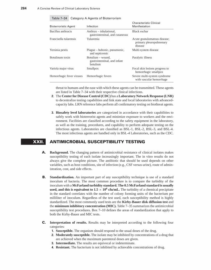
P1: OSO
LWBK192-07 LWBK192-Hubbard November 27, 2008 7:48
284 A Concise Review of Clinical Laboratory Science
Table 7–34 Category A Agents of Bioterrorism
Characteristic Clinical
Bioterroristic Agent Infection Manifestation
Bacillus anthracis Anthrax – inhalational,gastrointestinal, and cutaneous
Black eschar
Francisella tularensis Tularemia Acute granulomatous disease;primary pleuropulmonarydisease
Yersinia pestis Plague – bubonic, pneumonic,and septicemic
Multi-system disease
Botulinum toxin Botulism – wound,gastrointestinal, and infantbotulism
Paralytic illness
Variola major virus Smallpox Focal skin lesions progress tohemorrhagic smallpox
Hemorrhagic fever viruses Hemorrhagic fevers Severe multi-system syndromewith vascular hemorrhage
threat to humans and the ease with which these agents can be transmitted. These agentsare listed in Table 7–34 with their respective clinical infections.
2. The Center for Disease Control (CDC) has a Laboratory Network Response (LNR)to decentralize testing capabilities and link state and local laboratories with advanced-capacity labs. LRN reference labs perform all confirmatory testing on biothreat agents.
3. Biosafety level laboratories are categorized in accordance with their capabilities tosafely work with bioterrorist agents and minimize exposure to workers and the envi-ronment. Facilities are classified according to the safety equipment in the laboratory,as well as the training, procedures, and capability to perform adequate testing on theinfectious agents. Laboratories are classified as BSL-1, BSL-2, BSL-3, and BSL-4.The most infectious agents are handled only in BSL-4 Laboratories, such as the CDC.
XXII. ANTIMICROBIAL SUSCEPTIBILITY TESTING
A. Background. The changing pattern of antimicrobial resistance of clinical isolates makessusceptibility testing of each isolate increasingly important. The in vitro results do notalways give the complete picture. The antibiotic that should be used depends on othervariables, such as host conditions, site of infection (e.g., CSF versus urine), route of admin-istration, cost, and side effects.
B. Standardization. An important part of any susceptibility technique is use of a standardinoculum of bacteria. The most common procedure is to compare the turbidity of theinoculum with a McFarland turbidity standard. The 0.5 McFarland standard is usuallyused, and this is equivalent to 1.5 × 10⁸ cfu/mL. The turbidity of a chemical precipitatein the standard correlates with the number of colony forming units of the bacterium permilliliter of inoculum. Regardless of the test used, each susceptibility method is highlystandardized. The most commonly used tests are the Kirby-Bauer disk diffusion test andthe minimum inhibitory concentration (MIC). Table 7–35 summarizes the antimicrobialsusceptibility test procedures. Box 7–10 defines the areas of standardization that apply toboth the Kirby-Bauer and MIC tests.
C. Interpretation of results. Results may be interpreted according to the following fourcategories:
1. Susceptible. The organism should respond to the usual doses of the drug.2. Moderately susceptible. The isolate may be inhibited by concentrations of a drug that
are achieved when the maximum parenteral doses are given.3. Intermediate. The results are equivocal or indeterminate.4. Resistant. The bacterium is not inhibited by achievable concentrations of drug.
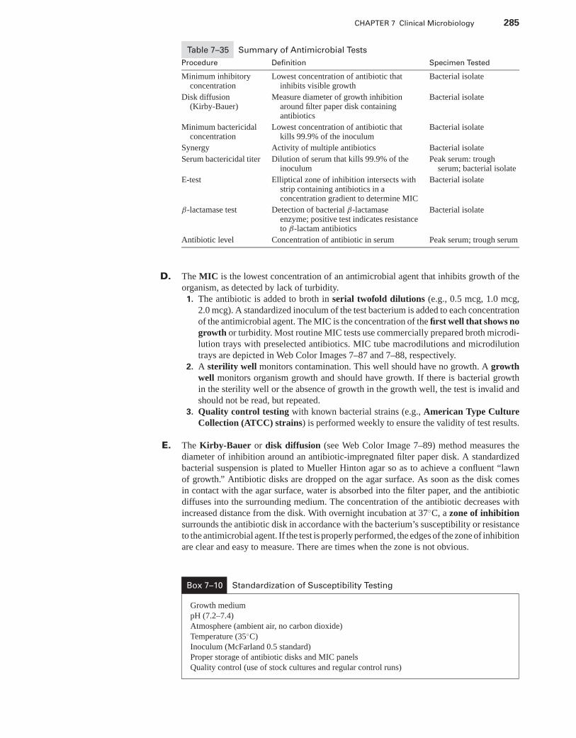
P1: OSO
LWBK192-07 LWBK192-Hubbard November 27, 2008 7:48
CHAPTER 7 Clinical Microbiology 285
Table 7–35 Summary of Antimicrobial Tests
Procedure Definition Specimen Tested
Minimum inhibitoryconcentration
Lowest concentration of antibiotic thatinhibits visible growth
Bacterial isolate
Disk diffusion(Kirby-Bauer)
Measure diameter of growth inhibitionaround filter paper disk containingantibiotics
Bacterial isolate
Minimum bactericidalconcentration
Lowest concentration of antibiotic thatkills 99.9% of the inoculum
Bacterial isolate
Synergy Activity of multiple antibiotics Bacterial isolate
Serum bactericidal titer Dilution of serum that kills 99.9% of theinoculum
Peak serum: troughserum; bacterial isolate
E-test Elliptical zone of inhibition intersects withstrip containing antibiotics in aconcentration gradient to determine MIC
Bacterial isolate
β-lactamase test Detection of bacterial β-lactamaseenzyme; positive test indicates resistanceto β-lactam antibiotics
Bacterial isolate
Antibiotic level Concentration of antibiotic in serum Peak serum; trough serum
D. The MIC is the lowest concentration of an antimicrobial agent that inhibits growth of theorganism, as detected by lack of turbidity.1. The antibiotic is added to broth in serial twofold dilutions (e.g., 0.5 mcg, 1.0 mcg,
2.0 mcg). A standardized inoculum of the test bacterium is added to each concentrationof the antimicrobial agent. The MIC is the concentration of the first well that shows nogrowth or turbidity. Most routine MIC tests use commercially prepared broth microdi-lution trays with preselected antibiotics. MIC tube macrodilutions and microdilutiontrays are depicted in Web Color Images 7–87 and 7–88, respectively.
2. A sterility well monitors contamination. This well should have no growth. A growthwell monitors organism growth and should have growth. If there is bacterial growthin the sterility well or the absence of growth in the growth well, the test is invalid andshould not be read, but repeated.
3. Quality control testing with known bacterial strains (e.g., American Type CultureCollection (ATCC) strains) is performed weekly to ensure the validity of test results.
E. The Kirby-Bauer or disk diffusion (see Web Color Image 7–89) method measures thediameter of inhibition around an antibiotic-impregnated filter paper disk. A standardizedbacterial suspension is plated to Mueller Hinton agar so as to achieve a confluent “lawnof growth.” Antibiotic disks are dropped on the agar surface. As soon as the disk comesin contact with the agar surface, water is absorbed into the filter paper, and the antibioticdiffuses into the surrounding medium. The concentration of the antibiotic decreases withincreased distance from the disk. With overnight incubation at 37◦C, a zone of inhibitionsurrounds the antibiotic disk in accordance with the bacterium’s susceptibility or resistanceto the antimicrobial agent. If the test is properly performed, the edges of the zone of inhibitionare clear and easy to measure. There are times when the zone is not obvious.
Box 7–10 Standardization of Susceptibility Testing
Growth medium
pH (7.2–7.4)
Atmosphere (ambient air, no carbon dioxide)
Temperature (35◦C)
Inoculum (McFarland 0.5 standard)
Proper storage of antibiotic disks and MIC panels
Quality control (use of stock cultures and regular control runs)
P1: OSO
LWBK192-07 LWBK192-Hubbard November 27, 2008 7:48
286 A Concise Review of Clinical Laboratory Science
1. Swarming by Proteus may result in a thin film of growth beyond the outer mar-gin. The zone of swarming should be ignored, and the outer margin should bemeasured.
2. Occasionally, colonies grow within the zone of inhibition. This represents either resis-tant mutants or a mixed culture. If the inoculum is a pure culture, the colonies representa mutant, and the isolate is considered resistant.
3. The disks must be placed so that the zones of inhibition do not overlap. Overlappingzones may interfere with accurate interpretation of the results.
4. Quality control testing is performed weekly using stock cultures with known suscep-tibility results (e.g., ATCC strains).
F. The minimum bactericidal concentration (MBC) measures the lowest concentration ofantibiotic that kills 99.9% of a bacterial isolate. It is used to demonstrate tolerance toan antibiotic. Tolerance is determined when the MBC endpoints are five or more twofolddilutions greater than the MIC.
G. Synergy describes the enhanced antibacterial activity achieved by using a combinationof two drugs rather than either drug separately. This is helpful information in treatingcases with combination drug therapy (e.g., Enterococcus). A checkerboard MIC plate isused. Antibiotics are tested separately and together at various concentrations with the testbacterium to determine what concentration of each drug results in synergy.
H. The serum bactericidal titer tests the bacterial isolate from the patient with the patient’sown serum (containing the antibiotic). The lowest dilution of patient serum that kills astandard inoculum of the bacterium is called the serum bactericidal level. The Schlichtertest is the most commonly used test protocol; however, it is not routinely performed.
I. E-test is similar to the disk diffusion test, but produces MIC results. Agar plates are in-oculated in the same manner as those for disk diffusion tests. A plastic strip containing agradient of the antimicrobial agent is placed on the plate. Following incubation, plates areexamined for an elliptical zone of inhibition to determine the MIC value (see Web ColorImage 7–90).
J. β-lactamase tests indicate that bacterial isolates produce β-lactamase, but cannot be usedto predict the organism’s susceptibility or resistance to a particular drug. In general, β-lactamase-positive bacteria are usually resistant to the β-lactam antibiotics. Negative β-lactamase tests do not mean that an organism is susceptible to the β-lactam antibiotics.Direct β-lactamase tests are not appropriate for the Enterobacteriaceae and Pseudomonasaeruginosa.
K. Antibiotic levels in serum (e.g., gentamicin, vancomycin) are measured using immunoas-says or other chemical methods to ensure that therapeutic levels of the antimicrobial agentare achieved in the serum. In addition, such monitoring ensures that toxic levels of theantibiotic are not present in the serum.
L. Antimicrobial agents include antiseptics, antibiotics, sterilants, and disinfectants. All havethe capacity to kill or suppress the growth of microorganisms. Antibiotics are used to treatinfections. These agents may be natural, semi-synththetic, or synthetic in accordance withhow the agents are produced. Antibiotics may be grouped in accordance with the target siteor mechanism of action. Antibiotic target sites include the following:
1. Interruption of structural integrity: interference with cell wall synthesis or cell mem-brane composition (e.g., penicillins, cephalosporins, carbapenems, vancomycin, andpolymyxins)
2. Inhibition of protein synthesis (e.g., aminoglycosides, tetracyclines, macrolides, chlo-ramphenicol)
3. Interference with nucleic acid metabolism (e.g., rifampin, metronidazole, nalidixicacid, fluorinated quinolones)
P1: OSO
LWBK192-07 LWBK192-Hubbard November 27, 2008 7:48
CHAPTER 7 Clinical Microbiology 287
4. Inhibition of essential metabolites (e.g., sulfonamides, trimethoprim, septra/bact-rim).
M. Some nonfastidious bacteria may require specials procedures to detect clinically significantantibiotic resistance.
1. Staphylococcus aureusa. Methicillin-resistant (oxacillin-resistant) S. aureus (MRSA or ORSA) are
resistant to the penicillinase-resistant penicillins, the drug class of choice fortreating staphylococcal infections. The resistance is due to the presence of apenicillin-binding protein (PBP2a or PBP’2) that is encoded by the mecA gene.Oxacillin screening with Mueller Hinton agar supplemented with 4% NaCl and 6mcg/mL oxacillin is used to detect MRSA. MRSA grows on the medium, whereasmethicillin-susceptible strains are inhibited.
b. Although uncommon, vancomycin-intermediate S. aureus (VISA) andvancomycin-resistant S. aureus (VRSA) have apparently acquired a plas-mid containing a plasmid containing the vanA vancomycin-resistant gene fromvancomycin-resistant enterococci. The CLSI recommends use of broth microdi-lution (MIC) tests or vancomycin screening agar (BHI with 6 mcg/mL van-comycin) for detection of these bacteria.
c. If a staphylococcal isolate is erythromycin-resistant but clindamycin-susceptible,the D-zone test must be performed for detection of inducible clindamycin resis-tance. The test is a disk diffusion test in which erythromycin and clindamycin disksare placed adjacent to each other on an inoculated agar plate. Following overnightincubation, a flattened clindamycin zone between the two disks indicates in-ducible clindamycin resistance due to the erm gene. The isolate must be reported asclindamycin-resistant. No flattening indicated the isolate is erythromycin-resistantonly (due to msrA gene), and susceptible to clindaymcin.
2. Enterococcus speciesa. Vancomycin-resistant enterococci (VRE) contain the vanA vancomycin-
resistant gene that encodes for resistance. These are detected by use of the van-comycin agar screen agar plate (BHI with 6 mcg/mL vancomycin).
b. High-level aminoglycoside resistance in enterococci is detected by use of broth,agar, or disk diffusion methods, testing for gentamycin and streptomycin resistance.
3. Resistance to later-generation penicillins, cephalosporins and aztreonam is due to ex-tended spectrum β-lactamases (ESBLs) in Klebsiella species and E. coli. ESBLproduction is detected by disk diffusion screening or MIC breakpoints with the usedof “indicator drugs,” followed by confirmatory testing. ESBL-producing strains shouldbe reported as clinically resistant to all cephalosporins, penicillins, and aztreonam,regardless of in vitro susceptibility test results.
XXIII. DISINFECTION AND STERILIZATION
A. Disinfection is the elimination of a defined scope of microorganisms, including somespores.
1. Disinfectants are chemical agents applied to inanimate objects.2. Antiseptics are applied to the skin to eliminate or reduce the numbers of bacteria
present. These agents do not kill spores.3. Pasteurization & boiling achieve disinfection but not sterilization, as endospores are
are not destroyed.
B. Sterilization refers to the destruction of all life forms, including bacterial spores. Thereare chemical and physical means of sterilization.1. Physical means of sterilization include moist heat (autoclaves–heat under steam pres-
sure) dry heat, filtration, and radiation.2. Chemical agents used for sterilization are known as chemosterilizers. These agents
damage the bacterial cytoplasmic membranes, destroy cellular proteins, or damagebacterial RNA and DNA.
P1: OSO
LWBK192-07 LWBK192-Hubbard November 27, 2008 7:48
288 A Concise Review of Clinical Laboratory Science
XXIV. MOLECULAR TESTING
A. The increased use of molecular diagnostic techniques in the clinical laboratory is, inpart, due to the increased sensitivity and specificity of these assays over routine culturetechniques. The techniques allow for rapid detection of microorganisms and rapid answersfor treatment options. Antibimicrobial resistance can be determined by the detection ofresistance genes (e.g., the vanA gene in vancomycin-resistant enterococci). Three basicdiagnostic techniques are as follows:
1. Nucleic acid hybridization techniques: detection of nucleic acid targets with labeledprobes
2. Amplicication techniques: exponential increase of the target nucleic acid or the signalthat binds to the target nucleic (e.g., PCR).
3. Strain typing techniques: used in epidemiologic studies to determine strain similari-ties in outbreaks
B. The simultaneous detection of Chlamydia trachomatis and Neisseria gonorrhoeae fromthe same specimen is one example of molecular testing in use in the clinical microbiologylaboratory. Refer to the chapter on molecular testing for a summary of diagnostic molecularprocedures in the clinical laboratory.
Online Menu for This Chapter
Go to thePoint.lww.com/Hubbard2e for the following:
� Web Color Images 7–1 through 7–90� Study Questions and Answers
P1: OSO
LWBK192-08 LWBK192-Hubbard November 27, 2008 7:50
CHAPTER 8
Clinical Parasitology,Mycology, and Virology
LORI RICE-SPEARMAN, MS, MT (ASCP) ANDLYNNE HAMILTON, PhD, MT (ASCP)
I. PARASITOLOGY
A. INTRODUCTION1. Definition. Parasitism is a symbiotic relationship in which one animal, the host, is
injured through the activities of the other animal, the parasite.2. Medical significance. Parasites of medical significance belong to four phyla: Protozoa,
Platyhelminthes, Aschelminthes, and Arthropoda.
B. Phylum protozoa. The protozoa are eukaryotic (unicellular) and have organelles that func-tion in nutrition or locomotion. The trophozoites (growing stage) feed by engulfing particlesand move by flagella, cilia, or pseudopodia. The cyst stage is more resistant and typicallyis the form shed in stools and transmitted by the fecal-oral route.
1. Sarcodina. This subphylum of Protozoa includes Amoeba, which is a group of organ-isms that move by means of cytoplasmic protrusions called pseudopodia. Entamoebahistolytica is the most medically significant species in this group. E. histolytica isthe agent of amebic dysentery, amebic liver abscess, and amebiasis. Other species,such as Entamoeba coli, Entamoeba hartmanni, Entamoeba gingivalis, Iodamoebabutschlii, and Endolimax nana, are considered commensals. Identifying structuresare summarized in Figure 8–1.
2. Sporozoa. This class is primarily tissue parasites with an involved life cycle thatalternates between sexual and asexual generations. Plasmodium species are foundas blood parasites and cause the disease malaria. Toxoplasma, Pneumocystis, andSarcocystis species are spread throughout various organs and tissues. Isospora speciescolonize the intestinal mucosa.a. Plasmodium. The anopheline mosquitoes are definitive hosts of these malarial par-
asites. The female mosquito first must bite a human who is infected with malaria.Sexual reproduction occurs within the mosquito with the development of sporo-zoites. Sporozoites enter the salivary glands of the mosquito and are inoculatedinto the next person bitten. The sporozoites enter the liver and begin an asexualmaturation stage. After several generations have developed, they invade the redblood cells (RBCs). The four species of Plasmodium are: P. vivax (benign ter-tian malaria), P. falciparum (malignant tertian or subtertian malaria), P. malariae(quartan malaria), and P. ovale (ovale malaria).
289
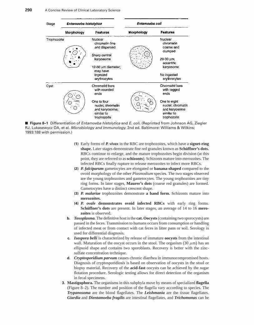
P1: OSO
LWBK192-08 LWBK192-Hubbard November 27, 2008 7:50
290 A Concise Review of Clinical Laboratory Science
■ Figure 8–1 Differentiation of Entamoeba histolytica and E. coli. (Reprinted from Johnson AG, Ziegler
RJ, Lukasewycz OA, et al. Microbiology and Immunology. 2nd ed. Baltimore: Williams & Wilkins;
1993:188 with permission.)
(1) Early forms of P. vivax in the RBC are trophozoites, which have a signet-ringshape. Later stages demonstrate fine red granules known as Schuffner’s dots.RBCs continue to enlarge, and the mature trophozoites begin division (at thispoint, they are referred to as schizonts). Schizonts mature into merozoites. Theinfected RBCs finally rupture to release merozoites to infect more RBCs.
(2) P. falciparum gametocytes are elongated or banana-shaped compared to theovoid morphology of the other Plasmodium species. The two stages observedare the young trophozoites and gametocytes. The young trophozoites are tinyring forms. In later stages, Maurer’s dots (coarse red granules) are formed.Gametocytes have a distinct crescent shape.
(3) P. malariae trophozoites demonstrate a band form. Schizonts mature intomerozoites.
(4) P. ovale demonstrates ovoid infected RBCs with early ring forms.Schuffner’s dots are present. In later stages, an average of 14 to 16 mero-zoites is observed.
b. Toxoplasma. The definitive host is the cat. Oocysts (containing two sporocysts) arepassed in the feces. Transmission to humans occurs from consumption or handlingof infected meat or from contact with cat feces in litter pans or soil. Serology isused for differential diagnosis.
c. Isospora belli is characterized by release of immature oocysts from the intestinalwall. Maturation of the oocyst occurs in the stool. The organism (30 μm) has anellipsoid shape and contains two sporoblasts. Recovery is better with the zinc-sulfate concentration technique.
d. Cryptosporidium parvum causes chronic diarrhea in immunocompromised hosts.Diagnosis of cryptosporidiosis is based on observation of oocysts in the stool orbiopsy material. Recovery of the acid-fast oocysts can be achieved by the sugarflotation procedure. Serologic testing allows for direct detection of the organismin fecal specimens.
3. Mastigophora. The organisms in this subphyla move by means of specialized flagella(Figure 8–2). The number and position of the flagella vary according to species. TheTrypanosoma are the blood flagellates. The Leishmania are the tissue flagellates.Giardia and Dientamoeba fragilis are intestinal flagellates, and Trichomonas can be
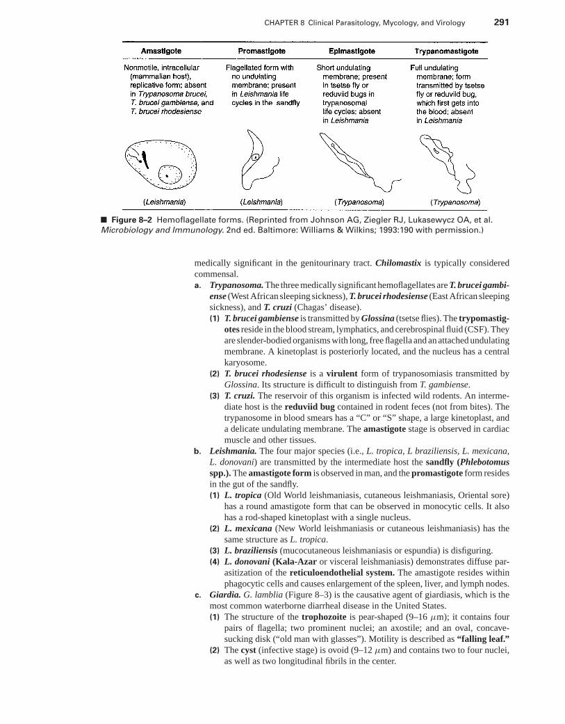
P1: OSO
LWBK192-08 LWBK192-Hubbard November 27, 2008 7:50
CHAPTER 8 Clinical Parasitology, Mycology, and Virology 291
■ Figure 8–2 Hemoflagellate forms. (Reprinted from Johnson AG, Ziegler RJ, Lukasewycz OA, et al.
Microbiology and Immunology. 2nd ed. Baltimore: Williams & Wilkins; 1993:190 with permission.)
medically significant in the genitourinary tract. Chilomastix is typically consideredcommensal.a. Trypanosoma. The three medically significant hemoflagellates are T. brucei gambi-
ense (West African sleeping sickness), T. brucei rhodesiense (East African sleepingsickness), and T. cruzi (Chagas’ disease).(1) T. brucei gambiense is transmitted by Glossina (tsetse flies). The trypomastig-
otes reside in the blood stream, lymphatics, and cerebrospinal fluid (CSF). Theyare slender-bodied organisms with long, free flagella and an attached undulatingmembrane. A kinetoplast is posteriorly located, and the nucleus has a centralkaryosome.
(2) T. brucei rhodesiense is a virulent form of trypanosomiasis transmitted byGlossina. Its structure is difficult to distinguish from T. gambiense.
(3) T. cruzi. The reservoir of this organism is infected wild rodents. An interme-diate host is the reduviid bug contained in rodent feces (not from bites). Thetrypanosome in blood smears has a “C” or “S” shape, a large kinetoplast, anda delicate undulating membrane. The amastigote stage is observed in cardiacmuscle and other tissues.
b. Leishmania. The four major species (i.e., L. tropica, L braziliensis, L. mexicana,L. donovani) are transmitted by the intermediate host the sandfly (Phlebotomusspp.). The amastigote form is observed in man, and the promastigote form residesin the gut of the sandfly.(1) L. tropica (Old World leishmaniasis, cutaneous leishmaniasis, Oriental sore)
has a round amastigote form that can be observed in monocytic cells. It alsohas a rod-shaped kinetoplast with a single nucleus.
(2) L. mexicana (New World leishmaniasis or cutaneous leishmaniasis) has thesame structure as L. tropica.
(3) L. braziliensis (mucocutaneous leishmaniasis or espundia) is disfiguring.(4) L. donovani (Kala-Azar or visceral leishmaniasis) demonstrates diffuse par-
asitization of the reticuloendothelial system. The amastigote resides withinphagocytic cells and causes enlargement of the spleen, liver, and lymph nodes.
c. Giardia. G. lamblia (Figure 8–3) is the causative agent of giardiasis, which is themost common waterborne diarrheal disease in the United States.(1) The structure of the trophozoite is pear-shaped (9–16 μm); it contains four
pairs of flagella; two prominent nuclei; an axostile; and an oval, concave-sucking disk (“old man with glasses”). Motility is described as “falling leaf.”
(2) The cyst (infective stage) is ovoid (9–12 μm) and contains two to four nuclei,as well as two longitudinal fibrils in the center.
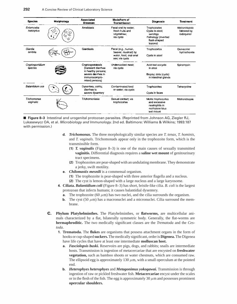
P1: OSO
LWBK192-08 LWBK192-Hubbard November 27, 2008 7:50
292 A Concise Review of Clinical Laboratory Science
■ Figure 8–3 Intestinal and urogenital protozoan parasites. (Reprinted from Johnson AG, Ziegler RJ,
Lukasewycz OA, et al. Microbiology and Immunology. 2nd ed. Baltimore: Williams & Wilkins; 1993:187
with permission.)
d. Trichomonas. The three morphologically similar species are T. tenax, T. hominis,and T. vaginals. Trichomonads appear only in the trophozoite form, which is thetransmissible form.(1) T. vaginalis (Figure 8–3) is one of the main causes of sexually transmitted
vaginitis. Differential diagnosis requires a saline wet mount of genitourinarytract specimens.
(2) Trophozoites are pear-shaped with an undulating membrane. They demonstratea jerky, swift motility.
e. Chilomastix mesnili is a commensal organism.(1) The trophozoite is pear-shaped with three anterior flagella and a nucleus.(2) The cyst is lemon-shaped with a large nucleus and a large karyosome.
4. Ciliata. Balantidium coli (Figure 8–3) has short, bristle-like cilia. B. coli is the largestprotozoan that infects humans; it causes balantidial dysentery.a. The trophozoite (60 μm) has two nuclei, and the cilia surrounds the organism.b. The cyst (50 μm) has a macronuclei and a micronuclei. Cilia surround the mem-
brane.
C. Phylum Platyhelminthes. The Platyhelminthes, or flatworms, are multicellular ani-mals characterized by a flat, bilaterally symmetric body. Generally, the flat-worms arehermaphroditic. The two medically significant classes are the Trematoda and the Ces-toda.
1. Trematoda. The flukes are organisms that possess attachment organs in the form ofhooks or cup-shaped suckers. The medically significant, order is Digenea. The Digeneahave life cycles that have at least one intermediate molluscan host.a. Fasciolopsis buski. Reservoirs are pigs, dogs, and rabbits; snails are intermediate
hosts. Transmission is ingestion of metacercariae that are encysted on freshwatervegetation, such as bamboo shoots or water chestnuts, which are consumed raw.The ellipsoid egg is approximately 130 μm, with a small operculum at the pointedend.
b. Heterophyes heterophyes and Metagonimus yokogawai. Transmission is throughingestion of raw or pickled freshwater fish. Metacercariae encyst under the scalesor in the flesh of the fish. The egg is approximately 30 μm and possesses prominentopercular shoulders.
P1: OSO
LWBK192-08 LWBK192-Hubbard November 27, 2008 7:50
CHAPTER 8 Clinical Parasitology, Mycology, and Virology 293
c. Clonorchis sinensis. The Chinese liver fluke is approximately 30 μm. Trans-mission occurs with the consumption of freshwater fish containing the encystedmetacercariae. The adult worm harbors in the liver and sheds eggs into the feces.The eggs resemble Heterophyes but have a small comma-shaped process at theabopercular end.
d. Fasciola hepatica. The sheep liver fluke infection occurs following the consump-tion of aquatic vegetation (metacercariae encyst watercress). Eggs are observedin the feces and are difficult to distinguish from Fasciolopsis. The eggs are oper-culated and measure 130 μm.
e. The three medically significant schistosomes that parasitize man are Schisto-soma mansoni, S. japonicum, and S. haematobium. The schistosomes are nothermaphroditic, they are diecious. The eggs are not operculated; the hatchedmiracidium swim in search of the appropriate snail host. The cercariae infecthumans. Schistosomes directly penetrate the skin to invade the circulatory sys-tem. The final locations differ in each host.(1) S. mansoni. Eggs are isolated from the feces. They are elongated, ovoid,
and possess a large lateral spine that is shaped like a thorn and projectsfrom the side of the egg near one end. These eggs measure approximately150 μm.
(2) S. japonicum. Eggs are isolated from the feces. They are spherical in shapeand possess a small lateral spine (sometimes this is absent or very difficult toview). These organisms measure approximately 65 μm.
(3) S. haematobium is the cause of urinary schistosomiasis. Eggs are isolatedin urine or in a biopsy of the bladder. The eggs have a terminal spine andmeasure approximately 150 μm.
f. Paragonimus westermani. The lung fluke is transmitted by the ingestion of rawor improperly cooked crayfish or fresh-water crabs encysted with the metacer-carial stage. The eggs are discharged into the bronchi or bronchioles; they can beexpectorated in sputum or, if swallowed, appear in the feces. The ovoid eggs haveraised opercular shoulders and measure approximately 100 μm.
2. Cestoda. The tapeworms have elongated segmented (proglottid) bodies with a scolex(specialized attachment organ). They inhabit and gain nourishment from the smallintestine. All cestodes of man have four muscular, cup-shaped suckers on the scolex,and a rostellum that can be armed with hooks. Transmission occurs when infectivelarvae are accidentally ingested with food.a. Diphyllobothrium latum selectively absorbs vitamin B12 from the host’s intestinal
tract. The primary intermediate host is the ciliated coracidium larvae (whichhas ingested the D. latum eggs). The larvae are ingested by copepods (D. latumdevelops and forms larvae). These infected cope-pods are then ingested by fish,and D. latum grows within the flesh of the fish to the plerocercoid larva stage. Onlyif the infected fish is ingested by man will the larva pass into the intestine anddevelop into adulthood.(1) The adult worm scolex is elongated, spoon shaped, and has two longitudinal
grooves. The uterus, within the proglottid, has a unique morphology and formsa rosette.
(2) The eggs are ovoid with an operculum and a thick, smooth shell. They areapproximately 70 μm.
b. Taenia solium. The pork tapeworm is transmitted by ingesting cured or under-cooked pork. Embryonated eggs are ingested by the intermediate host (the pig)and develop into the infective larval stage or cysticerci in the muscles. Humansingest the cysticerci; the larvae develop, and the scolex attaches to the intestinalwall and begins to develop proglottids (cysticercosis).(1) Structure. The adult worm scolex has four suckers and a double crown
of hooks (armed rostellum). Differential diagnosis is by observation of 7–13branches of the central uterine stem in a gravid segment.
(2) The egg contains the oncosphere. A thick coat, called the embryophore, is adark color, and is radially striated. Observation of Taenia eggs cannot differ-entiate T. solium from T. saginata.
P1: OSO
LWBK192-08 LWBK192-Hubbard November 27, 2008 7:50
294 A Concise Review of Clinical Laboratory Science
c. Taenia saginata. The beef tapeworm is transmitted by the ingestion of raw orinsufficiently cooked beef. Embryonated eggs are ingested by cattle and develop inthe flesh into the infective cysticercus stage. (T. saginata does not cause cystercosisof humans.)(1) The adult worm scolex has four suckers with a small rostellum that has no
hooks (unarmed rostellum). The gravid segment is longer in T. saginata, andthere are 15 to 20 branches off of the central uterine stem.
(2) Morphology of the eggs of T. saginata is similar to that of T. solium.d. Echinococcus granulosus. The minute tapeworm lives as an adult in the intestines
of the Canidae. Intermediate hosts are typically herbivores (e.g., sheep, goats). Thehuman is an accidental intermediate host. Humans can ingest the eggs, which hatchand develop into oncospheres. The oncospheres penetrate the intestinal mucosa,enter the circulation, and progress to the liver. A hydatid cyst begins to develop,with buds forming on the cyst. The cysts contain protoscolices, which appear ashydatid sand when the cyst begins to break down.(1) The adult worm has a scolex (with six hooklets) and three proglottids.(2) Structure. Eggs cannot be differentiated from Taenia eggs.
e. Hymenolepis nana. The dwarf tapeworm is transmitted to humans by ingestionof the egg, which develops into the adult worm in the intestines of the human. Thereservoir is the house mouse.(1) The adult worm has a scolex with four suckers and a rostellum armed with a
circle of hooks.(2) The eggs are ovoid and have a thin, smooth outer shell with two polar thick-
enings or knobs that have fine filaments (4–8) that are difficult to observe.The eggs are approximately 40 μm.
f. Hymenolepis diminuta transmission is through ingestion of infected insects fromflour contaminated by the droppings of infected rats. Larvae grow into adults inthe intestine of humans.(1) Adult worms have a scolex with four suckers and an “unarmed” rostellum.(2) Eggs are ovoid with a thick shell that also has two polar knobs, but no
filaments.
D. Phylum Aschelminthes. The medically significant class is Nematoda, the roundworms.They posses a stiff cuticle, and the sexes are separate. The roundworms can inhabit theintestines, the blood, or the tissues. The filariae are thread-like worms that inhabit thehuman lymphatic system, as well as subcutaneous and deep connective tissues. All of thefilariae require an arthropod intermediate host for transmission of infection. Some speciesdemonstrate periodicity. That is, they appear in the blood at specific times of the day.
1. Ascaris lumbricoides. The life cycle of Ascaris is complex. After the fully embry-onated eggs are ingested, they hatch in the intestines and begin to migrate (up to20 days) through the blood and lymphatic system. They finally reach the esophagusand once again enter the intestines, where the mature worm then begins to lay eggs.a. Adult worm
(1) The adult female worm is 30 cm in length and is as thick as a lead pencil.(2) The adult male worm is 25 cm in length and is slender with an “incurved tail.”
b. The eggs are broadly oval with a rough outer coating, an albuminoid coat. They arereferred to as fertilized corticated eggs. Decorticated eggs have a thick, smoothshell. Infertile eggs are narrow and have a thin shell (corticated or decorticated).
2. Enterobius vermicularis. The pinworm is the most common helminth parasite inmodern countries. Transmission is by ingestion of the eggs, which pass to the intestineand begin development. The females migrate down the intestinal tract to pass out of theanus and deposit their eggs. The cellophane tape preparation is useful for diagnosis.Typically, the eggs are not observed in the feces.a. Structure
(1) The adult female worm can reach 13 mm in length. It has a long, thin, sharplypointed tail.
(2) The adult male worm reaches 5 mm in length.
P1: OSO
LWBK192-08 LWBK192-Hubbard November 27, 2008 7:50
CHAPTER 8 Clinical Parasitology, Mycology, and Virology 295
b. The eggs are 50 μm and have a translucent shell that is flattened on one side(football appearance).
3. Trichuris trichiura. The whipworm is acquired through ingestion of embryonatedeggs. It can cause rectal prolapse and nutritional deficiencies in children.a. The female worm measures 3 to 5 cm and is typically longer than the male.b. The eggs are 50 μm and have bipolar plugs.
4. Ancylostoma duodenale. The Old World hookworm eggs are deposited into moistsandy soil, and within 24 to 48 hours rhabditiform larvae develop and hatch. Furtherdevelopment transforms the larvae into the infective filariform larvae. The filariformpenetrate the skin of humans. Humans may develop ground itch at the site of penetra-tion. Hookworm infestation is associated with iron deficiency anemia.a. Adult worm
(1) The female worm is grayish white with a buccal cavity that has four teeth.(2) The male worm is smaller than the female worm and has a prominent copulatory
bursa posteriorly.b. The eggs of A. duodenale and N. americanus cannot be differentiated. The eggs
are approximately 50 μm with a thin, smooth colorless shell. Clearly visible arethe two-cell, four-cell, or occasionally eight-cell stage of embryonic cleavage.
5. Necator americanus is the New World hookworm.a. Adult worm
(1) The female worm is 1 cm in length. The buccal cavity of Necator is armedwith a pair of cutting plates.
(2) The male worm is smaller than the female worm.b. Eggs (see Section I D 4 b)c. Rarely, hookworm eggs can hatch and release the rhabditiform larvae. The lar-
vae can be differentiated from Strongyloides larvae by observing the buccalcavities. The hookworm buccal cavity has a long capsule between the oralopening and the esophagus. The Strongyloides buccal capsule is comparativelyshort.
6. Strongyloides stercoralis. The threadworm is less prevalent than the hookworm andcan exist as a free-living nematode. Transmission and life cycle are similar to thehookworm.a. Adult worm
(1) The female worm is longer than 2 mm and is parthenogenic (capable ofunisexual reproduction, no fertilization required).
(2) The male worm is smaller, and it may be eliminated from the body early ininfection.
b. Structure. Eggs of the threadworm cannot be distinguished from the hookwormeggs. The eggs hatch in the intestine to form the rhabditiform larvae and are usuallypassed in the feces. Occasionally, a few of these larvae transform into the filariformlarvae, which penetrate the mucosa wall, enter the bloodstream, and begin themigratory cycle again. This is termed autoreinfection.
7. Trichostrongylus spp. This worm is very similar to the hookworm. The life cyclediffers in that Trichostrongylus is transmitted by ingestion of contaminated food. Theadult worms are much smaller than the hookworms. The eggs are much larger than thehookworm eggs (80 μm).
8. Trichinella spiralis. Trichinosis and trichinellosis are acquired by ingesting uncookedpork or bear meat that contains the encysted larvae. The larvae excyst and penetrateinto the intestinal mucosa of the host and develop into adult worms. The male andfemale worms mate, and more larvae are produced (intestinal phase). The larvae enterthe bloodstream and disseminate to the striated muscle, where they encyst. Diagnosisis based on observation of the encysted larvae in biopsied muscle or postinfectionserologic testing.
9. Dracunculus medinensis. The guinea worm is a parasite of dogs and other carnivoresin North America.a. Infection. The larvae are ingested by the intermediate host—the copepod
(crustacean)—and develop into the infective form. The infective larvae are
P1: OSO
LWBK192-08 LWBK192-Hubbard November 27, 2008 7:50
296 A Concise Review of Clinical Laboratory Science
ingested in drinking water and migrate in the host to the subcutaneous tissues.The larvae develop into the adult worm. The female worm migrates to the skinsurface and forms a papule to discharge large numbers of larvae.
b. Structure(1) The adult female worm is approximately 800 mm in length and can be removed
from the papule manually or excised surgically.(2) The adult male worm is approximately 40 mm in length.
10. Ancylostoma braziliense or A. caninum are the dog hookworms that occasionallyinfect humans and cause cutaneous larva migrans or creeping eruptions. The larvaepenetrate the skin and are unable to complete their migratory cycle; therefore, theybegin to migrate through the subcutaneous tissues.
11. Wuchereria bancrofti. Bancroftian filariasis, elephantiasis, or Bancrofti’s filariasis areassociated diseases.a. Infection. The infective larvae enter the human host through the proboscis of the
Culex or Anopheles mosquito. They migrate to the lymphatics (lymph nodes),where mating occurs and microfilariae are produced (can be observed in a bloodsmear).
b. Structure. The microfilariae are sheathed. The anterior is rounded, and the poste-rior tapers to a point. There are no nuclei in the tail.
12. Brugia malayi. Malayan filariasis or elephantiasis is transmitted by the Mansonia orAnopheles mosquito.a. Infection. Migration occurs through the lymphatics. Microfilariae can be observed
in the blood smear.b. Structure. The microfilariae are sheathed. There are two nuclei present in the tip
of the tail.13. Loa loa. The African eyeworm is transmitted by the Chrysops (fly) and migrates
through the subcutaneous tissues (especially across the conjunctiva of the eye). Calabarswellings also occur. The microfiliariae can be observed in the blood. They are sheathedand have a continuous row of posterior nuclei.
14. Onchocerca volvulus. Onchocerca is transmitted by the Simulium fly and migratesthrough the subcutaneous tissues. They usually settle in groups of two and becomeencapsulated to form nodules. These nodules can occur anywhere, but are typicallyobserved on the patient’s scalp. The microfilariae can be observed in “skin snips”taken from the nodules. The microfilariae are unsheathed, with no terminal nuclei, andtypically have straight tails.
15. Mansonellaa. M. ozzardi is typically a nonpathogenic filaria. The microfiliariae have no sheaths
and no nuclei in their tails.b. M. streptocerca (formerly Dipetalonema streptocerca) is pathogenic and transmit-
ted by Culicoides. The microfilariae are observed only in tissue scrapings. Theyhave no sheaths and have single nuclei in their tails.
c. M. perstans (formerly D. perstans) is nonpathogenic. The microfilariae have nosheaths and have paired nuclei in their tails.
E. Specimen collection, transport, and processing1. Collection and transport for fecal examination should include the following precau-
tions.a. The specimen should be collected in a clean container and must not be contaminated
with urine.b. If not examined within 1 hour, part of the specimen should be preserved in polyvinyl
alcohol fixative (PVA) or in 10% formalin.(1) PVA preserves cysts and trophozoites for permanent staining.(2) Formalin preserves eggs, cysts, and larvae for wet-mount examination and
concentration.c. Three specimens every other day is the optimal number for recovery of parasites.d. Saline purges are satisfactory to obtain specimens; castor and mineral oils make
examination for protozoa impossible.
P1: OSO
LWBK192-08 LWBK192-Hubbard November 27, 2008 7:50
CHAPTER 8 Clinical Parasitology, Mycology, and Virology 297
e. Specimens containing radiographic contrast media (e.g., barium salts) are inappro-priate.
2. Processing of specimens. The processing should include a macroscopic examination,a microscopic examination, and a concentration procedure.a. The process of macroscopic examination involves the following:
(1) Noting the consistency of the specimen (i.e., soft stools suggest the presenceof trophozoites or protozoa; formed stools suggest the presence of cysts)
(2) Examining the surface for tapeworm proglottids or adult pinworms(3) Examining for blood and mucus
b. The microscopic examination should include a direct saline and/or iodine mount.A calibrated ocular micrometer should be used routinely. Size differences of variousinternal structures and of whole organisms are important in differential diagnosis.
c. A fecal concentration technique increases the possibility of detecting parasiteswhen few are present in feces; this technique should be a routine part of the exam-ination process.(1) The Ritchie formalin-ether method is the most commonly used method for
concentrating eggs and cysts.(2) The zinc sulfate flotation method is not appropriate for operculated eggs,
schistosomes, or infertile ascaris eggs. This method is recommended for re-covery of oocysts.
3. The trichrome technique is a rapid procedure that gives good results for routineidentification of intestinal protozoa in fresh fecal specimens. The specimen must befixed with either PVA or Schaudinn’s solution.a. The cytoplasm of trophozoites and cysts appears light blue-green or light pink.b. Karyosomes stain ruby red.c. Degenerated organisms and the background material stain pale green.
II. MYCOLOGY
A. INTRODUCTION1. Definition. Mycology is the study of the eukaryotic fungi, including yeasts, molds,
and mushrooms.2. Characteristics of fungi include the following:
a. The eukaryotic fungi are non-motile and have definite cell walls. They lack chloro-phyll and absorb nutrients.
b. Hyphae are branching filaments of molds and mushrooms.The mycelium is the mass of hyphae constructing the thallus, the vegetative
body of a fungus.(1) Hyphae may be dematiaceous (dark) or hyaline (colorless).(2) Hyphae can be septate (with crosswalls) or aseptate. Aseptate fungi are often
referred to a coenocytic.(3) Vegetative, or food-absorbing, hyphae are either submerged or on top of the
agar substrate. Aerial hyphae extend above the agar surface and may supportreproductive structures, commonly called conidia.
c. Pseudohyphae are a series of elongated blastoconidia made by some yeast. Theseremain attached to each other and form a hyphal-like structure. Points of attach-ment and septations are constricted, while those of true hyphae are not constricted.(characteristic of Candida albicans).
d. Spores or conidia may be formed by either an asexual or a sexual process.(1) Asexual spores
(a) Conidia are asexual spores formed from a conidiogenous cell or at the endof conidiophores. The conidial types are used to identify the Fungi Im-perfecti, or phylum Deuteromycota. These fungi have no known sexualstage.
(b) Blastic conidiogenesis results in the production of blastoconidia thatbud from a mother cell. They can elongate to form pseudohyphae. These
P1: OSO
LWBK192-08 LWBK192-Hubbard November 27, 2008 7:50
298 A Concise Review of Clinical Laboratory Science
conidia are produced by yeasts and some fungi. Poroconidia, phialo-conidia, and annelloconidia are all produced by blastic conidiogenesis.
(c) In the process of thallic conidiogenesis, the daughter conidium developsafter the formation of a septum near the end of a parent cell. Macroconidiaand microconidia, characteristic of the dermatophytic fungi, are producedby thallic conidiogenesis.
(d) Arthric conidiogenesis results in the formation of arthroconidia, whichfragment from the hyphal strand at the septation points. This is a type ofthallic conidiogenesis.
(e) Chlamydoconidia, seen in molds, are thick-walled survival conidia thatcan occur either at a terminal site (end), an intercalary site (within thehyphae), or a sessile site (on the sides). These germinate and produceconidia under favorable environmental conditions.
(f) Sporangiospores are formed by internal cleavage of the contents of asporangium, a sac that is supported by a sporangiophore. This type offree cell formation is characteristic of the Zygomygetes.
(2) Sexual spores are produced by the Perfect Fungi.(a) Zygospores are formed by fusion of two compatible hyphal arms and are
surrounded by a thick walled, protective zygosporangium. These sexualspores are produced by fungi in the phylum Zygomycota.
(b) Ascospores are sexual spores produced by fungi in the phylum Ascomy-cota. Eight ascospores are inside an ascus that may be surrounded by aprotective ascocarp.
(c) Basidiospores are sexual spores that protrude out of a mother cell calleda basidium. The basidiospores and basidium may be protected by an outerbasidiocarp, as in the mushrooms, members of the phylum Basidiomy-cota.
B. Specimen collection and transport1. Collection of specimens for mycologic cultures must include:
a. Sterile techniqueb. Adequate amountc. Sample from the area most likely affected
2. Transporta. Blood and bone marrow are collected into a brain heart infusion (BHI) broth for
transportation to the laboratory. The use of Dupont Isolator tubes for transportand processing enhances fungal recovery from blood and bone marrow specimens.
b. CSF is aseptically collected and transported immediately to the laboratory.c. Hair, nails, and skin are initially cleaned with 70% alcohol to remove surface
contaminants. Scrapings and plucked hairs are placed in a sterile Petri plate fortransportation.
d. Respiratory tract specimens (e.g., sputum) should be collected in the morningand put into a sterile container for transport.
e. Tissues and biopsy specimens are aseptically collected and kept moist with sterilesaline for transport.
f. Vaginal and cervical specimens are typically collected on sterile swabs, thenplaced into transport media or broth.
g. Scrapings from wounds and lesions may be placed into sterile saline for transport.Aspiration specimens can be collected from deep cysts or abscesses by needle andsyringe.
h. Urine specimens are collected in the morning in sterile container and sent imme-diately to the laboratory. Clean catch or catheterized specimens are recommended.
C. Media and tests1. Sabouraud dextrose agar (SDA) inhibits the growth of many bacteria because its pH
is 5.6. However, this medium allows fungal contaminants and pathogenic fungi to grow.Histoplasma capsulatum and some strains of Nocardia asteroides fail to grow on thismedium.
P1: OSO
LWBK192-08 LWBK192-Hubbard November 27, 2008 7:50
CHAPTER 8 Clinical Parasitology, Mycology, and Virology 299
2. SDA with cycloheximide and chloramphenicol is reserved for skin, hair, and nailspecimens.
3. BHI agar (BHIA) is very nutritious and supports the growth of bacteria and fungi,including Histoplasma and Nocardia.a. BHIA with blood is recommended for converting dimorphic fungi from the mold
to the tissue (yeast) phase.b. BHIA with blood, cycloheximide, and chloramphenicol is very nutritious but
inhibits the growth of Nocardia. (NOTE: Chloramphenicol inhibits the yeast phaseof dimorphic fungi.) It supports the growth of dermatophytes, isolated from skin,hair and nail specimens.
4. Corn meal Tween 80 agar (CMT 80) is used for the demonstration of blastoconidia,pseudohyphae, arthroconidia, and chlamydospores in the identification of Candidaspecies and other yeasts.
5. Potato dextrose agar (PDA) is a medium used to enhance sporulation and pigmention.It is used as a subculture medium rather than a primary plating medium.
6. Inhibitory mold agar is used primarily to recover pathogenic fungi exclusive of der-matophytes.
7. Mycosel agar is used to recover dermatophytes8. Dermatophyte test medium (DTM) is a screening medium used for the isolation of
dermatophytes.9. Birdseed agar (Caffeic Acid Agar) is used for growth of Cryptococcus neoformans.
10. Trichophyton Test Agars are used for speciation of the Trichophyton species.11. Germ tube media (rabbit, fetal calf, or human serum) is used to demonstrate germ
tube production by the yeast Candida albicans.12. Urea agar slant is used to demonstrate urease production. A positive test is develop-
ment of a pinkish purple within 48 hours after inoculation.a. Trichosporon, Rhodotorula, and Cryptococcus are urease-positive.b. Geotrichum, Saccharomyces, and most Candida are urease-negative.
D. Microscopy of clinical specimens1. Saline wet mounts allow the observation of budding yeast, hyphae, conidia, and
hyphal filaments. These mounts are primarily used for the observation of vaginalspecimens.
2. Lactophenol cotton blue stain (LPCB) can be used for direct smears or stains offungal isolates. Tease mounts for permanent smears and scotch tape preps can be madeusing this stain.a. Phenol kills organisms.b. Lactic acid preserves the fungal structures.c. Cotton blue stains the chitin in the fungal cell walls.
3. Potassium hydroxide (KOH) is ideal for observing skin, hair, or nails. Ten percentpotassium hydroxide dissolves the keratin and enhances visibility of the fungi.
4. Gram’s stain. The fungi appear gram-positive or blue. This stain is used primarily toobserve yeast and pseudohyphae present in clinical specimens.
5. Acid-fast stain. The modified Kinyoun acid-fast stain is recommended for stainingthe partially acid-fast Nocardia. The fluorescent auromine-rhodamine acid-fast stainmay also be used.
6. India ink. This background stain is recommended for observing encapsulated yeast,especially Cryptococcus neoformans.
7. Periodic acid-Schiff (PAS) stains fungi (except Actinomycetes) magenta against a lightpink or green background.
8. Calcofluor white stain is observed using a fluorescence microscope. With this sensitivestain, fungal elements fluoresce against a dark background. Ten percent KOH may beadded to dissolve keratin in the specimen.
E. Opportunistic fungi are exogenous saprophytes capable of causing infections in immuno-compromised individuals. These fungi are routinely encountered in routine fungal culturesand must be differentiated from pathogenic fungi. This group of fungi includes the aseptateZygomycetes, as well as many septate Deuteromycetes and Ascomycetes. Characteristic
P1: OSO
LWBK192-08 LWBK192-Hubbard November 27, 2008 7:50
300 A Concise Review of Clinical Laboratory Science
asexual conidial forms are primarily used to identify and differentiate these fungi. Produc-tion of sexual spores may be used to differentiate the Zygomycetes.
1. Hyaline (light) opportunists–septate fungia. Acremonium species microscopically demonstrate tapering conidiophores that
support closely packed balls of sickle- or elliptical-shaped conidia.b. Aspergillus species have rough or smooth unbranched conidiophores with a foot
cell at the base and a vesicle (head) at the tip. Rows of flask-shaped phialides insingle or double rows support chains of rough or smooth phialoconidia.(1) A. fumigatus is the most common opportunistic pathogen in the genus. It grows
at 45◦C, and the phialides cover the upper two thirds of the vesicle. Coloniesare dark greenish to gray.
(2) A. niger has a black radiate head with phialides and chains of phialoconidiacovering the entire vesicle. Colonies are dark black.
(3) A. flavus has spiny (rough) conidiophores and phialides cover the entirevesicle. Colonies are usually a yellow-green color.
(4) A. terreus produces cinnamon-brown colonies.(5) A. clavatus has huge, elongated vesicles with short phialides.
c. Fusarium species have single or branching conidiophores that terminate in taperedphialides. Macrophialoconidia are banana or cylindrical-shaped with two to fivecells.
d. Gliocladium species produce terminal masses of phialoconidia supported by flask-shaped phialides. Terminal masses of phialoconidia are held together in a largeball by a gelatinous matrix. Mature colonies are a characteristic “green lawn” ofgrowth.
e. Paecilomyces species have single, short, or penicillus-type phialides with verydelicate chains of smooth or rough, oval conidia. This fungus resembles Penicil-lium.
f. Penicillium species colonies are characteristically blue-green with a white pe-riphery. Multiple, flask-shaped phialides support chains of round phialoconidia.Microscopically, the fungus has a “brush-like” appearance.(1) P. marneffei is a dimorphic fungus that produces yeast-like colonies at 37◦C.
In the mycelial form (25–30◦C), chains of lemon-shaped phialoconidia aresupported by wide phialides on broad metulae (conidiophores).
g. Scopulariopsis species have echinulate (spiny) lemon-shaped annelloconidia inchains, supported on flask-shaped annellides. Unbranched annellophores supportthe annellides.
h. Sepedonium species have simple or branched conidiophores supporting singleor clustered thick-walled, smooth or rough macroconidia. This hyaline fungusresembles the yeast phase of the dimorphic pathogen Histoplasma capsulatum.
2. Dematiaceous (dark) opportunists–septate fungia. Alternaria species colonies are dark brown to black, and the hyphae and conidia
are dematiaceous. Chained poroconidia with horizontal and vertical septa haveclub-shaped bases with tapered apices.
b. Bipolaris species have dark, septate hyphae with cylindrical, four- to five-celledporoconidia. “Bent-knee” shaped conidiophores support the poroconidia, withtruncate hila at the points of attachment. Bipolar germ tube formation is producedby these fungi.
c. Cladosporium species produce short chains of blastoconidia from repeatedlyforking shield cells. There is a scar at the point of attachment to the shield cells.
d. Curvularia species have large four- to five-celled poroconidia with an over-enlarged central cell and bent-knee conidiophores.
e. Exserohilum species have “bent-knee” conidiophores and protruding truncatehila at the ends of dark cylindrical poroconidia with 6 to 14 cells.
f. Epicoccum species have thick clusters of sporodchia (short conidiophores) thatsupport terminal dark, round conidia with horizontal and vertical septa.
g. Nigrospora species have dark hyphae and short fat conidiophores supporting sin-gle, oval, smooth-walled, black conidia.
P1: OSO
LWBK192-08 LWBK192-Hubbard November 27, 2008 7:50
CHAPTER 8 Clinical Parasitology, Mycology, and Virology 301
3. Zygomycetes–aseptate opportunists with large, ribbon-like hyphaea. Absidia species have stalk-like, branching sporangiophores with a columellae
(base) supporting pear-shaped sporangia (a sac). Asexual sporangiospores areproduced within the sporangia by free-cell formation. These aseptate Zygomyceteshave rhizoids, or root-like structures, between the interconnecting runners (stolons)connecting the sporangiophores.
b. Mucor species have single or branching sporangiophores supporting round, spore-filled sporangia. There are no rhizoids or stolons.
c. Rhizopus species produce black sporangia on unbranched sporangiophores op-posite rhizoids. Stolons connect the groups of rhizoids.
d. Saksenaea species have large, flask-shaped sporangia filled with elongated spo-rangiospores. Rhizoids are opposite the sporangiophores.
e. Cunninghamella species have branched sporangiophores supporting terminal,round vesicles. Denticles protrude from the vesicles and support one-celled roundsporangiola.
f. Syncephalastrum species produce terminal, round vesicles. Rows of round spo-rangiosporesare incylindricalmerosporangia that radiate thesurfaceof thevesicle.
4. Single-celled yeasts are normal flora of the skin, mucous membranes, and gastroin-testinal tract and can be opportunistic pathogens.a. Candida albicans is predominantly the causative agent of these opportunistic in-
fections.(1) C. albicans form chlamydospores, blastoconidia, and pseudohyphae on CMT
80 agar. Germ tubes are formed following incubation in germ tube media.(2) C. albicans assimilates the carbohydrate sucrose.
b. Torulopsis glabrata is normal vaginal flora and may be associated with vaginaland urinary tract infections.
c. Emerging opportunistic pathogens are C. tropicalis, C. parapsilosis, and C. krusei.
F. Opportunistic mycoses include infections ranging from mild and self-limiting to seriousdisseminated infections in severely immunocompromised individuals. These mycoses arecaused by exogenous, saprophytic fungi, and they are increasing as the number of immuno-compromised patients increases.1. Candidiasis is an acute-to-chronic fungal infection that can involve the mouth, vagina,
skin, nails, bronchi, lungs, alimentary tract, bloodstream, or urinary tract. Typically,the causative agent is Candida albicans, but other species of Candida are emergingas opportunistic pathogens. Candida albicans is part of the normal flora of the skin,mucous membranes, and gastrointestinal tract.a. Oral thrush is a yeast infection that forms white curd-like patches on the oral
muco-cutaneous membranes.b. Vulvovaginitis or vaginal thrush manifests as a thick yellow-white discharge. Dia-
betes, antibiotic therapy, oral contraceptives, and pregnancy predispose the patientto this condition.
c. Candidemias, primarily caused by C. parapsilosis, occur in patients who haveindwelling catheters.
d. Other opportunistic yeast infections in debilitated patients may be caused byGeotrichum candidum and Torulopsis glabrata. Infections range from oral andurinary tract infections to disseminated infections.
2. Malassezia furfur can be an opportunist, causing septicemia in patients who receiveintravenous lipid infusions. The fungus grows well in the sebaceous glands. It is alsothe causative agent of the superficial mycoses pityriasis versicolor.a. M. furfur requires a medium overlayed with a lipid, such as olive oil.b. Microscopically, this septate fungus appears as thick, round oval cells in clusters
with hyphae (spaghetti and meatballs).3. Cryptococcosis infections primarily involve the lungs and meninges and often occurs
in AIDS patients. The yeast Cryptococcus neoformans is the causative agent of cu-taneous and disseminated disease. Inhalation of fungal spores is usually the means oftransmission.
P1: OSO
LWBK192-08 LWBK192-Hubbard November 27, 2008 7:50
302 A Concise Review of Clinical Laboratory Science
a. C. neoformans has a capsule that is demonstrated with the India ink preparation.It is urease-positive.
b. C. neoformans is associated with pigeon and bird feces. It may also be associatedwith fruits, milk, or plants.
c. Capsular material in the CSF can be detected with an antigen latex agglutinationtest.
4. Aspergilloses are a variety of infections and allergic diseases primarily caused byAspergillus fumigatus. A. flavus and A. niger may also be causative agents of theseinfections. The Aspergillus species are ubiquitous fungi with airborne spores.a. Allergic aspergillosis involves inhalation of the conidia, colonization of the mu-
cous plugs in the lung, and subsequent asthma attacks. A high titer of IgE antibodyto Aspergillus is present.
b. Otomycosis (outer ear infections) is a characteristic form of aspergillosis.c. Fungus ball, a localized abscess that forms in the lung, is another form of as-
pergillosis. However, there is no tissue invasion.d. Invasive aspergillosis occurs in neutropenic patients. Initially, the patient has
sinusitis, followed by dissemination throughout the body (e.g., brain, bones)occurs.
5. Penicilliosis is caused by the penicillus-producing fungi, Penicillium, Scopulariopsis,and Paecilomyces. Infections may include otomycosis (outer ear), keratomycosis(cornea of the eye), infections of the nails. Dissemination occurs in debilitated patientswith transmission via inhalation of spores and spread of an initial pulmonary disease.The dimorphic fungus, P. marneffei, causes a disseminated form of penicilliosis withflu-like symptoms, followed by enlarged lymph node, spleen, and liver. Transmissionmay be via inhalation or ingestion of the fungal spores.
6. Zygomycoses, also known as phycomycoses or mucormycoses, are acute infectionscaused by the aseptate fungi in the phylum Zygomycota: Rhizopus, Absidia, and Mu-cor. Predisposed patients have uncontrolled diabetes, malnutrition, or are on steroidtherapy. Spores infect the nasal sinuses or orbital (eye) area and the infection dissemi-nates rapidly. Without treatment, death ensures within two to ten days after the initialinfection. Prompt treatment with amphotericin B is usually successful if initiated at thebeginning of the infection.
7. Other opportunistic fungal mycoses may include keratomycosis (corneal infections)and otomycosis (outer ear infections). These infections are usually the result of trauma.Acute or chronic sinusitis may also be caused by opportunistic fungi.
G. Systemic mycoses affect internal organs and disseminate to multiple organs in the body. Pri-mary or secondary cutaneous infections are often characteristic of these mycoses. Causativeagents are typically the slow-growing, dimorphic fungal pathogens. These fungi (grow asmolds at 25◦C and as yeast at 35◦C).
1. Histoplasmosis, or Darling’s disease, is a granulomatous fungal infection caused byHistoplasma capsulatum. The progressive disease is transmitted via inhalation or directinoculation into the skin, and is not contagious or transmissible via person-to-person.a. The yeast or tissue phase (37◦C) is small, single-budding yeast cells.b. The mold (25◦C–30◦C) demonstrates is a large tuberculate macroconidia and
small rough or smooth microconidia along the sides of the hyphae. This is thediagnostic stage. The fungus resides in soil enriched with bat or bird guano(inhalation transmission).
c. The major endemic region is the Ohio, and Mississippi River areas, and along theAppalachian Mountains.
2. North American blastomycosis, or Gilchrist’s disease, is caused by Blastomycesdermatitidis. Transmission is inhalation of the conidia. The primary respiratory diseasegradually disseminates hematogenously to other organs.a. The diagnostic tissue (37◦C) phase demonstrates large yeast with a double refrac-
tile wall and a broad-based bud.b. The mold phase (25◦C–30◦C) is single oval conidia at the ends of short conidio-
phores or directly on the hyphae.
P1: OSO
LWBK192-08 LWBK192-Hubbard November 27, 2008 7:50
CHAPTER 8 Clinical Parasitology, Mycology, and Virology 303
c. The major endemic areas are south of the Ohio River and east of the MississippiRiver.
3. Coccidioidomycosis is caused by Coccidioides immitis and is commonly known as“San Joaquin valley fever.” Transmission is via inhalation of the arthroconidia fromsand and dirt. The primary respiratory infection rapidly disseminated in immunocom-promised patients.a. The tissue phase (yeast) is large, thick-walled spherules filled with endospores.b. Alternating barrel-shaped arthroconidia between disjunctor cells are charac-
teristic of the mold phase.c. The major endemic area is the San Joaquin Valley and the lower Sonoran Desert
in the southwestern United States.d. Diagnosis is based on serologic and skin testing. In endemic areas, 3% of the
normal population convert to skin-test positive every year.4. South American Blastomycosis, or paracoccidioidomycosis, is caused by the sys-
temic fungus Paracoccidioides brasiliensis. Transmission is via inhalation and primaryinfections are pulmonary or mucocutaneous. The prognosis is death within 2–3 yearsof dissemination of the disease.a. The diagnostic tissue phase is large, thick-walled yeast with multiple buds,
resembling a “ship’s wheel.”b. Intercalary and terminal chlamydoconidia are observed in the mold phase.c. The endemic area is South America, especially in Brazil.
H. Superficial mycoses are noninvasive infections that affect the outermost layer of skin andhair.
1. Pityriasis versicolor is caused by Malassezia furfur, which infects the stratumcorneum epidermidis and causes hypopigmentation or hyperpigmentation on the trunkof the body. Scaly patches of skin fluoresce with a Wood’s lamp. Characteristic“sphagetti and meatballs” morphology is observed microscopically and a lipid oiloverlay is required for culture.
2. Tinea nigra is a superficial infection of the stratum corneum epidermidis caused by thedematiaceous fungus, Exophiala werneckii. The infection is characterized by brownto black, nonscaly patches on the palms of the handsa. Black, yeasty colonies develop short olive-gray mycelia with age.b. Microscopically, dark septate hyphae with one- or two-celled blastoconidia are
demonstrated in clusters along the hyphae. The fungus hydrolyzes casein.3. Piedra is an infection of the hair shaft that produces hair breakage. Treatment is cutting
or shaving infected hairs and use of topical antifungals. Infections may disseminate inimmunocompromised individuals.a. Trichosporon beigelli is the causative agent of white piedra and forms soft, light
brown nodules around the beard and mustache hairs. Microscopically, the fun-gus demonstrates hyaline hyphae with blastoconidia and arthroconidia on CMT80 agar.
b. Black piedra is caused by Piedraia hortai, which forms firmly attached hard,black nodules around the outside of scalp hairs. Microscopically, it produces darkthick-walled hyphae with swellings.
I. Cutaneous mycoses are typically caused by the dermatophytic fungi. Some cutaneousmycoses can be caused by Candida species. The cutaneous mycoses involve skin, hair,and nails. Infections may be acquired from animals (e.g., Trichophyton rubrum, Micro-sporum canis, Trichophyton verrucosum) or from humans (e.g., Epidermophyton flocco-sum, Microsporum audouinii). Diagnosis is made by microscopic examination of skin,hair, and nails with 10% KOH. Infections are commonly referred to as tinea infec-tions or dermatophytoses. “Ringworm” is another name for dermatophytic infections.Microscopically, the fungi demonstrate microconidia and macroconidia. Both conidiatypes or only one type may be demonstrated by a fungus. Diagnosis is made by macro-scopic and microscopic observations of the fungi and the location of the infection on thebody.
P1: OSO
LWBK192-08 LWBK192-Hubbard November 27, 2008 7:50
304 A Concise Review of Clinical Laboratory Science
1. Tinea capitis (head) is a dermatophytic infection of the scalp.a. Epidemic tinea capitis occurs in children and is extremely contagious. It is caused
by M. audouinii, which fluoresces under a Wood’s lamp. Microscopically, thefungus demonstrates rare irregular, rough-walled macroconidia with two to eightcells and rare club-shaped microconidia.
b. Zoophilic tinea capitis occurs primarily in children. Transmission of infection isthrough animals. It is most commonly caused by Microsporum canis with fewmicroconidia and numerous rough thick-walled, spindle-shaped macroconidiawith 6 to 15 cells. Trichophyton mentagrophytes is also a causative agent ofzoophilic tinea capitis. It demonstrates grape-like clusters of microconidia andnumerous pencil-shaped macroconidia with five to eight cells.
c. Black-dot tinea capitis is a chronic infection in adults caused by Trichophytontonsurans. Rare macroconidia and numerous microconidia are observed mi-croscopically.
d. Favus (tinea favosa) is a severe tinea capitis caused by Trichophyton schoen-leinii. Macroconidia are only observed if the fungus is grown on sterile ricegrains.
e. M. gypseum is transmitted via contact with contaminated soil is a causative agentof tinea capitis. Numerous rough, thin-walled, elliptical macroconidia with fourto six cells are produced by the fungus. Few microconidia are produced.
2. Tinea barbae is an infection of the beard, neck, or face.a. The causative agent Trichophyton verrucosum requires thiamine for growth and
microscopically produces rare three- to five-celled macroconidia with a rat-tail end.b. T. mentagrophytes causes tinea barbae.c. Trichophyton rubrum causes tinea barbae. It produces numerous club-shaped
microconidia, which occur singly along the hyphae. Rare to numerous smooth-walled, pencil-shaped macroconidia with three to eight cells are also produced.This dermatophyte is characterized by a deep red reverse.
3. Tinea corporis is a dermatophytosis of the body which usually affects the inside skinfolds. Candida species are often causative agents of this infection.a. T. rubrumb. T. mentagrophytesc. T. violaceum has characteristic violet colonies with a lavender reverse in 2–3 weeks.
Few microconidia are produced on thiamine-enriched media, in addition tochlamydoconidia in chains. No macroconidia are produced.
4. Tinea cruris is a dermatophytosis of the groin caused by the following fungi:a. Epidermophyton floccosum is a dermatophyte that produces two- to four-celled,
smooth, club-shaped macroconidia. These are produced singly or in clusters. Nomicroconidia are produced by this fungus.
b. T. rubrumc. T. mentagrophytesd. Candida species
5. Tinea pedis is an infection of the feet, commonly called athlete’s foot. It can be causedby the following fungi:a. T. rubrumb. T. mentagrophytes is the most common cause of athlete’s foot,c. E. floccosum causes epidemic tinea pedis.
J. Subcutaneous mycoses typically result from traumatic implantation (e.g., via thorn, splin-ter) of a fungus into the cutaneous and subcutaneous tissues. Subcutaneous fungi invadethe tissue and lymph nodes, and chronic, nonhealing, hard, lumpy, crusted ulcerationsare usually on the extremities. Endemic areas are the tropics, but distribution is world-wide. These infections are caused by hyaline and dematiaceous, as well as the fungus-likebacteria.
1. Sporotrichosis, or Rose Gardener’s disease, is caused by the dimorphic fungusSporothrix schenckii. Infections can be lymphocutaneous, fixed, or pulmonary.
P1: OSO
LWBK192-08 LWBK192-Hubbard November 27, 2008 7:50
CHAPTER 8 Clinical Parasitology, Mycology, and Virology 305
a. At 37◦C, microscopic growth is a cigar-shaped budding yeast, commonly referredto as cigar bodies. These are observed in tissue. Conversion to the yeast form isnecessary for identification.
b. At 25◦C–30◦C, the mycelial form of the fungus is a daisy head or floweret of mi-croconidia, which is produced at the end of unbranched conidiophores on delicatehyphae.
c. S. schenckii is found on plant material, such as rose thorn, sphagnum moss, andtimbers.
2. Eumycotic mycetoma, or madura foot, is a disease characterized by swelling, sinustract formation, and the presence of sulfur granules usually in the extremities. There islittle pain or bone destruction. It typically occurs in third world countries.a. Pseudoallescheria boydii, the teleomorph or sexual stage or of Scedosporium
apiospermum, is the most common cause of mycetoma in the United States. Asex-ual conidia or single or small, clustered annelloconidia produced terminally onlong or short annellophores. Sexual ascospores are contained asci within a largecleistothecia.
b. Madurella species most commonly cause mycetoma in Africa. An initial punctureor abrasion with contaminated material, usually on the feet, hands, and buttocks,progresses slowly to involve an entire extremity. Infected extremities are large anddeformed.
3. Chromoblastomycoses begin with traumatic implantation of the spore in a limb. Thedisease produces hard, dry, cauliflower-like lesions. Lymphatic channels are blocked bydrainage, causing elephantiasis, deformity and abnormal enlargement of the infectedlimb. Black dots, or sclerotic bodies, are observed in deep parts of the lesions. Sec-ondary bacterial infections may be a complication of the disease. Infections are causedby the dematiaceous fungi Phialophora, Fonsecaea, and Cladosporium and typicallyoccurs in third world countries. The conidial arrangement of these dematiaceous fungiis very similar. Conidiophores support branching chains of blastoconidia, multiple ar-rangements, or loosely organized conidial heads held together by a gelatinous matrix.
4. Phaeohyphomycosis ranges from a superficial infection to one with deep-organ in-volvement. Causative agents include the Phialophora, Exophiala, and Wangiella, all ofwhich produce slow-growing, black, yeasty colonies with a short mycelium. The dema-tiaceous Phialophora produce terminal balls of phialoconidia supported by phialides.Exophiala and Wangiella species demonstrate dark annelloconidia in clusters or termi-nal balls, respectively.
5. Actinomycotic mycetoma is caused by the anaerobic actinomycetes (fungus-like bac-teria). Sulfur granules may be observed in tissues, with symptoms including exudatesand painful bone involvement. Infections with the endogenous anaerobic oral flora re-sults in “lumpy jaw” or pulmonary and thoracic infections due to aspiration into thelungs. The aerobic actinomycetes, Nocardia, Streptomyces, and Actinomadura, causemycotic-like infections. These organisms are differentiated according to biochemicaltest and growth requirements.
K. Diagnostic tests for identification of the systemic fungi include antigenic skin tests,seromycology, and DNA testing. Several test methods can be used for serologic detec-tion of fungal antibodies. Empiric therapy is initiated with fungal infections. Antifungalagents, such as amphotericin B, fluconazole, and ketoconazole may be used for treatment.Fungal susceptibility testing can be performed in event of a failed response to empirictherapy. Fungal susceptibilities are usually preformed in reference laboratories.
III. VIROLOGY
A. Viruses (virions) are the smallest known form of infectious disease-causing agents.1. Size. Viruses are 1/10 the size of bacteria and 1/100 the size of eukaryotic cells. They
cannot be observed with a light microscope.
P1: OSO
LWBK192-08 LWBK192-Hubbard November 27, 2008 7:50
306 A Concise Review of Clinical Laboratory Science
2. Structure. Viruses of humans are composed of nucleic acid [deoxyribonucleic acid(DNA) or ribonucleic acid (RNA)].a. The nucleic acid is surrounded by a protein matrix referred to as the nucleocapsid.
The capsid surrounds the nucleocapsid and is composed of structural units calledcapsomers. Virus particles are either “naked” or have a lipoprotein structure, theenvelope, which surrounds the capsid.
b. Symmetry can be helical, icosahedral (20-sided polygon), or complex.3. Viral replication can occur only in living cells and involves many host-cell enzymes
and functions. The steps of replication are as follows.a. Attachment or adsorption occurs when the virion comes in contact with the host
cell.b. Penetration occurs when the virus passes through the plasma membrane.
(1) Enveloped viruses penetrate via fusion.(2) Naked viruses can penetrate transversely. Pinocytosis is the process of pinch-
ing through the plasma membrane, which results in viruses entering withincytoplasmic vacuoles.
c. Uncoating is typically mediated by cellular proteases and results in the separationof the capsid from the viral genome.
d. Synthesis of early proteins. These proteins are involved in genome replication.e. Synthesis of late proteins. These proteins are used in the development of structural
components of the virion.f. Assembly is the packaging of the new copies of the genome nucleic acid into the
capsid proteins.g. Release or egress of the progeny virus is the final step. The lipid envelope is acquired
along with the glycoproteins as the progeny viral nucleocapsid buds through themembrane.
B. Specimen collection, transport, and processing1. Time of collection. Specimens for viral identification are best collected during the time
of first presentation of symptoms.a. Virus titers (concentration of virus) are usually highest in the early part of the
illness.b. Serologically, a fourfold rise in the antibody titer between acute and convalescent
sera has been used to identify a particular infectious agent as the cause of a recentdisease.
2. Viral culture. The type of viral illness and the disease symptoms influence the speci-mens of choice for viral culture. Viral specimens are typically divided into nonsterilesites and sterile sites.a. Nonsterile sites include the following:
(1) Conjunctival(2) Skin (e.g., mouth, lips)(3) Vesicular(4) Nasal (aspirates or washes)(5) Throat/upper respiratory tract(6) Sputum(7) Urine(8) Genital (cervical, penile)(9) Stool/rectal swab
b. Sterile sites include the following:(1) Autopsy(2) Biopsy(3) CSF(4) Fine-needle aspirate(5) Blood(6) Bronchial alveolar (wash)(7) Pleural fluid
P1: OSO
LWBK192-08 LWBK192-Hubbard November 27, 2008 7:50
CHAPTER 8 Clinical Parasitology, Mycology, and Virology 307
3. The procedure for specimen collection is vital for recovery of the infectious virus.a. Specimens collected with a Dacron or rayon swab (cotton swabs are not recom-
mended for collection of viral samples because they are toxic to viruses) must notdry out during transport. The swab can be placed in a modified Stuart transportmedium for transport to the laboratory.
b. Nonsterile specimens that are not liquid, vesicular fluid, fine needle aspirates, ortissue biopsy specimens should be collected into viral transport media, such asHanks’ balanced salt solution, 0.2 M sucrose-phosphate, or 2% fetal calf serumwith Eagle’s minimum essential medium. Some transport mediums contain thefollowing ingredients:(1) Antibiotics to inhibit growth of normal bacterial or fungal flora(2) Gelatin media to stabilize protein
c. Urine for viral culture should not be collected into containers with preservatives.d. Blood for viral culture should be collected into heparin or ethylenediamine tetra-
acetic acid tubes.e. Stool specimens for viral culture should be collected into a clean container.
4. All specimens submitted for viral recovery regardless of source should be transportedat refrigerated temperatures (0◦C–4◦C).
5. If the inoculation of the specimen into tissue culture media will exceed 5 days, thespecimen should be quickly frozen at −70◦C.
6. Tissue culture is the inoculation of patient specimens into cells to determine the growthof a viral agent.a. Cells derived directly from the donor (animal or human sources) are known as
primary cultures.(1) Primary rabbit kidney is the first choice for herpes simplex viruses types 1
and 2.(2) Primary monkey kidney is the first choice for the following viruses:
(a) Influenza types A and B(b) Parainfluenza viruses types 1, 2, and 3(c) Mumps(d) Measles(e) Rubella(f) Enteroviruses
(3) Primary human embryonic kidney is the first choice for the followingviruses:(a) Parainfluenza virus(b) Measles(c) Rubella(d) Adenoviruses(e) Rhinoviruses(f) Herpes simplex viruses types 1 and 2
(g) Varicella zosterb. Cell lines are primary cultures that have been subcultured.
(1) Cell lines that have at least 75% of the cells with a normal chromosome com-plement are referred to as diploid (e.g., diploid human fibroblast cell line).Diploid cell lines usually die after 30 to 50 passages. The choices for culturingcytomegalovirus (CMV) are WI-38, MRC-5, or IMR-90.
(2) Cell lines that have a predominance of cells containing an abnormal numberof chromosomes are designated as heteroploid. Typically, these cell lines arecontinuous, that is, they have an indefinite number of passages.(a) A549 cells, which are derived from a human lung carcinoma, are used to
culture adenoviruses and varicella zoster.(b) HEp-2 cells, which are derived from a human laryngeal carcinoma, are
used to culture respiratory syncytial virus (RSV) and adenoviruses.(c) HeLa cells, which are derived from a human cervical adenocarcinoma, are
used to culture RSV, rhinoviruses, and adenoviruses.
P1: OSO
LWBK192-08 LWBK192-Hubbard November 27, 2008 7:50
308 A Concise Review of Clinical Laboratory Science
c. Order of inoculation. The diploid cell lines are inoculated first. The continuouscell lines are inoculated second. The primary cells are inoculated last.
d. Viral adsorption refers to the time point when the virus comes in contact with thetissue culture cells.(1) Stationary adsorption is the procedure of simply incubating the cell and virus
for 30 to 60 minutes at 35◦C.(2) The roller drum method refers to the gentle rotation of the culture tubes to
enhance adsorption.(3) The low-speed centrifugation method requires that shell vials be centrifuged
at 750 to 1,000 × g for 30 to 40 minutes at 25◦C.e. Viral detection in the culture cells depends on virus replication in the cells. Virus
may be detected by observing a characteristic cytopathogenic effect, such asswelling or inclusions.
7. Direct examination of clinical specimens for the presence of viral pathogens canbe done by immunohistochemical staining, nucleic acid hybridization, or solid-phaseimmunoassay.a. Immunohistochemical staining uses fixed or fresh specimens incubated with
chemically labeled (fluorescein) or enzymatically labeled (peroxidase) antibodiesto detect viral antigens.
b. Nucleic acid hybridization involves the detection of viral DNA or RNA sequencesin nucleic acid extracted from specimens.(1) Polymerase chain reaction is a technique that allows the viral genes to be
amplified to enhance detection.(2) The Western blot (dot blot) is a technique that employs single-stranded,
complementary nucleic acid probes for detection of human immunodeficiencyvirus (HIV) in the blood of seronegative individuals.
c. Solid-phase immunoassays use antibodies and radioimmunoassay or enzyme-linked immunosorbent assay (ELISA) to detect viral antigens.
C. DNA viruses1. The naked DNA viruses with an icosahedral symmetry are the Parvoviridae, Papil-
lomaviridae, Polyomaviridae, Adenoviridae, and Hepadnaviridae.a. Parvoviridae. Parvovirus B19 is spread by close physical contact. It causes ery-
thema infectiosum, or fifth disease.b. Papillomaviridae have two significant genera, papillomavirus and papovavirus.
(1) Human papillomavirus causes warts in humans and is associated with somegenital cancers.
c. Polyomaviridae produces histologically diverse tumors in various parts of thebody (JCV and BKV)
d. Adenoviridae have more than 47 serotypes of adenovirus. This virus causes infec-tion of the respiratory tract (nasopharyngitis); the eye (keratoconjunctivitis); andthe intestines (e.g., diarrhea, vomiting).
e. Hepadnaviridae have a tropism for infection of the liver.(1) Hepatitis B virus is also known as serum hepatitis and chronic hepatitis.
(a) Transmission of the virus is through the parenteral route (e.g., intra-venous drug abuse and contaminated blood products) and sexual inter-course.
(2) Hepatitis surface antigen (HBsAg) is present in early disease; anti-HBsAgindicates immunity.
2. The enveloped DNA virus with icosahedral symmetry is the Herpesviridae, whichhas three medically significant subfamilies: Alphaherpesvirinae, Betaherpesvirinae,and Gammaherpesvirinae. All have the ability to become latent after the primaryinfection.a. Within the group of Alphaherpesvirinae are herpes simplex 1 (HSV-1), herpes
simplex 2 (HSV-2), and varicella zoster virus.(1) HSV-1 is usually seen on the lips or skin or as an eye lesion. It may be diagnosed
by the Tzanck cell test.
P1: OSO
LWBK192-08 LWBK192-Hubbard November 27, 2008 7:50
CHAPTER 8 Clinical Parasitology, Mycology, and Virology 309
(2) HSV-2 affects the genital or lips area and is frequently transmitted sex-ually.(a) Neonatal herpes is a severe disease of the newborn caused by contact with
the virus during passage through an infected birth canal.(b) HSV-2 disease can be primary or recurrent.
(3) Varicella zoster virus causes a primary disease (chickenpox) and a recurrentdisease (shingles).(a) Chickenpox is a mild, self-limiting, highly infectious disease that occurs
mainly in children.(b) Shingles (or zoster) is caused by the reactivation of varicella zoster virus
that has been latent in the neurons. Severely painful vesicles form on thetrunk area.
b. The significant viral pathogen in the Betaherpesvirinae is CMV.(1) CMV is typically an inapparent disease of childhood. In immunosuppressed
patients, CMV can cause a generalized disease.(2) If CMV infection occurs in the uterus or soon after birth, it may cause fetal or
infant death.c. The viral pathogen in the Gammaherpesvirinae group is the Epstein-Barr virus
(EBV). EBV may cause infectious mononucleosis (IM) and is associated withBurkitt’s lymphoma.(1) IM, or “kissing disease,” produces atypical lymphocytes and immunoglob-
ulin (IgM) heterophile antibodies that are detected by the monospottest.
(2) Burkitt’s lymphoma patients have elevated titers of EBV antibodies.d. The human herpes virus type 6 causes a common exanthem disease (roseola
infantum), or sixth disease.3. The Poxviridae are naked DNA viruses with a complex symmetry. They include the
human viruses vaccinia, variola, and molluscum contagiosum.a. Vaccinia is a variant of the variola virus and produces a mild disease. Vaccinia is
used as the immunogen in the smallpox vaccination.b. The variola virus is the causative agent of smallpox, a disease that the World Health
Organization presumes to be eradicated.c. Molluscum contagiosum causes small wart-like lesions on the face, arms, but-
tocks, and genitals.(1) This virus can cause a disease that mimics genital herpes.(2) M. contagiosum forms eosinophilic inclusion bodies in infected cells.
D. RNA viruses1. The enveloped RNA viruses with helical symmetry are the Orthomyxoviridae,
Paramyxoviridae, Arenaviridae, Rhabdoviridae, Coronaviridae, and Bunyaviridae.a. The orthomyxoviruses have a hemagglutinin, a neuraminidase, and a matrix pro-
tein associated with the envelope.(1) The influenza viruses are classified as types A, B, and C.
(a) Types A and B are responsible for the epidemics of respiratory tract infec-tions.
(b) Reye’s syndrome is associated with influenza type B.(c) Influenza is diagnosed by viral isolation or the hemagglutination inhibition
test.(2) Antigenic drift is caused by a minor mutation in the hemagglutinin glycopro-
tein that leads to yearly epidemics.(3) Antigenic shift is caused by a major shift in the hemagglutinin glycoprotein
that leads to intermittent pandemics.b. Paramyxoviridae are divided into three genera: paramyxoviruses, morbilliviruses,
and pneumoviruses.(1) The paramyxoviruses are associated with the parainfluenza and mumps
viruses.(a) Parainfluenza virus causes croup in infants.
P1: OSO
LWBK192-08 LWBK192-Hubbard November 27, 2008 7:50
310 A Concise Review of Clinical Laboratory Science
(b) The mumps virus results in a generalized disease associated with enlarge-ment of the parotid glands.
(2) The morbillivirus is associated with the measles virus and causes a macu-lopapular rash, fever, and Koplik’s spots on the buccal mucosa.
(3) The pneumovirus responsible for bronchiolitis and pneumonia in in-fants is RSV. RSV is rapidly diagnosed by demonstration of viral anti-gens in nasal washings with fluorescent antibody or immunohistochemicaltechniques.
c. The Arenaviridae are not typically encountered in the United States. Includedin this family are the hemorrhagic fevers (e.g., Junin, Machupo, and Lassaviruses) and the flu-like illness caused by the lymphocytic choriomeningitisvirus.
d. Rhabdoviruses are bullet-shaped viruses associated with the human pathogenicrabies virus and bovine vesicular stomatitis virus (VSV).(1) The rabies virus produces a fatal disease following inoculation by an animal
bite or, occasionally, by inhalation.(a) The rabies virus produces specific cytoplasmic inclusion bodies called
Negri bodies in infected cells.(b) Postexposure prophylaxis consists of passive immunization with human
rabies immune globulin.(c) Diagnosis is verified by biopsies from skin, cornea impressions, or post-
mortem examination of brain tissue.(2) VSV is a mild disease associated with cattle.
e. The coronaviruses are the second most frequent cause of the common cold andhave been implicated in infant gastroenteritis. Severe acute respiratory syndromeis associated with SARS-CoV. These viruses are typically difficult to isolate in theearly stages in the laboratory.
f. Members of the family Bunyaviridae are Hanta virus, LaCrosse virus, and Cali-fornia virus.(1) The Hanta virus has been associated with severe life-threatening respira-
tory tract infections in the southwestern United States. Transmission has beenthrough exposure to infected deer mice droppings.(a) Thrombocytopenia(b) Left shift to myelocytes or earlier stages(c) Presence of blastic forms among lymphyocytes(d) Proteinuria
(2) The California and LaCrosse viruses produce encephalitis, mainly in theMississippi and Ohio River Valleys. Transmission is via an infected mosquitobite.
2. The enveloped RNA viruses with icosahedral symmetry are Togaviridae, Flaviviridae,and Retroviridae.a. The two medically significant genera in the Togaviridae are the alphavirus and the
rubivirus.(1) The alphaviruses are arboviruses with mosquito vectors and animal reser-
voirs, such as:(a) Eastern equine encephalitis(b) Western equine encephalitis(c) Venezuelan equine encephalitis
(2) The significant rubivirus is the rubella virus, which causes German measles,a systemic infection characterized by lymphadenopathy and a morbilliformrash.(a) The rubella virus can produce congenital infections that can cause severe
birth defects.(b) Diagnosis is by a serologic test for IgM antibodies.
b. The Flaviviridae include the dengue virus, hepatitis C, St. Louis encephalitis virus,yellow fever virus, and West Nile virus.
P1: OSO
LWBK192-08 LWBK192-Hubbard November 27, 2008 7:50
CHAPTER 8 Clinical Parasitology, Mycology, and Virology 311
(1) Dengue virus is an arbovirus transmitted by mosquitoes.(2) Hepatitis C virus, also known as non-A, non-B virus, causes 90% of the
hepatitis cases associated with blood transfusion or infected blood products.(3) St. Louis encephalitis virus is an arbovirus transmitted by mosquitoes.(4) Yellow fever virus is an arbovirus transmitted by mosquitoes.(5) West Nile virus is transmitted by mosquitoes and associated with contaminated
blood products, organs, and tissues.c. The Retroviridae contain a reverse transcriptase enzyme—RNA-dependent DNA
polymerase. This family is classified into three genera: lentiviruses, spumaviruses,and oncoviruses.(1) The lentivirus genus contains HIV-1 and HIV-2, which are the causative agents
of AIDS.(a) Transmission is through sexual intercourse, intravenous drug use, or
transplacental from mother to child.(b) AIDS is preceded by AIDS-related complex, which is characterized by
anorexia.(c) Full-blown AIDS is characterized by Kaposi’s sarcoma, Pneumocystis
carinii pneumonia, CMV, and AIDS-related dementia.(d) Antibodies to HIV are detected by enzyme immunoassay (EIA) and con-
firmed by the Western Blot technique. ELISA: simple screening tool for thepresence of HIV antibody. Western Blot: Patient, who is screen positive,must also demonstrate the bands of gp41, p24, and/or gp120/gp160. Thiscombination is regarded as confirmation of the presence of HIV virus. ViralLoad: is a more sensitive test that detects low quantities of HIV-1 RNA.CD4 Lymphocyte Counts: marker of disease progression.
(2) Human T-cell leukemia virus I (HTLV-I) is classified within the Type Concoviruses.(a) HTLV-I causes a neurologic degenerating disease.(b) Transmission is the same as for HIV.(c) Long-term effects include debilitation and paralysis.
3. The naked RNA viruses with icosahedral symmetry are Picornaviridae and Reoviri-dae.a. The medically significant picornaviruses are classified as enteroviruses or rhi-
noviruses.(1) The enteroviruses include the poliviruses, coxsackievirus types A and B,
echoviruses, enteroviruses, and the hepatitis A virus.(a) The poliovirus can cause a mild illness, aseptic meningitis, or poliomyeli-
tis (poliovirus types 1, 2, or 3).(i) Spinal cord motor neurons are killed, and flaccid paralysis results.(ii) Diagnosis is through serologic testing or cultivation of the virus from
throat culture or feces.(b) Coxsackievirus type A infections are associated with the following dis-
eases: hand-foot-and-mouth disease, hemorrhagic conjunctivitis, asepticmeningitis, and colds.
(c) Coxsackievirus type B infections are associated with the following dis-eases: herpangina, viral heart disease, and Bornholm disease (pleurodynia).
(d) Echovirus primarily infects the enteric tract but can cause a range of dis-eases from the common cold to meningitis and hemorrhagic conjunctivitis.
(e) Enterovirus infections are associated with respiratory tract infections, cen-tral nervous system disease, and hemorrhagic conjunctivitis.
(f) Hepatitis A virus (HAV) is also referred to as infectious hepatitis.(i) Transmission is via the fecal-oral route.
(ii) Infections can cause epidemics.(iii) HAV is diagnosed serologically by a rise in IgM as detected by ELISA.
(2) The rhinoviruses are the most frequent cause of the common cold. More than100 serotypes exist.

P1: OSO
LWBK192-08 LWBK192-Hubbard November 27, 2008 7:50
312 A Concise Review of Clinical Laboratory Science
b. The primary pathogen in the Reovirus group is the rotavirus, which is the primarycause of acute infantile diarrhea. Diagnosis is by demonstration of virus in thestool or by serologic testing (e.g., ELISA).
Online Menu for This Chapter
Go to thePoint.lww.com/Hubbard2e for the following:
� Study Questions and Answers
P1: OSO
LWBK192-09 LWBK192-Hubbard November 27, 2008 7:51
CHAPTER 9
Urinalysis and BodyFluid Analysis
BARBARA SAWYER, PhD, MT (ASCP), CLS (NCA)
I. THE RENAL SYSTEM
A. Structure and function1. Components. The urinary system consists of two bean-shaped kidneys, the ureters,
which carry urine into the bladder for storage, and the urethra, which transports urineoutside the body.
2. Kidneys. Each kidney weighs approximately 150 g and measures 5 cm by 12 cm.a. Anatomy
(1) Each kidney contains 1 to 1.5 million nephrons, which are the functional unitsof the kidney. A nephron is composed of a glomerulus, which is the filteringunit, and renal tubules, which are 30 to 40 mm in length.(a) A glomerulus (plural: glomeruli) is made up of tufts of blood vessels
formed from the afferent (entering) and efferent (exiting) arterioles.(b) The renal tubules include Bowman’s (glomerular) capsule, the proximal
convoluted tubules, the loops of Henle, and the distal convoluted tubules.(2) The cortex of the kidney contains the glomeruli and the proximal convoluted
tubules.(3) The medulla of the kidney consists of the loops of Henle, the distal convoluted
tubules, and the collecting tubules.(4) The calyx is the area where the collecting tubules join together and empty
freshly formed urine into the renal pelvis. From there, the urine flows into theureters, then to the bladder, and out the urethra.
b. Function. The kidneys maintain homeostasis by regulating fluid balance, acid-basebalance, and electrolyte balance. They are primarily excreters of waste productsand serve to maintain blood pressure and erythropoiesis.
B. Urine formation. In an adult, urine output volume ranges from 400 to 2,000 mL/day.1. Overview. Approximately 1,200 mL of blood per minute (i.e., 20%–25% of blood
volume) is supplied to the kidneys through the renal artery, which branches into theafferent arterioles and efferent arterioles.a. Glomerular filtration is accomplished through the thin walls of the afferent
and efferent arterioles. The difference in the size of the lumen of these two ves-sels produces an increase in the hydrostatic pressure within them. This pressureincrease forces the filtrate through the thin capillary epithelium and into the spaceof Bowman’s capsule, which surrounds the glomerulus (see Web Color Image 9–1).
313
P1: OSO
LWBK192-09 LWBK192-Hubbard November 27, 2008 7:51
314 A Concise Review of Clinical Laboratory Science
(1) The glomerulus functions as a sieve or filter. The capillaries retain blood cellsand serum proteins, whereas smaller molecules (ions, amino acids, glucose,urea, creatinine, uric acid, ammonia, or dissolved solutes with a molecularweight <70,000) and water filter into Bowman’s space.
(2) Approximately 20% of the volume of plasma (i.e., 120 mL/min) that passesthrough the glomerular tuft is caught in Bowman’s space and is referred to asthe glomerular filtrate. At this point, the filtrate is iso-osmotic with plasma andis called an ultrafiltrate. It has a specific gravity of 1.010 +/−0.002 and a pHof 7.4.
(3) The ultrafiltrate then passes into the tubular system, which consists of the prox-imal convoluted tubules, the loops of Henle, and the distal convoluted tubules.
b. Cellular transport mechanisms. Obviously, the body cannot lose 120 mL ofwater and essential substances every minute. Thus, when the ultrafiltrate entersthe proximal convoluted tubules (PCT), cellular transport mechanisms begin toreabsorb essential substances and water. Cellular mechanisms involved in tubularreabsorption can be active or passive.(1) Active transport occurs when substances to be reabsorbed combine with a
carrier protein contained in the membranes of the renal tubular cells. Electro-chemical energy produced by this interaction transfers the substance acrossthe cell membrane back into the bloodstream. Substances reabsorbed byactive transport include glucose, amino acids, and salts in the PCT, chlo-ride in the ascending loop of Henle; and sodium in the distal convolutedtubules.
(2) Passive transport moves molecules across a membrane based on concentrationgradient or electrical potential. The ascending loop of Henle is impermeable towater; therefore, passive reabsorption of water takes place in all other parts ofthe nephron. Urea is passively reabsorbed in the PCT and the ascending loopof Henle. Sodium is passively reabsorbed in the ascending loop of Henle.
C. Constituents of urine1. Urine is continuously formed by the kidneys. Depending on dietary intake, physical
activity, metabolism and endocrine function, concentrations of urine constituents vary.The largest component of urine is water.a. Urea accounts for half of the total dissolved solids in urine (6–18 g/24 h). It is
a metabolic waste product from the breakdown of protein and amino acids in theliver.
b. Other organic compounds in urine are creatinine (0.3–0.8 g/24 h) and uric acid(0.08–0.2 g/24 h). A fluid can be identified as urine if it contains a high concentrationof urea and creatinine.
c. Chloride (100–250 mEq/24 h) is the major inorganic solid dissolved in urine,followed by sodium (100–200 mEq/24 h) and potassium (50–70 mEq/24 h).
2. A small amount of protein, mainly albumin, is excreted (150 mg/d) in urine and uro-bilinogen is typically present at a concentration of 1 mg/dL [i.e., 1 Ehrlich unit (EU)].
3. In urinary sediment, a few squamous, transitional, and renal epithelial cells per high-power field (40X) as well as one to two red blood cells (RBCs) or one to five whiteblood cells (WBCs) are considered normal findings. Mucus and one to two hyalinecasts per low-power field are common. Sperm cells are occasionally observed in aurine specimen from a female, but not a male. Amorphous urate and phosphate crystals,calcium oxalate, triple phosphate, and uric acid crystals are common findings in urinesediment.
II. THE URINE SPECIMEN
A. Routine urinalysis testing describes the results of a series of screening tests capableof detecting (in a semi-quantitative manner) renal, urinary tract, metabolic and systemicdiseases. Urine is readily available and easy to collect.
P1: OSO
LWBK192-09 LWBK192-Hubbard November 27, 2008 7:51
CHAPTER 9 Urinalysis and Body Fluid Analysis 315
1. When there is disease of the kidney or bladder, kidney function may be impaired.Substances that are normally retained by the kidney may be excreted, and substancesthat are normally excreted may be retained. The routine urinalysis is a good screeningtest for the detection of changes in renal system.
2. Metabolic or systemic diseases may lead to the excretion of substances such as abnormalamounts of metabolic end products or substances specific for a particular disease thatcan be detected in urine. The amount of sodium or water that is excreted is also indicativeof systemic or metabolic disease.
3. All body fluid specimens should be considered infectious and collected, transported,and handled according to safety protocols.
4. Urine specimens should be analyzed within 1 hour of collection, or they must bestored in a dark refrigerator between 4◦C and 7◦C to preserve chemical and cellularconstituents.
B. Quality control in urinalysis1. Quality control (QC) is a system for monitoring analytic testing to ensure the reliability
and accuracy of each measurement performed on a specimen. Laboratory quality controlprocedures are designed and implemented to detect pre-analytical variables, such asimproper specimen collection or inappropriate specimen type or preservation, andanalytical variables including analytical (equipment or reagent problems) or technicalerrors during analysis to prevent reporting of incorrect individual results. By performingand adhering to QC procedures, a laboratory provides high quality service by removingthe need for recollection and retesting.
2. Pre-analytical variables are monitored in the urinalysis (and body fluid analysis) lab-oratory by distributing prepared guidelines for specimen collection, required specimentypes, and specimen preservation to all collection areas (e.g., physician’s offices, nurs-ing stations, outpatient facilities, etc.). These guidelines must be evaluated periodicallyto ensure that they continue to meet the standards of laboratory accreditation and theneeds of laboratory users.
3. Analytical variables in the urinalysis laboratory are monitored by analyzing controlmaterials (i.e., substances with known concentration intervals) and comparing the ob-served values with the expected values published for the controls. The control materialis analyzed exactly like the individual sample for the particular testing procedure. Inurinalysis, much of the analysis is qualitative or semi-quantitative in nature and specificreference intervals are not used.a. Numeric or qualitative limits are determined for each control. When tested, they
must fall within the control limits to ensure that the analytic system is functioningproperly.
b. Every testing method has a characteristic inherent variability. Thus, a control, if itis quantitative in nature, has a mean value ± two standard deviations, which is the95% confidence limit of parametric statistics.
c. Each day that a method is performed, quality control material must be analyzed.This is a regular responsibility of clinical laboratory scientists. The quality controlmaterial can be a frozen pooled sample, commercially available lyophilized pooledmaterial, or commercially available liquid preparations. It is important to followthe manufacturer’s instructions when preparing and using quality control materials.(1) The control materials should be labeled with the date of preparation, its expi-
ration date, and the initials of the person who prepared the control material.(2) The control material is analyzed in the same manner that unknown samples are
tested, but prior to the testing of unknown samples.(3) Two to three different levels of control substances are usually tested.
(a) A normal control contains constituents at concentrations within healthyreference intervals or descriptions.
(b) An abnormal control contains analytic constituents at concentrations out-side of the reference interval.
(4) A three-level control system usually contains a low, normal, and high referencerange when medically significant decisions are made at each level.
P1: OSO
LWBK192-09 LWBK192-Hubbard November 27, 2008 7:51
316 A Concise Review of Clinical Laboratory Science
(5) The clinical laboratory scientist must assure that the control material has vali-dated the testing procedure before any individual values are reported.
(6) Control data should be permanently recorded with the date and name of theclinical laboratory scientist who has done the testing as well as an inter-pretation of the data. If any values are outside of the supplied reference in-tervals, controls should be re-run and equipment troubleshooting should be-gin. Any notes regarding these procedures must be kept with the permanentrecord.
4. Specific QC procedures. In a urinalysis laboratory, the first quality control measure isto assure the identity and proper collection, handling and preservation of the submittedspecimens (pre-analytical variables). If there is no identifying label on the body of theurine container, the sample must be rejected. Collection, handling and preservation pro-cedures must be followed according to set laboratory guidelines. Control of analyticalvariables includes the following procedures:a. Proper performance of the refractometer (if used), reagent dipsticks for chemical
testing, automated equipment, microscopes, and any confirmatory testing methodsmust be assessed and the results must be recorded. Specific protocols for test-ing the performance of equipment must be located in the laboratory proceduremanual.
b. All controls and reagents have a lot number and expiration date. No reagents orcontrols are to be used past their expiration dates, even if they seem to be functioningproperly. Any reagent that looks unusual or reacts outside of the reference or controlinterval should not be used. All lot numbers and expiration dates are recorded inthe permanent quality control record.
c. Whenever a reagent is opened for the first time, the date and initials of the personplacing the control or reagent in use must appear on the container.
d. Reagent dipsticks, which are plastic strips containing pads impregnated with chem-icals for urine chemical testing, are subject to deterioration from moisture, heat, orlight exposure. Reagent dipsticks are packaged in opaque containers that contain adesiccant. They should be stored at room temperature in a dry place and checkedeach day of use with at least two levels of a control material.(1) Strips that are brown or discolored indicate that there has been contamination.(2) The container should be recapped promptly after removing the number of strips
needed for immediate testing.(3) The reagent pad areas should not be touched.(4) The strip should be dipped into a well-mixed specimen (control or individual)
to moisten all test areas. The strip should be removed promptly, drawing italong the edge of the urine container to remove excess urine, and placing ithorizontally on a clean piece of paper or in the automated strip reader. Excessivedipping or keeping the strip vertical can cause the reagents from one area to flowinto another. If performed manually, the strip should be held close to the colorchart on the container when the technician is reading results, and observationsshould be made in a well-lit area. If an automated strip reader is used, themanufacturer’s directions for equipment use should be closely followed. AllQC results of reagent strip tests should be maintained in a permanent record(hard copies or computer files) for a specified period of time.
(5) Whenever a new container of reagent strips or a new control is opened, thestrips should be tested using the new control material. All lot numbers andexpiration dates must be recorded.
(6) When testing unknown samples, any questionable reactions with the dipsticksshould be checked with the confirmatory tests according to laboratory proto-col.
(7) Interfering substances in the urine specimen or improper technique can leadto inaccurate results. The clinical laboratory scientist should be aware of thelimitations and interfering substances for each dipstick reaction.
e. If a refractometer is used, the specific gravity of distilled water (reference range,1.000 ± 0.001), 5% NaCl (1.022 ± 0.001), 3% NaCl (1.015 ± 0.001), 9% sucrose

P1: OSO
LWBK192-09 LWBK192-Hubbard November 27, 2008 7:51
CHAPTER 9 Urinalysis and Body Fluid Analysis 317
(1.034 ± 0.001), or other known substances should be measured. The results shouldbe recorded.(1) The specific gravity of a normal and abnormal control substance should be
measured and the results recorded.(2) Verification should be made that the results are all in range and that the ranges
are for that particular lot of control.5. Whenever a confirmatory test is performed (e.g., a Clinitest for reducing substances,
Ictotest for bilirubin, sulfosalicylic acid test for protein), the reagents must be checkedwith the normal and abnormal quality control material. Results must be recorded andverified as being within the reference interval before any unknown samples can beanalyzed.
6. Commercially available control materials can be used to assess the microscopic portionof urinalysis testing. It is important to follow the manufacturer’s instructions regardingthe procedure for preparing the control material to be microscopically assessed.
7. If a quality control result is out of control or out of the acceptable limits of the controlrange, the problem must be identified and corrected before any unknown testing canbe performed.
8. The following steps are useful in identifying problems related to QC:a. A newly opened bottle of reagent should be tested with the controls. If the new
reagent is acceptable, the old reagent can be discarded, and testing can continuewith the new reagent.
b. If the new reagent is also out of the control range, a new control can be opened.If the reactivity is now within range, the old control can be discarded, and testingcan continue.
c. If the reactivity is also out of the control range for the new reagent and the newcontrol, the supervisor should be notified before the work is continued.
d. All problems and resolutions should be noted in the permanent QC record of thelaboratory manual.
e. In general, potential inaccuracies that occur in urinalysis testing may be attributedto:(1) Failure to collect, handle, and preserve the specimen appropriately; failure to
maintain the identity of a specimen throughout the testing phases(2) Failure to follow recommended procedures of the laboratory and manufacturer
regarding reagents and control material; failure to observe expiration dates(3) Failure to use color charts for reagent dipstick and confirmatory testing(4) Failure to follow manufacturer’s instructions regarding use of automated equip-
ment(5) Expecting tests based on different methodologies to agree(6) Lack of good control procedures
C. Types of urine specimens1. To assess a individual’s metabolic state, it is often necessary to regulate certain aspects
of urine specimen collection, such as the time, length, and method of collection, aswell as the individual’s dietary and medicinal intake.
2. Types of urine specimens include: random, catheretic, first morning, and timed. Aspecial “three-glass” urine specimen for prostate infection can also be performed.Urine specimens used for evaluation of drug presence must be collected in a specialmanner. Descriptions of these specimen types are listed below.
D. Methods of collection1. The random urine specimen is the most commonly encountered specimen and the
most easily obtained.a. To obtain a random void specimen, the individual merely voids urine from his/her
bladder into a container. The container, usually a 100 to 200 mL cup, should besterile in case a bacterial, fungal, or viral culture may be needed. A random specimenmay be collected at any time of the day and is an ideal specimen to screen for manyabnormalities.
P1: OSO
LWBK192-09 LWBK192-Hubbard November 27, 2008 7:51
318 A Concise Review of Clinical Laboratory Science
b. A random midstream “clean catch,” in which contamination from the exter-nal genitalia and vagina is minimized, is desirable if a bacterial culture is to beperformed. Before this specimen is collected, the glans penis of the male or theurethral meatus of the female is thoroughly cleansed. To collect the specimen,the individual begins to void a few milliliters of urine into the toilet, then collectsthe midstream flow of urine into a sterile container. Any remaining urine is passedinto the toilet.
c. A catheter specimen is obtained after a sterile catheter is inserted by a physicianthrough the urethra into the bladder, which allows the urine to flow down thecatheter into a collection bag. These specimens can be random or timed. They areused for routine testing and microbial cultures. Urethral catheterization is also usedto examine the efficacy of a single kidney; dual-lumen urethral catheters retrieve aspecimen from each kidney. A catheter is inserted through the urethra and into theureter to obtain this type of specimen.
d. A suprapubic aspiration involves collecting urine directly from the bladder bypuncturing the abdominal wall and entering the bladder with a sterile needle andsyringe. The normally sterile urine is aspirated into the syringe and sent to thelaboratory for analysis. This specimen can be used to diagnose bacterial infectionsof the bladder, especially anaerobes, as well as for cytology studies and routinetesting. This technique may be used with infants to avoid fecal contamination ofthe specimen.
2. A first-morning specimen is collected immediately on arising and should be deliveredto the laboratory as quickly as possible. It is used to evaluate orthostatic proteinuriaand to detect low levels of substances that are difficult to observe in a random sample,such as hormones. Substances are more concentrated in a first morning specimen thanin most random specimens because the urine is retained in the bladder overnight.
3. Timed collections enable quantitation of various analytes in urine. Circadian or diurnalvariations in the excretion of various substances as well as the effect of exercise,metabolism, and hydration may necessitate collection of urine specimens at specifictimes, or for a specific amount of time (usually 2, 12, or 24 hours). Accurate timing isessential to ensure valid results. Depending on the substance being measured, a urinepreservative, as well as dietary restrictions, may be necessary.
4. Drug-testing programs require particularly exacting methods of urine collection thatfollow a standard “chain-of-custody” process. This process ensures that no specimencorruption has occurred. Proper identification of the specimen must be maintainedthroughout the collection, transport, and testing process. Signatures of all who come indirect contact with the specimen must be placed on the chain-of-custody form. Typi-cally, the urine specimen for drug testing is a random sample. Specimen collection maybe observed to guarantee that the sample is not being tampered with. To assess possibletampering, urine temperature can be measured within a period of 4 minutes followingcollection and must fall between 32.5◦C and 37.7◦C to be considered acceptable, oth-erwise the specimen may have been altered. Because of the exacting requirements thatmust be followed in drug collection procedures, it is best to review all procedures athttp://www.drugfreeworkplace.gov/DrugTesting/DTesting.aspx.
5. A so-called “three-glass” specimen involves collection of three separate urine samplesfrom a male. Urine is examined particularly for the differential presence of white bloodcells and bacteria in each of the three samples. The first sample is a control, the secondmidstream sample is used as a control for kidney and bladder infection, and the thirdcomponent of the sample follows prostatic massage. The third “glass” will containprostatic fluid, and in prostatic infection, will contain a white blood cell and bacterialcount at least ten times that of the first control specimen. If the second sample containsbacteria from a kidney or bladder infection, the results of the third sample are invalid.
E. Specimen Preservation. Because the changes in the composition of unpreserved urine canalter the physical, chemical, and microscopic results of a urinalysis, specimens must bedelivered to the laboratory promptly or be preserved using an approved method. Testing ofurine must occur within 2 hours of collection
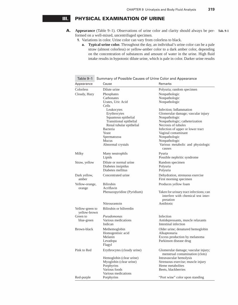
P1: OSO
LWBK192-09 LWBK192-Hubbard November 27, 2008 7:51
CHAPTER 9 Urinalysis and Body Fluid Analysis 319
III. PHYSICAL EXAMINATION OF URINE
A. Appearance (Table 9–1). Observations of urine color and clarity should always be per- Tab. 9-1
formed on a well-mixed, uncentrifuged specimen.1. Variations in color. Urine color can vary from colorless to black.
a. Typical urine color. Throughout the day, an individual’s urine color can be a palestraw (almost colorless) or yellow-amber color to a dark amber color, dependingon the concentration of substances and amount of water in the urine. High fluidintake results in hypotonic dilute urine, which is pale in color. Darker urine results
Table 9–1 Summary of Possible Causes of Urine Color and Appearance
Appearance Cause Remarks
Colorless Dilute urine Polyuria; random specimen
Cloudy, Hazy PhosphatesCarbonatesUrates, Uric AcidCells
LeukocytesErythrocytesSquamous epithelialTransitional epithelialRenal tubular epithelial
BacteriaYeastSpermatozoaMucusAbnormal crystals
NonpathologicNonpathologicNonpathologic
Infection; InflammationGlomerular damage; vascular injuryNonpathologicNonpathologic; catheterizationNecrosis of tubulesInfection of upper or lower tractVaginal contaminantNonpathologicNonpathologicVarious metabolic and physiologic
causes
Milky Many neutrophilsLipids
PyuriaPossible nephritic syndrome
Straw, yellow Dilute or normal urineDiabetes insipidusDiabetes mellitus
Random specimenPolyuriaPolyuria
Dark yellow,amber
Concentrated urine Dehydration, strenuous exerciseFirst morning specimen
Yellow-orange,orange
BilirubinAcriflavinPhenazopyridine (Pyridium)
Nitrourantoin
Produces yellow foam
Taken for urinary tract infections; caninterfere with chemical test inter-pretation
Antibiotic
Yellow-green toyellow-brown
Bilirubin or biliverdin
Green toblue-green
PseudomonasVarious medicationsIndican
InfectionAntidepressants, muscle relaxantsIntestinal infection
Brown-black MethemoglobinHomogentisic acidMelaninLevadopaFlagyl
Older urine; denatured hemoglobinAlkaptonuriaExcess production by melanomaParkinson disease drug
Pink to Red Erythrocytes (cloudy urine)
Hemoglobin (clear urine)Myoglobin (clear urine)PorphyrinsVarious foodsVarious medications
Glomerular damage; vascular injury;menstrual contamination (clots)
Intravascular hemolysisStrenuous exercise; muscle injuryHeme metabolitesBeets, blackberries
Red-purple Porphyrins “Port wine” color upon standing
P1: OSO
LWBK192-09 LWBK192-Hubbard November 27, 2008 7:51
320 A Concise Review of Clinical Laboratory Science
from reduced fluid intake resulting in a darker, hypertonic urine. Urine color isproduced by the presence of three pigments:(1) Urochrome, which is a yellow pigment that is present in the highest concen-
tration and is secreted by tubule cells;(2) Uroerythrin, which is a red pigment(3) Urobilin, which is an orange-red pigment from the oxidation of urobilinogen
b. Abnormal urine color may result from pathologic or nonpathologic conditions,such as medications and diet. Some dyes used to color food may cause changesin urine color, as can the ingestion of rhubarb and beets in genetically susceptibleindividuals.(1) Red or red-brown is the most commonly seen abnormal urine color. A
red color usually indicates the presence of blood (hematuria) or hemoglobin(hemoglobinuria).(a) If this color is seen in the urine of a female, contamination with menstrual
blood should be considered.(b) Other causes of red-colored urine include myoglobin (myoglobinuria),
porphyrins (porphyrinuria), food dyes, or ingestion of rhubarb or beets.(2) Dark brown to black urine can be caused by alkaptonuria or malignant
melanoma.(a) Alkaptonuria is a rare disorder caused by a lack of the enzyme homogentisic
acid oxidase. This enzyme is required for the catabolism of tyrosine andphenylalanine. In this disorder, homogentisic acid is excreted into the urine.The urine is normal in color when freshly voided but turns black on standingor when alkalinized.
(b) Individuals who have malignant melanoma excrete melanogen into theirurine. On exposure to light, this chromogen is converted to melanin, whichis black.
(3) Yellow-brown to yellow-green urine results from the excretion of bilirubin(oxidized to biliverdin) or bile pigments into the urine of individuals who haveobstructive jaundice.
(4) Colors ranging from green to blue typically indicate the presence of a drugmetabolite or dye; however, the presence of indican (resulting from abnormalbreakdown of tryptophan) will produce blue urine. Green urine may indicatea bacterial infection with Pseudomonas.
2. Urine appearance, transparency, clarity, turbidity or character indicates the presenceof particulate matter. It is important to examine a urine specimen at room temperaturefor clarity.a. Urine is essentially clear, although cloudy turbid urine does not always indicate a
pathologic condition.b. Turbidity is often caused by crystal precipitation, referred to as amorphous mate-
rial, when a specimen is refrigerated. This is a nonpathologic cause of turbidity.Other nonpathologic causes of turbidity include the presence of mucus, powders,squamous epithelial cells, and spermatozoa (in women). Pathologic causes of turbidurine include the presence of bacteria, yeast, cells, abnormal crystals, spermatozoa(in men), prostatic fluid, or lipids.
B. Odor indications in a urine sample. Odor is typically not reported on a urinalysis reportunless it is unusually prominent.
1. Normal odors. Urine has a faint, aromatic odor caused by the presence of volatile acidsproduced by the ingestion of various foods. Distinctive odors may result after ingestionof certain foods (e.g., asparagus, garlic, onions).
2. Abnormal odors may indicate pathologic conditions, improper handling, or improperstorage of the urine specimen. If allowed to stand, especially unrefrigerated, bacteriain a urine specimen break down urea to form ammonia and a strong ammoniacal odor.a. Urinary tract infections. Large numbers of bacteria present in urine specimens
from individuals who have urinary tract infections (UTIs) can result in a putrid,foul odor.
P1: OSO
LWBK192-09 LWBK192-Hubbard November 27, 2008 7:51
CHAPTER 9 Urinalysis and Body Fluid Analysis 321
b. Ketone bodies. Increased ketone bodies excreted by diabetic or individuals whoare on starvation diets give the urine a fruity odor.
c. Tubular necrosis. A lack of any odor may indicate acute tubular necrosis.d. Amino acid disorders produce characteristic smelling urines.
(1) Phenylketonuria is associated with a mousy-smelling urine.(2) Tyrosinuria produces a rancid odor.(3) Maple syrup urine disease produces a maple syrup-smelling urine.(4) Methionine malabsorption causes urine to smell like cabbage.(5) Isovaleric acid and glutaric acid in urine give it a sweaty feet smell.(6) Trimethylaminuria makes urine smell like rotting fish.
C. Specific gravity, refractive index, and osmolality measurements of urine indicate the amountof dissolved solids in urine or its concentration.
1. Specific gravity is a measure of the weight of a substance compared with an equalvolume of pure, solute-free water at the same temperature. Urine is water that containsdissolved substances, primarily urea, sodium, and chloride. The specific gravity of urineis a measure of its density, and it is influenced by the number and size of the particlespresent. Because the specific gravity of water is 1.000, and the specific gravity of urineis compared with it, the number is a ratio and has no units. For urine, the specific gravityis reported to the third decimal place with the healthy reference interval from 1.003to 1.035.
2. Specific gravity reflects the ability of the kidney to concentrate and dilute urine. Ordi-narily, the specific gravity of urine is inversely proportional to the volume. With diseaseof the kidney, this ability is lost, and the specific gravity is fixed at 1.010 (similar tothe initial plasma filtrate concentration of the glomerulus) and is termed isosthenuria.Urine specific gravity below 1.010 is termed hyposthenuric and above 1.010 is termedhypersthenuric.
3. There are three basic methods for determining the specific gravity of urine: byurinometer, refractometer, and reagent dipstick. The reagent strip is most commonlyused. Harmonic oscillation is used in automated instruments to determine specificgravity (see Section VI below).a. The urinometer is rarely used in a clinical laboratory and is included here only for
completeness. It is a glass float weighted with mercury with an air bubble abovethe weight and a graduated stem on top. The weighted float displaces a volume ofliquid equal to its weight and is calibrated at a specific temperature to sink to a levelof 1.000 in distilled water. Dissolved substances in urine provide additional massthat causes the float to displace a smaller volume of urine than distilled water. Thespecific gravity measurement is read at the bottom of the meniscus on the stem ofthe urinometer. Urinometry is considered an inaccurate method.
b. A refractometer is not often used in a clinical laboratory. It measures the refractiveindex of a solution, which is the ratio of the velocity of light in air to the velocity oflight in a solution. The velocity depends on the number of dissolved particles in thesolution and determines the angle at which light passes through the solution. Theclinical refractometer measures the angle and mathematically converts this angle tospecific gravity, which is read from a scale in the handheld instrument. Refractome-ters should be calibrated each day of use. Distilled water and sodium chloride orsucrose solutions of known concentration, as well as commercial controls, shouldbe measured and recorded.(1) The major advantages of the refractometer method are: a small volume of
specimen is required (1–2 drops); no temperature corrections are required; itis simple to operate; and it gives rapid, reliable results.
(2) Disadvantages include required corrections for large amounts of glucose andprotein, as with the urinometer, and a scale reading maximum of 1.035. Veryconcentrated specimens or urine samples contaminated with radiographic dyesneed to be diluted and remeasured.
c. Reagent strips can be used to measure the concentration of ions and give anindirect measure of specific gravity. The results are not identical to specific gravity
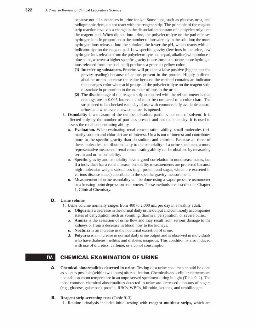
P1: OSO
LWBK192-09 LWBK192-Hubbard November 27, 2008 7:51
322 A Concise Review of Clinical Laboratory Science
because not all substances in urine ionize. Some ions, such as glucose, urea, andradiographic dyes, do not react with the reagent strip. The principle of the reagentstrip reaction involves a change in the dissociation constant of a polyelectrolyte onthe reagent pad. When dipped into urine, the polyelectrolyte on the pad releaseshydrogen ions in proportion to the number of ions already in the solution; the morehydrogen ions released into the solution, the lower the pH, which reacts with anindicator dye on the reagent pad. Low specific gravity (few ions in the urine, fewhydrogen ions released from the polyelectrolyte on the pad, alkaline) will produce ablue color, whereas a higher specific gravity (more ions in the urine, more hydrogenions released from the pad, acid) produces a green to yellow color.(1) Interfering substances. Proteins will produce a false positive (higher specific
gravity reading) because of anions present in the protein. Highly bufferedalkaline urines decrease the value because the method contains an indicatorthat changes color when acid groups of the polyelectrolyte on the reagent stripdissociate in proportion to the number of ions in the urine.
(2) The disadvantage of the reagent strip compared with the refractometer is thatreadings are in 0.005 intervals and must be compared to a color chart. Thestrips need to be checked each day of use with commercially available controlurines and whenever a new container is opened.
4. Osmolality is a measure of the number of solute particles per unit of solvent. It isaffected only by the number of particles present and not their density. It is used toassess the renal concentrating ability.a. Evaluation. When evaluating renal concentration ability, small molecules (pri-
marily sodium and chloride) are of interest. Urea is not of interest and contributesmore to the specific gravity than do sodium and chloride. Because all three ofthese molecules contribute equally to the osmolality of a urine specimen, a morerepresentative measure of renal concentrating ability can be obtained by measuringserum and urine osmolality.
b. Specific gravity and osmolality have a good correlation in nondisease states, butif a individual has a renal disease, osmolality measurements are preferred becausehigh-molecular-weight substances (e.g., protein and sugar, which are excreted invarious disease states) contribute to the specific gravity measurement.
c. Measurement of urine osmolality can be done using a vapor pressure osmometeror a freezing-point depression osmometer. These methods are described in Chapter1, Clinical Chemistry.
D. Urine volume1. Urine volume normally ranges from 400 to 2,000 mL per day in a healthy adult.
a. Oliguria is a decrease in the normal daily urine output and commonly accompaniesstates of dehydration, such as vomiting, diarrhea, perspiration, or severe burns.
b. Anuria is the cessation of urine flow and may result from serious damage to thekidneys or from a decrease in blood flow to the kidneys.
c. Nocturia is an increase in the nocturnal excretion of urine.d. Polyuria is an increase in normal daily urine output and is observed in individuals
who have diabetes mellitus and diabetes insipidus. This condition is also inducedwith use of diuretics, caffeine, or alcohol consumption.
IV. CHEMICAL EXAMINATION OF URINE
A. Chemical abnormalities detected in urine. Testing of a urine specimen should be doneas soon as possible (within two hours) after collection. Chemicals and cellular elements arenot stable at room temperature in an unpreserved specimen sitting in light (Table 9–2). Themost common chemical abnormalities detected in urine are increased amounts of sugars(e.g., glucose, galactose), protein, RBCs, WBCs, bilirubin, ketones, and urobilinogen.
B. Reagent strip screening tests (Table 9–3)1. Routine urinalysis includes initial testing with reagent multitest strips, which are
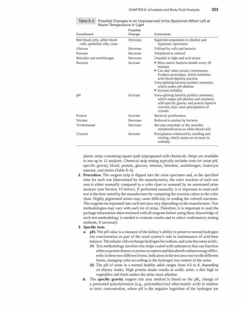
P1: OSO
LWBK192-09 LWBK192-Hubbard November 27, 2008 7:51
CHAPTER 9 Urinalysis and Body Fluid Analysis 323
Table 9–2 Possible Changes in an Unpreserved Urine Specimen When Left atRoom Temperature in Light
Possible
Constituent Change Comments
Red blood cells, white bloodcells, epithelial cells, casts
Decrease Rapid decomposition in alkaline andhypotonic specimens
Glucose Decrease Utilized by cells and bacteria
Ketones Decrease Volatilized or utilized
Bilirubin and urobilinogen Decrease Unstable in light and acid urines
Bacteria Increase � Most enteric bacteria double every 20minutes
� Can alter other urinary constituents:Produce peroxidase, which interfereswith blood dipstick reaction
Urea-splitting bacteria produce ammonia,which makes pH alkaline
� Increase turbidity
pH Increase Urea-splitting bacteria produce ammonia,which makes pH alkaline and interfereswith specific gravity and protein dipstickreaction; may cause precipitation ofcrystals
Protein Increase Bacterial proliferation
Nitrates Decrease Reduced to nitrites by bacteria
Trichomonad Decrease Become immobile or die; possiblemisidentification as white blood cells
Crystals Increase Precipitation enhanced by standing andcooling, which causes an increase inturbidity
plastic strips containing square pads impregnated with chemicals. Strips are availableto test up to 12 analytes. Chemical strip testing typically includes tests for urine pH,specific gravity, blood, protein, glucose, ketones, bilirubin, urobilinogen, leukocyteesterase, and nitrite (Table 9–4).
2. Procedure. The reagent strip is dipped into the urine specimen and, at the specifiedtime for each test (determined by the manufacturer), the color reaction of each testarea is either manually compared to a color chart or assessed by an automated urineanalyzer (see Section VI below). If performed manually, it is important to read eachtest at the time stated by the manufacturer by comparing the reaction colors to the colorchart. Highly pigmented urines may cause difficulty in reading the colored reactions.The reagents incorporated into each test area vary depending on the manufacturer. Testmethodologies may vary with each lot of strips. Therefore, it is important to read thepackage information sheet enclosed with all reagents before using them. Knowledge ofeach test methodology is needed to evaluate results and to select confirmatory testingmethods, if necessary.
3. Specific testsa. pH. The pH value is a measure of the kidney’s ability to preserve normal hydrogen
ion concentration as part of the renal system’s role in maintenance of acid-basebalance. The tubular cells exchange hydrogen for sodium, and urine becomes acidic.(1) Test methodology involves test strips coated with substances that can function
either as proton donors or proton acceptors and that absorb radiant energy differ-ently in these two different forms. Indicators in the test area react to the differentforms, changing color according to the hydrogen ion content of the urine.
(2) The pH of urine in a normal healthy adult ranges from 4.6 to 8, dependingon dietary intake. High protein intake results in acidic urine; a diet high invegetables and fruits makes the urine more alkaline.
b. The specific gravity reagent test area method is based on the pKa change ofa pretreated polyelectrolyte (e.g., polymethylvinyl ether/maleic acid) in relationto ionic concentration, where pH is the negative logarithm of the hydrogen ion
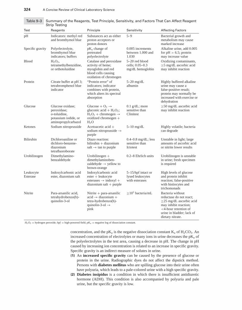
P1: OSO
LWBK192-09 LWBK192-Hubbard November 27, 2008 7:51
324 A Concise Review of Clinical Laboratory Science
Table 9–3 Summary of the Reagents, Test Principle, Sensitivity, and Factors That Can Affect ReagentStrip Testing
Test Reagents Principle Sensitivity Affecting Factors
pH Indicators: methyl redand bromthymol blue
Substances act as eitherproton acceptors orproton donors
5–9 Bacterial growth andmetabolism may causemarked increase
Specific gravity Polyelectrolyte,bromthymol blueindicators; buffers
pKa change ofpretreatedpolyelectrolyte
0.005 incrementsbetween 1.000 and1.030
Alkaline urine, add 0.005for pH > 6.5; proteinmay increase value
Blood H2O2,tetramethylbenzidine,or orthotoluidine
Catalase and peroxidaseactivity of heme;myoglobin and redblood cells causingoxidation of chromogen
5–20 red bloodcells; 0.05–0.3mg/dL hemoglobin
Oxidizing contaminants,≥5 mg/dL ascorbic acidmay inhibit reaction
Protein Citrate buffer at pH 3;tetrabromphenol blueindicator
“Protein error” ofindicators; indicatorcombines with protein,which alters its spectralabsorption
5–20 mg/dLalbumin
Highly buffered alkalineurine may cause afalse-positive result;protein may normally beincreased with exercise ordehydration
Glucose Glucose oxidase;peroxidase;o-toluidine,potassium iodide, oraminopropylcarbazol
Glucose + O2 →gluconic acid + H2O2;H2O2 + chromogen →oxidized chromogen +H2O
0.1 g/dL; moresensitive thanClinitest
≥50 mg/dL ascorbic acidmay inhibit reaction
Ketones Sodium nitroprusside Acetoacetic acid +sodium nitroprusside →purple
5–10 mg/dL Highly volatile; bacteriacan degrade
Bilirubin Dichloroaniline ordichloro-benzene-diazoniumtetrafluoroborate
Diazo reaction:bilirubin + diazoniumsalt → tan to purple
0.4–0.8 mg/dL; lesssensitive thanIctotest
Unstable in light; largeamounts of ascorbic acidor nitrite lower results
Urobilinogen Dimethylamino-benzaldehyde
Urobilinogen +dimethylaminoben-zaldehyde → yellow tobrown-orange
0.2–8 Ehrlich units Urobilinogen is unstablein urine; fresh specimenis required
LeukocyteEsterase
Indoxylcarbonic acidester, diazonium salt
Indoxylcarbonic acidester + leukocyteesterases → indoxyl +diazonium salt + purple
5–15/hpf intact orlysed leukocyteswith esterases
High levels of glucoseand protein inhibitreaction; false-positivewith histiocytes andtrichomonads
Nitrite Para-arsanilic acid,tetrahydrobenzo(h)-quinolin-3-ol
Nitrite + para-arsanilicacid → diazonium +tetra-hydrobenzo(h)-quinolin-3-ol →pink
≥105 bacteria/mL Bacteria withoutreductase do not react;≥25 mg/dL ascorbic acidmay inhibit reaction;<4-hour retention ofurine in bladder; lack ofdietary nitrate.
H2 O2 = hydrogen peroxide; hpf = high-powered field; pKa = negative log of dissociation constant.
concentration, and the pKa is the negative dissociation constant Ka of H2CO3. Anincreased concentration of electrolytes or many ions in urine decreases the pKa ofthe polyelectrolytes in the test area, causing a decrease in pH. The change in pHcaused by increasing ion concentration is related to an increase in specific gravity.Specific gravity is an indirect measure of solutes in urine.(1) An increased specific gravity can be caused by the presence of glucose or
protein in the urine. Radiographic dyes do not affect the dipstick method.Persons with diabetes mellitus who are spilling glucose into their urine oftenhave polyuria, which leads to a pale-colored urine with a high specific gravity.
(2) Diabetes insipidus is a condition in which there is insufficient antidiuretichormone (ADH). This condition is also accompanied by polyuria and paleurine, but the specific gravity is low.
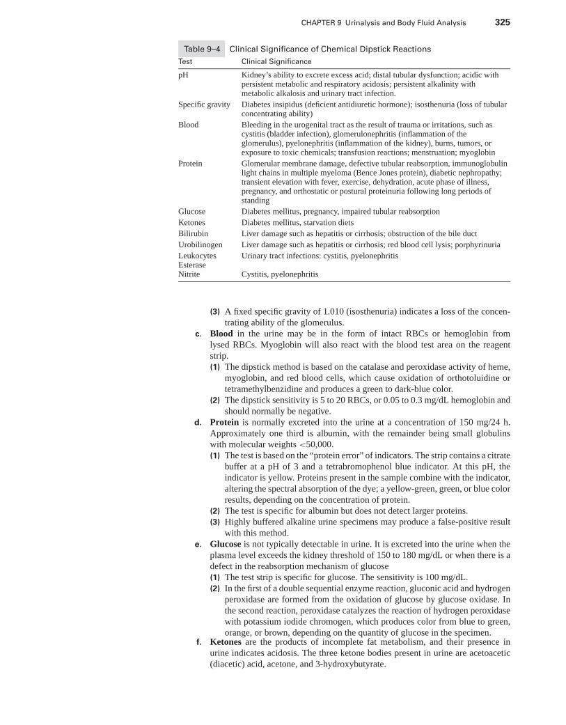
P1: OSO
LWBK192-09 LWBK192-Hubbard November 27, 2008 7:51
CHAPTER 9 Urinalysis and Body Fluid Analysis 325
Table 9–4 Clinical Significance of Chemical Dipstick Reactions
Test Clinical Significance
pH Kidney’s ability to excrete excess acid; distal tubular dysfunction; acidic withpersistent metabolic and respiratory acidosis; persistent alkalinity withmetabolic alkalosis and urinary tract infection.
Specific gravity Diabetes insipidus (deficient antidiuretic hormone); isosthenuria (loss of tubularconcentrating ability)
Blood Bleeding in the urogenital tract as the result of trauma or irritations, such ascystitis (bladder infection), glomerulonephritis (inflammation of theglomerulus), pyelonephritis (inflammation of the kidney), burns, tumors, orexposure to toxic chemicals; transfusion reactions; menstruation; myoglobin
Protein Glomerular membrane damage, defective tubular reabsorption, immunoglobulinlight chains in multiple myeloma (Bence Jones protein), diabetic nephropathy;transient elevation with fever, exercise, dehydration, acute phase of illness,pregnancy, and orthostatic or postural proteinuria following long periods ofstanding
Glucose Diabetes mellitus, pregnancy, impaired tubular reabsorption
Ketones Diabetes mellitus, starvation diets
Bilirubin Liver damage such as hepatitis or cirrhosis; obstruction of the bile duct
Urobilinogen Liver damage such as hepatitis or cirrhosis; red blood cell lysis; porphyrinuria
LeukocytesEsterase
Urinary tract infections: cystitis, pyelonephritis
Nitrite Cystitis, pyelonephritis
(3) A fixed specific gravity of 1.010 (isosthenuria) indicates a loss of the concen-trating ability of the glomerulus.
c. Blood in the urine may be in the form of intact RBCs or hemoglobin fromlysed RBCs. Myoglobin will also react with the blood test area on the reagentstrip.(1) The dipstick method is based on the catalase and peroxidase activity of heme,
myoglobin, and red blood cells, which cause oxidation of orthotoluidine ortetramethylbenzidine and produces a green to dark-blue color.
(2) The dipstick sensitivity is 5 to 20 RBCs, or 0.05 to 0.3 mg/dL hemoglobin andshould normally be negative.
d. Protein is normally excreted into the urine at a concentration of 150 mg/24 h.Approximately one third is albumin, with the remainder being small globulinswith molecular weights <50,000.(1) The test is based on the “protein error” of indicators. The strip contains a citrate
buffer at a pH of 3 and a tetrabromophenol blue indicator. At this pH, theindicator is yellow. Proteins present in the sample combine with the indicator,altering the spectral absorption of the dye; a yellow-green, green, or blue colorresults, depending on the concentration of protein.
(2) The test is specific for albumin but does not detect larger proteins.(3) Highly buffered alkaline urine specimens may produce a false-positive result
with this method.e. Glucose is not typically detectable in urine. It is excreted into the urine when the
plasma level exceeds the kidney threshold of 150 to 180 mg/dL or when there is adefect in the reabsorption mechanism of glucose(1) The test strip is specific for glucose. The sensitivity is 100 mg/dL.(2) In the first of a double sequential enzyme reaction, gluconic acid and hydrogen
peroxidase are formed from the oxidation of glucose by glucose oxidase. Inthe second reaction, peroxidase catalyzes the reaction of hydrogen peroxidasewith potassium iodide chromogen, which produces color from blue to green,orange, or brown, depending on the quantity of glucose in the specimen.
f. Ketones are the products of incomplete fat metabolism, and their presence inurine indicates acidosis. The three ketone bodies present in urine are acetoacetic(diacetic) acid, acetone, and 3-hydroxybutyrate.
P1: OSO
LWBK192-09 LWBK192-Hubbard November 27, 2008 7:51
326 A Concise Review of Clinical Laboratory Science
(1) The test method is based on the reaction of sodium nitroprusside with ace-toacetic acid. The test does not detect acetone or 3-hydroxybutyrate, but if oneketone is excreted, all are excreted. Positive reactions produce a maroon color.
(2) The test sensitivity is 5 to 10 mg/dL of acetoacetic acid.g. Bilirubin is not normally detectable in urine. The reagent strip method can detect
0.4 to 0.8 mg/dL bilirubin with the diazo reaction, in which 2,4 dichloroanilinediazonium salt reacts with bilirubin to produce a tan-to-purple color.
h. Urobilinogen is normally present in urine in small amounts up to 4 mg/24 h.(1) Paradimethylaminobenzaldehyde (Ehrlich’s reagent) reacts with urobilinogen
(and porphobilinogen, depending on the brand of reagent strips) to form colorsranging from light yellow to brown-orange.
(2) In a random urine specimen, the normal concentration on the test strip reflects0.2 and 1.0 Ehrlich units (EU). Increased concentrations can be detected bythe strip method, but the method is not suitable for detecting decreases inconcentration. Fresh specimens should be used for testing because urobilinogenis unstable in urine.
i. Leukocytes can be detected in urine with the leukocyte esterase reaction. Granulo-cytic leukocyte esterases catalyze the hydrolysis of the derivative pyrrole-N-tosyl-L-alanine ester to form pyrrole alcohol. The alcohol then reacts with a diazoniumsalt to produce a purple color.(1) The test strip can detect 5 to 15 leukocytes per high-power field and is even
sensitive to lysed granulocytes.(2) Vaginal cellular contamination or trichomonads can cause a false-positive re-
sult.j. The nitrite test provides a rapid screen for the detection of bacteria that are capable
of reducing nitrates to nitrite. The test results should be negative.(1) False-negative reactions may occur if:
(a) The bacteria in the specimen do not have the enzymes necessary to reducenitrates.
(b) The diet of the individual is deficient in nitrates.(c) The bacteria reduce the nitrate beyond the nitrate state to nitrogen or am-
monia.(d) The urine has not been in the bladder at least 4 hours, so the bacteria did
not yet reduce the nitrates.(2) On the reagent strip reaction, the Griess reaction detects nitrites that react
with an aromatic amine at an acid pH. The resultant azo product reacts with ahydrobenzoquinolin compound to form a pink color in the presence of nitrite.
C. Confirmatory Testing1. Glucose and other reducing substances, such as other disaccharides (e.g., fructose,
galactose, lactose, pentose) and others (e.g., ascorbic acid) in the urine can be detectedwith the copper reductase test (Clinitest).a. The test uses the reagents copper sulfate, sodium hydroxide, sodium carbonate,
and citric acid incorporated into a tablet.b. In the five-drop method, five drops of urine are added to 10 drops of distilled water
in a test tube. One Clinitest tablet is added, and the solution begins to boil. Coppersulfate reacts with reducing substances in urine, converting cupric ions to cuprousions in an exothermic process.(1) After 15 seconds, the tube is gently mixed and compared to a color chart. A neg-
ative reaction is blue. Depending on the concentration of reducing substance(s),the solution changes from green-blue to orange.
(2) It is important to watch the reaction because if a high glucose concentrationis present (>2 g/dL), the color reaction will pass from orange to a brownishcolor and back to blue (“pass-through” phenomenon). If this occurs, the urinemust be retested at a lower concentration with a two-drop method (same pro-cedure as the five-drop method, except two drops of urine are added to the testtube).
P1: OSO
LWBK192-09 LWBK192-Hubbard November 27, 2008 7:51
CHAPTER 9 Urinalysis and Body Fluid Analysis 327
c. The test sensitivity is 150 mg/dL, which is less sensitive than the dipstick method.Thus, at low glucose concentrations, it is possible to have a positive dipstick testand a negative Clinitest.(1) It is also possible to have a positive Clinitest and a negative dipstick test if the
sugar present is one other than glucose, or the urine contains another reducingsubstance (e.g., ascorbic acid).(a) Nonglucose mellituria can occur in certain conditions. Inherited enzyme
deficiencies can cause the accumulation of lactose, fructose, galactose,pentose, or sucrose in plasma. All of the sugar cannot be reabsorbed, andit appears in urine.
(b) Lactosuria can occur during late pregnancy or during lactation.(c) Pentosuria can occur following ingestion of certain fruits.(d) Fructosuria can occur with parenteral feedings with fructose.
(2) Sucrose is not a reducing sugar, so it does not react in the Clinitest.2. Precipitation tests for protein are more sensitive than are the reagent strip methods
and detect other proteins besides albumin.a. An aliquot (usually 1 mL) of 3% sulfosalicylic acid or trichloroacetic acid is added
to an equal amount of urine in a test tube and mixed by inversion. The tube isallowed to stand for 10 minutes and then is inverted twice.
b. The degree of precipitation is graded according to the following criteria:(1) Negative: no turbidity, ≤5 mg/dL protein(2) 1 +: faint turbidity but no discrete granulation; ≈50 mg/dL protein(3) 2 +: turbidity with granulation but no flocculation; ≈200 mg/dL protein(4) 3 +: turbidity with granulation and flocculation; ≈500 mg/dL protein(5) 4 +: clumps of precipitation; ≥1.0 g/dL protein
c. The test sensitivity is 5 to 10 mg/dL and detects albumin, globulins, glycoproteins,and Bence Jones protein. Radiographic dyes react with this method. When theradiographic dyes are present, the specific gravity of the urine is usually >1.035,and typical crystals are seen on the microscopic examination of the urine speci-men.
3. The Ictotest r© is a sensitive test for the detection of bilirubin in urine in which2,4-dichloro-benzene-diazonium tetrachlorozincate, sodium bicarbonate, and sulfos-alicylic acid are combined into a tablet.a. Five drops of urine are placed on an asbestos cellulose test pad, which concentrates
the specimen so that much smaller concentrations of bilirubin (0.05–0.1 mg/dL)can be detected as compared with the dipstick method.
b. The urine is allowed to absorb into the pad for 1 minute, and then a reagent tabletis placed on the wet area of the pad.
c. One drop of distilled water is placed onto the tablet. After 5 seconds, a second dropof distilled water is placed onto the tablet so that the drops run down the side ofthe tablet onto the test pad.
d. The tablet is removed, and the test pad is observed within 1 minute for a purplecolor, which indicates the presence of bilirubin. A pink or red color is a negativereaction.
V. MICROSCOPIC EXAMINATION OF THE URINE
A. Urine sediment preparation1. To ensure the accuracy and reproducibility of the urine microscopic examination, each
laboratory must establish a protocol for the preparation of the urine sediment. Bright-field microscopy is the most commonly used method of sediment examination. To seesediments with a low refractive index, such as hyaline casts, sediment should be ex-amined with decreased light obtained by decreasing the illumination, not by loweringthe condenser. Phase-contrast microscopy is used to produce a phase difference ofwavelengths of light, which gives better contrast when viewing low refractive indexsediments (hyaline and cellular casts, mucous threads, and Trichomonas). Polarizing
P1: OSO
LWBK192-09 LWBK192-Hubbard November 27, 2008 7:51
328 A Concise Review of Clinical Laboratory Science
microscopy involves the use of polarizing filters for observation of sediments that canrotate and refract light in two dimensions at 90◦ to each other (e.g., lipids and crystals).
2. Factors that must be considered in urinalysis to help standardize the microscopicexamination include the following:a. Appropriate specimen collection, preservation, and handling. It is best to examine
a specimen within 1 hour of individual voiding because many aspects of the chem-ical and microscopic examination change as the urine stands, especially at roomtemperature.
b. A standard amount of urine, usually 12 mL, is placed in a conical tube and cen-trifuged for a uniform amount of time and speed, typically 5 minutes at a relativecentrifugal force of 400.
c. The spun tube is decanted, which leaves approximately 1 mL of fluid in which toresuspend the urinary sediment. If a stain is used, it should be added at this time.
d. A small drop of well-mixed sediment is placed onto a microscope slide and shieldedwith a coverslip. Slides with uniform wells and coverslips are commercially avail-able for the microscopic examination of urine. Drop size determines the amountof sediment viewed.
e. A consistent method should be used to examine the urinary sediment. The slide isfirst viewed on low power (10×) to assess the overall composition of the specimenand to observe and count casts. Ten fields are examined at this magnification. Thelight must be very low to see hyaline casts. Other elements are counted on highpower (40×). An average count of sediments in ten fields is usually reported.
f. Microscopic results must be correlated with the color, appearance, and dipstickreactions for each specimen.
3. Microscopic sediment stains (supravital stains) can be used to aid the identificationof formed elements.a. The most commonly used stain is the Sternheimer-Malbin stain, which consists of
crystal violet and safranin O. This stain provides a more detailed visualization ofthe internal structure of cells and casts.
b. Another supravital urine sediment stain is 0.5% toluidine blue. It differentiallystains various cell components (e.g., the nucleus, cytoplasm) to help distinguishcells that may be similar in size, such as leukocytes and renal cells.
c. Sudan III or oil red O stains are used to confirm the presence of neutral lipids.Lipids or fats within renal cells or histiocytes (oval fat bodies) or free-floatingtriglycerides stain red or orange with these two stains.
d. The Prussian blue stain is used to confirm the presence of hemosiderin (iron) inepithelial cells and casts, as well as hemosiderin that is free-floating. The iron turnsa characteristic blue color.
e. Hansel stain consists of methylene blue and eosin-Y in methanol. It is used toidentify eosinophils.
f. One to two drops of 2% acetic acid added to a few drops of urine sediment can beused to differentiate RBCs from yeast cells, small WBCs, or epithelial cells. TheRBCs lyse; the yeast cells remain intact. The internal structures of WBCs and theepithelial cells are accentuated.
B. Normal and abnormal cells in urine (Figure 9–1)1. RBCs are small biconcave disks without a nucleus. They are 7–10 μm in diameter.
In a hypertonic urine they become crenated, appearing to have a crinkled border. In ahypotonic, alkaline urine, the RBCs swell and may lyse. These lysed cell membranesare called “ghost” or “shadow cells” and appear as faint, colorless circles. Normally, aurine specimen can contain 0 to 3 RBCs per high-power field; increased numbers mayindicate renal bleeding or glomerulonephritis (see Web Color Image 9–2).
2. WBCs, usually neutrophils, are larger than RBCs (10–15 μm diameter and containa distinct nucleus. A normal urine sample contains 0 to 5 WBCs per high-powerfield. An increase is called pyuria and indicates the presence of an infection or in-flammation in the genitourinary tract. Frequent causes of pyuria include bacterialinfections (e.g., cystitis, pyelonephritis, prostatitis, urethritis) or nonbacterial disor-
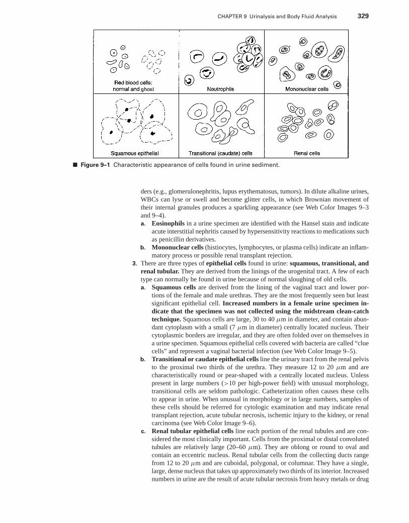
P1: OSO
LWBK192-09 LWBK192-Hubbard November 27, 2008 7:51
CHAPTER 9 Urinalysis and Body Fluid Analysis 329
■ Figure 9–1 Characteristic appearance of cells found in urine sediment.
ders (e.g., glomerulonephritis, lupus erythematosus, tumors). In dilute alkaline urines,WBCs can lyse or swell and become glitter cells, in which Brownian movement oftheir internal granules produces a sparkling appearance (see Web Color Images 9–3and 9–4).a. Eosinophils in a urine specimen are identified with the Hansel stain and indicate
acute interstitial nephritis caused by hypersensitivity reactions to medications suchas penicillin derivatives.
b. Mononuclear cells (histiocytes, lymphocytes, or plasma cells) indicate an inflam-matory process or possible renal transplant rejection.
3. There are three types of epithelial cells found in urine: squamous, transitional, andrenal tubular. They are derived from the linings of the urogenital tract. A few of eachtype can normally be found in urine because of normal sloughing of old cells.a. Squamous cells are derived from the lining of the vaginal tract and lower por-
tions of the female and male urethras. They are the most frequently seen but leastsignificant epithelial cell. Increased numbers in a female urine specimen in-dicate that the specimen was not collected using the midstream clean-catchtechnique. Squamous cells are large, 30 to 40 μm in diameter, and contain abun-dant cytoplasm with a small (7 μm in diameter) centrally located nucleus. Theircytoplasmic borders are irregular, and they are often folded over on themselves ina urine specimen. Squamous epithelial cells covered with bacteria are called “cluecells” and represent a vaginal bacterial infection (see Web Color Image 9–5).
b. Transitional or caudate epithelial cells line the urinary tract from the renal pelvisto the proximal two thirds of the urethra. They measure 12 to 20 μm and arecharacteristically round or pear-shaped with a centrally located nucleus. Unlesspresent in large numbers (>10 per high-power field) with unusual morphology,transitional cells are seldom pathologic. Catheterization often causes these cellsto appear in urine. When unusual in morphology or in large numbers, samples ofthese cells should be referred for cytologic examination and may indicate renaltransplant rejection, acute tubular necrosis, ischemic injury to the kidney, or renalcarcinoma (see Web Color Image 9–6).
c. Renal tubular epithelial cells line each portion of the renal tubules and are con-sidered the most clinically important. Cells from the proximal or distal convolutedtubules are relatively large (20–60 μm). They are oblong or round to oval andcontain an eccentric nucleus. Renal tubular cells from the collecting ducts rangefrom 12 to 20 μm and are cuboidal, polygonal, or columnar. They have a single,large, dense nucleus that takes up approximately two thirds of its interior. Increasednumbers in urine are the result of acute tubular necrosis from heavy metals or drug
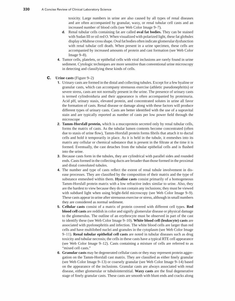
P1: OSO
LWBK192-09 LWBK192-Hubbard November 27, 2008 7:51
330 A Concise Review of Clinical Laboratory Science
toxicity. Large numbers in urine are also caused by all types of renal diseasesand are often accompanied by granular, waxy, or renal tubular cell casts and anincreased number of blood cells (see Web Color Image 9–7).
d. Renal tubular cells containing fat are called oval fat bodies. They can be stainedwith Sudan III or oil red O. When visualized with polarized light, these fat globulesdisplay a Maltese cross shape. Oval fat bodies often indicate glomerular dysfunctionwith renal tubular cell death. When present in a urine specimen, these cells areaccompanied by increased amounts of protein and cast formation (see Web ColorImage 9–8).
4. Tumor cells, platelets, or epithelial cells with viral inclusions are rarely found in urinesediment. Cytologic techniques are more sensitive than conventional urine microscopyin detecting and classifying these kinds of cells.
C. Urine casts (Figure 9–2)1. Urinary casts are formed in the distal and collecting tubules. Except for a few hyaline or
granular casts, which can accompany strenuous exercise (athletic pseudonephritis) orsevere stress, casts are not normally present in the urine. The presence of urinary castsis termed cylindroiduria and their appearance is often accompanied by proteinuria.Acid pH, urinary stasis, elevated protein, and concentrated solutes in urine all favorthe formation of casts. Renal disease or damage along with these factors will producedifferent types of urinary casts. Casts are better identified with the use of a supravitalstain and are typically reported as number of casts per low power field through themicroscope
2. Tamm-Horsfall protein, which is a mucoprotein secreted only by renal tubular cells,forms the matrix of casts. As the tubular lumen contents become concentrated (oftendue to stasis of urine flow), Tamm-Horsfall protein forms fibrils that attach it to ductalcells and hold it temporarily in place. As it is held in the tubule, it enmeshes into itsmatrix any cellular or chemical substance that is present in the filtrate at the time it isformed. Eventually, the cast detaches from the tubular epithelial cells and is flushedinto the urine.
3. Because casts form in the tubules, they are cylindrical with parallel sides and roundedends. Casts formed in the collecting ducts are broader than those formed in the proximaland distal convoluted tubules.
4. The number and type of casts reflect the extent of renal tubule involvement in dis-ease processes. They are classified by the composition of their matrix and the type ofsubstance enmeshed within them. Hyaline casts consist primarily of a homogeneousTamm-Horsfall protein matrix with a low refractive index similar to urine. Also, theyare the hardest to view because they do not contain any inclusions; they must be viewedwith subdued light when using bright-field microscopy (see Web Color Image 9–9).These casts appear in urine after strenuous exercise or stress, although in small numbersthey are considered as normal sediment.
5. Cellular casts consist of a matrix of protein covered with different cell types. Redblood cell casts are reddish in color and signify glomerular disease or physical damageto the glomerulus. The outline of an erythrocyte must be observed in part of the castto identify these (see Web Color Image 9–10). White blood cell (leukocyte) casts areassociated with pyelonephritis and infection. The white blood cells are larger than redcells and have multilobed nuclei and granules in the cytoplasm (see Web Color Image9–11). Renal tubular epithelial cell casts are noted in tubular diseases such as drugtoxicity and tubular necrosis; the cells in these casts have a typical RTE cell appearance(see Web Color Image 9–12). Casts containing a mixture of cells are referred to as“mixed cell casts.”
6. Granular casts may be degenerated cellular casts or they may represent protein aggre-gation on the Tamm-Horsfall cast matrix. They are classified as either finely granular(see Web Color Image 9–13) or coarsely granular (see Web Color Image 9–14) basedon the appearance of the inclusions. Granular casts are always associated with renaldisease, either glomerular or tubulointerstitial. Waxy casts are the final degenerativestage of finely granular casts. These casts are smooth with blunt ends and cracks along
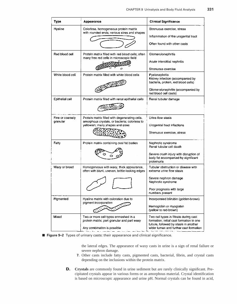
P1: OSO
LWBK192-09 LWBK192-Hubbard November 27, 2008 7:51
CHAPTER 9 Urinalysis and Body Fluid Analysis 331
■ Figure 9–2 Types of urinary casts: their appearance and clinical significance.
the lateral edges. The appearance of waxy casts in urine is a sign of renal failure orsevere nephron damage.
7. Other casts include fatty casts, pigmented casts, bacterial, fibrin, and crystal castsdepending on the inclusions within the protein matrix.
D. Crystals are commonly found in urine sediment but are rarely clinically significant. Pre-cipitated crystals appear in various forms or as amorphous material. Crystal identificationis based on microscopic appearance and urine pH. Normal crystals can be found in acid,
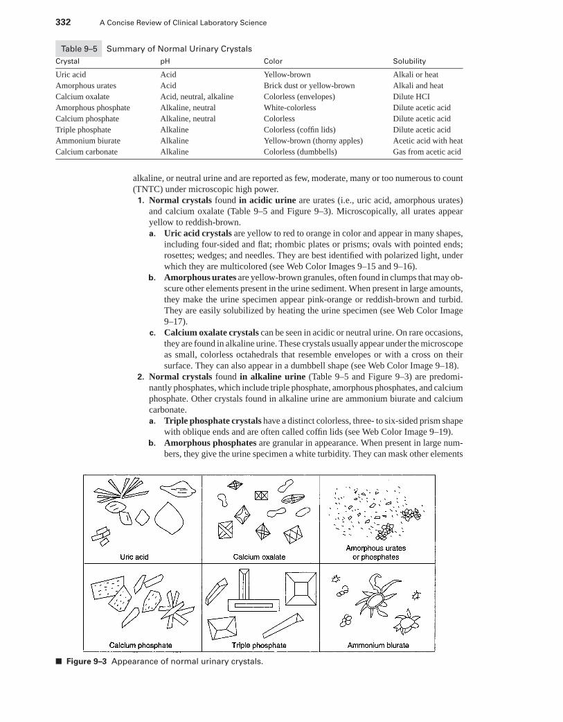
P1: OSO
LWBK192-09 LWBK192-Hubbard November 27, 2008 7:51
332 A Concise Review of Clinical Laboratory Science
Table 9–5 Summary of Normal Urinary Crystals
Crystal pH Color Solubility
Uric acid Acid Yellow-brown Alkali or heat
Amorphous urates Acid Brick dust or yellow-brown Alkali and heat
Calcium oxalate Acid, neutral, alkaline Colorless (envelopes) Dilute HCI
Amorphous phosphate Alkaline, neutral White-colorless Dilute acetic acid
Calcium phosphate Alkaline, neutral Colorless Dilute acetic acid
Triple phosphate Alkaline Colorless (coffin lids) Dilute acetic acid
Ammonium biurate Alkaline Yellow-brown (thorny apples) Acetic acid with heat
Calcium carbonate Alkaline Colorless (dumbbells) Gas from acetic acid
alkaline, or neutral urine and are reported as few, moderate, many or too numerous to count(TNTC) under microscopic high power.
1. Normal crystals found in acidic urine are urates (i.e., uric acid, amorphous urates)and calcium oxalate (Table 9–5 and Figure 9–3). Microscopically, all urates appearyellow to reddish-brown.a. Uric acid crystals are yellow to red to orange in color and appear in many shapes,
including four-sided and flat; rhombic plates or prisms; ovals with pointed ends;rosettes; wedges; and needles. They are best identified with polarized light, underwhich they are multicolored (see Web Color Images 9–15 and 9–16).
b. Amorphous urates are yellow-brown granules, often found in clumps that may ob-scure other elements present in the urine sediment. When present in large amounts,they make the urine specimen appear pink-orange or reddish-brown and turbid.They are easily solubilized by heating the urine specimen (see Web Color Image9–17).
c. Calcium oxalate crystals can be seen in acidic or neutral urine. On rare occasions,they are found in alkaline urine. These crystals usually appear under the microscopeas small, colorless octahedrals that resemble envelopes or with a cross on theirsurface. They can also appear in a dumbbell shape (see Web Color Image 9–18).
2. Normal crystals found in alkaline urine (Table 9–5 and Figure 9–3) are predomi-nantly phosphates, which include triple phosphate, amorphous phosphates, and calciumphosphate. Other crystals found in alkaline urine are ammonium biurate and calciumcarbonate.a. Triple phosphate crystals have a distinct colorless, three- to six-sided prism shape
with oblique ends and are often called coffin lids (see Web Color Image 9–19).b. Amorphous phosphates are granular in appearance. When present in large num-
bers, they give the urine specimen a white turbidity. They can mask other elements
■ Figure 9–3 Appearance of normal urinary crystals.
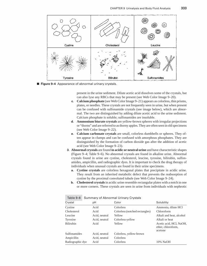
P1: OSO
LWBK192-09 LWBK192-Hubbard November 27, 2008 7:51
CHAPTER 9 Urinalysis and Body Fluid Analysis 333
■ Figure 9–4 Appearance of abnormal urinary crystals.
present in the urine sediment. Dilute acetic acid dissolves some of the crystals, butcan also lyse any RBCs that may be present (see Web Color Image 9–20).
c. Calcium phosphate (see Web Color Image 9–21) appears as colorless, thin prisms,plates, or needles. These crystals are not frequently seen in urine, but when presentcan be confused with sulfonamide crystals (see image below), which are abnor-mal. The two are distinguished by adding dilute acetic acid to the urine sediment.Calcium phosphate is soluble; sulfonamides are insoluble.
d. Ammonium biurate crystals are yellow-brown spheres with irregular projectionsor “thorns” and are referred to as thorny apples. They are often seen in old specimens(see Web Color Image 9–22).
e. Calcium carbonate crystals are small, colorless dumbbells or spheres. They of-ten appear in clumps and can be confused with amorphous phosphates. They aredistinguished by the formation of carbon dioxide gas after the addition of aceticacid (see Web Color Image 9–23).
3. Abnormal crystals are found in acidic or neutral urine and have characteristic shapes(Figure 9–4; Table 9–6). No abnormal crystals are found in alkaline urine. Abnormalcrystals found in urine are cystine, cholesterol, leucine, tyrosine, bilirubin, sulfon-amides, ampicillin, and radiographic dyes. It is important to check the drug therapy ofindividuals when unusual crystals are found in their urine specimens.a. Cystine crystals are colorless hexagonal plates that precipitate in acidic urine.
They result from an inherited metabolic defect that prevents the reabsorption ofcystine by the proximal convoluted tubule (see Web Color Image 9–24).
b. Cholesterol crystals in acidic urine resemble rectangular plates with a notch in oneor more corners. These crystals are seen in urine from individuals with nephrotic
Table 9–6 Summary of Abnormal Urinary Crystals
Crystal pH Color Solubility
Cystine Acid Colorless Ammonia, dilute HCl
Cholesterol Acid Colorless (notched rectangles) Chloroform
Leucine Acid, neutral Yellow Alkali and heat, alcohol
Tyrosine Acid, neutral Colorless-yellow Alkali or heat
Bilirubin Acid Yellow Acetic acid, HCl, NaOH,ether, chloroform,acetone
Sulfonamides Acid, neutral Colorless, yellow-brown
Ampicillin Acid, neutral Colorless
Radiographic dye Acid Colorless 10% NaOH
P1: OSO
LWBK192-09 LWBK192-Hubbard November 27, 2008 7:51
334 A Concise Review of Clinical Laboratory Science
syndrome or if the lymphatic system has been damaged. They are seen when urinespecimens have been refrigerated and in urine specimens with elevated protein (seeWeb Color Image 9–25).
c. Leucine appears as yellow-brown oily looking spheres that contain concentric cir-cles with radial striations (see Web Color Image 9–26). These crystals are observedin urine from individuals with liver failure.
d. Tyrosine crystals look like sheaths of fine needles. They are rare, but can occurin individuals who have severe liver disease. They are found in acidic or neutralurine (see Web Color Image 9–27).
e. Bilirubin can precipitate in acidic urine as yellowish spheres with spicules. Theyappear in urine from individuals with liver disease and are composed of the conju-gated form of bilirubin (see Web Color Image 9–28).
f. Sulfonamides most often look like colorless or yellow-brown bundles of wheatwith central bindings in acidic or neutral urine that has been refrigerated. Dependingon the form of the sulfonamide drug the individual is taking, these crystals canappear as rosettes, arrowheads, needles, petals, or round forms with striations.They usually appear when individuals are not adequately hydrated (see Web ColorImage 9–29).
g. Ampicillin crystals appear as long, fine colorless needles or form coarse sheavesafter refrigeration.
h. Radiographic dyes (contrast media) may have many colorless forms and can beconfused with uric acid crystals or calcium carbonate crystals. When radiographicdyes are present in a urine specimen, the specific gravity is very high (>1.040)(Web Color Image 9–30).
E. Microorganisms, artifacts, and miscellaneous1. Bacteria may or may not be significant, depending on the method of specimen col-
lection and how soon after collection the specimen is examined. If WBCs are alsopresent in the sediment with bacteria, an infection may exist. Bacteria will appear asrods, cocci, cocci in chains, or all of these and are typically reported as few, moderateor many under high power (Web Color Image 9–31).
2. Yeasts are usually found in the urine of individuals who have diabetes, but may alsohave gained access to the urine from places they usually reside (e.g., skin, vaginaltract) as the urine is voided. Airborne yeasts may also contaminate a urine specimenif it is left uncovered. Unless they are budding, yeasts can be confused with RBCs.To differentiate RBCs and yeast, it is best to add a drop of dilute acetic acid to theurine sediment and re-examine it. RBCs lyse; yeast cells remain intact. Occasionally,mycelial forms of Candida are seen (see Web Color Image 9–32).
3. The parasite Trichomonas vaginalis (8–20 μm) is seen in urine specimens as the resultof vaginal contamination. Small species can be confused with WBCs, but the parasitehas a characteristic undulating flagella.
4. Ova of the parasite Schistosoma haematobium are shed directly into urine. These arelarge (30 × 80 μm) ovals with a lateral spine (Chapter 9).
5. Occasionally, amoebae can find their way to the bladder through the lymphatics. Enta-moeba histolytica (cyst 10–20 μm) is usually accompanied by erythrocytes and leuko-cytes (Chapter 9).
6. Enterobius vermicularis (pinworm) eggs or ova (30 × 50 μm) can contaminate urinewhen the female migrates to the perianal fold to lay its eggs. Other intestinal parasitescan be seen in urine that has been contaminated with feces (Chapter 9).
7. Artifacts seen in urine include muscle fibers and vegetable cells seen with fecal con-tamination, hair, cotton fibers (see Web Color Image 9–33) from diapers or other clothmaterials, starch granules from surgical gloves (see Web Color Image 9–34), and oildroplets from lubricants used as catheter lubricants or vaginal creams. If glass particlesare on a slide or coverslip used to examine urine sediment, these may appear as bright,refractile, and irregularly shaped objects (see Web Color Image 9–35).
8. Mucus, a protein material produced by glands and epithelial cells in the urogeni-tal tract, is commonly observed in urine specimens, but has no clinical significance.
P1: OSO
LWBK192-09 LWBK192-Hubbard November 27, 2008 7:51
CHAPTER 9 Urinalysis and Body Fluid Analysis 335
Microscopically, mucus appears as threadlike structures with low refractive indexes,which requires subdued light for observation. The irregular appearance of mucus helpsto differentiate it from hyaline casts (see Web Color Image 9–36).
9. Spermatozoa may be found in a female urine specimen after sexual intercourse and areconsidered vaginal contaminants. Spermatozoa may be found in the urine of men afterrecent sexual activity, ejaculation or nocturnal emission. Disease of the genitourinarytract must also be considered if sperm are found in a male’s specimen (see Web ColorImage 9–37).
10. Unstained hemosiderin granules appear as coarse yellow-brown granules and are theresult of ferritin degradation following a severe hemolytic episode. Hemosiderin canbe confused with amorphous crystals. The Prussian blue stain is used to identifyhemosiderin granules.
VI. AUTOMATION IN THE URINALYSIS LABORATORY
A. Reflectance photometry1. Automated reagent strip readers measure the light reflected from the reagent strip
color pads and compare the amount of reflected light with a known standard. Theconcentration of a specific substance and concentration units are displayed on thereader’s display.
2. Principle of reaction: light reflected from the colored reagent pads decreases in a directproportion to the intensity of the color produced by the reaction with the specificsubstance in the urine sample (e.g., the darker the color, the less light reflected).
B. Automated urinalysis and microscopy1. Automated high-throughput urine analyzers utilize laser-based flow cytometry,
impedance, and light scatter to identify specific sediment elements in a urine sam-ple; harmonic oscillation is used to assess specific gravity and reflectance photometryreads the reagent strips. Hydrodynamic focusing is another method used to identifyspecific sediments.
2. Principles of reactionsa. Harmonic oscillation is a method based upon densitometry in which a sound wave
of a specific frequency changes in proportion to the density of the urine sample.The change in wave frequency is measured by a microprocessor and translates thereading to specific gravity.
b. Laser-based flow cytometry measures sediment conductivity and light scatter. Con-ductivity is based upon the impedance (the amount of resistance that occurs whena sediment passes through an electrical field) of sediments and counts the numbersof pulses (sediments). The size of the pulse indicates the size of the sediment.Light scattering characteristics of the sediments are determined by their movementthrough the laser light beam. Identification depends on how the light is scatteredby the sediment.
c. Hydrodynamic focusing involves the movement of single urine sediments past theoptics of a microscope to allow sediments to flow in several planes plane past themicroscope objective. A flow cell also measures sediment conductivity, size, andlight scattering traits. Questionable findings are viewed on a monitor for operatoridentification and confirmation.
VII. METABOLIC PRODUCTS IN THE URINE (Figure 9–5)
A. Homogentisic acid, a product formed in the metabolic pathway of tyrosine, is excreted inthe urine of individuals who lack a specific enzyme and develop alkaptonuria. Two screeningtests are used to detect homogentisic acid: the ferric chloride test and the silver nitrate test.
1. A transient, dark blue color is seen as two drops of 10% ferric chloride are added to 2mL urine.
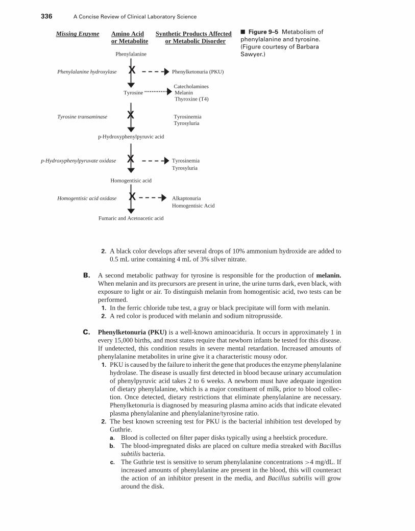
P1: OSO
LWBK192-09 LWBK192-Hubbard December 5, 2008 4:23
336 A Concise Review of Clinical Laboratory Science
Missing Enzyme Amino Acid Synthetic Products Affected
or Metabolite or Metabolic Disorder
Phenylalanine
Phenylalanine hydroxylase X Phenylketonuria (PKU)
Catecholamines
Tyrosine Melanin
Thyroxine (T4)
Tyrosine transaminase X Tyrosinemia
Tyrosyluria
p-Hydroxyphenylpyruvic acid
p-Hydroxyphenylpyruvate oxidase X Tyrosinemia
Tyrosyluria
Homogentisic acid
Homogentisic acid oxidase X Alkaptonuria
Homogentisic Acid
Fumaric and Acetoacetic acid
■ Figure 9–5 Metabolism of
phenylalanine and tyrosine.
(Figure courtesy of Barbara
Sawyer.)
2. A black color develops after several drops of 10% ammonium hydroxide are added to0.5 mL urine containing 4 mL of 3% silver nitrate.
B. A second metabolic pathway for tyrosine is responsible for the production of melanin.When melanin and its precursors are present in urine, the urine turns dark, even black, withexposure to light or air. To distinguish melanin from homogentisic acid, two tests can beperformed.
1. In the ferric chloride tube test, a gray or black precipitate will form with melanin.2. A red color is produced with melanin and sodium nitroprusside.
C. Phenylketonuria (PKU) is a well-known aminoaciduria. It occurs in approximately 1 inevery 15,000 births, and most states require that newborn infants be tested for this disease.If undetected, this condition results in severe mental retardation. Increased amounts ofphenylalanine metabolites in urine give it a characteristic mousy odor.
1. PKU is caused by the failure to inherit the gene that produces the enzyme phenylalaninehydrolase. The disease is usually first detected in blood because urinary accumulationof phenylpyruvic acid takes 2 to 6 weeks. A newborn must have adequate ingestionof dietary phenylalanine, which is a major constituent of milk, prior to blood collec-tion. Once detected, dietary restrictions that eliminate phenylalanine are necessary.Phenylketonuria is diagnosed by measuring plasma amino acids that indicate elevatedplasma phenylalanine and phenylalanine/tyrosine ratio.
2. The best known screening test for PKU is the bacterial inhibition test developed byGuthrie.a. Blood is collected on filter paper disks typically using a heelstick procedure.b. The blood-impregnated disks are placed on culture media streaked with Bacillus
subtilis bacteria.c. The Guthrie test is sensitive to serum phenylalanine concentrations >4 mg/dL. If
increased amounts of phenylalanine are present in the blood, this will counteractthe action of an inhibitor present in the media, and Bacillus subtilis will growaround the disk.
P1: OSO
LWBK192-09 LWBK192-Hubbard November 27, 2008 7:51
CHAPTER 9 Urinalysis and Body Fluid Analysis 337
3. Phenylpyruvic acid can be detected in urine with the ferric chloride reaction. Phenistixreagent strips are available for the detection of PKU. When dipped into urine containingphenylpyruvic acid, a permanent blue-gray to green-gray color is produced.
D. Other disorders of tyrosine metabolism can result from inherited or metabolic defects. Iftyrosine derived from the diet or from the metabolism of phenylalanine is not metabolized,it accumulates in the serum up to 100 times normal, producing tyrosinemia and overflowinto the urine (tyrosyluria).
1. Cirrhosis of the liver, renal dysfunction, and rickets are the principal clinical findingsin hereditary tyrosinemia, which is rare. More often, transient tyrosinemia, and thustyrosyluria, occur in low-birth-weight infants and must be distinguished from PKU.
2. Precipitated tyrosine crystals may be seen in urine sediment. A screening test to detecttyrosine in urine uses nitrosonaphthol, which forms red complexes with tyrosine andtyramine, but is nonspecific.
3. Chromatography should be used to confirm the presence of increased levels of tyrosinebecause normal urine contains some tyrosine.
E. Maple syrup urine disease (MSUD) is one of a group of disorders associated with abnormalbranched-chain amino acids.
1. Failure to inherit the gene for the enzyme necessary to produce oxidative decarboxy-lation of the keto acids in the metabolic pathways of leucine, isoleucine, and valineresults in their accumulation in the blood and urine. These excess keto acids give urinea characteristic maple syrup odor.
2. The most commonly used screening test for keto acids is the 2,4-dinitrophenylhydrazine(DNPH) reaction. Addition of DNPH to urine containing keto acids produces a yellowturbidity or precipitate.
F. Miscellaneous tests for metabolic products1. Dysfunction in the metabolism of tryptophan can result in an increase of indican or
5-hydroxyindoleacetic acid (5-HIAA) in the urine.a. In certain intestinal disorders, including obstruction, the presence of abnormal
bacteria, malabsorption syndromes, or a rare inherited disorder (Hartnup disease),increased amounts of tryptophan are converted to indole in the intestine.
b. The excess indole is then reabsorbed into the blood and converted to indican bythe liver and excreted into the urine.
c. Indican in urine, when exposed to air, is oxidized to indigo blue.d. Urinary indican is detected by an acidic ferric chloride solution, which reacts with
indican to form a deep-blue or purple color.2. In another metabolic pathway, tryptophan is converted to serotonin in the argentaffin
cells of the intestine.a. Malignant tumors of the argentaffin cells produce excess amounts of serotonin,
which results in elevated levels of the urinary degradation product 5-HIAA.b. 5-HIAA can be detected in urine with the addition of 1-nitroso-2-naphthol, which
produces a purple to black color, depending on the concentration of 5-HIAA.3. Cystinuria is characterized by defective tubular reabsorption of cystine and the amino
acids arginine, lysine, and ornithine after glomerular filtration.a. The demonstration of multiple amino acids not being reabsorbed rules out the possi-
bility of an error in metabolism, although the condition is inherited. Approximately65% of individuals who have cystinuria tend to form calculi.
b. A fresh, first morning urine specimen should be examined for cystine crystals.c. A chemical screening test for urinary cystine uses the cyanide-nitroprusside test.
Sodium cyanide reduces cystine, and the free sulfhydryl groups then react withnitroprusside to produce a red-purple color.
4. Homocystinuria is caused by deficiency of the liver enzyme cystathionine β-synthase.Homocysteine is rapidly oxidized to homocystine, which accumulates and is excretedin the urine. Children afflicted with this disease may have seizures and thromboses,and they may become mentally retarded.
P1: OSO
LWBK192-09 LWBK192-Hubbard November 27, 2008 7:51
338 A Concise Review of Clinical Laboratory Science
a. A fresh urine specimen should be tested for homocystine, because it is labile.b. The cyanide-nitroprusside reaction is positive.
G. Urine calcium1. Elevated urinary calcium may be seen in individuals who have renal calculi, hyper-
parathyroidism, osteoporosis, or multiple myeloma. Occasionally, individuals who takelarge amounts of calcium supplement might produce high urine calcium. Sulkowitch’stest is a quick qualitative test for increased levels of urinary calcium. Sulkowitch’sreagent consists of oxalic acid, ammonium oxalate, and glacial acetic acid. When re-acted with urinary calcium, calcium oxalate precipitates, producing turbidity that isgraded on a scale from 0 to 4.
2. If large amounts of clumped calcium oxalate or calcium carbonate crystals are notedin a freshly voided urine sample, it may be predictive of the presence of renal calculi.
H. Urine porphyrins1. Porphyrins are intermediate compounds in the production of heme.2. Porphyrias are a group of disorders resulting from defects in the heme synthesis path-
way.a. Inherited enzyme deficiencies and lead poisoning interrupt the heme synthesis path-
way and produce porphyrins, which result from the spontaneous and irreversibleoxidation of their respective porphyrinogens. These porphyrins cannot re-enter theheme synthesis pathway and are excreted in urine and stool.
b. Porphyrias are rare disorders. The most common type in North America is porphyriacutanea tarda.
3. The principle circulating porphyrins include uroporphyrin, coproporphyrin, and pro-toporphyrin.
4. Porphyrin precursors commonly found in urine are porphobilinogen and aminole-vulinic acid (ALA).
5. In their oxidative forms, uroporphyrin, coproporphyrin, and protoporphyrin are darkred or purple and fluorescent. The oxidative form of porphobilinogen—porphobilin—is dark red. Thus, urine containing porphobilinogen and porphobilin may appear darkred, often referred to as “port wine” color.
6. If test results are negative for blood in red-colored urine and the individual is not takingmedication that could color the urine, the specimen should be tested for porphyrinuria.
7. There are two screening tests for porphyrinuria: the Ehrlich’s reaction and fluores-cence under ultraviolet light from a Wood’s lamp (see 10 below). In Ehrlich’s reaction,urobilinogen and porphobilinogen react with p-dimethylaminobenzaldehyde (Ehrlich’sreagent) to produce a cherry-red color.
8. To detect ALA, acetylacetone is added to a urine specimen before testing to convertALA to porphobilinogen. Addition of Ehrlich’s reagent produces a cherry-red colorin a specimen that yields positive test results. Increased urinary ALA is a commonscreening test for lead poisoning.
9. If a cherry-red color is produced by a urine specimen after the addition of Ehrlich’sreagent, the Watson-Schwartz test is performed to differentiate urobilinogen andporphobilinogen based on solubility differences.a. Chloroform is added to the tube to extract urobilinogen, and porphobilinogen
remains in the aqueous phase. The tube is shaken vigorously, and the phases areallowed to separate.
b. If the red color resides only in the chloroform layer, increased amounts of uro-bilinogen are present. If the aqueous layer is red, porphobilinogen or other Ehrlich-reactive substances are present.
10. Fluorescence screening detects the presence of urobilinogen, coproporphyrin, andprotoporphyrin. These porphyrins must be extracted into a mixture of glacial aceticacid and ethyl acetate. The solvent layer is then examined with a Wood’s lamp. If thetest is positive, the solvent layer fluoresces as pink, violet, or red, depending on theconcentration of porphyrins present.

P1: OSO
LWBK192-09 LWBK192-Hubbard November 27, 2008 7:51
CHAPTER 9 Urinalysis and Body Fluid Analysis 339
11. The Hoesch screening test for porphobilinogen is a rapid screen for urinary porpho-bilinogen. Two drops of urine are added to the Hoesch reagent, which is Ehrlich’sreagent plus HCl. The uppermost part of the solution turns red in the presence ofporphobilinogen.
VIII. DISEASES OF THE KIDNEY
A. Glomerular diseases1. Four distinct morphologic changes of the glomeruli are recognized in glomerular
disease: cellular proliferation, leukocyte infiltration, glomerular basement-membranethickening, and hyalinization with sclerosis (hardening). Hyalinization is thedeposition of an albuminoid mass in tissues that typically appears semi-transparentin H&E stains.a. Cellular proliferation includes increased numbers of epithelial cells, mesangial
cells, and endothelial cells, which compose the capillary endothelium and accu-mulate in the glomerular tuft.
b. Leukocyte infiltration results from local chemotactic response or cellular prolif-eration. Neutrophils and macrophages can readily infiltrate glomeruli in responseto chemotactic factors.
c. Glomerular basement-membrane thickening usually results from deposition ofprecipitated proteins (e.g., immune complexes, fibrin).
d. Hyalinization of glomeruli results from the accumulation of a homogeneous,eosinophilic extracellular material in the glomeruli, which causes the glomerulito lose their structural detail and become sclerotic.
2. Immunologic disorders are the primary cause of glomerular injury. Circulatingimmune complexes created in response to either endogenous (e.g., cellular) or ex-ogenous antigens (e.g., microorganisms) become trapped in the glomeruli. A sec-ond immune response involves auto-antibodies that react directly with antigens onthe glomerular tissue. These may be cross-reacting antibodies formed against exoge-nous antigens. Glomerular injury results from the toxic substances (e.g., proteases,oxygen-derived free radicals, and arachidonic acid metabolites) produced by the com-plements, neutrophils, monocytes, platelets, and other factors at the site of antibodydeposition.
3. Glomerulonephritis is an inflammatory process that affects the glomerulus and in-cludes some or all of these clinical findings: hematuria, proteinuria, oliguria, azotemia,edema, and hypertension.a. Acute glomerulonephritis refers to a disease characterized by the rapid onset
of symptoms (e.g., fever, malaise, nausea, oliguria) caused by an inflammatoryprocess in the glomerulus. Blood, protein, and casts, particularly RBC casts, aredetected in urine.(1) Poststreptococcal glomerulonephritis is most frequently seen in children or
young adults following a respiratory tract infection caused by group A strep-tococci (e.g., Streptococcus pyogenes) in which treatment either did not occur,or it failed. As the bacteria proliferate and the body forms antibodies to them,opsonized bacteria form immune complexes that deposit on the glomerularmembrane.
(2) Approximately 95% of children and 60% of adults recover spontaneously orwith minimal therapy. The remainder recovers more slowly or develops chronicglomerulonephritis.
b. Crescentic (rapidly progressive) glomerulonephritis is a more serious type ofglomerulonephritis that often terminates in renal failure. Urinalysis findings includehematuria, proteinuria, oliguria; RBCs; and hyaline, granular, RBC, and WBCcasts.(1) It is characterized by cellular proliferation of epithelial cells inside Bowman’s
capsule that form “crescents,” which cause pressure changes in the glomerular
P1: OSO
LWBK192-09 LWBK192-Hubbard November 27, 2008 7:51
340 A Concise Review of Clinical Laboratory Science
tuft and can occlude the entrance to the proximal tubule. Fibrin deposition andleukocyte infiltration are present.
(2) Fibrin degradation products and IgA complexes eventually appear in urine,while extremely elevated serum protein occurs as the disease progresses.
c. Membranous glomerulonephritis is characterized by thickening of the glomeru-lar capillary basement membrane, which is usually caused by immune complexdeposition. The clinical course of this disease is variable, but may progress to anephrotic syndrome. Urinalysis findings include hematuria and proteinuria.
d. Mesangiocapillary (membranoproliferative) glomerulonephritis is character-ized by cellular proliferation and leukocyte infiltration that leads to thickening ofthe glomerular basement membrane. Immune complex deposition and complementactivation over many years leads to chronic renal failure. Laboratory findings mayinclude hypocomplementemia, hematuria, and proteinuria.
e. In focal glomerulonephritis, only a certain number of glomeruli are sclerotic.Characteristic pathology findings include immune complex deposition with hema-turia and proteinuria.
f. Chronic glomerulonephritis results from continual or permanent damage to theglomerulus associated with irreversible loss of renal tissue and chronic renal fail-ure. Clinical signs include edema, hypertension, anemia, metabolic acidosis, andoliguria that progresses to anuria. Urinalysis reveals hematuria, proteinuria, isos-thenuria, and many types of casts including broad or waxy.(1) Serum urea nitrogen, creatinine, phosphorous, and potassium levels are ele-
vated; serum calcium levels tend to be decreased.(2) Progression to end-stage renal disease may occur with further manifestation of
fatigue, anemia, edema and oliguria.g. Minimal change disease produces little cellular change in the glomeruli. Fre-
quently children are affected, probably through an allergic reaction. The disease isusually transient and is characterized by edema, heavy proteinuria, lipiduria, andhematuria.
B. Tubular and interstitial diseases1. Acute tubular necrosis results from destruction of renal tubular epithelial cells, which
is caused by decreased perfusion of the kidneys and ischemia (decreased blood flow)or certain agents (e.g., hemoglobin, myoglobin, drugs) or toxins (e.g., heavy metals,poisons, organic solvents) that produce ischemia.a. The resulting oliguria leads to acute renal failure.b. Urinalysis findings include proteinuria, hematuria, pyuria, and many types of casts,
depending on the phase of the disease.2. Tubular dysfunction can be a primary renal disease or secondary to some other disease
process.a. Fanconi’s syndrome is any condition that is characterized by a generalized prox-
imal tubular dysfunction. As a result, the reabsorption of water, glucose, phospho-rus, potassium, calcium, and amino acids is decreased, and these substances areexcreted into the urine.
b. Cystinosis is an inherited trait that results in the intracellular deposition of cystine.Every cell in the body is affected, including the proximal and distal tubular cells.The kidney can no longer concentrate or acidify urine. The extensive renal celldamage eventually requires dialysis or transplantation.
3. Tubulointerstitial disease results from other diseases that affect the renal tubules andeventually involve the interstitium. Causes of tubulointerstitial diseases are infections,toxins (e.g., drugs, heavy metals), vascular or metabolic diseases, irradiation, neoplasmsand organ transplant rejection.
4. Urethritis is microbial infection of the urethra. Cystitis is microbial infection of thebladder. Pyelitis is infection of the renal pelvis. Pyelonephritis is infection of the renalpelvis and the interstitium.
5. UTIs are more common in women than men, and 85% of UTIs are caused by gram-negative enteric bacteria. The most common pathogen is Escherichia coli. UTIs cause
P1: OSO
LWBK192-09 LWBK192-Hubbard November 27, 2008 7:51
CHAPTER 9 Urinalysis and Body Fluid Analysis 341
pain or burning on urination with urinary frequency and occasional back pain. Urinalysisfindings include: pyuria (i.e., presence of WBCs) and bacturia with mild hematuria andproteinuria possible as well. Microscopic examination of an upper UTI might revealWBC and granular casts; these are not seen in lower UTIs.
6. Acute pyelonephritis is an acute infection of the renal tubules, interstitium, and renalpelvis.a. There are two possible mechanisms leading to acute pyelonephritis:
(1) Bacteria can ascend the ureters from a lower UTIs; or(2) Bacteremia can occur with bacteria localizing in the kidney.
b. Bacteria multiply in the kidney, which causes inflammation that spreads to thetubules. Large numbers of neutrophil enzymes and bacterial toxins cause necrosisof the tubules.
c. Urinalysis reveals pyuria with WBCs often in clumps, large numbers of bacteria,leukocyte casts (and possibly granular, renal cell, or broad casts), proteinuria, andhematuria.
7. Yeast infections of the urinary tract usually result from inoculation of these organ-isms into the urinary tract by catheters or from the bloodstream, if it is infected withyeast.
8. Chronic pyelonephritis is a chronic inflammation of the tubulointerstitial tissues andresults in permanent scarring.a. Urinalysis findings include pyuria and proteinuria. Bacteria and casts may or may
not be present.b. Polyuria and nocturia develop as tubular function is lost, which results in low
specific gravity of urine.9. Acute interstitial nephritis is a condition of renal injury that results from various
drugs and toxins. Urinalysis results include hematuria, proteinuria, WBCs (particularlyeosinophils) and WBC casts with no bacteria.a. Drugs, such as penicillin and sulfonamides, can produce an immediate allergic
reaction in the renal interstitium, which is caused by the binding of the allergen tothe interstitial protein.
b. Heavy metals and aminoglycosides can cause direct damage to tubules.10. Acute renal failure (ARF) results from tubular necrosis with marked loss of renal
function. Prerenal causes of ARF (systemic disease such as cardiac or pulmonaryfailure) that produce decreased blood flow to the kidneys, renal causes (within thekidneys such as glomerular disease), and postrenal causes (obstruction of the renalsystem such as tumors or calculi) all can lead to ARF. Typical symptoms produced byARF include:a. Decreased glomerular filtration rate and oliguriab. Lack of renal concentrating abilityc. Increased serum BUN and creatinine levelsd. Microscopic findings of casts or renal tubular epithelial cells in prerenal disease,
RBCs and cellular casts in renal causes, and tumor cells or large numbers of clumpedcalcium oxalate crystals in obstructive postrenal disease.
11. Chronic renal failure results from a progressive chronic loss of renal function andcan be caused by progression from the original renal disorder. Urinalysis findingsconsist of isosthenuria at 1.010 (caused by the loss of kidney concentrating abil-ity), significant proteinuria, hematuria, and a telescoped urine sediment (includes alltypes of casts: granular, broad and waxy). Serum urea nitrogen and creatinine risegradually.
C. Other renal-related diseases1. Systemic diseases producing glomerular damage include systemic lupus erythematosus
(SLE), amyloidosis, and diabetes mellitus.a. In individuals who have SLE, immune complexes (e.g., DNA and anti-DNA) are
deposited in the glomerular membrane.(1) Complement is activated, inflammatory cells are called to the area, and the
cellular proteases destroy the surrounding tissue.
P1: OSO
LWBK192-09 LWBK192-Hubbard November 27, 2008 7:51
342 A Concise Review of Clinical Laboratory Science
(2) Disease progression may lead to recurrent hematuria, acute nephritis, ornephrotic syndrome.
(3) Chronic renal failure is a leading cause of death in SLE individuals who haverenal involvement.
b. Amyloidosis is characterized by deposition of amyloid between cells in any organor tissue of the body. Amyloid is a substance composed of 90% fibril protein and10% glycoprotein.(1) The deposition of amyloid within the glomeruli eventually destroys them.(2) Clinical findings include proteinuria and nephrotic syndrome. Eventually, renal
failure and uremia occur as more and more glomeruli are destroyed.c. Diabetes mellitus can result in renal disease. Renal capillaries, as are capillaries
in the retina and periphery, are markedly affected if the diabetes is uncontrolled.(1) Prolonged glycosemia is toxic to the kidney, particularly the glomerulus, and
causes thickening of the basement membrane.(2) These individuals have an excessive thirst (polydipsia), which leads to high
volume urine with a high specific gravity because of the glucose it contains.(3) Chronic renal failure eventually develops with persistent proteinuria.
2. Nephrotic syndrome is characterized by pronounced proteinuria (i.e., ≥3.5 g/day).Systemic diseases that can lead to nephrotic syndrome include SLE, amyloidosis,systemic shock, untreated infection, malignancies, and uncontrolled diabetes mellitus.Nephrotoxic agents such as drugs and poisons can also lead to nephrotic syndrome.a. Many plasma proteins, particularly albumin, are excreted into urine because of
increased permeability of the glomeruli.b. Other clinical findings include hypoproteinemia (particularly albumin), hyperlipi-
demia, lipiduria, and edema.c. Decreased plasma immunoglobulins and other low-molecular-weight molecules
(e.g., complement components, coagulation factors) make these individuals moresusceptible to infections and thrombosis.
d. Urinalysis findings include marked proteinuria; lipiduria with oval fat bodies;hematuria; and fatty, waxy, and renal tubular epithelial cell casts.
3. Renal calculi are solid aggregates of mineral salts that form within various glands.Calculi of the urinary tract are called kidney stones. They can be found in the renalcalyces, pelvis, ureters, or bladder.a. Calculi are usually composed of a mixture of elements. Most renal calculi contain
calcium and oxalate. Other frequently seen components are phosphates, uric acid,and cystine.
b. Causes of calculi formation include underlying metabolic or endocrine disorders,infections, or a fixed urinary pH. When the urine pH is optimal, and the urinebecomes supersaturated with a particular chemical component, renal calculi tendto form.
c. Classification of renal calculi(1) Urinary calculi are classified by location and chemical nature. In the United
States, most urinary calculi are found in kidneys or ureters. Multiple calculiand bladder calculi are less common.
(2) Calcium calculi are the most common in the United States.(3) Techniques for analyzing calculi include chemical and physical analysis, po-
larizing microscopy, and others.
D. Causes of various calculi compositions1. Calculi of calcium composition are caused by the following diseases or physiologic
conditions:a. Idiopathic hypercalciuriab. Primary hyperparathyroidismc. Bone diseased. Excessive milk, alkali, or vitamin D intakee. Renal tubular acidosisf. Sarcoidosis
g. Berylliosis
P1: OSO
LWBK192-09 LWBK192-Hubbard November 27, 2008 7:51
CHAPTER 9 Urinalysis and Body Fluid Analysis 343
2. Calculi of calcium oxalate composition are caused by the following diseases or phys-iologic conditions:a. Oxaluriab. Incomplete catabolism of carbohydratesc. Isohydria at pH 5.5 to 6.0d. Excessive glycogen breakdown
3. Calculi of calcium-phosphate composition are caused by the following diseases orphysiologic conditions:a. Same conditions as for calcium oxalateb. Alkaline infection (urea-splitting)c. Persistently alkaline urine
4. Calculi of magnesium ammonium phosphate hexahydrate composition are causedby alkaline infection with urea-splitting bacteria.
5. Calculi composed of uric acid and urate are caused by the following diseases orphysiologic conditions:a. Goutb. Polycythemiac. Leukemiad. Lymphomae. Liver diseasef. Acid isohydria
g. Theophylline and thiazide therapyh. Conditions associated with rapid protein catabolism
6. Calculi of cystine composition are caused by the following diseases or physiologicconditions:a. Transient acute phases of chronic renal diseasesb. Heavy metal nephrotoxicityc. Aminoaciduriad. Renal tubular acidosis syndromes
E. Values of pH can be associated with calculi formation.1. Urine consistently acid with a pH <5.5 favors the formation of uric acid, cystine, or
xanthine calculi.2. Urine consistently acid with a pH between 5.5 and 6.0 favors the formation of calcium
oxalate and apatite calculi.3. Urine consistently alkaline with a pH of >7.0 favors the formation of magnesium
ammonium phosphate or calcium phosphate calculi.
IX. RENAL SYNTHETIC PRODUCTS
A. Antidiuretic hormone, also called vasopressin, is a peptide hormone produced by theby the hypothalamus of the brain and stored in the posterior pituitary gland. It directlyaffects the cells of the distal convoluted and collecting tubules of the kidney to controlwater reabsorption.
1. Increased serum levels of ADH cause increased water reabsorption, which leadsto a decreased amount of concentrated urine. Increased ADH is typically caused byhypothalamic or pituitary tumor or adenoma. This is often referred to as the Syn-drome of Inappropriate ADH excretion (SIADH). Individuals exhibit hyponatremiaand water retention with low urine volume of high concentration (elevated specificgravity).
2. Decreased ADH levels cause decreased water reabsorption, which induces polydipsia(i.e., excessive thirst), polyuria and a large volume (diuresis) of very dilute urine witha low specific gravity. This is referred to as diabetes insipidus. Diabetes insipidus isa disease characterized by polyuria and polydipsia resulting from inadequate ADH se-cretion or inability of the renal tubules to respond to this hormone. Causes of decreasedADH include pituitary or hypothalamic insufficiency; toxic drugs; tumors, and braindisorders caused by trauma or neoplasms. Decreased renin also affects ADH
P1: OSO
LWBK192-09 LWBK192-Hubbard November 27, 2008 7:51
344 A Concise Review of Clinical Laboratory Science
B. Renin is a proteolytic enzyme (some consider it a hormone) formed and stored by juxta-glomerular cells of the kidney and released into the lymph and the renal venous blood. Reninconverts angiotensinogen into angiotensin products. Angiotensin II causes vasoconstrictionof renal arterioles and reabsorption of sodium. which directly stimulate the synthesis andsecretion of aldosterone by the adrenal cortex
1. Low plasma volume and a low sodium level stimulate renin secretion, resulting inaldosterone release, which causes sodium retention and reabsorption and potassiumloss. This results in water retention, which increases extracellular fluid volume andelevated blood pressure.a. As systemic pressure increases, renin production decreases. This produces a de-
crease in angiotensin and aldosterone levels.b. Potassium loss stimulates aldosterone secretion and suppresses renin release,
whereas increased potassium decreases renin and aldosterone levels.2. Lesions found in the kidney or its vascular supply can lead to increased renin levels,
resulting in increased aldosterone production and subsequent changes in sodium andpotassium excretion as well as possible hypertension.
3. Chronic renal failure can result in low, high, or normal renin levels.4. Increased aldosterone levels can result from adrenal adenoma, low renin production,
potassium wastage, sodium retention, nephrosis, cirrhosis, and heart failure.
C. Erythropoietin (EPO) is a glycoprotein hormone synthesized in specialized renal tubularfibroblasts that are sensitive to low oxygen levels. EPO stimulates bone marrow to producemore RBCs and thus increase the oxygen carrying capacity of the blood. It is also thoughtthat EPO increases the efficacy of iron insertion into heme.
D. Dihydroxycholecalciferol hydrolase is an enzyme synthesized in the juxtaglomerular cellsof the kidney that is responsible for activating vitamin D3. This enzyme is under the controlof parathyroid hormone.
X. URINE PREGNANCY TESTING
A. Molecular characteristics of chorionic gonadotropin (CG)1. The glycoprotein hormone CG is produced by the syncytiotrophoblast cells of the
placenta approximately 10 days after conception. CG stimulates the corpus luteum inthe ovary to synthesize progesterone during the first weeks of pregnancy. The moleculeconsists of two noncovalently bound polypeptide subunits: α and β.a. The α subunit of CG is nearly identical to the α subunit of luteinizing hormone (LH)
and follicle-stimulating hormone (FSH); therefore, this subunit of CG cross-reactswith LH in certain bioassay systems to give false-positive results.
b. The β subunit determines the biologic and immunologic specificity of CG. Urinecontains predominantly CGβ (core fragment) and to a lesser degree unmodifiedCG and CGn (nicked). The rate of clearance varies for the different CGs.
2. In the first 8 weeks of pregnancy, the CG concentration in maternal serum rises geo-metrically. Detectable amounts (∼5 IU/L) are present 8 to 11 days after conception,which is in the third week of pregnancy as measured from the last menstrual period.For women aged 18 to 40, serum CG concentrations of 5 IU/L or greater are consistentwith pregnancy. Subsequently CG concentrations start to decline in serum and urine,and by the end of the second trimester a 90% reduction from peak concentration hasusually occurred.
3. Pregnancy testing is not a direct test for pregnancy (i.e., detection of a fetus), but ratheris a test to detect the presence of CG.
B. Methods for urine pregnancy testing1. Home test kit assays are the most commonly used pregnancy tests in the United States.
Most kits provide a single test and use an immunochromatographic method to measureurine CG. Detection limits vary from 6.3 to 50 IU/L.
P1: OSO
LWBK192-09 LWBK192-Hubbard November 27, 2008 7:51
CHAPTER 9 Urinalysis and Body Fluid Analysis 345
2. First-morning urine specimens are preferred for qualitative urine pregnancy tests be-cause they are concentrated and contain abundant CG. Urine applied to the device isabsorbed into a nitrocellulose bed. The CG is concentrated into a narrow band as theurine migrates. A labeled anti-CG antibody in the device binds to the migrating CG andpasses through a zone having solid phase capture antibody to CG. The appearance of acolored line indicates a positive test. Positive controls are typically integrated into thetest system. False positive results occur in 1% of tests because of the presence in urineof interfering substances, such as proteins, drugs, bacteria, erythrocytes, or leukocytes.False negative results have occurred because of low CG concentration. About half ofthese qualitative tests will be positive on the day after the first missed menstrual period.Qualitative serum assays are available and tend to be more reliable than urine assaysbecause urine CG concentrations vary considerably.
XI. SEROUS BODY FLUIDS
A. Origin, anatomic relationship and functions1. The cavities of the body that hold abdominal organs, lungs, and the heart are lined by
two membranes consisting of mesothelial cells. The membrane that lines the cavityis referred to as the parietal membrane and the membrane that forms a sac aroundthe organs is the visceral membrane. The small amount of fluid between these twomembranes is serous fluid. Serous fluid is an ultrafiltrate of plasma and is produced andreabsorbed at a constant rate. This fluid functions as a lubricant between the membranesof the cavities and to allow free movement of the organs.a. The peritoneum encloses abdominal organs. The fluid contained between the vis-
ceral and parietal membranes is peritoneal fluid.b. The pleural cavity encloses the lungs. The fluid contained between the visceral and
parietal membranes here is pleural fluid.c. The pericardium encloses the heart. The fluid contained between the visceral and
parietal membranes in this cavity is pericardial fluid.2. Any disruption of the production and reabsorption of serous fluid because of an al-
teration in the hydrostatic and oncotic pressure in the capillaries of the cavities willcause an increase in fluid volume between the two membranes. This abnormal fluidbuildup is an effusion. Abnormal accumulation of fluid in any body cavity indicates anabnormality.
3. There are two kinds of effusions, transudates and exudates.a. Transudates result from excess filtration of blood serum across a physically intact
vascular wall due to disruption of reabsorption. This occurs in systemic diseasesthat alter the hydrostatic pressure of the capillaries and include congestive heartfailure, hepatic cirrhosis or nephrotic syndrome.
b. Exudates are the active accumulation of fluid within body cavities associated withinflammation of the membranes and vascular wall damage. Exudates, which arecloser to serum in chemical composition, are caused by the following conditions:(1) Inflammatory disorders,(2) Malignancies,(3) Infections.
4. Specimen collection of serous fluids is known as paracentesis and refers to the percu-taneous puncture of a body cavity for the aspiration and removal of fluid.a. Thoracentesis is the collection of pleural fluid.b. Peritoneocentesis is the collection of peritoneal fluid.c. Pericardiocentesis is the collection of pericardial fluid.
B. Analysis of serous body fluids1. Chemical examination of serous fluid differentiates between an exudate and a tran-
sudate (Table 9–7). These include:a. A total protein evaluation and fluid-to-serum protein ratio can help distinguish
between the physiologic basis of different fluids. A serous fluid with a protein
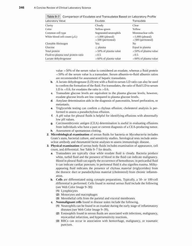
P1: OSO
LWBK192-09 LWBK192-Hubbard November 27, 2008 7:51
346 A Concise Review of Clinical Laboratory Science
Table 9–7 Comparison of Exudates and Transudates Based on Laboratory Profile
Laboratory Value Exudate Transudate
Clarity Cloudy Clear
Color Yellow-green Yellow
Common cell type Segmented neutrophils Mononuclear cells
White blood cell count (μL) >1,000 (pleural)>500 (peritoneal)
<1,000 (pleural)<300 (peritoneal)
Clottable fibrinogen Yes No
Glucose ≤ plasma Equal to plasma
Total protein >50% of plasma value <50% of plasma value
Fluid-to-plasma total protein ratio >0.5 <0.5
Lactate dehydrogenase >60% of plasma value <60% of plasma value
value >50% of the serum value is considered an exudate, whereas a fluid protein<50% of the serum value is a transudate. Serum albumin-to-fluid albumin ratiosare recommended for assessment of hepatic transudates.
b. A lactate dehydrogenase (LD) test with a fluid-to-serum LD ratio can also be usedto confirm the formation of the fluid. For transudates, the ratio of fluid LD to serumLD is <0.6; for exudates the ratio is >0.6.
c. Transudate glucose levels are equivalent to the plasma glucose levels; however,exudate glucose levels are low compared to plasma glucose levels.
d. Amylase determination aids in the diagnosis of pancreatitis, bowel perforation, ormetastasis.
e. Triglyceride testing can confirm a chylous effusion; cholesterol analysis is per-formed to assess a pseudochylous effusion.
f. A pH value for pleural fluids is helpful for identifying effusions with abnormallylow pH values.
g. Carcinoembryonic antigen (CEA) determination is useful in evaluating effusionsfrom individuals who have a past or current diagnosis of a CEA-producing tumor.
h. Assessment of spontaneous clotting.2. Microbiological examination of serous fluids for bacteria or Mycobacteria includes
Gram’s stain, bacterial culture, and sensitivity studies. Serological tests include antin-uclear antibody and rheumatoid factor analyses to assess immunologic disease.
3. Physical examination of serous body fluids includes examination of appearance, cellcount, and differential. See Table 9–7 for details.a. Transudates are typically clear while exudate fluid is cloudy. Bacteria produce
white, turbid fluid and the presence of blood in the fluid can indicate malignancy.Blood in pleural fluid can signify the occurrence of hemothorax; in pericardial fluidit can indicate cardiac puncture; in peritoneal fluid it also signifies trauma. Milky-appearing fluid indicates the presence of chylous material (triglycerides) fromthe thoracic duct or pseudochylous material (cholesterol) from chronic inflamm-ation.
b. Cells are differentiated using cytospin preparations. Typically, a 50- or 100-celldifferential is performed. Cells found in normal serous fluid include the following(see Web Color Image 9–38):(1) Lymphocytes(2) Monocytes and macrophages(3) Mesothelial cells from the parietal and visceral membranes
c. Nonmalignant cells found in disease states include the following.(1) Neutrophils can be found in an exudate during the early stage of inflammatory
diseases (see Web Color Image 9–39).(2) Eosinophils found in serous fluids are associated with infections, malignancy,
myocardial infarction, and hypersensitivity reactions.(3) RBCs can occur in association with hemorrhage, malignancy, or traumatic
puncture.
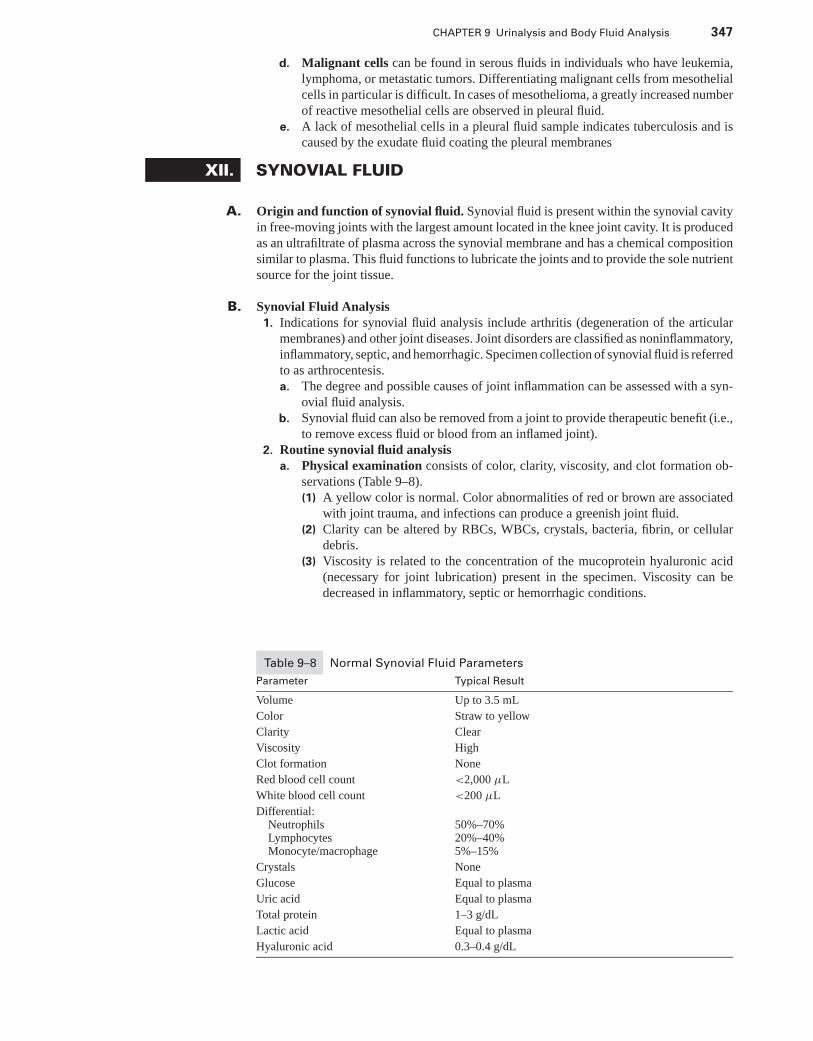
P1: OSO
LWBK192-09 LWBK192-Hubbard November 27, 2008 7:51
CHAPTER 9 Urinalysis and Body Fluid Analysis 347
d. Malignant cells can be found in serous fluids in individuals who have leukemia,lymphoma, or metastatic tumors. Differentiating malignant cells from mesothelialcells in particular is difficult. In cases of mesothelioma, a greatly increased numberof reactive mesothelial cells are observed in pleural fluid.
e. A lack of mesothelial cells in a pleural fluid sample indicates tuberculosis and iscaused by the exudate fluid coating the pleural membranes
XII. SYNOVIAL FLUID
A. Origin and function of synovial fluid. Synovial fluid is present within the synovial cavityin free-moving joints with the largest amount located in the knee joint cavity. It is producedas an ultrafiltrate of plasma across the synovial membrane and has a chemical compositionsimilar to plasma. This fluid functions to lubricate the joints and to provide the sole nutrientsource for the joint tissue.
B. Synovial Fluid Analysis1. Indications for synovial fluid analysis include arthritis (degeneration of the articular
membranes) and other joint diseases. Joint disorders are classified as noninflammatory,inflammatory, septic, and hemorrhagic. Specimen collection of synovial fluid is referredto as arthrocentesis.a. The degree and possible causes of joint inflammation can be assessed with a syn-
ovial fluid analysis.b. Synovial fluid can also be removed from a joint to provide therapeutic benefit (i.e.,
to remove excess fluid or blood from an inflamed joint).2. Routine synovial fluid analysis
a. Physical examination consists of color, clarity, viscosity, and clot formation ob-servations (Table 9–8).(1) A yellow color is normal. Color abnormalities of red or brown are associated
with joint trauma, and infections can produce a greenish joint fluid.(2) Clarity can be altered by RBCs, WBCs, crystals, bacteria, fibrin, or cellular
debris.(3) Viscosity is related to the concentration of the mucoprotein hyaluronic acid
(necessary for joint lubrication) present in the specimen. Viscosity can bedecreased in inflammatory, septic or hemorrhagic conditions.
Table 9–8 Normal Synovial Fluid Parameters
Parameter Typical Result
Volume Up to 3.5 mL
Color Straw to yellow
Clarity Clear
Viscosity High
Clot formation None
Red blood cell count <2,000 μL
White blood cell count <200 μL
Differential:Neutrophils 50%–70%Lymphocytes 20%–40%Monocyte/macrophage 5%–15%
Crystals None
Glucose Equal to plasma
Uric acid Equal to plasma
Total protein 1–3 g/dL
Lactic acid Equal to plasma
Hyaluronic acid 0.3–0.4 g/dL
P1: OSO
LWBK192-09 LWBK192-Hubbard November 27, 2008 7:51
348 A Concise Review of Clinical Laboratory Science
(a) Viscosity can be estimated by expelling a drop of the synovial fluid from asyringe. Synovial fluid of high viscosity (i.e., normal fluid) forms a “string”of 4 to 6 cm before breaking.
(b) Viscosity is also examined by looking at clot formation, which indicatesthe presence of fibrinogen when the synovial membrane is damaged orwhen there is hemorrhage or blood contamination during a traumatic tap.The test is referred to as a Ropes or mucin clot test, and is performed byadding synovial fluid to a 2% to 5% solution of acetic acid. Formation ofa solid clot indicates good viscosity.
b. Microscopic examination of synovial fluid includes a hemacytometer count ofRBCs and WBCs (Chapter 3), a cytospin differential to determine the types ofWBCs, and a wet preparation examination for crystals (Table 9–8 for normalvalues). Synoviocytes, which are cells that make up the synovial membrane, aresimilar to mesothelial cells and are a normal finding in synovial fluid. Other cells,such as LE, Reiter cells, and ragocytes, are indicative of specific syndromes.(1) An increase in RBCs indicates joint hemorrhage or traumatic tap.(2) A WBC count >2,000/μL, with a predominance of neutrophils, is associated
with bacterial arthritis, acute gouty arthritis, or rheumatoid arthritis (see WebColor Image 9–40). The presence of lymphocytes indicates nonseptic inflam-mation.
(3) Eosinophils may be present in cases of rheumatic fever or parasitic infestation.(4) LE cells are neutrophils that contain ingested cellular material and indicate
lupus erythematous; they are rarely seen in synovial fluid. Reiter cells are vac-uolated macrophages containing an ingested neutrophil and indicate Reiter’ssyndrome (systemic inflammation). Ragocytes are neutrophils containing pre-cipitated immunoglobulins, antinuclear antibodies, and rheumatoid factor andare common in septic and rheumatoid arthritis and gout.
(5) The presence of crystals can be diagnostic of joint disease.(a) Monosodium urate monohydrate crystals are fine, needle-like crystals as-
sociated with gouty arthritis. When examined under polarized light, thesecrystals appear as thin yellow needle-like structures (see Web Color Image9–41).
(b) Calcium pyrophosphate dihydrate crystals are rodlike or rhombic crystalsassociated with pseudogout. When examined under polarized light, thesecrystals appear as bluish plates and rods.
(c) Cholesterol crystals are flat, platelike crystals with notched corners. Theseare seen in conditions of chronic rheumatoid arthritis.
(d) Calcium hydroxyapatite crystals can also be present, but can be seen onlywith an electron microscope.
c. Chemical examination of synovial fluid includes only a few diagnostically usefultests (see Table 9–8 for normal values).(1) A decrease in synovial glucose that exceeds a plasma-fluid difference of >10
mg/dL indicates an inflammatory condition or septic disorder.(2) Increased protein levels indicate synovium permeability changes and damage
to the synovial membrane. Large molecules, such as protein, are not filteredthrough the synovial membrane.
(3) An increase in fluid uric acid levels helps diagnose gout when fluid crystalsare not found in the fluid.
(4) Increased lactate concentration is thought to occur from conditions of increasedanaerobic glycolysis in the synovial cavity, which is associated with severeinflammatory conditions, such as septic arthritis. Gonococcal arthritis producesnormal-to-low lactate levels.
d. A microbiologic examination can aid in the differential diagnosis of joint disease.(1) A Gram stain should be performed on all samples. A culture using enriched
medium is performed when Hemophilus or Neisseria infections are suspected.(2) The majority of infectious agents in septic arthritis are bacterial, but can also
include fungal, mycobacterium, or viral infections.
P1: OSO
LWBK192-09 LWBK192-Hubbard November 27, 2008 7:51
CHAPTER 9 Urinalysis and Body Fluid Analysis 349
XIII. SEMINAL FLUID ANALYSIS
A. Seminal fluid physiology1. Seminal fluid is composed of secretions from the testes, epididymis, seminal vesicles,
and prostate gland.a. Spermatozoa cells form approximately 5% of the ejaculate by volume.b. Seminal fluid is a complex solution of proteins and enzymes that include the fol-
lowing components:(1) Acid phosphatase (unique to prostatic fluid)(2) Citric acid(3) Zinc(4) Fructose(5) Fibrinogen-like coagulable proteins
2. Seminal fluid has the following mechanisms in fertilization:a. Activation of spermatozoal motilityb. Provision of nutritive substancesc. Conveyance of the spermatozoa to the female eggd. Provision of enzymes necessary to penetrate the ovum wall and achieve fertiliza-
tion3. Spermatozoa synthesis occurs in Sertoli cells and their maturation takes place in the
testes.a. Spermatozoa have a haploid complement of chromosomes.b. FSH (sometimes called “interstitial cell stimulating hormone”) from the pituitary
gland is necessary for the initiation of spermatogenesis. Inhibin (from Sertoli cells)inhibits FSH synthesis.
c. Testosterone (secreted by Leydig cells of the testes) is necessary for the subsequentstages of sperm maturation.
d. The stages of cellular maturation from the youngest to the most mature stage areas follows:(1) Spermatogonia(2) Spermatocytes(3) Spermatids(4) Spermatozoa
B. Indications for seminal fluid analysis1. Seminal fluid examination should be one of the first tests performed in any infertility
investigation.2. Qualification of donors for artificial insemination programs requires a complete seminal
analysis.3. Analysis can provide the necessary information for the documentation of completeness
for a vasectomy.4. Evaluation of semen quality is necessary for semen and sperm banking.5. Seminal fluid analysis often is necessary for forensic studies in sexually related crimes,
such as rape. Forensic studies of seminal fluid are also used in paternity allega-tions.
C. Routine analysis of seminal fluid involves a physical, chemical, and microscopic exami-nation of the specimen. Analysis of the specimen should not begin until after liquefactionhas occurred.
1. Physical examination includes an assessment of the appearance, volume, and viscosityof the ejaculate.a. The normal appearance of semen is gray-white and opalescent, while a brown or
red color may indicate blood and a yellowish color can signify lack of ejaculation(abstinence).
b. Normal volume for an ejaculate is 2 to 5 mL. Volumes outside this range can beassociated with infertility.
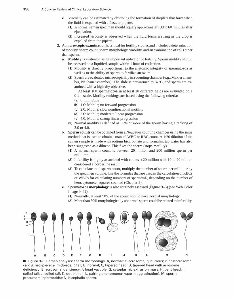
P1: OSO
LWBK192-09 LWBK192-Hubbard November 27, 2008 7:51
350 A Concise Review of Clinical Laboratory Science
c. Viscosity can be estimated by observing the formation of droplets that form whenthe fluid is expelled with a Pasteur pipette.(1) A normal semen specimen should liquefy approximately 30 to 60 minutes after
ejaculation.(2) Increased viscosity is observed when the fluid forms a string as the drop is
expelled from the pipette.2. A microscopic examination is critical for fertility studies and includes a determination
of motility, sperm count, sperm morphology, viability, and an examination of cells otherthan sperm.a. Motility is evaluated as an important indicator of fertility. Sperm motility should
be assessed on a liquified sample within 1 hour of collection.(1) Motility is directly proportional to the anatomic integrity of spermatozoa as
well as to the ability of sperm to fertilize an ovum.(2) Sperm are evaluated microscopically in a counting chamber (e.g., Makler cham-
ber, Neubauer chamber). The slide is prewarmed to 37◦C, and sperm are ex-amined with a high-dry objective.
At least 100 spermatozoa in at least 10 different fields are evaluated on a0-4+ scale. Motility rankings are based using the following criteria:(a) 0: Immobile(b) 1.0: Mobile; no forward progression(c) 2.0: Mobile; slow nondirectional motility(d) 3.0: Mobile; moderate linear progression(e) 4.0: Mobile; strong linear progression
(3) Normal motility is defined as 50% or more of the sperm having a ranking of3.0 or 4.0.
b. Sperm counts can be obtained from a Neubauer counting chamber using the samemethod that is used to obtain a manual WBC or RBC count. A 1:20 dilution of thesemen sample is made with sodium bicarbonate and formalin; tap water has alsobeen suggested as a diluent. This fixes the sperm (stops motility).(1) A normal sperm count is between 20 million and 200 million sperm per
milliliter.(2) Infertility is highly associated with counts <20 million with 10 to 20 million
considered a borderline result.(3) To calculate total sperm count, multiply the number of sperm per milliliter by
the specimen volume. Use the formulae that are used in the calculation of RBCsor WBCs for calculating numbers of sperm/mL, depending on the number ofhemacytometer squares counted (Chapter 3).
c. Spermatozoa morphology is also routinely assessed (Figure 9–6) (see Web ColorImage 9–42).(1) Normally, at least 50% of the sperm should have normal morphology.(2) More than 50% morphologically abnormal sperm could be related to infertility.
■ Figure 9–6 Semen analysis: sperm morphology. A, normal; a, acrosome; b, nucleus; c, postacrosomal
cap; d, neckpiece; e, midpiece; f, tail; B, normal; C, tapered head; D, tapered head with acrosome
deficiency; E, acrosomal deficiency; F, head vacuole; G, cytoplasmic extrusion mass; H, bent head; I,
coiled tail; J, coiled tail; K, double tail; L, pairing phenomenon (sperm agglutination); M, sperm
precursors (spermatids); N, bicephalic sperm.
P1: OSO
LWBK192-09 LWBK192-Hubbard November 27, 2008 7:51
CHAPTER 9 Urinalysis and Body Fluid Analysis 351
d. Viability is observed using supravital eosin staining of a sample of fresh semen.A smear is then made of the stained solution.(1) Viable sperm do not take up the stain, whereas dead sperm have damaged
membranes and absorb the stain. One hundred sperm are counted and evaluated.(2) The percentage of dead sperm should not exceed the percentage of immobile
sperm. Normally, 50% or more of the sperm should be viable.e. The stained seminal fluid smear should also be examined for the presence of cells
other than spermatozoa.(1) An ejaculate may normally contain leukocytes and urethral epithelial cells.(2) An increase of leukocytes >1 million/mL one million per milliliter of ejaculate
is suggestive of an inflammation.(3) The presence of RBCs or bacteria indicates a pathologic process.
3. A chemical analysis of seminal fluid is of limited usefulness. However, measurement ofthe pH, fructose level, and acid phosphatase activity can provide diagnostic information.a. The pH should be obtained from a fresh seminal specimen.
(1) A pH >7.8 could indicate an infectious process.(2) A pH <7.0 indicates either successful vasectomy or that the seminal fluid is
entirely prostatic fluid, which indicates tubule blockage or inflammation.b. Fructose is produced and secreted by the seminal vesicles.
(1) A fructose level of >13 μmol per ejaculate reflects normal secretory function.(2) A low fructose value can indicate ejaculatory duct obstruction or pathology of
the vas deferens.c. Acid phosphatase activity can determine secretory function of the prostate gland,
because this gland uniquely secretes high levels of this enzyme. Determination ofthis enzyme in forensic studies can be used to determine the presence of seminalfluid.
d. Citric acid and zinc levels can be determined to evaluate prostate gland function.Low levels of both substances are associated with prostatitis.
XIV. CEREBROSPINAL FLUID (CSF) ANALYSIS
A. CSF location, formation and functions1. The central nervous system (CNS) is bathed by a network of CSF-filled reservoirs
internally and externally. CSF is synthesized by the choroid plexus in the ventriclesand circulates out to the subarachnoid spaces surrounding the CNS (brain and spinalcord).
2. The CSF functions to protect and cushion the CNS, provide nutrients to neural tissue,and remove metabolic waste. Approximately 500 mL/day of CSF is produced, with atotal volume of 140 to 170 mL in adults.
B. Specimen collection is only for diagnosis or for the treatment of disease.1. Ventricular puncture to obtain CSF can be performed by a physician in special circum-
stances. Typically, sterile lumbar puncture between L2 and L3 vertebrae or betweenthe L3 and L4 vertebrae is the most common collection area. This collection taps CSFin the lumbar cistern of the subarachnoid space around the base of the spinal cord wherethere is no chance of piercing the cord itself.
2. Three specimen tubes are aseptically collected and labeled as 1, 2, and 3. Each tube isuniquely used for specific testing.a. Tube 1 is used for CSF chemistry analysis. This tube may contain tissue juice and
some cells from the spinal tap.b. Tube 2 is used for microbiology studies.c. Tube 3 is used for a hemocytometer cell count and cytospin differential. This tube
should contain no contaminating cells from surrounding tissue; therefore, any cellsfound in this tube are intrinsic to the CSF. Physical examination of the CSF samplemay be done using this tube.
3. All testing on CSF specimens should be performed immediately upon receipt of spec-imen because of rapid cellular degeneration.
P1: OSO
LWBK192-09 LWBK192-Hubbard November 27, 2008 7:51
352 A Concise Review of Clinical Laboratory Science
C. Pathologic diseases detected by and involving the CSF include the following conditions:1. Subarachnoid or intracerebral hemorrhages (i.e., strokes or trauma)2. Infections such as meningitis (e.g., bacterial, fungal, parasitic, or viral); abscesses and
encephalitis3. Malignant processes such as primary brain tumors, metastatic tumors with a primary
site elsewhere, or leukemias and lymphomas.4. Multiple sclerosis.
D. Routine CSF analysis typically includes a physical, chemical, and microscopic analysis.1. Gross examination includes a report of color and clarity of the specimen.
a. Normal CSF is clear and colorless.b. Turbidity is most often produced by the increased presence of WBCs (>200 cells
per milliliter), by increased RBCs (>400 RBCs per milliliter), or by microorgan-isms. Cellular turbidity is known as pleocytosis.
c. Abnormal specimen color is most commonly caused by a disease process.(1) Pink or red CSF is the result of RBC lysis and can be seen 4–10 hours after a
subarachnoid hemorrhage. This can also be caused by a traumatic tap.(2) Xanthochromic (i.e., yellow) specimens result from the following condi-
tions:(a) After pathologic bleeding caused by breakdown of hemoglobin and biliru-
bin formation in CSF or due to traumatic tap(b) When CSF protein is increased >250 mg/dL(c) Liver disease caused by increased total bilirubin levels
(3) Brown CSF specimens are the result of the following conditions:(a) Presence of methemoglobin(b) Subdural or intracerebral hematoma(c) Presence of melanin caused by a melanoma
d. Distinguishing between pathologic bleeding and traumatic tap is often necessary.The following criteria are used to distinguish between the two:(1) A serial decrease in RBCs in tubes 1 to 3 is seen with a traumatic tap.(2) A clotted specimen or clumped RBCs on microscopic examination indicates
traumatic tap.(3) The color of the supernatant of a bloody specimen after centrifugation can be
suggestive. A clear supernatant indicates a traumatic tap, whereas a pink, yel-low, or brown supernatant indicates pathologic bleeding. However, if a samplecontains bilirubin from lysed RBCs that have come from a traumatic tap, itmay have a yellowish color.
(4) A ratio of >500 RBCs for every WBC indicates traumatic tap.2. Chemical examination can include many analytes, but only a few have any diagnostic
value on a routine basis.a. Total protein in CSF represents a combination of prealbumin, albumin, transferrin,
and trace amounts of immunoglobulin G (IgG).(1) Normally, CSF protein ranges between 20 and 50 mg/dL, with albumin repre-
senting 50% to 70% of the total.(2) A CSF protein level provides information as to the integrity of the blood-brain
barrier since large protein molecules are kept out of CSF.(3) Increased protein levels in CSF can be the result of the following conditions:
(a) Contamination with peripheral blood on obtaining the specimen(b) Obstruction of CSF circulation (e.g., hydrocephalus or tumor)(c) Tissue degeneration(d) Increased permeability of the blood-brain membrane caused by drugs, tox-
ins, or infection (meningitis)(e) Intrathecal (by the brain) production of protein by tumors
(4) Recognizing increases in individual protein constituents in the CSF can beimportant.(a) Prealbumin is uniquely found in CSF specimens.(b) Any albumin present in a specimen is from the passage of plasma albumin
across a damaged blood-brain barrier (formed by tight junctions between
P1: OSO
LWBK192-09 LWBK192-Hubbard November 27, 2008 7:51
CHAPTER 9 Urinalysis and Body Fluid Analysis 353
endothelial cells). CSF albumin is increased when the permeability of theblood-brain barrier is compromised (i.e., normal CSF/serum albumin index<9.0).
(c) IgG is present in trace amounts but can originate from intrathecal produc-tion in the CNS.
(i) A CSF/serum IgG index <0.77 is normal.(ii) An IgG CSF/serum index >0.77 is highly indicative of multiple scle-
rosis.(d) CSF protein electrophoresis can be a useful tool to distinguish protein
content. Abnormal oligoclonal bands in the gamma region of the trac-ing are comprised of IgG and are highly diagnostic of multiple sclero-sis.
(e) Myelin basic proteins may be present with multiple sclerosis.b. CSF glucose is in equilibrium with plasma glucose.
(1) Normal values in the CSF range from 50 to 80 mg/dL or approximately 60%of plasma glucose levels.
(2) Levels are decreased in bacterial meningitis and fungal infections.(3) Levels are increased in hyperglycemia and in cases of a traumatic tap.
c. CSF enzyme levels can be detected and are elevated in a variety of pathologicconditions.(1) LDH concentrations can be elevated in the following conditions:
(a) Bacterial and viral meningitis(b) Subarachnoid hemorrhage(c) Lymphomas(d) Leukemias(e) Metastatic tumors
(2) Creatine kinase (CK) levels can be elevated in the following conditions:(a) Stroke(b) Multiple sclerosis(c) Degenerative disorders(d) Primary brain tumors(e) Viral and bacterial meningitis(f) Epileptic seizure
(3) Aspartate aminotransferase (AST) levels can be elevated in the following con-ditions:(a) Intracerebral hemorrhage(b) Subarachnoid hemorrhage(c) Bacterial meningitis
d. CSF lactic acid concentrations are unrelated to plasma values.(1) The normal concentration ranges from 10–22 mg/dL.(2) Lactate is increased with any disorder associated with increased metabolism
or ischemia in the CNS.e. CSF electrolytes reflect basically the same values as in serum, and their measure-
ment in CSF is of no diagnostic advantage.3. Cell count in CSF normally reflects a low number of cells (see Web Color Image 9–43).
a. Normally, mononuclear cells (i.e., lymphocytes, monocytes) predominate in a lowconcentration of 0 to 10 cells per microliter.(1) Neutrophils compose 0% to 6%.(2) Monocytes compose 15% to 45%.(3) Lymphocytes compose the majority of cell types: 40% to 80%.
b. RBCs should be absent. The presence of RBCs indicates either cerebral hemorrhageor traumatic tap.
c. Cell counts must be performed within 1 hour of collection.d. If increased WBCs are found, a cytospin smear is prepared, and the smear undergoes
Wright’s stain to determine a WBC differential count.e. Any other cell type or a change in differential percent could indicate a serious
disorder.(1) An increase in neutrophils is indicative of bacterial meningitis.
P1: OSO
LWBK192-09 LWBK192-Hubbard November 27, 2008 7:51
354 A Concise Review of Clinical Laboratory Science
(2) An increase in lymphocytes can be found in cases of viral, tubercular, or fungalmeningitis.
(3) Plasma cells may be found in individuals who have multiple sclerosis or chronicinflammatory conditions.
(4) An eosinophilia may be associated with parasitic or fungal disorders (see WebColor Image 9–44).
(5) Macrophages can be found in CSF in association with hemorrhage. The pres-ence of ferritin granules in the cytoplasm of CSF macrophages may indicatean older or chronic hemorrhagic condition.
(6) Malignant cells can be present as a result of a CNS tumor, metastatic tumor ora leukemic process.
4. Microscopic examination consists of a Gram’s stain or acid-fast stain, as well as amicrobiologic culture and sensitivity determination. Viral studies or fungal culturescan also be performed (see Chapter 7).
5. Meningitis-causing organisms include the following:(1) Cryptococcus spp.(2) Coccidioides immitis(3) Mycobacterium tuberculosis(4) Haemophilus influenzae(5) Neisseria meningitidis(6) Streptococcus pneumoniae(7) Staphylococcus aureus
XV. GASTRIC FLUID ANALYSIS
A. Physiology of gastric secretion and digestion1. Three physiologic functions of the stomach include:
a. Serves as an expandable reservoir for ingested food;b. Initiates protein digestion by pepsin;c. Secretion of intrinsic factor for binding and absorption of vitamin B12 in the ileum.
2. Three general phases of gastric secretion includea. The cephalic (neurogenic) phase involves vagus nerve stimulation caused by
stimuli such as taste, smell, or sight. Anticipation of food (i.e., hunger response)has four effects on the stomach.(1) Gastrin is secreted into the blood by specialized G cells in the pyloric gland
and stomach and by the delta cells of the pancreas.(2) Gastrin induces secretion of hydrochloric acid by parietal cells of the gastric
glands in the proximal body and fundus and secretion of pepsin by the chiefcells.
(3) Vagal excitation lowers the threshold of parietal cells to gastrin stimulation.(4) Gastric peristalsis and emptying is promoted.
b. The gastric phase involves stomach distention and continued gastric secretion ofdigestive juices.
c. The intestinal phase involves digestion in the small intestines.(1) Secretion of gastrin by G cells is inhibited by gastric inhibitory polypeptide
(GIP, a member of the secretin family), which is secreted by the duodenal glandsin the small intestine. GIP also causes insulin to be secreted and stimulateslipoprotein lipase.
(2) Gastrin secretion is reduced at a pH ≤ 3.0 and stops completely at a pH ≤ 1.5.When K cells in the distal duodenum and proximal jejunum come in contactwith fats, glucose, or amino acids, gastric GIP is produced.
B. Chemical composition of gastric fluid1. Hydrochloric acid (HCI) is present in varying amounts.
a. HCI is secreted by parietal cells in the fundus of the stomach.b. HCl converts pepsinogen to pepsin and hydrolyzes polypeptides and disaccharides.
2. Pepsin, which catalyzes proteolysis at a pH of 1.6–3.6, is also present.
P1: OSO
LWBK192-09 LWBK192-Hubbard November 27, 2008 7:51
CHAPTER 9 Urinalysis and Body Fluid Analysis 355
3. Mucus is present to protect the mucosa of the stomach from autodigestion.4. Miscellaneous substances are present such as water, various enzymes, proteins, and
intrinsic factor.
C. Gastric fluid analysis may be necessary for the diagnosis of digestive disorders or ulcers.1. Assessment of gastric acidity. Gastric acidity may fall into one of several categories.
a. Anacidity refers to the failure of the stomach acidity to fall lower than 6.0 in astimulation test.
b. Hypochlorhydria refers to the physiologic failure of pH to fall below 3.5, althoughit decreases 1.0 pH unit or more upon gastric stimulation.
c. Achlorhydria is the physiologic failure of pH to fall below 3.5 or 1.0 pH unit withgastric stimulation.
2. Evaluation of gastric fluid acidity may be performed to determine the proper surgicalprocedure to be used for peptic ulcer treatment.
3. Gastric analysis may aid in the diagnosis of Zollinger-Ellison syndrome, which usuallyresults from a neoplasm of the pancreas and causes increased gastric secretion of acid,secretory volume, and very high blood gastrin levels.
4. Determination of gastric acid secretion in response to insulin-induced hypoglycemiacan be beneficial to assess the completeness of surgical vagotomy.
5. A routine physical evaluation of gastric fluid includes the following measurements:a. Appearance (i.e., normal is translucent, pale gray, and slightly viscous)b. Volume (i.e., normal is 50 to 75 mL)c. Odor (i.e., normal is faintly pungent)d. Mucus (i.e., normal quantity varies)e. Microscopic examinationf. Measurement of gastric acid and pH
D. Gastric fluid collection is performed using several methods.1. The basal gastric secretion test measures fasting levels of gastric production. A Levin
tube is placed in the stomach for fluid collection.a. The gastric secretion collected represents the amount of secretion during a 15-hour
fast.b. Four 15- or 30-minute specimens are collected. On each gastric specimen, the
volume, pH, titratable acidity, and calculated acid output is determined.2. Stimulation of fluid formation involves the use of pentagastrin to stimulate maximal
secretion.3. Insulin-induced hypoglycemia test is often used to test for the completeness of vago-
tomy.a. Hypoglycemia stimulates the vagus to stimulate gastric acid secretion within 2
hours after insulin injection.b. Blood glucose measurements are also taken every 30 minutes (i.e., preinsulin and
postinsulin injection) to make sure that glucose falls to 50 mg/dL or lower.4. Tubeless gastric analysis is a noninvasive method to determine gastric acidity.
a. Dianex blue, which is a carboxylic acid cationic resin with an indicator dye (i.e.,azure A) coupled to it, is given orally to the individual.
b. Hydrogen ions in gastric secretion combine with the resin and release the azure Aions, which are absorbed into the bloodstream in the small intestine and excretedin the urine.
XVI. FECAL ANALYSIS
A. Abnormal fecal formation is often secondary to gastrointestinal disease.1. Diarrhea is defined as an increase in volume, liquidity, and frequency of bowel move-
ments as compared with normal.a. Secretory diarrhea is caused by increased intestinal secretion of a solute. This
condition can be caused by the following circumstances:(1) Endotoxin-producing bacteria
P1: OSO
LWBK192-09 LWBK192-Hubbard November 27, 2008 7:51
356 A Concise Review of Clinical Laboratory Science
(2) Mucosal damage(3) Drugs (e.g., caffeine, prostaglandins)
b. Osmotic diarrhea results from the increased ingestion of osmotically active solutesthat draw fluid into the gastrointestinal tract. This is seen in the following conditionsor treatments:(1) Maldigestion caused by a lactase deficiency or a lipase deficiency(2) Malabsorption (i.e., celiac disease, tropical sprue)(3) Laxative use(4) Parasitic infections(5) Small bowel resection
c. Increased intestinal motility is caused by the following conditions:(1) Parasympathetic nervous system activity (e.g., stress or stimulation with caf-
feine)(2) Laxatives (e.g., castor oil)(3) Cardiovascular drugs (e.g., digitalis)
2. Steatorrhea is defined as fecal fat excretion exceeding 3 g/day.a. Dietary fat has only a small effect on the total amount of fecal fat.b. With steatorrhea, feces appear pale, greasy, bulky, spongy, or pasty in consistency
and have a very strong odor.c. Steatorrhea can occur in combination with diarrhea and can result from either
maldigestion or malabsorption.(1) Maldigestion results from decreased levels of pancreatic enzymes (e.g., pan-
creatitis, pancreatic cancer) or decreased bile-acid formation (e.g., obstructivejaundice).
(2) Malabsorption results from diseases that damage intestinal mucosa (e.g., trop-ical sprue, celiac disease)
B. Specimen collection generally follows rules of common sense.1. Individuals must be instructed how to obtain a proper specimen.2. A specimen container can be any clean, nonbreakable, leakproof container that is large
enough to contain the specimen.3. The type and amount of fecal specimen depends on the type of test ordered.
a. For fecal occult blood, WBCs, or qualitative fat, only a small specimen is required.b. Quantitative fecal fat analysis requires a 72-hour specimen.
4. The technologist must be aware of contaminants such as urine, water, or paper.
C. Macroscopic examination includes observation of specimen color, consistency, andform.
1. Color changes of the stool specimen can result from gastrointestinal irregularities.Color changes of stool and their indications include the following:a. Brown—normalb. Gray—intestinal obstruction or bariumc. Red—blood or food dyesd. Black—blood from upper gastrointestinal tract, iron therapy, antacids, or charcoal
treatmente. Green—vegetables, biliverdin
2. Consistency can demonstrate the following variety:a. Formed—normalb. Hard—constipatedc. Watery—diarrhea or steatorrhea
3. Form of the stool specimen and changes in form can also be caused by gastrointestinalirregularities. The types of fecal form and their indications include the following:a. Cylindrical—normalb. Ribbon-like—intestinal strictures, such as tumor blockagec. Small, round—constipationd. Bulky and frothy—steatorrheae. Mucoid—colitis, constipation
P1: OSO
LWBK192-09 LWBK192-Hubbard November 27, 2008 7:51
CHAPTER 9 Urinalysis and Body Fluid Analysis 357
D. Chemical examination typically includes an examination for only blood, fat, or carbohy-drates.
1. Fecal blood determination is important as an early detector of colorectal cancer. Othercauses of blood in feces include hemorrhoids, anal fissures, colon polyps, ulcerativecolitis, and Crohn’s disease.a. The physician must be aware that any bleeding from the gums to the anus can result
in positive fecal blood.(1) Melena is defined as large amounts of fecal blood (i.e., 50–100 mL/d) that turn
the stool black.(2) Occult (hidden) blood refers to small amounts of fecal blood (i.e., 30–50
mL/d).b. Testing principles are designed for sensitivity and turnaround times.
(1) Hemoglobin-reduction methods are based on the reaction of hemoglobin (Hb)with hydrogen peroxide (H2O2) and an indicator. These methods are popularand very common because of their ease of operation and short testing time.The chemical reaction formula is demonstrated as follows:
H2O2 + Indicator → Oxidized indicator + H2O + (Hb) (blue-green color)
(a) The following are common indicators that are used (most sensitive to leastsensitive):
(i) Benzidine (carcinogenic)(ii) Orthotoluidine(iii) Guaiac (most common)
(b) There are a variety of interfering factors that can cause false-positive orfalse-negative test results.(i) False-positive results are seen with a diet of rare-cooked meats; some
vegetables (e.g., turnips, broccoli); some fruits (e.g., cantaloupes, ba-nanas); and drugs (e.g., aspirin).
(ii) False-negative results can be caused by vitamin C (ascorbic acid).(c) There are limitations to the sensitivity and specificity of this method.
(i) More than one fecal site needs to be tested for accuracy.(ii) If Hb has been degraded on standing or in the gastrointestinal tract, its
pseudoperoxidase activity has been lost, and it will not react with theindicator.
2. Fetal hemoglobin (Hb F) in feces must sometimes be determined, because differenti-ation between fetal and maternal blood is critical.a. Newborns may excrete stools (meconium) containing blood and other substances
originating from maternal blood that was ingested during delivery.b. Maternal RBCs can be distinguished from fetal RBCs in a stool sample with the
Apt test.(1) The test principle is based on the alkaline resistance of fetal Hb.(2) A fecal suspension is made, and 5 mL of the supernatant is mixed with 1 mL
of 0.25 mol/L sodium hydroxide.c. If the pink color of the blood changes to yellow or brown in 2 minutes, the Hb
present is Hb A (i.e., adult, maternal).d. If the pink color remains, the Hb present is Hb F.
3. Quantitative fecal fat is a definitive test for steatorrhea.a. Two days before collection, the individual is put on a normal diet to include adequate
fat and caloric intake.b. Typically, a 72-hour sample is needed.c. The test principle is based on the determination of fat content by either titrimetric
or gavimetric methods.d. The procedure involves first weighing the sample and then homogenizing it.
(1) Analysis by the titrimetric method involves the following basic steps:(a) Lipids are converted to free fatty acids before extraction with a solvent.(b) The extracted free fatty acids are titrated against sodium hydroxide.(c) This method measures up to 80% of the total fat content.
P1: OSO
LWBK192-09 LWBK192-Hubbard November 27, 2008 7:51
358 A Concise Review of Clinical Laboratory Science
(2) The gravimetric method of analysis quantitates up to 100% of fats.e. Normal values range between 1 and 6 g/day of fecal fat.f. Percent fat retention can be calculated using the following formula:
% Fat retention = Dietary gram fat fecal fat
Dietary gram fat
4. Fecal carbohydrate analysis may be necessary to differentiate disaccharidase enzymedeficiencies.a. If disaccharides from the diet are not enzymatically reduced to monosaccharides,
they will remain in the intestine and be osmotically active, producing an osmoticdiarrhea.
b. Causes of osmotic diarrhea due to an increase of disaccharides in the gastrointestinaltract include the following disorders:(1) Hereditary disaccharidase deficiency is rare.(2) Acquired disaccharidase deficiency occurs from malabsorption diseases (e.g.,
tropical sprue) or from drug effects (i.e., neomycin).(3) Lactose intolerance is the most common cause, and it is seen most often in
African or Asian populations.c. Osmotic diarrheal stools characteristically have an acid pH between 5.0 and 6.0.
Normal feces are alkaline (i.e., >7.0).d. Analysis for disaccharidase deficiency includes the following testing methods:
(1) Clinitest can be used to test for reducing sugars, but the specific disaccharideis not identified, and sucrose cannot be detected.
(2) Determination of specific enzyme deficiencies can be made by an oral tolerancetest using specific sugars (e.g., lactose, sucrose).(a) Normally, the disaccharide is converted to a monosaccharide (e.g., glucose,
galactose) in the small intestine.(b) An increase in the individual’s blood glucose or galactose 30 mg/dL greater
than the fasting level indicates adequate enzyme activity.
E. Microscopic examination of feces is necessary to identify stool WBCs, the qualitativepresence of increased fecal fat, and meat or muscle fibers.1. A determination of fecal WBCs aids in the differential diagnosis of diarrhea. A smear
is made on a glass slide and stained with Wright’s stain.a. The presence of stool WBCs, especially neutrophils, indicates an infectious or
inflammatory intestinal mucosal wall. Bacteria that cause increased fecal WBCsinclude Salmonella, Shigella, Campylobacter, Yersinia, and E. coli.
b. The finding of stool WBCs can indicate the following disorders:(1) Ulcerative colitis(2) Dysentery (bacterial)(3) Ulcerative diverticulitis(4) Intestinal tuberculosis(5) Abscesses
2. The presence of fecal fat can also be qualitatively determined microscopically.a. Sudan III, Sudan IV, or oil red O stains all stain fecal triglycerides orange to red in
suspension.(1) A suspension of the fecal preparation can also be placed on a slide with several
drops of ethanol.(2) Stain is added to the slide, and the wet preparation is shielded with a cover slip
and observed for stained fat globules.(3) Normal stool contains more than 60 globules per high-power field.(4) To a second slide, HCl and heat can be added, which also causes free fatty acids
to be stained. An increase in the number of fat globules is normally observed.(5) An increased number of globules >100 per high-power field, and large globules
measuring 40 to 80 μm, are indicative of steatorrhea.b. Differentiation. Cases of maldigestion can be differentiated from malabsorption
by comparing results from both slide preparations (Table 9–9).
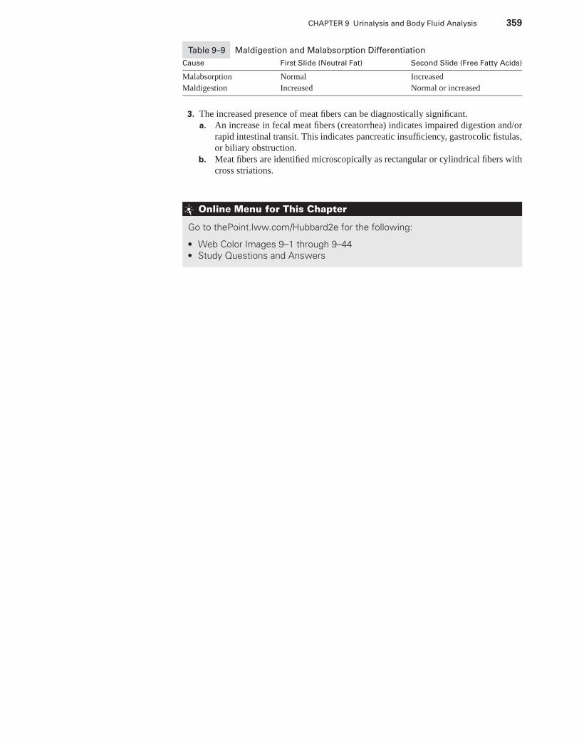
P1: OSO
LWBK192-09 LWBK192-Hubbard November 27, 2008 7:51
CHAPTER 9 Urinalysis and Body Fluid Analysis 359
Table 9–9 Maldigestion and Malabsorption Differentiation
Cause First Slide (Neutral Fat) Second Slide (Free Fatty Acids)
Malabsorption Normal Increased
Maldigestion Increased Normal or increased
3. The increased presence of meat fibers can be diagnostically significant.a. An increase in fecal meat fibers (creatorrhea) indicates impaired digestion and/or
rapid intestinal transit. This indicates pancreatic insufficiency, gastrocolic fistulas,or biliary obstruction.
b. Meat fibers are identified microscopically as rectangular or cylindrical fibers withcross striations.
Online Menu for This Chapter
Go to thePoint.lww.com/Hubbard2e for the following:
� Web Color Images 9–1 through 9–44� Study Questions and Answers
P1: OSO
LWBK192-10 LWBK192-Hubbard November 24, 2008 20:16
CHAPTER 10
Molecular Diagnostics
TOOTIE TATUM, PhD, CLSp (MB), MP (ASCP)
I. BIOLOGY OF THE CELL
A. Organelles. In contrast to bacterial cells, all eukaryotic cells contain membrane-boundorganelles.
1. Lipid bilayera. Cell membrane functions as the “shell” of each cell in the body; contains a hy-
drophilic and a hydrophobic surface; and encloses the contents of the cell.b. Membrane bound organelles within the cell are encased in a lipid bilayer.
2. Nucleus is the structure containing the majority of genetic material within thecell.a. DNA is the genetic material of the cell. It is organized into chromosomes.
3. Mitochondria are numerous within each cell. The number varies by cell type.a. Energy generation occurs within mitochondria.b. Oxidative phosphorylationc. Small amount of DNA
4. Endoplasmic reticulum is a structure that is continuous with the nucleus, but containsno DNA.a. It is the site of protein synthesis.
5. Golgi apparatusa. Protein modificationb. Protein transport to the cytosol
6. Lysosomes and peroxisomesa. Function in the removal of waste products within the cell
B. DNA is the genetic material of all cells and 99% is contained in the nucleus.1. Structure
a. Purine and pyrimidine bases: A, T, G, and Cb. A phosphodiester backbone links the bases together to form a chain.c. A deoxyribose sugar is also contained at each base position.d. Double helix. Two DNA chains are hydrogen bonded to one another between
adenine and thymine or cytosine and guanine. The two joined, antiparallel strandsthen twist into a double-helix (Figure 10–1).
e. Genes. Along the DNA strands are specific, short regions that encode for genes.2. Chromosomes. Human DNA is further arranged into 23 pairs of matched chromosomes.
a. Chromatids. The DNA strands are wound around histone proteins and these coiltogether to form a tightly packed chromatid.
b. Centromeres. The two sister chromatids are joined at the centromere. These twojoined chromatids form a chromosome.
360
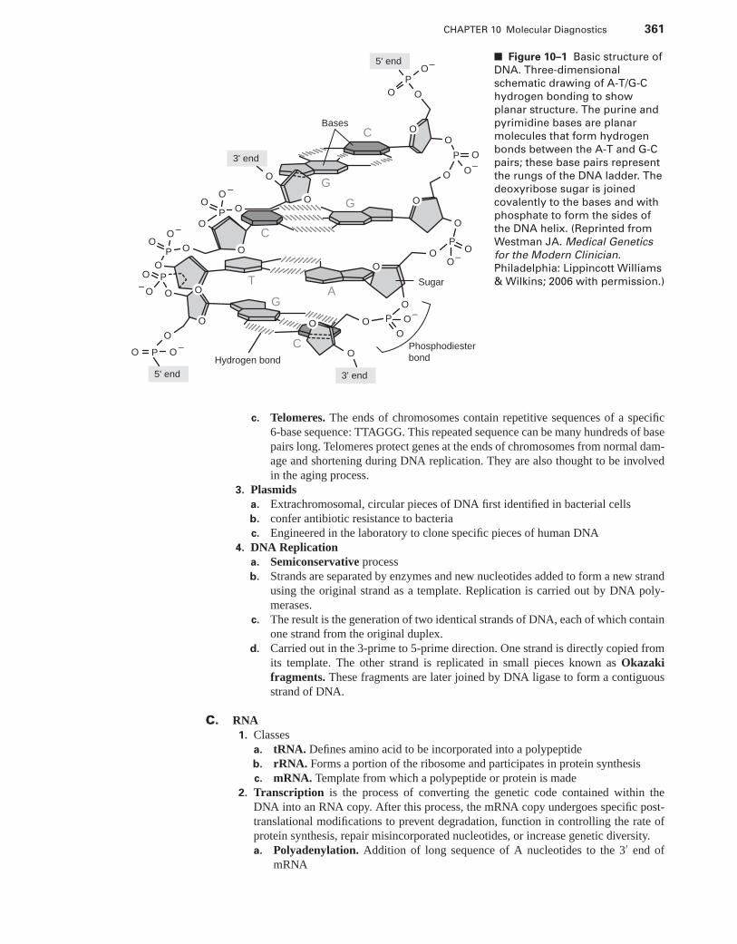
P1: OSO
LWBK192-10 LWBK192-Hubbard November 24, 2008 20:16
CHAPTER 10 Molecular Diagnostics 361
Bases
O O
O
OO
OO
OO
O
O
OO
O
P
P
P
P
–
–
–
–
5' end
5' end
3' end
3' end
O
O
O
T
G
G
G
C
A
C
C
O
O
OO
OP
P
P
P
O
O
OO
Sugar
PhosphodiesterbondHydrogen bond
O
O
O O
O
O
O
O
O
–
–
–
–
OO
O
O
■ Figure 10–1 Basic structure of
DNA. Three-dimensional
schematic drawing of A-T/G-C
hydrogen bonding to show
planar structure. The purine and
pyrimidine bases are planar
molecules that form hydrogen
bonds between the A-T and G-C
pairs; these base pairs represent
the rungs of the DNA ladder. The
deoxyribose sugar is joined
covalently to the bases and with
phosphate to form the sides of
the DNA helix. (Reprinted from
Westman JA. Medical Genetics
for the Modern Clinician.
Philadelphia: Lippincott Williams
& Wilkins; 2006 with permission.)
c. Telomeres. The ends of chromosomes contain repetitive sequences of a specific6-base sequence: TTAGGG. This repeated sequence can be many hundreds of basepairs long. Telomeres protect genes at the ends of chromosomes from normal dam-age and shortening during DNA replication. They are also thought to be involvedin the aging process.
3. Plasmidsa. Extrachromosomal, circular pieces of DNA first identified in bacterial cellsb. confer antibiotic resistance to bacteriac. Engineered in the laboratory to clone specific pieces of human DNA
4. DNA Replicationa. Semiconservative processb. Strands are separated by enzymes and new nucleotides added to form a new strand
using the original strand as a template. Replication is carried out by DNA poly-merases.
c. The result is the generation of two identical strands of DNA, each of which containone strand from the original duplex.
d. Carried out in the 3-prime to 5-prime direction. One strand is directly copied fromits template. The other strand is replicated in small pieces known as Okazakifragments. These fragments are later joined by DNA ligase to form a contiguousstrand of DNA.
C. RNA1. Classes
a. tRNA. Defines amino acid to be incorporated into a polypeptideb. rRNA. Forms a portion of the ribosome and participates in protein synthesisc. mRNA. Template from which a polypeptide or protein is made
2. Transcription is the process of converting the genetic code contained within theDNA into an RNA copy. After this process, the mRNA copy undergoes specific post-translational modifications to prevent degradation, function in controlling the rate ofprotein synthesis, repair misincorporated nucleotides, or increase genetic diversity.a. Polyadenylation. Addition of long sequence of A nucleotides to the 3′ end of
mRNA
P1: OSO
LWBK192-10 LWBK192-Hubbard November 24, 2008 20:16
362 A Concise Review of Clinical Laboratory Science
b. Capping. Addition of a methyl group to the 5′ end of mRNAc. Splicing. Inserting, deleting, or editing nucleotides after synthesis
D. Proteins1. Translation is the process of utilizing mRNA transcripts to produce a polypeptide (or
protein) represented by the original genetic code.a. The sequence of an mRNA is converted to a polypeptide.b. Occurs in the ribosome
2. Amino acidsa. Represented by a triplet codon sequenceb. Basic components of proteinsc. Contain an amino and a carboxyl groupd. Twenty amino acids found in proteins
3. Polypeptidesa. Chain of amino acidsb. α-helix or β-sheetc. Component or subunit of a mature protein
4. Enzymesa. All are proteinsb. Catalyze specific chemical reactions within or outside of the cellc. DNA synthesis proteins are common examples of enzymesd. Degradation of cellular componentse. Cell divisionf. Enzymes are also produced commercially for use in the laboratory.
II. MOLECULAR DIAGNOSTIC METHODS
A. Nucleic acid isolation1. Specimen collection
a. Whole blood. This is the most common type of molecular diagnostic specimen.Blood is most commonly collected in purple-top tubes. Heparin is a potent inhibitorof PCR and is, therefore, not used in molecular diagnostics. Blood should be storedat 4◦C until processing.
b. Tissue. Tissue specimens should be frozen immediately upon receipt or held incell culture medium at 37◦C until processing.
c. Buccal swabs. Often used for remote site testing; collection of DNA specimensfor parentage testing. Swabs should be air-dried before shipment to the labo-ratory.
d. Other specimens include those also commonly encountered in the clinical labincluding urine, feces, sputum, cerebrospinal fluid, genital swabs, semen, etc.Other molecular diagnostic specimens include sections of formalin-fixed paraffin-embedded tissues, bone, hair, and fingernails (usually confined to forensic speci-mens).
2. DNA isolationa. Phenol:Chloroform:Isoamyl alcohol method is the “gold standard.” Yields a
large mass of high quality DNA. Can be used to isolate DNA from any specimentype. This method normally requires a large mass of starting material.
b. Manufacturer (Qiagen) kits. Provide ease of use and high degree of reproducibil-ity. Yields smaller amounts of DNA. DNA is usually very pure. Most often used toisolate DNA from blood. This is the most commonly encountered clinical DNAisolation method.
c. Quick-extract solutions. Fastest methods. Low quality, low mass DNA recovery.Usually used to isolate DNA from buccal swabs/FTA cards.
3. RNA isolationa. Acid phenol/LiCl salt isolations. Gold standard. Yields a large mass of high
quality RNA. Can be used on any specimen type.
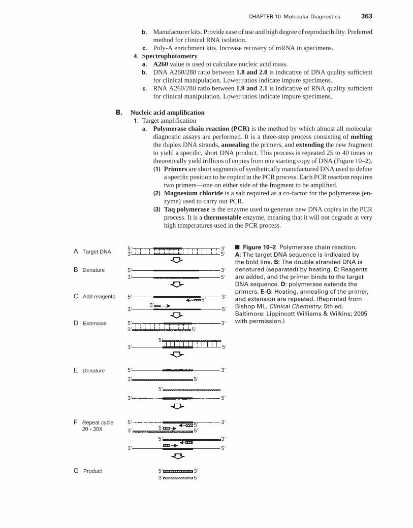
P1: OSO
LWBK192-10 LWBK192-Hubbard November 24, 2008 20:16
CHAPTER 10 Molecular Diagnostics 363
b. Manufacturer kits. Provide ease of use and high degree of reproducibility. Preferredmethod for clinical RNA isolation.
c. Poly-A enrichment kits. Increase recovery of mRNA in specimens.4. Spectrophotometry
a. A260 value is used to calculate nucleic acid mass.b. DNA A260/280 ratio between 1.8 and 2.0 is indicative of DNA quality sufficient
for clinical manipulation. Lower ratios indicate impure specimens.c. RNA A260/280 ratio between 1.9 and 2.1 is indicative of RNA quality sufficient
for clinical manipulation. Lower ratios indicate impure specimens.
B. Nucleic acid amplification1. Target amplification
a. Polymerase chain reaction (PCR) is the method by which almost all moleculardiagnostic assays are performed. It is a three-step process consisting of meltingthe duplex DNA strands, annealing the primers, and extending the new fragmentto yield a specific, short DNA product. This process is repeated 25 to 40 times totheoretically yield trillions of copies from one starting copy of DNA (Figure 10–2).(1) Primers are short segments of synthetically manufactured DNA used to define
a specific position to be copied in the PCR process. Each PCR reaction requirestwo primers—one on either side of the fragment to be amplified.
(2) Magnesium chloride is a salt required as a co-factor for the polymerase (en-zyme) used to carry out PCR.
(3) Taq polymerase is the enzyme used to generate new DNA copies in the PCRprocess. It is a thermostable enzyme, meaning that it will not degrade at veryhigh temperatures used in the PCR process.
A Target DNA
B Denature
C Add reagents
D Extension
E Denature
F Repeat cycle 20 - 30X
G Product
5�5�3�
5�
3�
5�
3�
5�3�
5�
3�
5�
3�
5�
5�
5�
5�
5�
5�5�
5�
5�
5�5�
3�
5�3�
3� 5�
5�
5�
5�
3�
3�
3�
3�
3�
3�
3�
3�
5�
3�
■ Figure 10–2 Polymerase chain reaction.
A: The target DNA sequence is indicated by
the bold line. B: The double stranded DNA is
denatured (separated) by heating. C: Reagents
are added, and the primer binds to the target
DNA sequence. D: polymerase extends the
primers. E-G: Heating, annealing of the primer,
and extension are repeated. (Reprinted from
Bishop ML. Clinical Chemistry. 5th ed.
Baltimore: Lippincott Williams & Wilkins; 2005
with permission.)
P1: OSO
LWBK192-10 LWBK192-Hubbard November 24, 2008 20:16
364 A Concise Review of Clinical Laboratory Science
(4) Buffers containing potassium chloride, sodium chloride, and other additivesare also included in the reaction to stabilize the enzymes and DNA during thePCR process.
(5) dNTPs (A, T, G, and C) are the individual building blocks of the DNA strand.(6) A very small mass of the genomic DNA specimen of the patient to be tested is
also added to the reaction. The PCR process only requires femtomolar quanti-ties of input DNA.
b. Transcription-based amplification (TMA) is an alternative method to PCR thatis not based upon direct amplification of the genomic DNA template.
2. Probe amplification is another method that does not rely on direct amplification of thetemplate DNA strand. Two commonly encountered clinical examples of this strategyinclude:a. Ligase chain reactionb. Multiplex ligation-dependent probe amplification (MLPA). This method is used
clinically to detect deletions of multiple entire exons in very long stretches of DNArepresenting large genes.
c. Strand displacement amplification3. Signal amplification is yet another method of detecting a DNA target without direct
amplification of the template DNA. It relies on detection of substrates bound to thetarget nucleic acid.a. Branched DNA amplificationb. Hybrid capture
C. Restriction digestion is a method used in many applications within the molecular diag-nostics laboratory. It is a method used to fractionate DNA at sequence-specific locationsusing restriction enzymes. Restriction digestion is used to generate size standards and toprepare genomic DNA for analysis by Southern blotting and hybridization.
D. Nucleic acid blotting and hybridization1. Southern blot is a method to analyze the DNA contained in an entire genome through a
process of restriction digestion, electrophoresis, transfer to a membrane, and hybridiza-tion to a sequence-specific probe.
2. Northern blot is analogous to the process of Southern blotting, but is performed onRNA specimens.
3. Probes are small fragments of DNA used to detect a specific gene or mutation in theprocess of Southern or Northern blotting.
4. Hybridization dynamics are conditions, such as salt concentration, temperature, timeand buffer composition, that control the stringency of the hybridization procedure.
E. Gel electrophoresis is a method used to separate DNA fragments onto a solid matrix basedupon size.
1. Agarose gel electrophoresis is most often performed to resolve DNA fragments largerthan 100 bp in size.
2. Polyacrylamide gel electrophoresis is the method of choice to resolve small DNAfragments. Very high concentrations of polyacrylamide matrix can resolve down to 1 bpdifferences in size.
3. Capillary gel electrophoresis is a modification of polyacrylamide gels used in auto-mated DNA sequencing and genotyping platforms.
F. Quantitative real-time PCR is a PCR method starting the mRNA as a template. Thismethod is capable of quantifying the copy number of mRNA transcripts present in a speci-men.
1. Reverse transcriptase is the enzyme used to make a copy DNA, or cDNA, fromthe mRNA transcript contained in a specimen. This process is referred to as cDNAsynthesis.
2. Real-time PCR is the general term for the PCR method used to observe the productionof new DNA fragments in real time on an automated platform.a. SYBR green is a dye often used in quantitative real-time PCR assays to detect
newly generated DNA fragments.
P1: OSO
LWBK192-10 LWBK192-Hubbard November 24, 2008 20:16
CHAPTER 10 Molecular Diagnostics 365
b. TaqMan chemistry is an alternate system of amplification and detection of frag-ments in quantitative real-time PCR using dye-labeled probes.
G. DNA sequencing allows determination of each base pair in a single strand of DNA.1. Sanger dideoxy sequencing is the original method of DNA sequencing and the basis
of all DNA sequencing used in the clinical environment today. It is based upon theincorporation of dideoxy nucleotides in random positions in the newly synthesizedDNA strand.
2. Fluorescent DNA sequencing is a method by which DNA sequence is determinedbased upon incorporation of a dye-labeled dideoxy nucleotide and resolution on acapillary electrophoresis instrument. Each dideoxy nucleotide is labeled with a specificdye and when incorporated, is excited by a laser and detected by a CCD camera on anautomated platform.
H. Microarray technologies are the newest area of use in molecular diagnostics. They fallinto two general categories:
1. Single nucleotide polymorphism arrays are those designed to detect single base orother small changes in DNA sequence across the entire genome simultaneously. Manythousands of positions can be interrogated in one experiment.
2. Gene expression arrays are those designed to assess global changes in expression ofspecific genes in a given tissue at a given time or under a specific set of treatmentparameters.
III. INHERITED GENETIC DISEASE
A. Fragile-X is characterized by the following:1. Trinuceotide repeat expansion2. X-linked3. Mental retardation4. Primary affects males
B. Duchenne muscular dystrophy is characterized by the following:1. Deletion of entire exons2. Dystrophin gene3. X-linked4. Muscle degeneration
C. Myotonic dystrophy is characterized by the following:1. Trinucleotide repeat expansion with anticipation (the worsening of clinical symptoms
with increasing repeat size in successive generations)2. Autosomal dominant3. Males and females4. Multisystem disease
D. Cystic fibrosis (CF) is characterized by the following:1. Many individual mutations in the CFTR gene may give rise to CF alone or in combi-
nation.2. Delta F508 mutation accounts for over 70% of CF cases.3. Autosomal recessive4. Although it primarily affects the lungs, it is a multisystem disease.5. Most common genetic disease in Caucasians
IV. MOLECULAR ONCOLOGY
A. Minimal residual disease detection is a commonly performed assay in molecular diagnos-tics. It is the determination of the effectiveness of a therapy or the tracking of a patient’s
P1: OSO
LWBK192-10 LWBK192-Hubbard November 24, 2008 20:16
366 A Concise Review of Clinical Laboratory Science
disease status based upon the presence of specific mRNA transcripts known to causeleukemia or lymphoma.
1. Chronic myeloid leukemia (CML) is characterized by the presence of the Philadel-phia chromosome or 9;22 translocation. The transcript can be detected by quantitativereal-time PCR and the size determined by fluorescent capillary electrophoresis.
B. In molecular oncology, clonality is the presence of a specific T or B cell clone originatingfrom a lymphoma. The clone is detected using PCR and fluorescent capillary electrophore-sis.
C. Microsatellite instability is a genetic phenomenon observed in cells undergoing changesin specific cells. It is the expansion of short, repetitive stretches of DNA to much longerstretches that change gene expression within the cell.
1. HNPCC is a hereditary form of colon cancer that is marked by microsatellite instability.
D. Loss of heterozygosity is the nature of some tumors to exhibit loss of function of oneallele in a specific gene in a cell in which the other gene was already inactivated. The mostcommon example of a cancer caused by this process is retinoblastoma, in which expressionof a tumor suppressor gene is lost.
E. Drug metabolism and sensitivity can be determined through genotyping of DNA or geneexpression analysis.
1. Cytochrome P450 (P450) genotyping is performed to determine drug sensitivity. Thetest is carried out on a microarray platform.
V. MOLECULAR INFECTIOUS DISEASE
A. Bacterial identification. The following organisms can be easily detected (qualitative iden-tification) using PCR:
1. Chlamydia trachomatis2. Neisseria gonorroheae3. MRSA4. VRE
B. Viral identification may also be performed in a quantitative manner. The following may bequantitatively detected:
1. Viral load—Quantitativea. HCVb. HIVc. CV
VI. DNA-BASED HUMAN IDENTIFICATION
There are a number of ways by which human identification may be performed. The mostwidely used system is based upon determination of the length of repetitive DNA in specificregions of chromosomes across the human genome. This is termed DNA-based humanidentification.
A. HLA polymorphisms are not as diverse as those observed using other systems of identifi-cation. This is an older system of human identification.
B. RFLP analysis was the first DNA-based identification system. It is based upon poly-morphism in the length of restriction enzyme-cut products across the genome. It utilizesSouthern blotting and hybridization as a method of laboratory testing.
C. STR analysis1. Most widely used system for identification of individuals
P1: OSO
LWBK192-10 LWBK192-Hubbard November 24, 2008 20:16
CHAPTER 10 Molecular Diagnostics 367
2. Very high power of discrimination3. Common alleles are typed and compared to the CODIS database for criminal investi-
gations.4. Forensic DNA typing of older specimens or “cold cases” utilizes this system when
the specimens are in acceptable condition.5. Parentage testing uses the same STR system as those used in the CODIS allele
database.6. Bone marrow engraftment is a clinical application of this chemistry.
a. STR analysis may be performed to track donor versus recipient DNA componentsfollowing transplant.
7. Other uses of DNA-based human identification include QC of histological specimensto ensure that a patient matches a specific specimen.
D. Mitochondrial DNA analysis is identification of individuals based upon their mitochon-drial haplotype.
1. Primarily used on degraded specimens2. Only capable of determining maternally inherited alleles with this system, because
mitochondria are passed from mothers to all offspring.3. Often used in “cold cases,” because there are many hundreds of copies of mitochondrial
DNA present for every one copy of nuclear DNA. This allows for a greater possibilityof success in analysis.
E. Y-chromosome analysis is a newer system for human identification.1. Only capable of determining paternally inherited alleles2. System only used to type males3. Based upon repeat length polymorphism
Online Menu for This Chapter
Go to thePoint.lww.com/Hubbard2e for the following:
� Study Questions and Answers
P1: OSO
LWBK192-11 LWBK192-Hubbard November 24, 2008 20:21
CHAPTER 11
Current Issues inLaboratory Management
WADE REDMAN, MBA, MT (ASCP) DLM, LORI RICE-SPEARMAN,MT (ASCP), MS, AND HAL S. LARSEN, PhD
I. MANAGEMENT AND ORGANIZATIONAL THEORY
A. Management (administration) can be defined as: the organizing and controlling of theaffairs of a business or a sector of a business or “working with and through people toaccomplish a common mission.”
B. Management/leadership theories. One factor that impacts leadership success is manage-ment style. There are primarily five leadership theories:
1. Tannenbaum and Schmidt theory. Styles can be plotted on a continuum from author-itative to democratic.
2. McGregor X and Y theory. McGregor X theory relates to authoritative leader,whereas the Y theory relates to the democratic leader
3. Blake and Mouton theory. Describes five types of management situations:a. Impoverished management. Low concern for people and productionb. Authority-compliance. Low concern for people, but high concern for produc-
tionc. Middle-of-the-road management. Medium concern for people and productiond. Country club management. High concern for people, but low concern for pro-
ductione. Team management. High concern for people and production
4. Fielder theory. This theory indicates that the style used by the leader may vary ac-cording to the situation with a very favorable or very unfavorable situation requiring atask-oriented leader and a moderately favorable or moderately unfavorable situationrequiring a relationship-oriented leader.
5. Hersey-Blanchard theory. Four leadership situations:a. Employee new to job.b. Employee has mastered some of the job, but needs supervision.c. Employee has mastered the job, but needs verification.d. Employee has mastered the job and is confident.
C. Skills of managers1. Organizational skills. Conceptualize and apply management process, systematize
work flow, make decisions, and communicate with coworkers.2. People skills. Understands basic theories of human needs and work motivation.
368
P1: OSO
LWBK192-11 LWBK192-Hubbard November 24, 2008 20:21
CHAPTER 11 Current Issues in Laboratory Management 369
3. Financial skills. Effective use of and accounting for the monetary assets of the com-pany.
4. Technical skills. Involve the synthesis of the first three skills above and the managementof physical resources (supplies, equipment, facilities) into the operational parameters(products and services).
D. Management theories1. Scientific management. Attempts to apply a systematic or scientific approach to the
study of organizations developed by Frederick Taylor, the father of scientific theory.Applies cause-and-effect analysis, breaking down each task into segments to improveefficiency.
2. Bureaucracy management. Examines the organizational aspects of the company andits work flow to explain how institutions function and how to improve their structuralprocess. Hierarchical chain of command is an example. Special attention is given tothe bureaucratic characteristics as they relate to rules, regulations, impersonality, anddivision of labor.
3. Behavioral science. Focuses on the performance and interaction of people withinthe organization by utilizing psychology and sociology, as well as managementtheories.
4. Systems analysis. An outgrowth of management science, which views the organizationas a continuous process interacting within itself and with its environment.a. Involves Peter Drucker’s five basic management operations:
(1) Setting objectives(2) Organizing(3) Motivating and communicating(4) Establishing standards (measurement of performance)(5) Developing people, including managers
E. Management process. The concept of management as a continuous process of interactingfunctions, each dependent on the success of the other.
1. Four main functions:a. Planningb. Organizingc. Directingd. Controlling
F. Management by objectives (MBO). A program that embodies all of the concepts in themanagement process (planning, operating, directing, and controlling). First introduced byPeter Drucker in the 1960s. The core of MBO is communications. Success or failure de-pends on how well employees understand the company’s mission and objectives. Managerswho allow employees to share in the decision-making process will have effective MBOprograms. Managers must understand and be skilled at writing and monitoring object-ives.
G. Motivation. The driving forces that make people act as they do. This also can be de-fined as incentives, motives, purpose, drive, desires, and needs. Includes three majortheories:1. Maslow’s hierarchy of needs. Identified physiological and safety needs as basic phys-
ical needs that would motivate if not satisfied. Satisfaction of physical needs is requiredbefore higher level social need, such as esteem and love, will motivate
2. Herzberg’s two-factor theory. Herzberg (1959) defined two factors:a. The basic needs, hygiene factors, which correspond primarily with Maslow’s
physical and social needs.b. Motivators correspond primarily with Maslow’s growth needs.
3. Vroom’s expectancy theory. Effort will bring reward. The effort expended will berelated to the perceived value of the reward. People make a choice between alternativecourses of actions according to how they perceive the resulting benefits.
P1: OSO
LWBK192-11 LWBK192-Hubbard November 24, 2008 20:21
370 A Concise Review of Clinical Laboratory Science
II. PROFESSIONALISM
A. Definition. Professionalism is judged by the professional quality with which one practicesthe profession and includes areas such as altruism, honor, integrity, caring, compassion,respect, responsibility, accountability, scholarship, and leadership.
B. Definition. Professional ethics is the study of standards of conduct and moral judgmentswhich provide a basis for right and wrong conduct. The American Society for ClinicalLaboratory Science (ASCLS) has developed and published the Code of Ethics for ClinicalLaboratory Science comprised of duty to the patient, duty to colleagues, and duty to society.
C. Job Description1. Purpose. The job description defines the functions and purpose of a specific position
for an employee.2. The job description should include:
a. Name of organizationb. Job titlec. Summary of duties for the positiond. Essential functions required for the positione. Immediate supervisor (chain of command)f. Working conditions/environment
g. Required qualifications and experience
D. Resume1. A resume is a tool for a candidate to demonstrate their strengths, education, and
accomplishments when seeking employment. The resume should include the following:a. Candidates name (legal)b. Contact information (including e-mail)c. Career objectived. Educational qualificationse. Clinical experiencef. Military experience
g. Certificationsh. Professional society membershipsi. Honorsj. Publications/research activities/special projectsk. Volunteer workl. Statement regarding references
2. A cover letter should accompany a resume to focus attention on the applicant. It isbest to address the cover letter to a specific individual.
E. Employee Interview and selection1. The purpose of the interview process if for the employer to confirm that the provided,
written information gathered from the resume and/or job application is valid. Addition-ally, the interview provides the employer with insight into the candidate’s interpersonalcommunication skills. The interview also provides the candidate with the opportunityto discuss information in more detail and to ask questions pertinent to the specificposition.
2. The selection process should be conducted after the interview and should include anevaluation of the candidate’s resume/application and information from the interview.The manager is seeking to select the candidate with the appropriate skills, certifications,and personality to fit the institution and the position.
F. Employee evaluation1. The employee evaluation process is a method of recognizing job excellence and deter-
mining areas for development and training. The job evaluation is directly linked to the
P1: OSO
LWBK192-11 LWBK192-Hubbard November 24, 2008 20:21
CHAPTER 11 Current Issues in Laboratory Management 371
job description and should be formative and summative in nature and conducted oftenenough to provide feedback and direction for the employee.a. Criteria-based evaluations indicate specific performance expectations that are
linked to the job description.b. Competency-based evaluations are used to validate competency of skills.
G. Corrective action1. The purpose of corrective action is to provide the employee with specific goals to
enhance their effectiveness and performance within the institution.a. The first step is verbal counseling to make the employee aware of the situation and
to open the lines of communication.b. The second step is to issue a verbal warning that specifically involves stating the
problem, performance in question, and offering resources and assistance (processis documented).
c. The third step is a formal written warning that states the problem, expected im-provement in performance, and a timeline for re-evaluation of performance. Thisdocument is signed by the immediate supervisor and employee. Failure to meet thestated requirements typically results in dismissal of the employee.
d. Most institutions have a policy that allows the employee to seek an appeal of thesituation to a higher level.
H. Communication and interpersonal relationships1. Communication is an ongoing process of interpreting symbols, decoding speech
and phrases, interpreting ideas and feelings, and encoding that information to for-mulate an appropriate response. Communication typically serves the following pur-poses:a. Exchange of knowledgeb. Expression of feelingsc. Expression of ideas and thoughtsd. To attempt to influence another’s thoughts and actionse. To meet social expectations
2. Barriers to effective communication are:a. Nonverbal expressions–body languageb. Cultural differences–unfamiliar phrasesc. Paraverbal issues–tone and pitch of voiced. Environmental–temperature, neatness of appearancee. Overcommunication–too much information offered at one timef. Undercommunication–not enough information provided for understanding the is-
sueg. time issues
I. Team building is essential for optimal employee performance and effectiveness of thesection, department, and institution.
1. Tuckman (1965) suggests the following phases of team development:a. Identifying the team membersb. Team members selecting their rolesc. Determining standards of behavior and expectationsd. Working together towards a common goal
III. QUALITY ASSURANCE
A. Introduction1. Quality assurance (QA) is a comprehensive set of policies, procedures, and prac-
tices that are followed to ensure that a laboratory’s results are reliable. QA evaluatesthe quality of the services provided. QA provides a way to prevent problems as wellas deal with problems that occur. It differs from quality control (QC) in that QC is
P1: OSO
LWBK192-11 LWBK192-Hubbard November 24, 2008 20:21
372 A Concise Review of Clinical Laboratory Science
included as part of a QA program. QC ensures that a particular test method is workingproperly and that results of that test are reliable.
2. QA includes record keeping, maintenance and calibration of equipment, profi-ciency testing, quality control, and training of personnel. QA is a commitment toquality. Physician and patient relations are a part of this plan.
B. Quality assurance plan1. All policies and procedures are in writing and available.2. Staff performance is routinely evaluated.3. Continuing education is used and documented.4. A safety program is in place for both staff and patients.
a. Occupational Safety and Health Administration (OSHA) standards are met.b. Hazardous materials are identified and labeled.c. Material Safety Data Sheets (MSDS) are available for each hazardous material.d. Employee safety training is documented.e. Records of any accidents (e.g., needle stick) are kept and reported to appropriate
authorities.f. Hazardous waste is properly disposed.
g. Universal precautions are strictly followed.h. Safety equipment (e.g., fire extinguishers, fire blankets, safety showers, goggles)
is available to staff, and training in the use of this equipment is performed anddocumented.
i. Electrical checks of all equipment are routinely performed.5. Specimen collection and handling (pre-analytical) as well as correct patient prepara-
tion follows written guidelines and is documented.6. A quality control (QC) program is in place (analytical). This ensures that the in-
struments, reagents, and personnel are functioning properly. A QC program comprisesseveral areas:a. A written QC program is required that specifies:
(1) Frequency of performing controls(2) Number of controls to be used(3) Type of controls(4) Acceptable limits for control results(5) Corrective action required if controls are out of range
b. QC plots of control results are used to detect shifts (a sudden upward or downwardchange of four or more consecutive values) and trends (the tendency of results togradually increase or decrease over a period of time).
c. Equipment maintenance is performed and documented.d. Temperatures of incubators, certain instruments, and the room are noted and
documented.7. Checklists are used to ensure that scheduled activities and duties are performed.8. The laboratory must participate in and document results of proficiency testing
(PT). This allows comparison of results from a number of laboratories. Specimensfrom PT sponsors are to be treated exactly like patient specimens.
9. The laboratory must be accredited by an appropriate agency. This gives officialapproval and states that the laboratory follows all of the guidelines set forth by theaccrediting agency. Inspectors representing the accrediting agency view records, doc-uments, and visit the laboratory.
IV. LABORATORY REGULATIONS
A. Clinical Laboratory Improvement Amendments of 1988 (CLIA 88)1. Background. CLIA 88 is intended to establish regulations for all laboratories,
regardless of size or location, where clinical testing is performed for the purpose ofdiagnosis, treatment, or monitoring of patients’ health. The law was published in theFebruary 28, 1992 issue of the Federal Register. The law became effective for alllaboratories on September 1, 1992.
P1: OSO
LWBK192-11 LWBK192-Hubbard November 24, 2008 20:21
CHAPTER 11 Current Issues in Laboratory Management 373
a. Modifications to the law list the complexity of tests.b. Additional modifications, define alternative routes for personnel qualifications.
2. Laboratory accreditationa. All laboratories must have a CLIA certificate to perform testing and receive
federal reimbursement.b. Certification is based on the level of test complexity.
(1) Sites performing only waived tests comprise 58% (105,138) of the approxi-mately 180,000 laboratory testing sites in the United States. Waived testingperformed in these sites is often wellness testing, screening tests, or other crit-ical testing that introduces a large population of persons into the health caresetting. Although the testing performed in Certificate of Waiver sites accountsfor <10% of the total U.S. testing volume, this percentage has been increas-ing each year since the CLIA program began. Types of direct, unprocessedspecimens suitable for waived testing are listed below:(a) Whole blood (fingerstick or anticoagualted blood collected via venipunc-
ture)(i) Glucose, cholesterol, prothrombin time, infectious mononucleosis, ery-
throcyte sedimentation rate, hemoglobin-copper sulfate method, spunhematocrit HIV antibody
(b) Urine(i) Dipstick urinalysis and pregnancy test (hCG)
(c) Throat swab(i) Group A streptococcal antigen
(d) Nasopharyngeal swab(i) Influenza
(e) Stool(i) Occult blood
(f) Saliva(i) Alcohol
(g) Oral fluid(i) HIV antibody
(h) Gastric biopsy(i) H. pylori
(2) Provider-performed microscopy (PPM) includes:(a) Wet mount, including preparations of vaginal, cervical or skin specimens(b) Potassium hydroxide (KOH) preparation(c) Pinworm slides(d) Fern test(e) Urine sediment(f) Vaginal smears
(g) Nasal smear examinations for granulocytes(h) Fecal leukocyte examinations(i) Semen analysis
(i) Qualitative examination(ii) Presence or absence(iii) Motility
(3) Moderately complex testing includes:(a) Hematology(b) Chemistry(c) Serology(d) Coagulation(e) Therapeutic drug monitoring(f) Blood gases
(g) Urine colony counts(h) Blood type and Rh factor(i) Limited immunology(j) Manual differential (limited identification; normal cells only)
(k) Gram’s stain
P1: OSO
LWBK192-11 LWBK192-Hubbard November 24, 2008 20:21
374 A Concise Review of Clinical Laboratory Science
(i) Urethral for gas chromatography (GC)(ii) Cervical for GC
(l) Throat culture screen only(i) Hemolysis
(ii) Bacitracin(iii) Strep select agar
(m) Microscopic urinalysis (non-PPM)(n) Rapid kits
(4) Highly complex testing includes:(a) Microbiology(b) Immunohematology(c) Bacteriology(d) Cytology(e) Histopathology(f) Mycology(g) Manual hematology counts(h) Manual differential
(i) Identification of abnormal cells(ii) Complete identification(iii) Nonautomated chemistry tests
c. The certificate fee and biennial inspection fee, which are based on the size ofthe laboratory and number of tests performed annually, must be paid.
d. Accreditation by a private organization is an alternative option for certifica-tion.(1) The Health Care Financing Administration (HCFA) may deem a laboratory to
meet all of the CLIA requirements if the laboratory is accredited by a private,nonprofit organization that is certified as having deemed status. This meansthat the certifying agency must:(a) Provide reasonable assurance to HCFA that it requires the laboratories it ac-
credits to meet all of the requirements equivalent to the CLIA requirementsin the law
(b) Inspect its laboratories per condition level requirements of the law(2) Approved state laboratory programs are responsible for accreditation of lab-
oratories in their state, if this is a requirement of the department of health ofthat state.
3. Personnel requirements must be met by laboratories performing moderately or highlycomplex testing. These positions can be held by the same person or by different personswho meet the qualifications.a. Moderately complex laboratories must have the following personnel:
(1) Laboratory director(2) Technical consultant(3) Clinical consultant(4) Testing personnel
(a) Each person performing moderately complex testing must:(i) Follow laboratory procedures
(ii) Maintain records(iii) Adhere to policies(iv) Identify problems(v) Document corrective actions
(b) Testing personnel must have documented training in all areas of testingperformed.
b. Highly complex laboratories are required to have the same personnel as a mod-erately complex laboratory, but must also have:(1) General supervisor(2) Technical supervisor
4. Quality control (QC) must be performed on all moderately and highly complex test-ing.
P1: OSO
LWBK192-11 LWBK192-Hubbard November 24, 2008 20:21
CHAPTER 11 Current Issues in Laboratory Management 375
a. General QC procedures are performed on:(1) All specialties and subspecialties as specified within the Federal Register(2) Procedures not found in the Federal Register, which must be conducted
according to the manufacturer’s protocol that has been approved by the Foodand Drug Administration
b. The laboratory must establish and follow written QC procedures for monitoringand evaluating the test process to assure accuracy and reliability of results.
c. There must be documentation of all calibration procedures.d. A minimum of two levels of control materials are run on each day of testing.e. All QC records must be retained for 2 years.f. QC must be tested in a manner that provides results within the laboratory’s stated
performance requirements.g. The laboratory must have instruments, reagents, and supplies sufficient for the
type and volume of tests performed.h. There must be defined criteria for proper storage of reagents and specimens.i. A procedure for remedial actions must be established when QC fails.j. All reagents, solutions, medias, QC materials, and standards must be labeled.k. Different lot numbers are not to be interchanged unless specified by the manu-
facturer.5. Proficiency testing (PT) is mandatory for all moderately and highly complex tests
listed on the Compiled list of clinical laboratory systems, assays, and examinationscategorized by complexity, published in the Federal Register.a. There are numerous CMS-approved PT programs available.b. Laboratories receive between three and five challenges for each analyte that re-
quires PT.c. Challenges are received three times a year. The same personnel who routinely
perform the test procedures must perform the PT.d. Laboratories are required to score 80% on all analytes to be considered in compli-
ance.e. Blood bank specimens require a score of 100%.
6. A procedures manual must be written to include all testing methods used in thelaboratory. It must be available and followed by all testing personnel.a. The manual must include:
(1) The principle and methodology of each test(2) Criteria for specimen collection, processing, and rejection (pre-analytical
steps)(3) Instructions for microscopic examination procedures(4) Test procedures (analytical)
(a) Step-by-step instructions(b) Result interpretation
(5) Preparation of materials(a) Required solutions (e.g., reagents, controls, calibrators, stains, slides) and
their location(b) Step-by-step preparation of all materials needed(c) Storage after preparation(d) Labeling and dating instructions(e) Safety precautions needed(f) Solutions used in preparation (e.g., deionized water, diluent)
(6) Calibration procedures(a) Step-by-step instructions(b) Verifications of calibration results(c) Concentration and number of calibrators used(d) Calibration schedule
(7) Control procedures(a) Materials used, name, lot number, level, frequency used(b) reparation of materials(c) Instructions for testing controls
P1: OSO
LWBK192-11 LWBK192-Hubbard November 24, 2008 20:21
376 A Concise Review of Clinical Laboratory Science
(d) Control limits(e) Description of how and where control results are recorded(f) Corrective actions taken when controls are not within limits
(8) Actions taken when results deviate from expected values:(a) Recalibration(b) Troubleshooting(c) Repeats(d) Dilutions
(9) Limitations(a) Interfering substances(b) Common sources of error
(10) Result reporting (postanalytical)(a) Reference ranges
(i) Range for different specimens (e.g., serum, plasma, urine)(ii) Demographic variables (e.g., age, sex)
(b) Panic values(i) Life-threatening results
(ii) Results needing special attention (critical results)(11) References
(a) Manufacturer’s product literature (e.g., inserts, manuals)(b) Literature (e.g., textbooks, professional magazines)
(12) Specimen retention(a) How long specimen is kept(b) Where specimen is stored
(13) System for reporting patient results (postanalytical)(a) Unacceptable results(b) How results are reported to physician(c) Protocol for panic and critical values
(14) Course of action for problems(15) Specimen referral
(a) To whom or where specimen is referred(b) How the specimen is sent(c) How results are received
b. The manual must be approved. To obtain approval, the manual must be:(1) Signed by the laboratory director(2) Reviewed by all personnel using the procedure(3) Reviewed and updated by the supervisor or director
c. The manual must be rewritten when new procedures are implemented.d. The manual must be retained for future reference.
7. A quality assurance (QA) plan must be established and followed to monitor andevaluate the quality of tests.a. The program must be an ongoing process.b. It must evaluate all facets of the testing process.c. The program must extend to the interactions of other health care professionals
in the ordering of tests.d. It must allow for actions to be taken to correct errors or problems.e. The program must monitor and evaluate:
(1) Specimen requirements(2) Collection(3) Handling and processing(4) Corrective actions taken in QC and PT(5) Result reporting(6) Employee competence
f. All QA activities must be documented and evaluated monthly.8. Inspections are conducted every 2 years by the Department of Health and Human
Services (HHS) or its designee.a. Laboratory inspectors have the right to:
P1: OSO
LWBK192-11 LWBK192-Hubbard November 24, 2008 20:21
CHAPTER 11 Current Issues in Laboratory Management 377
(1) Interview all employees(2) Access all areas of a facility(3) Observe all testing(4) Review all information and data records(5) Determine that the laboratory is operated in a safe manner(6) Determine that the laboratory is performing tests only within their complexity
category(7) Evaluate any complaints from the public
b. The inspector may also:(1) Conduct unannounced inspections at any time during hours of operation(2) Ask personnel to perform testing on test samples that the inspector supplies(3) Re-inspect a laboratory at any time to evaluate accurate and reliable results
9. Sanctions may be imposed on laboratories that are found to be out of compliance withone or more of the conditions for Medicare coverage.a. Suspension, limitations, or revocation of a certificate can be imposed.b. A civil suit can be brought against a laboratory to forbid testing.c. Imprisonment or fines can be levied against persons convicted of violations.d. HHS publishes annually a list of laboratories that have been sanctioned.
B. Occupational Safety and Health Administration (OSHA) regulations for OccupationalExposure to Bloodborne Pathogens require laboratories to establish workplace safetypractices to prevent accidental exposure to etiologic agents in blood.
1. The regulations limit occupational exposure to blood and other potentially infectiousmaterials that can transmit bloodborne pathogens and include but are not limited to:a. Hepatitis B virus (HBV)b. Human immunodeficiency virus (HIV)
2. The basis for all preventive measures is the practice of universal precautions. Theseare recommendations from the Centers for Disease Control and Prevention (CDC).a. Laboratory personnel must treat all human blood and other potentially infec-
tious body fluids as if they were infected with HIV, HBV, and other bloodbornepathogens.
b. This includes materials that are contaminated with blood.3. A written exposure control plan must be in place that describes the procedures es-
tablished to minimize employee exposure to bloodborne pathogens.a. There must be identification of job classification, tasks, and procedures in which
there is the potential of exposure.(1) A determination must be made of the job types in which exposure could occur.(2) All tasks must be included in the job description.
b. An explanation of personal protective equipment (PPE) available and in usemust be included.(1) Appropriate protective equipment must be supplied to employees at no cost
to them.(2) Equipment is considered appropriate only if it does not permit blood or
other potentially infectious materials (PIMs) to pass through to an employee’sclothes, skin, mouth, or other mucous membranes.
(3) Types of PPE are gloves, gowns, laboratory coats, face shields or masks, andeye protection.
(4) All employers are responsible for providing, maintaining, laundering, dis-posing of, replacing, and assuring the proper use of all PPE.
c. Engineering controls must be in place to minimize exposure.(1) These are devices that isolate or remove the pathogen hazard from the work-
place.(2) These devices must be made available to all employees.(3) Examples of engineering controls include:
(a) Sharps containers and biosafety bags(b) Splash guards and self-sheathing needles(c) Biosafety cabinets and fume hoods
P1: OSO
LWBK192-11 LWBK192-Hubbard November 24, 2008 20:21
378 A Concise Review of Clinical Laboratory Science
(d) Mechanical pipetting devices(e) Handwashing facilities, eyewash stations, and showers
d. Work practices must be developed to reduce or eliminate employee exposure toblood or PIMs during the execution of their work tasks.(1) Employees must fully understand these procedures.(2) The procedures must be implemented, when appropriate.(3) Work practice procedures include, but are not limited to:
(a) Handwashing(b) No recapping of contaminated needles(c) No food or drink allowed in the laboratory(d) No mouth pipetting(e) Warning labels affixed where needed, and biohazard symbols placed ap-
propriately(f) A dress code that ensures safety
e. Housekeeping procedures are necessary for maintaining a clean and sanitary worksite, which is a responsibility of employers.(1) All equipment and work surfaces must be decontaminated.(2) Sharps containers must be puncture resistant.(3) Containers for biohazard materials must be constructed to contain all contents
and prevent leakage during handling, storage, transport, and shipping.(4) Procedure for disposal of biohazard materials will be based on federal, state,
and local regulations.(5) All housekeeping personnel will handle contaminated materials with the use
of PPE.f. An HBV vaccination program must be established.
(1) HBV vaccine and vaccination series must be made available to all employeeswho are identified as being exposed to PIMs.
(2) The vaccination series will be offered within 10 days of the employee beingassigned to an area of possible exposure.
(3) The vaccination series must be offered at no cost to the employee.(4) The program must be established using the protocol outlined in the Federal
Register.(5) The program must include procedures for evaluation of the circumstances of
an exposure incident to prevent further incidents.(6) Exemptions to the HBV vaccination program are:
(a) Employees who have already completed the HBV series(b) Employees whose antibody tests reveal an immunity(c) Employees for whom vaccination is contraindicated for medical reasons
(7) Employees who decline the HBV series must sign the declination statementpublished in Addendum A of the Federal Register.
(8) All records are part of the employee’s permanent file and are confidentialinformation.
g. Postexposure and follow-up evaluations are available at no cost to any employeewho sustains an exposure.
h. The communication of hazards to employees is required by OSHA regulations.(1) All employees must receive free biosafety training during working hours.(2) Training sessions must be presented in a way that can be clearly under-
stood.(3) These sessions must be designed for the particular situation and institution
involved.(4) Generic information and signing of a statement will not satisfy the require-
ments.(5) Instructors must be knowledgeable and must:
(a) Demonstrate use and location of all safety devices(b) Document all training(c) Ensure that employees understand how to cope with hazards they may
come in contact with during their employment
P1: OSO
LWBK192-11 LWBK192-Hubbard November 24, 2008 20:21
CHAPTER 11 Current Issues in Laboratory Management 379
i. A record-keeping policy must be in place.(1) All documentation of training and exposure incidents must be a part of each
employee’s permanent record.(2) All files are confidential information.(3) Medical records are transferred when an employee leaves.(4) All records are kept on file for the duration of employment plus 30 years.
C. OSHA regulations for Occupational Exposures to Hazardous Chemicals in Laborato-ries are published in the Federal Register.
1. The laboratory must develop a systematic approach to significant risk determination.a. The standard defines “employee” as an individual employed in a laboratory work-
place who may be exposed to hazardous chemicals in the course of his or herassignments.
b. The standard defines “toxic or hazardous chemicals” as any substance thathas the capacity to produce personal injury or illness to man through ingestion,inhalation, or absorption through any body surface.
c. The significant risk findings must be based on the following factors:(1) Epidemiologic information relating to disease and mortality rates(2) Evidence that shows significant risk for specific substances that are used in
the laboratory(3) General recognition that safe work practices are necessary to prevent adverse
health effects(4) Reported information about adverse health effects resulting from exposures
to substances commonly used in the laboratory(5) Relevant policy considerations
2. Exposure monitoring of employees is required only when there is reason to believethat permissible exposure limits (PEL) or action levels for a substance are routinelyexceeded.
3. The standard has a provision for a written chemical hygiene plan (CHP) to beformulated and implemented by the employer.a. Under the law, employees have the right to know any operations in their work
area where hazardous chemicals are present.b. The location and availability of the written hazard communication program and
material safety data sheet (MSDS) are required.c. A designated chemical hygiene officer (CHO) oversees the development, imple-
mentation, and administration of the CHP. The duties of the CHO include:(1) Developing a standard operating procedure that is relevant to the safety
and health of persons who work with hazardous chemicals (This need not bedeveloped for each chemical. A generic approach that groups chemicals bytheir physical and chemical properties is acceptable.)
(2) Setting criteria that will be used by the laboratory to determine the need for andthe implementation of measures to control and reduce employee exposures(The criteria should address engineering controls and PPE.)
(3) Describing the laboratory’s employee information and training program(4) Explaining the laboratory’s program to provide OSHA-required medical ex-
aminations and consultations(5) Designating specific operations that cannot be performed without prior ap-
proval(6) Making provisions for providing additional protection for work with partic-
ularly hazardous materials, including:(a) Selected carcinogens(b) Reproductive toxins(c) Substances with a high acute toxicity
(7) Labeling all chemicals to reflect the degree of hazard involved in their use
D. OSHA’s final rule for Occupational Exposure to Formaldehyde was published on May27, 1992.
P1: OSO
LWBK192-11 LWBK192-Hubbard November 24, 2008 20:21
380 A Concise Review of Clinical Laboratory Science
1. The risk of exposure in the laboratory is great without proper engineering controlsand good work practices.
2. A policy to maintain the permissible exposure limit (PEL) must be in place.a. PEL at or below 0.75 parts per million (ppm)b. Short-term exposure limit of 2.0 ppm per 15 minutes of exposurec. Action level is 0.5 ppm
3. The formaldehyde exposure control plan must include:a. Monitoring of employees prone to exposureb. Limitation of access to regulated areas to authorized persons who have been
trained to recognize the hazardsc. Establishment of identified and located engineering controls and work practicesd. Provision of the necessary PPE and clothing at no cost to the employeee. A quick-drench shower and eyewash facility located within the immediate work
areaf. Visual inspections for spills as a housekeeping procedure and cleaning up spills
using formaldehyde-resistant clothingg. Performance by a licensed physician of all medical surveillance procedures with-
out cost to the employeeh. Training of employees at the time of employment, annually, and whenever a new
exposure to formaldehyde is introduced into the work areai. Environmental monitoring record keeping for 30 years
E. Guidelines from OSHA for Preventing the Transmission of Mycobacterium tuberculosisin Health Care Facilities are published in the Federal Register.
1. Each laboratory is responsible for having a policy on prevention of M. tuberculosistransmission, based on their own personnel’s exposure to tuberculosis.
2. This policy must include but is not limited to the following:a. Control measures must include the use of administrative measures to reduce the
risk of exposure to persons who have infectious tuberculosis by developing riskassessments.
b. Engineering controls include preventing the spread and reducing the concentra-tions of infectious droplets, as well as using personal respiratory tract protectiveequipment in areas where there is a risk of exposure. The most efficient of theseis the high-efficiency particulate air (HEPA) filter.
c. Risk assessments must be performed to identify and manage personnel who havetuberculosis. Management includes early treatment and isolation.
d. A tuberculosis screening program for employees must be developed and mustinclude the use of skin tests and follow-up treatment of all employees who havepositive test results.
e. All employees must be trained and educated in the use of PPE, hygiene, andhandling of patients who have tuberculosis.
f. The evaluation of tuberculosis infection control programs must include theeffectiveness of the program and the outcome of exposures.
3. Three levels for preventing the transmission of tuberculosis include the following:a. The first level is to develop and implement policies for rapid identification. If
a person is seen by a health care worker, who, from a history and examination,suspects tuberculosis, there should be a way to alert all other health care workersof the possibility. Each facility must determine its own policy for rapid identi-fication.
b. The second level is the use of PPE and respiratory tract protection (e.g., HEPA filter)to protect health care workers from the disease. This includes education and trainingprograms for all employees, as well as the prompt evaluation of possible exposuresto tuberculosis, including purified protein derivative skin tests and radiographs, ifnecessary.
c. The third level is the coordination of activities with the local public health de-partment. This includes the reporting of all cases, as well as continuation andcompletion of therapy.
P1: OSO
LWBK192-11 LWBK192-Hubbard November 24, 2008 20:21
CHAPTER 11 Current Issues in Laboratory Management 381
F. Health Insurance Portability and Accountability Act (HIPAA)1. Issued by the U.S. Department of Health and Human Services, which addresses the use
and disclosure of individuals’ health information called “protected health information.”HIPPA created the first national standard to protect individuals’ medical records andother personal health information.
2. The major goal of the Privacy Rule is that the “individual’s health information isproperly protected while allowing the flow of health information needed to provide andpromote the public’s health and well being.”
3. Applies to all health plans, health care clearinghouses, and any health care providerwho transmits health information in electronic form.
4. For patients, “it means being able to make informed choices when seeking care andreimbursement for care based on how personal health information may be used.”
5. Each entity is responsible for HIPPA compliance via training and ensuring properprotocol is followed.
V. FINANCIAL MANAGEMENT
A. Budget process usually begins in advance of the implementation date and is prepared inphases dependent upon the fiscal year which consists of four phases.
1. Development of goals. Laboratory’s goals must be in line with those of the organizationand must be achievable and measurable.
2. Budget assumptions. The organization provides a forecast of available money basedon past earnings, cash flow, and changes in state or federal laws affecting reim-bursement. Budget assumptions are based on annual test volume and revenue gener-ated
3. Forecast of expenses. Capital expenditure is an investment and, thus, the rate of returnmust be evaluated and used as a criterion for budgeting decisions
4. Monitoring. After operational budget is made and implemented, reports are reviewedmonthly for variances
B. Employee scheduling. One of the most challenging aspects of being a supervisor/manager,which has a huge impact on the departmental budget.
1. Factors that impact scheduling.a. Test menub. Education and experience Level of staffc. Number of employees needed for test volumed. Number of emergency areas servede. Turnaround time expectations (TAT)f. Level of test complexity
C. Health care reimbursement1. Diagnostic related group (DRG) is a system to classify hospital cases into one of
approximately 500 groups. The system was developed for Medicare as part of theprospective payment system. DRGs are assigned by a “grouper” program based ondiagnoses, procedures, age, sex, and the presence of complications or comorbidities.DRGs have been used since 1983 to determine how much Medicare pays the hospital,since patients within each category are similar clinically and are expected to use thesame level of hospital resources.
2. Current procedural terminology (CPT) is a listing of descriptive terms and identi-fying codes for reporting medical services and procedures. The purpose of CPT is toprovide a uniform language that accurately describes medical, surgical, and diagnosticservices, and thereby serves as an effective means for reliable nationwide communi-cation among physicians, and other health care providers, patients, and third parties.This system of terminology is the most widely accepted medical nomenclature usedto report medical procedures and services under public and private health insuranceprograms.
P1: OSO
LWBK192-11 LWBK192-Hubbard November 24, 2008 20:21
382 A Concise Review of Clinical Laboratory Science
3. Medicare is a social insurance program administered by the United States government,providing health insurance coverage to people who are either age 65 and over, or whomeet other special criteria.
4. Medicaid is the United States health program for individuals and families with lowincomes and resources. It is an entitlement program that is jointly funded by the statesand federal government, and is managed by the states. Among the groups of peopleserved by Medicaid are eligible low-income parents, children, seniors, and people withdisabilities.
5. Third-party payment is the practice of an insurer paying providers directly for servicesrendered to an insured, as opposed to an indemnity contract that pays the insured personfor the services incurred.
VI. LABORATORY INFORMATION SYSTEMS (LIS)
A. Benefits of an LIS1. Reduces time-consuming paper work2. Stores patient data3. Stores QC data4. Provides immediate access to results5. Reduces the chance of an assay being ran on the wrong patient when barcode is
used6. Managerial reports (tests per month, workload units, TATs)
B. Minimum requirements for LIS1. Access codes or passwords per user for access to system2. Assign a unique accession number3. Provide a unique file for each assay (sample type, volume required, ranges)4. Record time collected, received, and unique identifier of who received it, and who
released results5. Print bar-code labels (ISBT for immunohematology/transfusion services)6. Interface with instruments and other information systems
VII. INSTRUMENT SELECTION PROCESS
A. Needs assessment1. Characteristics of the individual laboratory2. Test volume (Random access vs. batch testing)3. Throughput (TAT for STATs)4. Connectivity (LIS)5. Test menu for present and future needs6. FDA approved7. Maintenance downtime (mean time between downtime)8. Sample dilution (manual vs. automatic)9. Ease of troubleshooting
10. Special electrical or plumbing requirements11. Waste management12. Amount of heat generated13. Safety features
B. Minimum standards for quality1. Linearity2. Sensitivity3. Specificity4. Accuracy5. Precision
P1: OSO
LWBK192-11 LWBK192-Hubbard November 24, 2008 20:21
CHAPTER 11 Current Issues in Laboratory Management 383
6. Stability of hardware/software (bar coding and ID features)7. Stability and availability of reagents
C. Cost1. Instrument itself2. Reagents3. Contracts (service, labor, travel)4. Interface costs (LIS)
VIII. PROBLEM SOLVING
A. Define the problem
B. Create scenarios
C. Implement solution
D. Examine solution
E. Reflect (on problem/solution)
IX. DELIVERY OF EDUCATION UNIT
A. A goal is a general statement which describes the instructor’s purpose for a particularinstructional activity.
B. Objectives are statements that describe the specific learning outcomes of an instructionalactivity and are written in greater detail than goals.
1. Cognitive. Intellectual learning outcome such as the recall of information, the com-prehension of that information, and the processes of analysis, synthesis, and evalua-tion.
2. Affective. Emphasize values, attitudes, and interests.3. Psychomotor. Require neuromuscular coordination such as performing a task.
C. Learning activities allow the student to master the objectives and are comprised of lec-tures, laboratory session, demonstration, computer assisted instruction, internet resources,tutorials, and online courses.
D. Evaluation determines the effectiveness of the learning activity and provides feedbackregarding the learner’s attainment of the objectives for the course.
X. OUTREACH PROGRAM
A. Physicians are treating patients more often on an outpatient basis; therefore, outpatientvolumes can potentially reach upwards of 60% of a laboratory’s total test volume.
B. Added-value services1. Phlebotomy2. Turn around times TATs (Outpatient volumes can cause TATs to increase for inpatient
testing)3. Supplies4. Courier service5. Reports (electronic, hand delivered, or both)6. Customer service staff’s (knowledge of the outreach program and its goals are critical)

P1: OSO
LWBK192-11 LWBK192-Hubbard November 24, 2008 20:21
384 A Concise Review of Clinical Laboratory Science
C. Know your competition1. SWOT analysis (strengths, weaknesses, opportunities, and threats)
D. Pricing. One of the most important aspects of an outreach program.
Online Menu for This Chapter
Go to thePoint.lww.com/Hubbard2e for the following:
� Web Color Images 3.1 through 3.18� Study Questions and Answers
P1: OSO
LWBK192-INDEX LWBK192-Hubbard December 8, 2008 18:55
INDEX
Note: Italic page numbers indicate figures and page numbers followed by t indicate tables.
A phenotype, 209
A549 cells, 307
AB phenotype, 209
ABO blood group system, 208–210
antibodies, 209
and Rh typing, 220
Abscesses, collection of specimens from, 244
Absidia species, 301
Absolute anemia, 120
Absolute polycythemia, 119, 120Absorbance, 5
Absorption, 12
Absorption spectrophotometry, 5
Absorptivity molar, 5
Abuse, substances of, 48
Acanthocytes, 114
Acetaminophen, 47
Acetoacetic acid, 326
Acetone, 326
Acetylcholinesterase (AChe), 34
Achlorhydria, 355
Acid phosphatase (ACP), 32, 156
Acid-base balance, 10, 18–19
blood gas analyzers in, 19
buffer systems in, 19
disorders of, 19
effect of tubular diseases on, 11
Henderson-Hasselbalch equation in, 19
in renal system, 19
in respiratory system, 19
Acid-fast stain, 241, 299
Acidified serum test, 143
Acidosis
nonrespiratory, 19
respiratory, 19
Acinar cells, 13
Acinetobacter species, 270
Acquired B phenomenon, 210
Acquired disaccharidase deficiency, 358
Acquired extrinsic hemolysis, 144–145
Acquired hemochromatosis, 28
Acquired hemolytic anemias, 114
Acquired idiopathic sideroblastic anemia, 131
Acquired immunity, 183
Acquired immunodeficiency syndrome (AIDS),
192–193
see also Human immunodeficiency virus
effects on immune system, 192–193
full-blown, 311
laboratory detection of, 192
stages of, 192
virus causing, 167, 311
Acquired pure red cell aplasia, 125
Acridine orange stain, 241
Actin dysfunction, 191
Actinomyces israelii, 282
Activated clotting time (ACT), 72
Activated protein-C resistance (APCR), 75
Acute intermittent porphyria (AIP), 26, 126–127
Acute lymphocytic leukemia (ALL), 173–175
Acute megakaryoblastic leukemia, 163
Acute myeloblastic leukemia, 161
Acute myelomonocytic leukemia (AMML), 162–163
Acute myeloproliferative leukemia (AML), 161
Acute myocardial infarction, 33, 34
Acute promyelocytic leukemia (APL), 162
Acute renal failure, 11
Acute stem cell leukemia, 163
Acute tubular necrosis, 340
ADCC assays, 195
Addison’s disease, 40, 41
Adenine, 219
Adenosine monophosphate (CAMP) test, 253
Adenosine triphosphate (ATP), 108–109
Adenoviridae, 308
Adenylate kinase (AK), 33
Adrenal cortex, 39
Adrenal endocrine function, 39–40
Adrenal glands, 45
cortex, 39
diseases associated with, 40
endocrine function, 39–40
hormones, 39
laboratory analysis of function, 40–41
medulla, 39
disorder of, 40
Adrenocorticotropic hormone (ACTH), 21, 38
Adsorption chromatography, 9
Aerobic gram-positive bacilli
Bacillus anthracis, 255
Bacillus cereus, 256, 256t
Corynebacterium diphtheriae, 255, 255t, 256t
Corynebacterium jeikeium, 255
Corynebacterium urealyticum, 255
385
P1: OSO
LWBK192-INDEX LWBK192-Hubbard December 9, 2008 0:0
386 Index
Aerobic gram-positive bacilli (Cont.)Erysipelothrix rhusiopathiae, 254
general characteristics, 254
Listeria monocytogenes, 254
Aerococcus, 250t, 253
Aeromonas, 267, 269t
Affinity, 177
Afibrinogenemia, 76–77
African eyeworm, 296
Agar
bacteroides bile esculin, 279
BHI, 298
blood, 245
buffered charcoal yeast extract, 271
chocolate, 245
corn meal Tween 80, 299
egg-yolk, 279
inhibitory mold, 299
kanamycin-vancomycin laked blood, 279
Lowenstein-Jensen, 274
mannitol salt, 248
mycosel, 299
phenylethyl alcohol blood, 248, 279
sabouraud dextrose, 298–299
sheep blood, 278
triple sugar iron, 263
Agglutination, 177, 208
Agglutination assays, 193–194
AIDS, see Acquired immunodeficiency syndrome
(AIDS)
AIDS-related complex (ARC), 192, 311
AIDS-related dementia, 311
Alanine aminotransferase (ALT), tests for, 220
Alanine transaminase (ALT), 32–33
Albumin, 14, 352
analysis of, 15
Alcoholism, detection of, 34
Alcoholism, liver disease associated with, 141
Alder-Reilly anomaly, 153
Aldosterone, 18, 39
Alkaline phosphatase (ALP), 29, 32, 195
Alkalosis
nonrespiratory, 19
respiratory, 19
Alkaptonuria, 320, 335
Allergic aspergillosis, 302
Alloantibodies, 177, 207
α cells, 13
α1-antitrypsin, 197
deficiency, 17
α-fetoprotein (AFP), 25
α1-globulins, 14
α2-globulins, 14
α-granules, 52, 53
deficiencies, 63
Alpha heavy-chain disease, 170
Alphaherpesvirinae, 308
α2-macroglobulin, 314
α-tocopherol, 25
Alphaviruses, 310
Alveolar macrophages, 149
Amebic dysentery, 289
American Association of Blood Banks (AABB), and donor
selection, 217
Amines, 36
Amino acids, 110
aminoacidopathies, 14
derivatives, 36
disorders involving, 321
essential, 14
ketoacids, 14
Aminoacidopathies, 14
aminolevulinic acid (ALA), 338
δ-aminolevulinic acid (ALA) synthase, 26
Ammonia
analytic methods, 21
increased levels of, 21
Ammonia electrode, 21
Ammonium biurate crystals, 332
Amoeba, 289
Amorphous phosphates, 332
Amorphous urates, 332
Amperometric-coulometric titration, 18
Amperometry, 6
Amphetamines, 48
Ampicillin crystals, 334
Amplitude, 4
Amylase (AMS), 13, 34
Amyloidosis, 342
ANA testing by immunohistochemistry, 190
Anaerobes, 242
atmospheric requirements, 277–278
characteristics of, 280–282
direct examination of, 278, 278t
media for, 278–279
tests for, 279–280
Analgesics
acetaminophen, 47
salicylate, 47
Anaphylactic reactions, 182, 230
Anaphylaxis (SRS-A), 148
Ancylostoma braziliense, 296
Ancylostoma duodenale, 295
Androgens, 44
Androstanedione, 45
Anemias, 27, 119–120, 121–125
absolute, 120
acquired hemolytic, 114
acute posthemorrhagic, 145
aplastic, 123–125
autoimmune hemolytic, 145, 233–234, 233t,
234t
Blackfan-Diamond, 125
of blood loss, 145
characteristics of, 119
of chronic disorders, 121–122
chronic posthemorrhagic, 145
drug-induced hemolytic, 234
drug-induced immune hemolytic, 145
of endocrine disease, 123
hemolytic, 106, 142–145, 144t
hypersensitive aplastic, 124
hypochromic-microcytic, 160
immune hemolytic, 145
iron deficiency, 129–130, 295
isoimmune hemolytic, 145
of liver disease, 123
macrocytic, 117, 138–139, 140t
megaloblastic, 138–139, 139, 140–142, 140t
microangiopathic hemolytic, 144
microcytic, 117
myelophthisic, 123
pernicious, 140
physiologic effects of, 119
pyridoxine-responsive sideroblastic, 131
red blood cell indices in diagnosis of, 117–118, 118t
refractory, 125
relative, 120
P1: OSO
LWBK192-INDEX LWBK192-Hubbard December 9, 2008 0:0
Index 387
of renal insufficiency, 122
sickle cell, 114
sideroblastic, 130–131
spherocytic, 118
and tissue hypoxia, 111
toxic aplastic, 124
types of, 120
Angel dust, 48
Anion gap, 18
Anisocytosis, 113
Anode, 6
Anode/cathode system, 6
Anopheles mosquito, 296
Anopheline mosquitoes, 289
Antiarrhythmic drugs, 49
Antibiotic disks, 279
Antibiotic levels, 286
Antibiotic susceptibility, 249
Antibiotic-associated diarrhea, 281
Antibodies, 8
allo, 177, 207
antigen reactions with, 177, 178, 208
anti-I, 214
anti-i, 214
anti-K, 215
anti-M, 213
anti-N, 213
anti-P1, 214
anti-S, 213
anti-U, 213
auto, 189, 207
Donath-Landsteiner, 214, 234
FY, 214
hetero, 177, 207
IgA, 207
lgG, 207
1gM, 207
monoclonal, 177
polyclonal, 177
tests for, 193
unexpected, 225–228, 226t, 228, 228t
xeno, 177
Antibody screen, 220
Anticonvulsants, 49–50
Antidepressants, 50
Antidiuretic hormone, 343
Anti-DNAse B (AD-B), 201
Antigen (ag), 8, 177, 207, 212, 213, 215
development of, 208–209
inheritance of, 208
presentation of, 182
processing of, 182
Antigen-antibody reactions, 177, 178, 208
Antigenic shift, 309
Antiglobulin testing, 224–225, 224t
Anti-I antibodies, 214
Anti-M antibodies, 213
Antimicrobial susceptibility testing, 285t
background, 284
interpretation of results, 284
Kirby-Bauer or disk diffusion method, 285
MIC, 285
minimum bactericidal concentration (MBC),
286
standardization, 284
synergy, 286
Anti-N antibodies, 213
Antineoplastic drugs, 50
Antinobacillus actinomycetemcomitans, 261
Antipsychotic drugs, 50
Antistreptolysin O (ASO) titer, 200–201, 250
Antithrombin III deficiencies, 82
Anuria, 322
Apheresis donors, 218–219
Aplasia
acquired pure red cell, 125
congenital red blood cells, 125
pure red cell, 125
thymic, 167
Aplastic anemia, 123–125
Aplastic crises, 132
Apoenzyme, 30
Arachidonic acid metabolites, 182
Arboviruses, 310
Arenaviridae, 310
Argentaffin cells of intestine, 337
Arsenic, 48
Arterial blood, 19
Arthritis
diagnosis of, 347
gonococcal, 348
rheumatoid, 348
septic, 348
Arthroconidia, 298
Arthropod-borne infection, 282–283
Arthus reaction, 187
Artifacts, 334–335
Arylsulfatase activity, 276
Asbestos cellulose test pad, 327
Ascaris lumbricoides, 294
Aschelminthes, 294–296
Ancylostoma braziliense, 296
Ancylostoma duodenale, 295
Ascaris lumbricoides, 294
Brugia malayi, 296
Dracunculus medinensis, 295
Enterobius vermicularis, 294–295
Loa loa, 296
Mansonella, 296
Necator americanus, 295
Onchocerca volvulus, 296
Strongyloides stercoralis, 295
Trichinella spiralis, 295
Trichostrongylus spp., 295
Trichuris trichiura, 295
Wuchereria bancrofti, 296
Ascorbic acid, 25
Ascospores, 298
AS-D chloroacetate esterase stain, 155
Aspartate aminotransferase (AST) levels, 353
Aspartate transaminase (AST), 32
Aspergilloses, 302
Aspirin, 47
Asplenia, 96
Asthma, bronchial, 187
Athlete’s foot, 304
Athletic pseudonephritis, 330
Atomic absorption spectrophotometry, 5, 18
Atropine sulfate, 47
Auer bodies, 161
Auramine-rhodamine stain, 241
Autoantibodies, 177, 189, 207
Autoimmune hemolytic anemias (AIHA), 145, 233–234, 233t,
234t
Autoimmunity, 188–190, 188t, 190t
Autologous donor processing, 220
Autologous transfusion, compatibility testing for, 229
Avidity, 177
P1: OSO
LWBK192-INDEX LWBK192-Hubbard December 8, 2008 18:55
388 Index
Azotemia
postrenal, 20
prerenal, 20
renal, 20
Azurophil granules, 152
B complex vitamins, 25
B lymphocytes, 96, 164–166, 182
B phenotype, 209
Babesiosis, 235
Bacillus anthracis, 255
Bacillus cereus, 256, 256t
Bacitracin, 253
susceptibility, 249, 251
Bacteremia, 247, 261
Bacteria, 238–239, 334
cell structure, 239–240
growth, 238
meningitis, 353
metabolism, 239
prokaryotic organism, 238
vaginosis, 282
Bacteroides bile esculin (BBE) agar, 279
Bacteroides fragilis, 279
Balantidium coli, 292
Barbiturates, 47
Bare-lymphocyte syndromes, 192
Basidiospores, 298
Basophilia, 152
Basophilic leukemia, 163
Basophilic normoblasts, 104
Basophilic stippling, 115
Basophils, 92, 182
and mast cells, 148
B-cell differentiation factor (BCDF), 166
B-cell growth factor (BCGF), 166
Beef tapeworm, 294
Beer’s law, 5
Bence Jones proteinuria, 170
Bernard-Soulier syndrome (BSS), 59–60, 61t
β cells, 13
β-galactosidase test, 265
β-globulins, 14
β-hemolytic group A streptococcus, 250
Betaherpesvirinae, 308
BHI agar (BHIA), 299
Bicarbonate, 19
values of, 18
Bifidobacterium, 282
Bile, 11
Bile canaliculi, 11
Bile solubility test, 250
Bilirubin, 11, 28, 232, 326, 334
conjugated, 110
unconjugated, 110
Biology of the cell, 360–362, 361Biosafety level laboratories, 284
Bioterroristic agent, 283–284, 284t
Biotin, 25
Biuret method of protein determination, 15
Black piedra, 303
Black precipitate, 254
Black-dot tinea capitis, 304
Blackfan-Diamond anemia, 125
Bladder, 313
Blake and Mouton theory, 368
Blastic transformation, 181
Blastoconidia, 297
Blood
collection of specimens from, 244
components of and component therapy, 221–223, 221t
in urine, 320, 325
Blood agar, 245, 253
Blood cell histograms, 87–90, 88, 89, 90Blood coagulation, 66–76, 67
disorders of, 76–84, 76laboratory diagnosis matrix for coagulation disorders, 81t
laboratory testing of, 70–71
natural inhibitors of, 69–70
vascular factors in, 75–76
Blood donor
processing of, 219–221
selection and blood collection from, 217–218
Blood gas analyzers, 19
Blood gas levels, clinical significance of, 19
Blood group antigens, 207
see also Antigens
Blood group systems
ABO, 208–210
Cartwright (Yt), 216
Colton (Co), 216
Cromer, 216
Diego (Di), 216
Dombrock (Do), 216
Duffy, 214–215
I, 214
Kell, 215
Kidd, 215
Lewis, 212–213
Lutheran, 215–216
MNS, 213
P, 214
Rh, 210–212, 211t
Scianna (SC), 216
Xg, 216
Blood loss, anemia of, 145
Blood pressure, 217
Blood tests, 27
Blood urea nitrogen (BUN), 66
Bloodborne pathogens, OSHA regulations for occupational
exposure to, 377–379
B-lymphocyte immunodeficiencies, 191–192
Bohr effect, 101
Bombay phenotype, 209
Bone fraction, 32
Bone marrow
examination of, 98–99
functions of, 97–98, 98structure and development of, 97
transplantation, 236
Bordetella, 259
Bordetella bronchiseptica, 259
Bordetella parapertussis, 260
Bordetella pertussis, 259–260
Borrelia, 273
Borrelia burgdorferi, 202–203, 273
Borrelia recurrentis, 273
Bowman’s capsule, 313
Bowman’s space, 314
Boyden chamber, 195
Brom-cresol green, 15
Brom-cresol purple, 15
Bromelin, 226
Bronchial asthma, 187
Bronchodilators, 50
Brucella, 260
P1: OSO
LWBK192-INDEX LWBK192-Hubbard December 8, 2008 18:55
Index 389
Brucella abortus, 260
Brucella canis, 260
Brucella melitensis, 260
Brucella suis, 260
Brucellosis, 202
Budget process, 381
Brugia malayi, 296
Bruton’s agammaglobulinemia, 192
Buffer, ionic strength of, 7
Buffer systems, 19
Buffered charcoal yeast extract agar, 271
Bunyaviridae, 309
Burkholderia (Pseudomonas) cepacia, 269
Burkitt’s lymphoma, 176, 309
Burns, 268
Burr cells, 114, 122
CA-19, 25
Cabot rings, 115
Calcitonin, 43
Calcium, 43
Calcium calculi, 342
Calcium carbonate crystals, 333
Calcium dependent, 53
Calcium hydroxyapatite crystals, 348
Calcium oxalate, 332, 343
Calcium phosphate, 333, 343
Calcium pyrophosphate dihydrate crystals, 348
Calcofluor white stain, 241, 299
Calculi, 342
Calibrated ocular micrometer, 297
California virus, 310
Campylobacter, 265–267
267, 267t
Candida albicans, 299
Candidaspecies, 304
Candidiasis, 301
Cannabinoid compounds, 48
Capnocytophaga, 261
Capsid, 306
Capsomers, 306
Carbamazepine, 50
Carbohydrates
classification of, 21–22
factors that affect glucose levels, 21
glucose disorders, 22
metabolism of, 21
methods of glucose analysis, 22
Carbohydrate-associated antigen (CA-125), 25
Carbolfuchsin stain, 241, 274
Carbon dioxide (CO2), transport of, 102
Carbon monoxide (CO), 47
Carbonic acid-bicarbonate system, 19
Carbonic anhydrase (CA), 101
Carcinoembryonic antigen (CEA), 25, 346
Carcinoma, pancreatic, 13
Cardioactive drugs, 49
Cardiobacterium hominis, 261
Carrier of microorganisms, 242
Cartwright (Yt) system, 216
Catalase, 276
Catalase test, 279
Catalytic mechanism, 30
Catecholamines, 39, 40
Catheterized specimen, 318
Cathode, 6
Caudate epithelial cells, 329
C antigen, 211
c antigen, 211
Cell membrane, 239
Cell wall, 239
Cell-mediated immunity, 183
Cell-mediated monocytolysis, 195
Cellular assays, 195–196
Cellular transport mechanisms, 314
Cellulitis, 200
Central nervous system, 351
Cephalothin, 234
Cerebrospinal fluid (CSF)
analysis of, 351–354
collection of specimens from, 244
lactic acid, 353
protein electrophoresis, 353
Ceruloplasmin, 17, 197
Cestoda, 293–294
CGD, 152
Chancroid, 258
Checkerboard MIC plate, 286
Chediak-Higashi syndrome (CHS), 62, 191
Chediak-Steinbrinck-Higashi anomaly, 153
Chemical hygiene officer (CHO), 379
Chemical hygiene plan (CHP), 379
Chemicals, OSHA regulation of occupational exposures to
hazardous, 379
Chemiluminescent labels, 8
Chemotaxins, 182
Chemotherapy for polycythemia, 121
Chest fluid, collection of specimens from, 244
Chickenpox, 309
Chilomastix, 291
Chilomastix mesnili, 292
Chinese liver fluke, 293
Chlamydia pneumoniae, 272
Chlamydia psittaci, 272
Chlamydia trachomatis, 272
Chlamydoconidia, 298
Chloramphenicol, 124, 299
Chloride
disorders of, 18
regulation of, 18
in urine, 314
Chloroform, 338
Chloroma, 163
Chocolate agar, 245
Cholecalciferol, 25
Cholesterol crystals, 333–334, 348
Cholesterol levels, 24
Cholinesterase, 34–35
Chorionic gonadotropin (CG), molecular characteristics of, 344
Choroid plexus, 351
Chromaffin cells, 39
Chromatography, 8–9
adsorption, 9
gas, 9
gas-liquid, 9
gas-solid, 9
high-performance liquid, 9
ion-exchange, 9
partition, 9
thin-layer, 9
Chromobacterium, 271
Chromoblastomycoses, 305
Chronic disorders, anemia of, 121–122
Chronic granulomatous disease (CGD), 148, 191
Chronic immune thyroiditis, 42
Chronic intravascular hemolysis, 143
P1: OSO
LWBK192-INDEX LWBK192-Hubbard December 8, 2008 18:55
390 Index
Chronic lymphocytic leukemia (CLL), 171–172
Chronic myelomonocytic leukemia (CMML), 161
Chronic porphyrias, 127
Chronic posthemorrhagic anemia, 145
Chronic renal failure, 11
Chrysops, 296
Chylomicrons, 23
Cirrhosis, 12
Citrate, 264
Citrate toxicity, 231
Citrate-phosphate-dextrose (CPD), 219
Citric acid, 351
Clean catch urine specimen, 318
Clearance, 10
Clinical chemistry
electrochemistry in, 6–7
laboratory automation and computer systems in, 3–4, 4t
laboratory math in, 1–2
osmometry in, 6–7
pre-analytical variables, 7
post-analytical interpretation, 7
special methods in
chromatography, 8–9
electrophoresis, 7–8
immunoassay, 8, 9t
spectrophotometry in, 4–5
statistical concepts in, 2–3
Clinical Laboratory Improvement Amendments (1988)
(CLIA 88), 372–377
Clinical microbiology
aerobic gram-positive bacilli in, 254–256, 256t
anaerobes in, 277–282, 278t, 280t, 281t
antimicrobial susceptibility testing in, 284–287
bacteria, 238–239
bacterial cell structure in, 239–240, 240bioterroristic agent, 283–284, 284t
Campylobacterin, 265–267, 267, 267t
Chlamydiain, 272
Chromobacterium in, 271
collection and handling of specimens in, 243–246
disinfection, 287
Enterobacteriaceaein, 262–265, 262t, 263t
Gardnerella vaginalis in, 271
gram-negative bacilli in
Antinobacillus actinomycetemcomitans, 261
Bordetella, 259–260
Francisella tularensis, 260
Haemophilus, 258–259
Pasteurella multocida, 259
gram-negative nonfermentative bacilli in, 268–271, 271t
Helicobacter in, 267
infection pathogenesis, 242–243
laboratory identification in, 253–254
Legionella in, 271
Micrococcaceae in
antibiotic susceptibility, 249
Coagulase-negative staphylococci, 248
general characteristics of, 246
Staphylococcus aureus, 246–248
molecular testing, 288
Moraxella catarrhalis in, 258
Mycobacteria in, 274–277, 275t, 276t, 277t
Mycoplasma in, 272
Neisseria gonorrhoeae in, 257, 288
Neisseria meningitidis in, 257
normal flora in, 241–242
spirochetes in, 273–274
stains in, 241
sterilization, 287
Streptocossus in
Enterococcus, 250t, 252, 253
Group D, 252
Groups C and G, 251
nutritionally variant streptococci, 252
Streptococcus agalactiae, 250t, 251
Streptococcus pneumoniae, 249–250, 250t
Streptococcus pyogenes, 250–251, 250t
treatment of infections from, 253
viridans streptocci, 252
streptocossus-like organisms in, 253
Vibrionaceae in, 267–268, 268t, 269t
Clinical specimens
collection of, 243–245
handling of, 245–246
Clinitest, 326, 358
Clonidine suppression test, 41
Clonorchis sinensis, 293
Clostridium botulinum, 281
Clostridium difficile, 281
Clostridium species, 281
identification of, 281t
Clostridium tetani, 281
Clusters of differentiation (CD), 181
Coagulase test, 246, 248
Coagulase-negative staphylococci, 248
Cocaine, 48
Coccidioidomycosis, 303
Coefficient of variation (CV), 3
Coenzymes, 30
Coffin lids, 332
Cold autoagglutinins, 212
Collecting ducts, 10
Collecting tubules, 10
Colligative properties, 6
Colorimetric method, 24
Colton (Co) system, 216
Combined B- and T-lymphocyte immunodeficiencies, 191–192
Commensals, 242
Compatibility testing, 228–230, 229t, 230t
Complement proteins, 208
Compound antigens, 211–212
Concentration, 1–2
Confirmatory drug tests, 49
Congenital adrenal hyperplasia, 40, 41
Congenital erythropoietic porphyria, 26, 127
Congenital red blood cells aplasia, 125
Congenital rubella syndrome, 202
Congenital syphilis, 196
Conidia, 297
Conjugated bilirubin, 110
Conjugated proteins, 15
Conjunctivitis, 258, 270
Conn’s syndrome, 40, 41
Contact dermatitis, 188
Continuing education, 372
Continuous flow analyzers, 3
Coombs’ control cells, 224
Coproporphyria, 26
hereditary, 127
Coproporphyrin, 26, 338
Coproporphyrinogen, 127
Coproporphyrinogen oxidase, 127
Cords, 11
Corn meal Tween 80 agar, 299
Coronaviruses, 310
Corpuscular constants, 117
P1: OSO
LWBK192-INDEX LWBK192-Hubbard December 8, 2008 18:55
Index 391
Cortex, 10
Cortisol, 39
Corynebacterium diphtheriae, 241, 255
Corynebacterium jeikeium, 255
Corynebacterium urealyticum, 255
Coulometry, 6
Coumarin, 69
Countercurrent immunoelectrophoresis (CIE), 194
Coupled enzyme assay, 20, 21, 32, 33
Coxsackievirus type A infections, 311
Coxsackievirus type B infections, 311
CR3 (iC3b receptor) deficiency, 191
C-reactive protein (CRP), 198
Creatine, 20
abnormal levels, 20
analytic methods of, 20
Creatine kinase (CK), 33, 353
Creatine kinase (CK) chromatography, 33
Creatine kinase (CK) electrophoresis, 33
Creatine kinase (CK) immunoassay, 33
Creatine kinase CK-BB, 33
Creatine kinase CK-MB, 33
Creatine kinase CK-MM, 33
Creatinine, 20
clearance, 20
in urine, 314
Cromer system, 216
Cryoprecipitate, 223
Cryptococcosis, 301
Cryptococcus neoformans, 299
Cryptosporidium parvum, 290
Crystal violet stain, 241
Crystals, in urine sediment, 331–334, 332, 333t
Culex mosquito, 296
Cushing’s disease, 38
Cushing’s syndrome, 40, 41
Cutaneous hepatic porphyria, 128
Cutaneous mycoses, 303–304
Cutaneous porphyrias, 26
Cutaneous T-cell lymphoma, 176
Cyanocobalamin, 25, 139–140
Cyclic AMP (AMP-c), 36
Cylindroiduria, 330
Cystic fibrosis, 13, 268, 365
Cystine, 333, 340, 343
Cystinosis, 340
Cystinuria, 337
Cystitis, 340
Cytochemical stains as diagnostic tools, 154–157
Cytogenetic abnormalities, 157, 158t
Cytokines, 184
Cytomegalovirus (CMV) infection, 168–169, 220, 235,
309, 311
Cytotoxic T cells, 181
D antigen, 211
Darkfield microscopy, 273
Davidsohn differential test, 201
D-deletion genes, 212
Decarboxylases, 265
Decolorizer, 241
Decreased serum albumin, 17
Deglycerolized frozen red blood cells, 222
Dehydrogenase, glucose-6-phosphate deficiencies, 143–144
Delayed-type hypersensitivity reactions, 188
Delayed-type hypersensitivity T cells, 181, 188
δ cells, 13
Delta hepatitis, 12
Dengue virus, 311
Dental infections, 261
Deoxyribonucleic acid (DNA) disorders, 138–142
Descriptive statistics, 2–3
Detoxification function, 11
Deuterium lamps, 4
Dexamethasone suppression test, 40
Diabetes insipidus, 39, 324, 343
Diabetes mellitus, 13, 22, 324, 342
Diacetic acid, 325
Diarrhea, 355–356
antibiotic-associated, 281
osmotic, 356
secretory, 355–356
Diazonium salts, 32
Diego system, 216
Dientamoeba fragilis, 290
Differentiated monoblastic leukemia (M5a), 162
Diffuse large-cell lymphoma, 176
Digenea, 292
Di-George syndrome, 167, 192
Digestion, 12
Digoxigenin, 49
Dihydroxycholecalciferol hydrolase, 344
Dilutions, 2
Dinitrophenylhydrazine, 32
2,4-Dinitrophenylhydrazine (DNPH) reaction, 337
Diphyllobothrium latum, 293
Dipsticks, 316
Direct antiglobulin test (DAT), 224, 224t
Direct bacterial agglutination, 193
Direct fluorescent antibody, 271
Direct method, 20
Disaccharides, 21
Discrete analyzers, 3
Disinfection, 287
Disopyramide, 49
Disseminated intravascular coagulation (DIC), 65, 118
Distal convoluted tubule, 313
Distal tubules, 10, 10DNA
chromosomes, 360–361
isolation, diagnosis of, 362
plasmids, 361
replication, 361
sequencing, 365
structure, 360, 361viruses, 308–309
analysis of, 26
tests for, 193
DNA-based human identification, 366–367
DNAse B, 250
Dog hookworms, 296
Dohle’s inclusion bodies, 152
Dombrock system, 216
Donath-Landsteiner antibody, 214, 234
Dosage effect, 213
Double immunodiffusion, 194
Doubly heterozygous β-hemoglobinopathies, 134
Dracunculus medinensis, 295
Drug allergies, 187
Drug screen, 49
Drug-induced immune hemolytic anemias, 145, 234, 234t
Drug-induced sideroblastic anemia, 131
Duchenne muscular dystrophy, 365
Duffy blood group system, 214–215
Dwarf tapeworm, 294
Dwarfism, 38
P1: OSO
LWBK192-INDEX LWBK192-Hubbard December 8, 2008 18:55
392 Index
Dye-binding method, 15
Dysfibrinogenemia, 77
E antigen, 211
e antigen, 211
Early antigen-diffuse (EA-D) antigens, 201
Early antigen-restricted (EA-R) antigens, 201
Education unit for laboratory, delivery of, 383
Echinococcus granulosus, 294
Echovirus, 311
Egg-yolk agar, 279
Ehrlich’s reaction, 338
Ehrlich’s reagent, 338
Eikenella corrodens, 270
Electric cell counting, 85–87, 86, 86t
Electrical impedance, 85–87
Electrical resistance, 86
Electrochemical cell, 6
Electrochemical measurements, 6
Electrochemistry, 6
Electroimmunodiffusion, 194
Electrolytes
anion gap, 18
bicarbonate, 18
chloride
disorders, 18
regulation, 18
determination of, 18
potassium
disorders, 18
regulation of, 18
sodium
disorders, 17
regulation of, 17
Electromagnetic radiant energy, 4
Electrometric measures, 35
Electrophoresis, 7–8, 34, 194
isoenzyme, 8
protein, 7–8
Elek test, 255
Elephantiasis, 296
Elevated levels of α1-fetoprotein, 17
Elliptocytosis, 114
Ellman technique, 35
Embden-Meyerhof pathway, 108, 108Employee
communication, 371
correction, 371
evaluation, 370
interpersonal relationships, 371
interview, 370
scheduling, 381
team building, 371
Endemic typhus, 282
Endocarditis, 252, 258, 261
subacute bacterial, 252
Endocrine disease, anemias of, 123
Endocrine disorders, 189
Endocrine function, 13
Endocrinology
adrenal glands, 39–41
cortex, 39
diseases associated with, 40
disorder of medulla, 40
endocrine function, 39–40
hormone, 39
laboratory analysis of function, 40–41
medulla, 39
hormone, 35–36, 35t
parathyroid, 42–44
reproductive, 44–46, 45t
hypothalamus, 37–39
pituitary gland, 37–39
thyroid gland, 41–42
Endothelial leukocyte adhesion molecule (ELAM-1), 183
Endpoint measurements, 31
Engineering controls, 377–378, 380
Entamoeba histolytica, 289
Enterobacteriaceae
culture media and tests, 262–265, 263t
general characteristics, 262, 262t
Enterobius vermicularis, 294–295, 334
Enterococcal infection, treatment of, 253
Enterococcus, 250t, 252, 253, 254, 286, 287
Enterococcus avium, 252
Enterococcus durans, 252
Enterococcus faecalis, 252
Enterococcus faecium, 252
Enteroviruses, 311
Enzymatic method, 24
Enzyme cofactos, 25
Enzyme labels, 8
Enzyme testing, 13
Enzyme-linked immunosorbent assay (ELISA), 193, 195, 311
Enzymes, 30–31
classifications of, 30
cofactors, 30
composition of, 30
kinetics, 30–31, 30structure of, 30
Enzymology, enzymes, 30–31
Eosin, 146
Eosinopenia, 150
Eosinophil chemotactic factor of anaphylaxis (ECF-A), 148
Eosinophilia, 152
Eosinophilic leukemia, 163
Eosinophils, 148, 182, 329, 348
Epidemic tinea capitis, 304
Epidemic typhus, 283
Epidermophyton floccosum, 304
Epiglottitis, 258
Epinephrine, 21
Epithelial cells, in urine, 329Epitope, 177
Epstein-Barr nuclear antigen (EBNA), 201
Epstein-Barr virus (EBV), 309
serology, 201
Erysipelas, 200
Erysipelothrix rhusiopathiae, 254
Erythema infectiosum, 308
Erythroblastosis fetalis, 231
Erythroblasts, 97
Erythrocytes and erythropoiesis, 104–116
life cycle, 109–110
maturation, 104–106, 105t
metabolism, 107–109
red blood cell morphology, 113–116
red blood cell production and destruction, 110–113, 111regulation of production, 106–107
structure, 107
Erythrohepatic protoporphyria, 127
Erythroid marrow, functional capacity of, 111–112
Erythroid maturation, 104–106, 105t
Erythroleukemia, 163
Erythropoiesis, ineffective, 123
Erythropoietin, 10, 344
P1: OSO
LWBK192-INDEX LWBK192-Hubbard December 8, 2008 18:55
Index 393
Escherichia coli, 340
Essential amino acids, 14
Essential thrombocythemia, 160
Esterase cytochemical stains, 155
Estradiol, 44
Estriol, 45
Estrone, 45
Ethanol, 48
Ethanol testing, 49
Ethosuximide, 50
Eubacterium, 282
Eumycotic mycetoma, 305
Exanthem disease, 309
Excitation, 4–5
Excretory function, 11
Exocrine function, 13
External otitis, 269
Exudates, 345
Factor II deficiencies, 80
Factor V deficiencies, 80
Factor VII deficiencies, 80
Factor X deficiencies, 80
Factor Xa activation, 69
Factor XI deficiencies, 80
Factor XIII
acquired inhibition of, 80
deficiency of, 80
Falling leaf, 291
Familial polycythemia, 121
Fanconi’s syndrome, 340
Fasciola hepatica, 293
Fasciolopsis buski, 292
Fatty acid derivatives, 36
Favus, 304
Febrile agglutinins, 202
Febrile disease serology, 202
Fecal analysis, 355–359
Fecal blood determination, 357
Fecal carbohydrate analysis, 358
Fecal concentration technique, 297
Fecal fat, presence of, 357–358
Fecal tests, 27
Feedback, types of, 37
Feedback loop, 37
Ferritin, 27, 99, 110, 128
Ferrochelatase, 128
Ferrochelatase deficiency, 128
Fetal hemoglobin (Hb F) in feces, 357
Fibrin, 53
clot formation, 69
distinguishing inherited, 78
Fibrin degradation products (FDP), 74
Fibrinogen, 76–77, 198
Fibrinolysis, 66–77
laboratory tests for, 74–75
Ficin, 208, 226
Fielder theory, 368
Filtrate, 313
glomerular, 313–314
Fimbriae, 240
Financial management of laboratory, 381–382
budget process, 381
employee scheduling, 381
health care reimbursement, 381
medicaid, 382
medicare, 382
third-party payment, 382
Fingerstick, 217
First-morning urine specimen, 318
First-order kinetics, 31, 51
Fisher-Race nomenclature, 210
Five-drop method, 326
Flagella, 240
Flatworms, 292
Flaviviridae, 310
Flavobacterium meningosepticum, 270
Fletcher medium, 274
Flocculation tests, 193
Flow cytometry, 196
steps of, 91
Flukes, 292–293
Fluorescein isothiocyanate, 195
Fluorescence screening, 338
Fluorescent antinuclear antibody test (FANA), 190
Fluorescent labels, 8
Fluorescent treponemal antibody absorption test
(FTA-ABS), 197
Fluorometry, 5
Fluoxetine, 50
Folates, 25
diagnosis of deficiency, 142
Folic acid, deficiency of, 140–142
Follicle-stimulating hormones (FSHs), 38, 44, 349
Follicular cells, 41
Food allergies, 187
Food and Drug Administration (FDA), and donor selection,
217
Food poisoning, 247
Formaldehyde, OSHA’s final rule for occupational exposure to,
379–380
Formyl peptides, 182
Forward grouping, 210
Fragile-X, 365
Francisella tularensis, 260
Fredrickson’s classification, 24
Free erythrocyte porphyrins (FEP), 26
Free thyroxine index (FT4I), 42
Freezing point, 6
Freezing point depression, 6
Frequency, 4
Fresh frozen plasma (FFP), 222
Fructose, 349
Fructosuria, 327
Functional assays, 195
Fungal killing, 191
Furazolidone susceptibility, 249
Fusobacterium necrophorum, 280
Fusobacterium nucleatum, 280
G antigens, 211
Galactorrhea, 38
γ -glutamyl-transferase (GGT), 34
Gamma heavy-chain disease, 170
γ -interferon (IFN-γ ), 166, 181
γ -globulins, 14
Gammaherpesvirinae group, 309
Gardnerella vaginalis, 271
Gas chromatography (GC), 9, 49
Gas-liquid chromatography, 9, 49
Gas-solid chromatography, 9
Gastrectomy, 140
Gastric fluid
analysis of, 12, 354–355
chemical composition of, 354–355
collection of, 355
P1: OSO
LWBK192-INDEX LWBK192-Hubbard December 8, 2008 18:55
394 Index
Gastric fluid (Cont.)secretion of
inhibition of, 12
stimulation of, 12
Gastrin, 12
Gastrointestinal (GI) tract
disorders of, 189
function tests, 12
gastric functions, 12
normal flora of, 242
Gaucher’s disease, 153
Gaussian distribution, 2, 3Gel electrophoresis, 364
Gelatin hydrolysis, 264
Genetic disease, inherited, 365
Genital tract, collection of specimen from, 244–245
Genitourinary tract, normal flora of, 242
Gerbich collection of antigens, 216
Germ tube media, 299
German measles, 202, 310
Giardia, 290, 291
Giemsa stain, 271
Gigantism, 38
Glanzmann thrombasthenia, 59, 60, 61t
Glass-membrane electrodes, 6
Globin chain disorders, 131–138, 133t
Globin chain synthesis, 100
Globulins, 14
Glomerular diseases, 11, 339–340
Glomerular filtration, 313–314
Glomerular filtration rate (GFR), 10
Glomerular function, 10
Glomerulonephritis, 11, 251, 339–340
Glomerulus, 10, 313
Glossina, 291
Glucagon, 13, 21
Glucocorticoids, 21, 39
Gluconeogenesis, 21
Gluconic acid, 325
Glucose, 325, 353
Glucose metabolism tests, 22
Glucose oxidase, 325
Glucose oxidase methods, 22
Glucose tolerance test (GTT), 22
Glucose-6-phosphate dehydrogenase (G6PD), 109, 191
Glutaric acid, 321
Glutathione reduction, 143
Glycated hemoglobin methods, 22
Glycocalyx, 52
Glycogen granules, 52
Glycogenesis, 21
Glycogenic ketoacids, 14
Glycogenolysis, 21
Glycolysis, 21
Glycoprotein, 52, 59
Glycosuria, 22
Glycotransferases, 208
Golgi apparatus, 360
Gonadotropins, 38
Gonococcal arthritis, 348
Gonorrhea, 257
Goodpasture’s syndrome, 189
Graft-versus-host disease (GVHD), 223, 231
Gram-negative cell wall, 239
Gram-negative nonfermentative bacilli, 268–271, 271t
Gram-positive cell wall, 239
Gram’s stain, 241, 271, 278, 299
Granulocyte colony stimulating factor (G-CSF), 149
Granulocyte/monocyte colony stimulating factor (GM-CSF),
149, 181
Granulocytes, 223
function of, 146–149
Granulocytic hyperplasia, 175
Granulocytosis, 150, 151
Granulopoiesis, 149
Graves’ disease, 42, 189
Gregory antibodies, 216
Grey platelet syndrome (GPS), 63
Group O secretors, 209
Group A secretors, 209
Group A streptococci, 339
Group A streptococcus, 250
Group AB secretors, 209
Group B secretors, 209
Group B streptococcus, 253
Growth hormone (GH), 21, 38
Guinea worm, 295
Gummata, 196
H antigens, 209
Haemophilus, 258–259
Haemophilus aphrophilus, 258
Haemophilus ducreyi, 258
Haemophilus influenzae, 242, 257
Haemophilus parainfluenzae, 257
Hageman factor, 66
Hairy-cell leukemia, 172–173
Hairy-cell lymphocytes, 172
Hansel stain, 328, 329
Hanta virus, 310
Haplotype, 184
Hapten, 177
Haptoglobin, 17, 198
Hashimoto’s disease, 42
Hashimoto’s thyroiditis, 189
Hassall’s corpuscles, 96
Hay fever, 187
Hazardous waste, disposal of, 372
Hb AS, 132
Hb C β-thalassemia, 137
Hb D disease and trait, 133–134
Hb E disease, 134
Hb E β-thalassemia, 137
Hb H disease, 138
HDL cholesterol analysis, 24
Health care reimbursement, 381
Health Insurance Portability and Accountability Act
(HIPAA), 381
Heavy-chain diseases, 170
Heinz bodies, 109
HeLa cells, 307
Helicobacter, 267
Helper-inducer T lymphocyte lymphokines,
166
Hemacytometer, 53
Hemagglutination, 193, 208
Hemagglutination inhibition (HAI) assays, 194
Hematocrit (HCT), 86
Hematologic disorders, 189
anemias, 121–125, 122heme, 125–131, 126leukocyte
blood film white cell morphology and differential,
145–146
leukemias, 154–156, 155t
myeloproliferative, 157–163, 157, 158t
P1: OSO
LWBK192-INDEX LWBK192-Hubbard December 8, 2008 18:55
Index 395
phagocyte function and kinetics, 146–150, 147t
quantitative and qualitative abnormalities, 150–154, 150lymphocyte physiology
development, 164
function and regulation of immune response, 166
pathophysiology, 166–176, 167, 171t, 174t
structure, 164–166
polycythemias, 120, 121
red blood cell, 118–120, 120red blood cell indices and their use in diagnosis of anemia,
117–118, 118t
stem cell
deoxyribonucleic acid, 138–142
globin chain, 131–138, 133t
survival, hemolytic anemias, 142–145, 144t
Hematology, 85–116, 88see also Immunohematology
carbon dioxide transport, 102
erythrocytes and erythropoiesis, 104–116
globin chain synthesis, 100
hematopoietic tissues, bone marrow, 94–98
embryonic and fetal, 94
lymphatic, 96–97
spleen, 95
heme synthesis, 99–100, 99hemoglobin synthesis, structure, and function,
99–104
laboratory analysis, blood cell histograms, 85–93, 88electric cell counting, 85–87, 86, 86t
laser scatter counting and flow cytometry, 91–93
X-bar-B (XB) statistical analysis, 90
laboratory counting
blood cell histograms, 87–90, 88, 89, 90multiparameter cell counters, 85
oxygen transport, 102
structure and function of hemoglobin, 100–102,
101Hematopoietic tissues, 94–98
bone marrow, 97–99
embryonic and fetal, 94
lymphatic, 96–97
spleen, 95–96
Hematuria, 320, 339, 340
Heme disorders, 125–131, 126Heme-oxygenase, 109
Heme synthesis, 99–100, 99Hemochromatosis, 27, 130, 135
primary familial, 130
Hemoglobin, 19
structure and function of, 100–102, 101Hemoglobin C disease, 133
Hemoglobin hematocrit, 217
Hemoglobinopathies, 131–135
β-Hemoglobinopathies, double heterozygotes for, 137
Hemoglobin-reduction methods, 357
Hemoglobins, unstable, 134
Hemoglobin synthesis, structure, and function, 99–104
Hemoglobinuria, 320
Hemolysis, 122
caused by red blood cells metabolic disorders, 143–144
chronic intravascular, 143
Hemolytic anemia, 106, 142–145, 144t
Hemolytic disease of newborn (HDN), 187, 231–233
Hemolytic disorders, 111
Hemolytic transfusion reactions (HTR), 187, 213
Hemolytic-uremic syndrome, 66
Hemopexin, 110
Hemophilia A, 78
Hemophilia B, 78–79
Hemorrhagic fevers, 310
Hemosiderin, 99, 110, 128, 335
Hemosiderosis, 27
transfusion-induced, 231
Hemostasis and coagulation, 52–84
blood coagulation, 66–76
coagulation disorders, 76–84
fibrinolysis, 66–76
platelet pathophysiology, 55–66
platelet physiology, 52–55
Hemostasis, balance of, 82t
Henderson-Hasselbalch equation, 19
Henle’s loop, 10
HEp-2 cells, 307
Hepadnaviridae, 308
Heparin, 182
as anticoagulant, 219
Heparin-induced thrombocytopenia, 65
Hepatic jaundice, 12
Hepatic system, see Liver
Hepatitis, 12, 198–200, 220, 234
Hepatitis A virus (HAV), 12, 198, 234, 311
Hepatitis B virus (HBV), 12, 198–199, 234, 308
vaccination program for, 378
Hepatitis C virus (HCV), 12, 199, 234, 311
Hepatitis D virus (HAD), 200, 234
Hepatitis E virus, 200
Hepatitis surface antigen (HBsAg), 308
Hereditary coproporphyria, 127
Hereditary disaccharidase deficiency, 358
Hereditary elliptocytosis, 114, 142–143
Hereditary hemochromatosis, 27
Hereditary pyropoikilocytosis (HPP), 143
Hereditary spherocytosis, 142
Hereditary stomatocytosis, 143
Hermansky-Pudlak syndrome (HPS), 61
Herpesviridae, 308
Hersey-Blanchard theory, 368
Herzberg’s two-factor theory, 369
Heteroantibodies, 177, 207
Heterophile antibodies, 201
Heterophyes heterophyes, 292
Heterozygous β-thalassemia, 136
Hexokinase methods, 22
Hexose monophosphate shunt, 109
High salt challenge, 40
High-density lipoproteins (HDL), 23
High-efficiency particulate air (HEPA) filter, 380
High-performance liquid chromatography (HPLC), 9
High-titer low-avidity (HTLA) antigens, 216
Hirsutism, 46
Histamines, 182, 187
Histograms, blood cell, 87–90, 88, 89, 90Histoplasmosis, 302
HIV, see Human immunodeficiency virus (HIV)
Hodgkin’s type of lymphoma, 175–176
Holoenzyme, 30
Homocystinuna, 14, 337
Homogentisic acid, 320, 335
Homologous donor processing, 219–220
Homologous transfusion, compatibility testing for,
228
Homozygous β-thalassemia, 135–136
Hormone binding, 36
Hormone release, 36
Hormone transport proteins, 36
Hormone/receptor interaction, 36
P1: OSO
LWBK192-INDEX LWBK192-Hubbard December 8, 2008 18:55
396 Index
Hormones
adrenal, 39
classification of, 35–36, 35t
hypothalamus, 38
parathyroid, 42–44
pituitary gland, 38
reproductive, 44–46
Housekeeping procedures, 378
Howell-Jolly bodies, 106, 115
HRP, 195
HSV-1, 308
HSV-2, 309
Human chromosome 6, 184
Human herpes virus type 6, 309
Human immunodeficiency virus (HIV), 167, 220, 235
see also Acquired immunodeficiency syndrome (AIDS)
isolation technique, 193
laboratory detection, 193
Human leukocyte antigens (HLA), 235–236
Human papillomavirus, 308
Human placental lactogen (hPL), 26
Human T-cell leukemia virus I, 311
Human T-cell lymphotrophic virus-1 (HTLV-l), 235
Human T-cell lymphotrophic virus-2 (HTLV-II), 235
Human T-cell lymphotrophic virus-3 (HTLV-3), 167
Humoral immunity, 183–184
Hyaluronic acid, 347
Hyaluronidase, 250
Hydatid cyst, 294
Hydration, 2
Hydrochloric acid (HCL), 354
Hydrogen lamps, 4
Hydrogen peroxidase, 325
Hydrogen sulfide (H2S), 264
Hydrolases, 30
Hydrolyze hippurate, 253
Hydrops fetalis with Hb Bart’s, 138
5-Hydroxyindoleacetic acid (5-HIAA), 337
21-hydroxylase deficiency, 40
Hymenolepis diminuta, 294
Hymenolepis nana, 294
Hyperadrenalism, 40
Hyperaldosteronism, 40
Hyperbilirubinemia, 232
Hypercalcemia, 43
Hyperchloremia, 18
Hypercoagulability disorder, 82
Hyperfunction, 46
Hypergammaglobulinemias, 169–170
Hyperglycemia, 22
Hypergonadism, 45–46
Hypergranular promyelocytes, 162
Hyperimmunoglobulin E, 191
Hyperkalemia, 18, 231
Hyperlipidemia, 23–24
Hyperlipoproteinemia, 24, 24t
Hypernatremia, 18
Hyperplasia, granulocytic, 175
Hyperproteinemia, 17
Hypersecretion, 38
Hypersensitive aplastic anemia, 124
Hypersensitivity, 187–188, 187t
Hypersplenism, 96
Hyperthyroidism, 42
Hyperviscosity, 118
Hypervolemia, 119
Hyphae, 297
Hypoadrenalism, 40
Hypocalcemia, 43
Hypochloremia, 18
Hypochlorhydria, 355
Hypochromia, 113
Hypochromic-microcytic anemia, 160
Hypocomplementemia, 340
Hypofunction, 46
Hypogammaglobulinemia, 166–168, 167, 171, 192
Hypoglycemia, 22
Hypogonadism, symptoms of, 45
Hypogranular promyelocytes, 162
Hypokalemia, 18
Hypolipoproteinemia, 24
Hypoproteinemia, 17
Hyposecretion, 38
Hypothalamus, 37
Hypothermia, 231
Hypothyroidism
anemia of, 123
symptoms of, 41–42
Hypoxia, tissue, 106, 111
I blood group system, 214
Icosahedral symmetry, 308
Idiopathic hypoparathyroidism, 43
Idiopathic (immunologic) thrombocytopenia purpura
(ITP), 57, 57t
IFA, 195
Immune glomerulonephritis, 187
Immune hemolytic anemias, 144–145
Immune response (IR) gene products, 182
Immune system, cells and tissues of, 181–183, 181t
Immune thrombocytopenia, 187
Immune thrombocytopenia purpura (ITP), 189
Immunity, acquired, 183
natural, 183
specific, 183
Immunoassay, 8, 9t
Immunochemical labels, 8
Immunocytochemical techniques, 195
Immunodeficiencies, 191–193
Immunoelectrophoresis (IEP), 17, 194
Immunofixation electrophoresis (IFE), 16, 194
Immunofluorescence assay (IFA), 193, 195
Immunoglobulin A, 179
Immunoglobulin A (IgA) antibodies, 207
Immunoglobulin D (IgD), 179
Immunoglobulin E (IgE), 145, 179
Immunoglobulin G (IgG), 184, 352
Immunoglobulin G (IgG) antibodies, 207
Immunoglobulin M (IgM), 178, 179, 184, 193
Immunoglobulin M (IgM) alloantibodies, 210
Immunoglobulin M (IgM) antibodies, 207
Immunoglobulins, 17, 179, 179t, 183–184, 207
Immunohematology, see also Hematology
antigen-antibody reactions, 208
antiglobulin testing, 224–225, 224t
autoimmune hemolytic anemias, 233–234, 233t, 234t
blood components and component therapy, 221–223, 221t
blood group systems
ABO, 208–210
Cartwright (Yt), 216
Colton (Co), 216
Cromer, 216
Diego (Di), 216
Dombrock (Do), 216
Duffy, 214–215
I, 214
P1: OSO
LWBK192-INDEX LWBK192-Hubbard December 8, 2008 18:55
Index 397
Kell, 215
Kidd, 215
Lewis, 212–213
Lutheran, 215–216
MNS, 213
P, 214
Rh, 210–212, 211t
Scianna (SC), 216
Xg, 216
compatibility testing, 228–230, 229t, 230t
complement proteins, 208
donor processing, 219–221
donor selection and blood collection, 217–219
hemolytic disease of newborn, 231–233
human leukocyte antigens, 235–236
immunoglobulins, 207
major histocompatibility complex, 235–236
transfusion reactions, 230
transfusion-transmitted diseases, 234–235
unexpected antibodies, 225–228, 226t, 228, 228t
Immunohistochemical staining, 308
Immunology and serology, 177–206
acquired or specific immunity, 183
acute phase proteins, 197–198
autoimmunity, 188–190, 188t, 190t
cells and tissues of immune system, 181–183, 181t
Epstein-Barr virus, 201
febrile disease, 202
hepatitis, 198–200
hypersensitivity, 187–188, 187t
immune response, 183–184
immunodeficiencies, 191–193
natural immunity, 183
rubella, 202
streptococcal, 200–201
syphilis, 196–197
techniques in, 193–196
Immunosuppression, 24
Impetigo, 200, 251
In situ hybridization and filter hybridization, 193
Incompatible crossmatches, 229, 229t
Independent low-frequency antigens, 216
India ink, 299
Indicator electrode, 6
Indirect antiglobulin testing (IAT), 224
Indole, 259
Infection pathogenesis, 242–243
criteria for success, 243
immunity, 243
resistance, 242–243
transmission, 243
Infectious disease, 366
Infectious lymphocytosis, 167
Infectious mononucleosis (IM), 201, 309
Infertility, 46
Influenza type B, 309
Influenza viruses, 309
Inherited genetic disease, 365
Inhibitory mold agar, 299
Inspections, 376–377
Instrument selection for laboratory, 382–383
Insulin, 13, 21, 22
Insulin-dependent diabetes mellitus, 22, 189
Insulin-induced hypoglycemia, 355
Interleukin-1 (IL-1), 183
Interleukin-2 (IL-2), 181
Interleukin-3 (IL-3), 181
Interleukin-6 (IL-6), 183
Interleukins (IL), 149
International normalized ratio (INR), 70–71
International unit (U) of enzyme activity, 31
Intoxication
acetaminophen, 47
amphetamines, 48
arsenic, 48
barbiturate, 47
carbon monoxide, 47
cocaine, 48
lead, 47–48
mercury, 47
methanol, 48
narcotics, 47
pesticides, 47
phencyclidine, 48
salicylate, 47
Invasive aspergilloma, 302
Iodine, 241
Ion-exchange chromatography, 9
Ionic balance, 10
Ion-selective electrodes, 6, 18
Ictotest, 327
Iron, 27–28
analysis, 28, 28t
disease states, 27–28
excess, disorders of, 130–131
free, 105
intracellular, 106
metabolism, 27
Iron deficiency anemia (IDA), 27, 129–130, 295
Iron deficiency states, 27
Iron incorporation, 105–106
Iron metabolism disorders, 128–131
Iron overload, 27
Iron uptake, 276
Iron-binding capacity (IBC), 28, 28t
Irradiation of blood products, 224–225
Islets of Langerhans, 13
Isoenzyme electrophoresis, 8
Isoenzyme tests, 32
Isoimmune hemolytic anemia, 145
Isomerases, 30
Isomers, 30
Isospora, 289
Isospora belli, 290
Isosthenuria, 321, 340
Isovaleric acid, 321
Jaffe reaction, 20
Jaundice, 11, 12, 320
hepatic, 12
posthepatic, 12
prehepatic, 12
Job description, 370
Job syndrome, 191
Jordan’s anomaly, 153
Kanamycin-vancomycin laked blood agar, 279
Kaplow count, 156
Kaposi’s sarcoma, 311
Karmen method, 32
Karyorrhexis, 106
Kell blood group system, 215
Keratoconjunctivitis, 308
Kernicterus, 232
Ketoacids, 14
Ketone bodies, 321
P1: OSO
LWBK192-INDEX LWBK192-Hubbard December 8, 2008 18:55
398 Index
Ketones, 321, 325
Kidd blood group system, 215
Kidney stones, 11
Kidneys
diseases of, glomerular, 339
disorders of, 11
functions of, 10, 313
physiology of, 10
structure of, 10, 313
Killer cells, 182
Kinetics, 54–55
Kinetic measurements, 31
Kinetic reaction, 20
Kingella kingae, 261
Kinyoun acid-fast stain, 241, 274
Kirby-Bauer disk diffusion, 285–286
Kjeldahl’s method, 15
Kleihauer-Betke stain, 232
Koplik’s spots, 310
Kupffer’s cells, 149
Labeling immunoassays, 195
Laboratory
analysis of routine hematology, 85–93
analysis of therapeutic drugs, 51
automation and computer systems, 3–4, 4t
automation and urinalysis laboratory, 335
information system in, 3–4, 382
Laboratory management
education unit, delivery of, 383
information system in, 382
instrument selection, 382–383
manager skill, 368
math for, 1–2
motivation, 369
outreach program, 383–384
problem solving, 383
process, 368
professionalism, 370–371
quality assurance in, 371–372
Clinical Laboratory Improvement Amendments of 1988
(CLIA 88) and, 372–377
guidelines for preventing transmission of Mycobacteriumtuberculosis, 380
Occupational Safety and Health Administration (OSHA)
and, final rule
for occupational exposure to formaldehyde, 379–380
for occupational exposures to bloodborne pathogens,
377–379
for occupational exposures to hazardous chemicals, 379
skill, 368
theory of, 368–369
LaCrosse virus, 310
Lactase deficiency, 356
Lactate dehydrogenase (LDH), 111
diagnostic significance of, 33
distribution of, 33
isoforms of, 33–34
measurement of total activity, 33
Lactobacillus, 242
Lactophenol cotton blue stain, 299
Lactose fermenters, 263
Lactose intolerance test, 12
Lactosuria, 327
LAP staining, 156
Laser scatter counting and flow cytometry, 91–93
Latex agglutination test, 193
Latex bead ingestion, 195
Lazy leukocyte syndromes, 152, 191
LE cell test, 190
LE factor, 190
Lead chelates, 47
Lead intoxication, 128
Lead poisoning, 27, 47–48, 338
Lecithin cholesterol acyltransferase (LCAT), 107
Leg ulcers, 132
Legionella, 271
Leishmania, 290, 291
Leishmania braziliensis, 291
Leishmania donovani, 291
Leishmania mexicana, 291
Leishmania tropica, 291
Lentivirus, 311
Leptospira interrogans, 274
Leptospirosis, 274
Leucine, 334
Leuconostoc, 253
Leukapheresis, 217
Leukemias, 154–156, 155t
acute stem cell, 163
basophilic, 163
common characteristics of, 153
eosinophilic, 163
Leukemoid reaction, 151
Leukocyte disorders, 145–163
blood film white cell morphology and differential,
145–146
leukemias, 154–156, 155t
myeloproliferative, 157–163, 157, 158t
phagocyte function and kinetics, 146–150, 147t
quantitative and qualitative abnormalities, 150–154, 150Leukocyte-poor red blood cells, 222
Leukocytes, 326
infiltration, 339
maturation of, 146, 147t
quantitative abnormalities of, 150
Leukoerythroblastic reaction, 123
Leukotrienes, 187
Levy-Jennings graph, 90
Lewis antibodies, 213
Lewis blood group system, 212–213
Lewis group, 107
LH, 38, 44
Lidocaine, 49
Ligases, 30
Lineweaver-Burk plot, 31, 31Lipase, 13
Lipase deficiency, 356
Lipids
disorders of, 23
and food composition, 23
lipoproteins, 23
methods of analysis, 24
physiology of, 23
specific lipid physiology, 23
Lipiduria, 340
Lipopolysaccharide (LPS) layer, 239
Lipoproteins, 23
Listeria monocytogenes, 254
Lithium, 50
Liver
disease of
anemia of, 123
associated with alcoholism, 141
disorders of, 12
fraction, 32
P1: OSO
LWBK192-INDEX LWBK192-Hubbard December 8, 2008 18:55
Index 399
physiology of, 11–12
structure of, 11
Loa loa, 296
Lobule, 11
Loops of Henle, 10, 313
Low-density lipoprotein (LDL) cholesterol, 23
Lowenstein-Jensen agar, 274
Low-speed centrifugation method, 308
L-pyrrolidonyl-β-naphthylamide (PYR) test, 252
Luebering-Rapaport pathway, 109
Lumbar puncture, 351
Lung fluke, 293
Lung injury, transfusion-related acute, 230
Lupus-like anticoagulant, 65
Luteinizing hormone (LH), 44
Lutheran blood group system, 215–216
LW antigens, 212
Lyases, 30
Lyme disease, 203, 274
Lymph nodes, 96–97
Lymphocytes
physiology and disorders, 164–176
development, 164
function and regulation of immune response, 166
pathophysiology, 166–176, 167, 171t, 174t
structure, 163–166
production in, 97
proliferation in, 97
transformation tests, 195
Lymphocytopenia, nonmalignant disorders of, 166–168, 167Lymphocytosis, 168–169
infectious, 168
Lymphoid tissues, 183
Lymphokine-activated killer cells, 182
Lymphokines, 184
Lympholysis tests, 195
Lymphoma, 170
Lymphomas, 175–176
Lymphopoiesis, 96, 97
Lymphoproliferative diseases, 170
acute lymphocytic leukemia, 173–175
chronic lymphocytic leukemia, 171–172
hairy-cell leukemia, 172
lymphomas, 175–176
lymphosarcoma cell leukemia, 173
prolymphocytic leukemia, 173
Lymphosarcoma cell leukemia, 173
M protein, 250
Macrocytic anemia, 117
Macrocytosis, 113, 117
Macroglobulinemia, 120
Macrophage chemotaxin, 182
Macrophage migration inhibition factor (MIF), 181
Macrophages, 96, 149, 182–183, 184, 339
Madura foot, 305
Magnesium ammonium phosphate hexahydrate calculi,
343
Major histocompatibility complex (MHC) proteins, 181,
184–187, 185t, 235–236
Malabsorption, 356
Malaria, 235
Malassezia furfur, 301
Maldigestion, 356
Management by objectives (MBO), 369
Mancini technique, 16
Mannitol salt agar (MSA), 248
Manometric techniques, 34–35
Mansonella, 296
Mansonella ozzardi, 296
Mansonella perstans, 296
Mansonella streptocerca 296
Mansoniamosquito, 296
Maple syrup urine disease (MSUD), 14, 321, 337
Marijuana, 48
Maslow’s hierarchy of needs, 369
Mass spectrometer, 9
Mass spectrometry, 49
Mastigophora, 290–292, 291Material Safety Data Sheets (MSDS), 372
Maurer’s dots, 290
May-Hegglin anomaly, 153
McCoy antigens, 216
McFarland standard, 284
McGregor theory, 368
Mean, 2
Mean cell volume (MCV), 117
Mean corpuscular hemoglobin concentration (MCHC), 87, 118
Mean corpuscular hemoglobin (MCH), 87, 117
Mean peroxidase index (MPXI), 92
Mean platelet volume (MPV), 85
Medicaid, 382
Medicare, 382
Medulla, 10
Megakaryocyte, 53, 97
granular, 54
maturation, 54, 54Megakaryocytopoiesis development, 54
Megaloblastic anemias, 56, 114, 138, 139Megaloblastosis, 106
Melanin, 336
Melanoma, 320
Melena, 357
Membrane attack complex, 180–181
Meningitis, 250, 253, 254, 259, 352
bacterial, 353
meningococcal, 258
neonatal, 270
organisms causing, 258, 354
viral, 353
Meningitis-causing organisms, 354
Meningococcal meningitis, 262–265, 262t, 263t
Meningococcemia, 258
Menopause, 46
Menstrual cycle, 45
Mepacrine, 64
Mercury, 48
Mercury lamps, 4
Mercury salts, 48
Metabolite, 49
Metacercariae, 292
Metagonimus yokogawai, 292
Metal poisoning, 47–48
Metanephrine, 40
Metarubricytes, 105
Methanol, 48
Methemalbumin, 110
Methemoglobin, 110
Methicillin, 249
Methionine malabsorption, 321
Methyl red-Voges-Proskauer (MR-VP), 264
Methylene blue stain, 145–146, 241
Michaelis-Menten constant, 30, 30Michaelis-Menten kinetics, 51
Microangiopathic hemolytic anemia, 144
Microarray technologies, 365
P1: OSO
LWBK192-INDEX LWBK192-Hubbard December 8, 2008 18:55
400 Index
Micrococcaceae
antibiotic susceptibility, 249
coagulase-negative staphylococci, 248
general characteristics, 246, 246t
laboratory identification, 247–248
Staphylococcus aureus, infections, 246–248
Micrococcus, 242, 246, 246t, 247Microcytic anemia, 117
Microcytosis, 113, 117
Microcytotoxicity studies, 195
Microcytotoxicity test, 186
Microfilament, 52
Microhemagglutination assay for Treponema pallidum(MHA-TP), 197
Microscopic sediment stains, 328
Microtubule, 52
Middlebrook, 274
Migration inhibition, 196
Migration rate, 7
Mineral corticosteroids, 39
Minimum bactericidal concentration (MBC), 286
Minimum inhibitory concentration (MIC), 284, 285
Minute tapeworm, 294
Mitochondria, 52
Mitogenic substances, 181
Mixed-lymphocyte cultures, 196
Mixed-lymphocyte reaction (MLR), 186–187
MNS blood group system, 213
Modified oxidase test, 246
Molar absorptivity, 5
Molarity, 1
Mole, 1
Molecular diagnostics, 360–367
biology of the cell, 360–362, 361DNA-based human identification, 366–367
inherited genetic disease, 365
molecular diagnostic methods, 362–365, 363molecular infectious disease, 366
molecular oncology, 365–366
Molecular testing, 288
Molluscum contagiosum, 309
Monochromator, 4
Monoclonal antibodies, 177
Monoclonal anti-D reagents, 212
Monocyte/macrophage colony stimulating factor (M-CSF), 149
Monocyte-macrophages, 166
Monocytes, 149, 182–183
function of, 146–149
Monocytosis, 152
Monokines, 149, 184
Mononuclear cells, 329, 329, 353
Monosaccharides, 21
Monosodium urate monohydrate crystals, 348
Monospecific reagents, 224
Monospot test, 201
Moraxella, 270
Moraxella catarrhalls, 257t, 258
Moraxella lacunata, 270
Morbillivirus, 310
Motility, 350
Motivation, in laboratory, 369
Mouth, normal flora of, 242
MPO deficiency, 191
Mu heavy-chain disease, 170
Mucor species, 301
Mucus, 334
Multiparameter cell counters, 85
Multiple myeloma (MM), 116, 120, 169–170
Multiple sclerosis, 353
Multiple sclerosis (MS), 189
Myasthenia gravis, 189
Mycetoma, eumycotic, 305
Mycobacteria, 274–277, 275t, 276t, 277t
Mycobacterial photoreactivity groups, 275, 275t
Mycobacterium tuberculosis, OSHA guidelines for preventing
transmission of, 380
Mycology
characteristics
hyphae, 297
pseudohyphae, 297
spores, 297–298
cutaneous mycoses, 303–304
definition of, 297
media and tests, 298–299
microscopy of clinical specimens, 299
opportunistic mycoses, 301–302
specimen collection and transport, 298
subcutaneous mycoses, 304–305
superficial mycoses, 303
Mycoplasma, 272
Mycoplasma hominis, 272
Mycoplasma pneumoniae, 272
Mycosel agar, 299
Myeloblastic sarcoma, 163
Myeloblasts, 146
Myelodysplastic syndromes (MDS), 125, 130, 160–161
Myelofibrosis with myeloid metaplasia, 114
Myeloid metaplasia (MMM), 159
Myeloid/erythroid (M:E) ratio, 99
Myeloma, multiple, 116, 169–170
Myeloperoxidase (MPO), 148, 182
Myelophthisic anemia, 123
Myeloproliferative disorders, 152
chronic and acute, 157–163, 157, 158t
Myocardial infarction, 33, 34
Myoglobin, 325
Myotonic dystrophy, 365
N-acetylcysteine, 47
NaCl tolerance, 276
Nagler’s test, 281
Naloxone, 47
α-Naphthyl acetate esterase stain, 155
Narcotics (opioids), 47
Nasopharynx, collection of specimens from, 244
Natural immunity, 183
Natural killer cells, 182
NBT reductase tests, 196
Necator americanus, 295
Necrotizing fasciitis, 251
X-bar-B (XB) statistical analysis, 90
Negri bodies, 310
Neisseria gonorrhoeae, 257–258
culture of, 257, 257t
direct microscopic examination of, 257
infections, 257
Neisseria meningitidis, 242, 258
direct microscopic examination, 258
infections, 258
specimens for culture, 257t, 258
Nematoda, 294
Neonatal hypogammaglobulinemia, 192
Neonatal meningitis, 270
Neonatal sepsis, 251
Neonatal transfusion, compatibility testing for, 229
Nephelometry, 6, 16, 195
P1: OSO
LWBK192-INDEX LWBK192-Hubbard December 8, 2008 18:55
Index 401
Nephritis, acute interstitial, 341
Nephrogenous cAMP, 44
Nephrons, 10, 313
Nephrotic syndrome, 11, 342
Neurologic porphyrias, 26
Neuromuscular disorders, 189
Neuropenia, 150, 151
Neutralization tests, 195
Neutrophilia
pathologic, 151
physiologic, 151
Neutrophils, 182, 329, 339, 348
congenital and acquired qualitative disorders of, 152–153
hereditary hypersegmentation of, 153
New World hookworm, 295
Newborn, hemolytic disease of, 231–233
Nezelof syndrome, 192
Niacin, 25
accumulation of, 276
Niemann-Pick disease, 154
Nitrate reduction, 263, 276
Nitrite test, 326
Nitroblue tetrazolium (NBT) reductase test, 191
NK cells, 184
Nocardia asteroides, 256
Nocturia, 322
Nodular small cleaved-cell lymphoma, 176
Nodular-sclerosis Hodgkin’s lymphoma, 175
Non-B lymphocytes, 182
Nonglucose mellituria, 327
Nongonococcal urethritis, 272
Non-Hodgkin’s lymphomas, 176
Non-insulin-dependent diabetes mellitus, 22
Nonlactose fermenters, 263
Nonprotein nitrogen, 10
ammonia
analytic methods, 21
increased levels, 21
carbohydrates
classification, 21
factors that affect glucose levels, 21
glucose disorders, 22
metabolism, 21
methods of glucose analysis, 22
creatine
abnormal levels, 20
analytic methods of, 20
urea, 20
disorders of, 20
uric acid
abnormal, 21
analytic methods, 20
Nonrespiratory acidosis, 19
Nonrespiratory alkalosis, 19
Non-T lymphocytes, 182
Nontreponemal antibody detection, 197
Normal flora
of gastrointestinal tract, 242
general characteristics, 241–242
of genitourinary tract, 242
of mouth, 242
of respiratory tract, 242
of skin, 242
Normality, 2
Normetanephrine, 40
Normoblastic and megaloblastic maturation, 106
Normoblasts, 123
Normocytosis, 111
North American blastomycosis, 302
NRBCs, 115
Nuclear chromatin, 54
Nucleic acid
amplification, 276, 276, 363–364
blotting, 364
hybridization, 308, 364
isolation, 362–365, 363Nucleocapsid, 306
5′-Nucleotidase (5′-NT), 35
Null lymphocytes, 166
Nutritionally variant streptococci (NVS), 252–253
O phenotype, 209
Occult blood, 357
Occupational Safety and Health Administration (OSHA)
final rule for occupational exposure to formaldehyde,
379–380
guidelines for preventing transmission of Mycobacteriumtuberculosis, 380
regulations for occupational exposure to bloodborne
pathogens, 377–379
regulations for occupational exposures to hazardous
chemicals, 379
standards, 372
Okazaki fragments, 361
Old World hookworm, 295
Oligosaccharides, 21
Oliguria, 322, 340
Oliver-Rosalki assay, 33
Onchocerca volvulus, 296
Oncofetal antigens, 25
Oncology, 365–366
Oocysts, 290
Open canalicular system, 52
Ophthalmia neonatorum, 257
Opportunistic mycoses, 301–302
Optochin (ethylhydrocuprein hydrochloride), 253
Oral thrush, 301
Organelles, 360
Organ transplantation, 235–236
Organ-specific autoimmune diseases, 188
Ornithine decarboxylase, 259
Orthochromic normoblasts, 105, 115
Orthomyxoviruses, 309
Orthonitrophenyl-B-D-galactopyranoside (ONPG) test,
265
Osmolal gap, 7
Osmolality, 6–7, 322
Osmometry, 6
Osmotic diarrhea, 356
Osmotic pressure, 6
Osteomyelitis, 247
Otitis, external, 269
Otitis media, 252, 258, 270
o-Toluidine (nonenzymatic method), 22
Ouchterlony method, 194
Outreach program for laboratory, 383–384
Ovalocytosis, 114
Ovaries, 44
abnormalities of function, 46
Oxidoreductases, 30
Oxygen saturation, 19
Oxygen transport, 102
P antigen, 214
P blood group system, 214
Packed red blood cells, 221–222
P1: OSO
LWBK192-INDEX LWBK192-Hubbard December 8, 2008 18:55
402 Index
Pancreas
cancer of, 13
disorders of, 13
functions of, 13
tests of exocrine function, 13
Pancreatitis, 13
Pancytopenia, 121, 172
Pancytosis, 121
Pantothenic acid, 25
Papain, 208, 226
Papillomaviridae, 308
Pappenheimer bodies, 115–116
Paracentesis, 345
Paradimethylaminobenzaldehyde, 326
Parafollicular cells, 41
Paragonimus westermani, 293
Parainfluenza, 309
Paramyxoviridae, 309
Paramyxoviruses, 309
Parasites, 242
Parasitology
aschelminthes
Ancylosfoma braziliense, 295
Ancylostoma duodenale, 295
Ascaris lumbricoides, 294
Brugia malayi, 295
Dracunculus medinensis, 295
Enterobius vermicularis, 294–295
Loa loa, 295
Mansonella, 295
Necator americanus, 295
Onchocerca volvulus, 295
Strongyloides stercoralis, 295
Trichinella spiralis, 295
Trichostrongylus spp., 295
Trichuris trichiura, 295
Wuchereria bancrofti, 295
definition of, 289
medical significance, 289
platyhelminthes
cestoda, 293–294
trematoda, 292–293
protozoa, 289
Balantidium coli, 292
mastigophora, 290–292, 291sarcodina, 289, 290sporozoa, 289
specimen collection, transport, and processing, 296–297
Parathyroid hormone, 42–44
Paratyphoid fever, 202
Paroxysmal cold hemoglobinuria (PCH), 189, 214
Paroxysmal nocturnal hemoglobinuria (PNH), 125, 143
Partition chromatography, 9
Parvoviridae, 308
PAS, 155–156
Passive latex agglutination, 202
Pasteurella multocida, 259
Paternity studies, 213
Paternity testing, 236
Pathologic neutrophilia, 151
Paul-Bunnell test, 202
PC deficiencies, 82, 83pCO2, 19
levels, 19
PCP, 48
Pediococcus, 253
PEG, Impetigo, 225
Pelger-Huet anomaly, 153
Pelvis, 10
Penicillinase-producing Neisseria gonorrhoeae, 257
Pentagastrin, 355
Pentosuria, 327
Peptide bonds, 14, 14Peptide hormones, 36
Peptostreptococcus, 242
Peptostreptococcus anaerobius, 282
Percent solution, 1
Pericardial fluid, 345
Pericardial fluid, collection of specimens from, 244
Pericardiocentesis, 345
Periodic acid (HIO4), 155
Periodic acid-Schiff, 299
Peritoneal fluid, 345
Peritoneocentesis, 345
Peritoneum, 345
Pernicious anemia, 140, 189
Peroxidase, 155
Personal protective equipment (PPE), 377
Pertussis, 168
Pesticides, 47
pH, 19
of buffer, 7
indicator dye, 20
meter, 6
of seminal fluid, 351
of urine, 323
Phagocyte function and kinetics, 146–147, 147t
of granulocytes and monocytes, 146–149
granulopoiesis, 149
leukocyte maturation, 146, 147t
Phagocytes, 182
Phagocytic cell deficiencies, 191
Phagocytosis assays, 196
Phagosome, 147
Pharmacokinetics, 51
Pharyngitis, 251
Phencyclidine, 48
Phenobarbital, 50
Phenotypes, 209, 209t
Phenylalanine deaminase, 264
Phenylethyl alcohol (PEA) sheep blood agar, 248, 279
Phenylketonuria (PKU), 14, 321, 335–337
Phenylpyruvic acid, 337
Phenytoin, 50
Pheochromocytoma, 41
Philadelphia chromosome, 158
Phosphatases
acid phosphatase, 32
alkaline phosphatase, 32
Phosphofructokinase, 109
Phospholipid A2, 53
Phospholipids, 23
Phosphoric acid-phosphate system, 19
Phosphorus, 42
Photodetector, 4
Photometric method, 35
Photons, 4
Phylloquinone, 25
Picornaviruses, 311
Piedra, 303
Pili, 240
Pinocytosis, 306
Pinworm, 294–295
Pituitary adenoma, 38
Pituitary gland, 37
Pityriasis versicolor, 303
P1: OSO
LWBK192-INDEX LWBK192-Hubbard December 8, 2008 18:55
Index 403
Placental fraction level, 32
Placental proteins, 25–26
Plasma cortisol, 40
Plasma derivatives, 223
Plasma ferritin, 28
Plasma proteins, 14
Plasma renal flow, 10
Plasma transferrin, 110
Plasmapheresis, 218
Plasminogen, 68
Plasmodium falciparum, 289–290
Plasmodium malariae, 289–290
Plasmodium ovale, 289–290
Plasmodium species, 289–290
Plasmodium vivax, 289–290
Platelet
activities, laboratory measurements of, 55
drug induced abnormalities, 66
function, 53–54
histograms, 88, 90
ineffective production of, 56
pathophysiology, 55–66
physiology, 52–55
quantitative platelet disorders, 55, 56, 59
ultrastructure, 52–53, 53Platelet activating factor (PAF), 148
Plateletpheresis, 219
Platelets, 86, 223
Platyhelminthes, 292
Plesiomonas, 267, 269t
Pleural fluid, 345
Plug formation, 53
Pneumocystis, 289
Pneumocystis carinii, 311
Pneumocystis carinii pneumonia, 311
Pneumonia, 251, 253, 271, 290
Pneumocystis carinii, 311
staphylococcal, 247
Pneumovirus, 310
Poikilocytosis, 114
Poisoning
carbon monoxide, 47
food, 247
lead, 27, 47–48, 338
metal, 47–48
Poliomyelitis, 311
Poliovirus, 311
Polyagglutination, 210
Polybrene, 225
Polychromatic erythroblasts, 104–105
Polychromatophilia, 115
Polyclonal antibodies, 177
Polycystic ovary syndrome, 46
Polycythemia
familial, 121
secondary absolute, 121
types of, 119
Polycythemia vera (PV), 121, 159
Polycythemias, 118–119, 120, 121
Polydermatomyositis, 190
Polydipsia, 342, 343
Polymerase chain reaction (PCR), 193, 308, 363–364, 363Polymorphonuclear leukocytes (PMNs), 182
Polymyositis, 189
Polyomaviridae, 308
Polysaccharides, 21
Polyspecific reagents, 224
Polyuria, 322, 343
Pork tapeworm, 293
Porphobilinogen, 338
Porphyria cutanea tarda, 27
Porphyrias, 26–27, 126–128
acute intermittent, 126–127
chronic, 127
congenital erythropoietic, 127
variegate, 127
Porphyrin
clinical importance, 26
methods of analysis, 27
synthesis, 26
Porphyrin test, 259
Porphyrinemia, 27
Porphyrinuria, 27, 338
Porphyromonas, 280
Positive direct antiglobulin test (DAT), 212
Posthepatic jaundice, 12
Postpartum fever, 251
Postpartum infections, 272
Postrenal azotemia, 20
Post-streptococcal glomerulonephritis, 200, 339
Potassium
disorders of, 18
regulation of, 18
in urine, 314
Potassium hydroxide, 299
Potentiometric methods, 6
Poxviridae, 309
Prealbumin, 14, 352
Precipitation assays, 194
Precipitation tests for protein, 327
Precursor cells, 98
Pregnancy testing
methods for, 344–345
molecular characteristics of CG, 344
Prehepatic jaundice, 12
Prerenal azotemia, 20
Prevotella, 280
Primary biliary cirrhosis, 189
Primary familial hemochromatosis, 130
Primary hyperaldosteronism, 40
Primary hyperparathyroidism, 43
Primary hypoparathyroidism, 43
Primary hypothyroidism, 41
Primary syphilis, 196
Primidone, 50
Problem solving in laboratory, 383
Procainamide, 49
Procedures manual for laboratory, 375–376
Professionalism, in laboratory management, 370–371
Proficiency test, 372
Progenitor (committed) cells, 98
Progesterone, 45
Prohormones, 36
Prokaryotic organism, 238
Prolactin (PRL), 38
Prolymphocytic leukemia (PLL), 173
Promegakaryocytes, 54
Promyelocytes, 146
Pronormoblasts (rubriblasts), 104
Propionibacterium, 282
Propionibacterium acnes, 242
Propranolol, 49
Prorubricytes, 104
Prostacyclin, 54
Prostaglandins, 10
Prostate-specific antigen (PSA), 25
P1: OSO
LWBK192-INDEX LWBK192-Hubbard December 8, 2008 18:55
404 Index
Protein electrophoresis, 7–8
Proteins, 14, 325, 362
amino acids, 362
analysis, 15–17
catabolism, 15
classification, 15
disorders of, 17
enzymes, 362
functions of, 15
hormone transport, 36
polypeptides, 362
precipitation tests for, 327
structure of, 14
synthesis, 15
translation, 362
in urine, 17, 314
Proteinuria, 339, 340, 341, 342
Prothrombin time (PT), 373
Protoporphyria, 27
erythrohepatic, 127
Protoporphyrin, 338
Protozoa, 289
Balantidium coli, 292
mastigophora, 290–292, 291sarcodina, 289, 290sporozoa, 289–290
Provider-performed microscopy (PPM), 373
Proximal convoluted tubule (PCT), 314
Proximal tubules, 10
Prozone, 208
Prussian blue stain, 99, 328
PS deficiencies, 83, 83, 83t
Pseudoallescheria boydii, 305
Pseudocholinesterase (PChe), 35
Pseudohyphae, 297
Pseudohypoparathyroidism (PHP), 43
Pseudomembranous enterocolitis, 281
Pseudomonas aeruginosa, 268
identification of, 268
Pseudonephritis, athletic, 330
Pseudo-Pelger Huet, 161
Psychotropic drugs, 50
Pure red cell aplasia, 125
PT ratio, 70
Pyelitis, 340
Pyelonephritis, 340
Pyoderma, 251
Pyridoxal, 253
Pyridoxine, 25
Pyridoxine-responsive sideroblastic anemia,
131
Pyruvate kinase (PK) deficiency, 144
Pyuria, 340, 341
Q fever, 202
Qualitative dipstick tests, 17
Quality assurance
in laboratory management, 371–372
plan for, 376
Quality control, 374–375
plots for, 372
program for, 372
in urinalysis, 315–317
Quantitative fecal fat, 357–358
examination, 13
Quantitative real-time PCR, 364
Quantitative turbidimetric methods, 17
Quinidine, 49
Rabies virus, 310
Radial immunodiffusion (RID), 16, 194
Radiant energy, 4
Radioallergosorbent test (RAST), 195
Radioimmunoassay (RIA), 193
Radioimmunoprecipitation assay, 193
Radioimmunosorbent test (RIST), 195
Radio-iodine saturated albumin (RISA), 113
Radioisotope labels, 8
RAEB in transformation (RAEBIT), 161
Random midstream urine specimen, 318
Range, 2
Rapid plasma reagin (RPR) test, 197, 273
Rappaport classification of non-Hodgkin’s lymphomas, 176
Reagent strip test, 321
Rebound thrombocytopenia, 55
Rebound thrombocytosis, 55
Red blood cells (RBCs), 86, 346, 347
abnormal forms of, 114–116
channel, 91
deglycerolized, 222
disorders, 118–120, 120hemolysis caused by membrane disorders, 142–143
histograms, 89, 91indices, 86, 117
intravascular destruction of, 110
leukocyte-poor, 222
measurements of, 110–113, 111morphology, 113–116
packed, 221–222
in urine, 328, 329washed, 222
Red blood count histograms, 89, 91Red cell distribution width (RDW), 85, 86, 86t
Reduviid bug, 291
Reed-Sternberg cells, 175
Reference electrode, 6
Reflectance photometry, 336
Refractive index, 321
Refractometer, 316, 321
Refractometry methods, 15
Refractory anemia, 125, 160
Relapsing fever, 273
Relative anemia, 120
Relative polycythemia, 119
Releasing factors, 36
Renal, see Kidneys
Renal azotemia, 20
Renal calculi, 11, 342
Renal disorders, 189
Renal failure, 11
acute, 11, 341
chronic, 11, 341
Renal insufficiency, anemia of, 122
Renal pelvis, 313
Renal synthetic products, 343
Renal system, 19, 313–314
normal constituents of urine, 314
structure and function, 313
urine formation, 313–314
Renal tubular epithelial cells, 329–330
Renal tubules, 313
Renin, 10, 343, 344
Renin-ADH system, 17
Renin-aldosterone system, 17
Reovirus group, 312
Reproductive hormones, 44–46, 44t
P1: OSO
LWBK192-INDEX LWBK192-Hubbard December 8, 2008 18:55
Index 405
Reptilase time (RT), 72
Respiratory acidosis, 19
Respiratory alkalosis, 19
Respiratory infections, 258
Respiratory system, 18–19
Respiratory tract, normal flora of, 242
Restriction digestion, 364
Resume, 370
Reticulocyte count, 111, 123
Reticulocyte index (RI), 112
Reticulocytes, 95, 105
Reticulocytosis, 123, 145
Retroviridae, 310–311
Reverse cAMP test, 279
Reverse grouping, 210
Reverse transcriptase enzyme, 311
Reye’s syndrome, 12, 21, 309
Rh blood group system, 210–212, 211t
antibodies in, 212
Rh immune globulin (RhIG), 233
Rh29, 212
Rhabdoviruses, 310
Rh-antigen-detection reagents, 212
Rheumatic fever (RF), 200, 251
Rheumatoid arthritis, 189, 348
Rhinoviruses, 311
Rhizopus species, 301
RHnull, 212
RIA, 195
Riboflavin, 25
Rickettsial infections, 282–283, 283t
Ritchie formalin-ether method, 297
Ritter’s disease, 247
RNA, 361–362
isolation, diagnosis of, 362–363
viruses, 309–312
tests for viruses, 193
Rocket electrophoresis, 194
Rocky Mountain spotted fever, 202, 282
Roller drum method, 308
Rosenfield nomenclature, 210
Rosette formation, 190
Rosetting test, 232
Rouleaux formation, 116
Rouleaux, 210, 212
Roundworms, 294
Rubella serology, 202
Rubella syndrome, congenital, 202
Rubella virus, 310
Rubivirus, 310
Rubriblasts, 104
Rubricytes, 105
Sabouraud dextrose agar (SDA), 298
Safety program, 372
Safranin O, 241
St. Louis encephalitis, 311
Salicylate, 47
Salicylate intoxication, 47
Salivary amylase, 21
Salmonella, 262
Sandfly Phlebotomus spp., 291
Sarcocystis, 289
Satellite phenomenon, 259
Scalded skin syndrome, 247
Scarlet fever, 200, 251
Schistocytes, 114
Schistocytosis, 114
Schistosoma haematobium, 293, 334
Schistosoma japonicum, 293
Schistosoma mansoni, 293
Schlichter test, 286
Schuffner’s dots, 290
Scianna system, 216
Scleroderma, 190
SDA with cycloheximide, 299
Se gene, 209
Secondary absolute polycythemia, 121
Secondary hyperparathyroidism, 43
Secondary hypothyroidism, 42
Secondary syphilis, 196
Secretin test, 13
Secretors, 209
Secretory diarrhea, 355–356
Secretory function, 11
Seminal fluid, analysis of, 349–351, 350Sensitivity, 177
Sepsis, 251
Septic arthritis, 348
Serologic tests for syphilis, 220
Serology, see Immunology and serology
Serous body fluids, 345–347, 346t
Serum bactericidal titer, 286
Serum protein electrophoresis (SPE), 15–16, 16Serum sickness, 187
Severe combined immunodeficiency disease,
192
Sheehan’s syndrome, 46
Sheep blood agar, 278
Shigella, 262
Shingles, 309
Sickle cell anemia, 114
Sickle cells, 114
Sickle-cell disease, 131–132
Sideroblastic anemia, 27, 130–131
Sideroblasts, 99
Simple proteins, 15
Simulium fly, 296
Single dose-response curve, 51Sinusitis, 258
Sinusoids, 11
Sixth disease, 309
Skin
normal flora of, 242
and wound infections, 246
Small lymphocytic lymphoma, 176
Sodium
disorders, 17
regulation of, 17
in urine, 314
Sodium nitroprusside, 326
Solid-phase immunoassays, 308
Somatostatin, 13
Southern blot hybridization, 193
Specific granule deficiency, 191
Specific immunity, 183
Specificity, 177
Specimen collection and handling, 372
Spectrophotometer, 4
Spectrophotometry, 4–5, 363
absorption, 5
atomic absorption, 5, 18
types of, 5
Sperm counts, 350
Spermatozoa, 335
Spherocytes, 114
P1: OSO
LWBK192-INDEX LWBK192-Hubbard December 8, 2008 18:55
406 Index
Spherocyte anemia, 118
Spherocytosis, 114
Sphingolipids, 23, 153
Spirochetes, 273–274
Treponema pallidum, 273
Spleen
asplenia, 96
functions of, 95–96
internal structure, 95
lymphatic tissues, 96–97
splenomegaly, 96
Splenectomy, for polycythemia, 121
Splenic trapping, 109
Splenomegaly, 96
Sporangiospores, 298
Spores, 240, 298
Sporotrichosis, 304–305
Sporozoites, 289
Spot indole test, 279
SPS disk, 279
Sputum specimen, 244
Squamous cells, 329, 329Stains
acid-fast, 241
acridine orange, 241
auramine-rhodamine, 241
calcofluor white, 241
Gram’s, 241
methylene blue, 241
Standard curve, 5
Standard deviation (SD), 2–3
Staphylococcal pneumonia, 247
Staphylococcus, 242, 246, 246t
Staphylococcus aureus, 242
infections, 246
Staphylococcus epidermidis, 242
Staphylococcus pneumoniae, 242
Staphylococcus spp., 242
Stationary adsorption, 308
Statistics, descriptive, 2–3
Steady-state drug level, 51
Steatorrhea, 356
Stem cell erythropoiesis, 121
Stem cells, 98
disorders from unipotential production of, 120
anemias, 121–125
deoxyribonucleic acid, 138–142
globin chain, 131–138
polycythemias, 120, 121
Stenotrophomonas (Xanthomonas) maltophilia, 270
Sterilization, 287
Sternheimer-Malbin stain, 328
Steroids, 36
Stomatococcus, 246, 246t, 247Stool specimen, 244
Streptobacillus moniliformis, 271
Streptococcal infection, treatment of, 253
Streptococcal serology, 200–201
Streptococcal TSS, 251
Streptococcus agalactiae, 250t, 251
Streptococcus anginosus, 251
Streptococcus bovis, 252
Streptococcus canis, 251
Streptococcus equinus, 252
Streptococcus equisimillis, 251
Streptococcus milleri, 251, 252
Streptococcus mitis, 252
Streptococcus mutans, 252
Streptococcus pneumoniae, 249–250, 250t, 253
Streptococcus pyogenes, 250–251, 250t, 253
Streptococcus salivarius, 252
Streptococcus sanguis, 252
Streptococcus spp., 242
Streptococcus zooepidemicus, 251
Streptococcus-like organisms, 253
Streptolysin O (SLO), 200, 250
Streptolysin S, 200
Strong autoantibodies, 210
Strongyloides stercoralis, 295
Stypven time, 70
Subacute bacterial endocarditis, 252
Subcutaneous mycoses, 304–305
Substances of abuse, 48
Succinylcoenzyme A (SCA), 99, 99–100
Sucrose hemolysis test, 143
Sudan black B stains, 154–155
Sulfamethoxazole-trimethoprim (SXT), 253
Sulfonamides, 334
Sulkowitch’s test, 338
Superficial mycoses, 303
Suprapubic aspiration, 318
Surface markers, 181
Surface receptors, 183
Survival disorders, hemolytic anemias, 142–145, 144t
Sweat electrolytes, 13
Symbionts, 242
Syndrome of inappropriate ADH secretion (SIADH), 38
Synergy, 286
Synovial fluid, 347–348
collection of specimens from, 244
Synthetic function, 11
Syphilis, 235, 273
tests for, 197, 220
Syphilis serology, 196–197
Systematic lupus erythematosus (SLE), 189–190, 341
Systemic mycoses, 302–303
Szasz assay, 34
Tannenbaum and Schmidt theory, 368
T helper cells (TH), 181
T lymphocytes, 96, 181–182, 183
T suppressor cells, 181
T3 resin uptake, 42
Taenia saginata, 294
Taenia solium, 293
Tamm-Horsfall protein, 330
Tanzer-Gilvarg assay, 33
Tapeworms, 293–294
Tart cells, 190
Tear drop cells, 114
Terminal deoxynucleotidyl transferase (TdT), 182
Tertiary hypothyroidism, 42
Tertiary syphilis, 196
Testes, 45
Testicular function, abnormalities of, 45–46
Testosterone, 45, 349
Testosterone secretion, deficiency in, 123
Tetrahydrocannabinol, 48
α-Thalassemia, 137–138
β-Thalassemia, double heterozygotes for, 137
Thalassemia major, 135–136
Thalassemia minor, 135, 136
α-Thalassemia, minor, 138
Thalassemias, 131, 135–138
THC, 48
Theophylline, 50
P1: OSO
LWBK192-INDEX LWBK192-Hubbard December 8, 2008 18:55
Index 407
Theory of laboratory management, 368–369
Therapeutic apheresis, 219
Therapeutic drugs
anticonvulsants, 49–50
antineoplastic, 50
antipsychotic, 50
bronchodilators, 50
cardioactive, 49
monitoring of, 50–51
psychotropic, 50
Thiamine, 25
Thin-layer chromatography (TLC), 9
Thiophen-2-carboxylic acid hydrazide, susceptibility to, 276
Third-party payment, 382
Thoracentesis, 345
Threadworm, 295
3-Hydroxybutyrate, 326
Throat, collection of specimens from, 244
Thrombin, 52, 53
Thrombin clotting time (TCT), 71
Thrombocythemia, essential, 160
Thrombocytopenia, 55, 56, 63, 64, 96, 145
Thrombocytosis, 58, 121
Thrombomodulin, 69
Thrombotic thrombocytopenia purpura (TTP), 57
Thromboxane A2, 53, 64
Thrush, oral, 301
Thymic aplasia, 167
Thymus, 96
Thyroid antibody screens, 42
Thyroid function, assays for, 42
Thyroid gland
disorders of, 41–42
hormones of, 21, 41
Thyroid hormones, 36
Thyroiditis, 42
Thyrotoxicosis, 38, 42
Thyrotropin releasing hormone (TRH), stimulation test
for, 36, 37
Thyrotropin (TSH), 41
Thyroxine-binding globulin (TBG), 41
Timed collections, 318
Tinea barbae, 304
Tinea capitis, 304
Tinea corporis, 304
Tinea cruris, 304
Tinea mentagrophytes, 304
Tinea nigra, 303
Tinea pedis, 304
Tinea rubrum, 304
Tissue hypoxia, 106, 111
Tn activation, 210
Togaviridae, 310
Total cholesterol analysis, 24
Total iron binding capacity (TIBC), 122
Total nitrogen determination, 15
Toxic aplastic anemias, 124
Toxic shock syndrome (TSS), 247
Toxicants, 46
specific drugs considered to be, 46–48
Toxicity, 46
Toxicology, 46
Toxoplasma, 289, 290
Toxoplasmosis, 169
Transaminases
alanine transaminase, 32–33
distribution, 33
Transferases, 30
Transferrin, 17, 27, 28, 128
Transfusion reactions, 230–231
Transfusion-induced hemosiderosis, 231
Transfusion-transmitted diseases,
234–235
Transmittance, 5
Transplants, 203–205
Transudates, 345
Trematoda, 292
Treponema pallidum, 273
Treponemal antibody detection, 197
Trichinella spiralis, 295
Trichomonas, 291, 292
Trichomonas vaginalis, 292, 334
Trichophyton mentagrophytes, 304
Trichophyton rubrum, 304
Trichophyton verrucosum, 304
Trichosporon beigelli, 303
Trichostrongylus spp., 295
Trichrome technique, 297
Trichuris trichiura, 295
Triglycerides, 23
analysis of, 24
levels of, 23
Trimethylaminuria, 321
Triple phosphate, 332
Triple sugar iron agar, 263
Trophozoites, 289, 292
Trypanosoma, 291
Trypanosoma brucei gambiense, 291
Trypanosoma brucei rhodesiense, 291
Trypanosoma cruzi, 291
Trypomastigotes, 291
Trypsin, 13, 226
Tubeless gastric, analysis, 355
Tuberculin skin test, 188
Tuberculosis screening program, 380
Tubular diseases, 11
Tubular dysfunction, 340
Tubular function, 10
Tubular necrosis, 321
Tubulointerstitial disease, 340
Tubulo-interstitial nephritis, 189
Tuftsin deficiency, 191
Tularemia, 202, 260
Tumor, 205–206, 205t
Tumor markers, 25–26, 205, 205t
Tumor-necrosis factor (TNFα), 183
Tungsten lamps, 4
Turbidimetry, 5, 195
Turbidity, 320
Turner’s syndrome, 46
Tween hydrolysis, 276
2-Hour postprandial blood glucose, 22
Type 1 diabetes mellitus, 22
Type 2 diabetes mellitus, 22
Tyrosine, 334
metabolism disorders, 337
Tyrosinemia, hereditary, 337
Tyrosinuria, 321
Ulcers, leg, 132
Ultracentrifugation, 24
Unexpected antibodies, 225–228, 226t, 228, 228t
Unipotential stem cell (CFU-Meg), 54
Universal precautions, in collection and handling of clinical
specimens, 244
Unstable hemoglobins, 135
P1: OSO
LWBK192-INDEX LWBK192-Hubbard December 8, 2008 18:55
408 Index
Urea, 20, 264
disorders of, 20
in urine, 314
Urea agar slant, 299
Ureaplasma urealyticum, 272
Urease, 260
Urease test, 276
Uremia, 20
Ureters, 313
Urethra, 313
Urethritis, 340
Uric acid
abnormal, 21
analytic methods, 20
calculi, 343
crystals, 332
in urine, 314
Urinalysis, 322
automation of laboratory, 335
quality control in, 315–317
routine, 314–315
Urinary proteins, 17
Urinary schistosomiasis, 293
Urinary tract infections (UTIs), 11, 252, 320, 340
Urine
collection of specimens from, 244
formation of, 313–314
normal constituents of, 314
specimen of
chemical examination of, 322–327, 323t, 324t, 325t
collection methods of, 317–318
metabolic products in, 335–339
casts, 330–331, 331, 340
normal and abnormal cells in, 328–330
sediment preparation, 327–328
physical examination of, 319–322, 319t
quality control in urinalysis, 315–317
routine urinalysis, 314–315
types of, 317
Urine calcium, 338
Urine casts, 330–331, 331, 340
Urine porphobilinogen, 27
Urine porphyrins, 27, 338–339
Urine sediment, preparation of, 327–328, 329Urine tests, 27
Urinometer, 321
Urobilin, 320
Urobilinogen, 11, 111, 326, 338
in urine, 314
Urochrome, 320
Uroerythrin, 320
Uroporphyrin, 26, 338
Uroporphyrinogen III cosynthetase, 127
Uroporphyrinogen III decarboxylase, 128
Urticaria, 187
UV absorption, 15
Vaccinia, 309
Vaginitis, 292
Vaginosis, bacterial, 282
Valproic acid, 50
Vanillylmandelic acid (VMA), 40
Vapor pressure, 6
Vapor pressure depression, 6
Varicella zoster virus, 309
Variegate porphyria, 26, 127
Variola virus, 309
Vaso-occlusive crises, 132
Vasopressin (ADH), 38, 343
Veillonella, 282
Vel antigens, 216
Venereal Disease Research Laboratory (VDRL) slide test,
197, 273
Venipuncture, 217
Ventricular puncture, 351
Very low-density lipoproteins (VLDLs), 23
Vesicular stomatitis virus (VSV), 310
Vibrio alginolyticus, 267
Vibrio cholerae, 267
Vibrio parahaemolyticus, 267
Vibrio vulnificus, 267
Vibrionaceae, 267
Viral adsorption, 308
Viral capsid antigen (VCA), 201
Viral culture, 306
Viral detection, 308
Viral meningitis, 353
Viridans streptococci, 252
Virology
DNA viruses, 308–309
RNA viruses, 309–312
specimen collection, transport, and processing, 306–308
viruses, 305–306
Virus titers, 306
Viruses
replication of, 306
size of, 305–306
structure of, 306
Viscosity of supporting medium, 7
Visible agglutination, 208
Visible spectrum, 4
Vitamin A, 24
Vitamin B12, 139–140
Vitamin C, 24
Vitamin D, 24
Vitamin E, 24
Vitamin K, 24
Vitamin K–dependent factors, 69
Vitamins
fat-soluble, 24–25
water-soluble, 25
Voges-Proskauer test, 264
Voltage, 86
von Willebrand’s disease (vWD), 79
von Willebrand’s factor (vWF), 52, 60
Vroom’s expectancy theory, 369
Vulvovaginitis, 301
Wacker method, 33
Waldenstrom’s macroglobulinemia (WM), 170
Warm autoimmune hemolytic anemias (AIHAs), 189
Warts, 308
Washed red blood cells, 222
WAS protein gene (WASp), 62
Water balance, 10
Watson-Schwartz test, 338
Wavelengths, 4
WBC/myeloperoxidase (MPO) channel, 91–92
Weil-Felix test, 202
Weil’s disease, 274
Western blot assays, 193, 308, 311
White blood cells (WBCs), 86
histogram, 87–88
in urine, 328
Whole blood, 221
Whooping cough, 259

P1: OSO
LWBK192-INDEX LWBK192-Hubbard December 8, 2008 18:55
Index 409
Widal’s test, 202
Wiener nomenclature, 210
Wiskott-Aldrich syndrome, 62
Work practices, 378
Wright’s stain, 52, 98, 146
Wroblewski-Ladue method, 33
Wuchereria bancrofti, 296
Xanthomas, 23
Xenoantibodies 245, 177
Xg system, 216
X-linked agammaglobulinemia (Bruton type), 167
X-linked G6PD deficiency, 109
Yeast infections, 341
Yeasts, 334
Yellow fever, 310
Zero-order kinetics, 31, 51
Ziehl-Neelsen acid-fast stain, 274
Zinc sulfate flotation method, 297
Zollinger-Ellison syndrome, diagnosis of, 355
Zoonotic infections, 282–283, 283t
Zoophilic tinea capitis, 304
Zygomycoses, 302
Zygomycota, 298, 302
Zygospores, 298

P1: OSO
LWBK192-INDEX LWBK192-Hubbard December 8, 2008 18:55
410

P1: OSO
LWBK192-INDEX LWBK192-Hubbard December 8, 2008 18:55
411

P1: OSO
LWBK192-INDEX LWBK192-Hubbard December 8, 2008 18:55
412

P1: OSO
LWBK192-INDEX LWBK192-Hubbard December 8, 2008 18:55
413

P1: OSO
LWBK192-INDEX LWBK192-Hubbard December 8, 2008 18:55
414