Embed Size (px)
Citation preview
CHAPTER SEVEN Prevention and control of
zoonotic diseases
Outline
Instructional ObjectiveIntroductionSpecific description of zoonosis
Instructional objective
At the end of this session students will able to
1. List at least four common zoonotic disease
2. Describe the common means transmission
3. Discuss common prevention and control
methods of zoonotic disease
4. Discuss management approaches of zoonotic
disease accordingly
IntroductionZoonosis
Communicable diseases common to man and
animals
Causes significant morbidity and mortality.
For most the diseases, human is a dead end
of the transmission cycle.
Introduction cont’d common types of zoonotic disease When animals used as a food
TaeniasisBrucellosis Trichinellosis or trichinosisToxoplasmosis
Animal Bite diseasesRabies
Direct contact diseases Anthrax
Animal reservoir diseasesLeishmaniasisAfrican Trypanosomiasis
Anthrax An acute bacterial disease primary of grass eating animals, and occasionally infect human. In human ,it primary affects skin but very rarely lower respiratory tract ,oropharynx, mediastinum or intestinal tract.
CauseBacillus anthracis
ReservoirAnimal normally herbivores
Anthrax-Mode of transmission
Occupational Hazard of workers who process
Hides
Hair
Bone and bone products
Anthrax-Mode of transmission Coetaneous anthrax
¨ contact with tissue of animals dying of anthrax
¨ Bite of flies that had partially feed on such animal
¨ Contaminated materials.
In halation Anthrax
Inhalation of spores in risky industrial process
Intestinal or oropharyngeal anthrax
Ingestion of contaminated meat
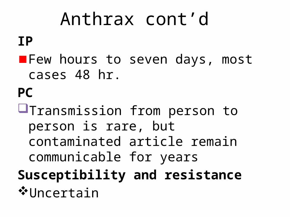
Anthrax cont’d IP
Few hours to seven days, most cases 48 hr.PCTransmission from person to person is rare,
but contaminated article remain communicable for years
Susceptibility and resistance Uncertain
Anthrax-C/M
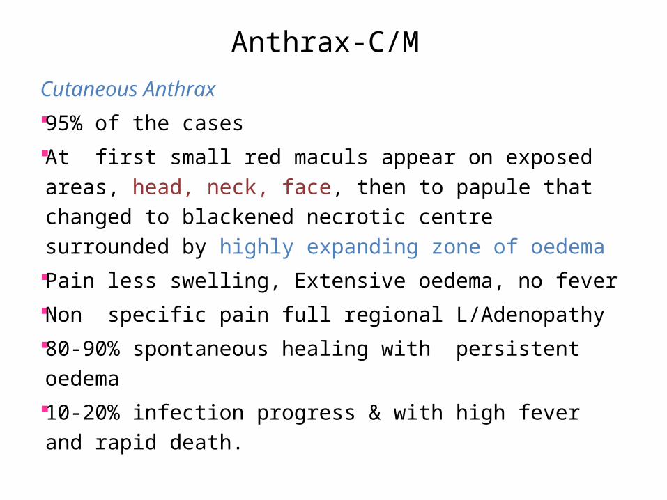
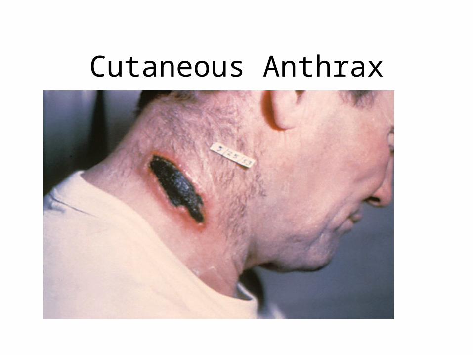
Cutaneous Anthrax 95% of the cases At first small red maculs appear on exposed areas, head,
neck, face, then to papule that changed to blackened necrotic centre surrounded by highly expanding zone of oedema
Pain less swelling, Extensive oedema, no fever Non specific pain full regional L/Adenopathy 80-90% spontaneous healing with persistent oedema 10-20% infection progress & with high fever and rapid
death.
Cutaneous Anthrax
Anthrax-C/M
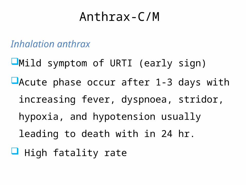
Inhalation anthrax
Mild symptom of URTI (early sign)
Acute phase occur after 1-3 days with increasing
fever, dyspnoea, stridor, hypoxia, and hypotension
usually leading to death with in 24 hr.
High fatality rate
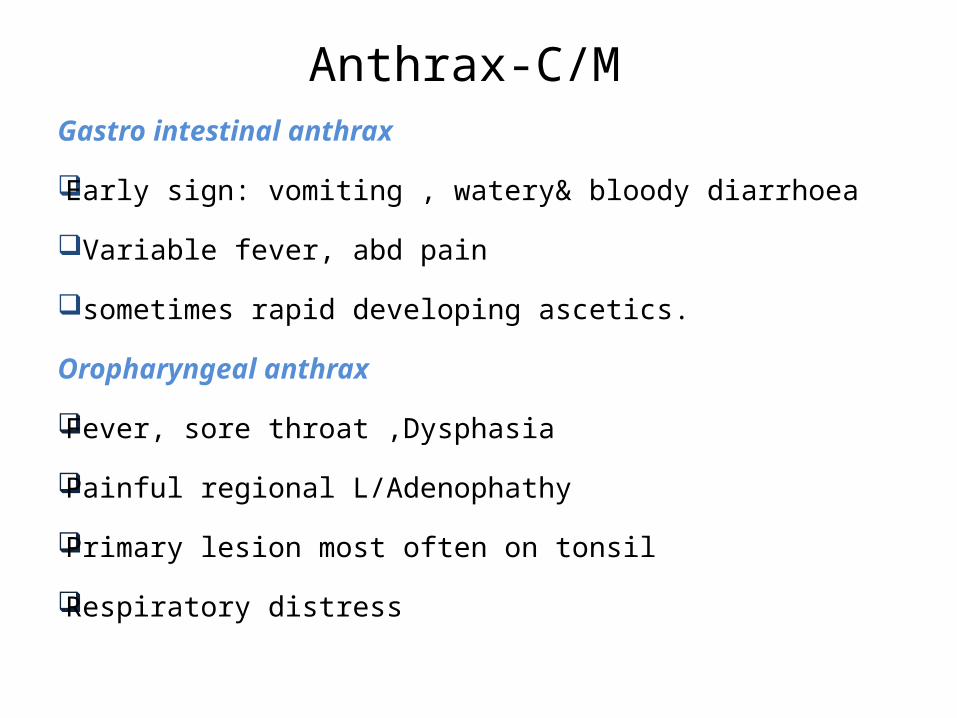
Anthrax-C/M Gastro intestinal anthrax
Early sign: vomiting , watery& bloody diarrhoea
Variable fever, abd pain
sometimes rapid developing ascetics.
Oropharyngeal anthrax
Fever, sore throat ,Dysphasia
Painful regional L/Adenophathy
Primary lesion most often on tonsil
Respiratory distress
Anthrax-Dx
Clinical data
Lab Dx from fluid of vesicle, sputum and stool
Gram stain
Culture
Anthrax-RX
Penicillin G 10Mu IV daily
or
TTC 2gm PO daily for two wks
Clean & cover the Cutaneous lesion
Prevention & Control of Anthrax ¨ Decontamination of wool and goat’s hair or
others ¨ Improvement of working condition of animal
product ¨ Vaccination of susceptible groups & animals ¨ Treat all exposed animal.¨ HE
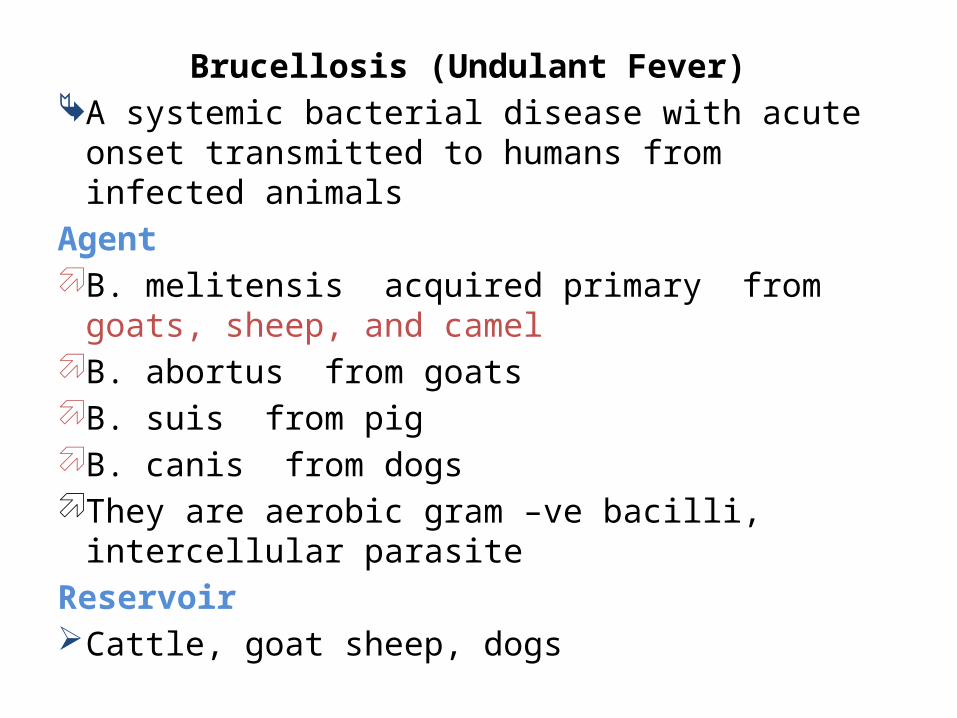
Brucellosis (Undulant Fever) A systemic bacterial disease with acute onset
transmitted to humans from infected animalsAgent B. melitensis acquired primary from goats, sheep, and
camel B. abortus from goats B. suis from pig B. canis from dogs They are aerobic gram –ve bacilli, intercellular parasite Reservoir Cattle, goat sheep, dogs
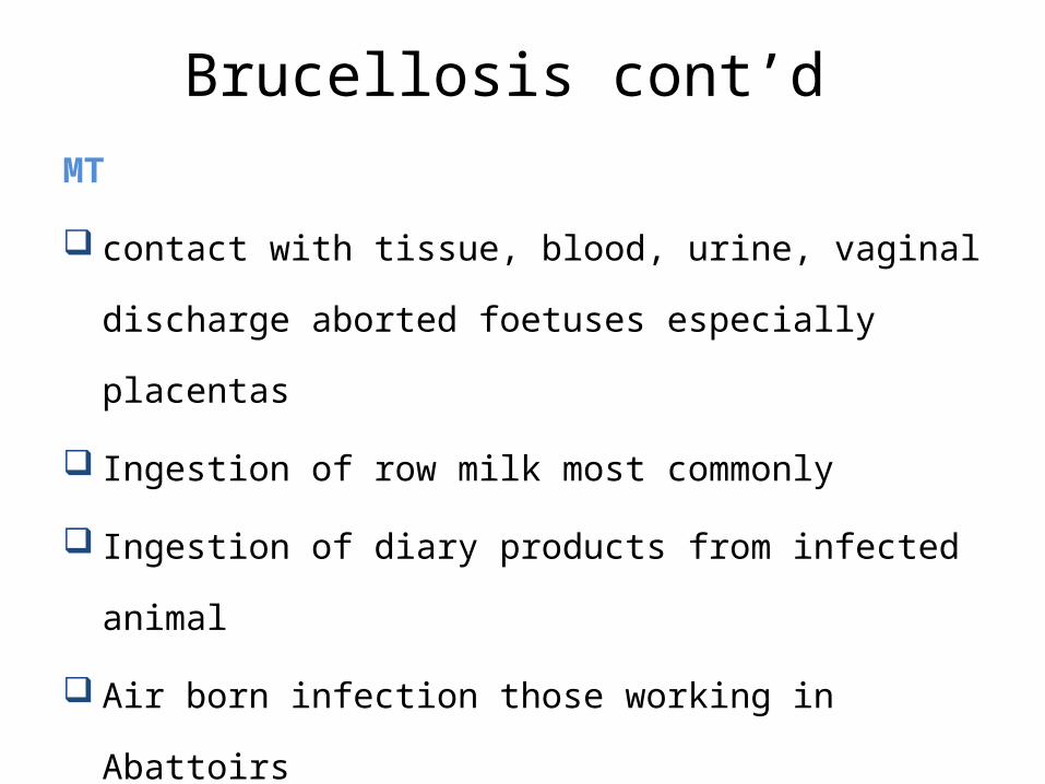
Brucellosis cont’d MT
contact with tissue, blood, urine, vaginal discharge aborted
foetuses especially placentas
Ingestion of row milk most commonly
Ingestion of diary products from infected animal
Air born infection those working in Abattoirs
Brucellosis cont’d
IP
1-3 wks or longer
PC
No evidence from person to person
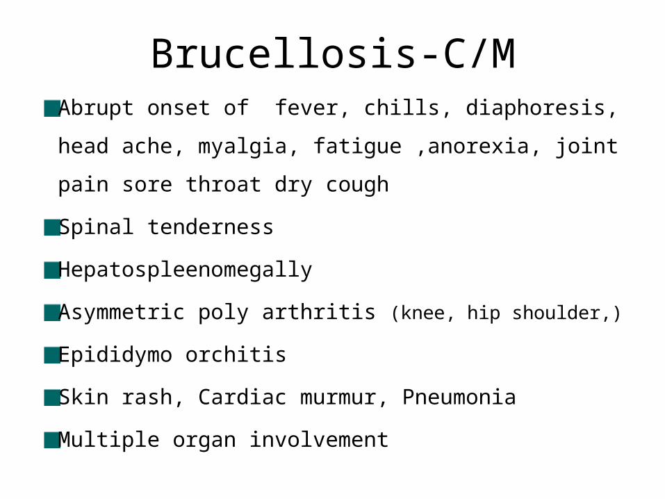
Brucellosis-C/MAbrupt onset of fever, chills, diaphoresis, head ache,
myalgia, fatigue ,anorexia, joint pain sore throat dry cough
Spinal tenderness
Hepatospleenomegally
Asymmetric poly arthritis (knee, hip shoulder,)
Epididymo orchitis
Skin rash, Cardiac murmur, Pneumonia
Multiple organ involvement
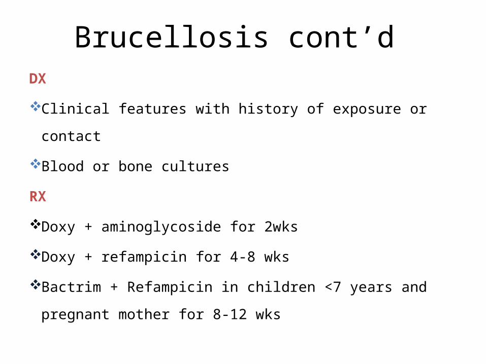
Brucellosis cont’d DX
Clinical features with history of exposure or contact
Blood or bone cultures
RX
Doxy + aminoglycoside for 2wks
Doxy + refampicin for 4-8 wks
Bactrim + Refampicin in children <7 years and pregnant mother
for 8-12 wks
Prevention & Control Brucellosis
Elimination of disease in domestic animals
HE
Proper disposal system
Animal examination and Rx
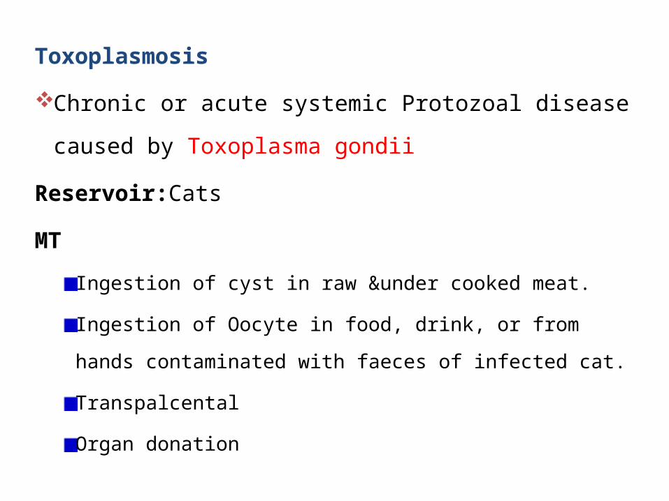
Toxoplasmosis
Chronic or acute systemic Protozoal disease caused by
Toxoplasma gondii
Reservoir:Cats
MT
Ingestion of cyst in raw &under cooked meat.
Ingestion of Oocyte in food, drink, or from hands contaminated
with faeces of infected cat.
Transpalcental
Organ donation

Toxoplasmosis cont’d
IP
10-23 days
PC
Not directly from person to person except
transpalcental
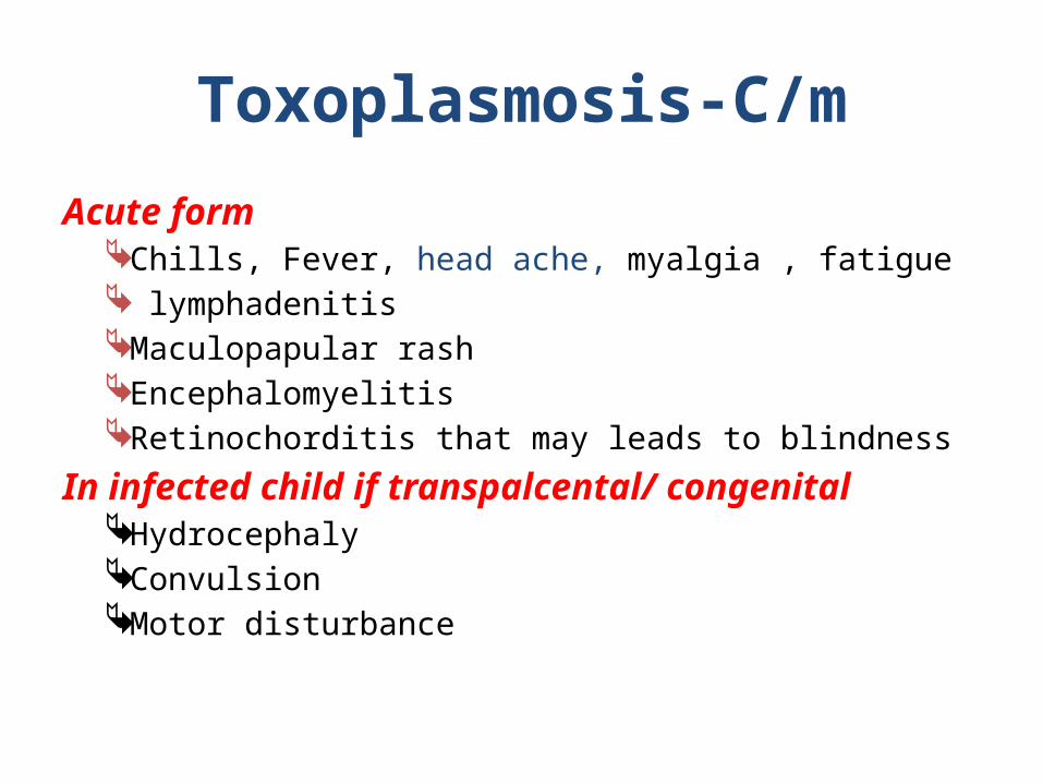
Toxoplasmosis-C/m
Acute form Chills, Fever, head ache, myalgia , fatigue lymphadenitis Maculopapular rash Encephalomyelitis Retinochorditis that may leads to blindness
In infected child if transpalcental/ congenital Hydrocephaly Convulsion Motor disturbance
Toxoplasmosis cont’d DX C/m Cell cultureRX:-Pyrimethamine 100-200mg PO daily followed by
maintenance dose 25mg PO/d for 4 wks. Note – Rx indicated in sick immunocompromised caseFolinic acidPrevention & Control
Eat cooked meat and animal products (60c)
Rabies An acute viral disease of CNS invariably fatal Affect all animals Transmitted by infected secretion mainly saliva.
AgentRabies virus (lyssavirus)ReservoirDog, rats
Rabies cont’d
PC :Usually 3-7 days before the onset of the disease and
throughout the course of disease.
MT
¨ Transmitted with saliva of infected animal and introduced to
body by bite or scratch
¨ Occasionally inhalation, ingestion and tissue transplantation
but unusual
IP: 3-8wks (40 days)
Rabies cont’d
Pathogenesis
A bullet shaped virus which has many strain recovered from
rabid street dog called “street virus”
The virus multiplies in muscle at site of inoculation then
ascends along the nerve to CNS where from it subsequently
spread to all parts of the body via emerging net work of ANS.
Entrance to saliva propagates transmission
Infected animal identified as Negri body in 80% cases
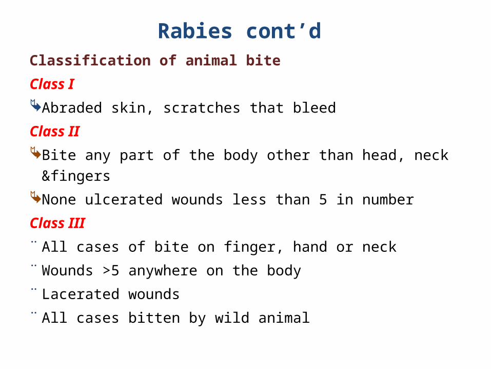
Rabies cont’d Classification of animal bite
Class I Abraded skin, scratches that bleed
Class II Bite any part of the body other than head, neck &fingers None ulcerated wounds less than 5 in number
Class III ¨ All cases of bite on finger, hand or neck ¨ Wounds >5 anywhere on the body ¨ Lacerated wounds ¨ All cases bitten by wild animal
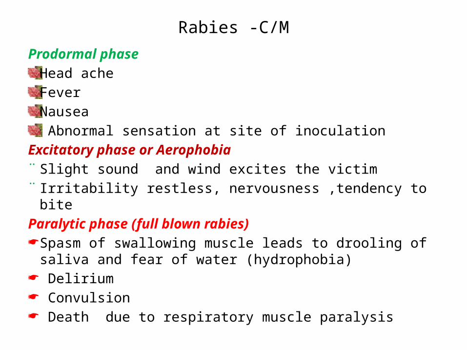
Rabies -C/MProdormal phase
Head ache FeverNausea Abnormal sensation at site of inoculation
Excitatory phase or Aerophobia ¨ Slight sound and wind excites the victim ¨ Irritability restless, nervousness ,tendency to bite Paralytic phase (full blown rabies) Spasm of swallowing muscle leads to drooling of saliva and fear of
water (hydrophobia) Delirium Convulsion Death due to respiratory muscle paralysis
Rabies -DX
History of bite by known rabid animal Nigri bodies
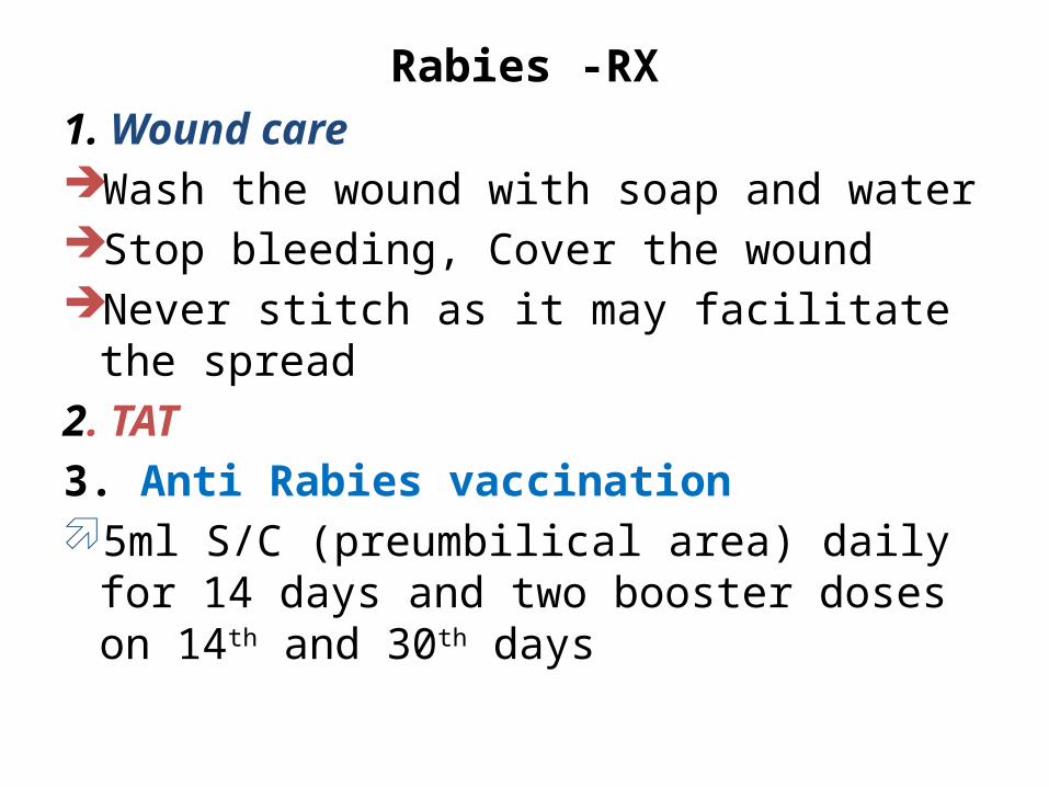
Rabies -RX1. Wound care Wash the wound with soap and water Stop bleeding, Cover the wound Never stitch as it may facilitate the spread 2. TAT 3. Anti Rabies vaccination 5ml S/C (preumbilical area) daily for 14 days
and two booster doses on 14th and 30th days
Rabies -RX
Indication for anti rabies vaccination Bite from known rabid animal or contact with
salve to lesioned tissue The animal escaped The animal show clinical pictures after 10 days
observation The animal died
4.Sadation if necessary
Rabies -Prevention and control
Immunize all dogs and cats
Detain & clinically observe for 10 days any unhealthy
appearing dog or cat known to have bitten a person
Post exposure prophylaxis
Keep dogs and cats at home
Destroy stray animals where rabies is endemic
Kill rabid animal.
Trichinellosis or Trichinosis Trichinosis is a disease caused by intestinal round worm whose
larvae (trichinae) migrates to and become encapsulated in the
muscles
Infectious agent
Trichinella spiralis, an intestinal nematode
Epidemiology
Occur worldwide, but variable incidence, depending in part on
practices of eating and preparing pork or wild animal meat.
Trichinellosis-cont’d
Reservoir
Swine, dogs, cats, horses, rats and many wild
animals, including fox wolf etc
Mode of transmission
By eating raw or insufficiently cooked flesh of
animals containing viable encysted larvae
Trichinellosis-cont’d
I/P
Systemic symptoms usually appear about 8-15
days after ingestion of infected meat.
Susceptibility and resistance
¨ Susceptibility is universal.
¨ Infection results in partial immunity
Trichinellosis-Clinical manifestations
Infection ranges from mild febrile illness to a severe progressive illness with multiple system involvement.
Fever(low-high grade)
Muscle pain mainly up on movement
oedema and spasm (periorbital and facial|)
photophobia and conjunctivitis
Weakness ; pain on swallowing
Dyspnoea , coughing and hoarseness
subconjuctival, retinal and nail splinter haemorrhage and rashes
Diarrhoea abdominal cramps, nausea and vomiting.
Trichinellosis-Clinical manifestationsInflammatory reactions around larvae that
reach tissues other than muscles may result in: ¨ Meningitis¨ Encephalitis ¨ Myocarditis¨ Bronchopneumonia¨ Nephritis¨ Peripheral ¨ Cranial nerve disorders
Trichinellosis - Diagnosis
Hx of ingestion of raw or inadequately cooked pork
Larvae in muscle biopsyPositive serologic test Oesinophilia
Trichinellosis -Treatment
Hospitalization of the PtMebendazole or Albendazole or
ThiabendazoleHigh dose of corticosteroids for 1-2 days
followed by lower doses for several days or weeks. But not for intestinal stage.
Trichinellosis-Prevention and control
Educate the public on the need to cook all fresh pork and pork products and meat from wild animals
Freezing of pork and its products inactivates trichinae

Summary
1. List at least four common zoonotic disease
2. Describe the common means transmission
3. Discuss common prevention and control
methods of zoonotic disease
4. Discuss management approaches of
zoonotic disease accordingly