Embed Size (px)
Citation preview

INTRODUCTION
COMPONENTS
COURSE OF CRANIAL ROOT
COURSE OF SPINAL ROOT
DISTRIBUTION OF SPINAL ACCESSORY NERVE
CLINICAL FINDINGS
LESIONS
XI- CRANIAL NERVE
(ACCESSORY)

• The Accessory nerve [XI] carries GSE ( General Somatic
Efferent) fibers.
• Innervates the STERNOCLEIDOMASTIOD and TRAPEZIUSmuscles.
• Unique cranial nerve - - - roots arise from motor neurons of the
UPPER FIVE SEGMENTS [C1-C5] OF THE CERVICAL SPINAL CORD.


COMPONENTS:
I- Cranial Root. (Nucleus Ambiguous)
II- Spinal Root. ( Spinal Nucleus of Ant. Grey
columns b/w C1 - C5)

CRANIAL ROOT:
• Arises from the lower part of NucleusAmbiguous.
• Accessory to the Vagus nerve [X].
• Distributed through the Branches of Vagus nerve.

Course of Cranial Root:
• Rootlets arising from Caudal part of the Medulla Oblongata on the
Anterolateral surface, just inferior to the rootlets arising to form Vagus
Nerve.
• Leaving the Medulla, Cranial roots course with the “spinal” root of
Accessory nerve into the Jugular foramen, and again separates outside
the foramen.
• Join the Vagus nerve [Inf. Ganglion] after exiting the Jugular
foramen, supplying the pharyngeal musculature supplied by Vagus Nerve.

Course of the Spinal Root:
• Fibers arising from the motor cells in the lateral part of the Anterior
column of grey substance of the medulla spinalis as low as fifth cranial
nerve[C1-C5].
• Joining together as they ascend.
• Enters the Cranial Cavity through Foramen Magnum.
• Continues through the Posterior Cranial Fossa, laterally towards
Jugular foramen
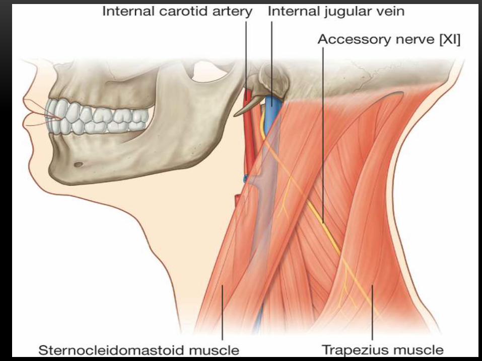
Extra-cranially:
• Exits through Jugular foramen.
• Descends in the neck, Medial to the Int. Jugular Vein.
• B/w the Angle of Mandible and Mastoid process.
• Lies under the Stylohyoid and Post. Belly of Digastric muscle.

• Crosses the Int. Jugular Vein laterally in 66%, and passes behind in
33.3% of cases.
• Disappear either into or beneath the Ant. Border of
Sternocleidomastoid muscle.
[NO BRANCHES IN ANT. TRIANGLE OF THE NECK]
• Continues its descend & Enters the Post. Triangle of the Neck.
• Still moving obliquely and downward, within the Investing layer of the
Cervical fascia.
• Reaches the Ant. Border of Trapezius muscle, terminates by
innervating the muscle.


Distribution:
→ Motor Innervation to;
• Sternocleidomastoid muscle.
• Trapezius muscle.

CLINICAL FINDINGS:
• Paralysis of Sternocleidomastoid and Trapezius muscle.
• Drooping of the Shoulder.
• Inability to turn chin to opposite side.
• Inability to draw head forward.
• Irritation of the nerve during biopsy of enlarged caseous lymph
nodes, may produce TORTICOLLIS or WRY NECK.
LESIONs:
• Penetrating injury to the Posterior Triangle of the Neck.
• Superficial location of the nerve in Post. Triangle of the neck makes it
susceptible to injury.