Embed Size (px)
Citation preview

CASE PRESENTATION
Dr. ADEEL AHMAD.
PGR WMW.

Name Sobia
Age 35 years
Sex Female
Resident Faisal Abad
Presented
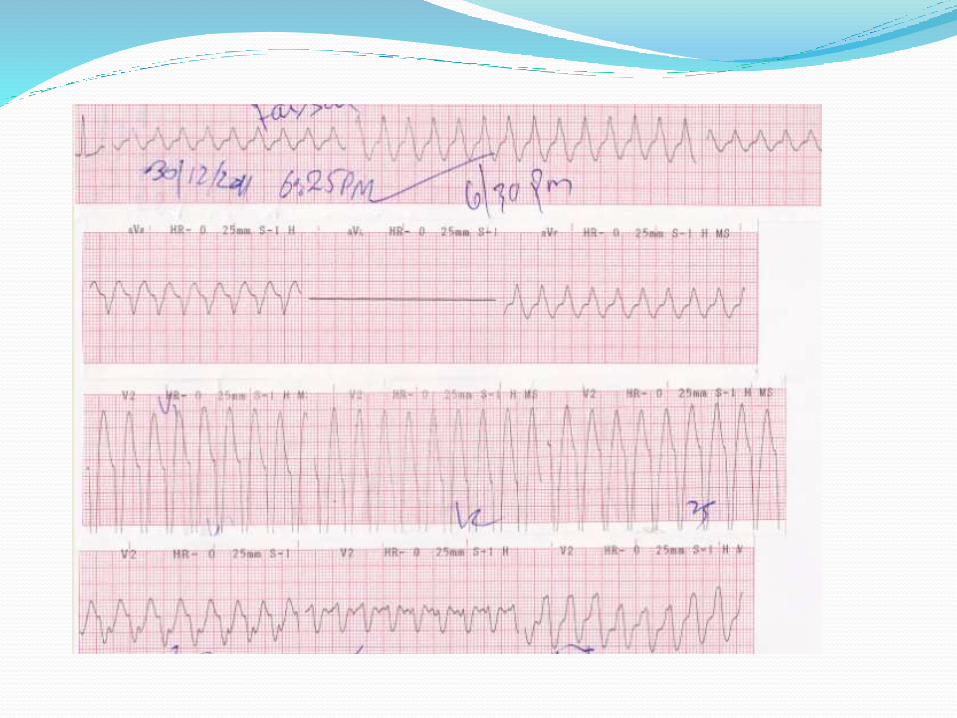
“Palpitation and SOB 6 hours”.
Patient HTN+ 3 months (on regular medication) nonDMpresented in emergency with H/O palpitation that started 6hrs back. She complains of such a palpitation for last 3 months on and off, palpitation can occur any time that have no relation to exercise or work load.She, along with palpitation, also complains of SOB for up to a similiarperiod. SOB can occur any time,no diurnal variation,noseasonal history. There is no H/O orthopnea and PND.

Past History: Except HTN not significant
Personal History: Not significant
Family History: She is married and living a happy life.
Treatment History:
She was on
Losartan 50 mg 1 po OD and
Tenormin 50 mg BD

VitalsBP 170/100 mmHg
Pulse 120 beat/m
RR 20/M
Fever A/F

Pallor -ve Cyanosis -ve Clubbing -ve Koilonychia -ve S /hemorrhage -ve Osler nodes absent Heberden nodes absent Boucard nodes absent Palmer erythema -ve Dupuytren contrature -ve Skin rash -ve Axillary nodes not palpable JVP not raised Thyroid normal Ankle edema absent Sign of dehyration -ve
GPE
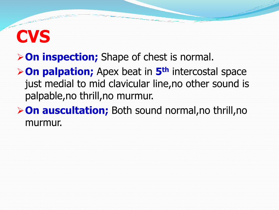
CVSOn inspection; Shape of chest is normal.
On palpation; Apex beat in 5th intercostal space just medial to mid clavicular line,no other sound is palpable,no thrill,no murmur.
On auscultation; Both sound normal,no thrill,nomurmur.

Respiratory system
On inspection; Rate 20/m thoraco abdominal.
On palpation; Trachea is central,apex beat in 5th
medial to midclavicular line,chest movement and expansion within normal range,no vocal fremitus.
On purcussion; upper border of liver is in 5th
intercostal space.
On auscultation; Breath sounds are normal,noronchi,no crepts.

GIT
On Inspection; Oral hygiene is good,
On Palpation; Not done
On Purcusion; Not done.
On Auscultation; B/S are audible,no bruit is present.

CNS
HMF Normal.
Speech Normal
Cranial nerves Intact.
Motor Intact
Sensory Intact
Cerebellar Intact


Diagnostic criteria:The QRS complex duration is >0.11 second; in
approximately 20% of individuals, the QRS complex may not be >0.11 second.
PR is <0.12 second.
A delta wave is prominent, often in V3 through V6,
In type A WPW syndrome, a tall R wave present in V1
In type B WPW , the QRS complex is predominately negative in V1 through V3 and upright in V5 and V6.


HistoryIn 1930, Wolff, Parkinson and White described a
distinct (ECG) pattern in healthy young people with short bursts of tachycardia.
In 1944, doctors confirmed the presence of extra pathways

Prevalence:WPW is a congenital heart abnormality
WPW occurs randomly in the general population
1 to 3 per 1,000 persons.
Men have a higher incidence of WPW than women.
Some cases of WPW are inherited.
7 to 20 percent of patients with WPW also have congenital defects within the heart.

Symptoms of WPWAny age, from infancy to adult years.
Heart palpitations
Racing feeling in your chest
Dizziness
Shortness of breath (dyspnea)
Anxiety
Rarely, cardiac arrest (sudden death)
Some people have WPW without any symptoms at all.

Types of WPWPractical concept is that a negative delta wave usually
signals where the AP is:
A negative delta wave in a left-side, I and aVLindicates a left-side AP.
A negative delta in a right-side lead such as V1 predicts a right-side AP.
A negative delta in the inferior leads (II, III, and aVF) indicates a posteroseptal AP.
A positive delta in the inferior leads predicts an anteroseptal AP.
An isoelectric delta in V1 predicts an anteroseptal AP.

Left lateral wall - Negative delta waves in lead I
and aVL; positive or isoelectric in II, III, aVF and V1-4; and negative or isoelectric delta waves in V5-6
Right free wall - Positive delta waves in I and II,
negative delta waves in aVR, isoelectric or negative delta wave in aVF, isoelectric delta wave in V1, isoelectric or positive delta waves in V2-3, and positive delta waves in V4-6

Left posterior free wall - Positive delta waves in lead I and aVL; negative delta waves in II, III, and aVF; positive delta waves in V1-5; and negative or isoelectric delta wave in V6
Posteroseptal - Positive delta waves in lead I and aVLwith negative delta waves in II, III, and aVF; isoelectricwaves in V1; and positive delta waves in the rest of the precordial leads
Left anteroseptal - Positive delta waves in I, II, and aVF; negative delta wave in aVR; isoelectric or positive delta wave in V1; and positive delta waves in V2-6
Right anteroseptal - Positive delta waves in I, II, and aVF; negative delta wave in aVR; negative or isoelectricdelta waves in V1-3; and positive delta waves in V4-6

TreatmentDDAVNRT
Orthodromic AVRT
Antidromic AVRT


Narrow complex
Orthodromic AVRT and AVNRT blocking AV node conduction
Vagal maneuvers (eg, Valsalva maneuver, carotid sinus massage, splashing cold water or ice water on the face)
IV adenosine 6-12 mg via a large-bore
IV verapamil 5-10 mg or diltiazem 10 mg

Wide complex
Antidromic AVRT
Procainamide or
Amiodarone or
Flecainide if wide-complex tachycardia is present, if patient hemodynamically stable
Ibutilide

Unstable patient
Synchronized electrical cardioversion,
A level of 100 J initially
If necessary, a second shock with higher energy (200 J or 360 J)
Pregnancy Sotalol

Radiofrequency Ablation Indication
Patients with symptomatic AVRT
Patients with AF
Patients with AVRT or AF with rapid ventricular rates found incidentally during EPS,RR interval during AF <250 ms
Asymptomatic patients who would endanger the public safety
Patients with WPW and a family history of sudden cardiac death

RFAIn RF ablation, platinum-tipped 3.5- to 8-mm
steerable multielectrode catheters are advanced via the femoral artery or vein to locate and ablate the AP by delivering thermal RF energy

Surgical treatmentSurgical treatment is replaced by RFA
Patients in whom RF catheter ablation (with repeated attempts) fails
Patients undergoing concomitant cardiac surgery

Long-term antiarrhythmic therapyOral medication is the mainstay of therapy in patients
not undergoing RFA. Choices include the following:
Dual-drug therapy (eg, procainamide and verapamil[class Ia and IV])
Class Ic drugs (eg, flecainide, propafenone), typically used with an AV nodal blocking agent
Class III drugs (eg, amiodarone, sotalol)
In pregnancy, sotalol (class B) or flecainide (class C)
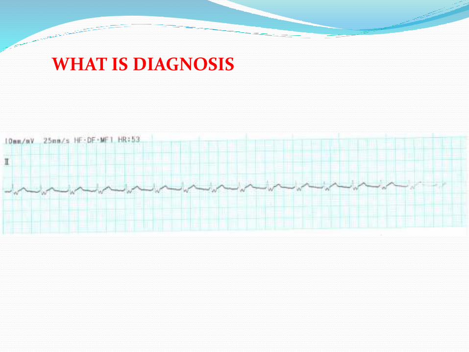
WHAT IS DIAGNOSIS

WHAT IS DIAGNOSIS


WHAT IS DIAGNOSIS

THANKS