Embed Size (px)
Citation preview
OA in the ‘Young”
HTO & Uni Knee
Professor Deiary Kader
Consultant Orthopaedic & Trauma Surgeon
Knee Surgeon
Newcastle Nuffield Hospital
Postgraduate OrthopaedicsFRCS(Tr&Orth) Revision Course
Newcastle Upon Tyne 16-21 March 2015
•
Professor Deiary KaderConsultant Orthopaedic & Trauma Surgeon
Knee Surgeon
Nuffield Hospital Newcastle
NGMV Charity
PostGrad Orth Deiary Kader
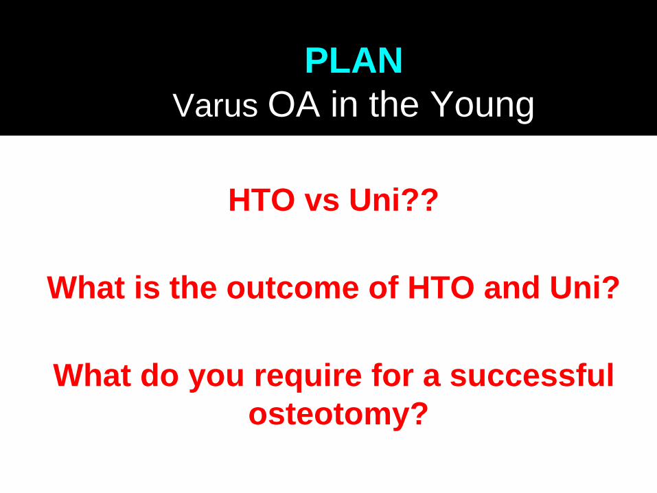
HTO vs Uni??
What is the outcome of HTO and Uni?
What do you require for a successful
osteotomy?
PLAN
Varus OA in the Young

Must Know
What is the
None-operative
Treatment for OA?

OA Nonoperative treatment
Strategies may include
Weight loss
Exercise
Patient education
Analgesia, (NSAIDs)
Bracing
Intra-articular (IA) injections. Cochrane reviews
Steroids (better than placebo but not longer than 4wks
HA more prolonged effect than steroids
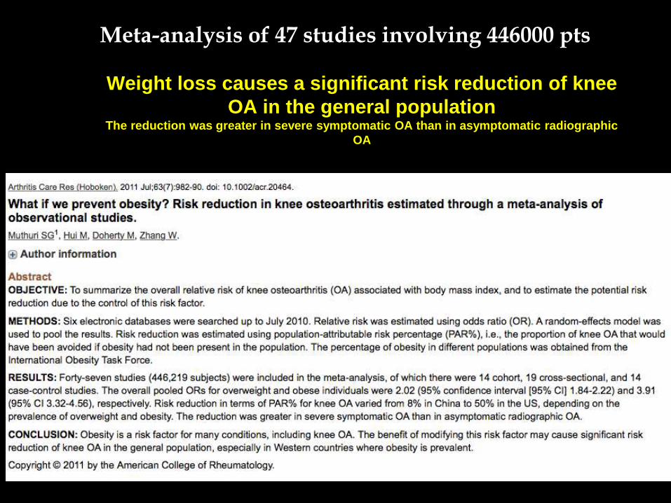
Weight loss causes a significant risk reduction of knee
OA in the general populationThe reduction was greater in severe symptomatic OA than in asymptomatic radiographic
OA
Meta-analysis of 47 studies involving 446000 pts
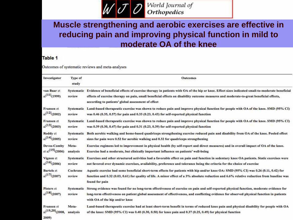
m,Muscle strengthening and aerobic exercises are effective in
reducing pain and improving physical function in mild to
moderate OA of the knee
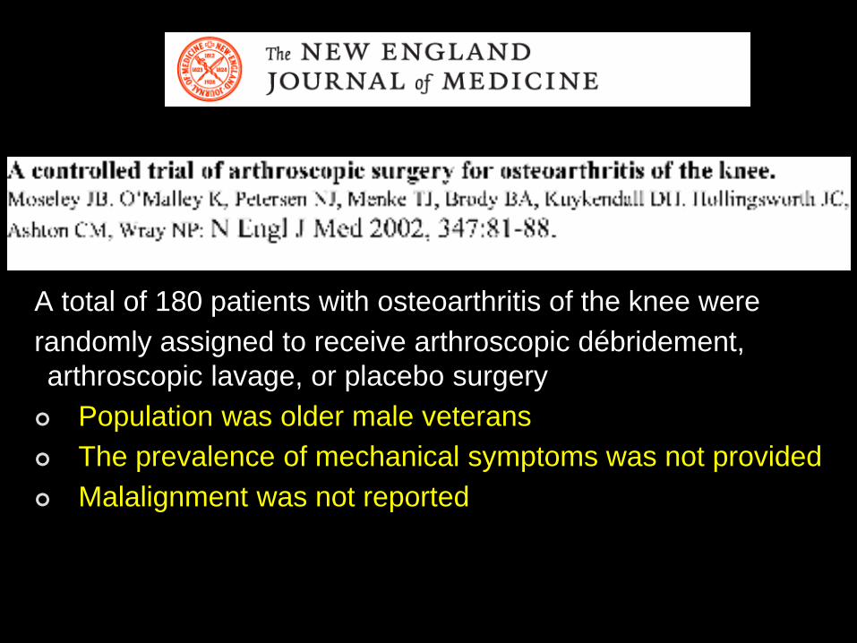
A total of 180 patients with osteoarthritis of the knee were
randomly assigned to receive arthroscopic débridement,
arthroscopic lavage, or placebo surgery
Population was older male veterans
The prevalence of mechanical symptoms was not provided
Malalignment was not reported
OSTEOTOMY
9
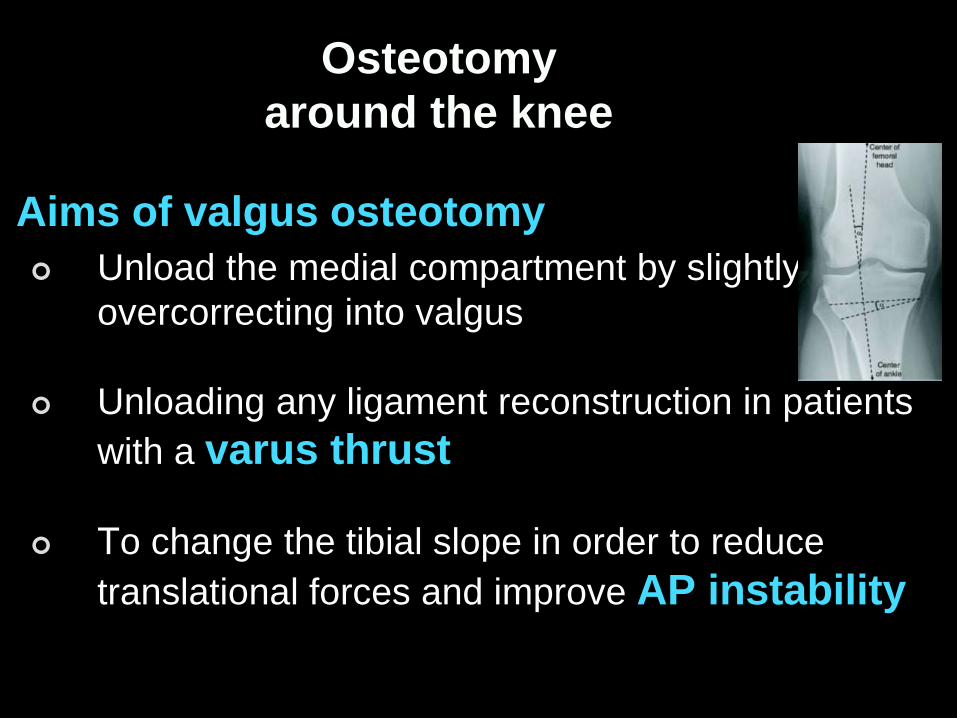
Osteotomy
around the knee
Aims of valgus osteotomy
Unload the medial compartment by slightly
overcorrecting into valgus
Unloading any ligament reconstruction in patients
with a varus thrust
To change the tibial slope in order to reduce
translational forces and improve AP instability

HTO for varus Malalignment
PostGrad Orth Deiary Kader
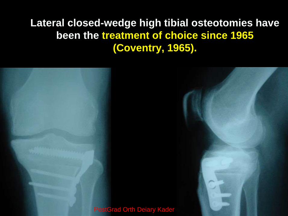
Lateral closed-wedge high tibial osteotomies have
been the treatment of choice since 1965
(Coventry, 1965).
PostGrad Orth Deiary Kader
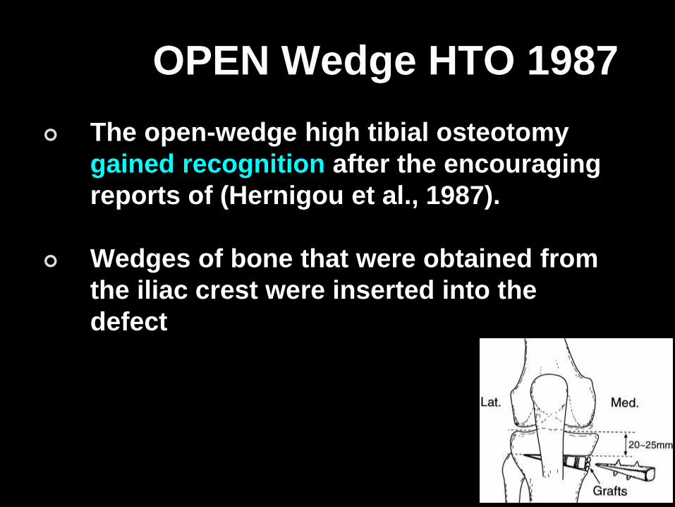
OPEN Wedge HTO 1987
The open-wedge high tibial osteotomy
gained recognition after the encouraging
reports of (Hernigou et al., 1987).
Wedges of bone that were obtained from
the iliac crest were inserted into the
defect
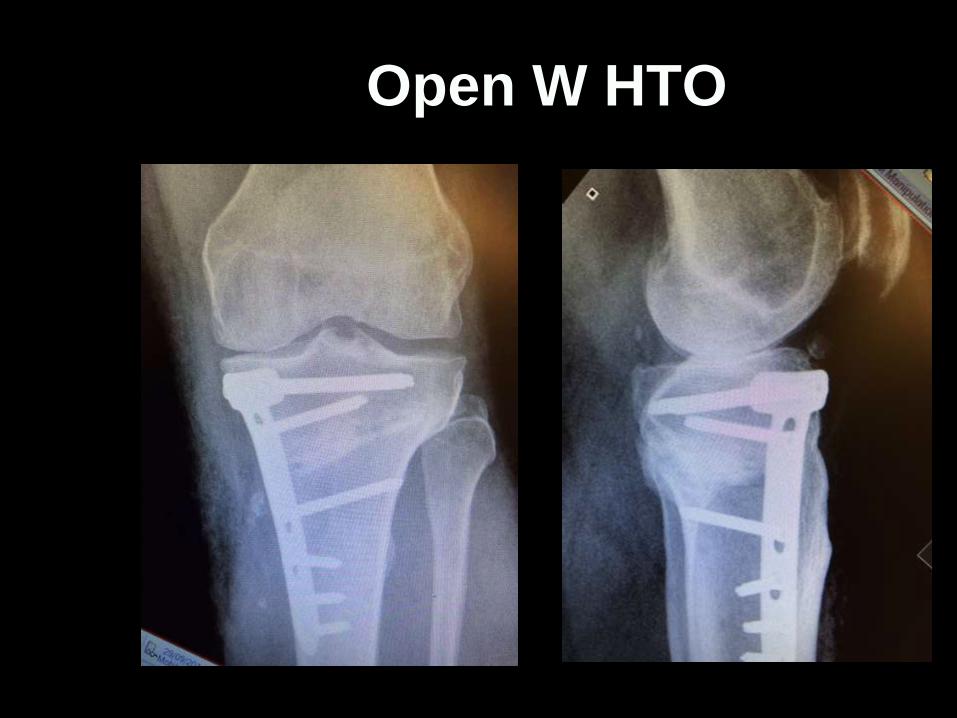
Open W HTO
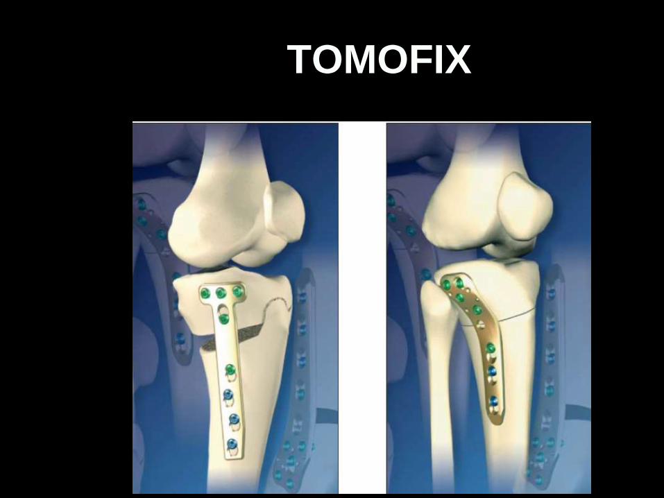
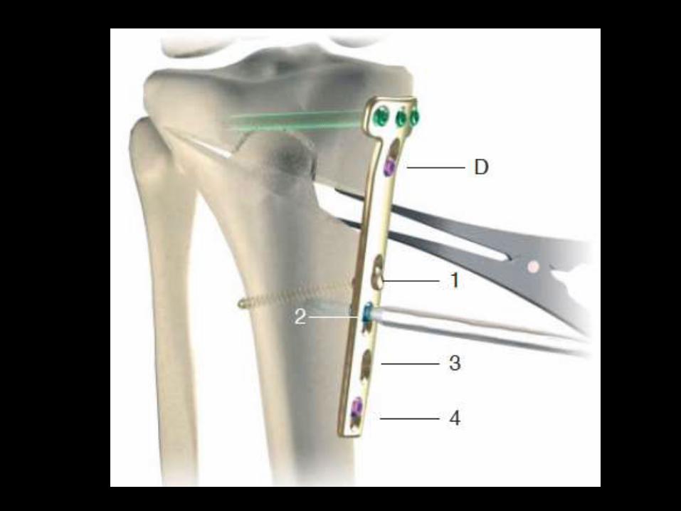
TOMOFIX

Proximal or High Tibial Osteotomy (HTO)
The IDEAL candidate for HTO
Age <60 years
Isolated medial OA
Good ROM
Less than 5° FFD knee
>120° flexion knee
Patients should be
able to use crutches
Have no major varicose veins or peripheral vascular disease
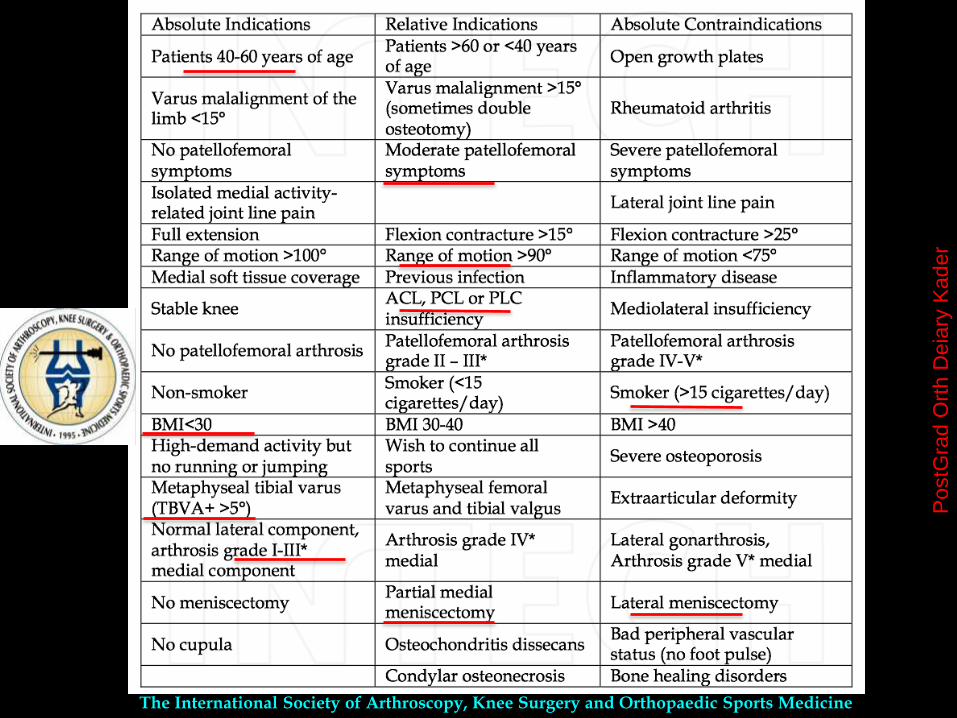
The International Society of Arthroscopy, Knee Surgery and Orthopaedic Sports Medicine
PostG
rad
Ort
h D
eia
ry K
ade
r
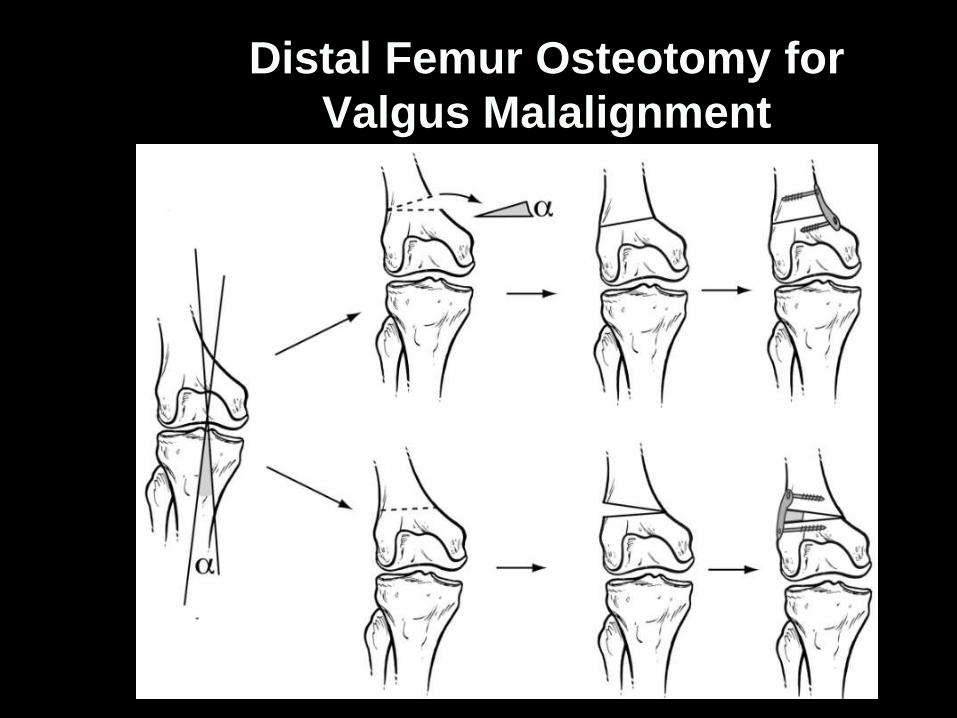
Distal Femur Osteotomy for
Valgus Malalignment
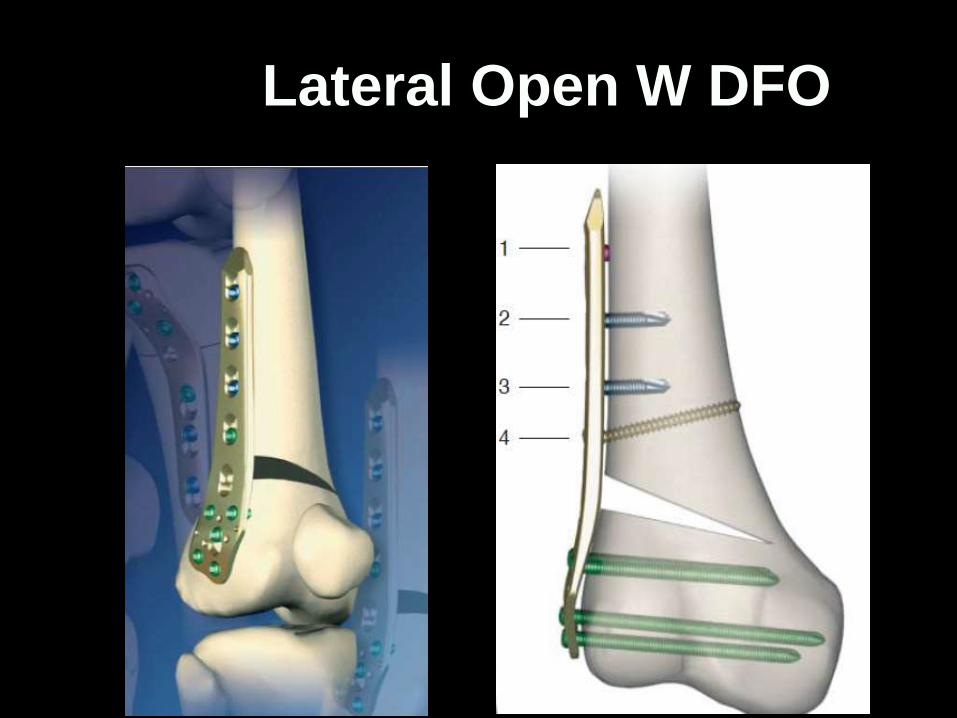
Lateral Open W DFO
Distal Femur Osteotomy
Valgus deformity of 12º or more needs distal femoral
varus producing osteotomy to address a lateral femoral
condyle deficiency and to prevent joint line obliquity and
gradual lateral tibial subluxation.
≈
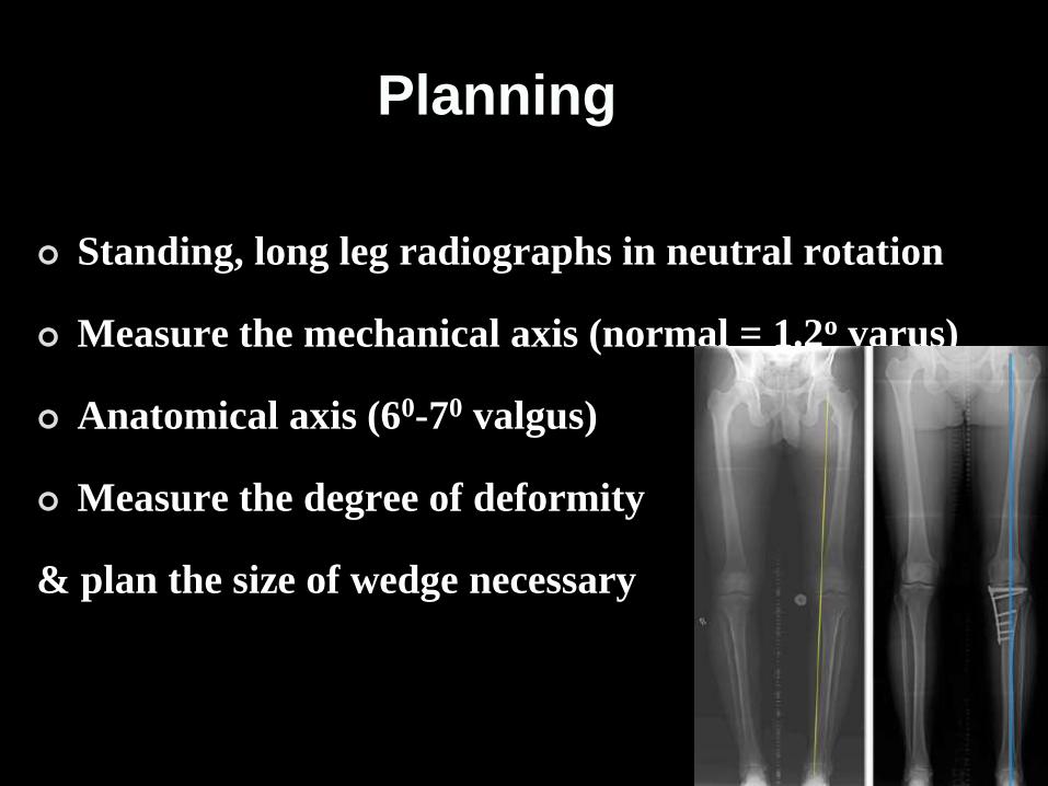
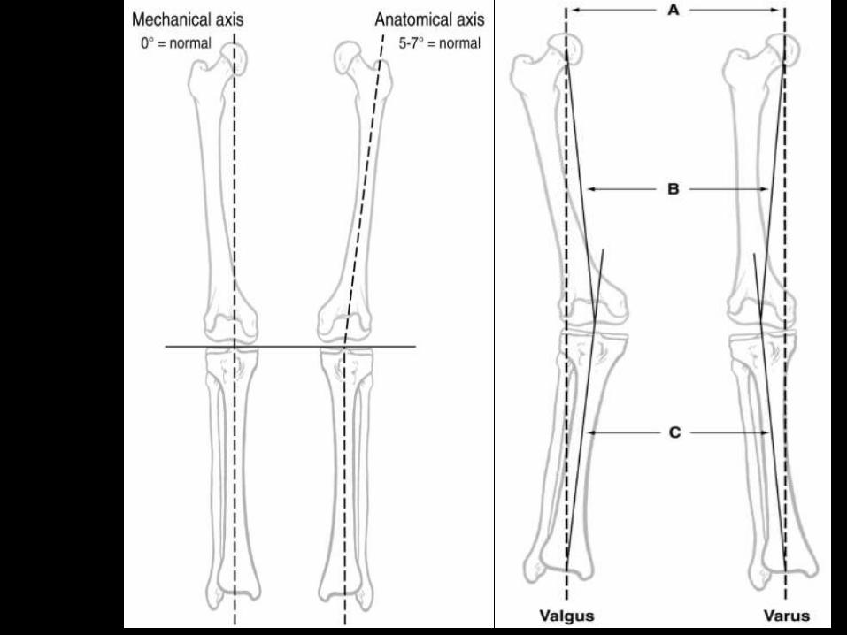
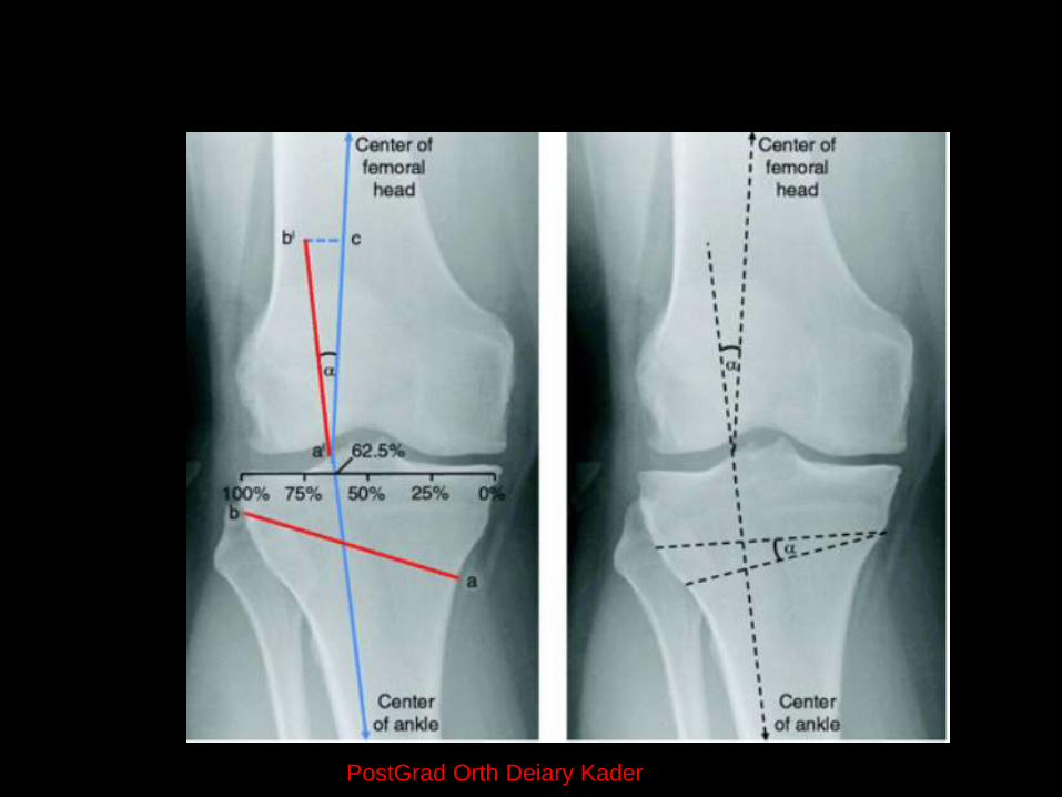
Planning
Standing, long leg radiographs in neutral rotation
Measure the mechanical axis (normal = 1.2o varus)
Anatomical axis (60-70 valgus)
Measure the degree of deformity
& plan the size of wedge necessary
Planning
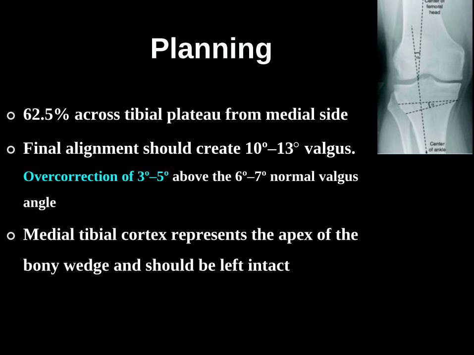
62.5% across tibial plateau from medial side
Final alignment should create 10º–13 valgus.
Overcorrection of 3º–5º above the 6º–7º normal valgus
angle
Medial tibial cortex represents the apex of the
bony wedge and should be left intact
PostGrad Orth Deiary Kader
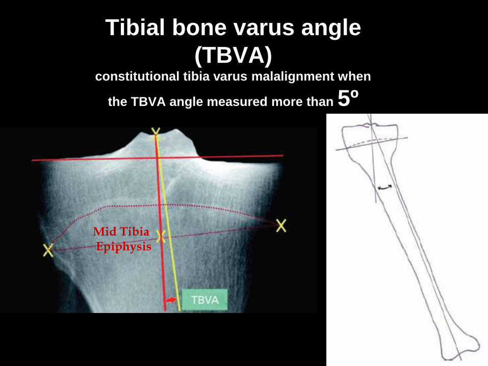
Tibial bone varus angle
(TBVA) constitutional tibia varus malalignment when
the TBVA angle measured more than 5º
Mid TibiaEpiphysis
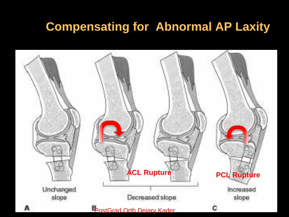
Compensating for Abnormal AP Laxity
ACL Rupture PCL Rupture
PostGrad Orth Deiary Kader
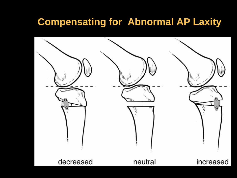
Compensating for Abnormal AP Laxity

PostGrad Orth Deiary Kader
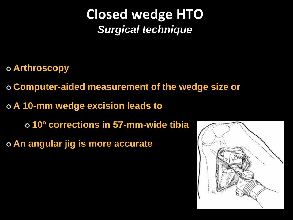
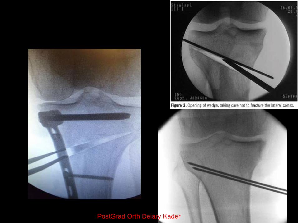
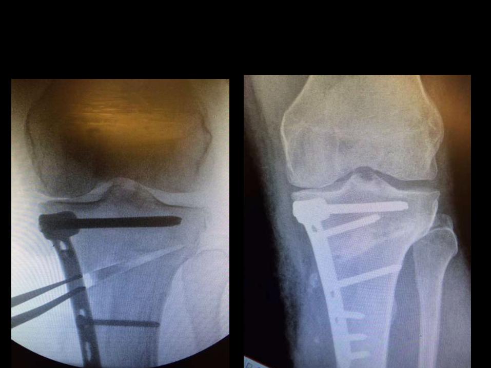
Closed wedge HTOSurgical technique
Arthroscopy
Computer-aided measurement of the wedge size or
A 10-mm wedge excision leads to
10º corrections in 57-mm-wide tibia
An angular jig is more accurate
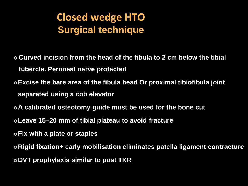
Closed wedge HTOSurgical technique
Curved incision from the head of the fibula to 2 cm below the tibial
tubercle. Peroneal nerve protected
Excise the bare area of the fibula head Or proximal tibiofibula joint
separated using a cob elevator
A calibrated osteotomy guide must be used for the bone cut
Leave 15–20 mm of tibial plateau to avoid fracture
Fix with a plate or staples
Rigid fixation+ early mobilisation eliminates patella ligament contracture
DVT prophylaxis similar to post TKR
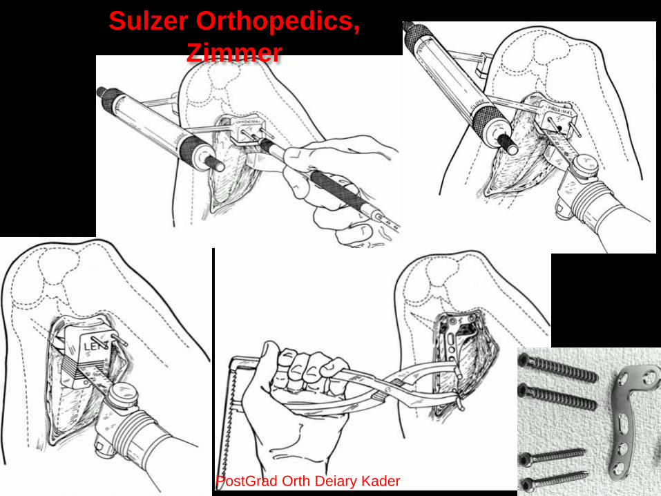
Sulzer Orthopedics,
Zimmer
PostGrad Orth Deiary Kader

PostGrad Orth Deiary Kader
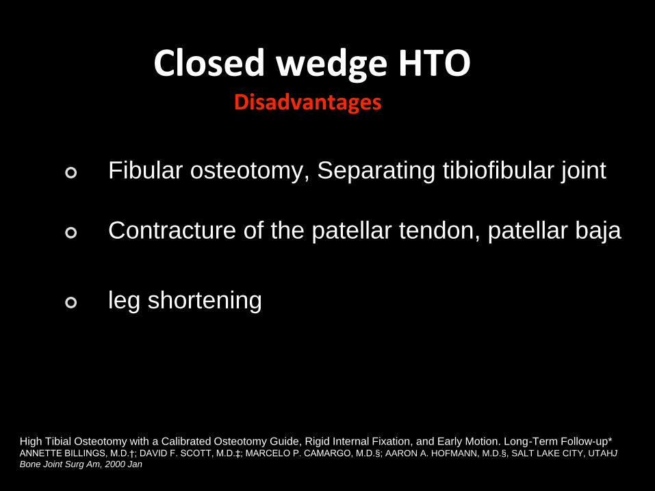
Fibular osteotomy, Separating tibiofibular joint
Contracture of the patellar tendon, patellar baja
leg shortening
High Tibial Osteotomy with a Calibrated Osteotomy Guide, Rigid Internal Fixation, and Early Motion. Long-Term Follow-up*ANNETTE BILLINGS, M.D.†; DAVID F. SCOTT, M.D.‡; MARCELO P. CAMARGO, M.D.§; AARON A. HOFMANN, M.D.§, SALT LAKE CITY, UTAHJ
Bone Joint Surg Am, 2000 Jan
Closed wedge HTODisadvantages
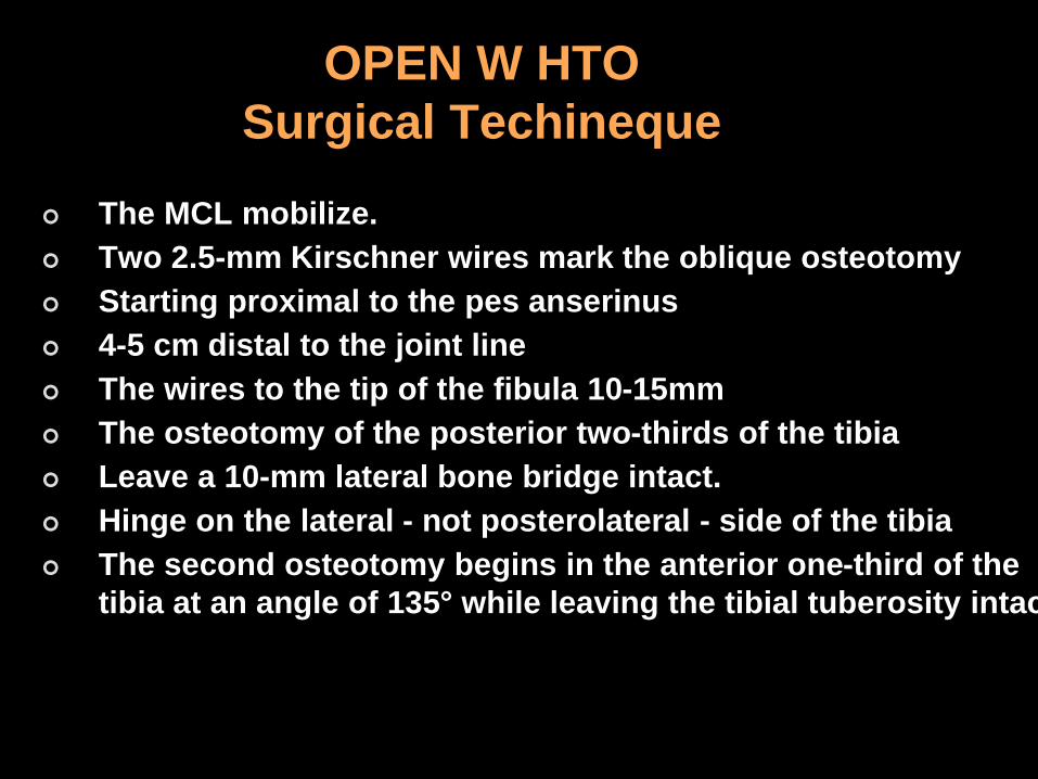
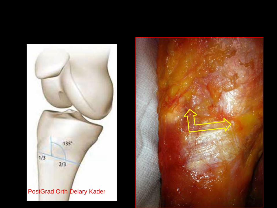
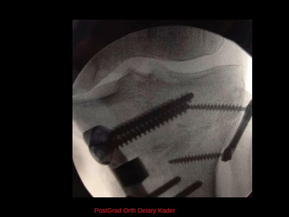
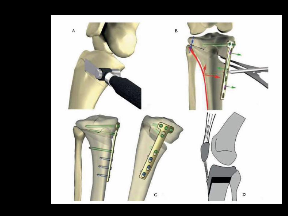
OPEN W HTO
Surgical Techineque
The MCL mobilize.
Two 2.5-mm Kirschner wires mark the oblique osteotomy
Starting proximal to the pes anserinus
4-5 cm distal to the joint line
The wires to the tip of the fibula 10-15mm
The osteotomy of the posterior two-thirds of the tibia
Leave a 10-mm lateral bone bridge intact.
Hinge on the lateral - not posterolateral - side of the tibia
The second osteotomy begins in the anterior one-third of the
tibia at an angle of 135° while leaving the tibial tuberosity intact
PostGrad Orth Deiary Kader
PostGrad Orth Deiary Kader
PostGrad Orth Deiary Kader
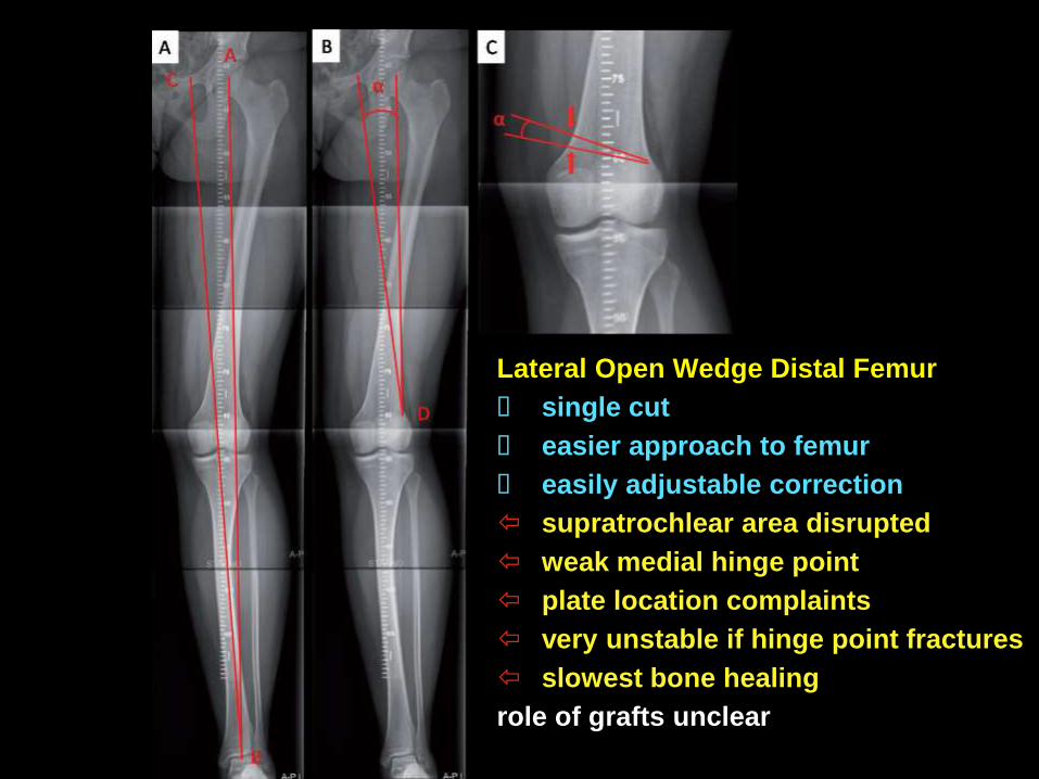
Lateral Open Wedge Distal Femur
single cut
easier approach to femur
easily adjustable correction
supratrochlear area disrupted
weak medial hinge point
plate location complaints
very unstable if hinge point fractures
slowest bone healing
role of grafts unclear
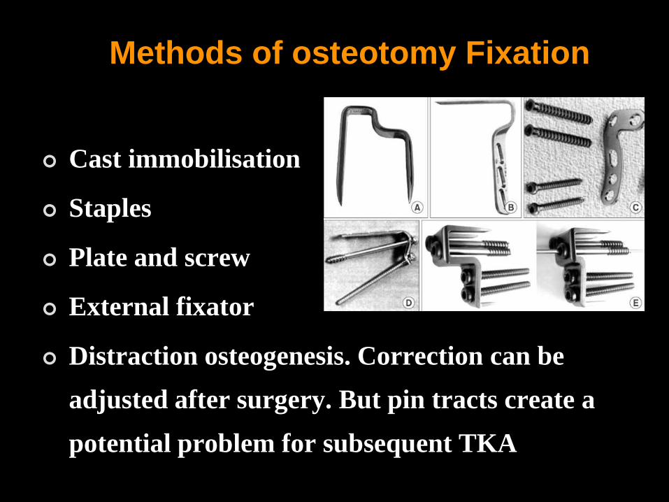
Methods of osteotomy Fixation
Cast immobilisation
Staples
Plate and screw
External fixator
Distraction osteogenesis. Correction can be
adjusted after surgery. But pin tracts create a
potential problem for subsequent TKA

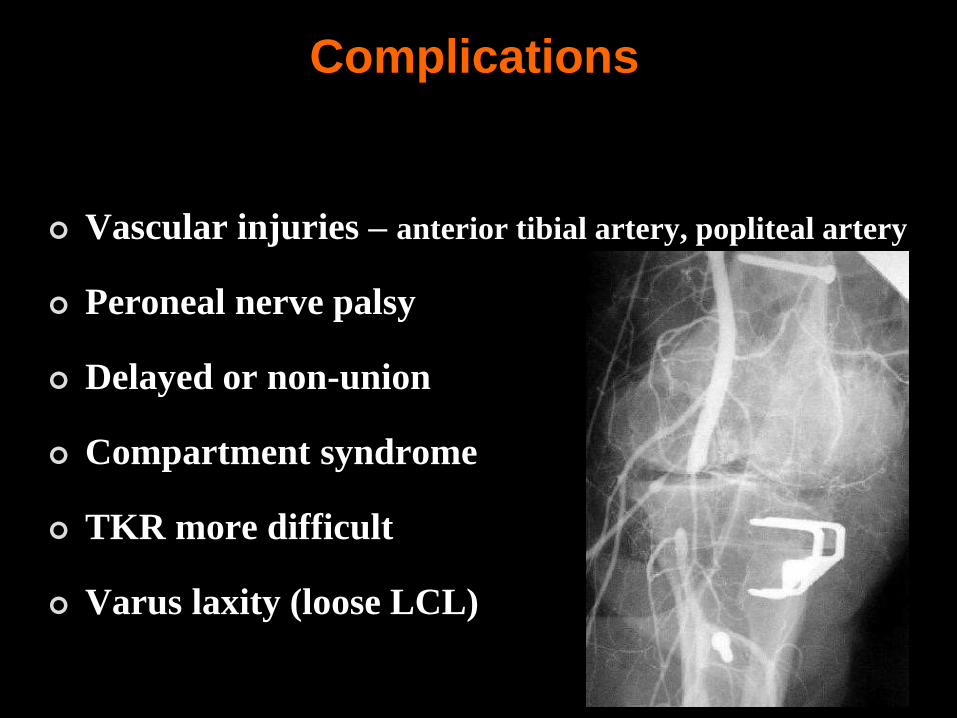
Complications
Inadequate valgus correction
Overcorrection – PFJ derangement
Alteration in patella height
Intra-articular fracture
Osteonecrosis of the tibial plateau
Complications
Vascular injuries – anterior tibial artery, popliteal artery
Peroneal nerve palsy
Delayed or non-union
Compartment syndrome
TKR more difficult
Varus laxity (loose LCL)
Open wedge HTO Advantages
Easier to achieve precise angular correction
Preserves bone stock (subsequent TKR is technically easier)
Makes tightening of the MCL easier
Preserve the lateral side for LCL or posterolateral
reconstruction if insufficient
No risk to peroneal nerve
Less dissection
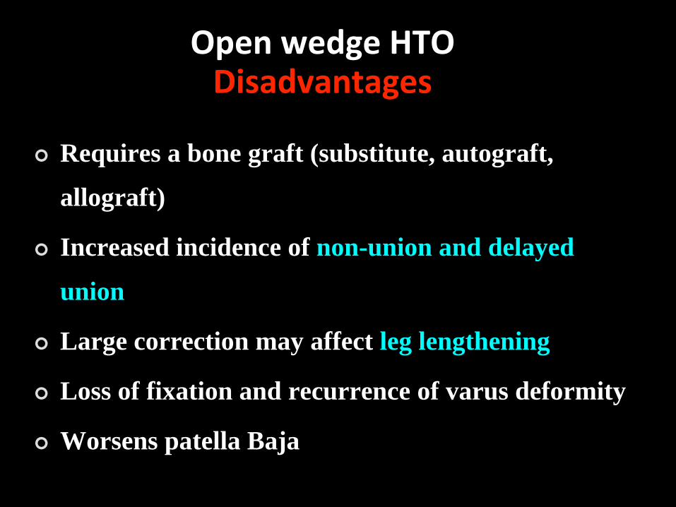
Requires a bone graft (substitute, autograft,
allograft)
Increased incidence of non-union and delayed
union
Large correction may affect leg lengthening
Loss of fixation and recurrence of varus deformity
Worsens patella Baja
Open wedge HTODisadvantages
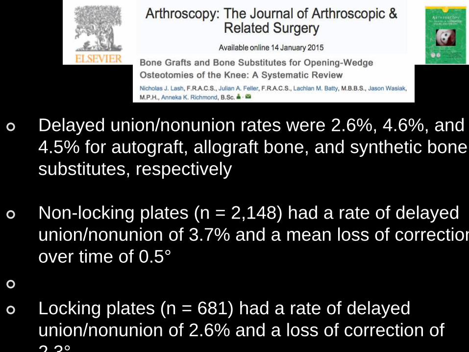
OW-HTO
Delayed union/nonunion rates were 2.6%, 4.6%, and
4.5% for autograft, allograft bone, and synthetic bone
substitutes, respectively
Non-locking plates (n = 2,148) had a rate of delayed
union/nonunion of 3.7% and a mean loss of correction
over time of 0.5°
Locking plates (n = 681) had a rate of delayed
union/nonunion of 2.6% and a loss of correction of
2.3°.
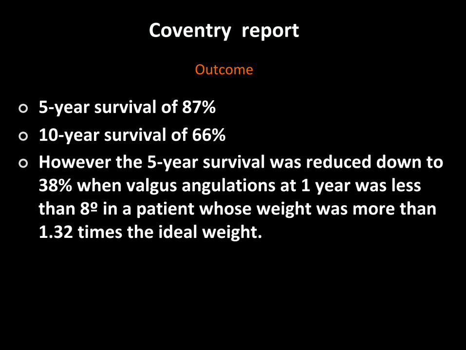
Coventry report
Outcome
5-year survival of 87%
10-year survival of 66%
However the 5-year survival was reduced down to 38% when valgus angulations at 1 year was less than 8º in a patient whose weight was more than 1.32 times the ideal weight.
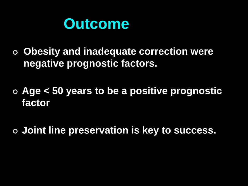
Outcome
Obesity and inadequate correction were
negative prognostic factors.
Age < 50 years to be a positive prognostic
factor
Joint line preservation is key to success.
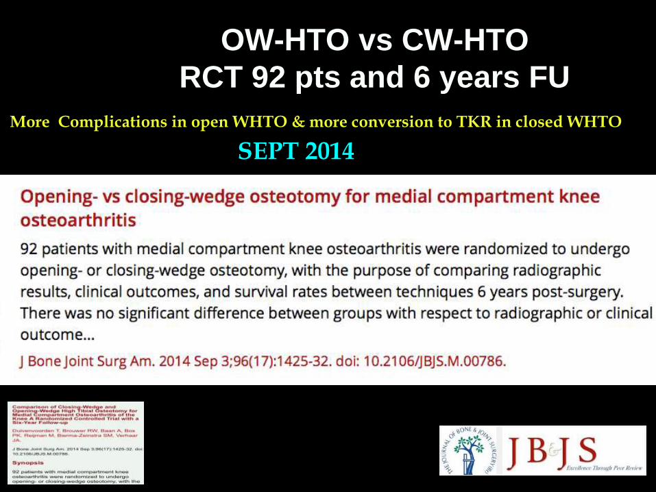
OW-HTO vs CW-HTO
RCT 92 pts and 6 years FU
More Complications in open WHTO & more conversion to TKR in closed WHTO
SEPT 2014
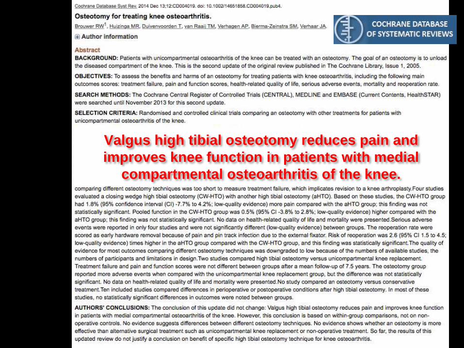
Valgus high tibial osteotomy reduces pain and
improves knee function in patients with medial
compartmental osteoarthritis of the knee.
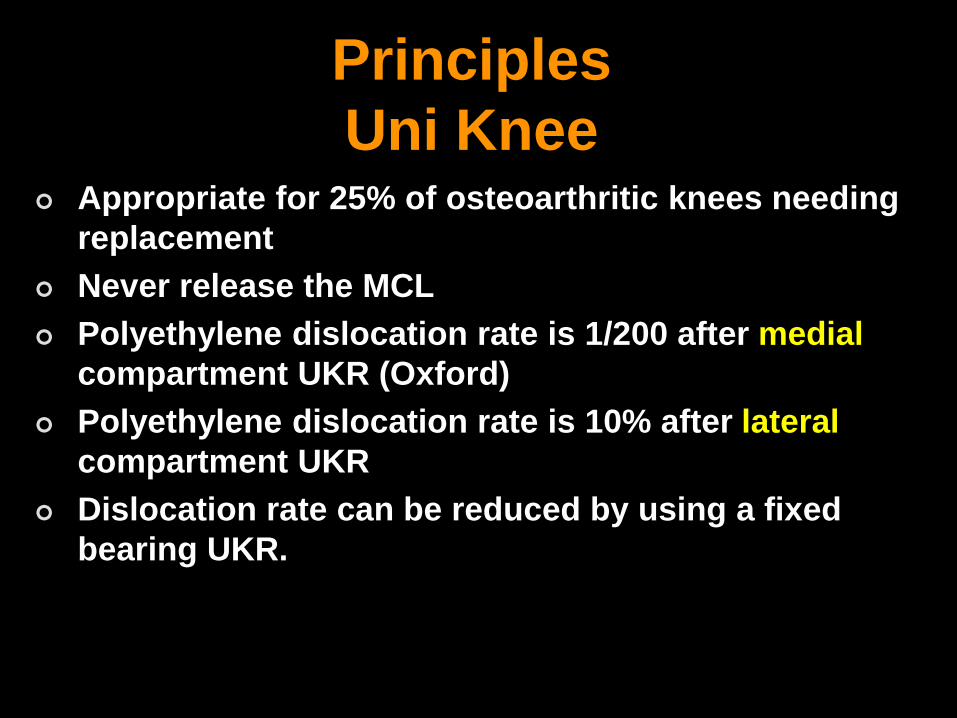
Principles
Uni Knee Appropriate for 25% of osteoarthritic knees needing
replacement
Never release the MCL
Polyethylene dislocation rate is 1/200 after medial
compartment UKR (Oxford)
Polyethylene dislocation rate is 10% after lateral
compartment UKR
Dislocation rate can be reduced by using a fixed
bearing UKR.

?
What are the Absolute contraindications
for Unicompartmental knee replacement?
What are the Advantages and
disadvantages?
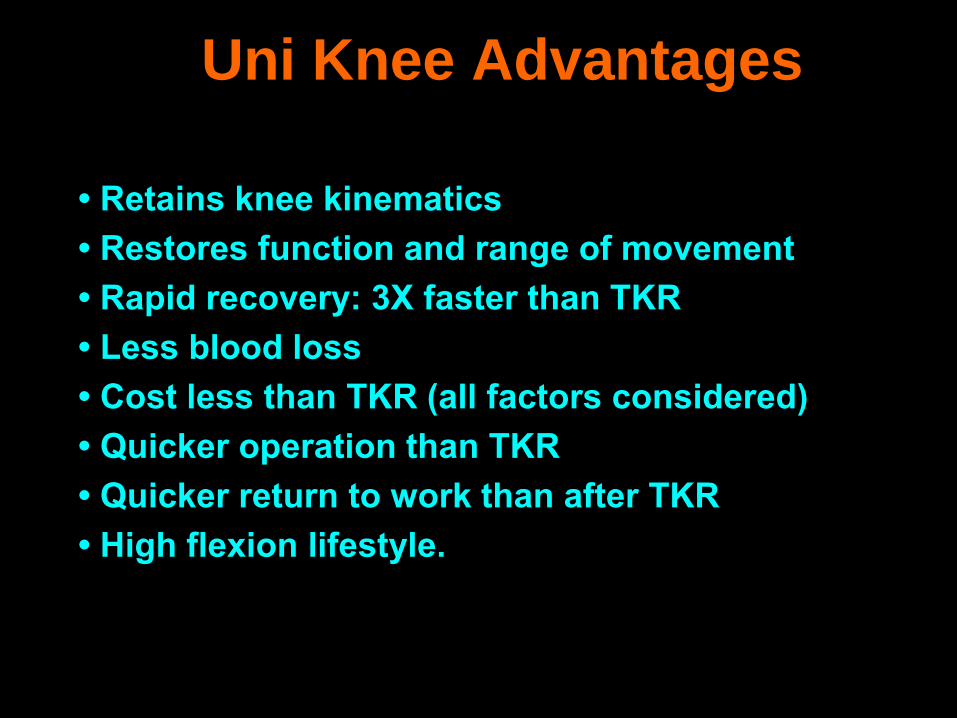
Uni Knee Advantages
• Retains knee kinematics
• Restores function and range of movement
• Rapid recovery: 3X faster than TKR
• Less blood loss
• Cost less than TKR (all factors considered)
• Quicker operation than TKR
• Quicker return to work than after TKR
• High flexion lifestyle.

Uni Knee
Advantages
• Lower infection rate (halved) compared with TKR
• Allows minimally invasive approach
• Easier to revise than HTO?
• No patellar fractures or dislocations
• Maximises the longevity of total knee arthroplasty
• Reduced incidence of DVT
• Reduced mortality from pulmonary embolism
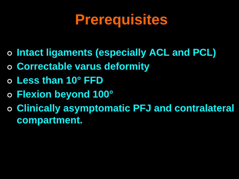
Prerequisites
Intact ligaments (especially ACL and PCL)
Correctable varus deformity
Less than 10° FFD
Flexion beyond 100°
Clinically asymptomatic PFJ and contralateral
compartment.
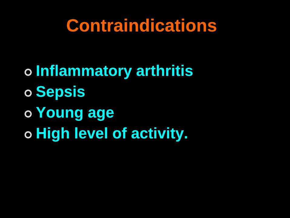
Contraindications
Inflammatory arthritis
Sepsis
Young age
High level of activity.

Relative contraindications
ACL degeneration
Chondrocalcinosis
Lateral meniscectomy
Osteonecrosis
Combined obesity and small bone
size in some women.
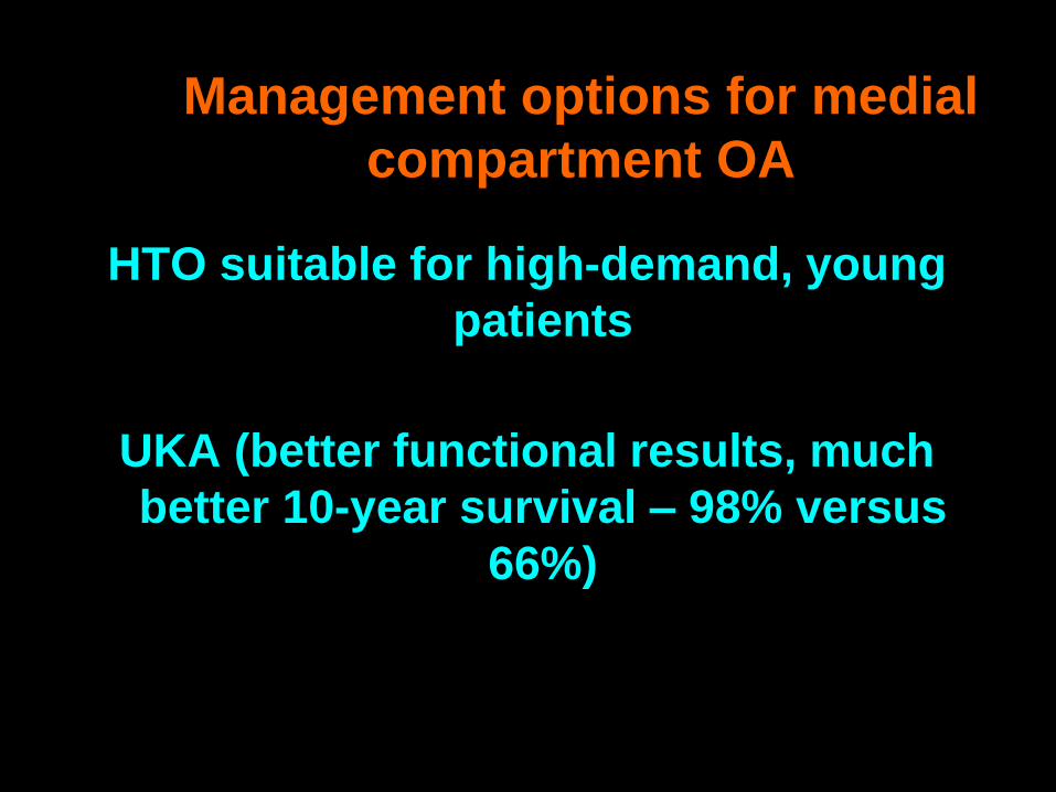
Management options for medial
compartment OA
HTO suitable for high-demand, young
patients
UKA (better functional results, much
better 10-year survival – 98% versus
66%)

THANK YOU


![Qi webinar HTO Oct 2019 [Autosaved] - Synergy Science · Title: Microsoft PowerPoint - Qi webinar HTO Oct 2019 [Autosaved] Author: Trevor Created Date: 10/16/2019 9:28:36 PM](https://img.pdfslide.us/doc/110x75/5f0f51517e708231d4439066/qi-webinar-hto-oct-2019-autosaved-synergy-science-title-microsoft-powerpoint.jpg)