Embed Size (px)
Citation preview
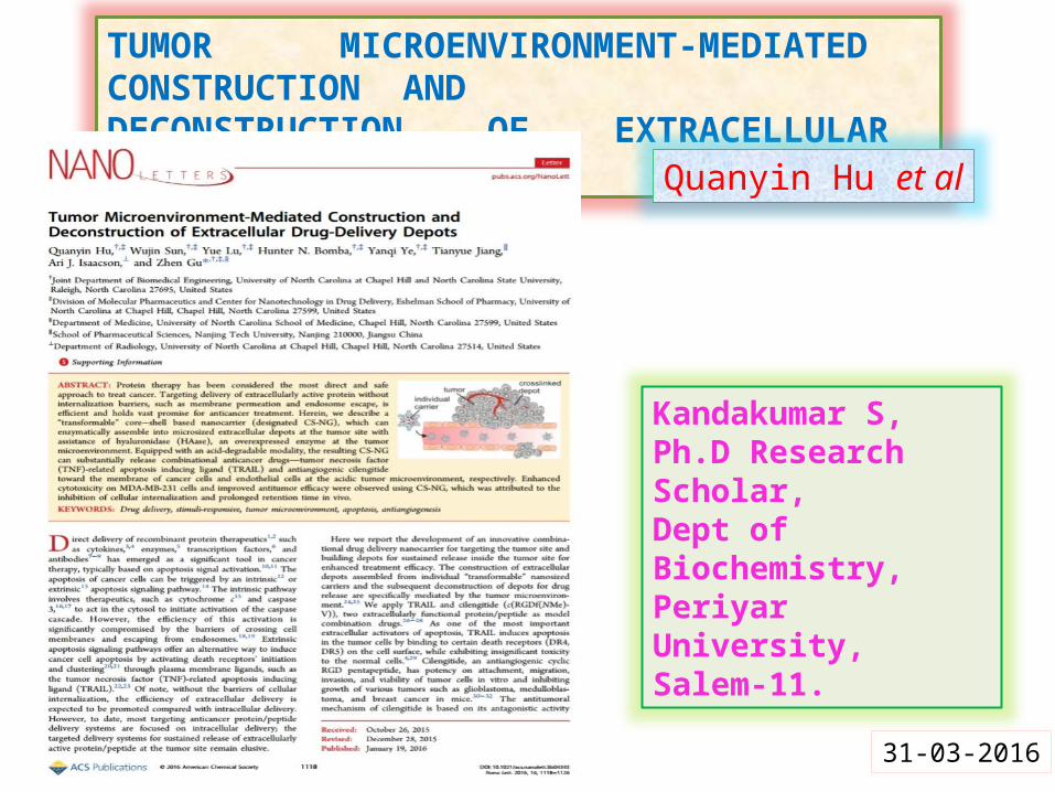
TUMOR MICROENVIRONMENT-MEDIATED CONSTRUCTION ANDDECONSTRUCTION OF EXTRACELLULAR DRUG-DELIVERY DEPOTS
Kandakumar S,Ph.D Research Scholar,Dept of Biochemistry,Periyar University,Salem-11.
Quanyin Hu et al
31-03-2016
INTRODUCTION
Protein therapy has been considered the most direct and safe approach to treat cancer. Targeting delivery of extracellularly active protein without internalization barriers, such as membrane permeation and endosome escape, is efficient and holds vast promise for anticancer treatment.Direct delivery of recombinant protein therapeutics such as cytokines, enzymes, transcription factors, and antibodies has emerged as a significant tool in cancer therapy, typically based on apoptosis signal activation. The apoptosis of cancer cells can be triggered by an intrinsic or extrinsic apoptosis signaling pathway. The intrinsic pathway involves therapeutics, such as cytochrome c and caspase 3 to act in the cytosol to initiate activation of the caspase cascade.The efficiency of this activation is significantly compromised by the barriers of crossing cell membranes and escaping from endosomes. Extrinsic apoptosis signaling pathways offer an alternative way to induce cancer cell apoptosis by activating death receptors’ initiation and clustering through plasma membrane ligands, such as the TNF-related apoptosis inducing ligand (TRAIL). The targeted delivery systems for sustained release of extracellularly active protein/peptide at the tumor site remain elusive.
The key advantages of nanoparticles (1) improved bioavailability by enhancing aqueous solubility, (2) increasing resistance time in the body (increasing specificity for its cognate receptors and (3) targeting drug to specific location in the body (its site of action) Herein, these researchers (Quanyin Hu et al) describe a “transformable” core−shell based nanocarrier (designated CS-NG), which can enzymatically assemble into microsized extracellular depots at the tumor site with assistance of hyaluronidase (HAase), an overexpressed enzyme at the tumor microenvironment. Equipped with an acid-degradable modality, the resulting CS-NG can substantially release combinational anticancer drugs - tumor necrosis factor (TNF)-related apoptosis inducing ligand (TRAIL) and antiangiogenic cilengitide toward the membrane of cancer cells and endothelial cells at the acidic tumor microenvironment, respectively. Enhanced cytotoxicity on MDA-MB-231 cells and improved antitumor efficacy were observed using CS-NG, which was attributed to the inhibition of cellular internalization and prolonged retention time in vivo.
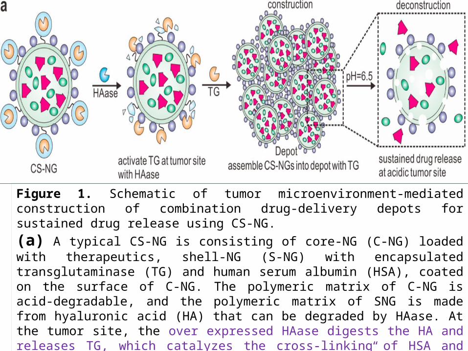
Figure 1. Schematic of tumor microenvironment-mediated construction of combination drug-delivery depots for sustained drug release using CS-NG.(a) A typical CS-NG is consisting of core-NG (C-NG) loaded with therapeutics, shell-NG (S-NG) with encapsulated transglutaminase (TG) and human serum albumin (HSA), coated on the surface of C-NG. The polymeric matrix of C-NG is acid-degradable, and the polymeric matrix of SNG is made from hyaluronic acid (HA) that can be degraded by HAase. At the tumor site, the over expressed HAase digests the HA and releases TG, which catalyzes the cross-linking of HSA and assembles CS-NG into the “drug-delivery depots”. The matrix of C-NG can be gradually degraded and subsequently release the encapsulated therapeutics at the acidic tumor microenvironment.
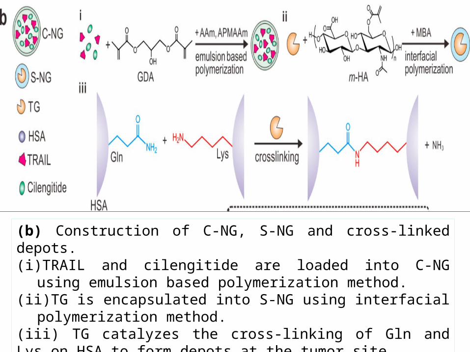
(b) Construction of C-NG, S-NG and cross-linked depots.(i) TRAIL and cilengitide are loaded into C-NG using emulsion based
polymerization method. (ii) TG is encapsulated into S-NG using interfacial polymerization method. (iii) TG catalyzes the cross-linking of Gln and Lys on HSA to form depots at the tumor site.
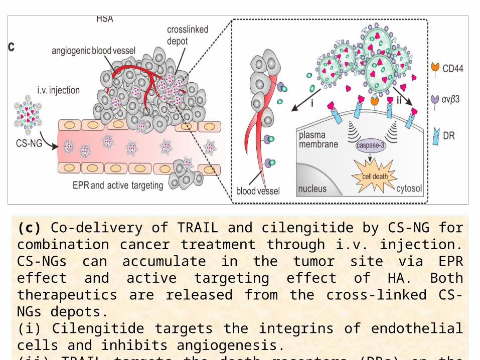
(c) Co-delivery of TRAIL and cilengitide by CS-NG for combination cancer treatment through i.v. injection. CS-NGs can accumulate in the tumor site via EPR effect and active targeting effect of HA. Both therapeutics are released from the cross-linked CS-NGs depots. (i) Cilengitide targets the integrins of endothelial cells and inhibits angiogenesis. (ii) TRAIL targets the death receptors (DRs) on the cell membrane and triggers the apoptosis.
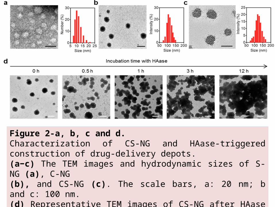
Figure 2-a, b, c and d. Characterization of CS-NG and HAase-triggered construction of drug-delivery depots.(a−c) The TEM images and hydrodynamic sizes of S-NG (a), C-NG (b), and CS-NG (c). The scale bars, a: 20 nm; b and c: 100 nm. (d) Representative TEM images of CS-NG after HAase treatment for 0, 0.5, 1, 3, and 12 h. Scale bar, 100 nm.
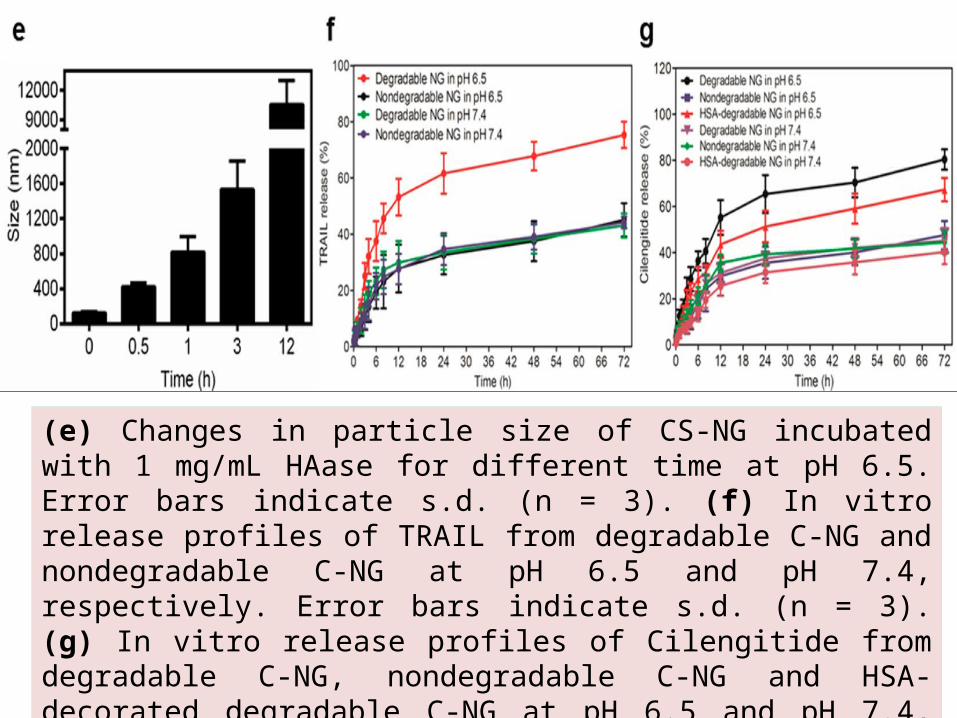
(e) Changes in particle size of CS-NG incubated with 1 mg/mL HAase for different time at pH 6.5. Error bars indicate s.d. (n = 3). (f) In vitro release profiles of TRAIL from degradable C-NG and nondegradable C-NG at pH 6.5 and pH 7.4, respectively. Error bars indicate s.d. (n = 3). (g) In vitro release profiles of Cilengitide from degradable C-NG, nondegradable C-NG and HSA-decorated degradable C-NG at pH 6.5 and pH 7.4, respectively. Error bars indicate s.d. (n = 3).
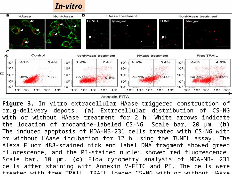
Figure 3. In vitro extracellular HAase-triggered construction of drug-delivery depots. (a) Extracellular distribution of CS-NG with or without HAase treatment for 2 h. White arrows indicate the location of rhodamine-labeled CS-NG. Scale bar, 20 μm. (b) The induced apoptosis of MDA-MB-231 cells treated with CS-NG with or without HAase incubation for 12 h using the TUNEL assay. The Alexa Fluor 488-stained nick end label DNA fragment showed green fluorescence, and the PI-stained nuclei showed red fluorescence. Scale bar, 10 μm. (c) Flow cytometry analysis of MDA-MB- 231 cells after staining with Annexin V-FITC and PI. The cells were treated with free TRAIL, TRAIL loaded CS-NG with or without HAase treatment at the TRAIL concentration of 50 ng/mL for 12 h. The cells incubated with drug free DMEM served as a control.
In-vitro
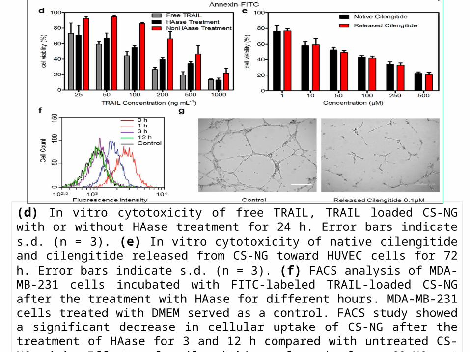
(d) In vitro cytotoxicity of free TRAIL, TRAIL loaded CS-NG with or without HAase treatment for 24 h. Error bars indicate s.d. (n = 3). (e) In vitro cytotoxicity of native cilengitide and cilengitide released from CS-NG toward HUVEC cells for 72 h. Error bars indicate s.d. (n = 3). (f) FACS analysis of MDA-MB-231 cells incubated with FITC-labeled TRAIL-loaded CS-NG after the treatment with HAase for different hours. MDA-MB-231 cells treated with DMEM served as a control. FACS study showed a significant decrease in cellular uptake of CS-NG after the treatment of HAase for 3 and 12 h compared with untreated CS-NG (g) Effect of cilengitide released from CS-NG at concentration of 0.1 μM on HUVEC cells tube formation for 8 h. HUVEC cells incubated with drug free DMEM served as a control. Scale bar, 100 μm.
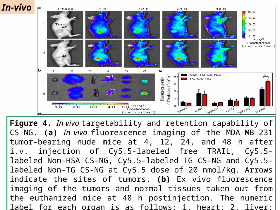
Figure 4. In vivo targetability and retention capability of CS-NG. (a) In vivo fluorescence imaging of the MDA-MB-231 tumor-bearing nude mice at 4, 12, 24, and 48 h after i.v. injection of Cy5.5-labeled free TRAIL, Cy5.5-labeled Non-HSA CS-NG, Cy5.5-labeled TG CS-NG and Cy5.5-labeled Non-TG CS-NG at Cy5.5 dose of 20 nmol/kg. Arrows indicate the sites of tumors. (b) Ex vivo fluorescence imaging of the tumors and normal tissues taken out from the euthanized mice at 48 h postinjection. The numeric label for each organ is as follows: 1, heart; 2, liver; 3, spleen; 4, lung; 5, kidney; 6, tumor. (c) Region-of-interest analysis of fluorescent signals from the tumors and normal tissues.
In-vivo
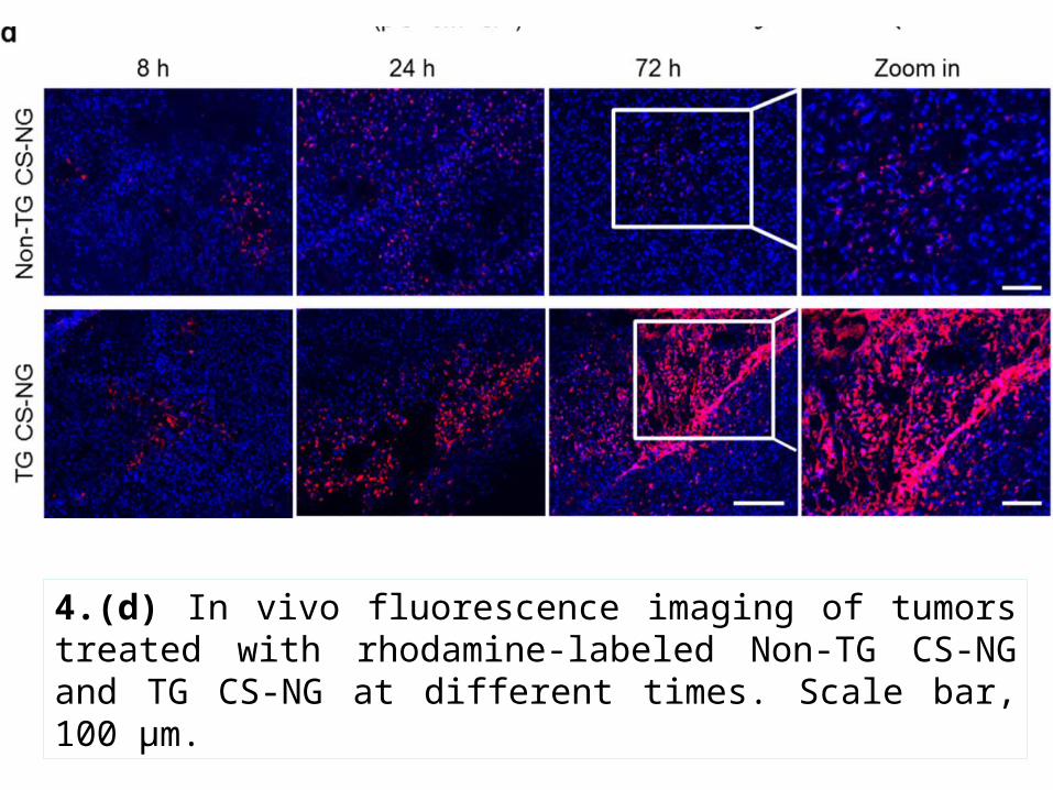
4.(d) In vivo fluorescence imaging of tumors treated with rhodamine-labeled Non-TG CS-NG and TG CS-NG at different times. Scale bar, 100 μm.
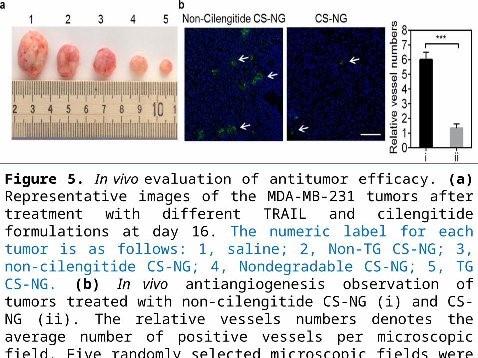
Figure 5. In vivo evaluation of antitumor efficacy. (a) Representative images of the MDA-MB-231 tumors after treatment with different TRAIL and cilengitide formulations at day 16. The numeric label for each tumor is as follows: 1, saline; 2, Non-TG CS-NG; 3, non-cilengitide CS-NG; 4, Nondegradable CS-NG; 5, TG CS-NG. (b) In vivo antiangiogenesis observation of tumors treated with non-cilengitide CS-NG (i) and CS-NG (ii). The relative vessels numbers denotes the average number of positive vessels per microscopic field. Five randomly selected microscopic fields were quantitatively analyzed on ImageJ. Arrows indicate the sites of tumor blood vessels (green). Scale bar, 200 μm.
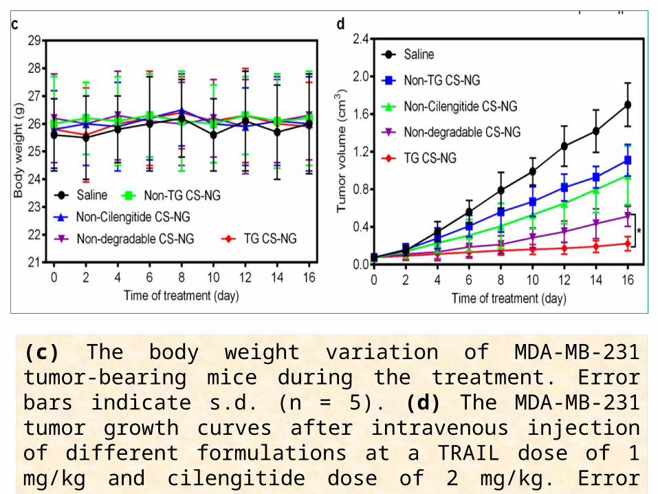
(c) The body weight variation of MDA-MB-231 tumor-bearing mice during the treatment. Error bars indicate s.d. (n = 5). (d) The MDA-MB-231 tumor growth curves after intravenous injection of different formulations at a TRAIL dose of 1 mg/kg and cilengitide dose of 2 mg/kg. Error bars indicate s.d. (n = 5).
The in vivo antitumor efficacy was evaluated on the MDA-MB- 231 tumor-bearing nude mice model. The growth of the tumor was significantly inhibited after the treatment with different TRAIL formulations, including Non-TG CS-NG, Non-Cilengitide CS-NG, nondegradable CS-NG, and TG CSNG, compared with saline control group (Figure 5a). It was worth noting that cilengitide played a significant role on suppressing tumor growth, which was validated by the decreased tumor vascularization at the tumor site treated with cilengitide-loaded CS-NG compared with that treated with Non-Cilengitide CS-NG
Conclusion
In this study, these researchers (Quanyin Hu et al) have developed a new drug delivery strategy by using in situ extracellularly built depots assembled from individual nanocarriers for sustained release of combination therapeutics toward cell membranes to inhibit tumor proliferation. Both the construction and the deconstruction of depots are activated in the tumor microenvironment by overexpressed enzyme (HAase) and the acidic condition, respectively.This tumor microenvironment-responsive delivery system makes such “transformable” formulation highly selective, while the resulting extracellular depots enable highly sustained release of the membrane-associated therapeutics towards to their most active destinations.This formulation can be further adapted as a general platform for delivery of a variety of other therapeutics, such as small molecule anticancer drugs, anti-inflammation drugs, and vaccine agents, because of its targeting specificity, transformable behavior, and biodegradable nature.