Embed Size (px)
Citation preview
RADIONUCLIDE THYROID IMAGING
INDICATIONS
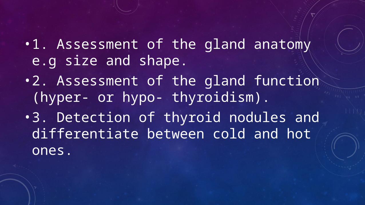
• 1. Assessment of the gland anatomy e.g size and shape.• 2. Assessment of the gland function (hyper- or
hypo- thyroidism).• 3. Detection of thyroid nodules and differentiate
between cold and hot ones.
• 4. Post-operative (e.g. post-thyroidectomy) assessment• 5. detection of functioning metastatic tissues
in known cases of thyroid malignancy 6. Detection of Retrosternal Goiter
CONTRAINDICATIONS
• 1 .pregnancy • 2 .Patients with Hypersensitivity reaction to iodine• 3 .Pertechnetate is excreted in breast milk, so
breastfeeding women are advised to express and discard breast milk for 26 hours after injection.

RADIOPHARMACEUTICALS
99MTC PERTECHNETATE :
• Used mainly in anatomical assessment• It carries the properties of 99m Tc • Gamma-emitter = 140 Kev• Physical half life = 6 hrs.• Dose: 3-5 mCi.• Route: IV.• Advantages: cheap and available
131 I -SODIUM IODIDE:
• Used in full Assessment (Function & Anatomy)• Gamma-emitter = 364 Kev• Half life: 8 days.• Dose: 3 - 5 mCi • Route: Orally.• Disadvantage: Relatively expensive , Difficult preparation & B
particle emitter
123 I-SODIUM IODIDE
• Used in full Assessment (Function & Anatomy)• Gamma-emitter = 159 Kev• Half life: 13 hours.• Dose: 100 mCi• Route: orally.• Advantage: Pure Gamma emitter (Does not emit B particles)• Disadvantage: Very expensive with limited availability
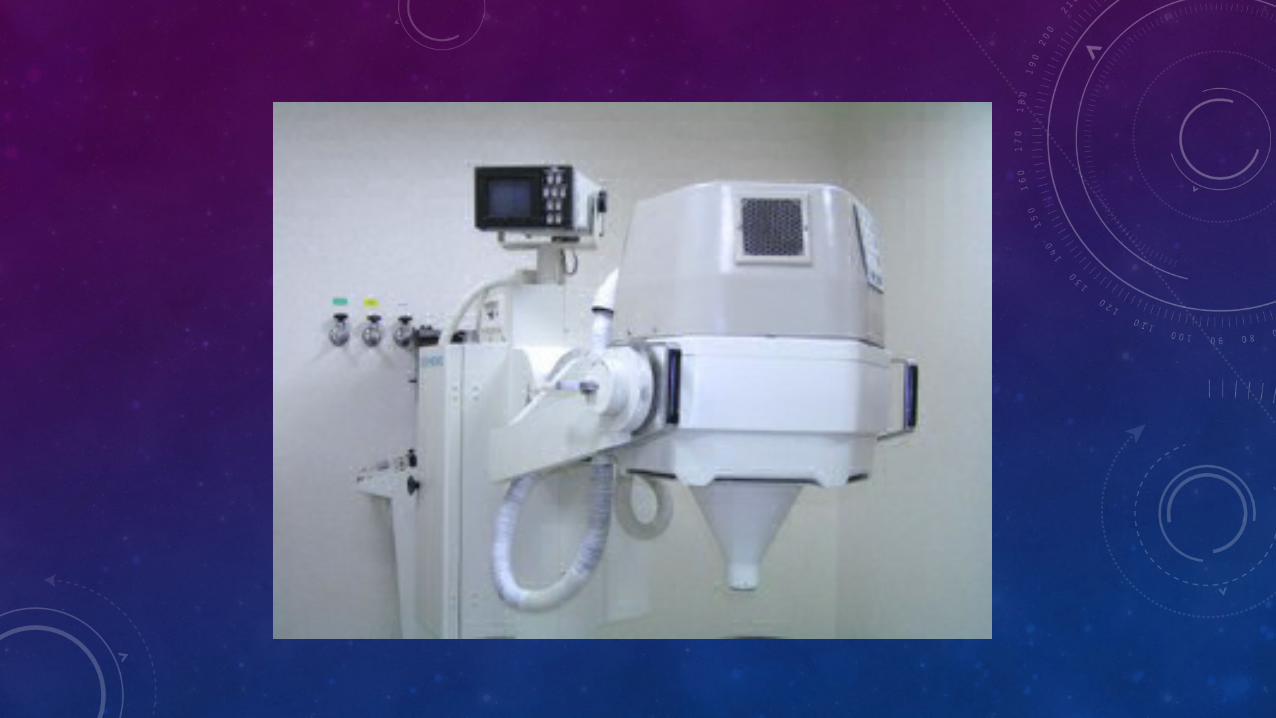
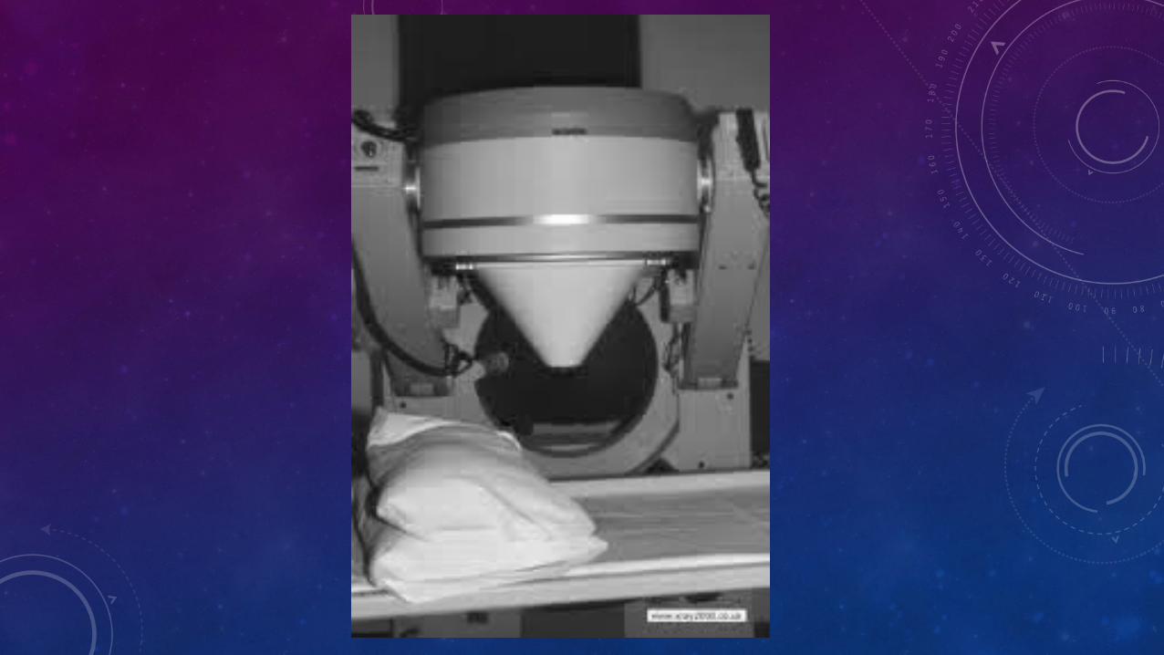
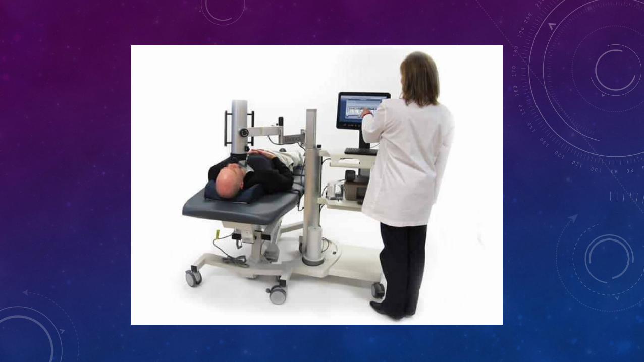
EQUIPMENT:
• Gamma camera with pinhole collimator

PATIENT PREPARATION
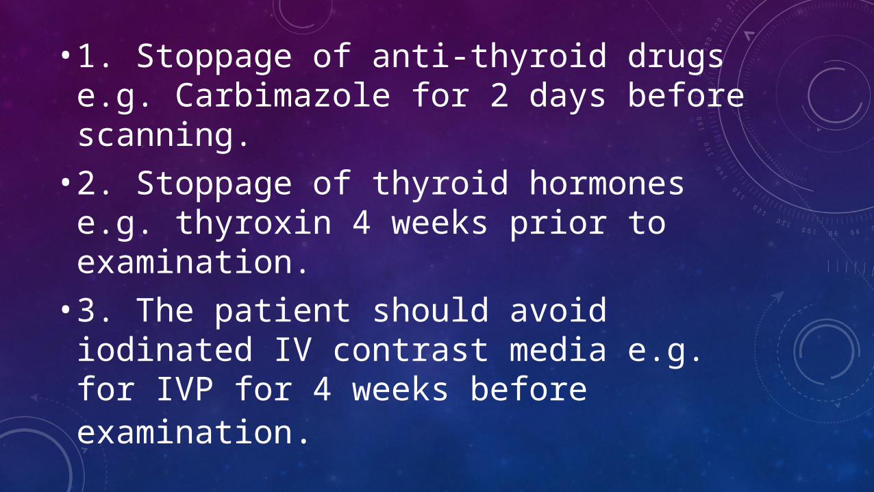
• 1. Stoppage of anti-thyroid drugs e.g. Carbimazole for 2 days before scanning.• 2. Stoppage of thyroid hormones e.g. thyroxin 4
weeks prior to examination.• 3. The patient should avoid iodinated IV contrast
media e.g. for IVP for 4 weeks before examination.
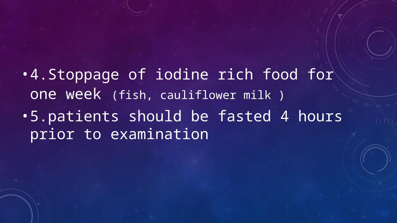
• 4.Stoppage of iodine rich food for one week (fish, cauliflower milk )
• 5.patients should be fasted 4 hours prior to examination
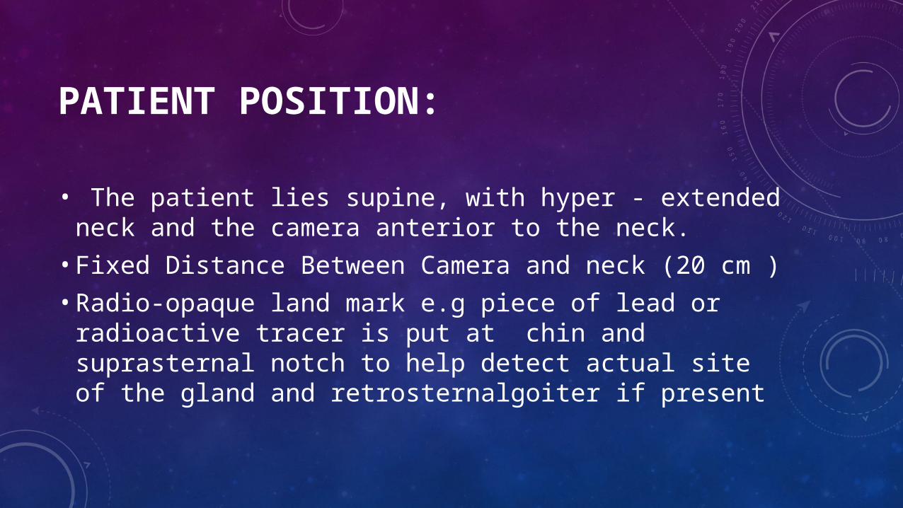
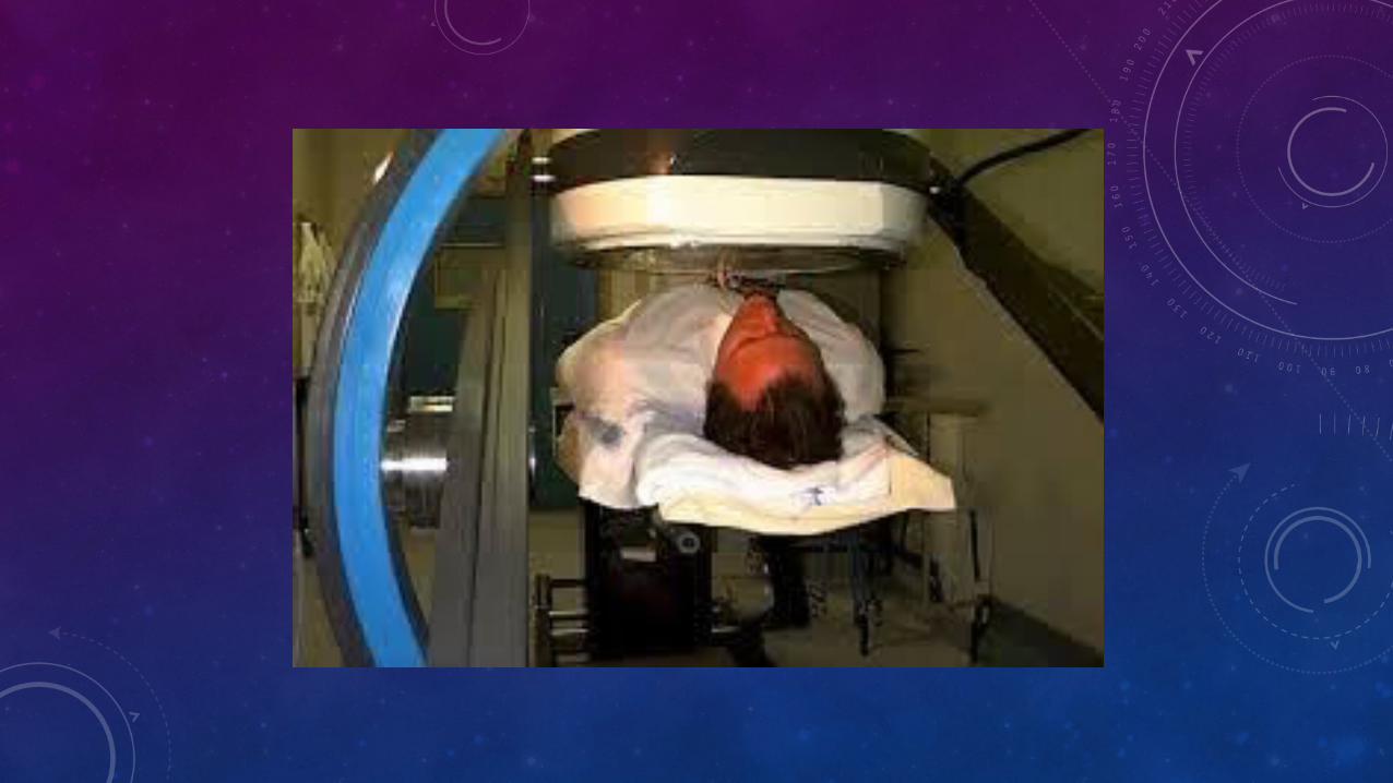
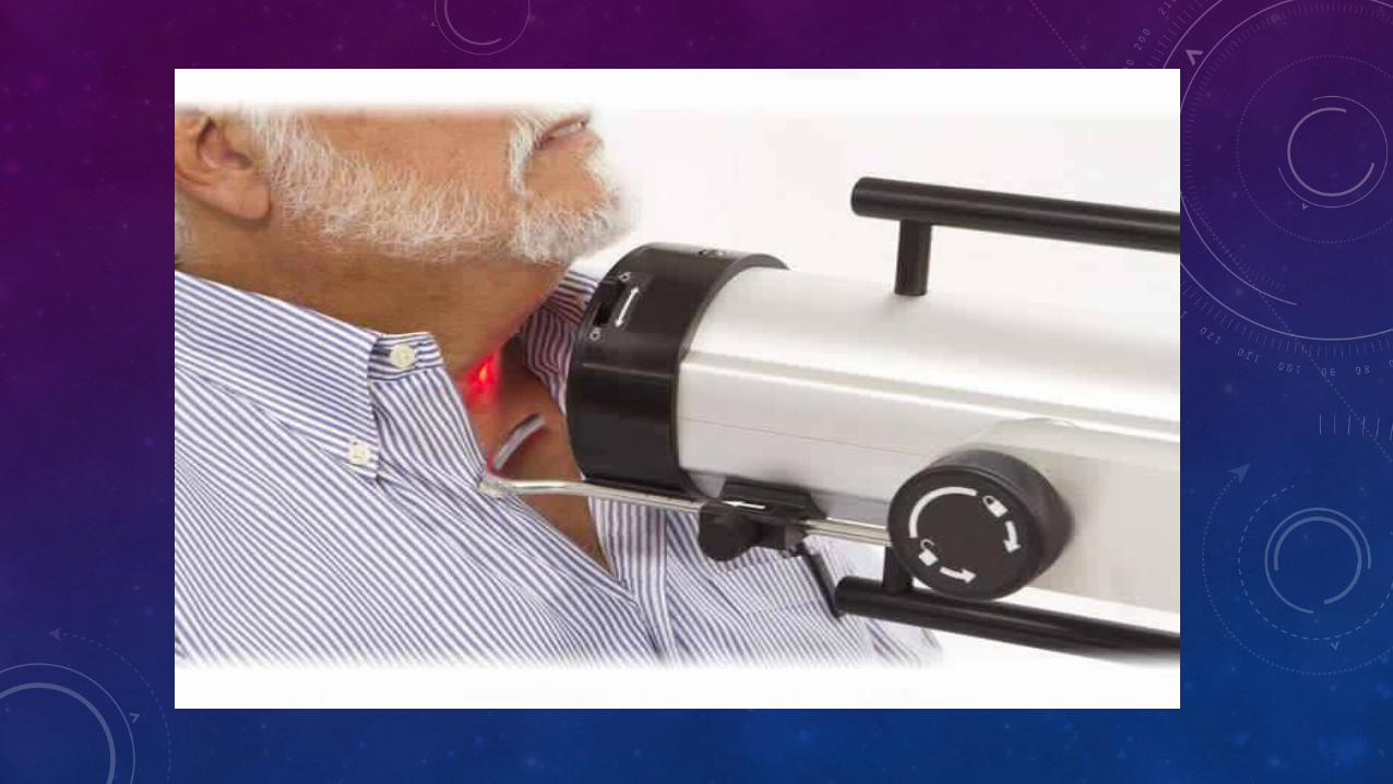
PATIENT POSITION:
• The patient lies supine, with hyper - extended neck and the camera anterior to the neck.• Fixed Distance Between Camera and neck (20 cm )• Radio-opaque land mark e.g piece of lead or radioactive
tracer is put at chin and suprasternal notch to help detect actual site of the gland and retrosternalgoiter if present
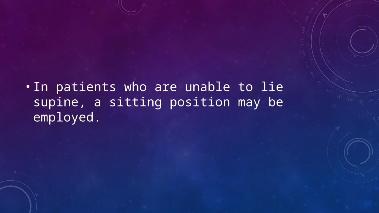
• In patients who are unable to lie supine, a sitting position may be employed.
VIEWS :
• Anterior• Right & Left oblique• Whole body scan only in detection of functioning
metastatic tissues in known cases of thyroid malignancy ( Anterior- Posterior –oblique)
SCANNING TIME :
• 1. When Tc-99m is used, imaging should begin 15–30 min after injection.• 2. When I-131 is used, the images should be
obtained at 2h, 24 hr & 3- 4 days after ingesting the radioiodine.• 3.When I-123 is used, images can be obtained as
early as after 4-6 H, then at 16 – 24 H
INTERPRETATION
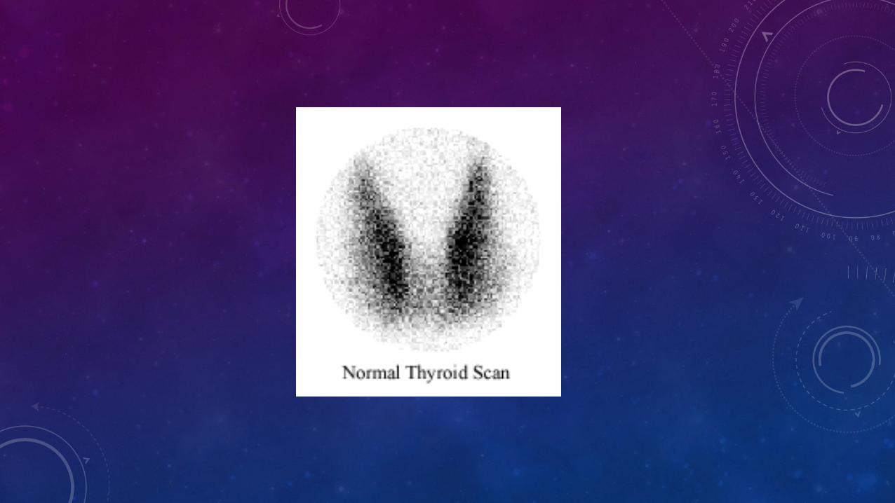
NORMAL
• 99 Tc normal uptake : 1-3 % of total dose• Radioactive iodine 131 I normal uptake :
• Early uptake after 2 h : 10-20 % of total dose• Late uptake after 24 hours : 20 – 60% of total dose• In case of evaluating functioning cancer thyroid metastasis
in whole body scan uptake after 3-4 days will appear as Hot spot any where in the body
• Gland Should be situated mid-way between symphysis menti & suprasternal notch• butterfly shaped• Right lobe is somewhat larger than the left• In 10% of the patients pyramidal lobe may be present.• Homogenous uniform symmetrical uptake all over the
gland
ANTERIOR R ANT. OBLİQUE L ANT. OBLİQUE
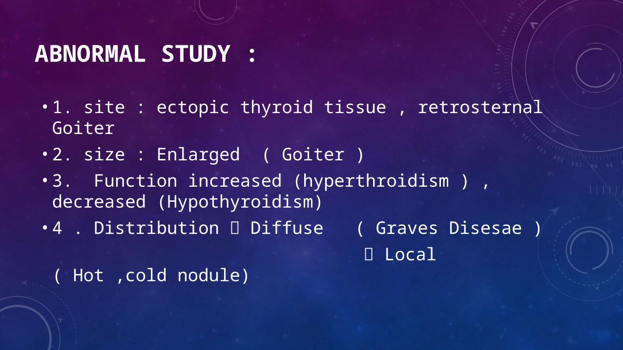
ABNORMAL STUDY :
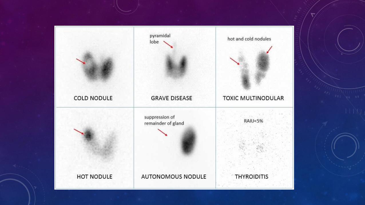
• 1. site : ectopic thyroid tissue , retrosternal Goiter• 2. size : Enlarged ( Goiter )• 3. Function increased (hyperthroidism ) , decreased
(Hypothyroidism)• 4 . Distribution Diffuse ( Graves Disesae ) Local ( Hot ,cold nodule)
• Cold Nodule : 80 % (cyst , abcess ,hematoma ) 20% Tumor benign ( Adenoma ) ,Malignant (Non functioning carcinoma)• Hot nodule : Solitary (Autonomous nodule, functioning
adenoma or adenocarcinoma ) Multiple ( multiple nodular goiter )
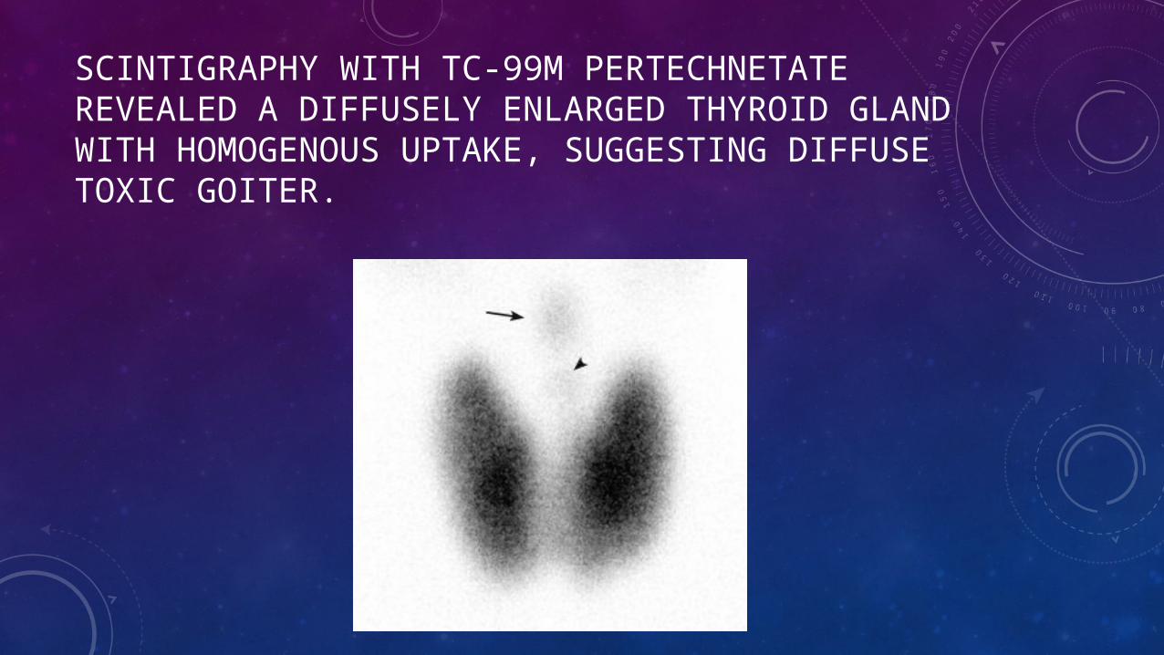
SCINTIGRAPHY WITH TC-99M PERTECHNETATE REVEALED A DIFFUSELY ENLARGED THYROID GLAND WITH HOMOGENOUS UPTAKE, SUGGESTING DIFFUSE TOXIC GOITER.
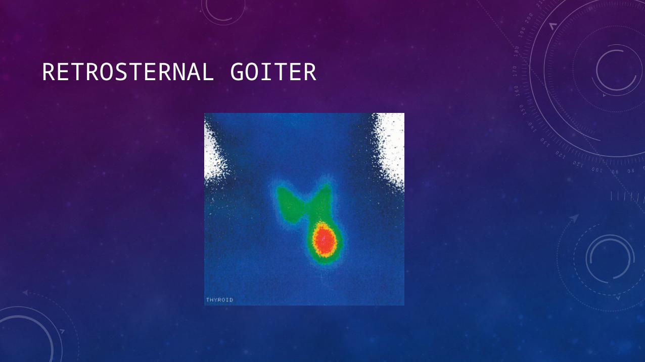
RETROSTERNAL GOITER
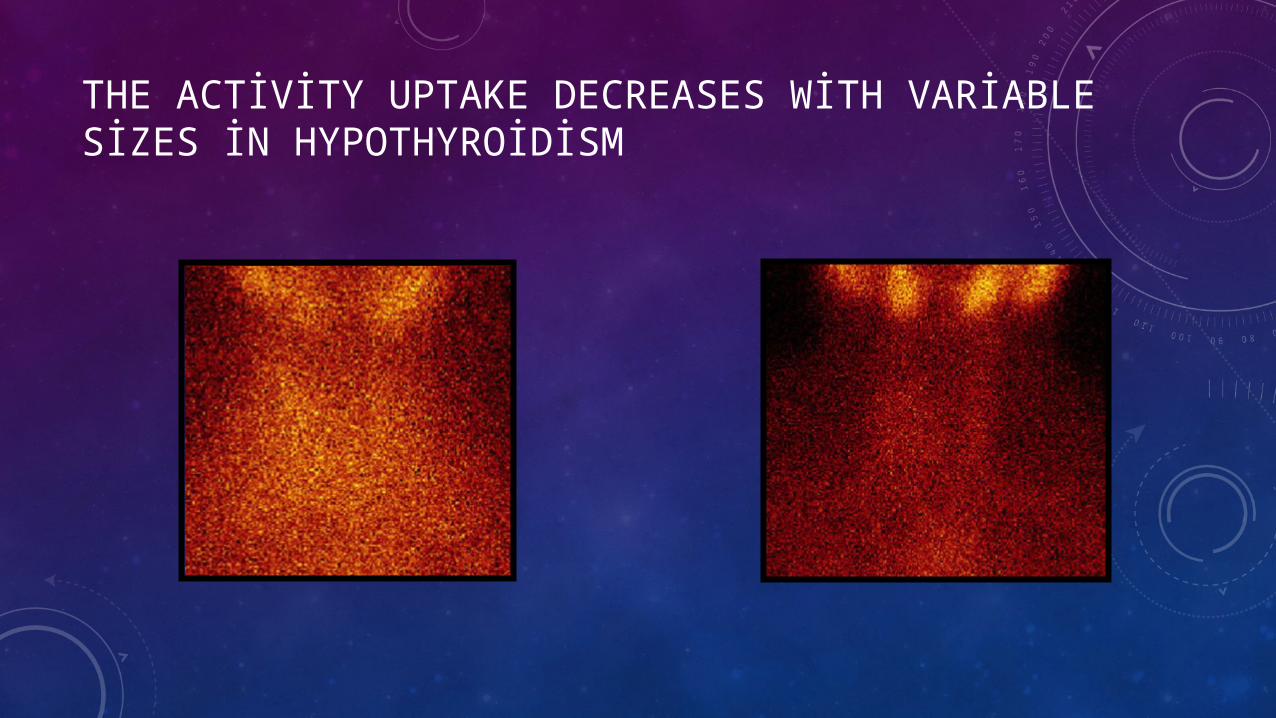
THE ACTİVİTY UPTAKE DECREASES WİTH VARİABLE SİZES İN HYPOTHYROİDİSM
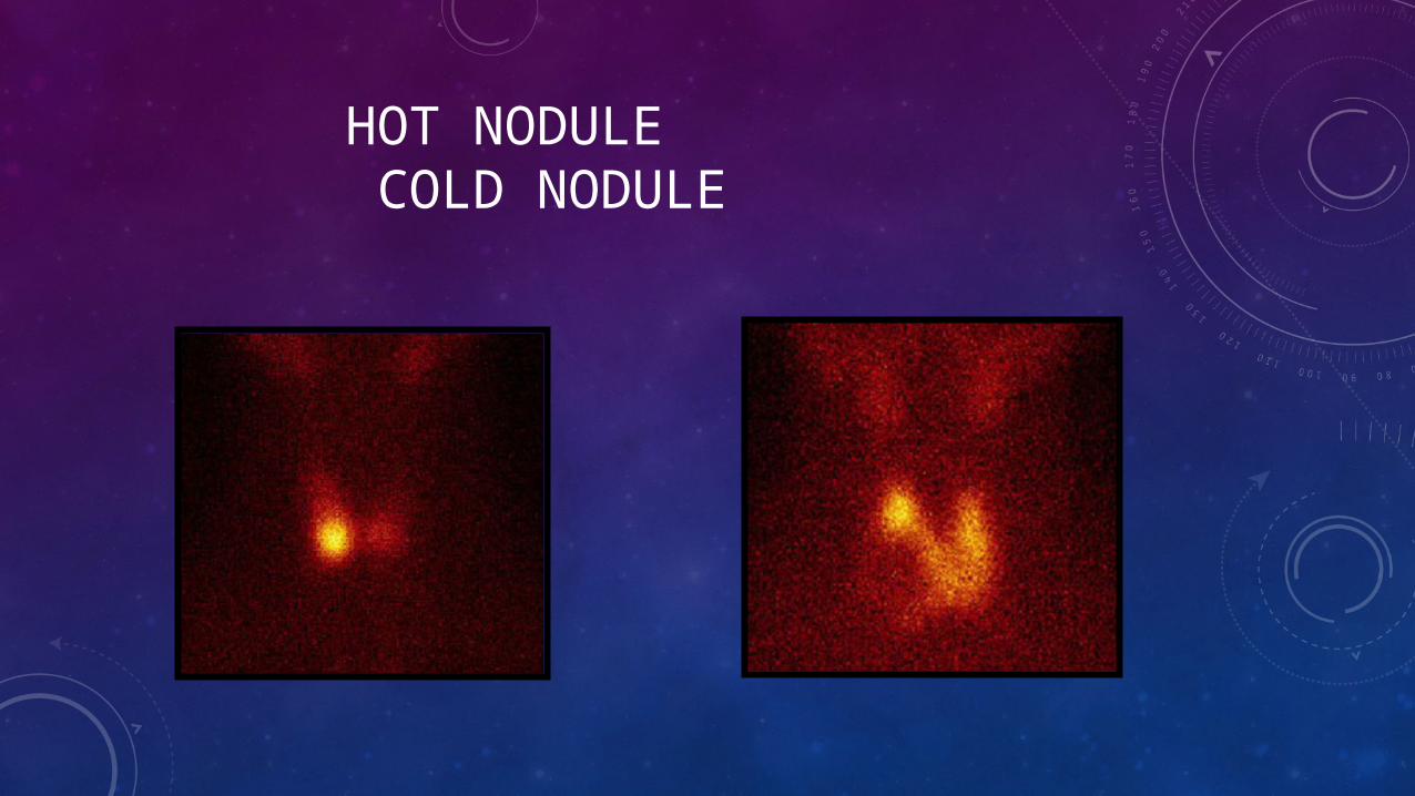
HOT NODULE COLD NODULE
• Radioactive iodine Uptake may increase in following :• Hyperthyroidism ,Iodine deficiency ,Pregnancy ,Recovery phase of subacute,
silent or postpartum thyroiditis ,Rebound after withdrawal of antithyroid medication ,Lithium carbonate therapy ,Hashimoto thyroidites
• May decrease in the following :
Hypothrodism , Destructive thyroiditis (subacute thyroiditis, silent thyroiditis, postpartum thyroiditis)
Because of the large radiation dose to the thyroid(approximately one to three rads per mCiadministered), the use of I-131 for thyroidscintigraphy should be discouraged (exceptwhen a subsequent treatment with I-131 isplanned). (Society of nuclear medicine procedureguide lines for thyroid scintigraphy)
I-131 is usually spared for metastatic disease screening for its high radiation dose and inferior image quality.
Due to its mode of beta decay, iodine-131 is notable for causing mutation and death in cells that it penetrates, and other cells up to several millimeters away. For this reason, high doses of the isotope are sometimes less dangerous than low doses, since they tend to kill thyroid tissues that would otherwise become cancerous as a result of the radiation. For example, children treated with moderate dose of I-131 for thyroid adenomas had a detectable increase in thyroid cancer, but children treated with a much higher dose did not. Likewise, most studies of very-high-dose I-131 for treatment of Graves disease have failed to find any increase in thyroid cancer, even though there is linear increase in thyroid cancer risk with I-131 absorption at moderate doses.[2]
Thus, iodine-131 is increasingly less employed in small doses in medical use (especially in children), but increasingly is used only in large and maximal treatment doses, as a way of killing targeted tissues. This is known as "therapeutic use."
• Dr.Mohamed Nader Fouad• Dr.Asmaa Youssry Alkasaby