Embed Size (px)
Citation preview
Temporal Bone Anatomy
Dr. Apoorv Pandey
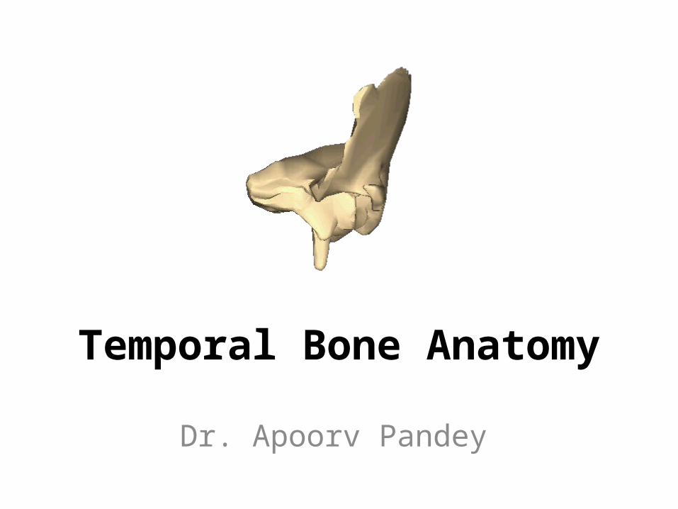
Temporal bones are paired bones located in posterolateral floor of middle & posterior cranial
fossae
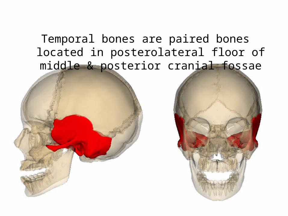
1. Squamous2. Petrous3. Mastoid4. Tympanic5. Styloid

• Squamous: Forms lateral wall of middle cranial fossa
• Mastoid: Aerated posterolateral part and contains mastoid antrum
• Petrous: Pyramidal shape forms the medial part containing inner ear, internal auditory canal & petrous apex
• Tympanic: V-shaped bone forming bony EAC
• Styloid: Forms styloid process
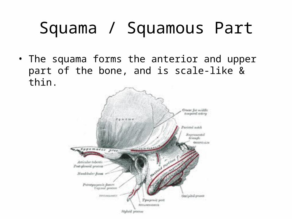
Squama / Squamous Part
• The squama forms the anterior and upper part of the bone, and is scale-like & thin.
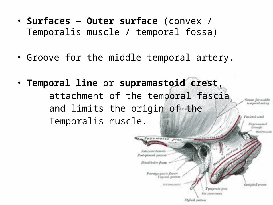
• Surfaces — Outer surface (convex / Temporalis muscle / temporal fossa)
• Groove for the middle temporal artery.
• Temporal line or supramastoid crest, attachment of the temporal fascia and limits the origin of the Temporalis muscle.
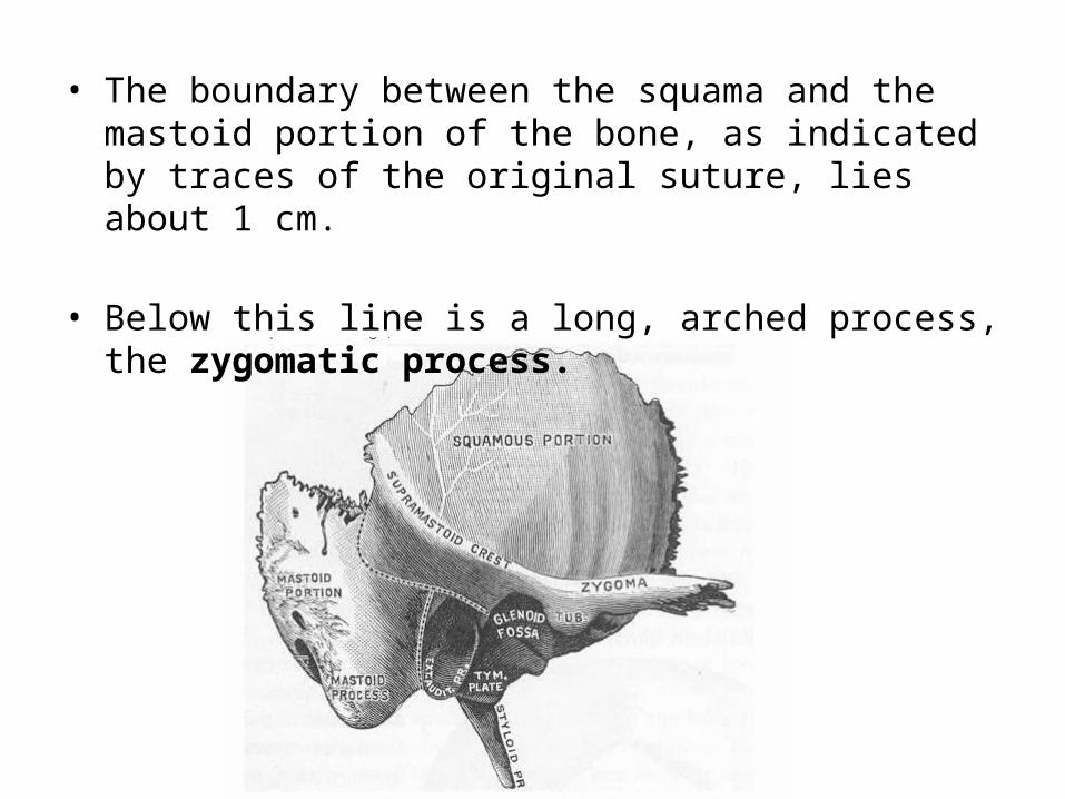
• The boundary between the squama and the mastoid portion of the bone, as indicated by traces of the original suture, lies about 1 cm.
• Below this line is a long, arched process, the zygomatic process.

• Superior border of zygomatic procress: Attachment of the temporal fascia.
• Inferior & medial surface border attaches fibers of the Masseter.
• The anterior end is deeply serrated and articulates with the zygomatic bone
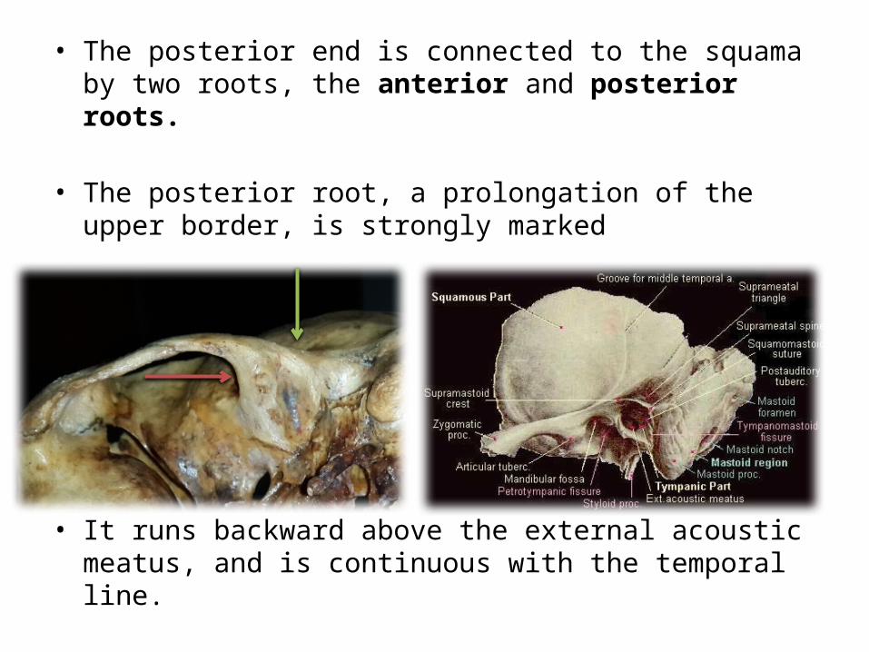
• The posterior end is connected to the squama by two roots, the anterior and posterior roots.
• The posterior root, a prolongation of the upper border, is strongly marked
• It runs backward above the external acoustic meatus, and is continuous with the temporal line.
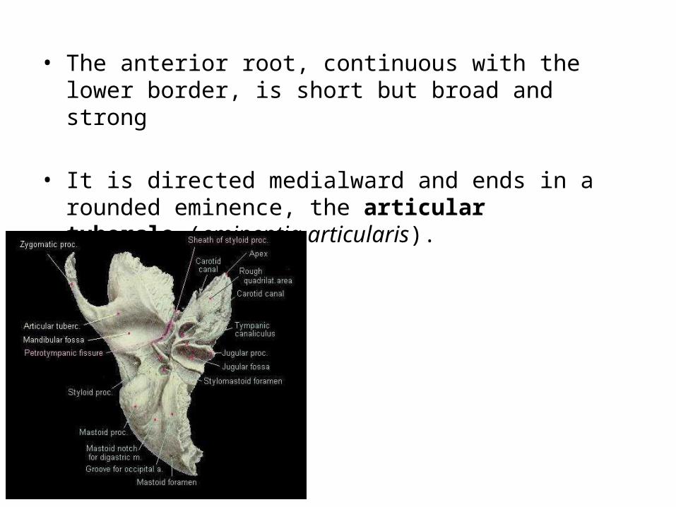
• The anterior root, continuous with the lower border, is short but broad and strong
• It is directed medialward and ends in a rounded eminence, the articular tubercle (eminentia articularis).

• Suprameatal triangle (Macewen), or mastoid fossa, through which an instrument may be pushed into the tympanic antrum.
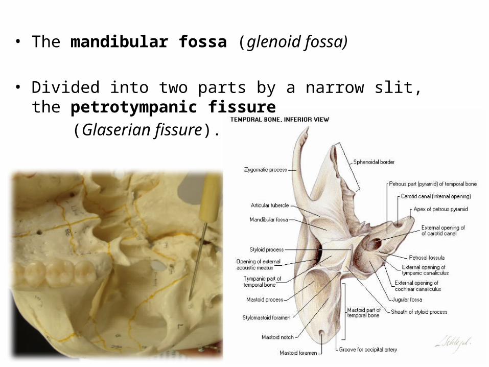
• The mandibular fossa (glenoid fossa)
• Divided into two parts by a narrow slit, the petrotympanic fissure (Glaserian fissure).

• The internal surface of the squama is concave; it presents depressions corresponding to the convolutions of the temporal lobe of the brain, and grooves for the branches of the middle meningeal vessels.
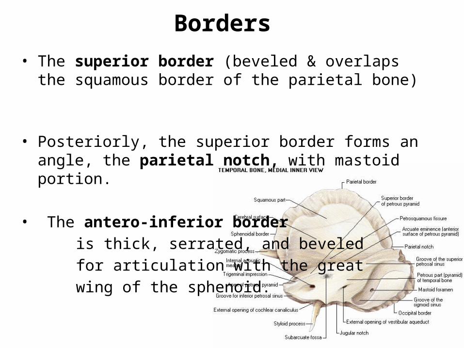
Borders• The superior border (beveled & overlaps the squamous border
of the parietal bone)
• Posteriorly, the superior border forms an angle, the parietal notch, with mastoid portion.
• The antero-inferior border is thick, serrated, and beveled for articulation with the great wing of the sphenoid.
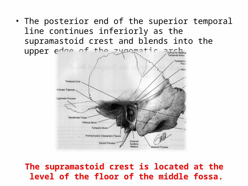
• The posterior end of the superior temporal line continues inferiorly as the supramastoid crest and blends into the upper edge of the zygomatic arch.
The supramastoid crest is located at the level of the floor of the middle fossa.
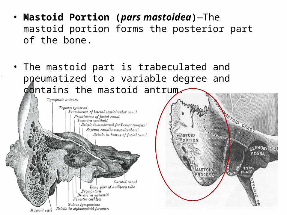
• Mastoid Portion (pars mastoidea)—The mastoid portion forms the posterior part of the bone.
• The mastoid part is trabeculated and pneumatized to a variable degree and contains the mastoid antrum.
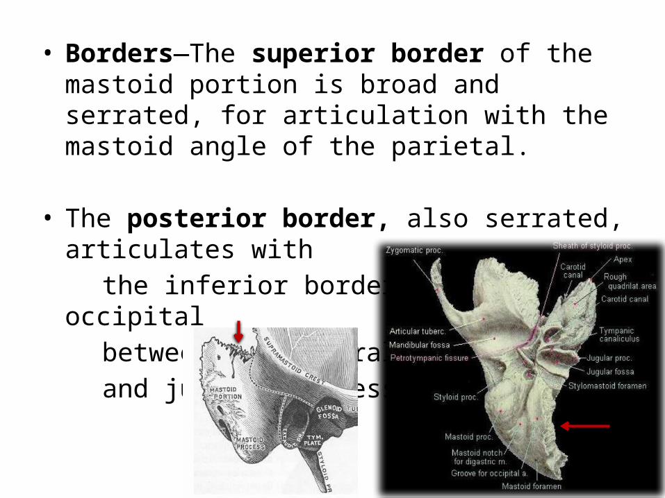
• Borders—The superior border of the mastoid portion is broad and serrated, for articulation with the mastoid angle of the parietal.
• The posterior border, also serrated, articulates with the inferior border of the occipital between the lateral angle and jugular process.

• Anteriorly the mastoid portion is fused with the descending process of the squama above
• Below it enters into the formation of the external acoustic meatus and the tympanic cavity.
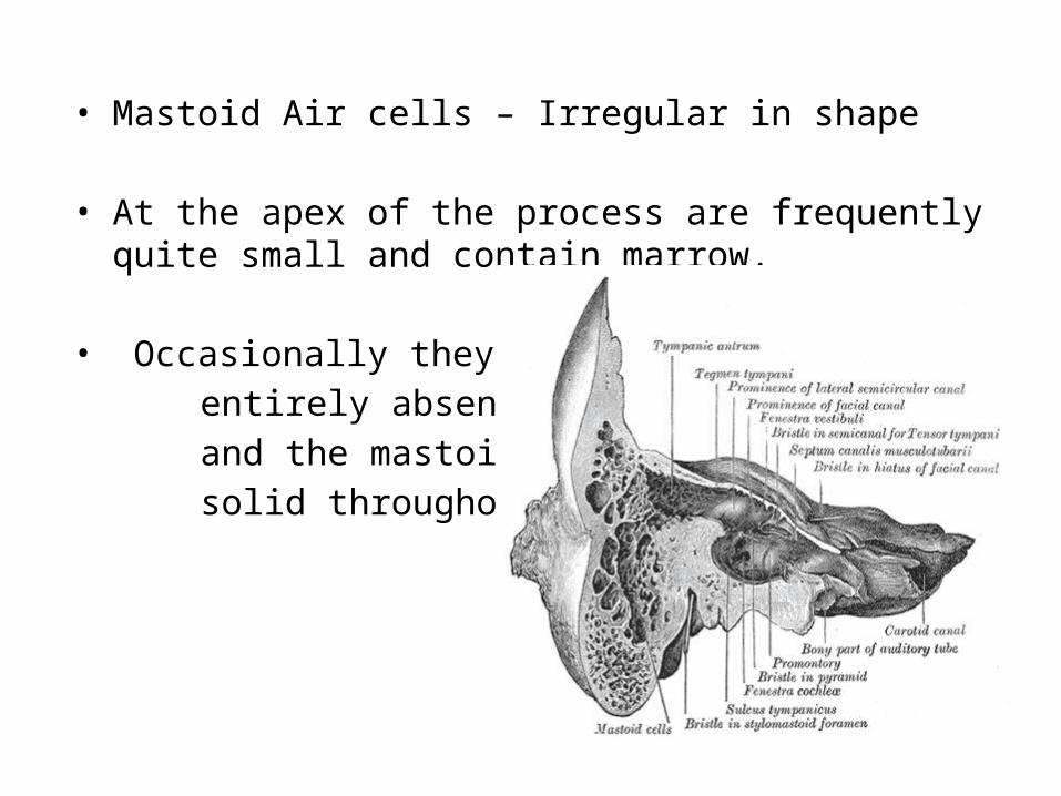
• Mastoid Air cells – Irregular in shape
• At the apex of the process are frequently quite small and contain marrow.
• Occasionally they are entirely absent and the mastoid is then solid throughout
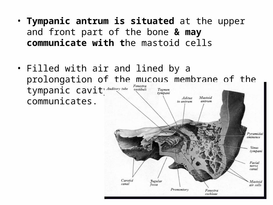
• Tympanic antrum is situated at the upper and front part of the bone & may communicate with the mastoid cells
• Filled with air and lined by a prolongation of the mucous membrane of the tympanic cavity, with which it communicates.
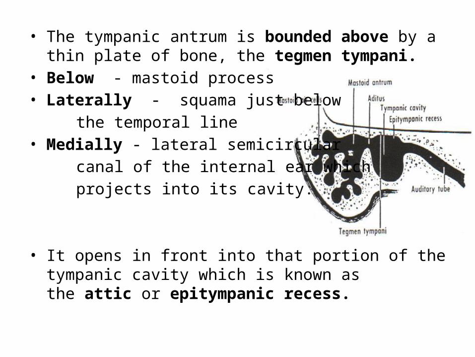
• The tympanic antrum is bounded above by a thin plate of bone, the tegmen tympani.
• Below - mastoid process• Laterally - squama just below the temporal line• Medially - lateral semicircular canal of the internal ear which projects into its cavity.
• It opens in front into that portion of the tympanic cavity which is known as the attic or epitympanic recess.

• The tympanic antrum is a cavity of some considerable size at the time of birth;
• Mastoid air cells may be regarded as diverticula from the antrum, and begin to appear at or before birth
• By the fifth year they are well-marked, but their development is not completed until toward puberty.

Petrous Portion (pars petrosa [pyramis])
• The petrous portion or pyramid is pyramidal and is wedged in at the base of the skull between the sphenoid and occipital.
• Directed medially, forward, and a little upward
• Has a base, an apex, three surfaces, and three angles, and contains, in its interior, the essential parts of the organ of hearing.

Base• The base is fused with the internal surfaces of the squama and mastoid portion.
Apex • Rough and uneven, is received into the angular interval
between the posterior border of the great wing of the sphenoid and the basilar part of the occipital
• It has the anterior or internal orifice of the carotid canal, and forms the postero-lateral boundary of the foramen lacerum.
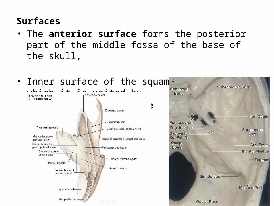
Surfaces• The anterior surface forms the posterior part of the middle
fossa of the base of the skull,
• Inner surface of the squamous portion, to which it is united by petrosquamous suture

It is marked by depressions for the convolutions of the brainPoints for examination:
An eminence (eminentia arcuata) at the centre which indicates the situation of the superior semicircular canal
Little lateral to this eminence, a depression indicating the position of the tympanic cavity (tegmen tympani)
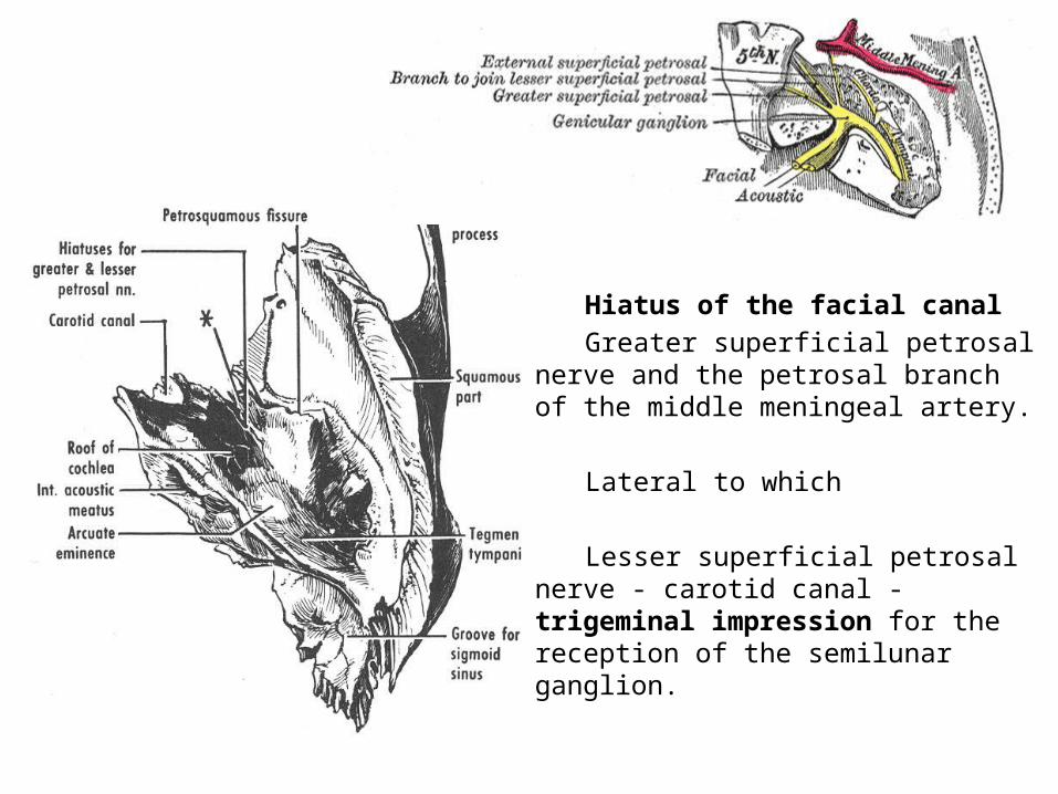
Hiatus of the facial canal Greater superficial petrosal nerve
and the petrosal branch of the middle meningeal artery.
Lateral to which
Lesser superficial petrosal nerve - carotid canal - trigeminal impression for the reception of the semilunar ganglion.
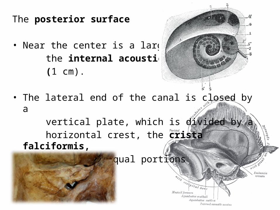
The posterior surface
• Near the center is a large orifice, the internal acoustic meatus (1 cm).
• The lateral end of the canal is closed by a vertical plate, which is divided by a horizontal crest, the crista falciformis, into two unequal portions
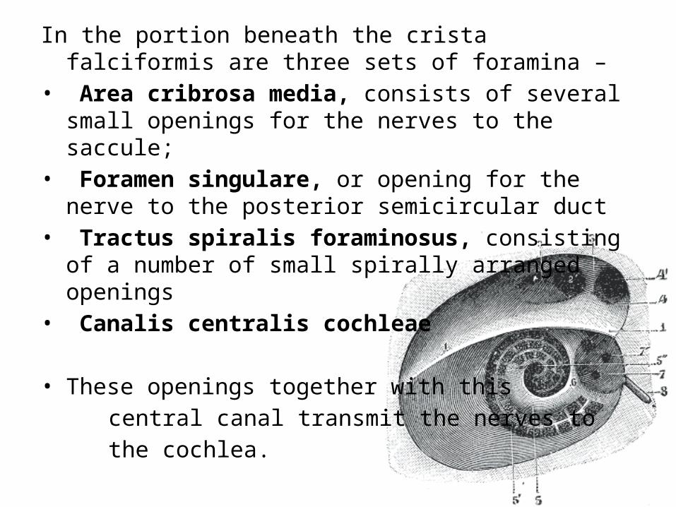
In the portion beneath the crista falciformis are three sets of foramina –
• Area cribrosa media, consists of several small openings for the nerves to the saccule;
• Foramen singulare, or opening for the nerve to the posterior semicircular duct
• Tractus spiralis foraminosus, consisting of a number of small spirally arranged openings
• Canalis centralis cochleae
• These openings together with this central canal transmit the nerves to the cochlea.
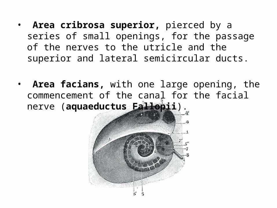
• Area cribrosa superior, pierced by a series of small openings, for the passage of the nerves to the utricle and the superior and lateral semicircular ducts.
• Area facians, with one large opening, the commencement of the canal for the facial nerve (aquaeductus Fallopii).
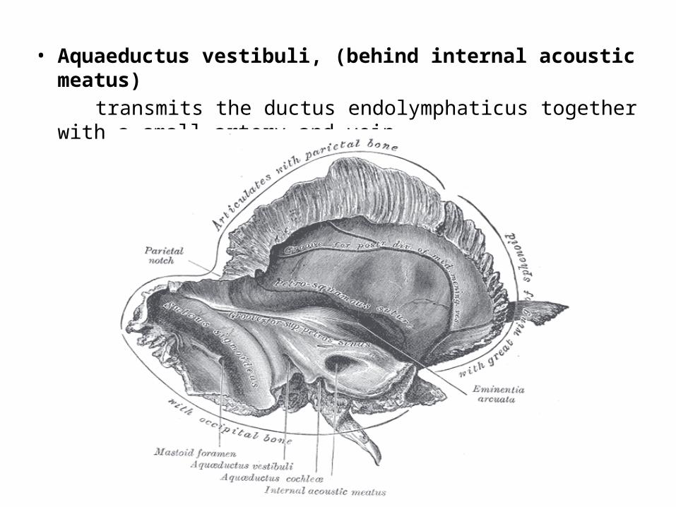
• Aquaeductus vestibuli, (behind internal acoustic meatus) transmits the ductus endolymphaticus together with a small
artery and vein.
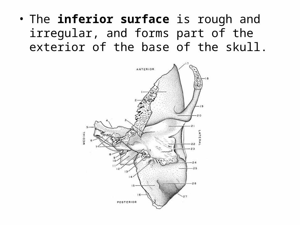
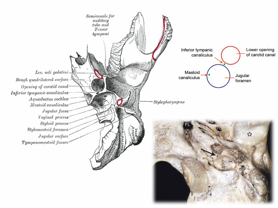
• The inferior surface is rough and irregular, and forms part of the exterior of the base of the skull.
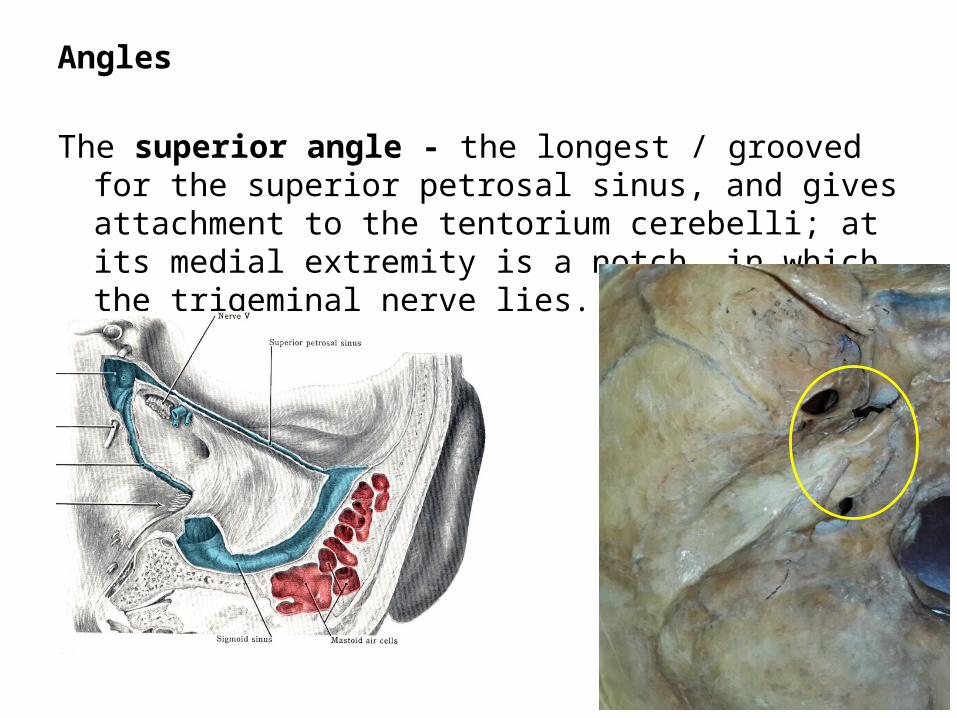
Angles
The superior angle - the longest / grooved for the superior petrosal sinus, and gives attachment to the tentorium cerebelli; at its medial extremity is a notch, in which the trigeminal nerve lies.
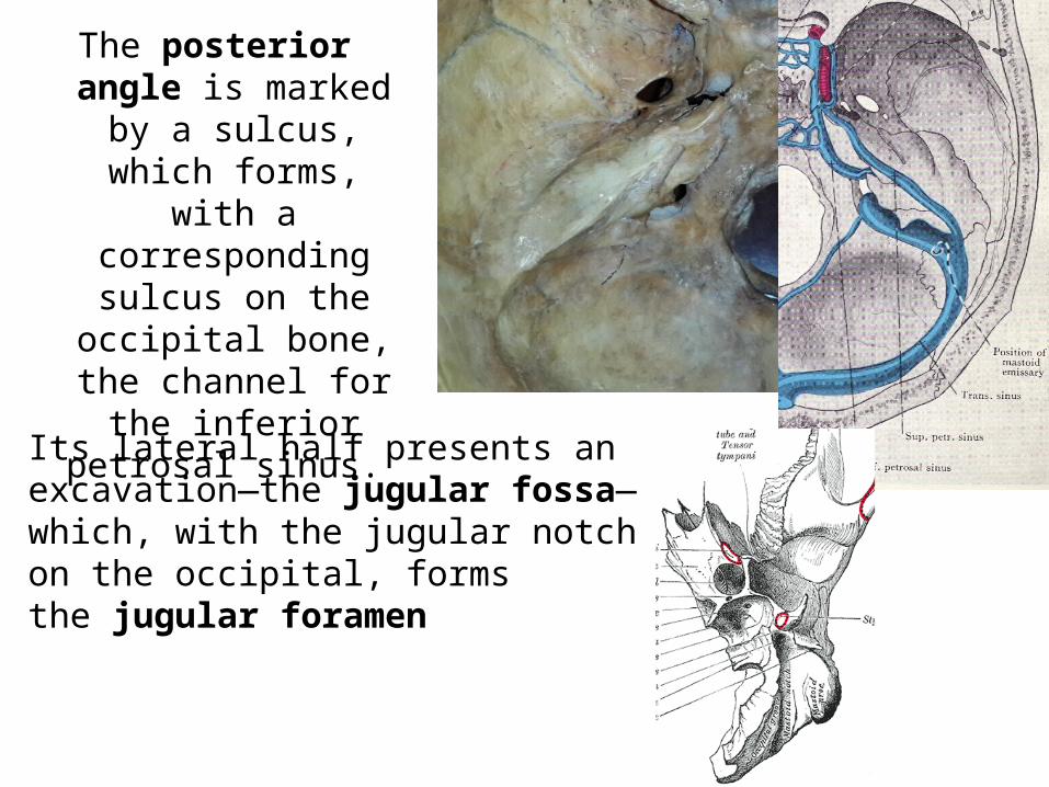
The posterior angle is marked by a sulcus, which forms, with a
corresponding sulcus on the occipital bone, the channel for the inferior
petrosal sinus.
Its lateral half presents an excavation—the jugular fossa—which, with the jugular notch on the occipital, forms the jugular foramen
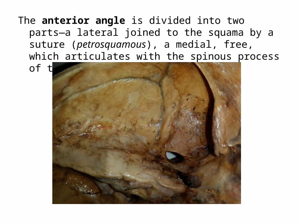
The anterior angle is divided into two parts—a lateral joined to the squama by a suture (petrosquamous), a medial, free, which articulates with the spinous process of the sphenoid.
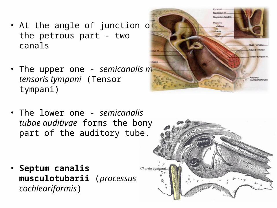
• At the angle of junction of the petrous part - two canals
• The upper one - semicanalis m. tensoris tympani (Tensor tympani)
• The lower one - semicanalis tubae auditivae forms the bony part of the auditory tube.
• Septum canalis musculotubarii (processus cochleariformis)
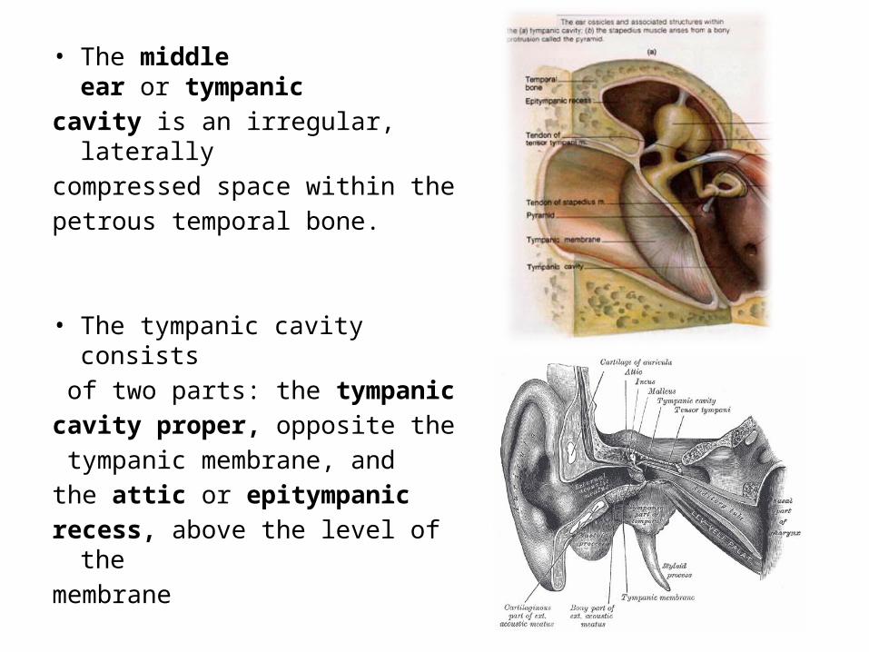
• The middle ear or tympanic cavity is an irregular, laterally compressed space within the petrous temporal bone.
• The tympanic cavity consists of two parts: the tympanic cavity proper, opposite the tympanic membrane, and the attic or epitympanic recess, above the level of the membrane
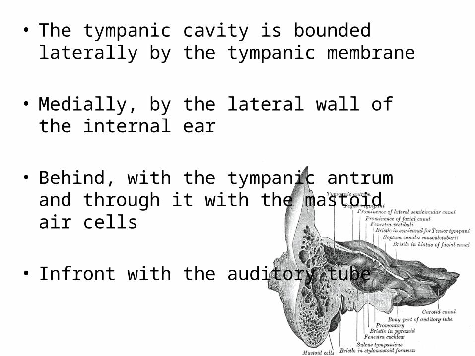
• The tympanic cavity is bounded laterally by the tympanic membrane
• Medially, by the lateral wall of the internal ear
• Behind, with the tympanic antrum and through it with the mastoid air cells
• Infront with the auditory tube
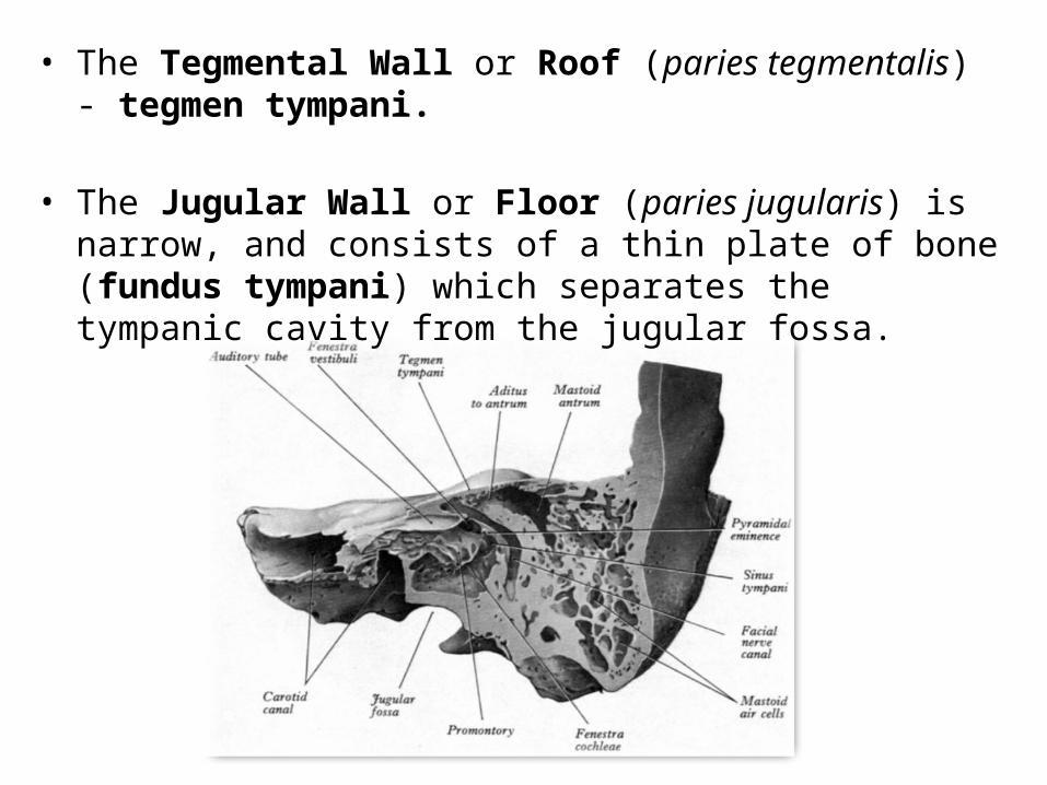
• The Tegmental Wall or Roof (paries tegmentalis) - tegmen tympani.
• The Jugular Wall or Floor (paries jugularis) is narrow, and consists of a thin plate of bone (fundus tympani) which separates the tympanic cavity from the jugular fossa.
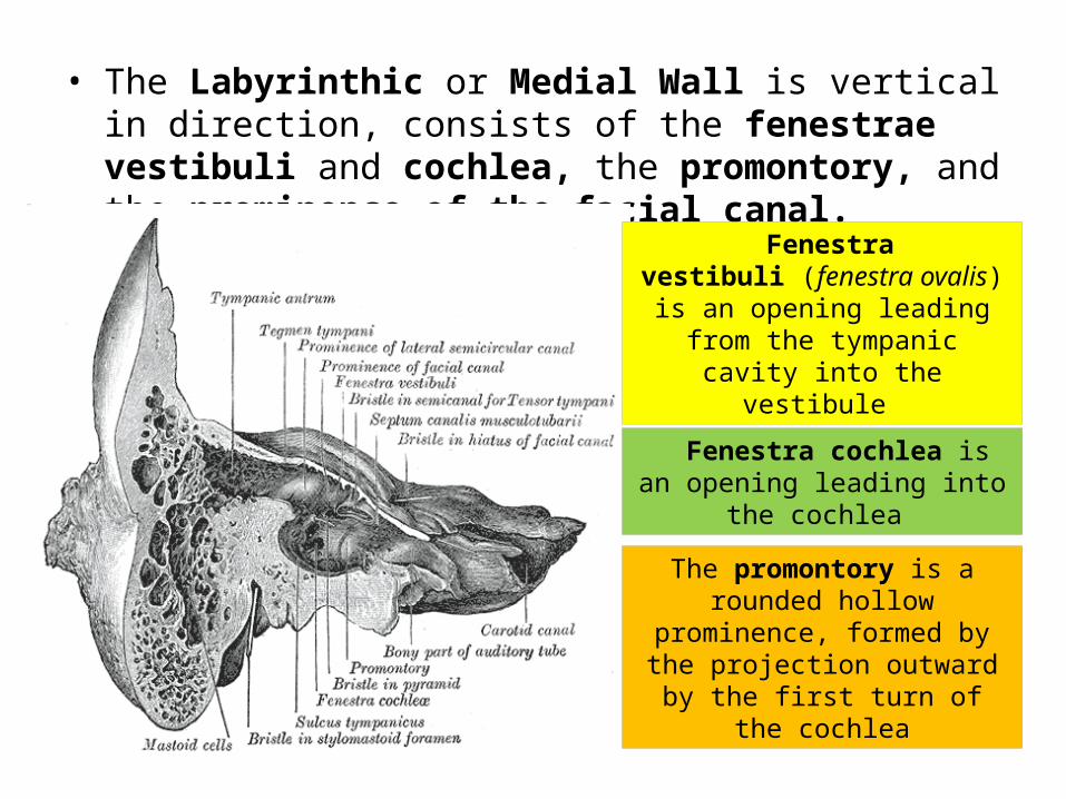
• The Labyrinthic or Medial Wall is vertical in direction, consists of the fenestrae vestibuli and cochlea, the promontory, and the prominence of the facial canal.
Fenestra vestibuli (fenestra ovalis) is an opening leading from the
tympanic cavity into the vestibule
Fenestra cochlea is an opening leading into the cochlea
The promontory is a rounded hollow prominence, formed by the
projection outward by the first turn of the cochlea
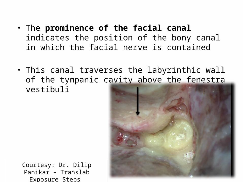
• The prominence of the facial canal indicates the position of the bony canal in which the facial nerve is contained
• This canal traverses the labyrinthic wall of the tympanic cavity above the fenestra vestibuli
Courtesy: Dr. Dilip Panikar – Translab Exposure Steps
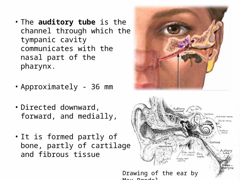
• The auditory tube is the channel through which the tympanic cavity communicates with the nasal part of the pharynx.
• Approximately - 36 mm
• Directed downward, forward, and medially,
• It is formed partly of bone, partly of cartilage and fibrous tissue
Drawing of the ear by Max Brodel
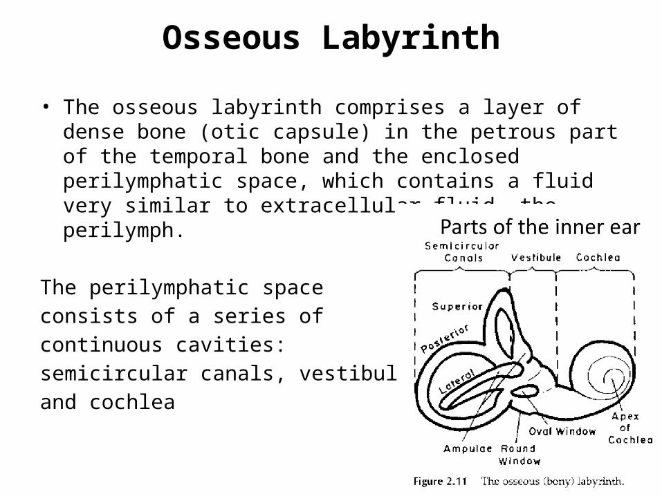
Osseous Labyrinth
• The osseous labyrinth comprises a layer of dense bone (otic capsule) in the petrous part of the temporal bone and the enclosed perilymphatic space, which contains a fluid very similar to extracellular fluid, the perilymph.
The perilymphatic space consists of a series of continuous cavities: semicircular canals, vestibule, and cochlea

Semicircular canals
The Superior, posterior, and lateral semicircular canals are at right
angles one to another
The semicircular ducts provide sensory input
for experiences of rotary movements.
Grey`s Anatomy
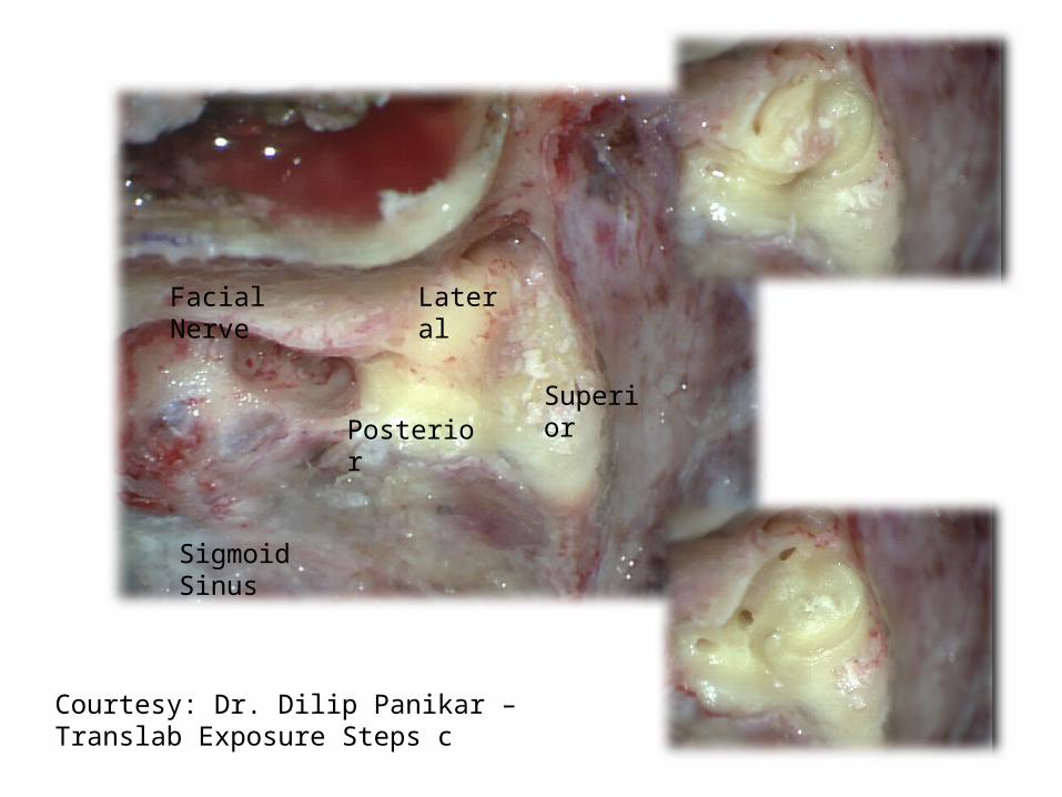
Lateral
PosteriorSuperior
Facial Nerve
Sigmoid Sinus
Courtesy: Dr. Dilip Panikar – Translab Exposure Steps c

Vestibule
• The vestibule is the central part of the osseous labyrinth, and is situated medial to the tympanic cavity, behind the cochlea, and in front of the semicircular canals.
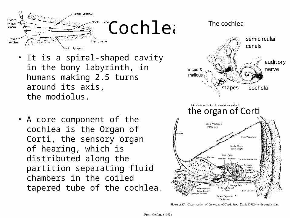
Cochlea
• It is a spiral-shaped cavity in the bony labyrinth, in humans making 2.5 turns around its axis, the modiolus.
• A core component of the cochlea is the Organ of Corti, the sensory organ of hearing, which is distributed along the partition separating fluid chambers in the coiled tapered tube of the cochlea.
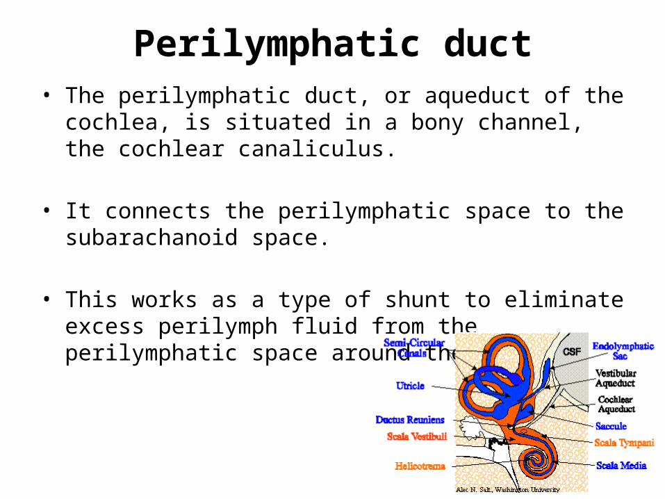
Perilymphatic duct• The perilymphatic duct, or aqueduct of the cochlea, is situated
in a bony channel, the cochlear canaliculus.
• It connects the perilymphatic space to the subarachanoid space.
• This works as a type of shunt to eliminate excess perilymph fluid from the perilymphatic space around the cochlea.

Tympanic Part (pars tympanica)
• The tympanic part is a curved plate of bone lying below the squama and in front of the mastoid process.
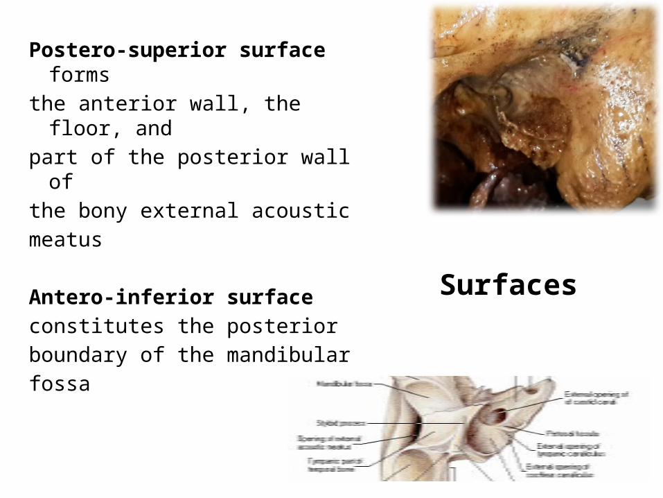
Surfaces
Postero-superior surface forms the anterior wall, the floor, and part of the posterior wall of the bony external acoustic meatus
Antero-inferior surface constitutes the posterior boundary of the mandibular fossa
Borders
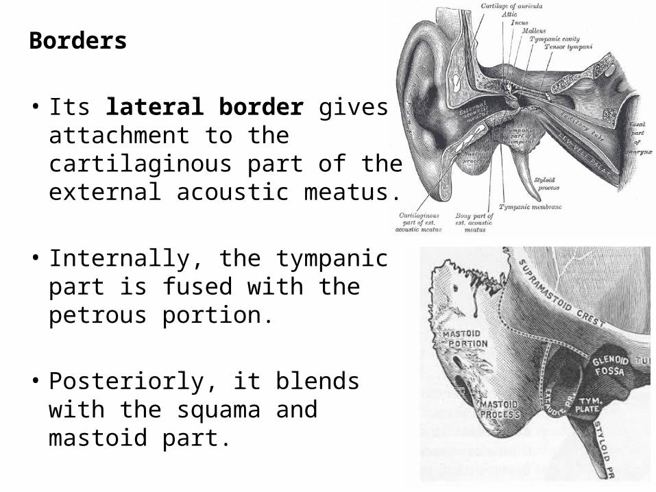
• Its lateral border gives attachment to the cartilaginous part of the external acoustic meatus.
• Internally, the tympanic part is fused with the petrous portion.
• Posteriorly, it blends with the squama and mastoid part.
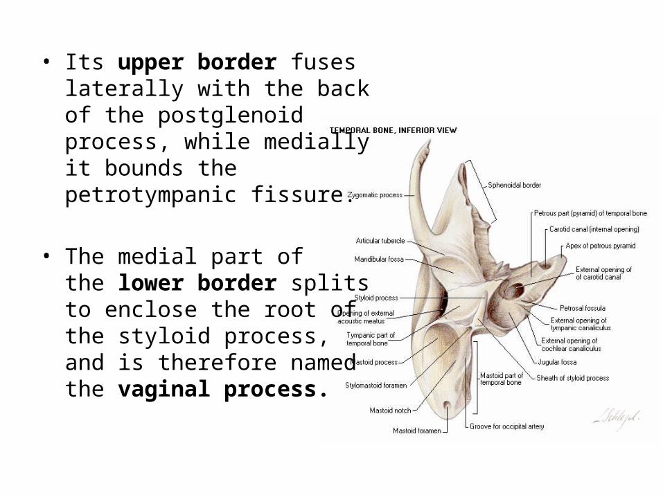
• Its upper border fuses laterally with the back of the postglenoid process, while medially it bounds the petrotympanic fissure.
• The medial part of the lower border splits to enclose the root of the styloid process, and is therefore named the vaginal process.
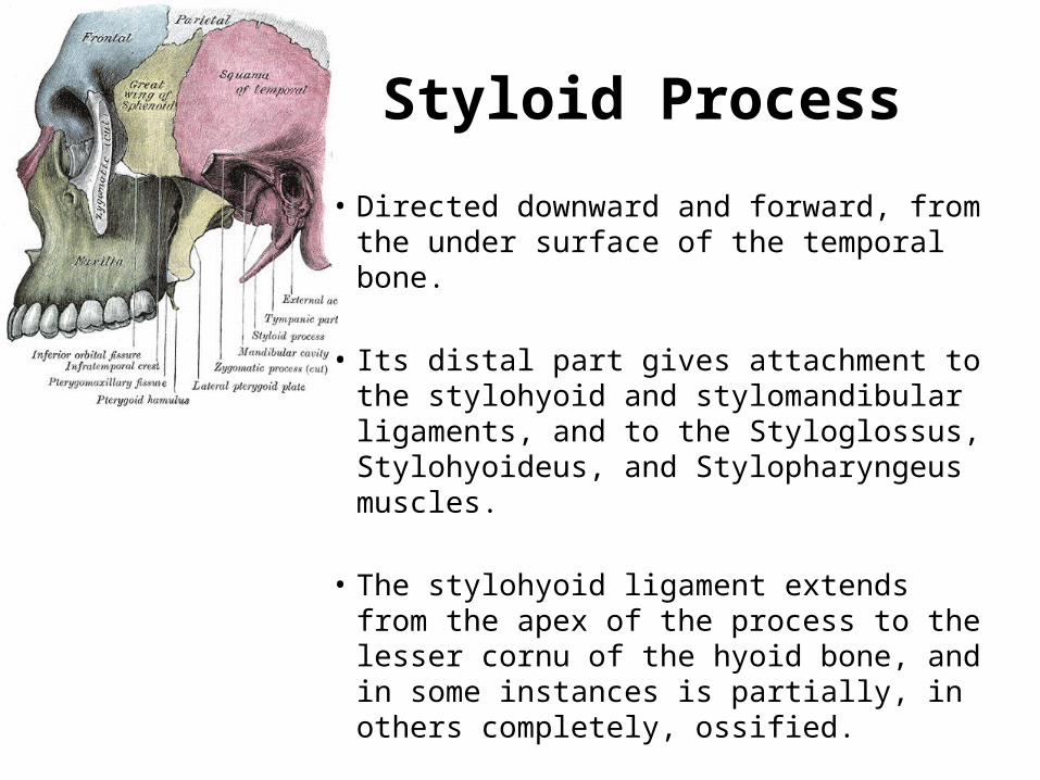
Styloid Process
• Directed downward and forward, from the under surface of the temporal bone.
• Its distal part gives attachment to the stylohyoid and stylomandibular ligaments, and to the Styloglossus, Stylohyoideus, and Stylopharyngeus muscles.
• The stylohyoid ligament extends from the apex of the process to the lesser cornu of the hyoid bone, and in some instances is partially, in others completely, ossified.
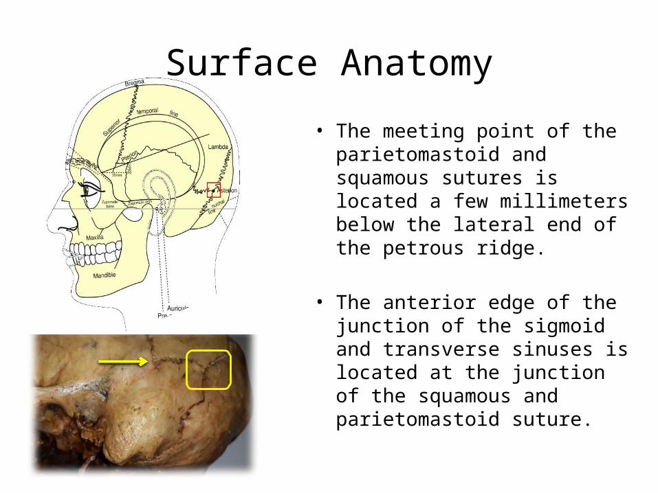
Surface Anatomy
• The meeting point of the parietomastoid and squamous sutures is located a few millimeters below the lateral end of the petrous ridge.
• The anterior edge of the junction of the sigmoid and transverse sinuses is located at the junction of the squamous and parietomastoid suture.
THANK YOU