Embed Size (px)
Citation preview
SICKLE CELL DISEASE &ITS COMPLICATIONS(COMPLETE INFO. PPT)
Dr. Armaan Singh
CASE A 20 years old male reported with history of
delayed puberty, decreased growth, severe joint pain, severe weakness and cough. He also have defective vision
Past history reveals episodes of jaundice, severe body aches and pains, and gall stones
In his childhood he used to have frequently fever swelling of the hands and feet and pain in the chest, abdomen, limbs, and joints and nosebleeds and frequent upper respiratory infections
feb
, 17
, 20
15
2
Dr. A
rma
an
Sin
gh
CASE
O/E; Decreased growth, delayed signs of puberty severely anemic, mildly jaundiced, ulcers on right leg, inflamed gums
Enlarged spleen. Temp 39.1°C, diaphoretic, and uncomfortable. HR of 90, BP 116/84 mm Hg, RR 26 O2 Sat 89% and improved to 94% with 6 L/min
via face mask. Family history: similar problem in one of his cousin
who died at the age of 30, who used to receive blood transfusions
feb
, 17
, 20
15
3
Dr. A
rma
an
Sin
gh
CASE
WBC of 17 500/μL 62% neutrophils 25% lymphocytes 9% monocytes 2% eosinophils 1% basophils 1% atypical lymphocytes.
Hb was 8 g/dL reticulocyte 25% platelet 206 000/μL.
feb
, 17
, 20
15
4
Dr. A
rma
an
Sin
gh
CASE
Which lab test you will advise? What is most probable cause of patients problem? What complications the patient may have? Design therapeutic objectives for this patient?
feb
, 17
, 20
15
5
Dr. A
rma
an
Sin
gh
feb
, 17
, 20
15
6
Dr. A
rma
an
Sin
gh
HEMOGLOBIN: INTRODUCTION Normal: alpha gene at Chr 16, beta at Chr.11
HbA: 2 Alfa + 2 beta97-98% Hb A2: 2 Alfa + 2 delta 2-3% Hb F; 2 alfa + 2 gamma >1% Hb S: Glutamic acid at 6 in beta chain replaced with Valine HbC:
……………………………………………………………………….Lysine Thalassemia:
Thalassemia describes a group of inherited disorders characterized by reduced or absent amounts of hemoglobin
Alfa: less alfa chain Chr.16 Beta: Chr.11: less beta, beta thalassemia minor
or no beta, all alfa chain beta thalassemia major :
feb
, 17
, 20
15
7
Dr. A
rma
an
Sin
gh
An autosomal recessive genetic disease of Hb synthesis Result of a single–amino acid substitution in the β-
globin chain of the Hb molecule, valine for glutamate at position 6
Sickle cell trait: Pt. with hetrozygous genotype Epidemiology in KSA:
“The prevalence of SCD in Saudi Arabia varies significantly in different parts of the country, with the highest prevalence is in the Eastern province, followed by the southwestern provinces. The reported prevalence for sickle-cell trait ranges from 2% to 27%, and up to 2.6% will have SCD in some areas”
Ann Saudi Med. 2011 May-Jun; 31(3): 289–293.
feb
, 17
, 20
15
8
Dr. A
rma
an
Sin
gh
SICKLE CELL DISEASE
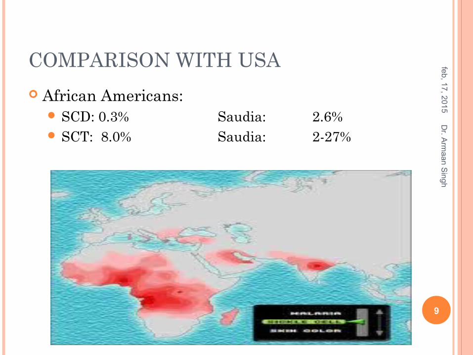
COMPARISON WITH USA
African Americans: SCD: 0.3% Saudia: 2.6% SCT: 8.0% Saudia: 2-27%
feb
, 17
, 20
15
9
Dr. A
rma
an
Sin
gh

feb
, 17
, 20
15
10
Dr. A
rma
an
Sin
gh
feb
, 17
, 20
15
11
Dr. A
rma
an
Sin
gh
feb
, 17
, 20
15
12
Dr. A
rma
an
Sin
gh
feb
, 17
, 20
15
13
Dr. A
rma
an
Sin
gh
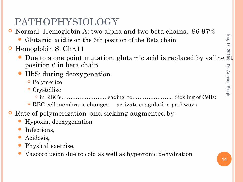
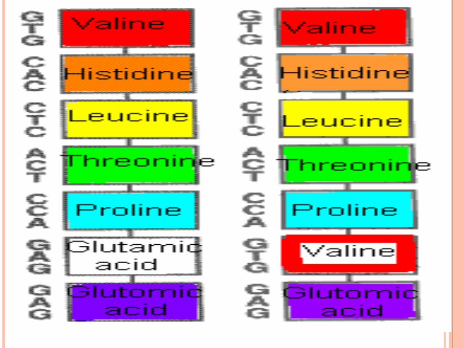
PATHOPHYSIOLOGY Normal Hemoglobin A: two alpha and two beta chains, 96-97%
Glutamic acid is on the 6th position of the Beta chain Hemoglobin S: Chr.11
Due to a one point mutation, glutamic acid is replaced by valine at position 6 in beta chain
HbS: during deoxygenation Polymerize Crystellize
in RBC’s…………………….leading to………………….. Sickling of Cells: RBC cell membrane changes: activate coagulation pathways
Rate of polymerization and sickling augmented by: Hypoxia, deoxygenation Infections, Acidosis, Physical exercise, Vasoocclusion due to cold as well as hypertonic dehydration
feb
, 17
, 20
15
14
Dr. A
rma
an
Sin
gh
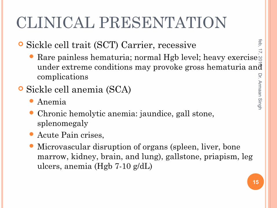
CLINICAL PRESENTATION Sickle cell trait (SCT) Carrier, recessive
Rare painless hematuria; normal Hgb level; heavy exercise under extreme conditions may provoke gross hematuria and complications
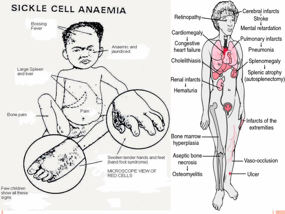
Sickle cell anemia (SCA) Anemia Chronic hemolytic anemia: jaundice, gall stone,
splenomegaly Acute Pain crises, Microvascular disruption of organs (spleen, liver, bone
marrow, kidney, brain, and lung), gallstone, priapism, leg ulcers, anemia (Hgb 7-10 g/dL)
feb
, 17
, 20
15
15
Dr. A
rma
an
Sin
gh
CLINICAL PRESENTATION Sickle cell hemoglobin C:
Painless hematuria Aseptic necrosis of bone: less common Vaso-occlusive crises less common, occur late in life Pregnancy-related problems; mild anemia (Hb 10–12 g/dL)
Sickle cell β-thalassemia Rare crises; milder severity than sickle cell disease because of
production of HbA; Hb 10–14 g/dL with micro-cytosis
Sickle cell Alfa-thalassemia or β0 Thalassemia No HbA production; severity similar to sickle cell anemia; Hb
7–10 g/dL with microcytosis
feb
, 17
, 20
15
16
Dr. A
rma
an
Sin
gh
DIAGNOSIS
Laboratory findings RBC’s: 5-50 % sickled Low hemoglobin; 7-10%; HbA; 0%; HbS 85-98% Increased reticulocytes: 10-25%, platelet, and
leukocyte counts; and sickle forms on the peripheral smear
Routine neonatal screening programs: DNA from fetal cell for mutation
feb
, 17
, 20
15
17
Dr. A
rma
an
Sin
gh
GOALS OF THERAPY
To reduce Hospitalizations, Complications, Mortality
feb
, 17
, 20
15
18
Dr. A
rma
an
Sin
gh
TREATMENT
GENERAL PRINCIPLES No Treatment for the primary disease Lifelong multidisciplinary care general measures, preventive strategies, treatment of complications and acute crises.
Routine immunizations plus influenza, meningococcal, and pneumococcal vaccinations.
Prophylactic penicillin for children with sickle cell disease until they are 5 years old. Penicillin V potassium, 125 mg orallytwice daily until 3 years of age
and then 250 mg twice daily, Benzathine penicillin, 600,000 units intramuscularly every 4 weeks.
Folic acid, 1 mg daily, is recommended in adult patients, pregnant women, and patients of all ages with chronic hemolysis.
feb
, 17
, 20
15
19
Dr. A
rma
an
Sin
gh
FETAL HEMOGLOBIN STIMULATORS AND OTHER STRATEGIES Hydroxyurea, a chemotherapeutic agent
Stimulate HbF by stimulating erythropoiesis In patients with frequent painful episodes, severe symptomatic anemia, acute
chest syndrome, or other severe vasoocclusive complications.
Butyrate and 5-aza-2-deoxycytidine. Chronic transfusion every 3 to 4 weeks The optimal duration is
unknown to prevent stroke and stroke recurrence in children. Maintain HbS of less than 30% of total hemoglobin.. Risks include, hyperviscosity, viral transmission (requiring hepatitis A and B
vaccination), volume and iron overload, and transfusion reactions.
Allogeneic hematopoietic stem cell transplantation The only therapy that is curative. Best candidates are
younger than 16 years of age, With severe complications, Have HLA-matched donors. Risks: mortality, graft rejection, and secondary malignancies
feb
, 17
, 20
15
20
Dr. A
rma
an
Sin
gh
STEM CELLS IN THE TREATMENT OF SCD
Skin stem cells cure mice of sickle cell anemia
Success is proof that technique has potential to cure diseasehttp://www.msnbc.msn.com/id/22136029/
feb
, 17
, 20
15
21
Dr. A
rma
an
Sin
gh
COMPLICATIONS Acute Chest Syndrome Septicemia Stroke or CVA Acute splenic sequestration crisis (ASSC) Aplastic Crisis VasoOcclusive pain: Sickle cell crisis
Severe pain is an emergency called acute sickle cell crisis
Osteomyelitis
feb
, 17
, 20
15
22
Dr. A
rma
an
Sin
gh
SICKLE CELL CRISIS
Rapid diagnosis and treatment are necessary to minimize morbidity and mortality.
feb
, 17
, 20
15
23
Dr. A
rma
an
Sin
gh
CASE 1
A 16-year-old boy with a history of SCD presented to the ED with a 3-day history of fever, cough, and SOB.
Five days prior, he had been evaluated and treated for severe pain in his legs and arms.
He complained of persistent and worsening pain in both his lower extremities and pain in his chest, in spite of oral narcotic therapy.
feb
, 17
, 20
15
24
Dr. A
rma
an
Sin
gh
His medical history included multiple, vasoocclusive, painful crises, including an episode of priapism, and he had received multiple blood transfusions over his lifetime.
feb
, 17
, 20
15
25
Dr. A
rma
an
Sin
gh
Case 1
CASE -1
On examination Temp 39.1°C, diaphoretic, and uncomfortable. HR of 80, BP 116/84 mm Hg, RR 26 O2 Sat 89% and improved to 94% with 6 L/min
via face mask. Conjunctivae were icteric Mucous membranes were moist
feb
, 17
, 20
15
26
Dr. A
rma
an
Sin
gh
Cardiovascular II/VI systolic ejection murmur.
labored respiration with suprasternal and intercostal retractions.
decreased breath sounds in the right midzone and lower zone, and scattered crepitations on the right side.
no lower extremity edema Abdominal examination Normal CNS Normal
feb
, 17
, 20
15
27
Dr. A
rma
an
Sin
gh
Case 1
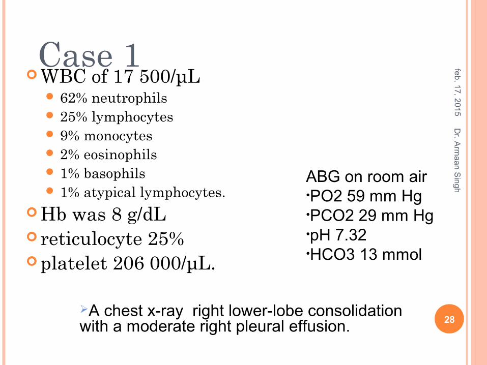
WBC of 17 500/μL 62% neutrophils 25% lymphocytes 9% monocytes 2% eosinophils 1% basophils 1% atypical lymphocytes.
Hb was 8 g/dL reticulocyte 25% platelet 206 000/μL.
feb
, 17
, 20
15
28
Dr. A
rma
an
Sin
gh
ABG on room air •PO2 59 mm Hg•PCO2 29 mm Hg•pH 7.32•HCO3 13 mmol
A chest x-ray right lower-lobe consolidation with a moderate right pleural effusion.
Case 1
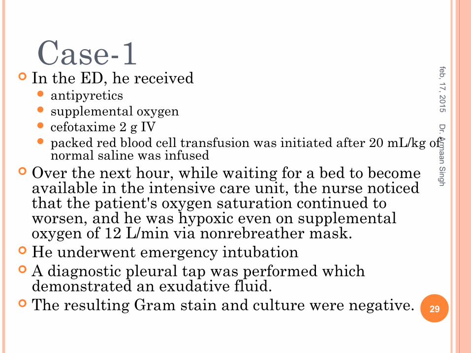
In the ED, he received antipyretics supplemental oxygen cefotaxime 2 g IV packed red blood cell transfusion was initiated after 20 mL/kg of
normal saline was infused Over the next hour, while waiting for a bed to become
available in the intensive care unit, the nurse noticed that the patient's oxygen saturation continued to worsen, and he was hypoxic even on supplemental oxygen of 12 L/min via nonrebreather mask.
He underwent emergency intubation A diagnostic pleural tap was performed which
demonstrated an exudative fluid. The resulting Gram stain and culture were negative.
feb
, 17
, 20
15
29
Dr. A
rma
an
Sin
gh
Case-1

CASE 1
What is it
feb
, 17
, 20
15
30
Dr. A
rma
an
Sin
gh
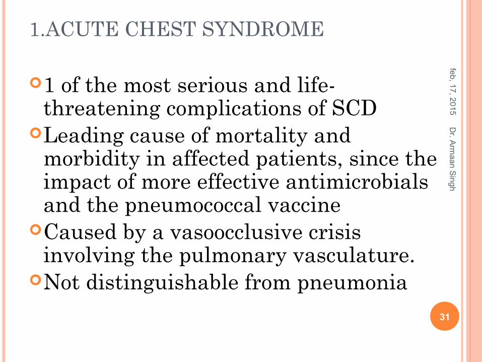
1.ACUTE CHEST SYNDROME
1 of the most serious and life-threatening complications of SCD
Leading cause of mortality and morbidity in affected patients, since the impact of more effective antimicrobials and the pneumococcal vaccine
Caused by a vasoocclusive crisis involving the pulmonary vasculature.
Not distinguishable from pneumonia
feb
, 17
, 20
15
31
Dr. A
rma
an
Sin
gh
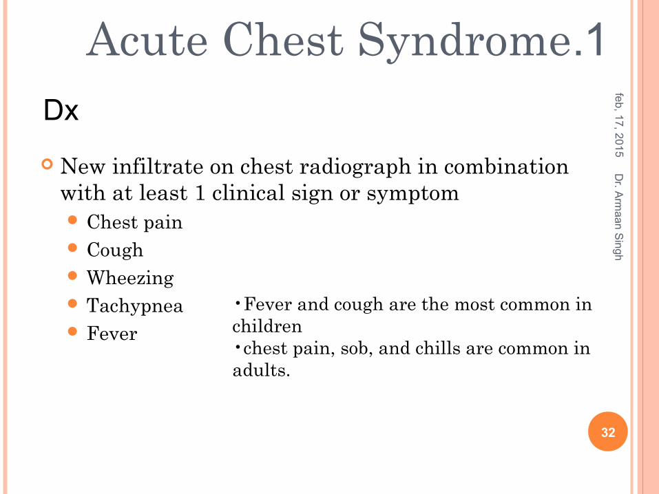
New infiltrate on chest radiograph in combination with at least 1 clinical sign or symptom Chest pain Cough Wheezing Tachypnea Fever
feb
, 17
, 20
15
32
Dr. A
rma
an
Sin
gh
•Fever and cough are the most common in children •chest pain, sob, and chills are common in adults.
1.Acute Chest Syndrome Dx
Common causesPulmonary infection: Mycoplasma pneumoniae more commonly associated with acute chest syndrome
ThromboemboliFat emboliRib infarction
Infection and fat emboli were the most common identifiable causes.
Vichinsky EP, Neumayr LD, Earles AN, et al. Causesand outcomes of the acute chest syndrome in sickle cell disease. National Acute Chest Syndrome Study Group [published erratum appears in N Engl J Med 2000; 343:824]. N Engl J Med 2000;342:1855–65.
1.Acute Chest Syndrome
Possible causesIatrogenic: excessive hydration or narcotic use
feb
, 17
, 20
15
Dr. A
rma
an
Sin
gh
33
Therapeutic Modalities Supportive measures
Oxygen for hypoxia Appropriate hydration Appropriate pain control
Antibiotics: third-generation cephalosporin + macrolides
Transfusion therapy: Reports of dramatic improvement in clinical condition
after initiation of transfusion Simple transfusion Exchange transfusion
Experimental therapy Nitric oxide Corticosteroids
feb
, 17
, 20
15
34
Dr. A
rma
an
Sin
gh1.Acute Chest Syndrome
Bodo I, Khoury H, Blinder M. Rapid resolution of the acute chest syndrome of sickle cell disease after automated red cell exchange. Blood 1997;90 Suppl 1:23b

2.SEPTICEMIA SCD pts have impaired immunologic function that is caused
by splenic dysfunction. Impairment of splenic function can occur in infants as young
as 3 months. High risk for encapsulated organisms such as S pneumoniae
and H influenzae. Recommended antibiotic
Third-generation cephalosporin; ceftriaxone, or cefotaxime Vancomycin should be added to protect against penicillin-
resistant strains of S pneumoniae if suspected until culture results become available
All SCD patients with fever must be managed with extreme caution because of the risk of overwhelming bacteremia which can rapidly lead to septic shock
feb
, 17
, 20
15
35
Dr. A
rma
an
Sin
gh
3.STROKE OR CVA Major complication of SCD Is a leading cause of death in both and disability
children and adults The most common is blockage of the intracranial
internal carotid and middle cerebral arteries. Patients with stroke usually present with obvious
signs such as acute hemiparesis, aphasia or dysphasia, seizures, severe headaches, cranial nerve palsy, altered mental status, or coma.
The most common tends to be hemiparesis. Can be very subtle, such as a slight limp
feb
, 17
, 20
15
36
Dr. A
rma
an
Sin
gh
TREATMENT: STROKE OR CVA Initial therapy is exchange transfusion in an ICU setting to reduce Hb
S to less than 30% of total Hb. After acute clearance of symptoms should be started
on a long-term transfusion therapy. If not on a long-term transfusion program have an 80%
chance of recurrent stroke within 3 years of the initial event
Long-term transfusion involves regularly scheduled blood transfusions aimed at reducing the percentage of Hb S and not at normalizing the Hb level.
feb
, 17
, 20
15
37
Dr. A
rma
an
Sin
gh
CASE 2A 44-year-old diabetic presented to the ED complaining of nonexertional dyspnea and severe back pain for 12 hours before presentation. The patient reported malaise, fatigue, weakness that started 3 days before, chronic blurred vision, insomnia, and anxiety. The remainder of the review of systems was unremarkable.
feb
, 17
, 20
15
Dr. A
rma
an
Sin
gh
38
O/E
HR 101 bpm RR 31/min Temp 37C BP 148/62 mm Hg
o2 sat 99%. The patient was awake, alert, and oriented He was motionless to avoid back pain.
feb
, 17
, 20
15
39
Dr. A
rma
an
Sin
gh
Case 2
Normal S1 and S2 Chest Normal Strength was 4/5 in all 4 extremities. Deep tendon reflexes were normoactive. Normal flexor plantar response was obtained, and no
meningismus
feb
, 17
, 20
15
40
Dr. A
rma
an
Sin
gh
Case 2O/E
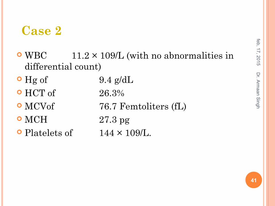
WBC 11.2 × 10⁹/L (with no abnormalities in differential count)
Hg of 9.4 g/dL HCT of 26.3% MCVof 76.7 Femtoliters (fL) MCH 27.3 pg Platelets of 144 × 109/L.
feb
, 17
, 20
15
41
Dr. A
rma
an
Sin
gh
Case 2
Total bilirubin level of 2.3 mg/dl
Direct bilirubin level of 0.8 mg/dL
ESR 54 mm/h C-reactive protein level of 2.3 mg/dL. ECG Normal MRI of the lumbar spine was Normal
feb, 17, 201 5
42
Dr. A
rmaa n
Sin
gh
• Blood glucose 267 mg/dL• AST 79 U/L• ALT 30 of U/L• ALK Ph 475 U/L
Case 2
Despite aggressive narcotic treatment of back pain, the pain continued to increase
CT abdomen: an enlarged spleen1 hour later hypotension of 90/50 mm Hg. The new CT scan of the abdomen revealed an
increasing splenomegaly compared with the previous one
feb
, 17
, 20
15
43
Dr. A
rma
an
Sin
gh
Case 2
Despite transfusion therapy, the patient's Hb progressively dropped to a level of less than 4 mg/dL over the course of 3 hours, with thrombocytopenia (<50 × 109/L).
immediately transferred to an ICU. altered mental status. Airway protection with intubation and mechanical
ventilation were initiated. As the patient was rapidly deteriorating, an emergent
splenectomy was performed The patient recovered every organ function and, 6
months later, has resumed his normal activities
feb
, 17
, 20
15
44
Dr. A
rma
an
Sin
gh
Case 2
4.ACUTE SPLENIC SEQUESTRATION CRISIS (ASSC)
Clinical Presentation:
Sudden impounding of red blood cells by the spleen
Characterized by the rapid fall in hemoglobin concentration, rise in reticulocyte count, and splenomegaly
Requires prompt recognition and treatment.
In the adult patient, ASSC is extremely rare.
Hypotension caused by large volumes of blood (mainly sickled cells) entrapped in the spleen.
Hb levels may fall acutely more than 2 g/dL less than the patient's normal value, causing circulatory compromise
Treatment: Prompt diagnosis and therapy with RBC transfusions Surgical splenectomy may be indicated in certain patients to prevent
recurrences
feb
, 17
, 20
15
45
Dr. A
rma
an
Sin
gh
5.APLASTIC CRISIS Temporary cessation of red cell production with a
corresponding decrease in the reticulocyte count. Approximately 80%, are thought to be caused by human
parvovirus B19 infection Diagnosis is made by comparing baseline blood and
reticulocyte counts to those obtained during the acute illness.
Sign Symptoms: , tachypnea, tachycardia, or hypoxia Treatment:
Simple blood transfusion to raise serum Hb back to the patient's baseline and to prevent heart failure secondary to severe anemia.
Parvovirus B19 is contagious, affected persons should be isolated from pregnant women, who are at risk for miscarriage with infection, and from immuno-compromised patients and those with chronic illness
feb
, 17
, 20
15
46
Dr. A
rma
an
Sin
gh

6.OSTEOMYELITIS
Most commonly caused by Salmonella species or Staphylococcus aureus
Bone pain or joint pain with localized swelling and decreased range of motion, along with fever, should alert the physician to the possibility of osteomyelitis.
Increased white blood cell count and elevated ESR
Broad-spectrum antibiotic:Ceftriaxone:
feb
, 17
, 20
15
47
Dr. A
rma
an
Sin
gh
7.PRIAPISM Painful prolonged erection of the penis Caused by sickling of the red blood cells producing venous
stasis in the erectile tissue of the penis. The resulting stasis causes ischemia, hypoxia, and pain. Treatment:
Initial treatment involves intravenous hydration and analgesia. Antianxiety agents Vasoconstrictors to force blood out of corpus cavernosum:
Phenyl ephedrine Epinephrine
Vasodilators: to relax smooth muscles: Terbutaline Hydrallazine
Episodes refractory to this initial management include direct irrigation of the corporeal bodies of the penis
feb
, 17
, 20
15
48
Dr. A
rma
an
Sin
gh
VASO-OCCLUSIVE PAIN CRISIS
feb
, 17
, 20
15
Dr. A
rma
an
Sin
gh
49
8.VASO-OCCLUSIVE PAIN CRISES: SUMMARY Most common symptoms of SCD Severe pain Caused by sickle-shaped red blood cells trapped in small blood
vessels causing localized ischemia. Triggered by Dehydration, fever, cold exposure, and emotional stress Therapy
Intravenous/Oral hydration Pain management It is useful to assess pain in a standard manner using pain
measurement scales ……………..See next Causal Treatment: (treatment of the cause)
Poloxamer 188 (Flocor) a surfactant returns RBCs to a non adhesive state and blocks RBC aggregation to enhance blood flow in ischemic areas
feb
, 17
, 20
15
50
Dr. A
rma
an
Sin
gh
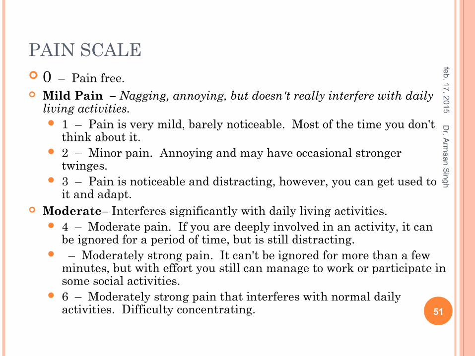
PAIN SCALE 0 – Pain free. Mild Pain – Nagging, annoying, but doesn't really interfere with daily
living activities. 1 – Pain is very mild, barely noticeable. Most of the time you don't
think about it. 2 – Minor pain. Annoying and may have occasional stronger
twinges. 3 – Pain is noticeable and distracting, however, you can get used to
it and adapt. Moderate– Interferes significantly with daily living activities.
4 – Moderate pain. If you are deeply involved in an activity, it can be ignored for a period of time, but is still distracting.
– Moderately strong pain. It can't be ignored for more than a few minutes, but with effort you still can manage to work or participate in some social activities.
6 – Moderately strong pain that interferes with normal daily activities. Difficulty concentrating.
feb
, 17
, 20
15
51
Dr. A
rma
an
Sin
gh
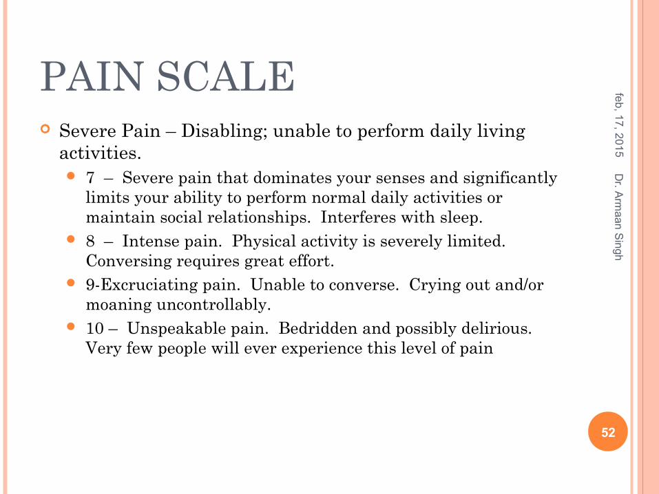
PAIN SCALE Severe Pain – Disabling; unable to perform daily living
activities. 7 – Severe pain that dominates your senses and significantly
limits your ability to perform normal daily activities or maintain social relationships. Interferes with sleep.
8 – Intense pain. Physical activity is severely limited. Conversing requires great effort.
9-Excruciating pain. Unable to converse. Crying out and/or moaning uncontrollably.
10 – Unspeakable pain. Bedridden and possibly delirious. Very few people will ever experience this level of pain
feb
, 17
, 20
15
52
Dr. A
rma
an
Sin
gh
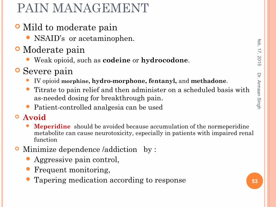
PAIN MANAGEMENT Mild to moderate pain
NSAID’s or acetaminophen. Moderate pain
Weak opioid, such as codeine or hydrocodone. Severe pain
IV opioid morphine, hydro-morphone, fentanyl, and methadone. Titrate to pain relief and then administer on a scheduled basis with
as-needed dosing for breakthrough pain. Patient-controlled analgesia can be used
Avoid Meperidine should be avoided because accumulation of the normeperidine
metabolite can cause neurotoxicity, especially in patients with impaired renal function
Minimize dependence /addiction by : Aggressive pain control, Frequent monitoring, Tapering medication according to response
feb
, 17
, 20
15
53
Dr. A
rma
an
Sin
gh
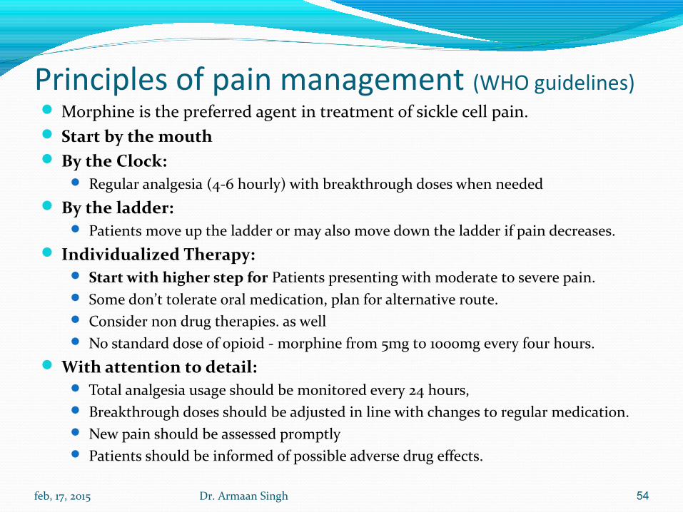
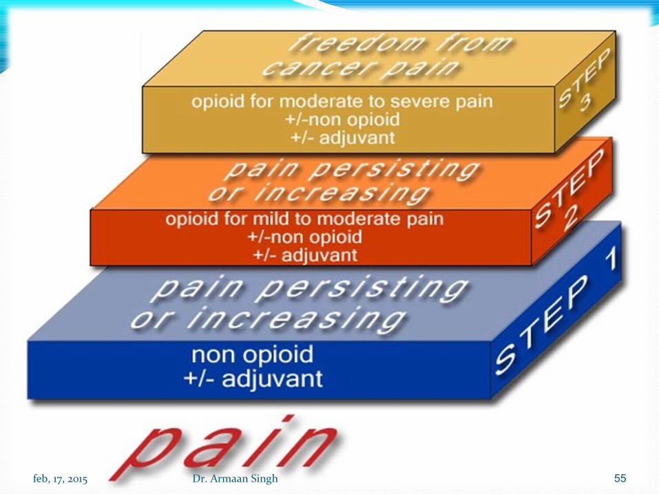
Principles of pain management (WHO guidelines) Morphine is the preferred agent in treatment of sickle cell pain. Start by the mouth By the Clock:
Regular analgesia (4-6 hourly) with breakthrough doses when needed By the ladder:
Patients move up the ladder or may also move down the ladder if pain decreases. Individualized Therapy:
Start with higher step for Patients presenting with moderate to severe pain. Some don’t tolerate oral medication, plan for alternative route. Consider non drug therapies. as well No standard dose of opioid - morphine from 5mg to 1000mg every four hours.
With attention to detail: Total analgesia usage should be monitored every 24 hours, Breakthrough doses should be adjusted in line with changes to regular medication. New pain should be assessed promptly Patients should be informed of possible adverse drug effects.
feb, 17, 2015 Dr. Armaan Singh 54
feb, 17, 2015 Dr. Armaan Singh 55
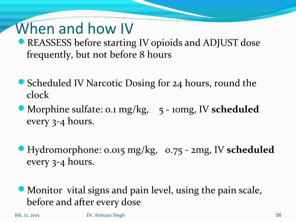
When and how IVREASSESS before starting IV opioids and ADJUST dose
frequently, but not before 8 hours
Scheduled IV Narcotic Dosing for 24 hours, round the clock
Morphine sulfate: 0.1 mg/kg, 5 - 10mg, IV scheduled every 3-4 hours.
Hydromorphone: 0.015 mg/kg, 0.75 - 2mg, IV scheduled every 3-4 hours.
Monitor vital signs and pain level, using the pain scale, before and after every dose
feb, 17, 2015 Dr. Armaan Singh 56
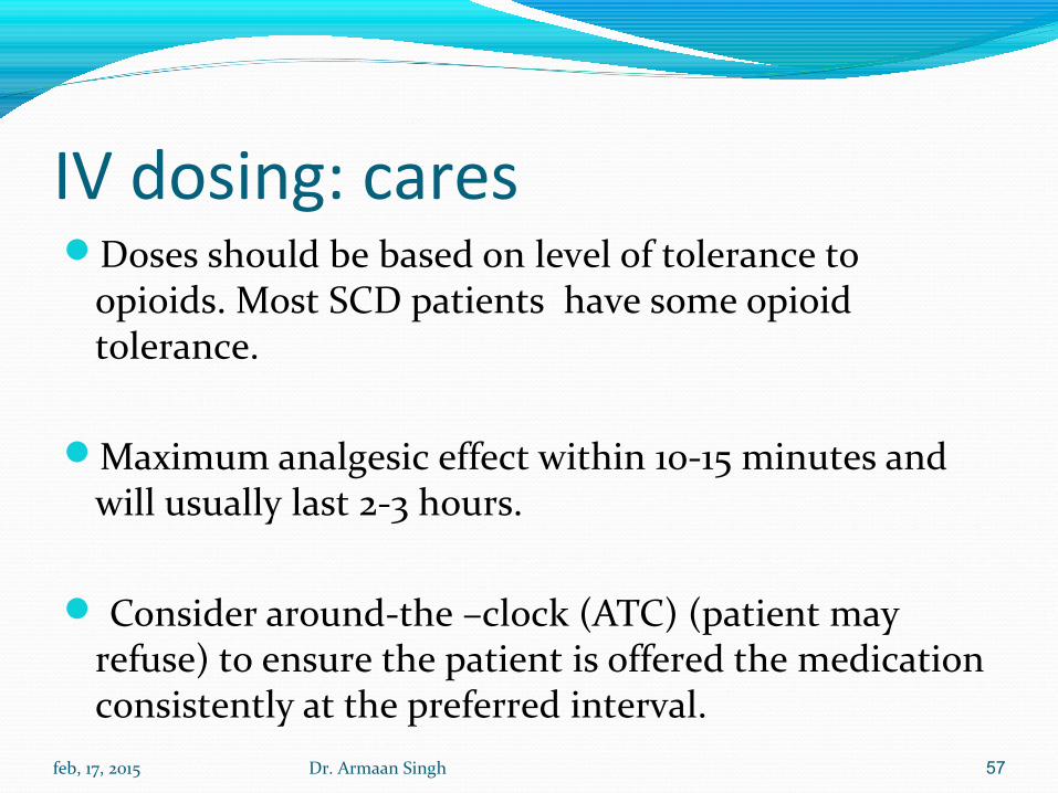
IV dosing: caresDoses should be based on level of tolerance to
opioids. Most SCD patients have some opioid tolerance.
Maximum analgesic effect within 10-15 minutes and will usually last 2-3 hours.
Consider around-the –clock (ATC) (patient may refuse) to ensure the patient is offered the medication consistently at the preferred interval.
feb, 17, 2015 Dr. Armaan Singh 57
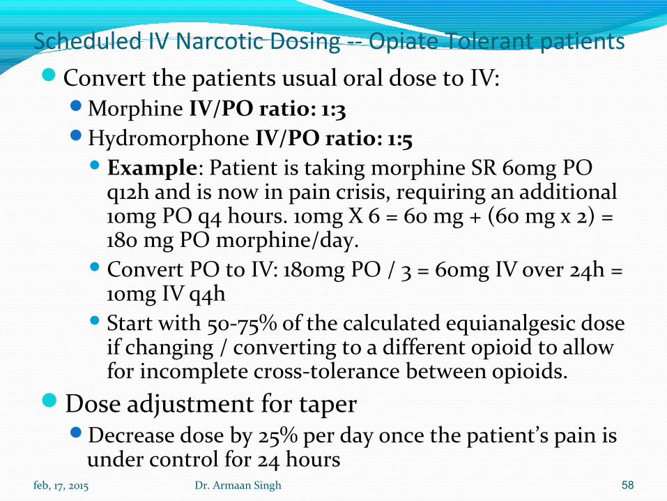
Scheduled IV Narcotic Dosing -- Opiate Tolerant patients Convert the patients usual oral dose to IV:
Morphine IV/PO ratio: 1:3 Hydromorphone IV/PO ratio: 1:5
Example: Patient is taking morphine SR 60mg PO q12h and is now in pain crisis, requiring an additional 10mg PO q4 hours. 10mg X 6 = 60 mg + (60 mg x 2) = 180 mg PO morphine/day.
Convert PO to IV: 180mg PO / 3 = 60mg IV over 24h = 10mg IV q4h
Start with 50-75% of the calculated equianalgesic dose if changing / converting to a different opioid to allow for incomplete cross-tolerance between opioids.
Dose adjustment for taper Decrease dose by 25% per day once the patient’s pain is
under control for 24 hours feb, 17, 2015 Dr. Armaan Singh 58
Monitoring the patientChest X-ray: Order for any patient with cardiopulmonary
complaints, hypoxia, know chronic lung disease, fever, tachycardia, or tachypnea.
Complete blood count q24 hours Comprehensive metabolic panel, magnesium,
phosphorous q48 hours Keep magnesium level > 2 mg/dL:
Magnesium < 1.8 mg/dL, replace with IV magnesiumMay need to follow with daily oral supplementation Magnesium > 1.8 mg/dL, replace with oral product
Lactic dehydrogenase (LDH) q72 hours
feb, 17, 2015 Dr. Armaan Singh 59
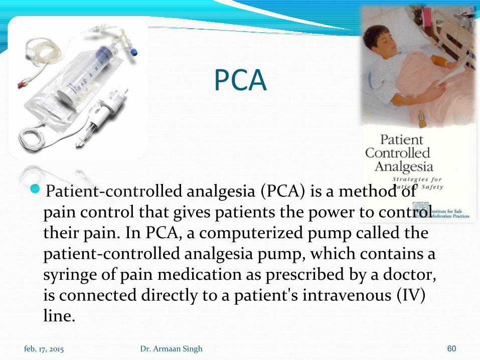
PCA
Patient-controlled analgesia (PCA) is a method of pain control that gives patients the power to control their pain. In PCA, a computerized pump called the patient-controlled analgesia pump, which contains a syringe of pain medication as prescribed by a doctor, is connected directly to a patient's intravenous (IV) line.
feb, 17, 2015 Dr. Armaan Singh 60
Patient Controlled Analgesia (PCA)For setting where scheduled IV dosing is not controlling the patient’s pain. There is no “PCA protocol.” Continuous opioid infusion
should not be used in opioid naive patients until assessed the needs over a given period of time (i.e. after 12 hrs of demand/bolus doses)
Only use a in patients with a known opioid requirement. Those patients taking daily opioids: calculate an equianalgesic dose of
currently used opioids over past 24 hrs and then convert to an equianalgesic basal rate
Example: Patient taking 120 mg extended release morphine Q 12 hrs now in crisis taking an additional 15 mg immediate release morphine q 4 hrs. 15 mg X 6 = 90 mg + (120 mg X 2)= 330 mg PO morphine/day. Convert to IV equivalent 330/3= 110 mg IV morphine/24 hrs = 4-5 mg/hr.
If changing/converting to a different opioid, start with 50-75% of the calculated equianalgesic dose to allow for incomplete cross-tolerance between opioids. feb, 17, 2015 Dr. Armaan Singh 61
Titration of DoseBasal infusions will take at least 8 hours to reach steady state.
Do not titrate the basal rate more frequently than every 8 hours.
Never increase basal rate by more than 100% at any one time.
Demand Doses: Adjust demand dose size every 30-60 minutes to quickly reach adequate analgesia. For mild-moderate pain increase dose by 25-50%. For moderate-severe pain increase dose by 50-100%.
feb, 17, 2015 Dr. Armaan Singh 62

Converting IV to Oral Pain ManagementOnce the IV dose has been tapered to 50% of the
initial dose, start oral morphine or hydromorphone: Morphine & Hydromorphone: Add total daily dose of
IV morphine received; multiply by 2-3 to determine total daily dose.
Immediate release formulations should be administered on a scheduled basis, every 4 hours.
Sustained release formulations should be administered every 12 hours.
Morphine to oral Oxycodone:Convert morphine 10mg IV q4h to oxycodone 30 mg PO
q6h.
feb, 17, 2015 Dr. Armaan Singh 63
Adjunct therapies Bowel regimen: All patients on opioids must also be
on a bowel regimen of stool softener and a cathartic. May administer Hydroxyzine 25-50 mg PO with each
narcotic dose. Itching:
Diphenhydramine 50mg IV/PO can be given with the initial dose of morphine and PRN
Diphenhydramine may be given in conjunction with opiates for additive effect.
Nausea: administer prochlorperazine 10mg PO PRN nausea.
feb, 17, 2015 Dr. Armaan Singh 64
EVALUATION OF THERAPEUTIC OUTCOMES
All patients should be evaluated regularly to establish change in baseline, parameters
Laboratory evaluations complete blood cell and reticulocyte counts HbF level. Kidney and Liver function tests and pulmonary function
Patients should be screened for retinopathy. The efficacy of hydroxyurea can be assessed by
monitoring the number, severity, and duration of sickle cell crises.
feb
, 17
, 20
15
65
Dr. A
rma
an
Sin
gh