Embed Size (px)
Citation preview

Root canal filling materials.
Root filling techniques.
Lecturer: Levkiv MarianaDepartment of Therapeutic DentistryTSMU

Purpose of root canal filling
To prevent bacteria and bacterial elements from spreading from (or through) the canal system to the periapical area,
the fully instrumented root canal has to be provided with a tight and long-lasting obturation.
A root canal filling material should, therefore, prevent infection/reinfection of treated root canals. Together with an acceptable level of biocompatibility (inert material) this will provide the basis for promoting healing of the periodontal tissues and for maintaining healthy periapical conditions.

Endodontic Endodontic MaterialsMaterials

Instruments for root canal filling
Lentulo spiral filler/rotary paste filler Function and features • Small flexible instrument used to place
materials into the canal • Fits into the conventional handpiece • Use with caution as it can be easily
broken • Different sizes available

Finger spreaderFunction, features and precaution• Used to condense gutta percha into the canal during obturation• Finger instrument with a smooth, pointed, tapered working end• Disposed of in the sharps’ containerVarietiesCan be of the hand instrument type (lateral condenser)

Endodontic pluggerFunctionWorking end is flat to facilitate plugging or condensing the gutta percha after the excesshas been removed by melting off with a heated instrumentVarieties• Different sizes of working ends are available•Available as hand or finger instruments

Gutta percha pointsFunction and features •Non-soluble, non-irritant points that are condensed
into the pulp chamber during obturation • Standardised type: follows same ISO classification
as endodontic files • Non-standardised: have a greater taper than the
standard ISO typeVarieties
• Can be packaged in single dose or bulk packages • Different sizes with different tapers available

OBTURATING MATERIALSCore Obturation Materials
Historically, a variety of materials have been employed to obturate the root canal, falling into three broad categories:
pastes(sealers)
semisolids
solids

Sealers
Sealers fill the space between the canal wall and core obturation material and may fill lateral and accessory canals, isthmuses, and irregularities in the root canal system.

Obturating materials Ideal properties of root canal filling materials:Ideal properties of root canal filling materials:
AntimicrobialAntimicrobial Biocompatible.Biocompatible. Good flowGood flow Adhesive in natureAdhesive in nature Dimensionally stableDimensionally stable Not affected by moistureNot affected by moisture Radio-opaqueRadio-opaque Good handlingGood handling Easily removed, post prep or retreatEasily removed, post prep or retreat Does not stain dentineDoes not stain dentine CheapCheap

The most popular sealers are grouped by type:
Zinc oxide-eugenol formulations
Calcium hydroxide sealers
Glass- ionomers Resins
Regardless of the sealer selected, all are toxic until they set. For this reason, extrusion of sealers into the periradicular tissues should be avoided.
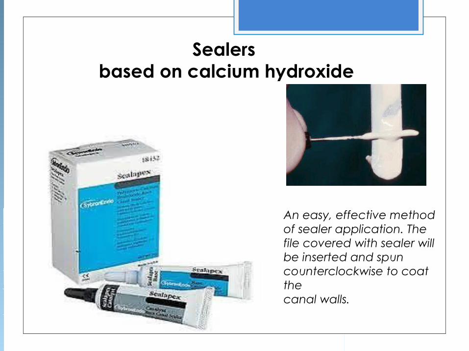
Sealers based on calcium hydroxide
An easy, effective method of sealer application. The file covered with sealer will be inserted and spun counterclockwise to coat thecanal walls.

Indications for the use of calcium hydroxide include:

Indications of calcium hydroxide sealer

Acceptable methods of placing the sealer in the canal include the following:
Placing the sealer on the master cone and pumping the cone up and down in the canalPlacing the sealer on a file and spinning it counter clockwisePlacing the sealer with a lentulo spiralUsing a syringeActivating an ultrasonic instrument
The clinician should use care when placing sealer in a canal with an open apex to avoid extrusion.

Solid materials
Silver cones met many of the criteria for filling materials but suffered from several deficiencies.
When leakage occurred and the points contacted tissue fluids, they corroded, further increasing leakage.

Semisolid material
Gutta-percha, a semisolid material, is the most widely used and accepted obturating material.
Typical composition of gutta-perchacones.
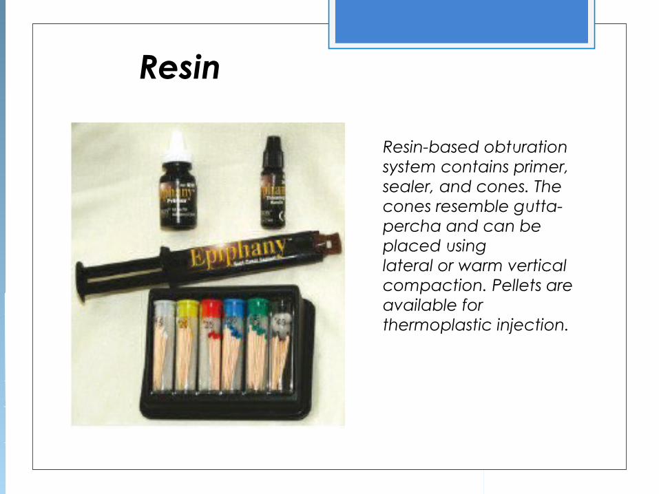
Resin
Resin-based obturation system contains primer, sealer, and cones. The cones resemble gutta-percha and can be placed usinglateral or warm vertical compaction. Pellets are available for thermoplastic injection.

Single cone
Warm lateral compaction
Warm vertical compaction
Lateral compaction
Thermomec-hanical
compaction
Injection-molded gutta-
percha
Core carrier
Root filling techniques
Solid core techniques
Softened core techniques

Root canal filling technique.Solid core technique
Single coneThe single-cone
technique consists of matching a cone to the prepared canal. For this technique a type of canal preparation is advocated so that the size of the cone and the shape of the preparation are closely matched.

Cold lateral condensation This is a commonly taught method of obturation and is the gold standard by which others are judged.
The technique involves placement of a master point chosen to fit the apical section of the canal. Obturation of the remainder is achieved by condensation of smaller accessory points. The steps involved are:

1. Select a GP master point to correspond with the master apical file instrument. This should fit the apical region snugly at the working length so that on removal a degree of resistance or 'tug-back' is felt. If there is no tug-back select a larger point or cut 1 mm at a time off the tip of the point until a good fit is obtained. The point should be notched at the correct working length to guide its placement to the apical constriction.
2 . Take a radiograph to confirm that the point is in correct position if you are in any doubt.
3. Coat walls of canal with sealer using a small file. 4. Insert the master point, covered in cement. 5 . Condense the GP laterally with a finger spreader to provide
space into which accessory points can be inserted until the canal is full.
6. Excess GP is cut off with a hot instrument and the remainder packed vertically into the canal with a cold plugger.

Sketch showing a cross-sectional cut through a root canal filled with a master cone and multiple accessory cones

Warm lateral condensation As above, but uses a warm spreader after the initial cold lateral condensation. Finger spreaders can be heated in a flame or a special electronically heated device (Touch of heat) can be used.

Diagram of the warm vertical condensation technique. A, After a heated spreader is used to remove the coronalsegment of the master cone, a cold plugger is used to apply vertical pressure to the softened master cone. B, Obturation of the coronal portion of the canal is accomplished by adding a gutta-percha segment. C, A heated spreader is used to soften the material. D, A cold plugger is then used to apply pressure to the softened gutta-percha.

Thermoplasticized injectable GP (e.g. Obtura, Ultrafil)
Thermomechanical compactio

Coated carriers (e.g. Thermafil) These are cores of metal or plastic coated with GP. They are heated in an oven and then simply pushed into the root canal to the correct length. The core is then severed with a bur. A dense filling results, but again apical control is poor and extrusions common. They are expensive and difficult to remove.

GuttaFlow is a cold flowable injection system that combines a silicone-based matrix with finely ground gutta-percha. It is used inconjunction with a master gutta-percha point without the need for compaction.

Glass ionomer–coated gutta-percha points (A) are used in conjunction with a glass ionomer sealer (B) to attempt to create a monoblock within the canal system.

A, Resin composite core buildup, with a ferrule incorporated into the preparation. B, Full crown as the definitive restoration.
A cast post and core provides the best foundation for restoring maxillary premolars.

THE CORONAL SEAL
Regardless of the technique used to obturate the canals,
coronal microleakage can occur through seemingly well-
obturated canals within a short time, potentially causing
infection of the periapical area. A method to protect
the canals in case of failure of the coronal restoration is to cover the floor of the pulp chamber with a lining of glass ionomer cement after the excess gutta-percha and sealer have been cleaned from the canal.

![Role of Dentin Compositional Changes and Structural Loss on … · 2015-08-27 · and to prevent reinfection using a root canal filling [8-10]. After accessing the root canal, mechanical](https://img.pdfslide.us/doc/110x75/5f063b3c7e708231d416f462/role-of-dentin-compositional-changes-and-structural-loss-on-2015-08-27-and-to.jpg)


![Evaluation of Root Canal Filling with a Bioceramic Sealer ... · [8]. So, the quality of root canal filling and coronal restoration after root canal treatment has a strong effect](https://img.pdfslide.us/doc/110x75/5ed56e8d11be98291d04238d/evaluation-of-root-canal-filling-with-a-bioceramic-sealer-8-so-the-quality.jpg)