Embed Size (px)
Citation preview

RESPIRATORY ACIDOSIS AND ALKALOSIS…
Moderator/Resource Faculty: Dr. Deebya Raj Mishra…Presented by: Nikhil Agarwal…

OBJECTIVES…• Introduction• Causes • Diagnosis• Management

INTRODUCTION…
• Respiratory acid-base disorders are those abnormalities in acid-base equilibrium initiated by a change in the arterial carbon dioxide tension (PaCO2 )--the respiratory determinant of acidity in the Henderson equation:
H+= 24 x PaCO2 / [HCO3-]• There are two respiratory acid-base disorders: – respiratory acidosis and – respiratory alkalosis.

RESPIRATORY ACIDOSIS…• Respiratory acidosis is the acid-base disturbance initiated by an increase in PaCO2.
• The level of PaCO2 is determined by the interaction of two factors, the rate of carbon dioxide production (VCO2) and the rate of alveolar ventilation (VA), as follows:PaCO2 = K x VCO2 / VA
where K is a constant.
• An increase in arterial pCO2 can occur by one of three possible mechanisms:– Presence of excess CO2 in the inspired gas – Decreased alveolar ventilation – Increased production of CO2 by the body
• By far, most cases of respiratory acidosis reflect a decrease in alveolar ventilation.
• Overproduction of carbon dioxide is usually matched by increased excretion (due to increased alveolar ventilation) such that hypercapnia is prevented.
• Secondary physiologic response – – Respiratory acidosis acidifies body fluids. – It elicits adaptive increments in plasma bicarbonate concentration
that attenuate the impact of hypercapnia on systemic acidity; these increments in plasma bicarbonate should be viewed as an integral part of the respiratory acidosis.

RESPIRATORY ACIDOSIS…• In acute respiratory acidosis, the PaCO2 is elevated above the
upper limit of the reference range (over 6.3 kPa or 45 mm Hg) with an accompanying acidemia (pH <7.36). Acute respiratory acidosis occurs when an abrupt failure of ventilation occurs.
• In chronic respiratory acidosis, the PaCO2 is elevated above the upper limit of the reference range, with a normal blood pH (7.35 to 7.45) or near-normal pH secondary to renal compensation and an elevated serum bicarbonate (HCO3
− >30 mm Hg).
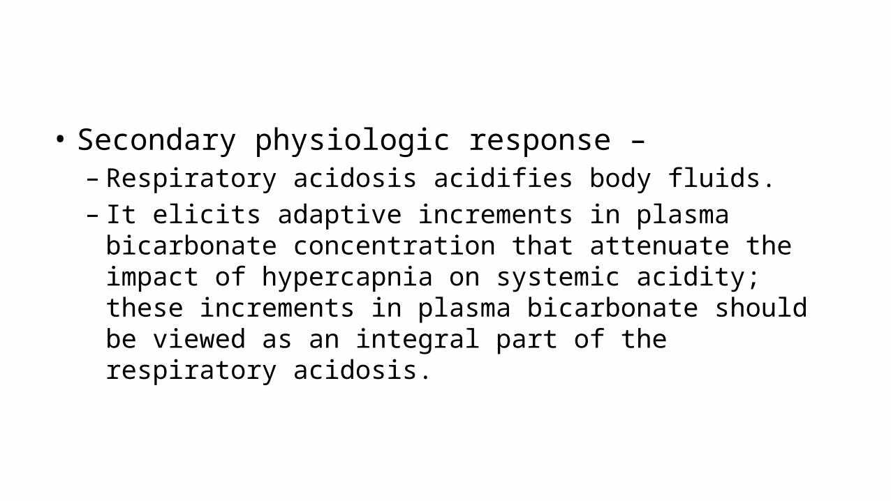
ACUTE CAUSES…Normal airway and lungs Abnormal airway and lungs
Central nervous system depressionGA/ Sedative overdoseHead trauma/Cerebrovascular accidentCerebral edemaBrain tumor/Encephalitis
Upper airway obstructionComa induced hypopharyngeal obstructionAspiration of foreign body or vomitusLaryngospasm or angioedemaObstructive sleep apnea
Neuromuscular impairmentHigh spinal cord injury, Guillain-Barre syndromeStatus epilepticus, Botulism, tetanusCrisis in myathenia gravisHypokalemic myopathyDrugs or toxic agents (curare, succinylcholine, aminoglycosides, organophosphates)
Lower airway obstructionGeneralized bronchospasmSevere asthmaBronchiolitis of infants and adults
Ventilatory restrictionRib fractures with flail chestPneumothorax, HemothoraxImpaired diaphragmatic function
Disorders involving pulmonary alveoliSevere bilateral pneumoniaAcute respiratory distress syndromeSevere pulmonary edema
Iatrogenic eventsMisplacement of airway cannula during mechanical ventilationBronchoscopy associated respiratory arrestIncreased CO2 production with constant mechanicalVentilation
Pulmonary perfusion defectCardiac arrest, Severe circulatory failureMassive pulmonary thromboembolism, Fat or air embolus

CHRONIC CAUSES…Normal airway and lungs Abnormal airway and lungsCentral nervous system depressionSedative overdoseMethadone/heroin addictionPrimary alveolar hypoventilationObesity-hypoventilation syndromeBrain tumorBulbar poliomyelitis
Upper airway obstructionTonsillar and peritonsillar hypertrophyParalysis of vocal cordsTumor of the cords or larynxAirway stenosis post prolonged intubationThymoma, aortic aneurysm
Neuromuscular impairmentPoliomyelitis Multiple sclerosisMuscular dystrophyAmyotrophic lateral sclerosisDiaphragmatic paralysisMyxedemaMyopathic disease
Lower airway obstructionChronic obstructive lung disease (bronchitis, Bronchiolitis, bronchiectasis, emphysema)
Ventilatory restrictionKyphoscoliosis, spinal arthritisObesityFibrothorax HydrothoraxImpaired diaphragmatic Function
Disorders involving pulmonary alveoliSevere chronic pneumonitisDiffuse infiltrative diseaseInterstitial fibrosis
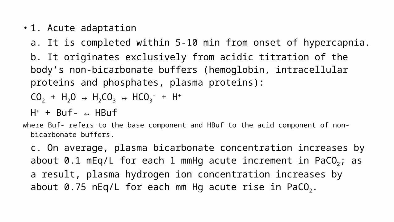
• 1. Acute adaptationa. It is completed within 5-10 min from onset of hypercapnia.b. It originates exclusively from acidic titration of the body’s non-
bicarbonate buffers (hemoglobin, intracellular proteins and phosphates, plasma proteins):
CO2 + H2O ↔ H2CO3 ↔ HCO3- + H+
H+ + Buf- ↔ HBufwhere Buf- refers to the base component and HBuf to the acid component of non-bicarbonate
buffers.
c. On average, plasma bicarbonate concentration increases by about 0.1 mEq/L for each 1 mmHg acute increment in PaCO2; as a result, plasma hydrogen ion concentration increases by about 0.75 nEq/L for each mm Hg acute rise in PaCO2.

• Chronic adaptation
a. It requires 3-5 days of sustained hypercapnia for completion.
b. It originates from up regulation of renal acidification mechanisms (both in the proximal and distal segments of the nephron) that result in:
i. A transient increase in urinary net acid excretion; andii. A persistent increase in the rate of renal bicarbonate reabsorption that maintains the
increased plasma bicarbonate level.
c. On average, plasma HCO3- concentration increases by about 0.3 mEq/L for each mm Hg
chronic increment in PaCO2; as a result, plasma H+ concentration increases by about 0.3 nEq/L for each mm Hg chronic rise in PaCO2.
Thus, at a given PaCO2 value, chronic adaptation provides better defense of systemic acidity than acute adaptation.
d. The renal response to chronic hypercapnia includes a transient increase in chloride excretion and generation of hypochloremia. This reduction in plasma chloride concentration balances the increase in plasma bicarbonate concentration, plasma anion gap remaining unchanged.
CLINICAL FEATURES…• Varies according to the severity and duration of respiratory acidosis, the
underlying disease, and whether there is accompanying hypoxemia.
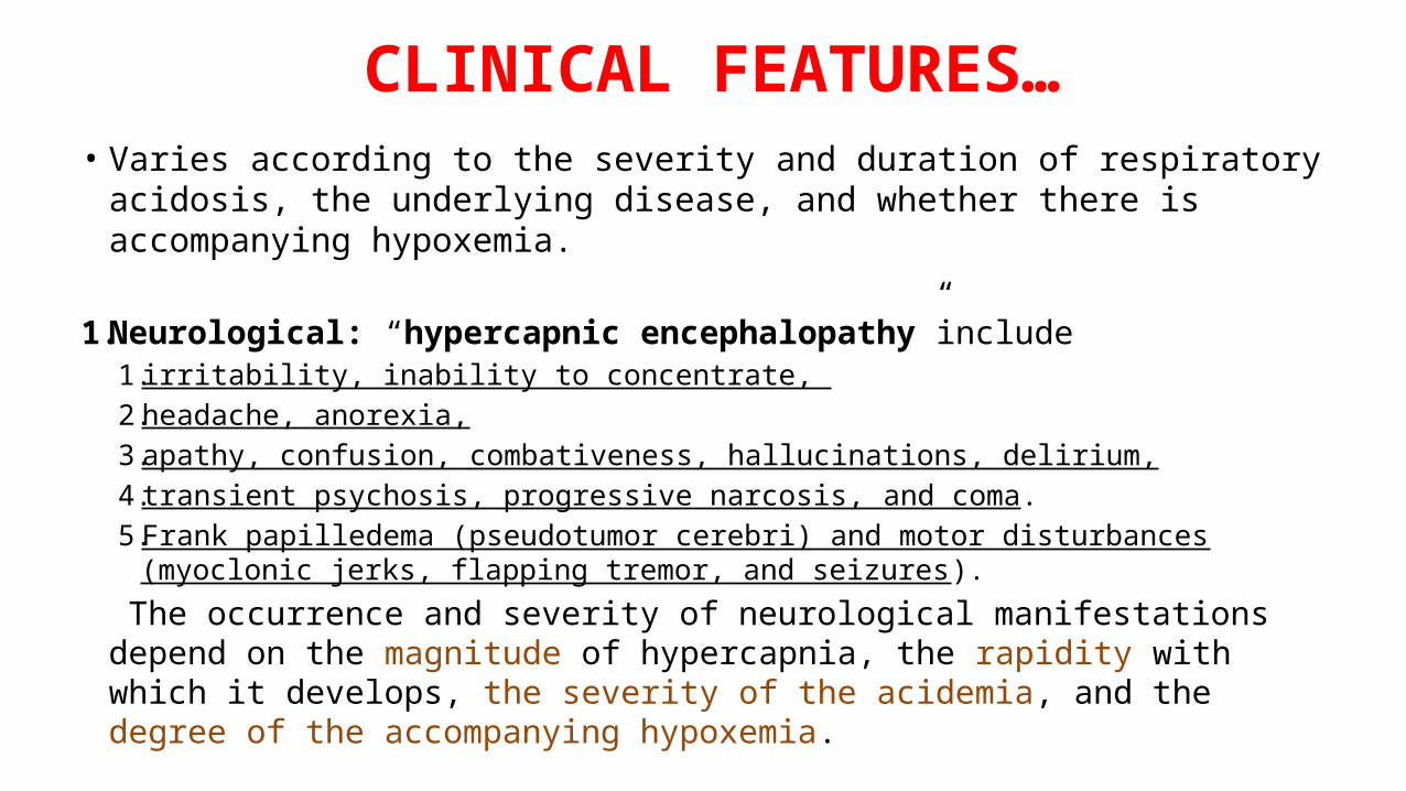
1. Neurological: “hypercapnic encephalopathy”include 1. irritability, inability to concentrate, 2. headache, anorexia,3. apathy, confusion, combativeness, hallucinations, delirium,4. transient psychosis, progressive narcosis, and coma. 5. Frank papilledema (pseudotumor cerebri) and motor disturbances (myoclonic
jerks, flapping tremor, and seizures).
The occurrence and severity of neurological manifestations depend on the magnitude of hypercapnia, the rapidity with which it develops, the severity of the acidemia, and the degree of the accompanying hypoxemia.
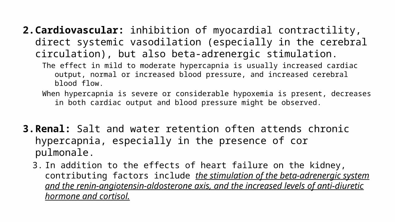
2. Cardiovascular: inhibition of myocardial contractility, direct systemic vasodilation (especially in the cerebral circulation), but also beta-adrenergic stimulation.
The effect in mild to moderate hypercapnia is usually increased cardiac output, normal or increased blood pressure, and increased cerebral blood flow.
When hypercapnia is severe or considerable hypoxemia is present, decreases in both cardiac output and blood pressure might be observed.
3. Renal: Salt and water retention often attends chronic hypercapnia, especially in the presence of cor pulmonale.
3. In addition to the effects of heart failure on the kidney, contributing factors include the stimulation of the beta-adrenergic system and the renin-angiotensin-aldosterone axis, and the increased levels of anti-diuretic hormone and cortisol.
DIAGNOSIS…
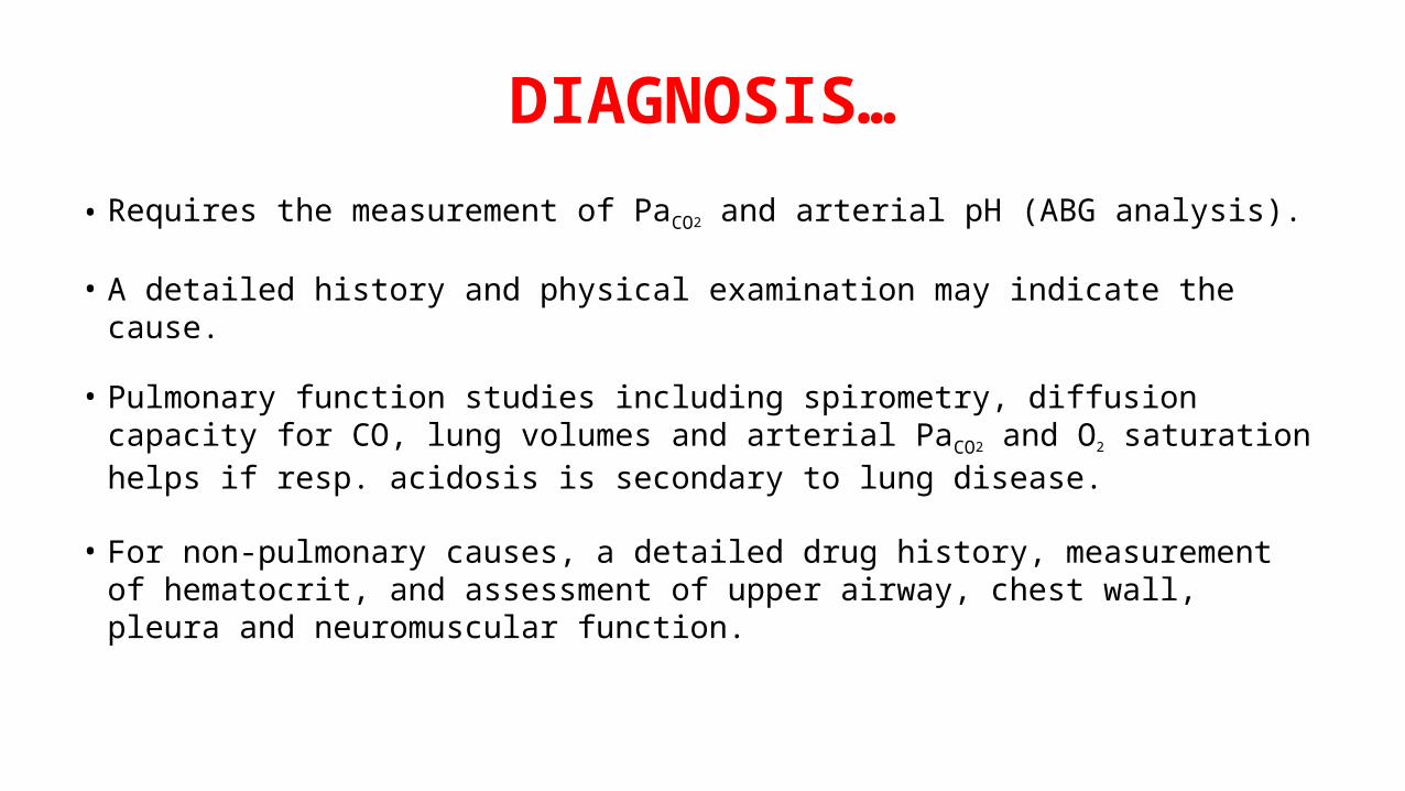
• Requires the measurement of PaCO2 and arterial pH (ABG analysis).
• A detailed history and physical examination may indicate the cause.
• Pulmonary function studies including spirometry, diffusion capacity for CO, lung volumes and arterial PaCO2 and O2 saturation helps if resp. acidosis is secondary to lung disease.
• For non-pulmonary causes, a detailed drug history, measurement of hematocrit, and assessment of upper airway, chest wall, pleura and neuromuscular function.
ARTERIAL FINDINGS IN RESP. ACIDOSIS…
• PCO2 is always raised.• In acute respiratory failure.– pH is low– HCO3
- is high normal or slightly raised as compensatory changes take sometimes to occur.
• In chronic respiratory failure.– Ph is normal or low, depending on chronicity(time for compensation
to occur)– HCO3
- is raised
MANAGEMENT…
• Primarily directed at the underlying disorder or patho-physiologic process.
• Caution should be exercised in the correction of chronic hypercapnia: too-rapid correction of the hypercapnia can result in metabolic alkalemia.
• Alkalization of the cerebrospinal fluid (CSF) can result in seizures.
Pharmacologic Therapy• Pharmacologic therapies are generally used as treatment of the underlying
disease process. • Bronchodilators: such as beta agonists (eg, albuterol and salmeterol),
anticholinergic agents (eg, ipratropium bromide and tiotropium), and methylxanthines (eg, theophylline) are helpful in treating patients with obstructive airway disease and severe bronchospasm. Theophylline may improve diaphragm muscle contractility and may stimulate the respiratory center.
• Drug antagonists: Drug therapy aimed at reversing the effects of certain sedative drugs may be helpful in the event of an accidental or intentional overdosage. Naloxone may be used to reverse the effects of narcotics. Flumazenil may be used to reverse the effects of benzodiazepines.
• Respiratory stimulants: Respiratory stimulants have been used but have limited efficacy in respiratory acidosis caused by disease.– Medroxyprogesterone increases central respiratory drive and may be
effective in treating obesity-hypoventilation syndrome (OHS). Medroxyprogesterone has also been shown to stimulate ventilation is some patients with COPD and alveolar hypoventilation.
– Acetazolamide is a diuretic that increases bicarbonate excretion and induces a metabolic acidosis, which subsequently stimulates ventilation.
– Theophylline increases diaphragm muscle strength and stimulates the central ventilatory drive. In addition, theophylline is a bronchodilator.
• Oxygen Therapy– Because many patients with hypercapnia are also hypoxemic, oxygen therapy may be
indicated. – Oxygen therapy is employed to prevent the sequelae of long-standing hypoxemia.– Hypercapnia is best avoided by titrating oxygen delivery to maintain oxygen saturation in
the low 90% range and partial arterial pressure of oxygen (PaO2) in the range of 60-65 mm Hg.
• Ventilatory Support– Therapeutic measures that may be lifesaving in severe hypercapnia and respiratory
acidosis include endotracheal intubation with mechanical ventilation and noninvasive positive pressure ventilation (NIPPV)(they help improve PaO2 and decrease the PaCO2 ) techniques such as nasal continuous positive-pressure ventilation (NCPAP) and nasal bilevel ventilation.
– Rapid correction of the hypercapnia by the application of external noninvasive positive-pressure ventilation or invasive mechanical ventilation can result in alkalemia and the development of sudden post- hypercapnic alkalosis with potential serious consequences.

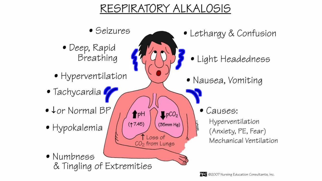
Respiratory alkalosis…• Respiratory alkalosis is the acid-base disturbance initiated by a reduction in
PaCO2.
• This occurs when there is excessive loss of CO2 by hyperventilation of lungs.
• Hypocapnia develops when a sufficiently strong ventilatory stimulus causes CO2 output in the lungs to exceed its metabolic production by the tissues.
• As a result, partial pressure of CO2 and H+ conc. falls and so there is a decrease in bicarbonate levels.
Pathophysiology…
• By far, most cases of respiratory alkalosis reflect an increase in alveolar ventilation.
• Primary decreases in CO2 production are generally attended by parallel decreases in alveolar ventilation, thus preventing expression of respiratory alkalosis.
• However, in the presence of constant alveolar ventilation (i.e., mechanical ventilation), decreased carbon dioxide production (e.g., sedation, skeletal muscle paralysis, hypothermia, hypothyroidism) can cause respiratory alkalosis.

Secondary physiologic response
• Respiratory alkalosis alkalinizes body fluids. It elicits adaptive decrements in plasma bicarbonate concentration that attenuate the impact of hypocapnia on systemic acidity; these decrements in plasma bicarbonate should be viewed as an integral part of the respiratory alkalosis.
• Acute adaptationa. It is completed within 5-10 min from onset of hypocapnia
b. It originates principally from alkaline titration of the body’s nonbicarbonate buffers (hemoglobin, intracellular proteins and phosphates, plasma proteins):
HBuf ↔ H+ + Buf-HCO3
- + H+ ↔ H2CO3 ↔ H2O + CO2
where HBuf refers to the acid component and Buf- to the base component ofnonbicarbonate buffers.
c. On average, plasma bicarbonate concentration falls by about 0.2 mEq/L for each mm Hg acute decrement in PaCO2; as a result, plasma hydrogen ion concentration decreases by about 0.75 nEq/L for each mm Hg acute reduction in PaCO2.
• Chronic adaptationa. It requires 2-3 days of sustained hypocapnia for completion.
b. It originates from downregulation of renal acidification mechanisms (both in the proximal and distal segments of the nephron) that result in
i. A transient decrease in urinary net acid excretion (mostly a fall in ammonium excretion and an early component of increased bicarbonate excretion) that reduces the body’s bicarbonate stores; and
ii. A persistent decrease in the rate of renal bicarbonate reabsorption that maintains the decreased plasma bicarbonate level.
c. On average, plasma bicarbonate concentration decreases by about 0.4 mEq/L for each mm Hg chronic decrement in PaCO2; as a result, plasma hydrogen ion concentration decreases by about 0.4 nEq/L for each mm Hg chronic reduction in PaCO2. Thus, at a given PaCO2 value, chronic adaptation provides better defense of systemic acidity than acute adaptation.
d. Chronic hypocapnia is characterized by an increase in plasma chloride concentration that balances most of the fall in plasma bicarbonate concentration, the remainder reflecting a small increase in the plasma anion gap.
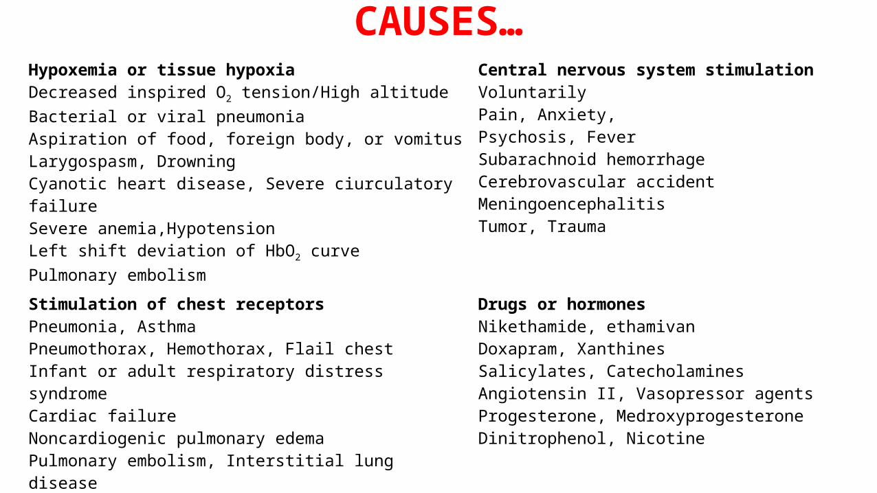
CAUSES…Hypoxemia or tissue hypoxiaDecreased inspired O2 tension/High altitudeBacterial or viral pneumoniaAspiration of food, foreign body, or vomitusLarygospasm, DrowningCyanotic heart disease, Severe ciurculatory failureSevere anemia,HypotensionLeft shift deviation of HbO2 curvePulmonary embolism
Central nervous system stimulationVoluntarilyPain, Anxiety, Psychosis, FeverSubarachnoid hemorrhageCerebrovascular accidentMeningoencephalitisTumor, Trauma
Stimulation of chest receptorsPneumonia, AsthmaPneumothorax, Hemothorax, Flail chestInfant or adult respiratory distress syndromeCardiac failureNoncardiogenic pulmonary edemaPulmonary embolism, Interstitial lung disease
Drugs or hormonesNikethamide, ethamivanDoxapram, XanthinesSalicylates, CatecholaminesAngiotensin II, Vasopressor agentsProgesterone, MedroxyprogesteroneDinitrophenol, Nicotine
MiscellaneousPregnancy, Sepsis, Hepatic failureMechanical hyperventilationHeat exposure, Recovery form metabolic acidosis
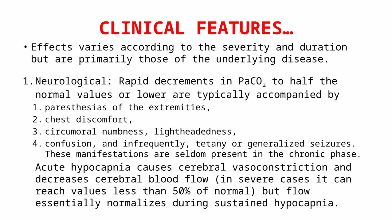
CLINICAL FEATURES…• Effects varies according to the severity and duration but are primarily those
of the underlying disease.
1. Neurological: Rapid decrements in PaCO2 to half the normal values or lower are typically accompanied by
1. paresthesias of the extremities, 2. chest discomfort, 3. circumoral numbness, lightheadedness, 4. confusion, and infrequently, tetany or generalized seizures. These manifestations
are seldom present in the chronic phase.
Acute hypocapnia causes cerebral vasoconstriction and decreases cerebral blood flow (in severe cases it can reach values less than 50% of normal) but flow essentially normalizes during sustained hypocapnia.
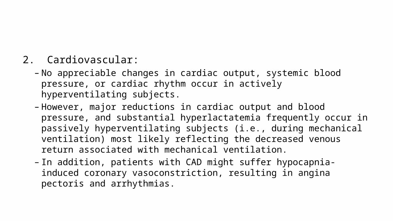
2. Cardiovascular: – No appreciable changes in cardiac output, systemic blood pressure, or
cardiac rhythm occur in actively hyperventilating subjects. – However, major reductions in cardiac output and blood pressure, and
substantial hyperlactatemia frequently occur in passively hyperventilating subjects (i.e., during mechanical ventilation) most likely reflecting the decreased venous return associated with mechanical ventilation.
– In addition, patients with CAD might suffer hypocapnia-induced coronary vasoconstriction, resulting in angina pectoris and arrhythmias.

DIAGNOSIS…
• Requires the measurement of PaCO2 and arterial pH.
• Plasma K + is often reduced and the Cl- is increased.
ARTERIAL FINDINGS IN RESP. ALKALOSIS…
• PCO2 is always reduced.• HCO3
- is low normal or low.• Ph is raised or normal.
TREATMENT…
• The treatment of respiratory alkalosis is primarily directed at correcting the underlying disorder. Respiratory alkalosis itself is rarely life threatening.
• Therefore, emergent treatment is usually not indicated unless the pH level is greater than 7.5. Because respiratory alkalosis usually occurs in response to some stimulus, treatment is usually unsuccessful unless the stimulus is controlled.
• If the PaCO2 is corrected rapidly in patients with chronic respiratory alkalosis, metabolic acidosis may develop due to the renal compensatory drop in serum bicarbonate.
• In mechanically ventilated patients who have respiratory alkalosis, the tidal volume and/or respiratory rate may need to be decreased. Inadequate sedation and pain control may contribute to respiratory alkalosis in patients breathing over the set ventilator rate.
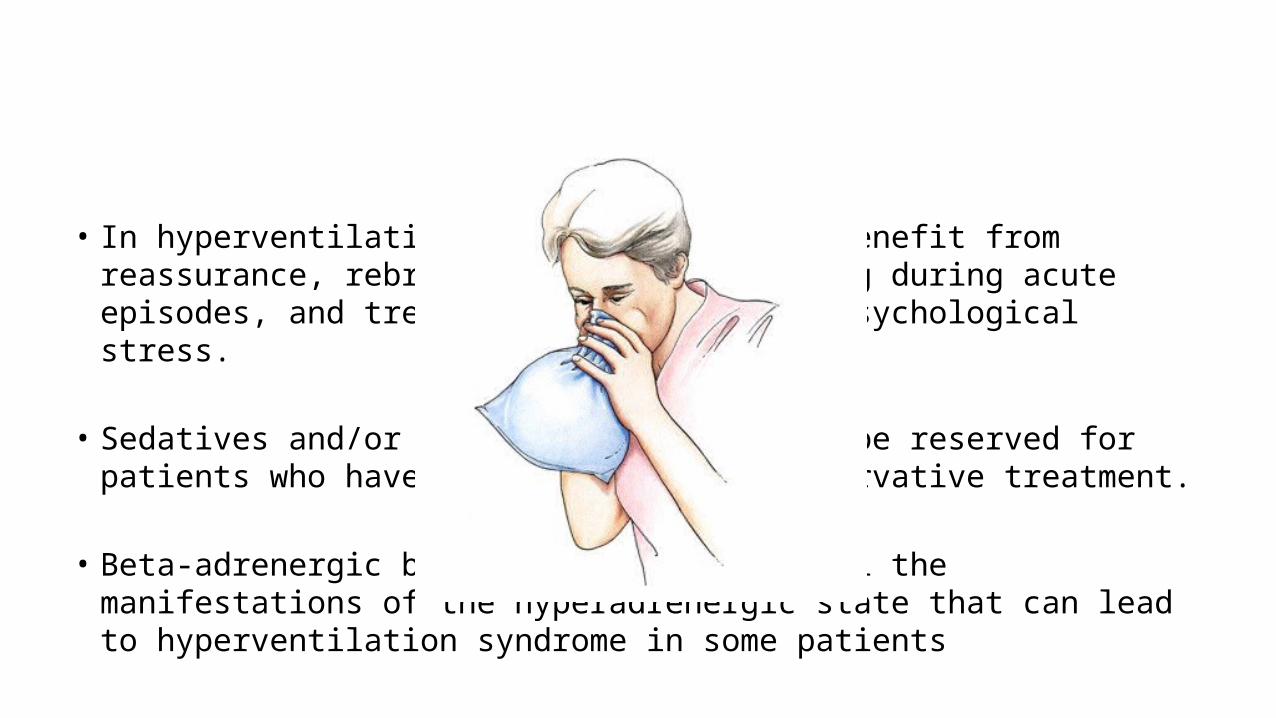
• In hyperventilation syndrome, patients benefit from reassurance, rebreathing into a paper bag during acute episodes, and treatment for underlying psychological stress.
• Sedatives and/or antidepressants should be reserved for patients who have not responded to conservative treatment.
• Beta-adrenergic blockers may help control the manifestations of the hyperadrenergic state that can lead to hyperventilation syndrome in some patients

References :• Longo, Fauci, Kasper et al Harrison’s principles of Internal Medicine, 18th Edition,
2012 (371-73)• Aggarwal P., Matthew K.G., Medicine Prep Manual for Undergraduates, 4th Edition,
2014, (715-16)• Alagappan R. Manual of Practical Medicine, 4th Edition, 2011, (424-26) • Nicolaos Madias, MD: Tufts University School of Medicine• Medscape