Embed Size (px)
Citation preview

Renal Physiology 1Renal Physiology 1
Dr. Bikesh Pandey

Body Fluid & KidneyBody Fluid & Kidney

3
Body fluidBody fluid (60% of body weight)(60% of body weight)
Interstit ial f luid Interstit ial f luid (15%)(15%)
Extracellular f luid Extracellular f luid (20%)
Intracellular f luidIntracellular f luid
( 40% )
PlasmaPlasma( 5 %)

ICF
plasma
interstitial

Fluid Input and OutputFluid Input and Output
Daily Input of Water
• Water is added to the body by two major sources:
– Ingested
• 2100 ml/day
– Synthesized in the body
• Result of oxidation of carbohydrates - 200 ml/day

Fluid Input and OutputFluid Input and OutputDaily Output of Water
• Water is lost by :
– Insensible water lossInsensible water loss
• Termed so, because we are not consciously aware of it
• Eg: Evaporation through skin & respiratory system
• 700 ml/day
– SweatingSweating
• 100 ml/day
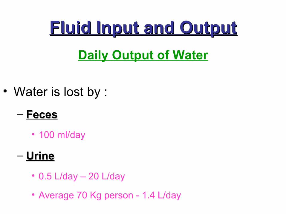
Fluid Input and OutputFluid Input and Output
Daily Output of Water
• Water is lost by :
– FecesFeces
• 100 ml/day
– UrineUrine
• 0.5 L/day – 20 L/day
• Average 70 Kg person - 1.4 L/day


3 PM

Anatomy & Histology of Urinary SystemAnatomy & Histology of Urinary System

Organization of Urinary SystemOrganization of Urinary System
It includes:It includes:
• Two kidneys
• Two ureters
• One bladder
• One urethra

Function of the Urinary SystemFunction of the Urinary System • Maintanance of fluid volume & thus blood
pressure.
• Regulation of electrolyte balance
– Eg: Sodium, Potassium, Calcium…….
• Removal of waste product from the body
– Eg: Urea, Uric acid…….

Function of the Urinary System Function of the Urinary System
• Regulation of “Acid-Base” balance.
• Production of “Erythropoietin” to stimulate production of RBC in bone marrow.
• Regulation of Vitamin D in our body ……………….

KidneyKidney
• Bean-shaped
• Retroperitoneal
• Right kidney is lower than the left one
• Adrenal gland rests upon each kidney

KidneyKidney
There are three layers of coverings surrounding each kidney from the outside to the inside :
• The Renal fascia (Also encloses adrenal gland)
• The Adipose capsule
• The Fibrous capsule

KidneyKidney
Each kidney consists of:
• An outer, Renal Cortex
• An inner, Renal Medulla

KidneyKidney
• Extension of renal cortex, the Renal Column projects into the inner aspect of the kidney, dividing the Renal medulla into triangular shaped Renal Pyramids.

KidneyKidney• Their apices form the Renal Papillae, which indent the Minor calices.
• Minor Calyces are 7-8 in number on each kidney.
• Minor calices unite to form a Major Calyx. They are 2-3 in number on each kidney.
• Two or three major calices unite to form the Renal Pelvis, which is the funnel shaped superior end of the Ureters.
• The Renal sinus is a space that extends into the kidney from the Hilum.
• The foramina On the tip of renal papillae are termed the papillary foramina. The urine formed in the kidney passes through these foramina into the minor calices.

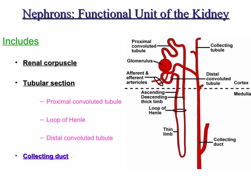
Nephrons: Functional Unit of the KidneyNephrons: Functional Unit of the Kidney
Includes
• Renal corpuscleRenal corpuscle
• Tubular sectionTubular section
– Proximal convoluted tubule
– Loop of Henle
– Distal convoluted tubule
• Collecting ductCollecting duct

Nephron Nephron Two types of nephrons
• Cortical nephron– 80-85% of nephrons are cortical nephrons.– Renal corpuscles are in outer cortex and loops of
Henle lie mainly in cortex.
• Juxtamedullary nephrons– 15-20% of nephrons are juxtamedullary nephrons.– Renal corpuscles close to medulla and long loops
of Henle extend into deepest medulla


Renal Corpuscle Renal Corpuscle
Provides for filtration of plasma from glomerular capillary.Provides for filtration of plasma from glomerular capillary.
• Renal glomerulus
• Bowman's capsuleBowman's capsule

Renal CorpuscleRenal Corpuscle

Renal glomerulusRenal glomerulus
• A tightly-coiled capillaries network.
• The endothelial cells are fenestrated.
• Capillaries are divided into :
– Afferent arteriole – Brings blood / wider
– Efferent arteriole – takes away blood / narrower

Bowman's CapsuleBowman's Capsule
Divided into two layers Divided into two layers
• Parietal or capsular layerParietal or capsular layer - simple squamous epithelium - simple squamous epithelium
• Visceral layerVisceral layer - podocytes - podocytes

PodocytesPodocytes
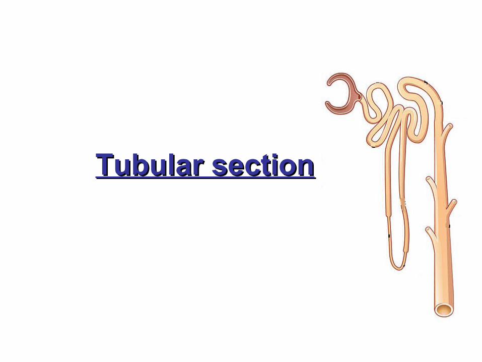
Tubular sectionTubular section

PCTPCT Loop of HenleLoop of Henle DCTDCT Collecting Collecting TubuleTubule
CuboidalRound nucleus Strong acidophilicBrush border
Thick descending Similar to PCT
Thin descending / Thin ascending
Simple squamous
Thick ascendingThick ascending Simple cuboidal
Cuboidal Lighter cytoplasmRound nucleusNo brush border Less microvilli
Simple cuboidal to columnarLight staining cytoplasmClear boundaryLumen is largest

Juxtaglomerular apparatusJuxtaglomerular apparatus located at the vascular pole of the renal corpuscles
consist of juxtaglomerular cells, macula densa
and extraglomerular mesangial (polar cushion)
cells
function: control water and electrolyte balance;
regulate blood pressure;
produce erythropoietin

Juxtaglomerular ApparatusJuxtaglomerular Apparatus
3 parts:3 parts:
(1)(1) Macula densaMacula densa - cells of distal - cells of distal tubuletubule
(2)(2) Juxtaglomerular (JG) cellsJuxtaglomerular (JG) cells - - modified smooth muscle cells modified smooth muscle cells (myoepithelioid) in arteriole. (myoepithelioid) in arteriole.
(3)(3) Extraglomerular mesangiumExtraglomerular mesangium
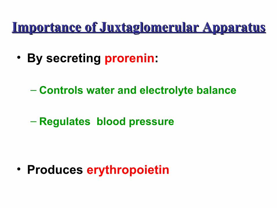
Importance of Juxtaglomerular ApparatusImportance of Juxtaglomerular Apparatus
• By secreting prorenin:
– Controls water and electrolyte balance
– Regulates blood pressure
• Produces erythropoietin

The - EndThe - End


![Exercise 9 - Renal System Physiology[1]](https://img.pdfslide.us/doc/110x75/553dd6134a7959502f8b47ca/exercise-9-renal-system-physiology1.jpg)
