Embed Size (px)
Citation preview
S e c t i o n 1
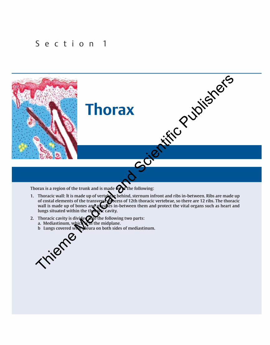
Thorax
Thorax is a region of the trunk and is made up of the following:
of costal elements of the transverse process of 12th thoracic vertebrae, so there are 12 ribs. The thoracic wall is made up of bones and muscles in-between them and protect the vital organs such as heart and lungs situated within the thoracic cavity.
2. Thoracic cavity is divided into the following two parts: a. Mediastinum, which is in the midplane. b Lungs covered with pleura on both sides of mediastinum.
Thieme M
edica
l and
Scie
ntific
Pub
lishe
rs
CHAPTER
1 Regions of the Thorax
superior aperture of the thorax or inlet of the thorax. The lower limit of the thorax is at the costal margin in
front and at the lower border of 12th rib behind. This is known as inferior aperture of the thorax or outlet of the thorax.
4. Because the inferior aperture of the thorax is formed by the diaphragm, which is flexible and moves downwards and upwards during inspiration and expiration, the abdomen extends upwards into the thorax.
5. There is a pelvic diaphragm (made up of muscles) which separates the pelvis above and perineum below.
In short, one region encroaches another region.
Thorax in Adults and Infants
Adults InfantsThorax is kidney shaped. Transverse diameter is greater than anteroposterior diameter
Thorax is circular
The ribs are oblique. Therefore, the respiration is of both types—thoracic and abdominalIn thoracic respiration, there is inspiration and expiration
The ribs are horizontal. Therefore, the respiration is abdominal by the action of diaphragm
The ribs are ossified and not elastic. There-fore, the ribs can be fractured by direct or indirect violence
The ribs are highly elastic. Therefore, the fractures of the ribs are rare
Superior Thoracic Aperture / Inlet of the Thorax (Figs. 1.1–1.3)
Introduction Inlet of the thorax is the area of communication between thorax and neck.
ShapeThe thorax is kidney shaped.
Dimension 1. Transverse diameter 4 to 5 in. 2. Anteroposterior diameter 2 in.
IntroductionThorax is the most important region of our body, which houses vital organs such as heart and the large vessels, lungs and lower respiratory tract, oesophagus, etc. Therefore, it is protected by the thoracic cage. Thoracic cage is the thoracic wall, formed by vertebral column behind, sternum in front and ribs with intercostal muscles in the intercostal spaces on either side. Above, the thorax communicates with the neck through the thoracic inlet that is covered by suprapleural membrane. Below, it is separated from the abdomen by a flexible diaphragm. Of course, there are apertures in the diaphragm for the passage of important structures between thorax and abdomen.In humans, vertebral column, which forms the axis of the body, has two types of curvatures: 1. Primary curvatures: The thoracic vertebrae and sacral
vertebrae which are concave in forward direction. 2. Secondary curvatures: The cervical vertebrae and
lumbar vertebrae which are convex in forward direction. The secondary curvature of cervical vertebrae is produced when a child raises his neck and head, whereas the secondary curvature of the lumbar vertebrae is produced when a child stands and begins to walk. The ribs in the thorax are not horizontally placed but they are oblique. The first rib is at the upper part of the manubrium sterni in front but posteriorly it articulates with the first vertebra, which is 1½ in. above in the neck.
One should understand the orientation and position of the regions of the body: 1. Head region begins at the lower border of the mandible
in front and external occipital protuberance and external occipital crest behind.
2. The upper limit of the neck is at the lower border of the mandible in front but at the level of external occipital protuberance and external occipital crest behind, so the neck extends high posteriorly.
The lower limit of the neck is the anterior end of the first rib in front and the seventh cervical vertebra behind. Commonly, the lower limit is taken as a circle passing from the upper border of manubrium sterni, clavicle, upper border of scapula and the inferior border of the second thoracic vertebra all around.
3. The upper limit of the thorax is at the anterior end of the first rib in front but at the upper border of the first thoracic vertebra behind which is in the neck. This is known as
Thieme M
edica
l and
Scie
ntific
Pub
lishe
rs
SECTION 1 Thorax4
Direction The plane of the inlet is directed at an angle of 45 degrees forwards and downwards. The direction is such that the first and second thoracic vertebrae lie in the neck and the upper border of the second (may be third in female) thoracic vertebra in male is at the level of the upper border of manubrium sterni.
Boundary It is bounded as noted below: 1. Anteriorly: upper border of manubrium sterni 2. Lateral sides: first rib with its costal cartilage 3. Posteriorly: superior border of the body of the first
thoracic vertebra
Structures Passing through the Inlet of the ThoraxSee table 1.1
Clinical Anatomy
Thoracic Inlet Syndrome (Fig. 1.2)The inlet of the thorax is bounded by the inner border of the first rib, and the apex of the axilla is bounded by the outer border of the first rib behind the clavicle. Between the upper surface of first rib and clavicle, subclavian vessels and the brachial plexus (C5, C6, C7, C8 and T1) are intimately related.
Therefore, at this point, either the nerves or the vessels or both may be compressed.
Causes 1. Cervical rib 2. Any other swelling in the region
ResultMost commonly, lower trunk of the brachial plexus (C8, T1) is involved. This results in: 1. Pain along the skin of the upper limb supplied by C8,
T1, that is medial side of the hand and forearm 2. Wasting of the intrinsic muscles of the hand 3. Possibility of deficient blood supply of the upper limb
due to pressure on the subclavian artery
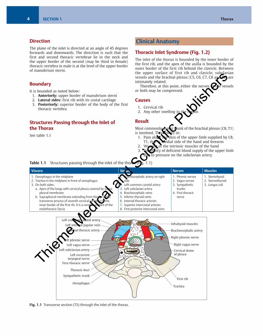
Table 1.1 Structures passing through the inlet of the thorax (Fig. 1.1)
Viscera Vessels Nerves Muscles1. Oesophagus in the midplane2. Trachea in the midplane in front of oesophagus3. On both sides:
a. Apex of the lungs with cervical pleura covered by supra-pleural membrane
b. Suprapleural membrane extending from the tip of the transverse process of seventh cervical vertebra to the inner border of the first rib. It is a condensed part of the endothoracic fascia
1. Brachiocephalic artery on right side
2. Left common carotid artery3. Left subclavian artery4. Brachiocephalic veins5. Inferior thyroid veins6. Internal thoracic arteries7. Superior intercostal arteries8. First posterior intercostal veins
1. Phrenic nerves2. Vagus nerves3. Sympathetic
trunks4. First thoracic
nerve
1. Sternohyoid2. Sternothyroid3. Longus coli
Left common carotid arteryLeft internal jugular vein
Internal thoracic artery
Left phrenic nerveLeft vagus nerve
Left subclavian arteryLeft recurrent
laryngeal nerveFirst thoracic nerve
Thoracic duct
Sympathetic trunk
Oesophagus
Infrahyoid muscles
Brachiocephalic artery
Right phrenic nerve
Right vagus nerve
Cervical domeof pleura
First rib
Trachea
Fig. 1.1 Transverse section (TS) through the inlet of the thorax.
Thieme M
edica
l and
Scie
ntific
Pub
lishe
rs

CHAPTER 1 Regions of the Thorax 5
Inferior Thoracic Aperture / Outlet of the Thorax
DescriptionThe outlet of the thorax is covered by the diaphragm which separates the thorax from the abdomen.
BoundaryIt is bounded as noted below: 1. Anteriorly by infrasternal angle which is made between
the right and left costal margins. 2. On lateral sides by the costal margins formed by the
costal cartilages of the 7th to 10th ribs and tips of the 11th and 12th ribs.
3. Posteriorly by inferior border of the body of the 12th thoracic vertebra. The inferior aperture or outlet of
the thorax is occupied by the diaphragm in which apertures are present for the passage of structures between thorax and abdomen. It will be described along with the muscles of the thoracic floor and posterior abdominal wall.
4. Inferiorly by diaphragm which presents many apertures for the passage of structures between thorax and abdomen.
Regions of the ThoraxThorax can be broadly divided into two regions, viz. thoracic wall and thoracic cavity. Each region comprises distinct elements, as listed in Table 1.2.
Scalenus mediusScalenus anteriorCervical domeof pleuraBrachial plexus(C8, T1)Lower trunk ofbrachial plexusSubclavian arterySubclavian vein
T1
T2
Fig. 1.2 Cervical dome of pleura (on left side) and its relationship.
C7
Scalenus medius
Scalenus anterior
Lower trunk ofbrachial plexus
Subclavian arteryCervical rib
Fig. 1.3 Thoracic inlet (as seen from above).
Table 1.2 Regions of the thorax
Regions ContentsThoracic wall 1. Bones: ribs, sternum and vertebrae
2. Muscles in the intercostal spaces3. Vessels: internal thoracic vessels, intercostal vessels, azygos and hemiazygos veins4. Nerves: intercostal nerves
Thoracic cavity
1. Movable structures Lungs with pleurae
2. Mediastinum Viscera: Arteries: Veins: Lymphatics: Nerves:
a. Superior mediastinum 1. Trachea2. Oesophagus3. Thymus
Arch of aorta 1. Brachiocephalic veins
2. Superior vena cava
Thoracic duct 1. Vagus nerves2. Phrenic nerves3. Cardiac nerves4. Left recurrent
laryngeal nerve
b. Inferior mediastinum
(i) Middle mediastinum Heart with pericar-dium
1. Ascending aorta2. Pulmonary trunk3. Pulmonary arteries
1. Pulmonary veins2. Superior vena
cava
Lymph nodes: tra-cheobronchial lymph nodes
1. Phrenic nerves2. Cardiac plexuses
(ii) Posterior mediastinum Oesophagus Descending thoracic aorta 1. Azygos vein2. Hemiazygos vein
Thoracic ductLymph nodes
1. Sympathetic trunk2. Splanchnic nerves3. Vagus nerves
(iii) Anterior mediastinum Thymus Lymph nodes
Thieme M
edica
l and
Scie
ntific
Pub
lishe
rs
ThoraxThe thorax is made up of thoracic wall and thoracic cavity.
1. Thoracic Wall (Described in Chapter 2)The wall consists of 12 thoracic vertebrae, 12 ribs, sternum and 11 intercostal spaces with three layered intercostal muscles.
2. Thoracic CavityThe cavity contains: 1. A median partition called mediastinum. 2. Laterally placed lungs covered by pleura that is
made up of visceral pleura which covers the lungs and parietal pleura lining the parities. Thus two pleural cavities are formed on each side of the thorax between the thoracic wall and the lungs.
Chapters on Thoracic Cavity Chapter 3: Thoracic Cavity Chapter 4: Respiratory System Chapter 5: Development of Cardiovascular System (CVS)
and Pericardium Chapter 6: Cardiovascular System (CVS) and Pericardium Chapter 7: Major (Large) Vessels of the Thoracic Cavity Chapter 8: Oesophagus and Thymus Chapter 9: Autonomic Nervous System (ANS)
Thieme M
edica
l and
Scie
ntific
Pub
lishe
rs