Embed Size (px)
Citation preview
HEAD AND NECK IMAGING EHAB ABOU ELFOTOUH. MD.
QUIZ

CASE 1:
CASE 1:

CASE 1:
CASE 1:
CASE 1:
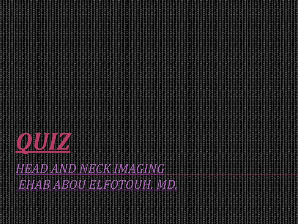
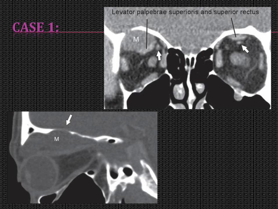
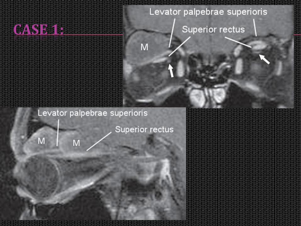
28-year-old man with right proptosis, with orbital MASS on CT & MRI :
A- Intra-conal.
B- Extra-conal.
C- Intra and extra-conal.
CASE 1:
This MASS was seen:
A- Infiltrating conal muscles.
B- Conal muscle origin.
C- Displacing conal muscle.
D- Intra-cranial extension.
CASE 1:
This MASS has:
A- Marginal enhancement.
B- No ehancement.
C- Intense enhancement.

CASE 1:
This MASS is likely related to:
A-Capillary hemangioma.
B- Thrombosed orbital varix.
C- Neuro-fibroma of supra-orbital nerve.
D- Rhabdomyosarcoma.

CASE 2:

CASE 2:
A 9-year-old girl born with a left orbital mass with dramatically enlargement:
A- Infantile (capillary) hemangiomas.
B- Orbital cellulitis.
C- Infantile Fibromatosis.
D- Venous-lymphatic malformation.

THANK YOU