Embed Size (px)
DESCRIPTION
Pulmonary sarcoidosis radiology
Citation preview
Sarcoidosis is a systemic disorder of unknown origin.
It is characterized by non-caseating granulomas in multiple organs, that may resolve spontaneously or progress to fibrosis.
Pulmonary manifestations are present in 90% of patients.
Systemic symptoms such as fatigue, night sweats and weight loss are common.
Löfgren's syndrome, an acute presentation of sarcoidosis, consists of arthritis, erythema nodosum, bilateral hilar adenopathy.
Two third of patients have a remission within ten years.
One third have continuing disease leading to clinically significant organ impairment.
Less than 5% of patients die from sarcoidosis usually as a result of pulmonary fibrosis.
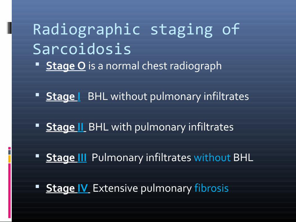
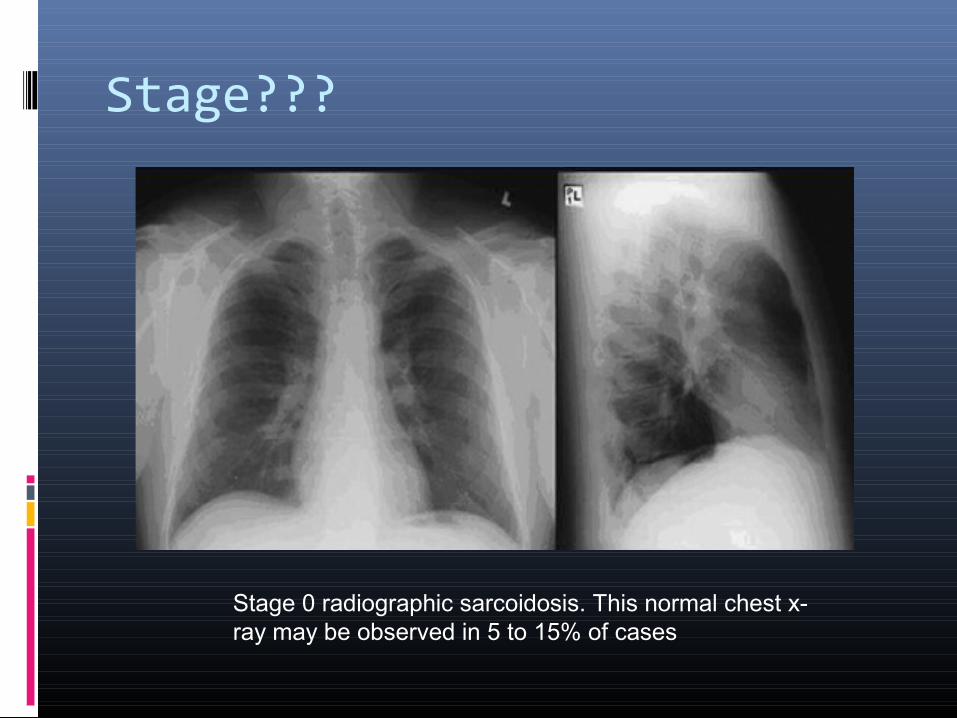
Radiographic staging of Sarcoidosis Stage O is a normal chest radiograph
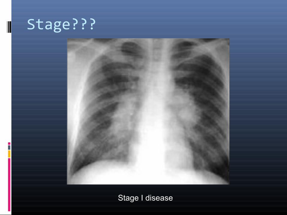
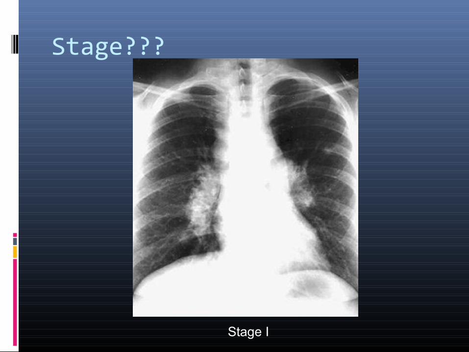
Stage I BHL without pulmonary infiltrates
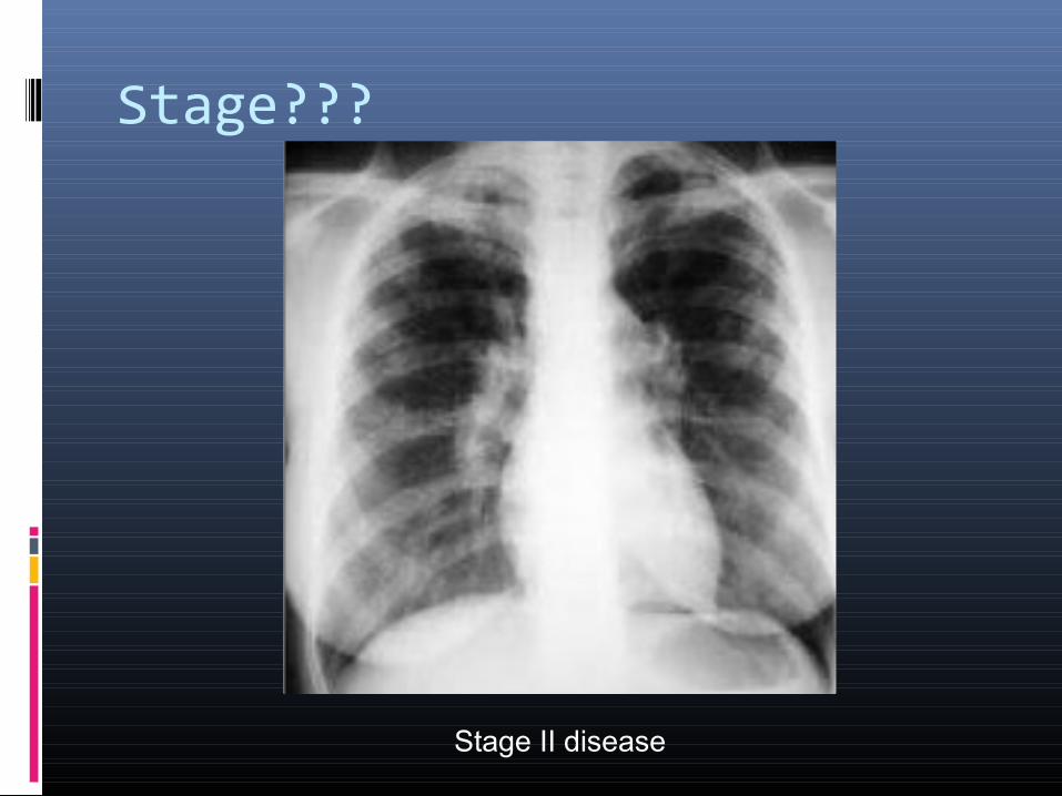
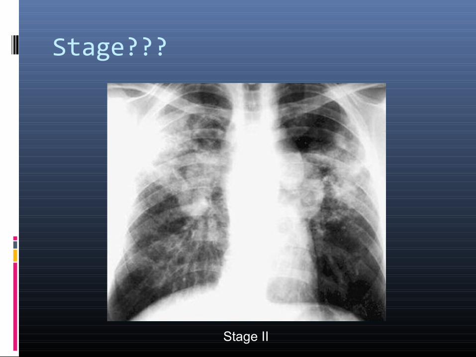
Stage II BHL with pulmonary infiltrates
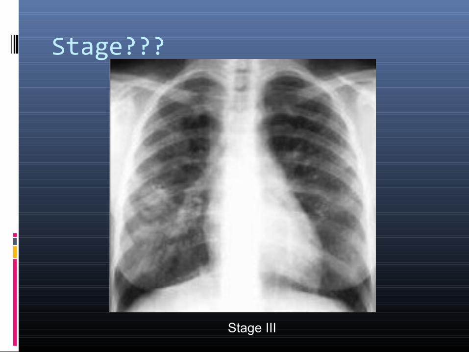
Stage III Pulmonary infiltrates without BHL
Stage IV Extensive pulmonary fibrosis
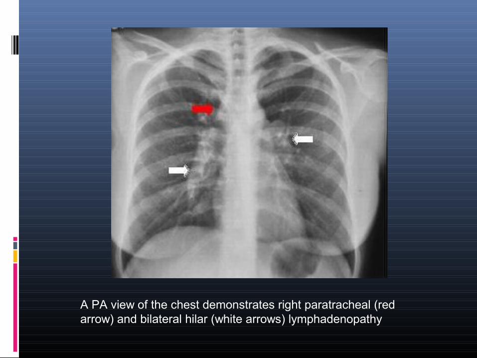
A PA view of the chest demonstrates right paratracheal (red arrow) and bilateral hilar (white arrows) lymphadenopathy
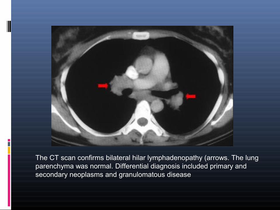
The CT scan confirms bilateral hilar lymphadenopathy (arrows. The lung parenchyma was normal. Differential diagnosis included primary and secondary neoplasms and granulomatous disease
Stage???
Stage I disease
Stage???
Stage II disease
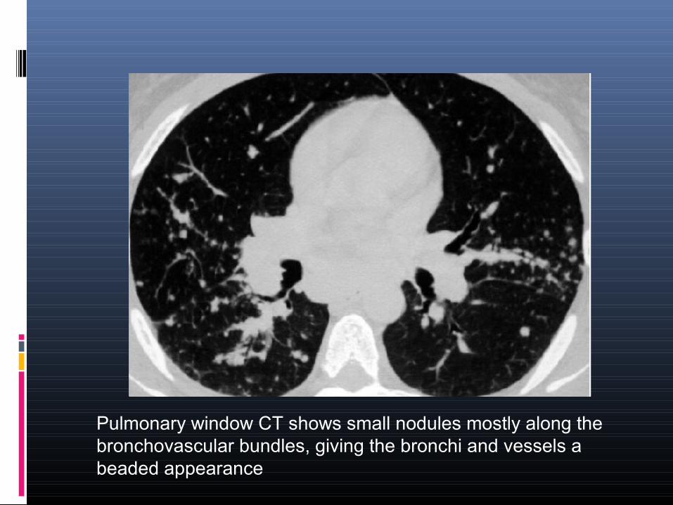
Pulmonary window CT shows small nodules mostly along the bronchovascular bundles, giving the bronchi and vessels a beaded appearance
Stage???
Stage III
Stage???
Stage I
Stage???
Stage II
Stage???
Stage 0 radiographic sarcoidosis. This normal chest x-ray may be observed in 5 to 15% of cases
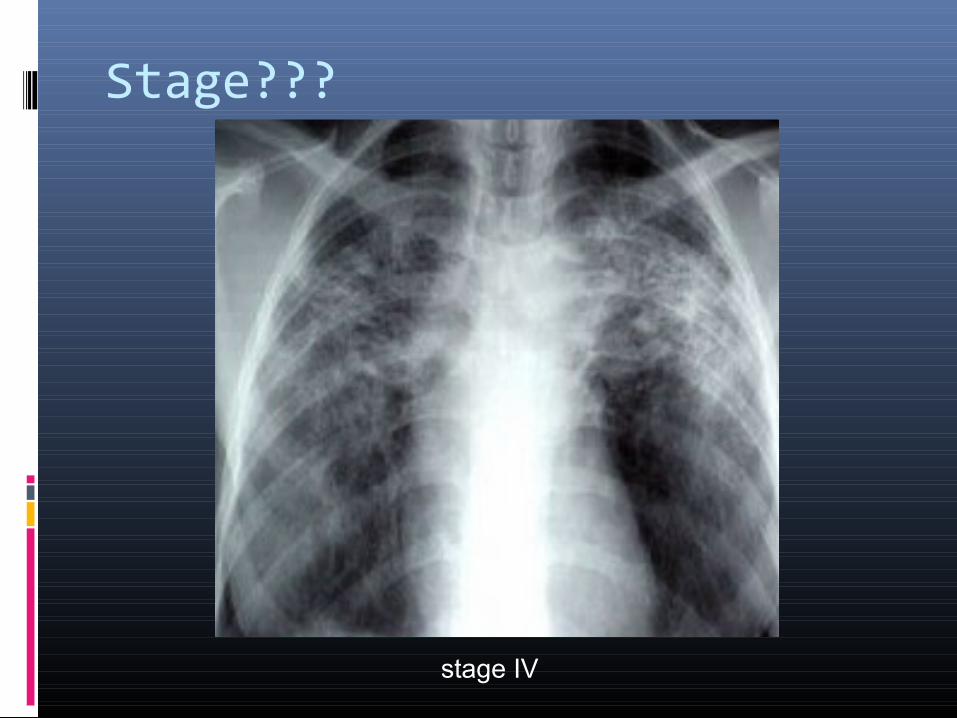
Stage???
stage IV
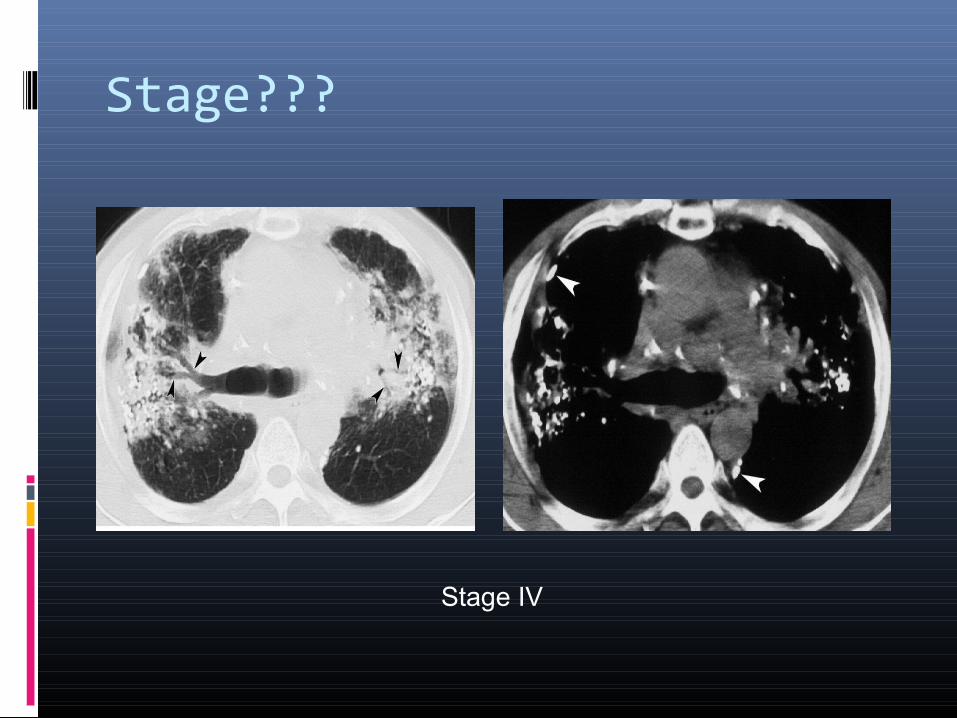
Stage???
Stage IV
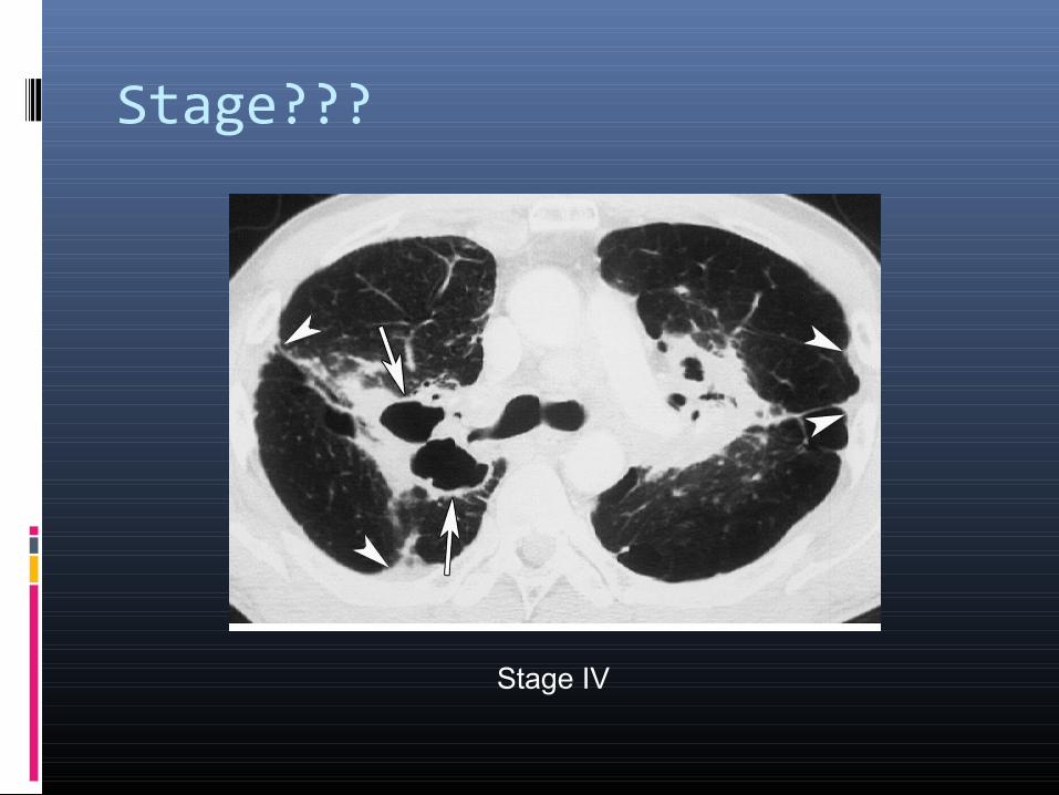
Stage???
Stage IV
Conventional chest radiographs
Over 90% of patients with sarcoidosis manifest abnormalities on chest radiographs
Commonest feature-BHL (50-80%)
Concomitant enlargement of right paratracheal lymph nodes is common
Pulmonary parenchymal infiltrates (25 to 50%)
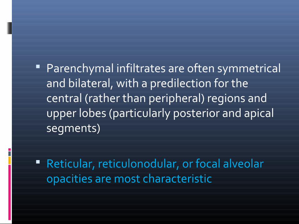
Parenchymal infiltrates are often symmetrical and bilateral, with a predilection for the central (rather than peripheral) regions and upper lobes (particularly posterior and apical segments)
Reticular, reticulonodular, or focal alveolar opacities are most characteristic
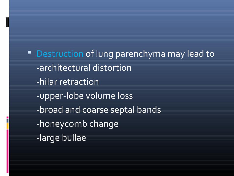
Destruction of lung parenchyma may lead to-architectural distortion-hilar retraction-upper-lobe volume loss-broad and coarse septal bands-honeycomb change-large bullae
With advanced stage III or IV sarcoidosis, enlarged pulmonary arteries (attributable to secondary pulmonary arterial hypertension) and bronchiectasis may be observed.
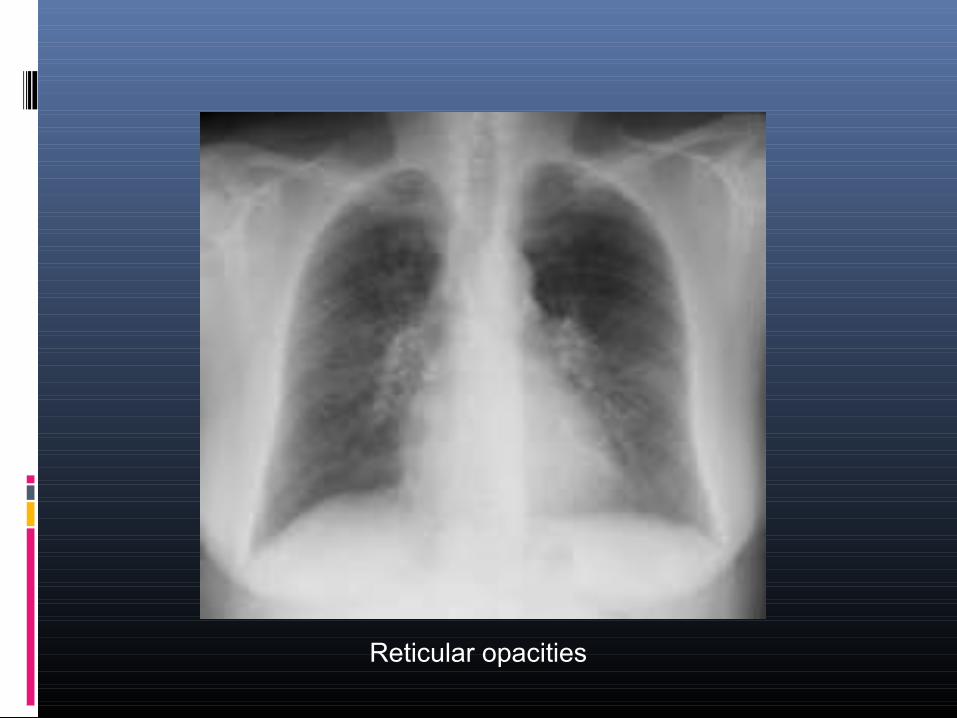
Reticular opacities
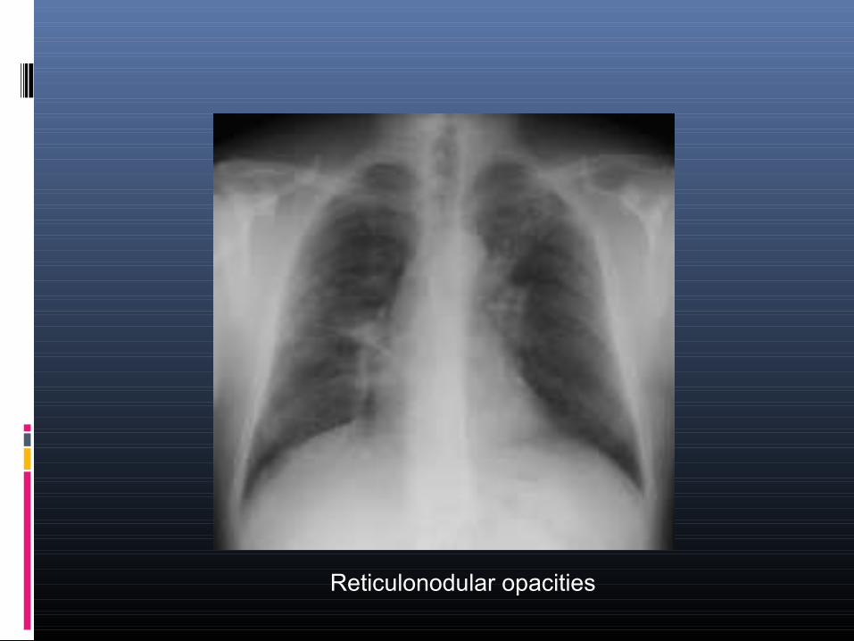
Reticulonodular opacities
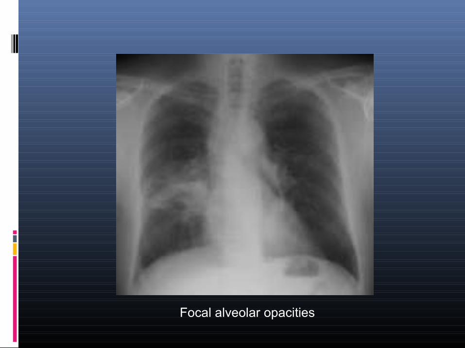
Focal alveolar opacities
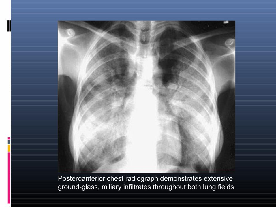
Posteroanterior chest radiograph demonstrates extensive ground-glass, miliary infiltrates throughout both lung fields
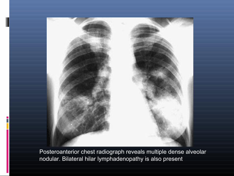
Posteroanterior chest radiograph reveals multiple dense alveolar nodular. Bilateral hilar lymphadenopathy is also present
Unusual chest radiographic features
The prevalence of atypical features is higher in sarcoid patients presenting after the age of 50
Pleural effusion Pleural thickening Pneumothorax Cavitation Bronchostenosis Vascular involvement (pulmonary vessels) Unilateral hilar lymphadenopathy
Computed tomographic scanning in Sarcoidosis INDICATIONS
(a) atypical clinical and/or chest radiographic findings.
(b) detection of complications of the lung disease.
(c) a normal chest radiograph but a clinical suspicion for sarcoidosis.
CT scan provide improved anatomic lung detail and are more sensitive than plain chest radiographs in delineating parenchymal, mediastinal, and hilar structures.
CT scan may detect enlarged lymph nodes or parenchymal infiltrates that are below the resolution of conventional chest radiographs
Enlarged lymph nodes are often observed in paratracheal, pretracheal, para-aortic, internal mammary, subcarinal, or axillary regions, which are not appreciated on chest radiographs.
Calcified hilar or mediastinal lymph nodes may be observed in patients with longstanding sarcoidosis
Calcified lymph nodes may also be observed in tuberculosis, silicosis, and other chronic granulomatous disorders.
The lymph nodes in sarcoidosis are larger than those in tuberculosis, more often focal, and less likely to be completely calcified
Lymph node calcification is bilateral in most of patients with sarcoidosis, but usually unilateral in most of patients with tuberculosis
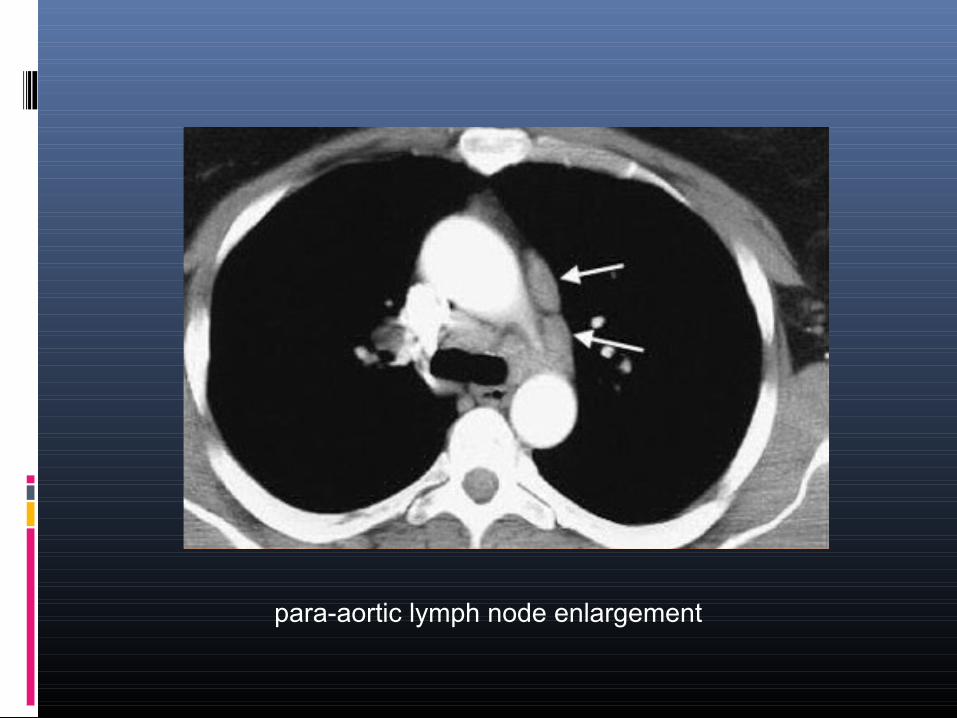
para-aortic lymph node enlargement
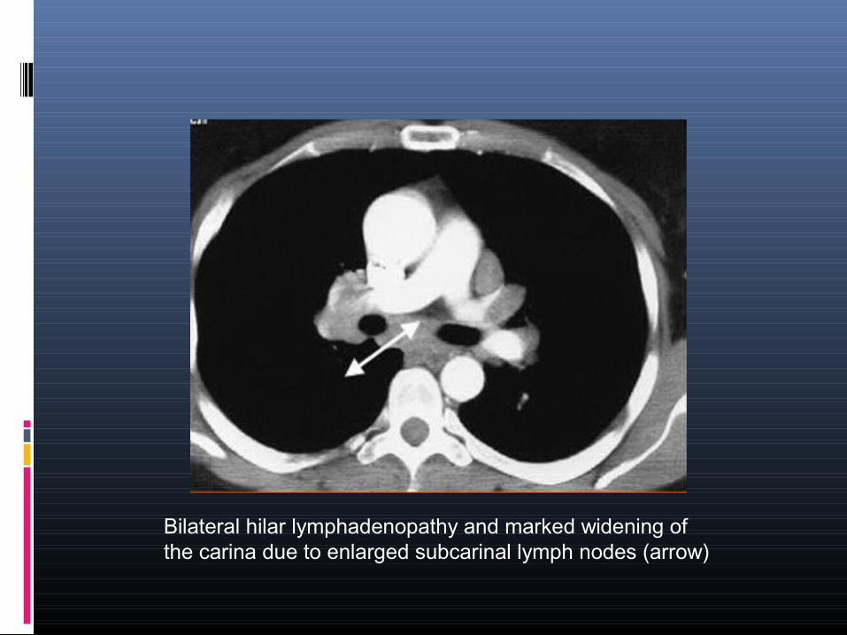
Bilateral hilar lymphadenopathy and marked widening of the carina due to enlarged subcarinal lymph nodes (arrow)
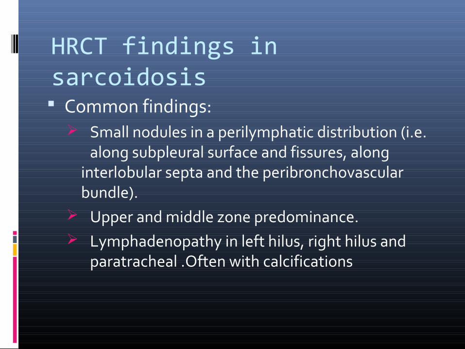
HRCT findings in sarcoidosis Common findings:
Small nodules in a perilymphatic distribution (i.e. along subpleural surface and fissures, along
interlobular septa and the peribronchovascular bundle).
Upper and middle zone predominance. Lymphadenopathy in left hilus, right hilus and
paratracheal .Often with calcifications
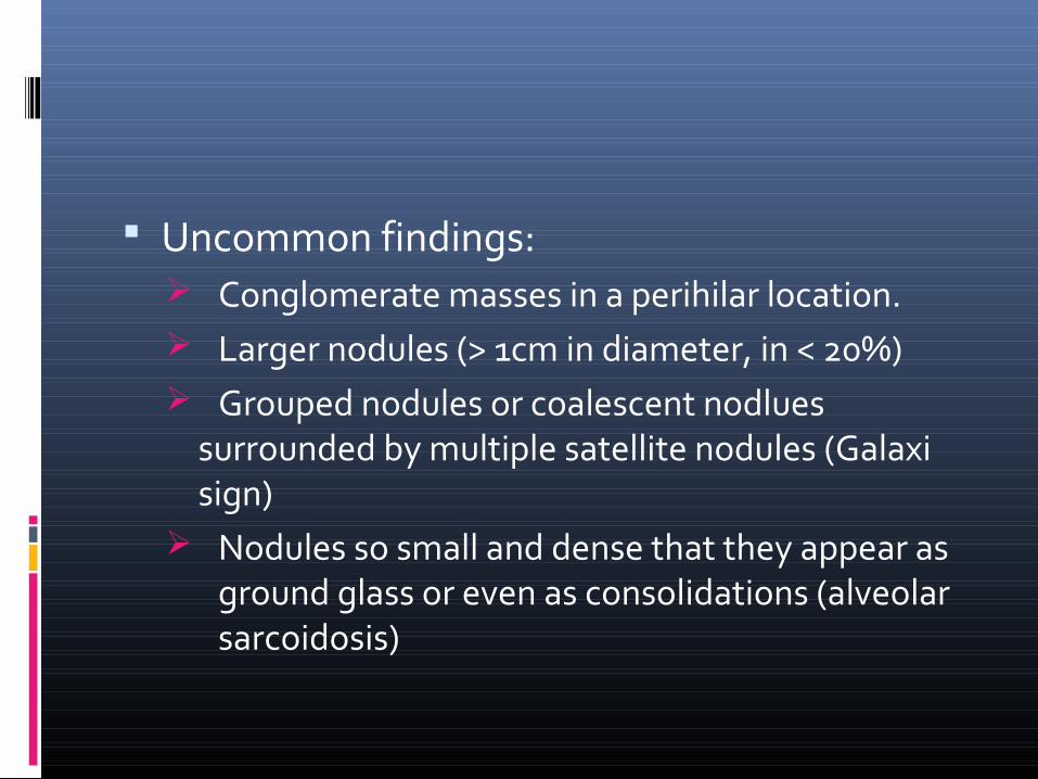
Uncommon findings: Conglomerate masses in a perihilar location. Larger nodules (> 1cm in diameter, in < 20%) Grouped nodules or coalescent nodlues
surrounded by multiple satellite nodules (Galaxi sign)
Nodules so small and dense that they appear as ground glass or even as consolidations (alveolar sarcoidosis)
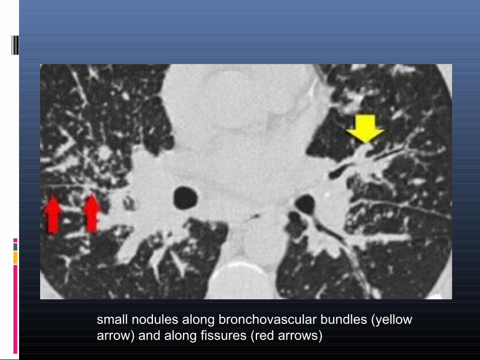
small nodules along bronchovascular bundles (yellow arrow) and along fissures (red arrows)
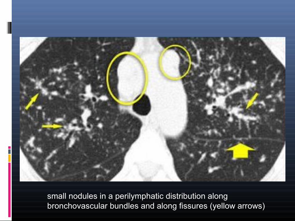
small nodules in a perilymphatic distribution along bronchovascular bundles and along fissures (yellow arrows)
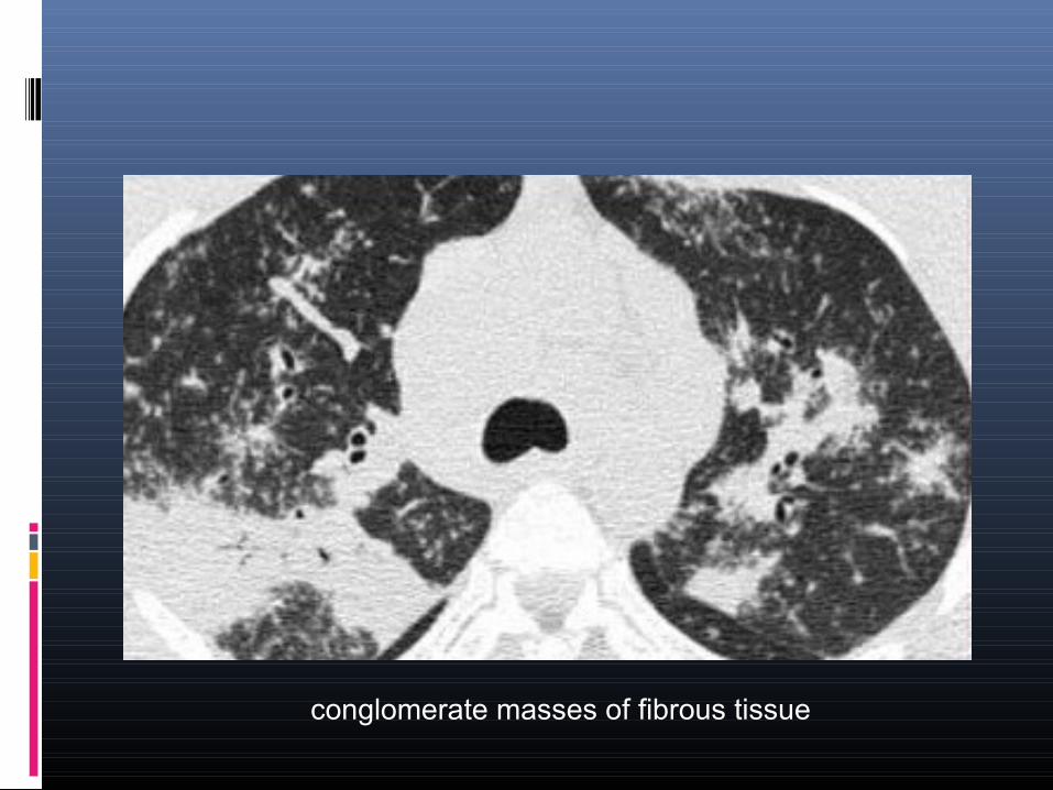
conglomerate masses of fibrous tissue
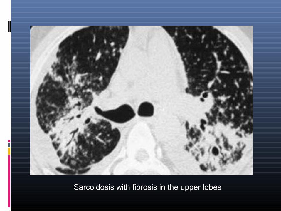
Sarcoidosis with fibrosis in the upper lobes
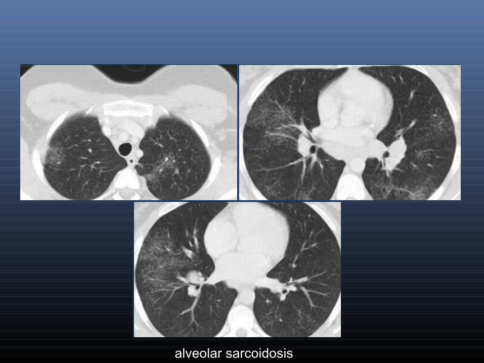
alveolar sarcoidosis
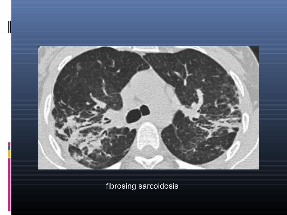
fibrosing sarcoidosis
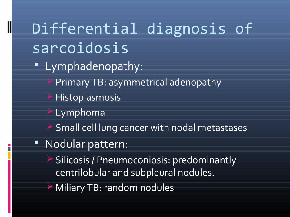
Differential diagnosis of sarcoidosis Lymphadenopathy:
Primary TB: asymmetrical adenopathy Histoplasmosis Lymphoma Small cell lung cancer with nodal metastases
Nodular pattern: Silicosis / Pneumoconiosis: predominantly
centrilobular and subpleural nodules. Miliary TB: random nodules
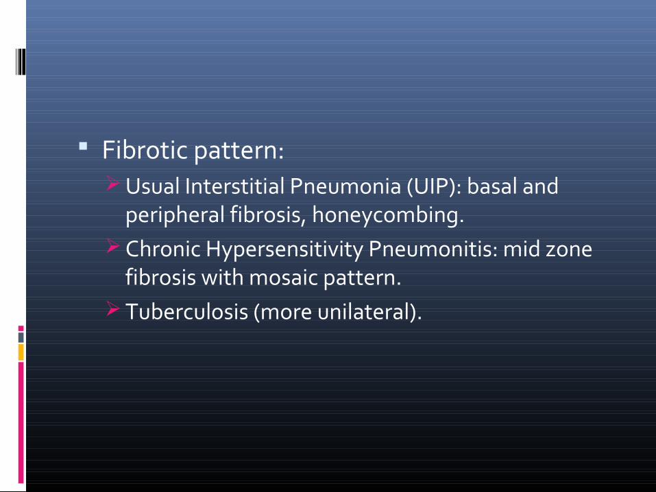
Fibrotic pattern: Usual Interstitial Pneumonia (UIP): basal and
peripheral fibrosis, honeycombing. Chronic Hypersensitivity Pneumonitis: mid zone
fibrosis with mosaic pattern. Tuberculosis (more unilateral).


![Ocular Manifestations of Biopsy-Proven Pulmonary ...downloads.hindawi.com/journals/joph/2018/9308414.pdf · ocular sarcoidosis [2–4]. Ocular sarcoidosis can occur with-out apparent](https://img.pdfslide.us/doc/110x75/60094e678ad2796c001b27fc/ocular-manifestations-of-biopsy-proven-pulmonary-ocular-sarcoidosis-2a4.jpg)