Embed Size (px)
Citation preview

Pulmonary Function Testing:The Basics
Dene W. Daugherty, DODepartment of Surgery

Pulmonary Function Testing
Tidal Volume (TV)

Pulmonary Function Testing
Inspiratory Reserve Volume (IRV)
Pulmonary Function Testing
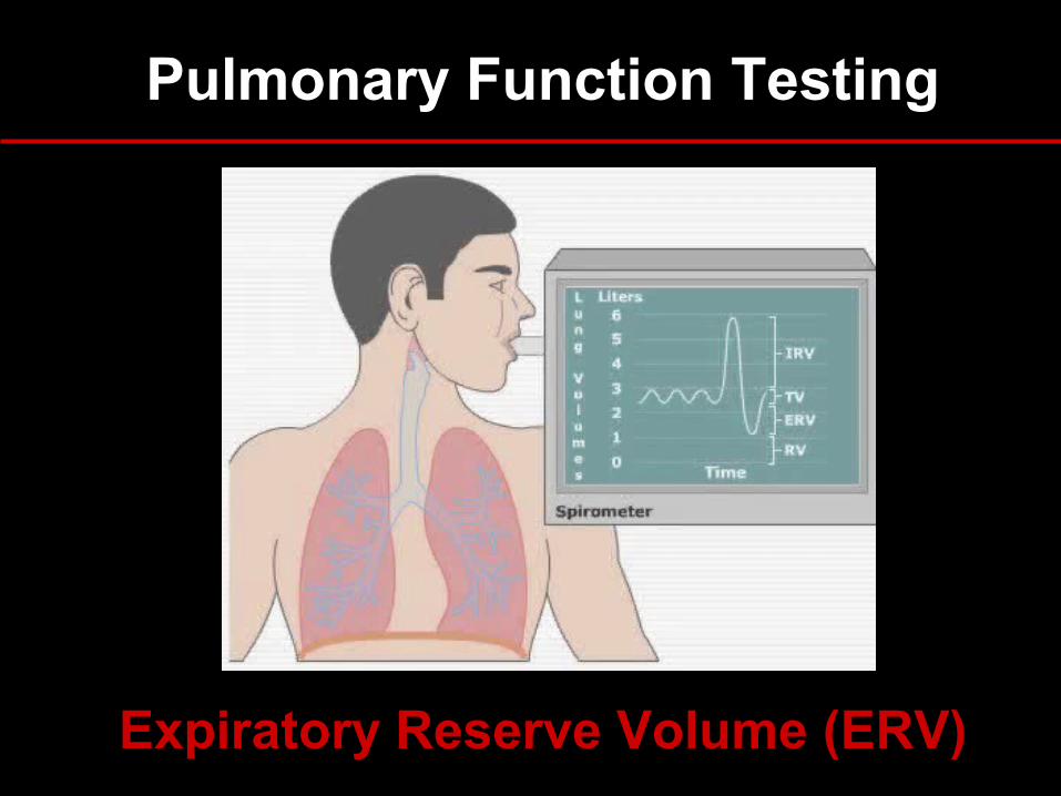
Expiratory Reserve Volume (ERV)

Pulmonary Function Testing
Vital Capacity (VC)

Pulmonary Function Testing
Total Lung Capacity (TLC)

Objectives
Identify the components
Describe the indications
Interpretation of results
Recognize common patterns
Clinical applications

The Purpose of PFT’s
To provide a quantifiable, reproducible measurement of lung
function

Description
Spirometry Flow Volume Loop Bronchodilator response Lung volumes Diffusion capacity (DLCO) Bronchoprovocation testing Maximum respiratory pressures Simple and complex cardiopulmonary exercise
testing

Indications — Diagnostic
Evaluation of signs and symptoms- SOB, exertional dyspnea, chronic cough
Screening at-risk populations
Evaluation of occupational symptoms
Monitoring pulmonary drug toxicity
Abnormal study- CXR, EKG, ABG, hemoglobin
Preoperative assessment
Indications — Prognostic
■ Assess severity
■ Follow response to therapy
■ Determine further treatment goals
■ Referral for surgery
■ Disability

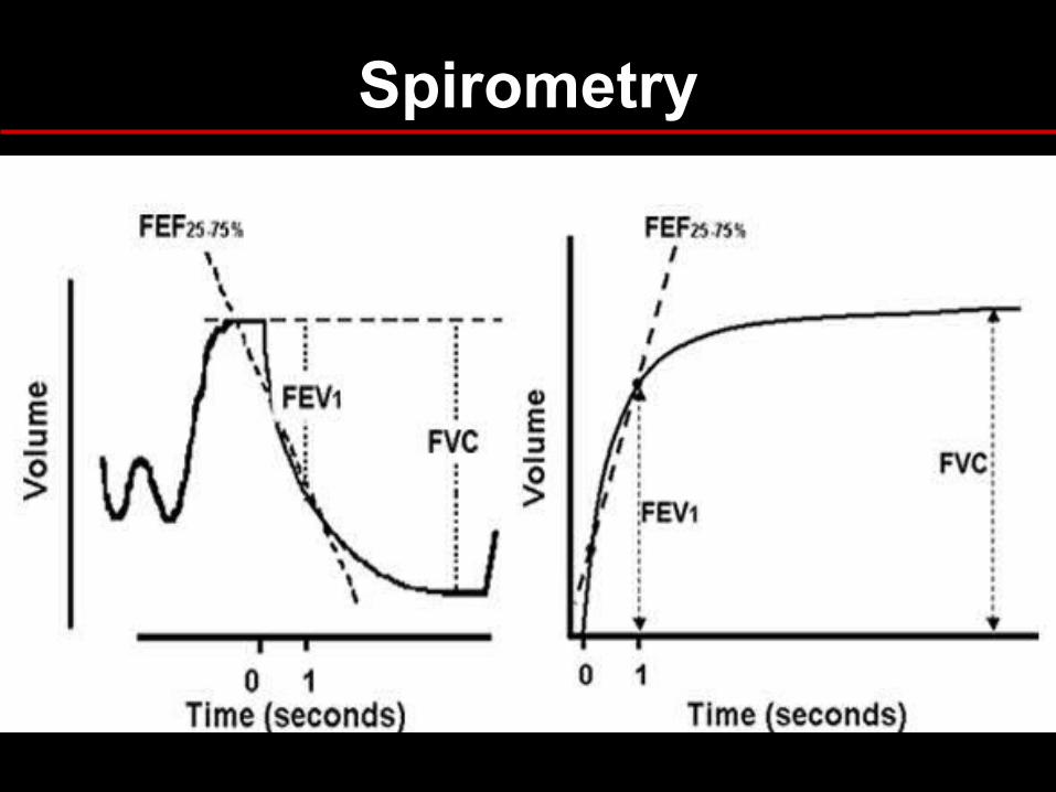
Spirometry
Simple, office-based
Measures flow, volumes
Volume vs. Time
Can determine:- Forced expiratory volume in one second (FEV1)- Forced vital capacity (FVC)- FEV1/FVC- Forced expiratory flow 25%-75% (FEF25-75)

Lung Volumes
Spirometry

Obstructive Pattern
■ Decreased FEV1
■ Decreased FVC
■ Decreased FEV1/FVC
- <70% predicted
■ FEV1 used to follow severity in COPD

Obstructive Lung Disease
Asthma
COPD - chronic bronchitis
- emphysema
Bronchiectasis
Bronchiolitis
Upper airway obstruction

Restrictive Pattern
Decreased FEV1
Decreased FVC
FEV1/FVC normal or increased

Restrictive Lung Disease
Pleural
Parenchymal
Chest wall
Neuromuscular

Spirometry Patterns

Bronchodilator Response
Degree to which FEV1 improves with inhaled bronchodilator
Documents reversible airflow obstruction
Considered a significant response if:
- FEV1 increases by 12% and >200ml
Request if obstructive pattern on spirometry

Flow Volume Loop
“Spirogram”
Measures forced inspiratory and expiratory flow rate
Augments spirometry results
Indications: evaluation of upper airway obstruction (stridor, unexplained dyspnea)

Flow Volume Loop

Upper Airway Obstruction
Variable intrathoracic obstruction
Variable extrathoracic obstruction
Fixed obstruction

Upper Airway Obstruction

Lung Volumes
Measurement:- helium- nitrogen washout- body plethsmography
Indications: - Diagnose restrictive component
- Differentiate chronic bronchitis from emphysema

Lung Volumes – Patterns
Obstructive
- TLC > 120% predicted
- RV > 120% predicted
Restrictive
- TLC < 80% predicted
- RV < 80% predicted

Diffusing Capacity
Diffusing capacity of lungs for Carbon Monoxide
Measures ability of lungs to transport inhaled gas from alveoli to pulmonary capillaries
Depends on:
- alveolar—capillary membrane
- hemoglobin concentration
- cardiac output

Diffusing Capacity
Decreased DLCO (<80% predicted)
Obstructive lung disease
Parenchymal disease
Pulmonary vascular disease
Anemia
Increased DLCO (>120-140% predicted)
Asthma (or normal)
Pulmonary hemorrhage
Polycythemia
Left to right shunt

DLCO — Indications
Differentiate asthma from emphysema
Evaluation and severity of restrictive lung disease
Early stages of pulmonary hypertension
Pre-operative assessment: < 60% correlates to poor prognosis following lung resection
Not done for routine evaluation or follow-up, it’s expensive

Bronchoprovocation
Useful for diagnosis of asthma in the setting of normal pulmonary function tests
Common agents:
- Methacholine and Histamine
Diagnostic if ≥20% decrease in FEV1

Quick Reference Obstructive Disease Algorithm
↓SYMPTOMS
PFTs
OBSTRUCTION?
YES NO
TREATBRONCHOPROVOCATION
Obstruction
TREAT
No Obstruction
Other Diagnosis
↓
↓
↓ ↓
↓
↓ ↓
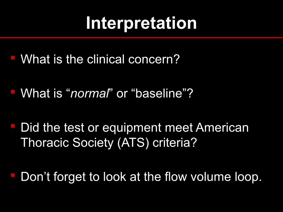
Interpretation
What is the clinical concern?
What is “normal” or “baseline”?
Did the test or equipment meet American Thoracic Society (ATS) criteria?
Don’t forget to look at the flow volume loop.

Obstructive Pattern — Evaluation
Spirometry FEV1, FVC: decreased
FEV1/FVC: decreased (<70% predicted)
FV Loop “scooped”
Lung Volumes TLC, RV: increased
Responds to Bronchodilator

Restrictive Pattern – Evaluation
Spirometry FVC, FEV1: decreased
FEV1/FVC: normal or increased
FV Loop “witch’s hat” pattern
DLCO decreased
Lung Volumes TLC, RV: decreased
Muscle pressures may be helpful

PFT Patterns
Emphysema
FEV1/FVC <70%
“Scooped” FV curve
TLC increased
Increased compliance
DLCO decreased
Chronic Bronchitis
FEV1/FVC <70%
“Scooped” FV curve
TLC normal
Normal compliance
DLCO usually normal
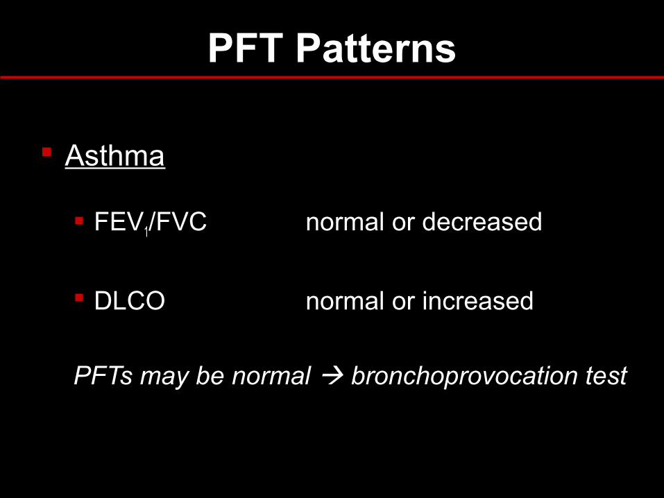
PFT Patterns
Asthma
FEV1/FVC normal or decreased
DLCO normal or increased
PFTs may be normal bronchoprovocation test

Question
Which of the following is used to follow disease severity in COPD patients?
a. Total lung capacity (TLC)
b. Degree of responsiveness to bronchodilators
c. Forced vital capacity (FVC)
d. Forced expiratory volume in 1 second (FEV1)
e. Diffusing capacity (DLCO)

Answer
Which of the following is used to follow disease severity in COPD patients?
a. Total lung capacity (TLC)
b. Degree of responsiveness to bronchodilators
c. Forced vital capacity (FVC)
d. Forced expiratory volume in 1 second (FEV1)
e. Diffusing capacity (DLCO)

Question
A 36yo F, non-smoker, presents to your office for follow-up of ‘recurrent bronchitis.’ You suspect asthma and decide to order spirometry. Which of the following would you include in your prescription for testing?
a. Diffusing Capacity (DLCO)b. If no obstruction present, perform trial of bronchodilatorc. If no obstruction present, perform methacholine challenged. Flow volume loope. b and c

Answer
A 36yo F, non-smoker, presents to your office for follow-up of ‘recurrent bronchitis.’ You suspect asthma and decide to order spirometry. Which of the following would you include in your prescription for testing?
a. Diffusing Capacity (DLCO)b. If no obstruction present, add trial of bronchodilatorc. If no obstruction present, perform methacholine challenged. Flow volume loope. b and c

Question
A 68yo M is admitted to the ICU with acute respiratory distress. A CXR obtained in the ED demonstrates bilateral pulmonary infiltrates, and his DLCO is elevated. What is the most likely diagnosis?
a. Pulmonary edemab. Aspiration pneumonitisc. Pulmonary embolid. Alveolar hemorrhagee. Interstitial lung disease

Answer
A 68yo M is admitted to the ICU with acute respiratory distress. A CXR obtained in the ED demonstrates bilateral pulmonary infiltrates, and his DLCO is elevated. What is the most likely diagnosis?
a. Pulmonary edemab. Aspiration pneumonitisc. Pulmonary embolid. Alveolar hemorrhagee. Interstitial lung disease

References
1. Aboussouan LS, Stoller JK: Flow volume loops. UpToDate, 2006.
2. Bahhady IJ, Unterborn J: Pulmonary function tests: an update. Consultant. 2003.
3. Barreiro, TJ, Perillo I: An approach to interpreting spirometry. Am Fam Physician. 2004 Mar 1;69(5):1107-14.
4. Chesnutt MS, Prendergast TJ. Current Medical Diagnosis and Treatment. New York: Appleton and Lange, 2006.
5. Enright PL: Diffusing capacity for carbon monoxide. UpToDate, 2007.
6. Enright PL: Overview of pulmonary function testing in adults. UpToDate, 2007.
7. Irvin CG: Bronchoprovocation testing. UpToDate, 2006.
8. West JB. Respiratory Physiology: The Essentials. Lippincot Williams & Wilkins, 2000.