Embed Size (px)
Citation preview
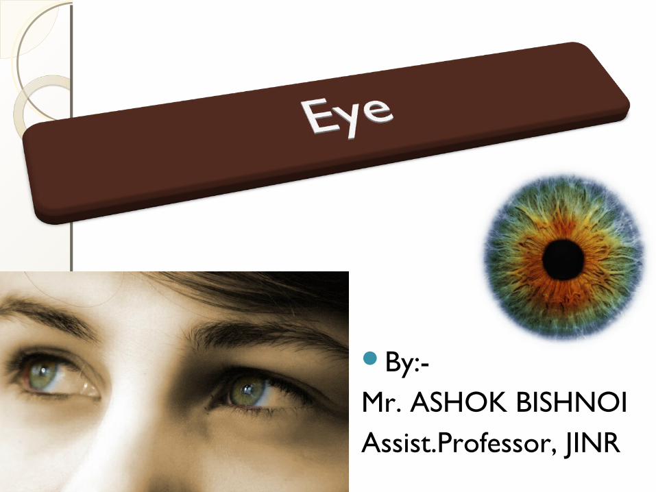
By:-Mr. ASHOK BISHNOIAssist.Professor, JINR
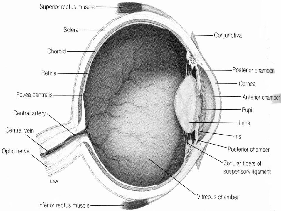
Eye Parts - Diagram
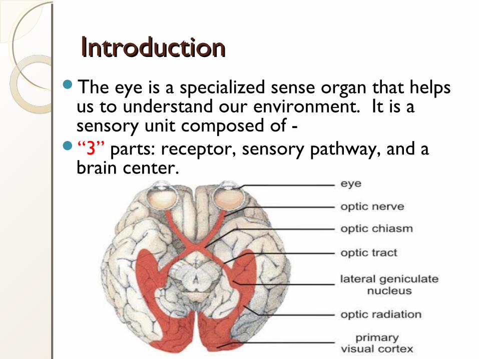
IntroductionIntroductionThe eye is a specialized sense organ that helps
us to understand our environment. It is a sensory unit composed of -
“3” parts: receptor, sensory pathway, and a brain center.
It is Spherical in shaped
It is about 2.5 cm in diameter
Situated in the orbital cavity
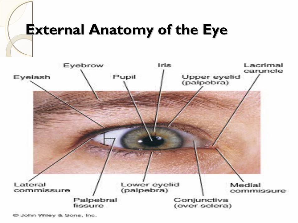
External Anatomy of the EyeExternal Anatomy of the Eye
Accessory Organs & Eye ProtectionAccessory Organs & Eye Protection
Orbital cavities (bony sockets) – house & protect the eye
• Adipose tissue – cushions the eye
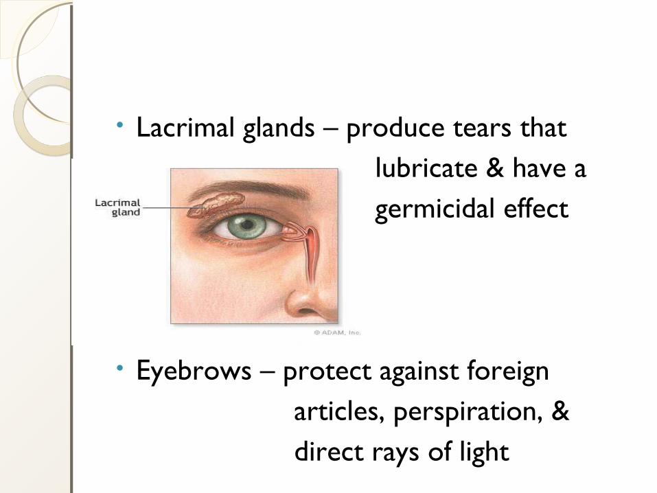
• Lacrimal glands – produce tears that lubricate & have a germicidal effect
• Eyebrows – protect against foreign articles, perspiration, & direct rays of light
Eyelids – folds of skin that cover the surface of the eye; close by reflex action when an object approaches
• Eyelashes – secrete oils that prevent lids from sticking together
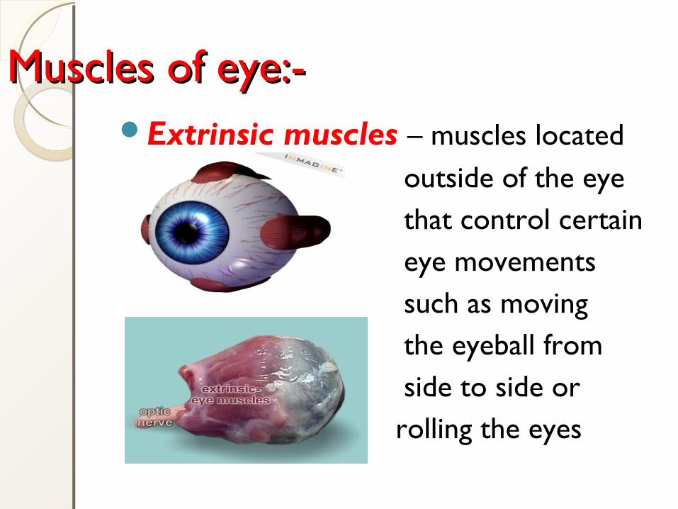
Muscles of eye:-Muscles of eye:-Extrinsic muscles – muscles located outside of the eye that control certain eye movements such as moving the eyeball from side to side or rolling the eyes
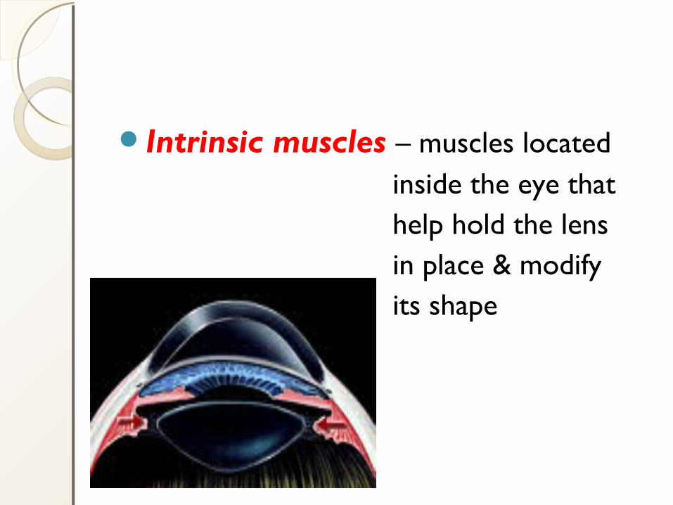
Intrinsic muscles – muscles located inside the eye that help hold the lens in place & modify its shape
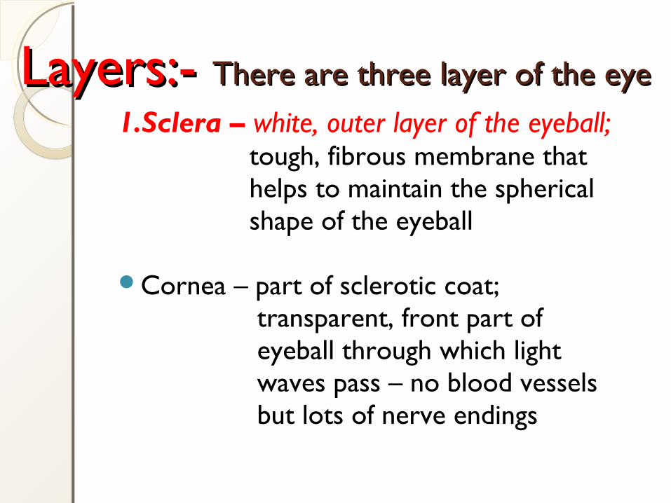
Layers:- Layers:- There are three layer of the eyeThere are three layer of the eye1.Sclera – white, outer layer of the eyeball; tough, fibrous membrane that helps to maintain the spherical shape of the eyeball
Cornea – part of sclerotic coat; transparent, front part of eyeball through which light waves pass – no blood vessels but lots of nerve endings
Canals of Schlem – venous passages that drain the fluid that accumulates behind the cornea; located where the sclera & cornea meet
Conjuctiva – thin, transparent mucous membrane that covers the eyeball
2.Choroid layer – middle layer of the eye; supplies blood vessels to the eye and contains dark pigment granules that prevent the reflection of light in the eye
Ciliary body – intrinsic muscle; smooth muscle fibers support & modify lens shape
Iris – colored portion of eye formed by circularly and radially arranged smooth muscle fibers; regulates amount of light entering they eye by constricting or dilating the pupil
Pupil – rounded opening of the iris through which light passes
• 3.Retina – innermost layer of the eye; lines its surface and contains photoreceptors (cells responsible for converting light into nerve impulses – rods & cones)
Eye PartsEye PartsRods – cylindrical photoreceptors found in greatest concentration on the edges of the retina; most common type of receptor; allow us to see in low light and provide for peripheral vision
Cones – Conical photoreceptors found in greatest concentration near the center of the retina; there are three varieties of cones, each most sensitive to a particular wavelength (color) of light – blue, green, & red; allow for visual acuity (sharp vision) and color vision
Fovea centralis – a depression, or pit, in the center of the retina that contains only cones; provides for the most acute vision & color sensitivity
Optic disk (blind spot) – area where optic nerve attaches to the retina; does not contain any photorecptors
Lens – flexible, biconvex, crystal-like structure that brings rays of light into focus and produces an image on the retina
• Suspensory ligament – holds the lens in place; attached to the ciliary body, which controls the amount of tension exerted on the lens
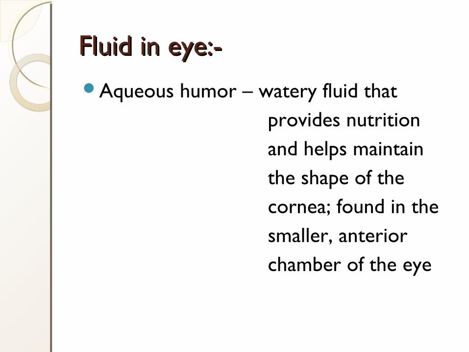
Aqueous humor – watery fluid that provides nutrition and helps maintain the shape of the cornea; found in the smaller, anterior chamber of the eye
Fluid in eye:-Fluid in eye:-
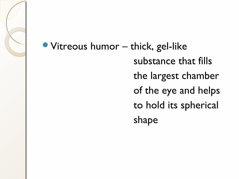
Vitreous humor – thick, gel-like substance that fills the largest chamber of the eye and helps to hold its spherical shape
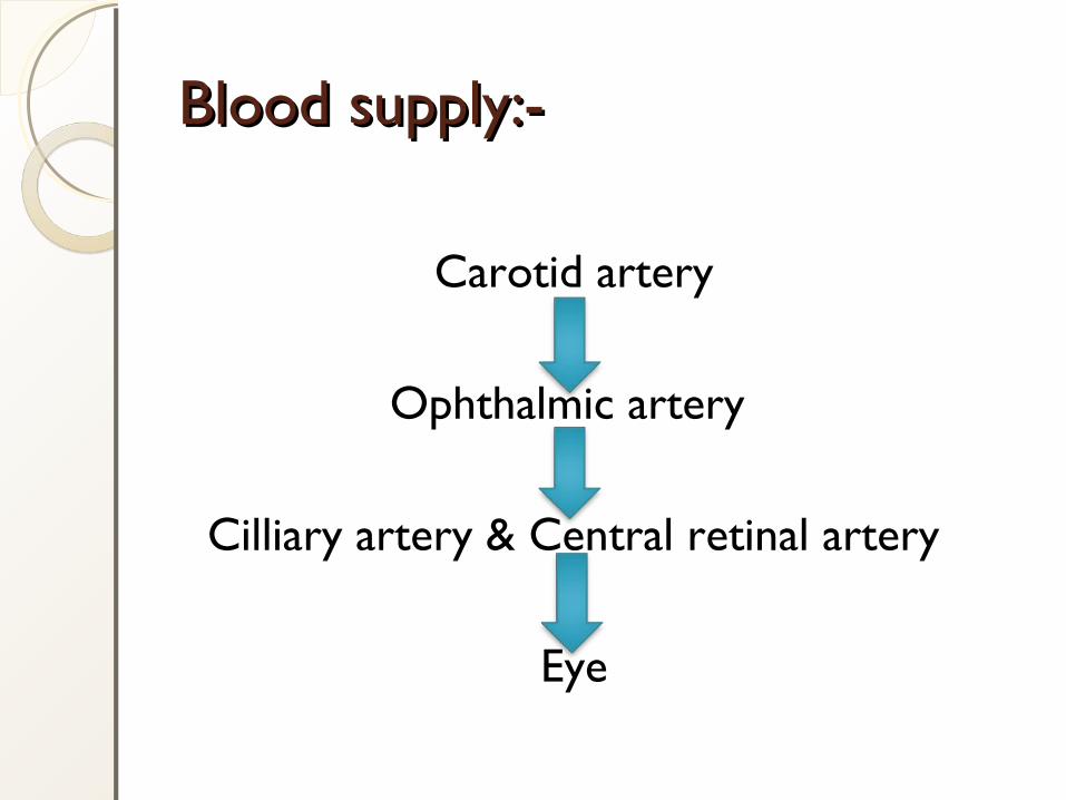
Blood supply:-Blood supply:-
Carotid artery
Ophthalmic artery
Cilliary artery & Central retinal artery
Eye
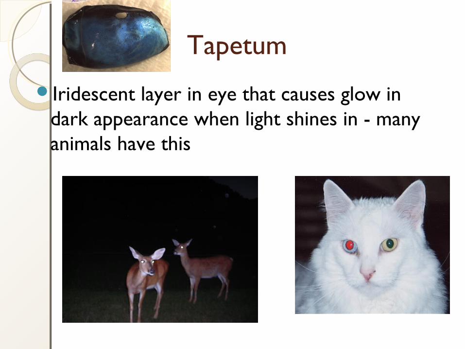
Tapetum
Iridescent layer in eye that causes glow in dark appearance when light shines in - many animals have this

The End
![[PPT]THE EYE - PowerPoint Presentations free to download ... Eye.ppt · Web viewTHE EYE Anatomy of the Eye Common Disease of the Eye Corneal Laceration James is a 22 yrs old martial](https://img.pdfslide.us/doc/110x75/5b2be91a7f8b9afd358bb692/pptthe-eye-powerpoint-presentations-free-to-download-eyeppt-web-viewthe.jpg)
![[PPT]BIONIC EYE · Web viewTitle BIONIC EYE Author EE516 Group Last modified by CASSINI Created Date 4/14/2002 5:41:09 PM Document presentation format On-screen Show (4:3) Company](https://img.pdfslide.us/doc/110x75/5ae28c057f8b9a7b218c12ca/pptbionic-viewtitle-bionic-eye-author-ee516-group-last-modified-by-cassini-created.jpg)