Embed Size (px)
Citation preview

OOHCA
The Cardiology Perspective
Ian WebbKing’s College Hospital

• Role of early coronary angiography and intervention • Literature review
• Additional considerations • The King’s Experience

• I’m an Interventional Cardiologist
• I don’t particularly like to get up in the middle of the night for OOHCA arrest patients with so many unknown clinical and prognostic variables • I have sympathy with colleagues who question the aetiology, the diagnostic work-up and early aggressive coronary strategy
• I believe that an artery with 0% stenosis is highly likely to improve ischaemia and ischaemia-related complications (like VF/VT/LV dysfunction)
• Can I prove it with Class IA data – No ...
Conflicts of Interest
• ........ And I still believe in therapeutic hypothermia

• Incidence of OOHCA in the European population estimated at about 40 per 100,000 per year1
• OOHCA resuscitations by UK Ambulance crews in 2013 ~ 28,000 cases2 (estimated 60,000 call-outs)
• LAS: 80% arrests occur at home, with 20% VF/VT the presenting rhythm3
• Outcomes in SHR better; mortality increases with every minute delay to BLS or DCCV4,5
• UK average survival to hospital discharge from decision to resuscitate 8.6% (cf Holland/Seattle/Norway ~20%) 2
• Leading causes of death are CV (acute) and brain injury (late)6,7
1. Atwood C et al. (2005) Incidence of EMS-treated out-of-hospital cardiac arrest in Europe. Resuscitation, 67: 75–802. www.england.nhs.uk/statistics/stasistical-work-areas/ambulance-quality-indicators3. London Ambulance Service Cardiac Arrest Annual Report 2012/2013 [www.londonambulance.nhs.uk]. 4. Lindner T et al. Resuscitation 2011; 82:1508-13. 5. 2010 European Resuscitation Council guidelines for resuscitation. Resuscitation 2010;81: 1219-76.6. Schoenenberger R et al. Survival after failed out-of-hospital resuscitation. Arch Intern Med. 1994;154:2433-7.7. Laver S et al. Mode of death after admission to an intensive care unit following cardiac arrest. Intensive Care Med 2004;30:2126–2128.

• OOHCA is the leading cause of death in IHD patients1
• ‘Obstructive’ coronary anatomy is common in angiographic and post- mortem series of OOHCA cohorts2-4
> 90% in STEMI / >40% in non-STEMI
• Good data to support early interventional strategy in non-OOHCA NSTEMI/STEMI5,6
• Little data to guide practice in comotose / I+V patients
- observational / registries / multiple variables •Observational literature that VT/VF burden reduced in revascularised ICD patients7,8
1. Zheng Z et al. (2001) Sudden cardiac death in the United States, 1989–1998. Circ.104:2158–21632. Spaulding C et al. (1997) Immediate Coronary angiography in survivors of OOHCA. NEJM 1997 336; 1629-16333. Davies M (1992) et al. Anatomic features in victims of sudden coronary death: coronary artery pathology. Circ ;85:S I:I-194. Dumas F et al.(2010) PROCAT registry. CCI, 3:200-207.5. 2014 ESC/EACTS Guidelines on Myocardial Revascularisation. Windecker S et al.6. 2012 ESC Guidelines for the management of acute myocardial infarction in patients presenting with ST-segment elevation. Steg P et al.7. Cook J et al. Am Heart J 2002; 143(5): 821-68. Gillis A et al. Circulation 2007; 116: II_534

•2010 International Consensus on Cardiopulmonary Resuscitation and Emergency Cardiovascular Care1
- ‘reasonable to perform immediate angiography and PCI in“selected” patients, despite the absence of STEMI or prior clinical findings such as chest pain
• 2012 ESC Guidelines support immediate CAG +/- revascularisation2
- STEMI (Class IB)- Suspected ACS irrespective of ECG (Class IIa B)
•2014 Invasive coronary treatment strategies for out-of-hospital cardiac arrest: a consensus statement from the European association for percutaneous cardiovascular interventions (EAPCI)/stent for life (SFL) groups3
1. 2010 International Consensus on Cardiopulmonary Resuscitation and Emergency Cardiovascular Care Science with Treatment Recommendations. Circulation . Hazinski M et al. 2. ESC Guidelines for the management of acute myocardial infarction in patients presenting with ST-segment elevation: Task Force Statement for ESC (2012) Steg P et al.3. Consensus statement from the European association for percutaneous cardiovascular interventions (EAPCI)/stent for life (SFL) groups. EuroIntervention 2014 May;10(1):31-7.

Common practice
OOHCA
STEMI PPCI or Lysis

Common practice
OOHCA
STEMI PPCI or Lysis
‘Not sure’
‘Could all be reactive ECG changes’
‘Nothing to hang your hat on’
‘Lets see if ‘he does’ and work him up after’
‘Normal’ ECG
Minor ST-segment changes
Moderate ST-segment or T wave changes

British Cardiac Society (Intervention Sub-group meeting) 2014
• 56-year old male
• Unprovoked OOHVF arrest
• Known IHD with prior PCI to his LAD following NSTEMI (several years ago)
• LBBB
• Prompt resuscitation
• Presents to A+E in stable condition / I+V
What would you do now?

BCS 2014
• ‘Well – the LBBB could well be old. There’s no clear mandate for CAG’
• ‘It’s unusual for it to be a primary coronary event if he had no preceding symptoms’
• ‘There’s certainly not enough evidence to rush in there and potentially de-stabilise the situation.’
• ‘You cannot tell if this is ischaemia or scar-related arrhythmia from his old MI’
BCS 2014
• Cooled
• Recurrent VF on ICU Day 2
• Commenced on amiodorone and taken to the Cath Labs
• Critical in-stent re-stenosis treated with a single DES in less than a minute

BCS 2014
• Cooled
• Recurrent VF on ICU Day 2
• Taken to the Cath Labs
• Critical in-stent re-stenosis treated with a single DES in less than a minute
• No further arrhythmias
• Extubated Day 4• Discharged to wards Day 6• Home Day 11

3 fundamental questions
• Is coronary disease a likely aetiological factor?
• What evidence is there for early diagnosis/intervention in an otherwise stabilised patient?
• What harm can an angiogram do?

Spaulding CM. N Engl J Med 1997;336:1629-33.
OOHCA associated with high incidence of IHD
Urgent CAG (84)
Normal 17 (20%)
Non-obstructive CAD 7 (8%)
Obstructive CAD 60 (71%)
Single vessel 22
Multivessel 37
Isolated LM 1
Acute Coronary occlusion 40 (48%)
Survivors to hospital in stable condition101 non-cardiac causes excluded80% VT/VFAll ECG’s

Positive Negative
Chest discomfort
and ST-elevation 87% 61%
Spaulding CM. N Engl J Med 1997;336:1629-33.
Predictive value
Absence of ST-segment elevation does not exlude acute coronary obstruction

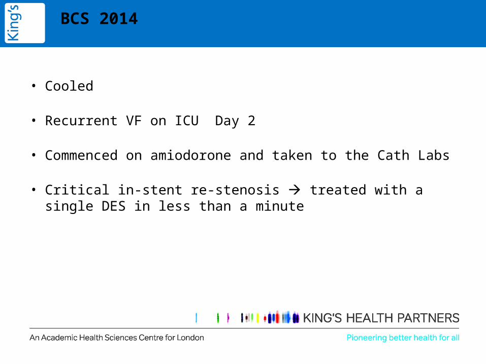
Patients post-ROSC admitted to the intensive care unit.
Florence Dumas et al. Circ Cardiovasc Interv. 2010;3:200-207
2003 – 2008
ROSC survivors to admission
All rhythms
PCI to lesions >50%
STEMI All other


• Prevalence of CAD is high – but is it causative?
• How can we prove and treat accordingly?
• Is there a role for early invasive angiography +/- revascularisation?
Evidence for acute invasive strategy?
• No RCT data
• Registry data (>3,500 patients)
• Multivariate analysis of observational registries

EuroIntervention 2014;10:31-37
Non-STEMI
Author n Multivariate predictor of survival
Spaulding 85 Successful PCI (OR 5.2; p=0.004)
Reynolds 241 CAG/PCI strategy (OR 2.16; p=0.02)
Nielsen 986 CAG/PCI strategy (OR 1.56; p=0.008)
Dumas 714 Successful PCI (OR 2.06; p=0.013)


Resuscitation 2014 85, 88-95DOI: (10.1016/j.resuscitation.2013.07.027)
ShockLV supportAntiplatelets etc..
32.7%
39.0%
Registry data754 consecutive comotose ROSC6 Tertiary care centresUnclear timeframe/historyVT/VF only STEMI excluded
Early <24hrsLate >24hrs

In non-STEMI VF/VT arrests:- 30% of patients had an acute coronary occlusion - 60-70% had significant bystander disease- 30-40% went on to have successful PCI
No angiographic differences between early and late CAG


Multivariate logistic regression analysis examining predictors of in-hospital mortality
Resuscitation 2014 85, 88-95DOI: (10.1016/j.resuscitation.2013.07.027)

Unclear if revascularisation itself is of benefit

CCI, 2010
2003 – 2008
ROSC survivors to admission n=714
PCI to lesions >50%
Patients post-ROSC admitted to the intensive care unit.
Florence Dumas et al. Circ Cardiovasc Interv. 2010;3:200-207
2003 – 2008
ROSC survivors to admission
PCI to lesions >50%

Florence Dumas et al. Circ Cardiovasc Interv. 2010;3:200-207
Survival rates according to the performance and outcome of PCI

Features associated with survival to hospital discharge
Younger age
Non-DM status
Arrest in public location
Prompt BLS and ROSC
VF/VT presentation
STEMI ECG pattern
Low admission lactate
Therapeutic hypothermia
Successful PCI OR 2.06 (1.16-3.66; p 0.013)
Only ‘successful’ PCI predictive of mortality benefit after multivariate analysis
Florence Dumas et al. Circ Cardiovasc Interv. 2010;3:200-207

DortmundPopulation >500,0002007-2008

PCI capable HospitalNon-PCI capable Hospital


Selection bias?
• Patients undergoing early CAG/revascularisation are generally:
- younger
- better CPC score post-arrest
- more likely to have STEMI
- more likely to have VF/VT
• Similarly, variability between Centres may not take into account
- sub-specialty input
- access to Ix / treatment options/pathways
- more formalised MDT approach to patient care
Are all coronary stenoses alike?

Are all coronary stenoses alike?

• STEMI – plaque rupture, heavy thrombus burden, impaired coronary flow
- clear mandate to restore flow
• Obstructive coronary disease with preserved TIMI flow
- take a view on the complexity of anatomy in conjunction with
the patient and their haemodynamics
- Surgeons rarely taken on emergency CABG in this setting
- Similarly, complex rotablation in heavily calcified vessels or
complex CTO revascularisation NOT an option acutely
No RCT Data
No information about revascularisation strategies from registry data
Little to guide us on timing (acute vs staged) – except shock and extent of revascularisation (cf non-comatose ACS patients)
But worth noting: PCI revascularisation levels high in STEMI cohorts and 30-50% in non-STEMI groups from registry data

What harm can an angiogram do?

What harm can an angiogram do?
*Major bleeding approximately 3.5%
*Peripheral ischaemic complications approx 4-11%
*Stroke rates approx 1%
------------------------------------
Radial vs femoral
Use of adjunctive LV support devices
Variable anticoagulation regimens
*IABP – SHOCK II study, NEJM 2012Early Revascularization in Acute Myocardial Infarction Complicated by Cardiogenic Shock, NEJM 1999 30-40% CPR/VT/VF
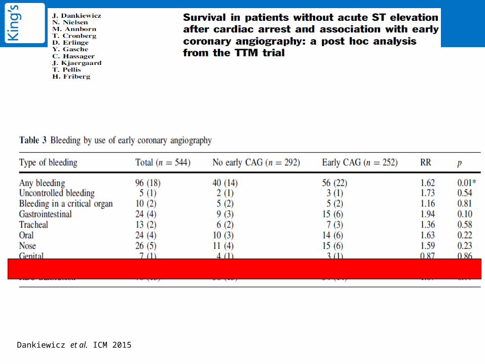
Dankiewicz et al. ICM 2015

What harm can an angiogram do?

LV support devices

IABP Impella Tandem Heart ECMO
LV support devices
14Fr8Fr 17/15Fr 14-29Fr

LV support devices
• Cardiogenic shock is present in 30-40% of OOHCA survivors
• Presents most often within 4-6 hours of the index event
• No device has shown prognostic benefit in comatose or non-comatose patients1
• Use of IABP/Impella observational series ranges from 10-42%2
• Higher incidence in patients treated emergently by PCI
• Future Role for Impella CP and/or ECMO...
• Principle of haemodynamic support vs vascular access and longer-term complications (sepsis, blood dyscrasias etc..)
1. ISAR Shock, IABP-SHOCK I and II, PROTECT II2. Cheng et al. 2009, Sjauw et al. 2009, Hovdenes et al. 2009

Pharmacotherapy

Acute Stent Thrombosis

Acute Stent Thrombosis
• Acute stent thrombosis occurs in 4.6% - 10.9% of ACS patients complicated by CA1,2
• OR for AST 12.9 following cardiac arrest (95%CI 1.3-124.6; p=0.027)2
• Mortality with AST is high (up to 45%) 3
• Likely factors - non-administration of antiplatelets / heparins2
- malabsorption and altered metabolism4
- highly procoagulant /inflammatory state- adverse haemodynamic status
1. Shah N et al. JACC 2015;65(10_S)2. Joffre J et al. Resuscitation 2014; 85(6):769-733. Buchanan G et al. Thrombosis 20124. Bjelland T et al. Resuscitation 2010;81:1627–31

Rosencher J et al. 2015
N=20OOHCAPCI

Cangrelor
• IV P2Y12 ADP receptor antagonist• High affinity, reversible• ½ life 3 minutes• Normal PLT function after 1hr
• Recent FDA approval


London Heart Attack Centre Network 2005
Harefield Royal Free Heart London Chest
Imperial
St. George’s
St. Thomas’s
King’s
8 HACApprox 30 Acute admission units7.5/10million people>600m2

• 9,805 OOHCA calls attended by LAS• Resuscitation attempted for 4,317 patients
- 92% declared dead on scene / 8% DNR in place
• Of those actively treated by LAS:
• Survival to discharge:
2013/2014
48.6% Witnessed
44.8% Bystander CPR performed
31% Sustained ROSC to Hospital admission
85.7% Primary cardiac aetiology
10.3% Witnessed
32.4% Utstein population (witnessed / Vf or VT / presumed cardiac)
47.6% Treated at HAC
58.8% Use of AED

78% Private
86% cardiac
65% transfer to hospital
31% sustained ROSC
December Monday mornings!

King’s College Hospital
• PPCI programme 2003• 1 million catchment• 4th busiest London unit• ~900 STEMI/yr• 120 OOHCA-I+V/yr

OOHCAOOHCA
STEMI
STEMI FAST-TRACK(Cath Labs)
Rapid evaluation of comorbidities
+ Cardiac arrest details
STEMI FAST-TRACK(Cath Labs)
Rapid evaluation of comorbidities
+ Cardiac arrest details
Coronary angiography
No STEMI
‘ER stop’‘ER stop’
Diagnostic work-up
Additional history
Additional history
EchoEcho
Laboratory values
Laboratory values
CT Imaging
CT Imaging
Obvious non-cardiac causes excluded

Coronary angiography
STEMI
Non-STEMI ECGClear culprit stenosis+
Non-STEMI ECGSignificant stenosis <70%
Good flow (TIMI III)
Non-STEMI ECGNon-obstructive coronary
disease
PCI of culprit artery
Consider bystander PCI if:
- unstable- low risk + prognostic
Consider PCI if:- clinical correlate
- < intermediate risk
STOPReview
+ Stenosis >90%, thrombus, <TIMI I/II flow, clinical correlate* DAPT and statin therapy per NGT
*
*

OOHCA
Cath Lab
Resus
ITU
Home
N = 331
N = 146 (44.1%)
N = 185 (55.9%)
N = 47 (25.4 %)
N = 144 (43.5 %)2011-2014OOHCA ROSCSurvivors to Cath-Lab +/or ICUAll I+V cases
Total number to Cath Lab =193
Data - R.Nerla/F.Jouhra

OOHCA presenting rhythms
Data - R.Nerla/F.Jouhra

Presumed causes of OOHCA
Data - R.Nerla/F.Jouhra

Primary Diagnosis
Data - R.Nerla/F.Jouhra

OOHCA
Cath Lab sub-group
Cath Lab
No angio
PCI
AngioN =181 (93.7%)
N = 12 (6.3%)
N = 117 (65%)
No PCI
N = 59 (32.6%)
N = 331
N = 193 (58.3 %)
Futile N = 6Other diagnosis more probable N = 6
CABG N = 5STEMI (92%)nonSTEMI (38%)
Data - R.Nerla/F.Jouhra
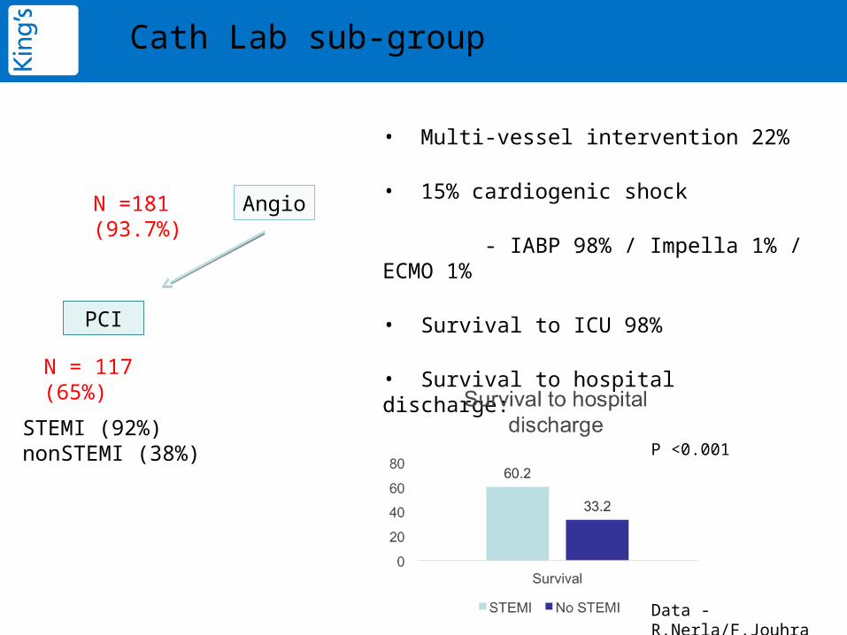
Cath Lab sub-group
• Multi-vessel intervention 22%
• 15% cardiogenic shock
- IABP 98% / Impella 1% / ECMO 1%
• Survival to ICU 98%
• Survival to hospital discharge:PCI
AngioN =181 (93.7%)
N = 117 (65%)
STEMI (92%)nonSTEMI (38%) P <0.001
Data - R.Nerla/F.Jouhra

Survival by diagnosis
P < 0.001
* Including all patients with a definite primitive cardiac cause for OOHCA
Data - R.Nerla/F.Jouhra
Overall survival to discharge 43.5%
Median ICU stay (d) 3 (0-40)Mean 6.8 + 8.2
Median Hospital stay 6 (0-87)

Work to do...
• Follow-up data
Mortality
Neurological outcomes
ICD implantation
• Improving post-arrest care
LV support (ECMO)
Neurocognitive prognostication
Neuro-rehab
Academic integration

Summary
• OOHCA is associated with significant mortality and morbidity
• The major gains in survival are likely to evolve through front-managed patient care
• The prevalence of coronary disease in OOHCA survivors is significant
• Whether this is causative or not is uncertain – but aggressive multi-disciplinary patient management – including early coronary angiography and revascularisation – appears safe and to confer better survival and neurological outcomes.