Embed Size (px)
Citation preview
1
Optimizing male infertility treatment in ART
Dr Parul Katiyar MDSenior Consultant, Infertility and Reproductive Medicine Max Hospitals, New Delhi and Gurgaon
Male factor infertility
Male factor is solely responsible for infertility in ~20% cases
Male factor contributes to infertility in another 30-40% cases
Semen analysis is the cornerstone for diagnosing male infertility
In many men with normal sperm parameters, sperms do not function in a manner necessary for fertility and can still cause infertility
How to improve treatment outcome in ART?
Improve the diagnostic evaluation Improve the sperm health Improve the fertilization technique Improve the sperm retrieval procedures
Improving diagnostic evaluation
Minimum evaluation of a man with suspected male infertility includes A complete medical history – surgery/ medications/
infections/ allergies/ lifestyle Full physical examination – secondary sexual
characters/ testes/ varicocele Two semen analyses done eight weeks apart and
assessed using WHO criteria
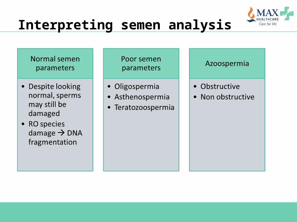
Interpreting semen analysis
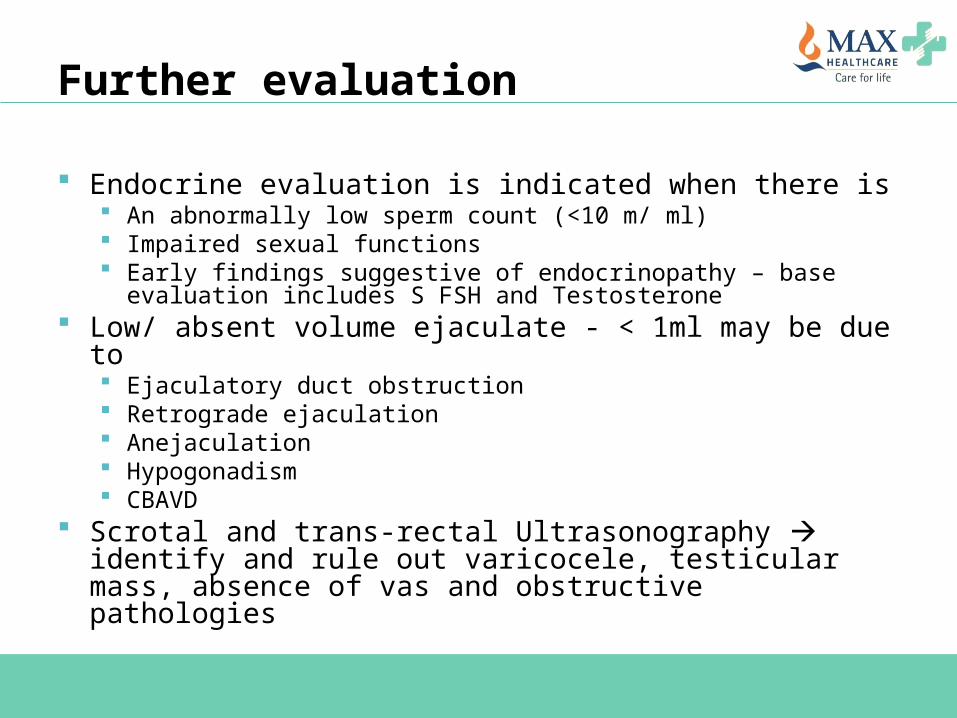
Further evaluation
Endocrine evaluation is indicated when there is An abnormally low sperm count (<10 m/ ml) Impaired sexual functions Early findings suggestive of endocrinopathy – base evaluation
includes S FSH and Testosterone Low/ absent volume ejaculate - < 1ml may be due to
Ejaculatory duct obstruction Retrograde ejaculation Anejaculation Hypogonadism CBAVD
Scrotal and trans-rectal Ultrasonography identify and rule out varicocele, testicular mass, absence of vas and obstructive pathologies
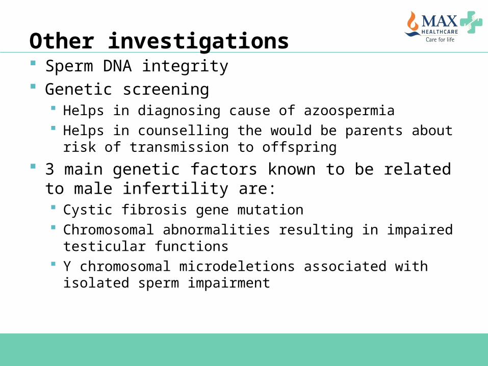
Other investigations Sperm DNA integrity Genetic screening
Helps in diagnosing cause of azoospermia Helps in counselling the would be parents about risk of
transmission to offspring 3 main genetic factors known to be related to male
infertility are: Cystic fibrosis gene mutation Chromosomal abnormalities resulting in impaired testicular
functions Y chromosomal microdeletions associated with isolated sperm
impairment
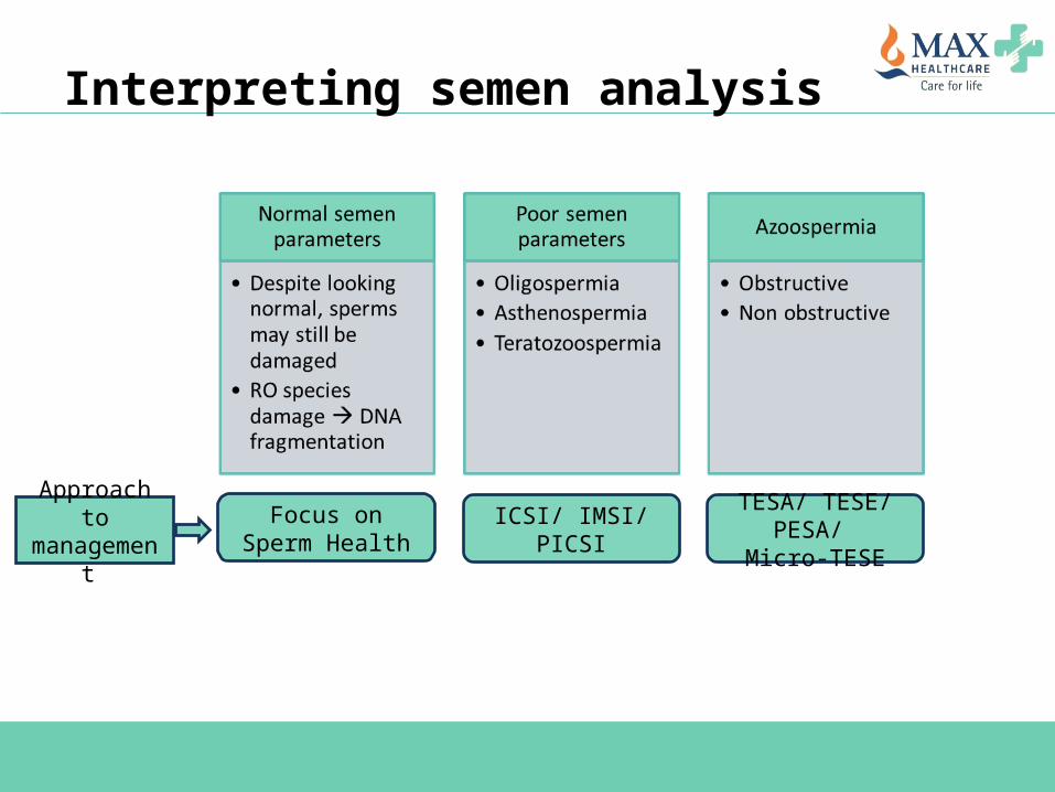
Interpreting semen analysis
Focus on Sperm Health ICSI/ IMSI/ PICSI TESA/ TESE/ PESA/
Micro-TESEApproach to
management
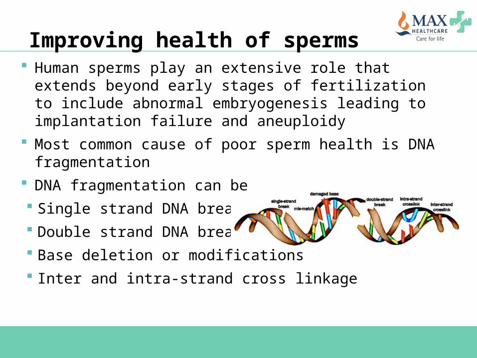
Improving health of sperms Human sperms play an extensive role that extends beyond
early stages of fertilization to include abnormal embryogenesis leading to implantation failure and aneuploidy
Most common cause of poor sperm health is DNA fragmentation
DNA fragmentation can be Single strand DNA break Double strand DNA break Base deletion or modifications Inter and intra-strand cross linkage
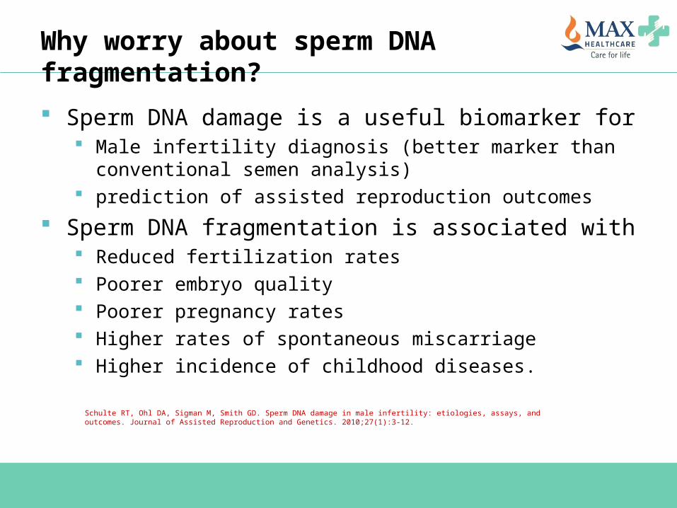
Why worry about sperm DNA fragmentation?
Sperm DNA damage is a useful biomarker for Male infertility diagnosis (better marker than conventional semen
analysis) prediction of assisted reproduction outcomes
Sperm DNA fragmentation is associated with Reduced fertilization rates Poorer embryo quality Poorer pregnancy rates Higher rates of spontaneous miscarriage Higher incidence of childhood diseases.
Schulte RT, Ohl DA, Sigman M, Smith GD. Sperm DNA damage in male infertility: etiologies, assays, and outcomes. Journal of Assisted Reproduction and Genetics. 2010;27(1):3-12.
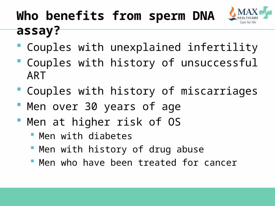
Who benefits from sperm DNA assay?
Couples with unexplained infertility Couples with history of unsuccessful ART Couples with history of miscarriages Men over 30 years of age Men at higher risk of OS
Men with diabetes Men with history of drug abuse Men who have been treated for cancer
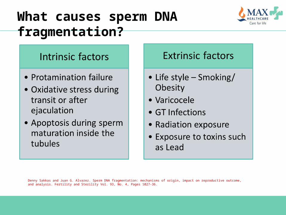
What causes sperm DNA fragmentation?
Denny Sakkas and Juan G. Alvarez. Sperm DNA fragmentation: mechanisms of origin, impact on reproductive outcome, and analysis. Fertility and Sterility Vol. 93, No. 4, Pages 1027-36.
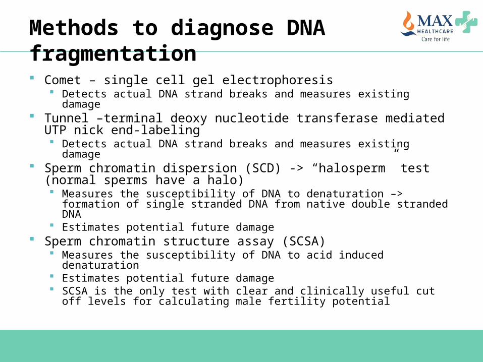
Methods to diagnose DNA fragmentation
Comet – single cell gel electrophoresis Detects actual DNA strand breaks and measures existing damage
Tunnel –terminal deoxy nucleotide transferase mediated UTP nick end-labeling Detects actual DNA strand breaks and measures existing damage
Sperm chromatin dispersion (SCD) -> “halosperm” test (normal sperms have a halo) Measures the susceptibility of DNA to denaturation –> formation of
single stranded DNA from native double stranded DNA Estimates potential future damage
Sperm chromatin structure assay (SCSA) Measures the susceptibility of DNA to acid induced denaturation Estimates potential future damage SCSA is the only test with clear and clinically useful cut off levels for
calculating male fertility potential

Sperm DNA fragmentation assays TUNEL assay -> Blue sperm are TUNEL
negative while green sperm are TUNEL positive indicating DNA fragmentation.
Sperm Chromatin Dispersion Test -> The two sperm in the center with non-fragmented DNA form large halos, while the sperm in the upper right hand corner has no halo indicating DNA fragmentation.
Results from both assays are expressed as percentage of sperm demonstrating DNA fragmentation.
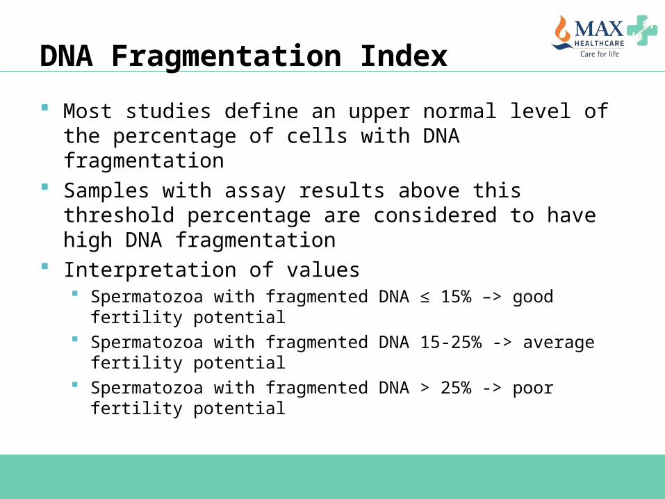
DNA Fragmentation Index
Most studies define an upper normal level of the percentage of cells with DNA fragmentation
Samples with assay results above this threshold percentage are considered to have high DNA fragmentation
Interpretation of values Spermatozoa with fragmented DNA ≤ 15% –> good fertility
potential Spermatozoa with fragmented DNA 15-25% -> average fertility
potential Spermatozoa with fragmented DNA > 25% -> poor fertility
potential

Management of sperm DNA fragmentation Lifestyle modifications
Cessation of smoking and alcohol intake Weight management for obese men
Antioxidants – should be used at least for 2-3 months for effect Vit C (500 mg/ day) Vit E (200 mg/ day) Folic Acid (2 mg/ day) Zinc (25 mg/ day) Selenium (26 mcg/ day)
Treatment of underlying pathological conditions Varicocele GU Infections
Avoid environmental exposure to toxins and judicial medical use of radiations
Reducing the abstinence period or serial ejaculation every 24 hours reduces SDF by up to 25%
In cases with uncorrected DNA fragmentation – consider surgical sperm retrieval
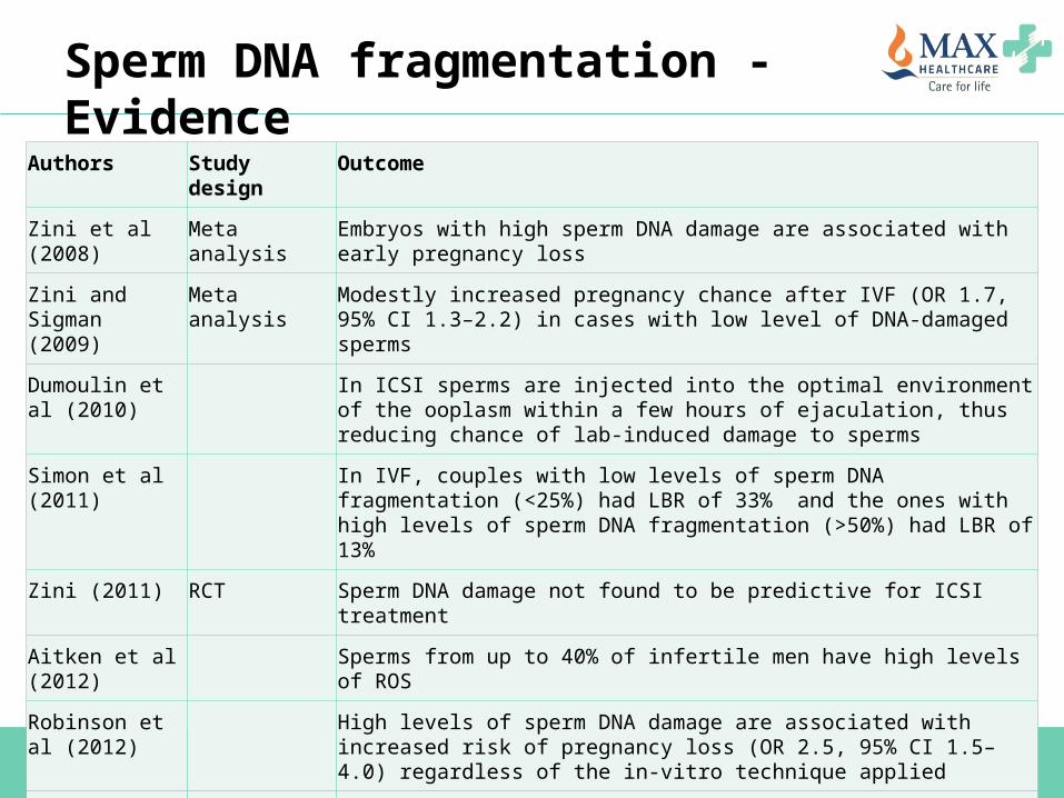
Sperm DNA fragmentation - EvidenceAuthors Study design Outcome
Zini et al (2008) Meta analysis Embryos with high sperm DNA damage are associated with early pregnancy loss
Zini and Sigman (2009)
Meta analysis Modestly increased pregnancy chance after IVF (OR 1.7, 95% CI 1.3–2.2) in cases with low level of DNA-damaged sperms
Dumoulin et al (2010)
In ICSI sperms are injected into the optimal environment of the ooplasm within a few hours of ejaculation, thus reducing chance of lab-induced damage to sperms
Simon et al (2011) In IVF, couples with low levels of sperm DNA fragmentation (<25%) had LBR of 33% and the ones with high levels of sperm DNA fragmentation (>50%) had LBR of 13%
Zini (2011) RCT Sperm DNA damage not found to be predictive for ICSI treatment
Aitken et al (2012) Sperms from up to 40% of infertile men have high levels of ROS
Robinson et al (2012)
High levels of sperm DNA damage are associated with increased risk of pregnancy loss (OR 2.5, 95% CI 1.5–4.0) regardless of the in-vitro technique applied
Simon et al (2013) OR of 76 (95% CI 8.7–1700) for clinical pregnancy when the mean DNA fragmentation per spermatozoon was < 52%
Osman et al (2015)Meta analysis LBR was lower for high sperm DNA fragmentation in IVF cycles. But, there was no difference in LBR between low and high sperm DNA fragmentation with ICSI
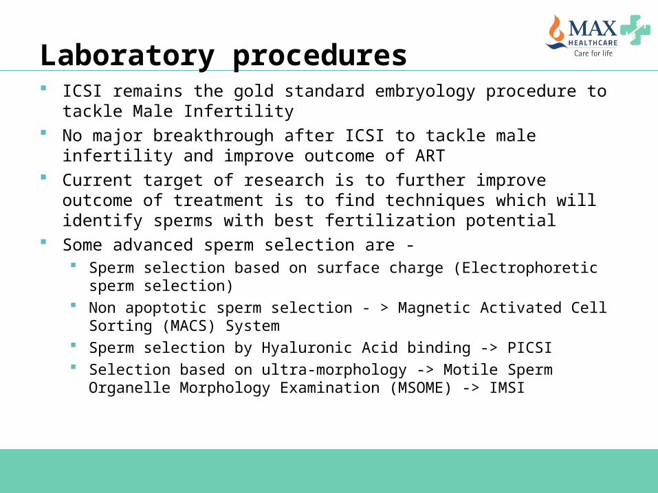
Laboratory procedures ICSI remains the gold standard embryology procedure to tackle Male
Infertility No major breakthrough after ICSI to tackle male infertility and improve
outcome of ART Current target of research is to further improve outcome of treatment is
to find techniques which will identify sperms with best fertilization potential
Some advanced sperm selection are - Sperm selection based on surface charge (Electrophoretic sperm selection) Non apoptotic sperm selection - > Magnetic Activated Cell Sorting (MACS)
System Sperm selection by Hyaluronic Acid binding -> PICSI Selection based on ultra-morphology -> Motile Sperm Organelle
Morphology Examination (MSOME) -> IMSI

Electrophoretic sperm selection
Positively (PCS) and negatively charged sperms (NCS) can be identified using micro- electrophoresis techniques.
DNA damage is inversely proportional to % NCS and directly proportional to the % PCS
Selection of Negatively charged sperms using this technique helps isolate sperms, which are relatively free of DNA damage, and can be used for ART
Simon et al. Micro-electrophoresis: a noninvasive method of sperm selection based on membrane charge. Fertility and Sterility, Volume 103, Issue 2, 2015, 361–366.e3
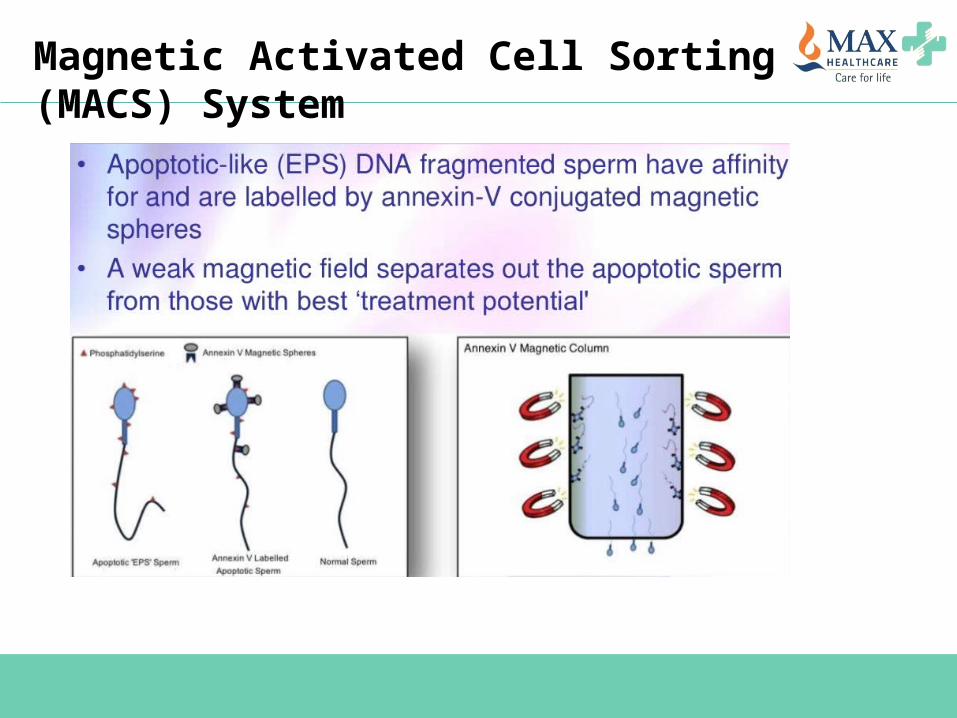
Magnetic Activated Cell Sorting (MACS) System
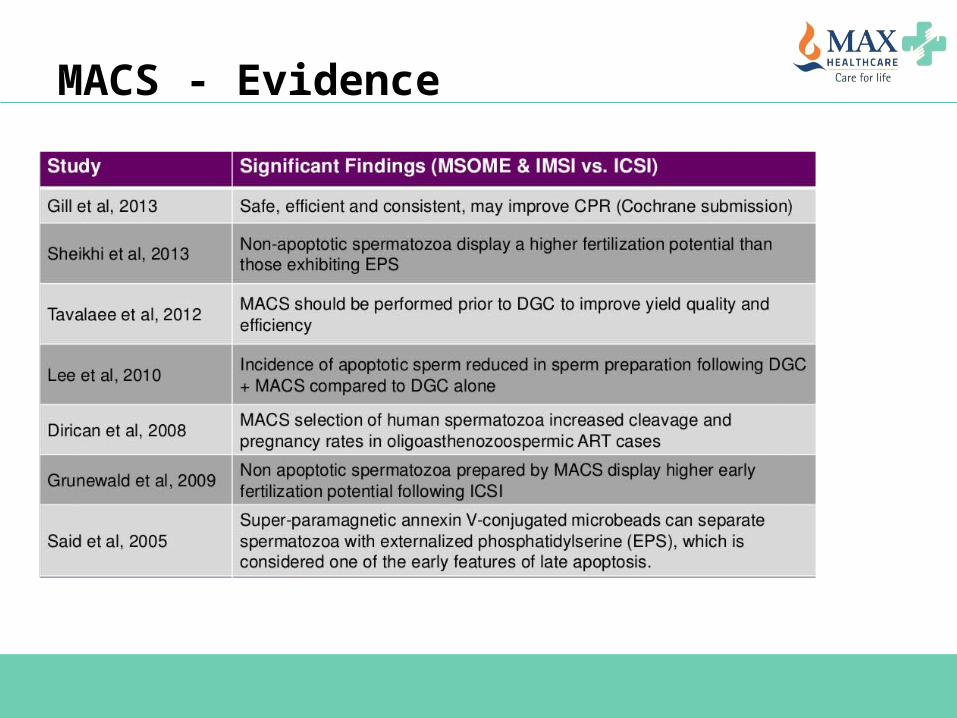
MACS - Evidence

Sperm selection by HA binding - PICSI
Formation of hyaluronic acid binding sites on the sperm plasma membrane is one of the signs of sperm maturity and forms the basis of sperm selection
PICSI dish has been developed by adding 4 marked spots of immobilized HA in a Falcon petri dish
One drop of washed spermatozoa is placed at the edge of HA spot and HA bound spermatozoa are collected after 15 minutes in an ICSI pipette and used for injection
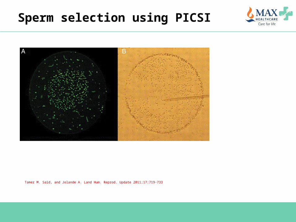
Tamer M. Said, and Jolande A. Land Hum. Reprod. Update 2011;17:719-733
Sperm selection using PICSI
Advantages & Effect on ART outcome
Advantages of PICSI PICSI ensures that the selected sperms are mature ->
Defined by Creatinine Kinase, HspA2 & Aniline Blue staining Lower risk of aneuploidy Better embryo quality and cleavage rates
Effects on ART outcome – what does the evidence tell us? Not much improvement in fertilization rates/ pregnancy rates
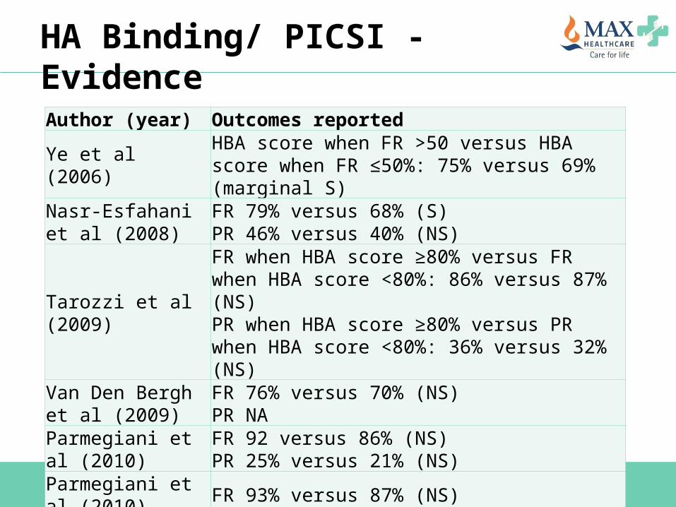
HA Binding/ PICSI - Evidence
Author (year) Outcomes reported
Ye et al (2006) HBA score when FR >50 versus HBA score when FR ≤50%: 75% versus 69% (marginal S)
Nasr-Esfahani et al (2008)
FR 79% versus 68% (S)PR 46% versus 40% (NS)
Tarozzi et al (2009)
FR when HBA score ≥80% versus FR when HBA score <80%: 86% versus 87% (NS)PR when HBA score ≥80% versus PR when HBA score <80%: 36% versus 32% (NS)
Van Den Bergh et al (2009)
FR 76% versus 70% (NS)PR NA
Parmegiani et al (2010)
FR 92 versus 86% (NS)PR 25% versus 21% (NS)
Parmegiani et al (2010) FR 93% versus 87% (NS)
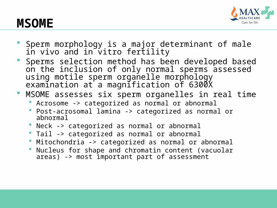
MSOME Sperm morphology is a major determinant of male in
vivo and in vitro fertility Sperms selection method has been developed based on
the inclusion of only normal sperms assessed using motile sperm organelle morphology examination at a magnification of 6300X
MSOME assesses six sperm organelles in real time Acrosome -> categorized as normal or abnormal Post-acrosomal lamina -> categorized as normal or abnormal Neck -> categorized as normal or abnormal Tail -> categorized as normal or abnormal Mitochondria -> categorized as normal or abnormal Nucleus for shape and chromatin content (vacuolar areas) ->
most important part of assessment
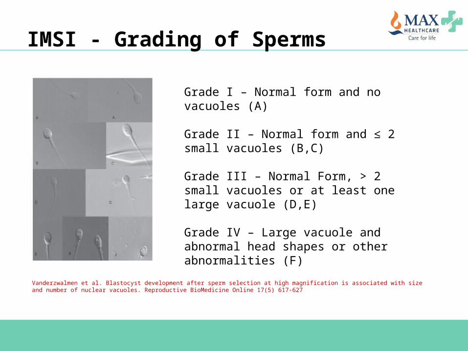
IMSI - Grading of Sperms
Grade I – Normal form and no vacuoles (A)
Grade II – Normal form and ≤ 2 small vacuoles (B,C)
Grade III – Normal Form, > 2 small vacuoles or at least one large vacuole (D,E)
Grade IV – Large vacuole and abnormal head shapes or other abnormalities (F)
Vanderzwalmen et al. Blastocyst development after sperm selection at high magnification is associated with size and number of nuclear vacuoles. Reproductive BioMedicine Online 17(5) 617-627
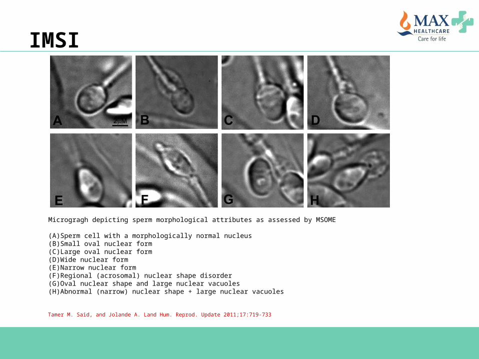
Microgragh depicting sperm morphological attributes as assessed by MSOME (A)Sperm cell with a morphologically normal nucleus(B)Small oval nuclear form(C)Large oval nuclear form(D)Wide nuclear form(E)Narrow nuclear form(F)Regional (acrosomal) nuclear shape disorder(G)Oval nuclear shape and large nuclear vacuoles(H)Abnormal (narrow) nuclear shape + large nuclear vacuoles
Tamer M. Said, and Jolande A. Land Hum. Reprod. Update 2011;17:719-733
IMSI
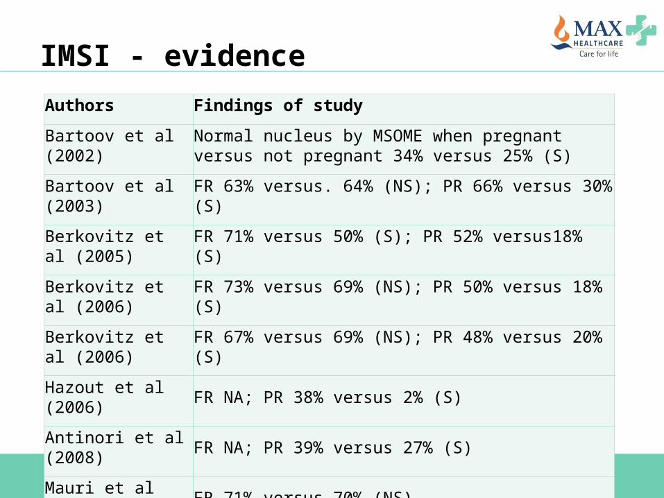
IMSI - evidenceAuthors Findings of study
Bartoov et al (2002) Normal nucleus by MSOME when pregnant versus not pregnant 34% versus 25% (S)
Bartoov et al (2003) FR 63% versus. 64% (NS); PR 66% versus 30% (S)
Berkovitz et al (2005) FR 71% versus 50% (S); PR 52% versus18% (S)
Berkovitz et al (2006) FR 73% versus 69% (NS); PR 50% versus 18% (S)
Berkovitz et al (2006) FR 67% versus 69% (NS); PR 48% versus 20% (S)
Hazout et al (2006) FR NA; PR 38% versus 2% (S)
Antinori et al (2008) FR NA; PR 39% versus 27% (S)
Mauri et al (2010) FR 71% versus 70% (NS)
Gianaroli et al (2008) FR 74% versus 72% (NS); PR 31% versus 21% (NS)
Gianaroli et al (2010) FR 69% versus 67% (NS); PR 55% versus 14% (S)
Role of IMSI - conclusion
Results from RCTs do not support the clinical use of IMSI There is no evidence of effect on live birth or miscarriage
and the evidence that IMSI improves clinical pregnancy is of very low quality
There is no indication that IMSI increases congenital abnormalities
Further trials are necessary to improve the evidence quality before recommending IMSI in clinical practice
Teixeira DM, Barbosa MAP, Ferriani RA, Navarro PA, Raine-Fenning N, Nastri CO, Martins WP. Regular (ICSI) versus ultra-high magnification (IMSI) sperm selection for assisted reproduction. Cochrane Database of Systematic Reviews 2013, Issue 7. Art. No.: CD010167.
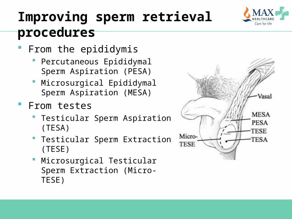
Improving sperm retrieval procedures
From the epididymis Percutaneous Epididymal Sperm
Aspiration (PESA) Microsurgical Epididymal Sperm
Aspiration (MESA) From testes
Testicular Sperm Aspiration (TESA) Testicular Sperm Extraction (TESE) Microsurgical Testicular Sperm
Extraction (Micro-TESE)
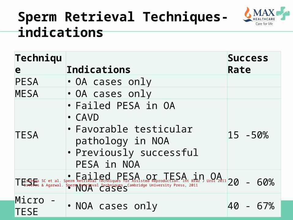
Sperm Retrieval Techniques- indications
Technique Indications Success RatePESA • OA cases onlyMESA • OA cases only
TESA
• Failed PESA in OA• CAVD• Favorable testicular pathology in NOA• Previously successful PESA in NOA
15 -50%
TESE• Failed PESA or TESA in OA• NOA cases 20 - 60%
Micro - TESE • NOA cases only 40 - 67%Esteves SC et al. Sperm Retrieval Techniques for Assisted Reproduction. Int Braz J Urol 2011Esteves & Agarwal. Sperm Retrieval Techniques. Cambridge University Press, 2011
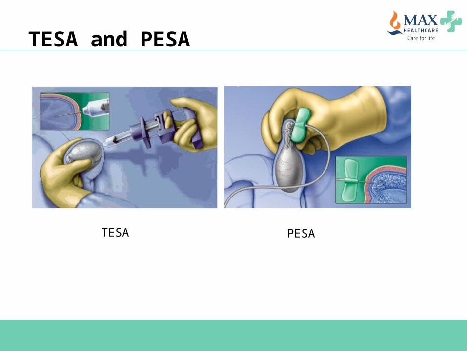
TESA and PESA
TESA PESA
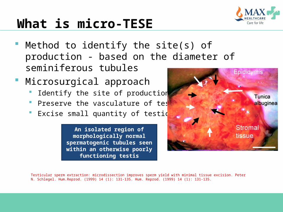
What is micro-TESE Method to identify the site(s) of production - based on
the diameter of seminiferous tubules Microsurgical approach
Identify the site of production Preserve the vasculature of testis Excise small quantity of testicular tissue
Testicular sperm extraction: microdissection improves sperm yield with minimal tissue excision. Peter N. Schlegel. Hum.Reprod. (1999) 14 (1): 131-135. Hum. Reprod. (1999) 14 (1): 131-135.
An isolated region of morphologically normal
spermatogenic tubules seen within an otherwise poorly
functioning testis
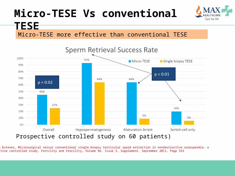
Micro-TESE Vs conventional TESE
Prospective controlled study on 60 patients)
Verza & Esteves, Microsurgical versus conventional single-biopsy testicular sperm extraction in nonobstructive azoospermia: a prospective controlled study, Fertility and Sterility, Volume 96, Issue 3, Supplement, September 2011, Page S53
Micro-TESE more effective than conventional TESE
Take home messages
Optimizing the outcome of ART in male infertility Normal semen parameters ≠ healthy spermsPoor ART outcome can be correlated with high DNA fragmentation in spermsHealthy lifestyle & correction of underlying pathology for healthier spermsNewer modalities of sperms selection like IMSI, PICSI and MACS will benefit a carefully selected segment of patients Micro-TESE is superior to TESE for sperm retrieval in NOA
Thank YouDr Parul Katiyar [email protected]