Embed Size (px)
Citation preview

Occipital Lobe Syndromes

CLASSIFICATION
• Visual field defects:-
Homonymous hemianopia
Blindsight
• Cortical blindness
• Visual anosognosia (Anton syndrome)
• Visual illusions (metamorphopsias)
• Visual hallucinations
• Occipital lobe epilepsy

Homonymous hemianopia
• It is hemianopia visual field loss on the same side of both eyes.
• Occurs because the right half of the brain has visual pathways for the left hemifield of both eyes, and the left half of the brain has visual pathways for the right hemifield of both eyes.
• When one of these pathways is damaged, the corresponding visual field is lost.

Homonymous hemianopia (homonymous hemianopia)
Homonymous hemianopia caused by lesion posterior to the optic chiasm.
Vision is lost in ipsilateral nasal field & contralateral temporal field.
2 types Complete
Incomplete
Incomplete – 2 types Incongruous – differently shaped defect in both eyes
lesion site – anterior lesion
(optic tract, LGN)
Congruous – similarly shape defect
lesion site – posterior (occipital lobe)
In complete hemianopia congruity can not be assessed
Homonymous hemianopiaCongruous
Incongruous

i\[[[[[[[[[[[[[[[[[[[[]\\\\\\\\\\\\\\f

Lesions of visual cortex
Congruous homonymous
hemianopia(sparing macula)
Occlusion of posterior cerebral artery
supplying anterior part of occipital cortex
Congruous homonymous macular
defect
Head injury/gun shot injury leading to lesions
of tip of occipital cortex+

Macular sparing• Macular sparing is when the
central 5-10 degrees of the visual field is unaffected in an otherwise hemianopic defect. it is due to
Separate blood supply[Smith and Richardson (1996)].
They demonstrated two interesting points:
• (i) in some individuals the occipital pole of the visual cortex is supplied by the middle cerebral artery rather than the posterior cereberal artery and
• (ii) in some patients, there is a horizontal border at the macular between the areas supplied by the posterior temporal artery ( a branch of the posterior cerebral artery) and the area supplied by the middle cerebral artery.
• Extent of macular representationThe final theory to explain macular sparing simply states that the macular area has such a large cortical representation that in any incomplete lesion there is a high probability that some of the macular fibres will be left intact.

Visual field defects
• Some visual perception is preserved in hemianopia field.
• OKN are usually spared in hemianopia of occipital origin.
• Coloured targets may be detected , achromatic one are not.
• Residual visual function –may be likely due to sparing of small
island of calcarine neurons.

CORTICAL BLINDNESS
• Due to B/L lesion of occipital lobe
• Total loss of vision in both eyes ,loss of reflex lid closure
to a bright light or threat
• Normal retina & ocular str., normal pupillary reflex and
maintenance of full extra ocular movements
• OKN –absent
• Alpha rhythm in EEG is lost
• Less complete lesion- variable perception

CORTICAL BLINDNESS (contd…)
ETIOLOGY :-
• Occlusion of PCA (Embolic or thrombotic)
• Hypertensive/eclamptic/hypoxic encephalopathy
• Schilder’s ds. & other leucodystrophies
• CJD
• PML
• Cerebral infarction following cardiac arrest
• B/L Glioma
TRANSITORY:-
• Head injury
• Migraine
• Antiphospholipidsyndrome
• Drugs:-IFN alpha,
cyclosporine

Anton–Babinski syndrome(Visual anosognosia)
• Denial of blindness who cannot see.
• The lesion extend beyond the striate cortex to involve visual association areas.
• Failing to accept being blind, the sufferer dismisses evidence of his condition and employs confabulation to fill in the missing sensory input.
• Lesion is in visual association areas superior to calcarine cortex.
Gabriel Anton

Blindsight:-– Pt. with no ability to discriminate
patterns in the hemianopic field,
nonetheless could still reach
accurately and look at a moving
light in blind field.
– Flashing lights & moving objects
can sometime be seen even
without patient’s full awareness
(Weiskrantz & colleagues)
– Attributed to preserved function
of retinocollicular or
geniculoprestriate cortical
connections
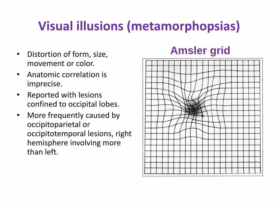
Visual illusions (metamorphopsias)
• Distortion of form, size, movement or color.
• Anatomic correlation is imprecise.
• Reported with lesions confined to occipital lobes.
• More frequently caused by occipitoparietal or occipitotemporal lesions, right hemisphere involving more than left.
Amsler grid

Visual illusions contd..
Different forms of illusions:-
• PHOSPHENES – flashes of light.
• METACHROMATOPSIA – colour changes into entirely different colour
• ALLESTHESIA – displacement of real object, person from its true geographic location
• PALLINOPSIA – persistence/reccurence of image after the stimulus has been removed
• METAMORPHOPSIA – distortion of shape
• MICROPSIA – object appear small, lilliputian vision
• MACROPSIA – magnification of object

Visual illusions contd…
Different forms of illusions:-
• TELEOPSIA – object appear distant.
• POLYOPIA – seeing single target as multiple
• ENTOMOPIA – multiple copies of same image in a grid like pattern
• SCINTILLATION – subjective sensation of sparks, flickering, or flashes of light
• UNFORMED – images not recognized as person or object
• FORMED – images of person or object

Visual hallucinations
SIMPLE/Elementary
(unformed) visual hallucination:-
points, stars, flames, flashes, wheels, circles and triangles, may be stationary/moving.
Lesion #Occipital lobes
Coloured in SEIZURE
Simplest black & white moving scintillation MIGRAINE

Complex Visual Hallucinations
Complex Hallucinations
• Complex hallucinations may feature images of people, faces, birds, animals or scenery.
• Lesion in Temporal or frontal lobes

Visual hallucinations
contd….• With hemianopia, visual hallucinations appear in the
defective field or move from intact field toward
hemianopic field.
• Also can occur during recovery from cortical blindness.
• Charles Bonnet syndrome:-Complex visual hallucination in elderly
Preservation of insight and other cognitive abilities
Almost always have poor vision
Visual hallucinations in blindness .

OCCIPITAL LOBE EPILEPSY
Characteristics:- Elementary visual hallucination:- flashing or steady spots or simple geomatric forms either
coloured or achromatic When lateralised – contralateral to side of lesion Stationary or move across the visual field Ictal amaurosis:- second most common symptom Blindness may limited to one visual field but often B/LIf spread outside occipital lobe
– Somatosensory aura– Complex visual or auditory hallucination
Aura of eye movement sensation without detectable eye movement.
Forced blinking or eye lid flutter at the beginning of seizure are objective evidence of occipital lobe origin.

OCCIPITAL LOBE EPILEPSY
Idiopathic occipital epilepsy:-
Gastaut type:-
• Age of onset is b/w 3 to 16 yrs of age.
• Characteristics:-visual hallucinations, ictal blindness,
phosphenes & tonic deviation of eyes.
• Seizure type- Hemiclonic, CPS, GTCS
• Duration usually <1 min
• Behavioral &autonomic features(eg:-vomitting) are
unusual
• Seizures resolves within 2 to 5 yrs in 50-60% of pt

OCCIPITAL LOBE EPILEPSY
• Panayiotopoulos syndrome:-• Age of onset 3 to 6yrs (upto 14yrs)• Characteristic :-• Esp nocturnal• Tonic deviation of the eyes• Autonomic and behavioral features common• e.g:- sweating, vomiting, pallor and irritability• Best classified as autonomic rather than occipital
epilepsy• Prognosis is good with remission in 1 to 2yrs• Treatment with AED is usually unnecessary .

OCCIPITAL LOBE EPILEPSY
Idiopathic photosensitive occipital epilepsy:-
• Age of onset 5 to 17 yrs
• Trigger agent:- watching TV, playing video games
• Semiology:-starts with moving colorful spots in peripheral visual field f/b head and eye version with visual blurring, nausea, vomiting, sharp pain in head or orbit & unresponsiveness
• Cognition, neurological examination and brain imaging are normal
• Need distinction from IGE with photosensitivity.

OCCIPITAL LOBE EPILEPSY
EEG finding :-• InterictalGastaut type & Panaiyotopoulos syn:-
• Runs of high amplitude 2 to 3 Hz sharp & slow wave complexes in post quadrants.
Idiopathic photosensitive occipital epilepsy:-
• B/l synchronous/asynchronous occiptal spikes & spike-wave complexes.
• Photic stimulation may induce an occipital photoparoxysmal response & generalized discharge.

OCCIPITAL LOBE EPILEPSY
Ictal EEG:-
• Gastaut type:- prominent occipital discharge
• Panayiotopoulos syndrome:- posterior
slowing
• Photosensitive epilepsy:- occipital
epileptiform activity which may shift from one
side to other.

OCCIPITAL LOBE EPILEPSY
Symptomatic form of ocipital epilepsy:-• Following conditions associated with
prominent occipital discharges:-
Lafora’s disease
Mitochondrial disorders
Malformations of occipital cortical development
Occipital epilepsy with b/l occipital calcifications
Celiac disease

SUMMARY
Effects of unilateral disease, either right or left
• Contralateral (congruent) homonymous
hemianopia, which may be central (splitting the
macula) or peripheral; also homonymous
hemiachromatopsia
• Elementary (unformed) hallucinations—usually
because of irritative lesions

Effects of left occipital disease :
• Left homonymous hemianopia
• With more extensive lesions, visual illusions (metamorphopsias) and hallucinations (more frequent with right-sided than left-sided lesions)
• Visual object agnosia
• Effects of right occipital disease :
• Left homonymous hemianopia
• With more extensive lesions, visual illusions (metamorphopsias) and hallucinations (more frequent with right-sided than left-sided lesions)
• Loss of topographic memory and visual orientation

Bilateral occipital disease
• Cortical blindness (pupils reactive)
• Anton syndrome (visual anosognosia, denial of
cortical blindness)
• Loss of perception of color (achromatopsia)
• Prosopagnosia (bilateral temporooccipital including
fusiform gyrus), simultanagnosia (parietooccipital)
• Balint syndrome (bilateral dorsal [high]
parietooccipital

THANKSA beautiful, fascinating and educational book of optical illusions and eye tricks, created by experts, Eye Tricks takes the reader on a journey around the brain, teaching why we see the way we do, and even how we think!

Occipital Lobe Syndromes

VISUAL AGNOSIA
• Impairment of ability to recognise objects visually in the
absence of loss of visual acuity and general intellectual
functions that would account for it .
• Two factor in object recognition
– Act of conscious perception of sensory impression
(perception)
– The act of linking the content of perception with
previously encoded percept thus acquiring meaning
(association)

VISUAL AGNOSIA
• Not able to name and function of
seen object ( neither by spoken,
written words nor by gesture).
• Visual acuity intact and not
aphasic.
• Can identify object by palpation,
smell or sound i.e if presented in
other sensory modality.
• Usually a/w alexia,
homonymous hemianopia and
prosopagnosia.
• Lesion usually bilateral.
One highly intelligent patient described by Oliver Sacks, when asked to identify a flower, described it as 'a convoluted red form, with a linear green attachment' but only recognised it as a rose when allowed to smell it

Visual AgnosiaNeurophysiology
• “What” pathway (located in the temporal lobe)
Perceive allocentric space (where objects are located with respect to other objects)
Visual perception-to-meaning processing
Object processing
Categories of knowledge about objects
• “Where” pathway (located in the parietal lobe) Perception of egocentric space (where objects are with respect to
the perceiver’s position)
Perception-to-spatial location processing
Conscious Space processing
‘Where’ and ‘How’

Ventral Stream – “WHAT ?”• Striate cortex to inferior temporal lobe• Activated by static images• Provides info about what an object is
Dorsal Stream – “WHERE ?”• Striate cortex to posterior parietal lobe• Activated by moving objects• Provides info about where an object is
Visual Perception (Higher visual pathways)
• Helps in visual recognition of objects, faces & perception of colour
• Lesions produces impaired pattern recognition & learning, producing
-Visual object agnosia-Prosopagnosia -Alexia-Colour agnosia-Ventral simultanagnosia
• Lesion produces—disorders of spatial temporal analysis & disturbances of visually guided eye & hand control
-Balint syndrome-Hemi neglect syndrome-Dorsal simultanagnosia

“Object centered”
a mental
representation of
what whole object
looks like
Basic 2D description, gives
information about the fundamental
elements such as contours, edges,
lengths and position
“Viewer-centred” picture from
viewers standpoint , adds texture,
figure-ground discrimination
and depth from cues of shading
Semantic interpretation
Object
Primal sketch
2 ½ D sketch
3-D model Object
Recognition unit
Give
meaning
to stimulus
Marr’s computational theory of visual processing

Object
Initial Representation
Viewer-centered representation
Object-centered representation
Object recognition units
Semantic Processing
Name Retrieval
Spoken name
Modern model of visual processing
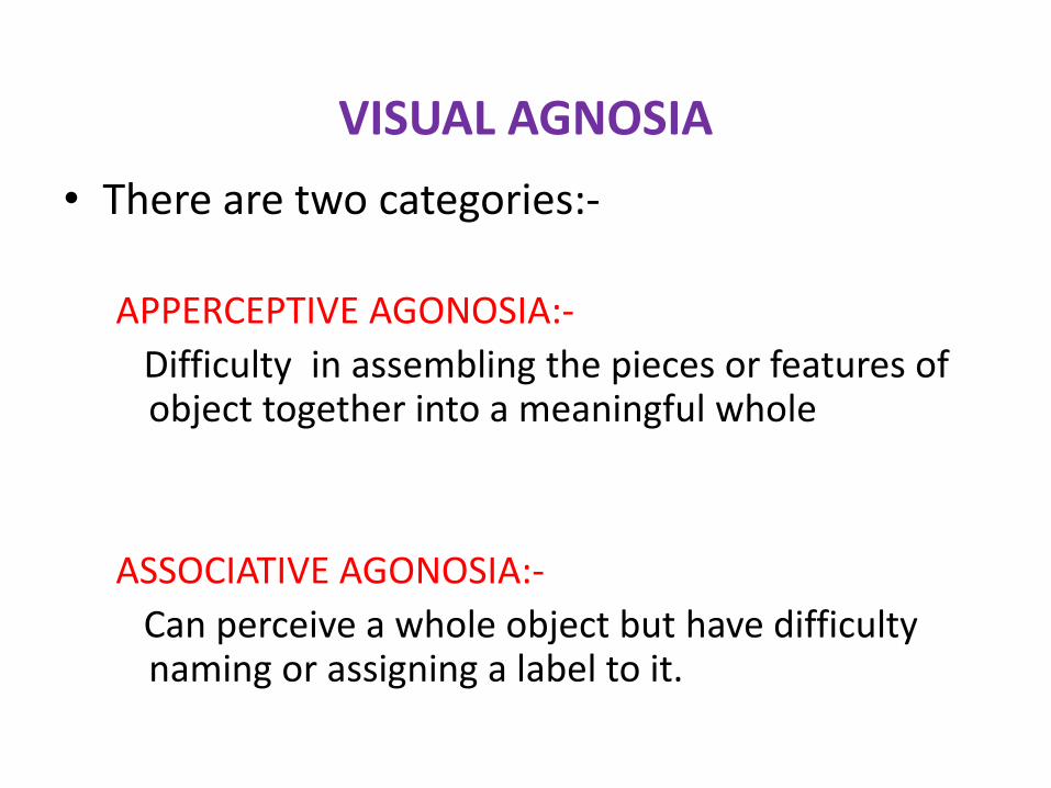
VISUAL AGNOSIA
• There are two categories:-
APPERCEPTIVE AGONOSIA:-
Difficulty in assembling the pieces or features of object together into a meaningful whole
ASSOCIATIVE AGONOSIA:-
Can perceive a whole object but have difficulty naming or assigning a label to it.

Apperceptive Visual Agnosia
• Defect in early stage of visual processing, preventing a
correct perception of the stimulus being formed.
• Can pick out features of an object correctly, such as
lines, angles, colors, or movements but fail to appreciate
the whole object.
• Object is not seen in a meaningful way.
• Common Causes:- Stroke, anoxia & carbon monoxide
poisoning.
• Often associated with diffuse, posterior lesions or stage
of recovery from complete cortical blindness.

Associative Visual Agnosia
• Patient can perceive objects presented visually but
cannot interpret, understand or assign meaning to the
object, face or word.
• Primary sensory & early visual processing are
preserved.
• More common than apperceptive visual agnosia
• Usually result of b/l damage to the inferior temporo-
occipital junction and subjacent white matter.

• Copying a drawing
– Apperceptive
agnosia
• Cannot copy the
drawing
• Cannot identify it
–
– Associative agnosia
• Can copy the
drawing
• Cannot identify it

Apperceptive Associative

Simultagnosia
• Term first used by Wolpert in 1924
• Defined as inability to grasp the scene of the multiple
components of a total visual scene despite retained ability
to identify individual detail.
• Inability to report all the items and relations in a complex
visual display despite unrestricted head & eye movements
• Describe the picture in a piecemeal manner.
Two types-
Dorsal simultanagnosia
Ventral simultanagnosia

DORSAL SIMULTANAGNOSIA
• Perception is limited to a single object without
awareness of the presence of other stimuli. Thus, being
able to see only one object at a time
• Patient may collide with various objects in a room being
unaware of them. Additionally, objects in motion appear
more difficult to perceive
• Appear blind to observers
• Lesion in B/L junction at parieto-occipital lobe.

Patients suffering from Simultanagnosia and Balint’s syndrome will only report the features they are presently looking at but will not be able to understand the contents of the scene.
Simultanagnosia

VENTRAL SIMULTANAGNOSIA
• Patients are able to see several objects at once, but their
recognition of objects is limited to one object at a time.
• Slowed visual processing speed causing difficulty in
simultaneously recognizing the individual parts of a
multipart object
• capable of navigating through a room without bumping
into furniture
• Don’t appear blind to observer
• Site of lesion—left inf. temporooccipital cortex

Ventral simultanagnosia
“Mixed figures” used in the assessment of ventral simultanagnosia

ProsopagnosiaGreek: "prosopon" = "face", "agnosia" = "not knowing"
• A case of a prosopagnosia is "Dr. P." in Oliver Sacks' 1985 book The Man Who Mistook His Wife for a Hat, though this is more properly considered to be one of a more general visual agnosia.
• Although Dr. P. could not recognize his wife from her face, he was able to recognize her by her voice.

Prosopagnosia
• Inability to recognize familiar faces or to learn and recognize new faces
• They can identify facial parts, recognize a face as a face but cannot recognize the person
• Semantic knowledge about people is intact (Can retrieve history of familiar persons when their name/other details are provided verbally)
• They can identify familiar people by nonvisual or nonfacialvisual cues
• Prosopagnosia may occur in isolation suggesting that there are specific areas of the brain that process visual information pertaining to face recognition.

Prosopagnosia• Many are also impaired in distinguishing specific types of
objects in a class (discriminating apple from a mango, a maruti car from a santro)
• Eventhough patients have difficulty in identification, usually they recognize the category of the visual object eg., fruit,car or bird. (vs patients with visual agnosia who fail to recognize the object category to which they belong)
• They may identify facial expression& emotion, sex, and age correctly, depending on the extent & location of the lesion
• In severe cases, the patient may not even recognize his own face

Prosopagnosia-Neurophysiology
• For face recognition, there are 2 stages beyond perception:Recognition units :
Store the abstract representations of familiar faces derived from our previous experience
Their activation by the perceptual processing raises a feeling of familiarity, although not yet permitting identification
Occurs mainly in visual association areas of right hemisphere
Identity nodes: Subsector of the semantic store that provides the information
concerning the biography and relationship between the observer & familiar persons and allows their identification. Then only the name of face can be retrieved
Carried out by left inferomedial temporal lobe

Prosopagnosia
Tests: Matching pictures of unknown faces
Asking the patient to describe a given face verbally or to recognize familiar faces or faces of well-known politicians /celebrities
Lesions site:B/L mesial occipito-temporal region & subjacent white matter involving the fusiform gyrus
Causes: Stroke (PCA infarct) Trauma, tumors, abscess, surgical resection Viral encephalitis, migraine, hypoxia, FTD etc

Patients with damage to the amygdala have difficulties in recognizing emotion from facial expression

Associative visual Agnosia related syndromes

Category specific Agnosia
• Impaired recognition of objects within a certain categories
- living ,nonliving,
- metals, fruits, and musical instruments
• Warrington and Shallice (1984) reported a patient who following an acute lesion to the left temporal lobe had a selective deficit when asked to name pictures from just one semantic category – living things
• By contrast he was able to name non-living objects very well including those with low frequency names such as ‘accordion’

Movement agnosia, motion blindness or akinetopsia
A patient is unable to perceive motion, while all other perceptual capabilities are intact.
He can still read books and see colors, but cannot discern which direction something is moving, or how fast it is.
Everyday actions, such as crossing a street ,are extremely difficult since she cannot judge how fast a car is approaching
Lesion site: B/L medial-temporal area
Motion Agnosia

Difficulty in naming visually presented objects
Convey their recognition by pantomiming or describing their use
Can name stimuli presented in modalities other than vision
Some consider it as distinct entity from associative agnosia, others consider it part of the later
Most commonly due to a left occipital lesion
Optic aphasia

Mirror Agnosia
• Mirror agnosia is the inability to differentiate between real and reflected objects, to mentally rotate objects, and to perform line orientation tasks
• Patients cannot correct their behavior even after they have been shown the real location of the object; they always believe the object to be behind or in the mirror itself and reach for it accordingly
• Lesions of either parietal lobe near the posterior angular gyrus/superior temporal gyrus (the temporo-parieto-occipital) junction

Pure alexia Alexia without agraphia / Pure word blindness
A perceptual disorder causing impairment in reading words and letters
Patient can write to dictation but is unable to read back what has been written
The patient can copy words and letters and in the act of copying the words or tracing out the letters may recognize the word or letter
Lesion site: Damage to pathways conveying
visual inputs from both hemispheres to the dominant angular gyrus
Combined lesions of dominant medial occipital region & inferior fibers of splenium of corpus callosum

Color Agnosia
Naming & recognition of colors may be selectively impaired
Colour matching & other aspects of colour perception are normal (as tested by Holmgrens colour sorting test)
Can’t point to the colour named by the examiner
Perform well in verbal-verbal tasks (e.g. “tell me the colour of the sky?”)
Lesion is commonly in inferomesial aspect of the occipital & temporal lobes of dominant hemisphere

Color Agnosia• Tests for a colour agnosia
showing patients incorrectly coloured objects.
• A patient who identifies an inappropriately coloured object as correct as to colour, may have colour agnosia.
• For example, a blue banana may seem quite normal to a colour agnosic

Assessment of visual agnosia
• First step :- establish the preservation of adequate elementary visual abilities—acuity, visual fields, colour vision
• Then assess for..
Object recognition—recognition & naming tested
Colour —recognition & naming
Face recognition—persons, photographs
Picture recognition—for simultanagnosia
Matching & Copying - to differentiate between apperceptive& associative agnosias

Assessment of visual agnosia
Test items Visual
agnosia
Optic aphasia Anomia Semantic
dementia
Visual naming Impaired Impaired, able
to pantomime,
circumloculate
Impaired, able
to pantomime,
circumloculate
Impaired
Tactile naming Normal Normal Impaired Impaired
Naming of
verbally
described
objects
Normal Normal Impaired Impaired
Object or
picture sorting
by semantic
category
Fails Normal Normal Fails
Error type Visual Semantic Paraphasic Semantic

Balint’s Syndrome
• Clinical triad described by Balint :-
– Optic Ataxia-- visually difficult to reach for objects;
may see, recognize object, but movement is usually
misdirected
– Psychic paralysis of gaze--difficulty in visual scanning;
not able to maintain fixation on an object - eyes will
begin to wander to another object
– Simultanagnosia - (can only “see” one object at a
time) not able to perceive more than one object at a
time
• Bilateral occipital-parietal lesions

Optic Ataxia
Inability to perform coordinated voluntary movements in order to reach for an object.
Kinematics of reaching is altered More prominent in periphery of extrapersonal
space A visuomotor problem, can reach out
properly in response to tactile/auditory cues Damage to the dorsal stream at unilateral or
bilateral parieto-occipital junction (Karnath) Unilat lesions: failure of reach to
contralateral side with either hand

Prerequisites for a diagnosis of Optic Ataxia (Garcin)
• A normal visual field
• Objects can be seen, recognised and named even in the field where the visuomotor deficits occur
• No defect in binocular stereopsis
• Normal proprioceptive function
• No intrinsic motor, oculomotor or cerebellar deficit

Optic Ataxia

Oculomotor apraxia – Psychic paralysis of gaze
An inability to move the eyes voluntarily to points in the visual field.
Difficulty in visual scanning as in reading a book or appreciating a painting. Failure of disengagement of fixation (?)
Reflexive movements may be spared.
Eye movements towards auditory or somatosensorystimuli spared.
Usually co-occurs with visuospatial deficits

Ocular apraxia


![Reduced occipital GABA in Parkinson disease with …occipital lobe (data available from Newcastle University e-prints [figure 1]: eprint.ncl.ac.uk/247552). Sequence parameters were](https://img.pdfslide.us/doc/110x75/5fd7cf36f78f8445ba57ba06/reduced-occipital-gaba-in-parkinson-disease-with-occipital-lobe-data-available.jpg)