Embed Size (px)
Citation preview

MRI artifacts &Artifact Remedy
Strategies
GGamal amal FFathalla athalla MM.. MMahdalyahdaly
[email protected][email protected]
Experta Medica

MR Image ArtifactsTechniqu
e-Related
Experta Medica
Patient-Related
System-Related
Experta Medica

Technique Related ArtifactsFat & water
protons exist in
the same Voxel(Chemical
Shift)
Fat & water protons exist in
•The same Voxel
(Gibbs –Fine Line or
Truncation)
Planning Fault
(Magic Angle & Cross Talk)
FOV smaller than the anatomy inside the receive coil
(Aliasing)
Experta Medica
Experta Medica

Artifacts
Wrap around, Fold over, Aliasing or Back folding :
Etiology : Anatomy inside the receive coil is greater than the prescribed
FOV. The coil, then receives signal from the anatomy outside the FOV. The signal of these data oversampled is plotted during readout in the opposite direction because the system fills the k-space lines in one direction. It fills the K-space lines first with the prescribed FOV. Then the oversampled area is then plotted on the already filled first lines.
Manifested as :Image data out side (FOV) wraps around it (in opposite side
of the image) Occurs in any of the 3 encoding axes, hence the names
phase-wrap, frequency-wrap & slice wrap. Slice-wrap occurs only in Fourier 3D acquisitions.
Experta Medica

Experta Medica

This case study shows a 3Dacquisition technique with aliasing )in the red circle( in the slice selection direction. The image of the upper leg wraps into the image of
the lower leg . Experta Medica

Remedy Tips:For frequency-wrap,Use larger FOV .Oversample the frequency.Use digital filters to eliminate signal with
frequency out side the (FOV).
For Phase-wrap, Use larger FOV- Phase oversampling- No Phase
WrapOption.
For 3D or Slice-wrap, Decrease the number of slices/slab, Use a larger
FOV. Experta Medica

Chemical shift - Chemical Mis-registration –India Ink Artifact
Etiology:Occurrence of water & fat Protons in the same Voxel as occurs in : Abdomen – Spine – Orbits where in low TE’s , fat & water
protons precess at nearby frequencies resulting in spin- spin resonance causing positional shift (Mis-registration) of fat signal along the frequency axis. This is especially evident in long TR sequences especially when TE is short ( PD WI).
Manifested as : Bright & Dark demarcation at fat/ water interface.Remedy Tips: Increase the band width to help separate the peaks of water & fat
signals.Decrease the ETL as every Echo contains its innate chemical shift
& increasing the ETL accumulates the shifts. Use a Lower field strength.Increase the TE & reduce the TR.
Experta Medica

Experta Medica

Chemical shift - Chemical Mis-registration - Odd Presented-Complicated with Susceptibility
Etiology:Dental fillings in the frequency direction as occurs in the brain where in
low TE’s , fat & water protons precess at nearby frequencies resulting in spin- spin resonance causing positional shift (Mis-registration) of fat signal along the frequency axis. This is further complicated by different magnetic susceptibility of dentures. This is especially evident in long TR sequences especially when TE is short ( PD WI & FLAIR).
Manifested as : Bright crescent near the middle of some images near the skull base.Remedy Tips: Increase the band width to help separate the peaks of water & fat
signals.Swap the frequency/phase axes.Decrease the ETL as every Echo contains its innate chemical shift &
increasing the ETL accumulates the shifts. Use a Lower field strength.Increase the TE & reduce the TR.
Experta Medica

Experta Medica

Gibbs, Fine line or Truncation :
Etiology:Periodic brain & visceral motion produces different phases
registered as fine lines parallel to the anatomy borders along the phase encoding axis.
Manifested as :- Bright and dark lines that may be seen parallel and
adjacent to boarders of the anatomy in areas containing both fat & water as the CSF/ spinal cord interface
The artifact occurs in the phase encoding direction Remedy Tips:Increase the matrix & use an isotropic pixel .Use a fine line filter Swap the Frequency/ Phase axes.Decrease the Echo Sampling time.Use even NEX.
Experta Medica

Experta Medica

Experta Medica

Magic angle artifacts:Manifested as:Bright signal commonly seen in tendons & ligaments of
the knee & shoulder joints that are oriented at magic angle (55°) to the Bo.
Etiology : Normally, signal from water molecules associated with
the tendon collagen fibers is not seen due to dipolar interactions that result in very short T2 Times…..But at an angle of about 55° to the main magnetic field, the dipolar interactions become zero, resulting in an increase of the T2 Times about 100 folds. This results in signal being visible in tendons with ordinary pulse sequences especially at short TE long TR SE & FSE sequences i.e. PD WI.
Remedy Tips:*Change the positioning and/or the planning angle.*Scan in multiple planes & multiple pulse sequences.*Increase the TE & reduce the TR in PD sequences.Experta Medica

Cross Talk artifacts:Manifested as:Dark signal commonly seen at areas of intersection
as in axial angulated spine.Etiology:At the areas of intersection as in axial angulated
spine, the excitation pulse of the 2nd image set saturates the signal of the spins at the areas of intersection which were already excited in the first image set.
Remedy Tips:Change the planning angle.Use FSE & fr-FSE.Increase the TE & reduce the TR in PD sequences. Use interleaved rather than sequential image
acquisition.Experta Medica

Regional Fat Sat Failure
Manifested as:Areas where fat appears bright in fat sat images (inhomogeneous fat saturation).
Etiology:Mal positioning of the shim volume.Using too large FOV leading to improper shimming.Improper use of the receivers of phase array coils.
Remedy Tips:Centralize the shim volume to the anatomy of interest.Decrease the FOV.Review the active receiver s of phased array coil . Slightly shift the central frequency or transmit gain .Change the chemical shift tuning factor (CSTUN).Experta Medica

Failed Fat Saturation
SAG FSE + Fat Sat: Autoshim On, without moving the Autoshim volume.Autoshim failed: Default shim values = 1 0 0
Experta Medica

AnnefactManifested as:Striated bands along the S/I axis in sag images.
Etiology:
Mal positioning of the FOV to the used receivers of phased array coil.
Improper selection of the receivers of phase array coils.
Remedy Tips:
Centralize the S/I FOV in front of the active receiver set.
Use the adequate number of receivers suitable for the FOV.

Annefact
Experta Medica

Patient Related ArtifactsMotion (Ghosting)
ArtifactsMetal (Susceptibility)
Artifact Physical & Physical &
Physiological Physiological Motion of the Motion of the
patient produces patient produces multiple time-multiple time-
resolved phases resolved phases that are manifested that are manifested
as Ghostsas Ghosts
Due to (microscopic gradients) or variations
in the magnetic field strength that occurs
near the interfaces of substance of different magnetic susceptibility
Experta Medica

Motion (Ghosting) Artifacts:
Etiology : Patient physical motion . Periodic physiological motions like: Cardiac motion ,Respiratory motion ,Swallowing,
Vascular & CSF pulsations & Peristalsis.Manifested as:Repeating contour figures oriented in the phase
direction as every motion creates a new different phase spatially shifted slightly apart from the preceding one.
Remedy Tips:Stabilize your Patient.Trigger the Pulse Sequence to the physiological motion
(Respiratory, cardiac & peripheral gating).Reduce the ETL in FSE to minimize phase shifting.Use antispasmodic drugs to scan the abdomen.Use breath hold pulse sequences.Use Flow Compensation.Use Spatial pre-saturation.Swap the Phase/ Frequency axes. Put the source of artifact in the phase direction.Experta Medica

Experta Medica

Experta Medica

Swallowing versus CSF Flow Pulsation Artifacts Experta Medica
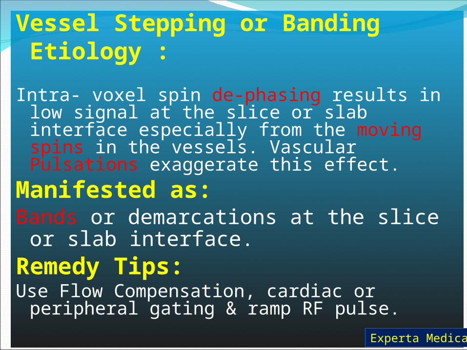
Vessel Stepping or Banding Etiology : Intra- voxel spin de-phasing results in low
signal at the slice or slab interface especially from the moving spins in the vessels. Vascular Pulsations exaggerate this effect.
Manifested as: Bands or demarcations at the slice or
slab interface. Remedy Tips:Use Flow Compensation, cardiac or peripheral
gating & ramp RF pulse.
Experta Medica

Experta Medica

Flow Artifacts Etiology : Fully magnetized spins entering the slice in the vessels
may possess brighter signal than the tissue spins ( Flow Enhancement) or
Fresh non excited spins may enter the imaging volume during readout producing signal void. Also, the excited protons may flow outside the imaging volume before read producing a signal void. These two states are known as TOF artifact.
Position encoding of the Voxel containing the vessel in the phase direction at a time factor of TE/2 before frequency encoding.
Manifested as: Either Flow enhancement or void or as ghosting.
Remedy Tips:Use Flow Compensation, cardiac or peripheral gating.Increase TE.Use SPGR.Use S/I Spatial saturation outside the FOV. Experta Medica

Experta Medica

Experta Medica

Experta Medica

Susceptibility Artifacts Etiology : Due to (microscopic gradients) or variations in the
magnetic field strength that occur near the interfaces of substance of different magnetic susceptibility. Large susceptibility artifacts are commonly seen surrounding ferromagnetic objects inside of diamagnetic materials (such as the human body). These gradients cause de-phasing of spins and frequency shifts of the surrounding tissues.
Manifested as: Bright and dark areas with spatial distortion of
surrounding anatomy especially with Long TE SE & GRE pulse sequences.
Remedy Tips:Use Fast Sequences ( fr-FSE, f-GRE & f-SPGR,
FIESTA,SWAN, SWIFT).Reduce the TE.Swap the Frequency/ Phase axes.Use S/I Spatial saturation outside the FOV or at the
different susceptibility area. Experta Medica
Experta Medica

Experta Medica

N/2 & Geometric Distortion Artifacts Etiology : Due to high B values used in DWI, the MPG pulse duration increases & hence minimum TE increases. With the following consequences:Increase of gradient amplitude/Voxel increasing Microscopic Gradients & thereby increasing the magnetic susceptibility artifacts.Enhancing of ghosting resulting from Periodic Brain Motion.Exaggerating the Phase Error Buildup due to Off- Resonance Spins These effects are further complicated by:Partial Volume Averaging due to occurrence of multiple signals in the same Voxel.Eddy Currents due to high amplitude gradients with resultant Feed through artifacts.Obligatory A/P phase direction to prevent Peripheral Nerve stimulation.Narrow Receive Bandwidth slows down the sampling rate thereby enhancing Ghosting & increasing Chemical Shift.Occurrence of tissues of different susceptibilities in the Frontal & Temporal regions. Experta Medica

Experta Medica

Geometric Distortion
N/2Ghosting
Note theSignal drop
Due to Different
Susceptibility
Experta Medica

N/2 & Geometric Distortion Artifacts
Manifested as: Bright and dark areas with huge spatial distortion of
the anatomy especially in the frontal area obscuring parts of the frontal & temporal lobes.
Remedy Tips:Use S/I Spatial saturation outside the FOV or at the
different susceptibility area. This increases the dielectric effect but reduces Susceptibility effects. ( Tradeoff: SAR increases).
Reduce the Echo Sampling Time by:1-Decreasing B value with trade off of Poor Diff weighting
& appearance of T2 Shine through artifact.2- Widening of RBW with trade off of low SNR.3-Optimizing TE to allow maximum gradient amplitude at
minimum possible TE.4-Ramp Sampling allowing echo sampling during ramp ups
& downs of the frequency encoding gradient, thereby reducing ech0spacing & allowing faster sampling with tradeoff of wide RBW & hence reduced SNR. Experta Medica

5-Using an additional refocusing pulse to reduce the eddy currents with a tradeoff of increasing the minimum TE.
6-Using Parallel Imaging techniques making use of spatial information related to the spatially varying sensitivity of different receive channels of the receive coil. Using these pre-measured data, sensitivity profiles of the coil helps reconstruct the K-Space trajectory missing lines which is under sampled during the actual scan .This Tech is called ASSET or SMASH.
• SENSE is another parallel imaging technique where K-Space is under sampled by widening the K-Space lines . The overflowing data / reduced number of lines leads to aliasing. Each pixel of an aliased receiver channel image reflects the signal from different spatial origins. The premeasured sensitivity profile helps allocate each signal to its spatial origin. The trade off is low SNR due to lossy K-Space.
7-Using PROPELLER where K-Space trajectory is rotated across the time domain to define the areas of K-Space where distortion occurred & subtract them from the data set with a tradeoff of limited SNR because of lossy K-Space. Experta Medica

Experta Medica

System Related ArtifactsShimmi
ng Related Artifacts
Gradient Related Artifacts
Radio-frequenc
y Related Artifacts

Shimming Related Artifacts Etiology : Defective shimming causes interaction with unwanted
waves of external sources that may cause spatial or intensity distortions or even both.
Manifested as: Spatial ( geometric ) or intensity distortions or both.Zebra or totally clipped images due to faulty K-
Space Pixel.
Remedy Tips:Use auto shim at the first sequence of the exam.Filter the currents in the field vicinity.Active Shimming may be advisable if problems
persist.

Clipped Image

Zebra Artifact


Gradient Related Artifacts Etiology : Faulty gradients cause local spatial or intensity
distortions or even both in the direction of the faulty gradient. Moiré Fringes are due to B0 inhomogeneity from one side of the body to the other where signals of different phases superimpose . The in-phase signals add to each other where the off-phase signals subtract.
Manifested as: Spatial ( geometric ) or intensity distortions or
both. Moiré fringes are manifested as 2 window image i.e. half the image being bright & the other half being dark.
Remedy Tips:Swap Freq/ Phase axes.Filter the currents in the field vicinity.Use FSE/ fr-FSE rather than SE or GRE.Use surface coils.

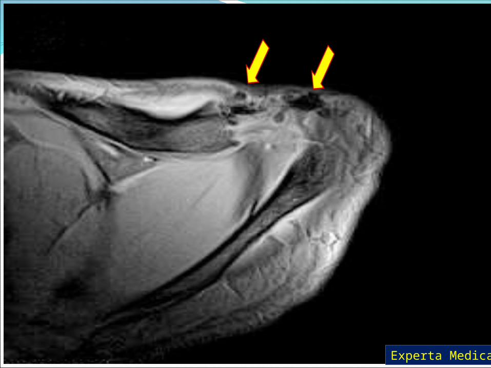
Gradient Fault: The artifact appears as bright noise here
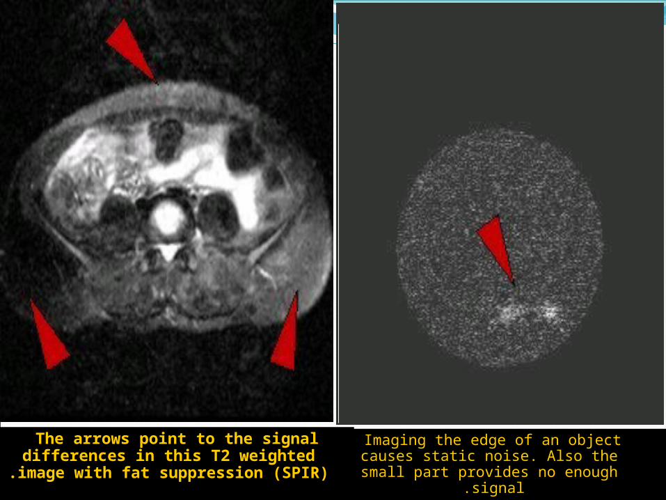
The arrows point to the signal differences in this T2 weighted
image with fat suppression (SPIR) .
Imaging the edge of an object causes static noise. Also the small part
provides no enough signal .

RF-Related Artifacts Etiology : Faulty RF- Amplifier or Receiver. Manifested as: Zipper (RF Feed through) artifacts in cases of faulty
RF amplifier. Normally, the RF amplifier should stop working when read out gradient is applied. When faulty, it works while readout( F. encoding gradient) is applied. Residual FID stimulated echoes may also produce zippers.
Bright spot of increasing intensity in the center of the image in cases of constant offset of the DC in the signal perception channels of the receiver coil. Narrow band noise appears perpendicular to the frequency encoding axis while broad band noise disfigures the image over a wide zone.
Remedy Tips:Coil tuning.Filter the currents in the field vicinity.Proper RF-shielding or active shielding ( Faraday’s
cage).Offset readout apart from the central artifact.

Zipper Artifact

RF Feed through
(Zipper )Artifact
Any Question???
Again Any Question???
Thank you!

Have a nice day