Embed Size (px)
Citation preview
M.P.
Editor: Dr. Meenu Chadha
Co-editor: Dr. Harsha Desai Phulambrikar

Anaesthesiology M.P.
1
CONTENTSExective Body of
M.P. State 2017-2018Exective Body of
M.P. State 2017-2018
EDITOR
CO-EDITOR
Dr. Harsha Desai Phulambrikar
Dr. Meenu Chadha Chief Anaesthetist CHL Hospital, Indore (M.P.) Mob. : 9977161035 E-mail : [email protected]
Consultant AnaesthesiologistGreater Kailash Hospitals, [email protected]
Dr. Harsha Desai Phulambrika
The Unmet Needs of Perioperative Prophylaxis of Venous Thrombosis 021
Dr. Jitendra Agrawal
stConference Report of 31 Isacon MP 2017 and Minutes of GBM 2017 - Bhopal 042
Dr. Kriti Vig, Dr. Kunal Waghmare
Atrial Fibrillation after Cardiac Surgery 194
Dr. Ruchi Tandon, Dr. Emendr Wahnel, Dr. Abhay Raj Yadav
Anesthetic Management in a Patient of Apert Syndrome 275
President
Vice President
Hon. Secretary
Hon. TreasurerDr. Gayatri Tanwar, Dr. Anju Grewal
Anesthetic Issues for Neurosurgical Procedures 093
Dr. Amisha Vats, Dr. Manish Kumari, Dr. Yugal Chandrakar, Dr. K.K. Arora
306Anticipated difficulties of Congenital Oral Swellings in Newborn
Dr. Suman Gupta, Dr. Preeti Goyal, Dr. Bhanu Coudhary
Do Conventional direct Laryngoscopy needs windup with the advent of Video laryngospopes? 357
Dr. Surendra Raikwar Associate Professor Department ofAnaesthesiology Gandhi Medical College, Bhopal (M.P.) Mob. : 9406533300, 8989118989 E-mail : [email protected]
Dr. Pradip Kumar Bhattacharya Director Emergency & Critical Care Chirayu Medical College, Bhopal (M.P.) Mob. : 9893181555 E-mail : [email protected]
Dr. Manu Gupta Chief Anaesthesiologist Aarogyadham Hospital, Gwalior (M.P.) Mob. : 7049852009 E-mail : [email protected]
Dr. Jitendra Agrawal Assistant Professor Department of Anaesthesiology G.R. Medical College, Gwalior (M.P.) Mob. : 9300009942 E-mail : [email protected]
Executive Members
Dr. Sandeep Shrivastava (Jabalpur-2016) Dr. Vikas Gupta (Bhopal-2016) Dr. Mamta Mahobe (Jabalpur-2016) Dr. Rajeev Divedi (Rewa-2016) Dr. Deepesh Gupta (Bhopal-2017) Dr. Subhash Agrawal (Rewa-2017)
Past President
Dr. Sanjay Khanna Mob. : 9826166423
Past Hon. Secretary
Dr. Surendra Raikwar Mob. : 9406533300, 8989118989
Past Hon. Treasurer
Dr. R. P. Kauhal Mob 9617377134

Anaesthesiology M.P.
2
EDITORIAL
enous thromboembolism (VTE) is a Vcommon and often untreated cause of preventable death, hospital readmissions, perioperative morbidity and complications. Contrary to the popular early belief, the Asian population is equally at risk to develop VTE as the west. Thromboembolism is an underrated problem in the Indian medical community and one finds very few articles on the Pubmed directed to the VTE problem in Indians. The absolute rates of DVT in Indian population have been found as 15-40% in pelvic surgeries, neurosurgery 40-60%, orthopedic and arthroplasty 40-50% and up to 60-80% in
1spinal cord trauma patients. A recent randomized controlled study compared clinically important VTE after isolated limb fractures. This study showed a 2.3% risk of CIVTE as compared to the ones that use venography as a tool. There was no pulmonary
2embolism (PE) in this group of 256 patients.
The pathogenesis of thrombosis is based on the “Virchow's triad” and the surgical patient has factors affecting all the three limbs of the triad. Venous thrombi are formed under low flow/stasis, low shear situations with fibrin strands and platelets trapped within. Hence they are best prevented by inhibitors of fibrin formation, i.e. anticoagulants. The therapeutic approach for prevention and treatment of DVT rel ies on pharmacological and non pharmacological methods, for which numerous guidelines have been proposed. The ACCP gu i de l i ne s o f pe r i ope ra t i ve
anticoagulation and the AAOS guidelines are most commonly followed. The limitation with the ACCP guidelines is that all studies which form the basis of its recommendation are done in patients with pre-existing atrial fibrillation, or in patients with intracardiac valves. Hence, they overrate the gravity of the situation.
The first step in effective deep vein thrombosis (DVT) prophylaxis is to identify the patient who is actually high risk for post operative DVT and PE. The Caprini risk model does an individualized risk assessment score based on presence or absence of over 35 risk factors. The Caprini model's ability to predict VTE risk has been validated for general, urologic, and vascular surgery patients, as well as those patients having post-bariatric body contouring surgery.
There is lack of evidence supporting most guidelines for DVT prophylaxis and most of them are based on expert opinions and consensus or limited evidence. Hence, the clinician must identify each patient category and manage venous thrombosis accordingly.
The old time tested heparin is still used frequently as subcutaneous dose or intravenous infusion. However, owing to unpredictable response, thrombocytopenia and heparin resistance, heparin has been gradually replaced by low molecular weight heparin. With a lower plasma protein affinity, LMWH has a more predictable dose response curve and better bio-availability. further, because of a longer half life they stay in
THE UNMET NEEDS OF PERIOPERATIVE
PROPHYLAXIS OF VENOUS THROMBOSISl Dr. Harsha Desai Phulambrikar

Anaesthesiology M.P.
3
circulation for a longer time and need only once a day dosing, with little need for repeated monitoring.
Warfarin is by far the most commonly used oral anticoagulant in the world, and also number three on the list of drugs implicated in causing hospital admission through adverse effects. The only advantage of warfarin is its easy reversibility and low cost. Other than that, warfarin has a very narrow therapeutic range, lot of individual variability in response, skin necrosis and hair loss. There are insufficient guidelines to reliably recommend the interval of monitoring INR in warfarin treated patients.
The novel oral anticoagulants seem to bring a paradigm shift in anticoagulant therapy. Rivaroxaban(Xarelto) is being widely prescribed for acute VTE. An Acute DVT study showed rivaroxaban as a promising therapy for primary prevention as well as acute VTE even in perioperative patients. Dabigatran also a safe oral anticoagulant especially in patients with impaired creatinine clearance.
The US Food and Drug Administration have granted potential reversal agents-idarucizumab for dabigatran and andexanet alfa for apixaban, edoxaban, and rivaroxaban. Both of these drugs are being tested in actual NOAC-treated bleeding patients, and in mid-2015, initial results for idarucizumab were
3published.
Bridging therapy is falling into disfavor with the contemporary approach as evidence shows higher risk of bleeding than of DVT in most patients on pre-operative anticoagulants.
One important deve lopment in perioperative anticoagulation is, mapping the bleeding risk in perioperative period. A Jtafur et al., have formulated a proper bleeding risk and DVT risk calculator usable on daily basis. Bleed MAP score assigns one point for each risk factor: history of prior bleeding (Bleed), mechanical mitral heart valve (M), active cancer (A), and low platelets (P). The score obtained thus, reliably predicts bleeding risk
4and thrombotic risk for individual patient.
is
On an average, the periprocedural bleeding to thrombosis ratio is approximately 13:1 with bridging and 5:1 without bridging. Thus bleeding is by far a bigger risk than VTE and poor outcome. Traditional anticoagulation practices are fast being replaced by novel oral anticoagulant regimes. Low DVT risk patients do not require any pharmacological modalities for VTE prevention during most surgeries. The guidelines of managing perioperative VTE prophylaxis for high risk patients are well lit paths. The only dilemma that remains is with intermediate risk patients, where the clinician needs to identify individual risk v/s benefit ratio of anticoagulant therapy. Over rating the VTE issue or under-treating with insufficient anticoagulation, both lead to poor outcomes.
References:
1. Venous thromboembolism: A problem in the Indian/Asian population? Sunil Agarwal, Arvind Dhas Lee,1 Ravish Sanghi Raju, and Edwin Stephen, Indian J Urol. 2009 Jan-Mar; 25(1): 11–16.
2. A double-blind, randomized controlled trial of the prevention of clinically important venous thromboembolism after isolated lower leg fractures. Selby R1, Geerts WH, Kreder HJ, Crowther MA, Kaus L, Sealey F; D-KAF (Dalteparin in Knee-to-Ankle Fracture) InvestigatorsJ Orthop Trauma. 2015 May;29(5):224-30
3. Idarucizumab for dabigatran reversal. N Engl J Med. 2015;373:511–520. doi: 10.1056/NEJMoa1502000. Pollack CV Jr., Reilly PA, Eikelboom J, Glund S, Verhamme P, Bernstein RA
4. Predictors of major bleeding in peri-procedural anticoagulation management.A. J. Tafur,R. McBane II,W. E. Wysokinski,S. Litin,P. Daniels,J. Slusser,D. Hodge,M. G. Beckman,J. A. Heit. First published: 2 February 2012DOI: 10.1111/j.1538-7836.2011.

Anaesthesiology M.P.
4
STCONFERENCE REPORT OF 31 ISACON MP 2017
AND MINUTES OF GBM 2017 - (BHOPAL)l Dr. Jitendra Agrawal - Hon. Secretary MP State ISA
espected Governing Council Members, RPresidents and Secretaries of the city branches, Respected president, Past President, Senior Anesthesiologists and my dear colleagues.
Greeting on behalf of ISA Mp State office Gwalior.
Wish you a Happy Deepawali, Merry christmas and Happy New year in advance.
Conference Report
stThe 31 ISACON Mp,2017 was held on 1 & nd2 october, 2017 and pre conference
workshops on 30th september, 2017 at Chirayu Medical College Bhopal, hosted by ISA Bhopal city Branch and organized by Department of Anaesthesia, Chirayu Medical College & Hospital Bhopal.
It was a very weil organized conference attended by approximatery 300 delegates from India and abroad.
thOn 30 Sep, 2017 workshops for
(1) Difficult Airway workshop by Dr. J.P. Sharma (AIIMS Bhopal) and Dr. Bharat Bhusan (Medical College Saifai, U.P.) & his Team.
(2) USG in Anaesthesiology by Dr. Virendra Mohan (AIIMS Delhi) and Dr Munir (Chirayu Medical college Bhopal) & his Team,
(3) Chronic Pain Management by Dr. Sanjay Khanna (Jabalpur), Dr. Dinesh Sahu (Bombay), Dr Jitendra Agrawal (Gwalior), Dr. Amit Jain (Sagar) and Dr. Jaideep Singh (Gandhi Medical College Bhopal) & his Team.
Approxmately 100 delegates attended these workshops.
st
stOn 1 Oct, 2017 ln Hall "A" Scientific session started with a guest lecture delivered by Dr. T.C. Kriplani from Jabalpur on "History of Indian society of Anaesthesiology M P Chapter." The session was chaired by Dr. V.M. Agnihotri and Dr. Aditya Agarwal.
nd2 lecture delivered by Dr. Pradeep Bhattacharya from Bhopal on "Goal directed perioperative fluid therapy".This session was chaired by Dr. (Col) R.C. Agarwal, and Dr. Shikha Mehrotra.
rd3 Dr. S.K. Mehta Oration was delivered by Dr. Mahendra Upadhyay from Baroda, Gujarat on "Low Flow Anaesthesia".Session was chaired by Dr. Sanjay Khanna and Dr. T. C. Kriplani.
th4 was delivered by Dr. Meenu Chadha from lndore on ''Anesthetic management of organ retrieval for transplant from a brain dead patient". This session was chaired by Dr. Surendra Raikwar and Dr. Lalit Mehndiratta.
The lnaugural ceremony started with the floral welcome of Mr. Vishwas Sarang Minister of State Public Selection Govt. of Madhya pradesh the Chief Guest of the inaugural function.
Mr. Azatshatru, Commissioner Bhopal was the Guest of Honour.
Dr. Ajay Goenka was the special guest.
After floral welcome, Lighting of Lamp was done by Mr. Vishwas Sarang Ji, Mr. Azatshatruji, Dr. Ajay Goenka, Dr. Sanjay Khanna, Dr. Surendra Raikwar and Dr. Pradeep Bhattacharya. Saraswati vandana was sung by Postgraduates students. Dr. Sanjay Khanna gave presidential speech and Dr. Surendra Raikwar delivered the welcome speech.
Dr. N.S. Ahluwalia Oration
Anaesthesiology M.P.
5
Felicitation of senior members Dr. (Col) R.C. Agarwal, Dr. V.M. Agnihotri,
Dr. T.C. Kriplani, Dr. L.D. Mishra, Dr. K.L. Krishnani, Dr. A. Zutshi, Dr. Deepak Zutshi, Dr. S.K. Mathur, Dr. Sanjay Dave, Dr. Aditya Agarwal and Dr. Shikha Mehrotra was done by giving them mementos.
Dr. Ajay Goenka, Mr. Azatshatru and Mr. Vishwas Sarang ji also delivered encouraging speeches.
Dr. Pradeep Bhattacharya, Organizing stSecretary of 31 ISACON MP, 2017 proposed
the vote of thanks followed by lunch.
The Second scientific session
Hall A - Papers for Late Dr. T.N. Jha and Dr. K.P. Chansoriya travel grant, which were 9 in numbers followed by papers for Dr. M.T. Bhatia Gold Medal for pain, which were 9 in number.
ln the competitive session for Late Dr. T.N. Jha and Dr. K.P. Chansoriya medal and travel grant was awarded to Dr. Raghav Sengupta of Shri Aurobindo Medical college & PG institute, Indore, for his paper presentation on ‘‘Comparison of oropharyngeal pack soaked in lignocaine with sodabicarb and lignocaine with dexamethasone in patients undergoing nasal surgeries’’, the session was judged by Dr. L.D. Mishra, Dr. C. Radha Krishnan Rao and Dr. Sanjay Agarwal.
Dr. M.T. Bhatia Gold medal was awarded to Dr. Surbhi Shekhar from NSCB Medical College Jabalpur for her paper on "Evaluation of the effect of preemptive anatgesia in laparoscopic gynecological surgeries", the session was judged by Dr. Sanjay Khanna, Dr. Sudhakar Dwidedi and Dr. Abhay Babar.
After this session General Body Meeting of Indian society of M.P. State chapter was held and various matters were discussed.
nd2 October 2017stHall A -1 lecture was delivered by Dr.
Aoron Abraham (lsrael) on "Perioperative noninvasive hemodynamic monitoring".
nd2 lecture was delivered by Dr. Rajesh Bhagchdndani (Bhopal) on "Perioperative Glucose control".
rd3 lecture was delivered by Dr. Tarun Bhagchandani (Dubai) on "Day Surgery concept- A changing paradigm" and
th4 lecture was delivered by Dr. Divya Gupta on "Occupational Health among Anesthesiologists".
This session was chaired by Dr. T. C. Kriprani, Dr. Ashish sethi, and Dr. Pradeep Bhattacharya.
Free papers were presented in Hall B and Hall C which were 36 in number
ln the free paper session, one best paper out of 06 papers was given prize:
Hall B
1. Dr. Sadiya Khan (GMC Bhopal)
2. Dr. Tanya Chhauda
3. Dr. Manisha Kumari
Hall C
1 Dr. Avani Tiwari (GMC Bhopal)
2 Dr. Urmila Keshri (Bhopal)
3. Dr. Pranchill Pandey from SSMC Rewa
Was Poster presentations were done which were 32 in number.
One best poster out of 10 posters was given prize:
Poster no. 1-10 - Best poster Dr. Divya Agarwal
Poster no. 11-20 - Best poster Dr. Nupur Khare
Poster no. 21-32- Best poster Dr. Shabad Ali
In the quiz competition, the first prized was bagged by Team of Gandhi Medial college Bhopal, and second prize was bagged by Team of M.G.MMedical college Indore. The quiz was conducted by Dr. Urmila Keshri.
Anaesthesiology M.P.
6
lSA Jabalpur city branch won the Dr. W.P. Thatte Best city branch award 2017, Judged by Dr. L.D. Mishra, Dr. Aoron Abraham and Dr. Mahendra Upadhyay
Minutes of the annual GBM of ISA MP State Chapter 2017
The annual general body meeting of MP stState chapter was held on 1 October 2017
stduring the 31 ISACON MP at Bhopal. The meeting was called by Dr. Sanjay Khanna President ISA MP State, Dr. Surendra Raikwar Hon. Secretary, lSA, MP State and Dr. R. P. Kaushal, Hon.Treasurer.
Agenda I
Dr. Sanjay Khanna president ISA MP chapter welcomed all the delegates attending the conference. He congratulated the organizer for the successful organization of the conference.
Hon. secretary, Dr. Surendra Raikwar congratulated the organizing secretary, Dr. Pradeep Bhattacharya and Organizing chairperson, Dr. Anoop Hajela for a well planned and well organized conference.
Agenda ll - ISA city branch Ujjain is inactive for last four years. ISA national has been informed about it.
Dr. Surendra Raikwar informed the house that he has talked personally with city branch President and Secretary regarding inactivity and branch may be dissolved but no action has been taken so far. Dr. Abhay Babar from Ujjain said that he will raise the matter in city branch but denied to become the Secretary of city branch and to take the lead in reviving Ujjain City Branch.
Dr V.M. Agnihotri said that this is not the loss of city branch this is a loss the MP state branch so he suggested the new secretary of MP state should take personal interest in reviving the branch and coordinate with Dr. Babar this was seconded by Dr. (Col) R.C. Agarwal.
Agenda III
Dr. Surendra Raikwar informed that city branches have not sent their audit reports hence share of city branch of new members has not released from national body All secretaries are requested to update their balance sheet and send audit report to lSA Head quarter, Secretary MP state and Dr. Lalit Mehndiratta .
Agenda IV
Our new website www.isampchapter.com is now fully functional and is updated with detairs regularly. Second issue of MP State Journal is available on website. Please visit our website and check your personal details updated on it. There is a column named IMPORTANT LINKS under which a portal named MEMBERS of MP State is present. You can check your detaits here. In case your details are incomplete or your name is missing from the list, please bring it to the secretary's notice.
Agenda V
We have received a list of inactive members of the ISAMP, from the ISANHQ, who have not updated their profile details. The list has been forwarded to the city branch secretaries. The city branch secretaries are requested to update the details and mail the updated list to [email protected], [email protected] or drjaqrawal@gmail. com at the earliest.
I also request you all to kindly update your details as soon as possible because now-a-days all communication is being done via email or whatsapp. You can update your details either with the city branch secretary, State Secretary, or on the web site. I would request you all to please register your name, email address, lSA number & mobile number by sending an email to [email protected] also. This will enable the member to be eligible for
st thelectronic voting to be held from 1 to 5

Anaesthesiology M.P.
7
November 2017. The last date to update thdetails is 30 september, 2017 and will again
thopen after election i. e. from 06 November 2017.
Agenda VI
Sagar city has 18 ISA members Dr. V.M. Agnihotri suggested to convert it into lSA city club and arrange meetings regularly. As soon as 25 members are made they should apply for city branch.
Agenda VII
Dr. Pradeep Bhattacharya informed that many postgraduate students were not having ISA membership and they want to become member on the spot in conference Dr. (col) R.C. Agarwal and Dr. V.M. Agnihotari suggested to have member ship counter where assistance, money transfer facility should be available in next state conference. Dr. Shikha Malhotra said it will require computers, internet and facility for money transfer which is a very tedious job. It is suggested that all postgraduates should register before registration of conference and if any difficulty is there contact city branch secretary and if needed state secretary.
Postgraduates are Associate Life members and after passing should submit degree and registration to become permanent life member. Dr. Shikha Mehrotra suggested that they should be motivated to convert themselves as Life member. Dr. V.M. Agnihotri suggested that should submit their pass certificate otherwise they wiil not get journal.
Agenda VII
Central zone has started our CZ Journal. The journal has not received any article for publication from MP till now. It is my humble request to all City branch President and Secretaries to encourage the members of their city branch to send minimum one article from each city branch for publication.
Dr. V.M. Agnihotri suggested all papers coming for presentation should have option of publication in central zone journal. There should be two categories in papers Thesis paper and non thesis paper as thesis paper cannot be published until thesis gets approved. Dr. shikha Mehrotra and Dr. Lalit Mehdiratta seconded it.
Agenda IX
Hon. Treasurer Dr. R. P. Kaushal presented the financial report for the year 2016-17.
It was approved by Dr. Aditya Agarwal and seconded by Dr. Shikha Mehrotra and unanimously approved by floor of the house.
Dr. V.M. Agnihotri said that please send this treasurer report to registrar ISA Society.
Agenda X
Dr (Col) R. C. Agarwal informed that ACLS and BLS course has to be started in each state, not only for doctors but for staff and public as well as for school going students also. One coordinator should be appointed and informed and give information by mail to national head quarter.
Dr (Col) R. C. Agarwal suggested Dr. Pradeep Bhattacharya is the best person to become a Coordinator. State Secretary need to communicate with persons who are qualified in BLS and ACLS instructor or provider, get all the data and the matter will be discussed in the next meeting about guidelines and manual in different languages.
Agenda Xl
Elections for the following posts for 2017 were made:
President
There was only one nomination of Dr. Surendra Raikwar, Associate Professor, Department of Anaesthesia, GMC Bhopal, for the post of president, proposed by Dr (Col) R. C. Agarwal Sir and Seconded by Dr. Anoop

Anaesthesiology M.P.
8
Hajela. As there was no other nomination, he was elected as president unanimously.
Vice President
There was no nomination for the post of Vice president - Dr. Ashish Sethi proposed the name of Dr. Pradeep Bhattacharya Director Emergency and Critical Care Chirayu Medical College, Bhopal and seconded by Dr. Ruchi Tondon. As there was no other nomination, he was elected as Vice President.
Honorary Secretary
There was no nomination for the post of honorary secretary-Dr. V.M. Agnihotri proposed name of Dr.Jitendra Agrawal Ass is tant professor Department of Anaesthesia GR Medical College Gwalior Seconded by Dr. Aditya Agarwal. As there was no other nomination he was elected as Honorary Secretary unanimously.
Hon. Treasurer
There was no nomination for the post of Treasurer, Dr. Jitendra Agrawal proposed the name of Dr. Manu Gupta, Chief Anaesthetist, Aarogydham Hospital, Gwalior seconded by Dr. (Col) R.C. Agarwal. As there was.no other nomination she was elected as Hon. Treasurer unanimously.
Editor lnchief
There was no nomination for the post of Editor in Chief and Dr. Meenu Chadha did not mind in continuing the post so house unanimously decided Dr. Meenu Chadha to continue proposed by Dr. Lalit Mehdiratta and seconded by Dr. V.M. Agnihotri.
Executive Members (02 posts) -
1. Dr. Deepesh Gupta from Bhopal was elected proposed by Dr. Aditya Agrawal and seconded by Dr. Urmila Keshri
2. Dr. Subhash Agarwal from Rewa was elected proposed by Dr.Sudhakar Dwivedi
and seconded by Dr. Jitendra Agrawal.
Agenda Xll
The venue for the next conference i.e. 32nd ISACON MP was discussed on floor of the house. By rotation, it was the turn of Ujjain but Dr. Babar denied as their city branch is not functioning.
Then it was the turn of ISA Indore city branch but unfortunately, no representation from Indore city branch was on floor of the house.
Finally, Dr. V.M. Agnihotri said that it is responsibility of State Secretary to decide the next conference venue in 15 days and inform Dr. R.C. Agarwal approved it.
Agenda Xlll
Decision was made by floor of the house in GBM that city branch office bearers must attend GBM. lt is the duty of city branch Secretaries, Hon Treasurer and Executive members to attend at least minimum two GBM during his/her tenure of three years. Dr. V.M. Agnihotri approved it and Dr. Lalit Mehdiratta seconded it.
Agenda XIV
Dr. Sanjay Khanna, Dr. V.M. Agnihotri and Dr. Jitendra Agrawal congratulated to Dr. Surendra Raikwar and his team by giving presidential medal.
Agenda XV
The meeting was adjourned with vote of thanks proposed by Dr. Surendra Raikwar to the Chair, Past President, organizers and delegates.
Anaesthesiology M.P.
9
rest, or 700ml/ min which equates to approximately 50ml/100gm of brain tissue per minute. This high flow rate is due to high metabolic rate of brain. It consumes 3.5ml of O2/100gm of brain tissue per minute that means 20% of total body O2 consumption.
Cerebral blood flow is affected by various factors which include:
1. Cerebral perfusion pressure (CPP)
2. Cerebral metabolic rate (CMR) and Cerebral metabolic oxygen requirement (CMRO )2
3. Arterial carbon dioxide and arterial oxygen tension
4. Various drugs
5. Intracranial pathology
Cerebral perfusion pressure (CPP): CPP is defined as the difference between mean arterial pressure (MAP) and intra cranial pressure (ICP).
CPP = MAP - ICP
Most anesthetic drugs can influence CPP as they effect both MAP and ICP. Goal of anesthetist is to maintain CPP >70 mmHg as reduced CPP leads to poor neurological outcome.
Autoregulation is the ability of brain to maintain CBF at constant level despite changes in CPP. It is an active vascular response characterized by arterial vasoconstriction in case of increase in blood pressure and vice versa. In normal patient, CBF is kept in a stable
ANESTHETIC ISSUES FOR
NEUROSURGICAL PROCEDURES1 2
l Dr. Gayatri Tanwar , Dr. Anju Grewal
Introduction:
Anesthesia for neurosurgical procedures is continually challenging due to the need for precise combination of thorough knowledge and skills. Advancements in neurosurgical Technique and neuroimaging increases the responsibility of an anesthesiologist. The development of newer minimally invasive procedures enhance emphasis on the provision of good operative conditions with preservation of neurocognitive functions, continuous electrophysiological monitoring, and smooth rapid recovery to facilitate early neurological examination postoperatively. Anesthesia for neurosurgical procedures requires understanding of the normal anatomy and physiology of central nervous system as well as changes that occur due to the presence of space occupying lesion, trauma or infection. Anesthesiologists should also have a good knowledge of effects of anesthetic drugs on normal physiology and changes in presence of pathology, as it can change the outcome of patient.
Basic concepts of neurophysiology:
The entire concept of monitoring and drug effects are based on neurophysiologic principles hence a thorough information about it is extremely important while giving good care to neurosurgical patient. Brain has some peculiarities: it constitutes 2% of total body weight, receives 12-15% of cardiac output at
1. Senior Resident, Dept of Anaesthesiology,
Dr Sampurnanand Medical College Jodhpur, Rajasthan, India.
2. Professor, Dept of Anaesthesiology,
Dayanand Medical College & Hospital, Ludhiana, Punjab, India

Anaesthesiology M.P.
10
level over a range of CPP between 50 – 150 mmHg.Intracranial pathology and anesthetic drugs can impair autoregulation of brain so our goal is to wisely choose drugs which minimally affect it. Autoregulation of CBF is altered in chronic hypertensive patients. Rapid hypotension can precipitate stroke while a gradual decrease in systolic blood pressure with antihypertensive drugs will shift autoregulation towards its original range. In normotensive patients, acute hypertensive episodes, as produced by direct laryngoscopy, surgical stimulation or emergence, can disrupt autoregulation and lead to poor neurological outcome.
Inhalational agents, on one hand, can cause dose dependent vasodilatation hence increasing CBF and ICP, while on the other hand they decrease CMR and CMRO which 2
leads to vasoconstriction and counteract direct 2vasodilatory effect . At low concentration (<1
MAC), CBF is minimally effected by inhalational agents as direct vasodilatory effect is counter
3balanced by decrease in CMR. Sevoflurane minimally affects autoregulation even in high
4concentration while isoflurane maintains auto 5,6regulation in concentration of < 1 MAC .
Intravenous agents do not have any direct effect on CPP but can affect it by decreasing
4MAP .
Cerebral metabolic rate (CMR) and Cerebral metabolic oxygen consumption (CMRO ): they are important factors to 2
maintain blood flow and prevention of ischemic damage to cerebral tissue. In normal circumstances, brain is capable of adapting to changes in blood flow to specific areas of brain which have higher CMR and CMRO . If this 2
does not occur, then ischemic damage can occur to those areas hence reduction in CMRO 2is also an important and effective measure to prevent ischemic damage. Volatile agents decrease CMRO thereby decreasing CBF and 2
ICP but it is countered by direct vasodilatory 2,3effects . Intravenous agents decrease CMRO 2
hence reduce CBF and ICP so are preferred in 4cases of increased ICP .
Arterial carbon dioxide (PaCO ) and
Arterial oxygen tension (PaO ): CBF increases
1ml/100gm/min for every 1 mmHg rise in PaCO while CBF reduces by 50 % when PaCO
is acutely reduced to 20 mmHg. The impact of PaCO on CBF is mediated by variation in CSF 2
pH. Increased PaCO causes reduction in pH
which results in cerebral vasodilatation hence increases CBF. Hypocapnia can reduce the direct vasodilatory effect of inhalational agents. Isoflurane and sevoflurane maintain carbon dioxide reactivity up to 1.5 MAC while halothane can decrease it at higher
7concentrations. Intravenous agents have no effect on it.
Intracranial pressure (ICP): Intracranial vault contains neural tissue, blood and cerebrospinal fluid (CSF) ,enclosed by dura mater and bone. The pressure within this space is called as ICP. Normal ICP is usually 5-15 mmHg. Increase in ICP follows Munroe Kelly doctrine which states that any increase in one component of intracranial vault must be compensated by decrease in other to prevent increase in ICP. Normally these changes are well compensated but eventually a point is reached where a small changes in intracranial volume result in large changes in ICP. If ICP rises> 20 mmHg, focal ischemia starts which
9can progress to global ischemia.
Inhalational anesthetic agents have direct vasodilatory effect, increasing CBF and hence increasing ICP. Some recent studies show that in space occupying lesions increase in ICP is not so significant if concentration of
10,11inhalational agents kept < 1 MAC . Intravenous agents have minimum effect on ICP but even they decrease it slightly.
Perioperative anesthetic management:
Anesthetic goals:
1. To maintain an adequate perfusion and oxygenation of normal brain tissues,
2
2
2 2
2
Anaesthesiology M.P.
11
2. To decrease and maintain a stable ICP,
3. To provide optimal surgical condition,
4. To ensure a rapid return of consciousness to facilitate neurologic assessment in postoperative period,
5. When needed to facilitate intraoperative electrophysiological monitoring.
12-14Preoperative assessment:
Purpose of preoperative assessment of neurosurgical patients is to review patient's neurological condition, medical history and special considerations directed towards identifying the presence of raised ICP. Symptoms of raised ICP include headache, nausea, vomiting, altered consciousness, blurred vision, mydriasis and decreased reactivity of pupils to light, papilledema, bradycardia, systemic hypertension, and breathing disturbances which are commonly associated with intracranial tumors. Evidence of midline shift > 0.5 cm on CT or MRI scan also suggests the presence of increased ICP.
In addition to routine assessment, emphasis should be placed on complete neurological history which includes mental status by Glasgow coma scale (GCS) and any neurological deficit should be documented. The presence of other comorbid conditions involving cardiac, respiratory, renal, and hepatic system should also be noted.
History of drugs should also be taken in detail as neurological drugs cause many adverse effects as well as have impact on anesthetic drugs. The patients with intracranial tumors are usually on steroids, antiepileptic drugs and diuretics. Steroids can cause hyperglycemia so blood glucose level monitoring is necessary in perioperative period. Mostly antiepileptics are microsomal enzyme inducers, and also they can enhance cerebral depression caused by anesthetic drugs so dosage should be adjusted accordingly. These patients are usually on diuretics like furesomide and mannitol so electrolyte assessment is mandatory. Sedative
premedications should be avoided as they can hamper neurological examination and also cause hypoventilation leading to increase in carbon dioxide level hence has deleterious effect on ICP.
14Clinical investigations:
Al l the rout ine base l ine b lood investigations should be done prior to elective surgery which include complete blood count, renal function test, serum electrolytes, c o agu l a t i o n p r o f i l e , c h e s t X- ra y, electrocardiogram (ECG), and blood sugar level. Blood loss is common in neurosurgeries so cross match should be done for blood and blood products.
Neurological imaging including computed tomography (CT), magnetic resonance imaging (MRI), and angiography must be reviewed to know about size, location, vascularity of tumour, involvement of surrounding structures, presence of hydrocephalus and any midline shift. However, in case of emergency, only CT of brain is needed as early decompression with evacuation of tumour or hematoma is required for positive outcome.
Anaesthetic techniques should be modified according to location of tumour as in case of supratentorial tumours, risk of raised ICP is high and advanced neuromonitoring techniques can be needed intraoperatively. Whereas, infratentorial tumours are associated with high risk of involvement of cranial nerves and vital centres which can lead to difficult extubation and continuation of
13mechanical ventilation after surgery.
Induction of anaesthesia:
Induction is very crucial in neurosurgery as sudden changes in blood pressure can severely affect CBF hence neurological outcome. Induction should be smooth and blood pressure should be maintained near preoperative value. Intravenous agents (thiopentone sodium, propofol) and opioids

Anaesthesiology M.P.
12
(fentanyl, morphine) are preferred for induction as they provide extra advantage of reduction of ICP. Intubation should be done with the use of nondepolarizing muscle relaxants. Depolarizing muscle relaxants should be avoided until strongly needed as in case of difficult airway. Recent studies suggest that succinylcholine increases ICP transiently which can be counterbalanced by hypocapnia prior to intubation and giving additional dose of induction agents or defasciculation dose of
1 6nondepo la r i z ing musc le re laxant . Laryngoscopic stimulation response can be blunted by short acting beta blocker like esmolol, additional boluses of propofol or opioids, intravenous l ignocaine and
17-18dexmedetomidine . Endotracheal tube should be secured by waterproof adhesive tapes meticulously as airway access during surgery is very limited after draping.
Monitoring:
All standard monitors should be attached include 5 lead ECG, non invasive blood pressure, pulse oxymetry, temperature, and end tidal carbon dioxide (EtCO ). ECG
monitoring is necessary to detect changes due to increased ICP, surgical retraction and manipulation of brain stem centres and cranial nerves. End tidal CO monitoring can
facilitate ventilation, PaCO management and
early detection of venous air embolism. Temperature should be maintained at normal level as hyperthermia and hypothermia both have adverse effect on neurophysiology. Urine output should be monitored on hourly basis by a Foley's catheter. Intra-arterial catheter placement is useful for continuous blood pressure monitoring and repeated sampling in case of large tumour as fluctuation in blood pressure can lead to deleterious effect.
A large bore cannula should be secured for intravenous fluid infusion and blood transfusion. Central venous catheter can be useful for large volume infusion, CVP monitoring as well as removal of intracardiac
2
2
2
air in case of venous air embolism. ICP monitoring can be useful in supratentorial tumours. Transoesophageal echocardiography can detect intracardiac air early in venous air embolism. Peripheral nerve stimulator is also useful for the monitoring of skeletal muscle weakness or paralysis as paretic extremity shows resistance to non-depolarizing muscle relaxant so the evoked response in that limb may show inadequate muscle relaxation erroneously. Recent advanced neurological monitoring like intraoperative neuroimaging and electro physiological monitoring can also be useful. Depth of anaesthesia can be monitored by Bispectral index and spectral
19, 20entropy.
Maintenance of anaesthesia:
Anaesthetic techniques used for maintenance during surgery should be based on balance between normal neurophysiology and depth of anaesthesia. ICP should be maintained at lower level until dura is opened. Total intravenous anaesthesia (TIVA) with propofol and short acting opioids like fentanyl and remifentanyl is preferred as they have minimum effect on neurophysiology and electrophysiological monitoring. Inhalational agents can be used in low concentration (< 1 MAC), but sevoflurane can be used even at high concentration as 1.2 MAC. Recent studies reported that inhalational agents can be safely used in their usual concentration with slight hypocapnia as direct vasodilatory effect is counter balanced with vasoconstrictive effect of hypocapnia. Overall, combination of intravenous and inhalational agents is preferred for maintenance of adequate depth of anaesthesia.
Positioning:
Optimum patient position is mandatory to reduce ICP and oedema simultaneously providing easy access to tumours and other lesions. Each and every position also has its own deleterious effects and anaesthetic

Anaesthesiology M.P.
13
implications. Various commonly used positions for neurosurgery and its effects are described below:
Supine pos i t ion i s u sed fo r cerebropontine tumours and cerebral aneurysms. Lateral rotation of head can cause stretching of jugular veins and brachial plexus. In aneurysm surgery a 10 degree reverse trendelenberg tilt position has been shown to decrease ICP, although CPP remains
23unchanged.
Prone position provides good access for posterior fossa and midline tumours. Head up tilt is used to decrease bleeding but it increases chances of venous air embolism. Chest and pelvis should be well supported to ensure free movement of abdomen during ventilation. All bony parts should be padded to avoid injury. Head should be stabilized with horseshoe or specially designed head stabilizers so that the pressure on face and eyes can be minimized. In children with space occupying lesions, cerebral edema and rise in ICP are more in prone
24position when compared with supine.
Lateral position is more suitable and efficient approach for cerebropontine angle tumours. A modification called the 'Park bench' position is also commonly used. Padding should be done beneath axilla to avoid brachial plexus injury. Excessive flexion of neck can cause pressure on internal jugular veins so should be avoided by ensuring gap between chin and sternal notch.
Sitting position provides good surgical access to midline structures with improvement
25in cerebrovascular intracranial compliance. Ventilation is also improved as it can cause increase in functional residual capacity and vital capacity. Inspite of being beneficial for both neurosurgeon and neuroanaesthetist, this position is rarely used as it is associated with various complications which includes cord compression, venous air embol ism, pneumocephalus and peripheral nerve injury. Despite of well recognised complication of sitting position, several case series have
established its relative safety in carefully 26,27selected patients.
Fluid therapy:
Goal of fluid therapy is to maintain balance between adequate tissue perfusion and cerebral edema. Relatively isoosmolar solution (0.9% normal saline, ringer lactate) are preferred. Although hyperosmolar solutions have documented favourable fluid in neurosurgical patients as it decreases cerebral edema but this effect can occur with intact blood brain barrier which is rarely present after
28neurological insult. Blood loss during surgery should be corrected with packed cells. Glucose containing fluids should be avoided as it can exacerbate neural injury in case of cerebral ischemia hence worsen the outcome.
Intraoperative complication and challenges:
Raised ICP: it is most common and most awful intra-operative complication which can cause brain swelling and lead to problems for the surgeon. Effective method to decrease ICP are:
lHead up position – 10-30 degree head up position can increase venous flow from brain and reduce ICP.
lHyperventilation – can cause normoc-apnia decrease in PaCO hence cause 2
vasoconstriction and lower ICP. Safe range of PaCO is in between 30-35 mmHg as 2
prolonged hypocapnia can cause cerebral hypoperfusion as well as rebound high ICP when restored.
lHyperosmolar drugs – such as mannitol can decrease ICP by drawing water from tissues including brain.
lAnaesthetic drugs- thiopentone and propofol can cause decrease in brain metabolic activity which can cause vasoconstriction hence reduce ICP.
lCorticosteroids – like dexamethasone or methyleprednisolone also decrease ICP.
lEnsure the patient is paralysed and depth of
29,30
31,32

Anaesthesiology M.P.
14
anaesthesia is adequate.
lCheck there no neck vein kinking or compression and abdomen is also free for respiratory movements.
lAvoid sudden increase in peak airway pressure by coughing or bucking.
Haemorrhage: it is also very common complication which can occur intraoperatively specially in tumour resection. Blood loss should be replaced by packed cells so cross-matched blood and blood products should be arranged preoperatively.
Emergence:
Extubation should be planned in consultantion with neurosurgeon and depends on preoperative consciousness, intraoperative bleeding and involvement of respiratory centres and cranial nerves. Goal of emergence is maintaining smooth and rapid recovery with avoidance of coughing to decrease chances of sudden fluctuation in blood pressure and ICP. Intraoperative infusion of narcotics and muscle relaxants should be tapered accordingly to facilitate rapid recovery and neurological examination after surgery. If patient needs mechanical ventilation on postoperative period then should be kept deeply sedated to maintain blood pressure and ICP.
Postoperative care and concerns:
Patients having tumour resection surgery may need intensive care or at least high dependency unit with standard monitoring.
Pain management: Patient should remain pain free in postoperative period as any stimulation can effect neurological outcome. Pain from craniotomy is mostly mild to moderate grade. Although several reviews have been done for postoperative pain management in craniotomy patients but no guideline is proposed for effective drugs and their side effects. Codeine based analgesics are usually ineffective, so multimodal
33approach should be preferred. Local anaesthesia infiltration at incision site
preoperatively and scalp block can be useful in 3 4pos topera t i ve pa in management .
Paracetamol and opioids in low dose may be quite useful. Non-steroidal anti-inflammatory drugs should be avoided as they have anti-platelet effect so have chances of bleeding and
35hematoma formation.
Nausea and vomiting: It is a common problem in postoperative period in neurosurgical patients, in spite of use of dexamethasone. Infra tentorial tumor surgery patients and young females are more prone for nausea and vomiting. Prophylaxis with long actin 5-HT3 antagonist like palonnsteron should be administered prior to extubation. A recent study suggests that ondanseteron, droperidol, and dexamethasone can each reduce the risk of nausea and vomiting by 25% while metoclopromide and scopolamine have
33also been used with some success. Antiemetics should be continued in postoperative period in high risk patients.
Newer techniques:
Awake craniotomy: It is a new modality and gaining popularity worldwide. It is used to preserve function during surgery mainly in epilepsy surgery and surgery near eloquent areas of brain (speech area, motor area). Goals of anaesthetists are to provide adequate sedation and analgesia, with safe airway and hemodynamic stability. Patient should remain calm, co-operative and alert for neurological examination. Commonly used technique include combination of scalp block with local anesthetic agents and supplementary analgesia under monitored sedation care.
M i n i m a l l y i n v a s i v e o r neuroendoscopic surgeries: With the a d v a n c e m e n t o f e n d o s c o p e s , neuroendoscopic surgeries are commonly used these days. It is used for third ventriculostomy, tumor biopsy or resection,
39cyst fenestration etc. These procedures also have some complications as raised ICP due to irrigation of the scope at high flows,
36
Anaesthesiology M.P.
15
bradycardia and arrhythmias.Anesthetic goals are same to maintain adequate depth of anesthesia with complete immobility and hemodynamic stability and rapid recovery.
Interventional neuroradiology: Commonly used procedures are coiling of cerebral aneurysm, devascularization of tumors and intra-arterial injection of papavarine and nimodipine for treatment of
43cerebral vasospasm. Most of procedures are done under monitored anesthesia care but some need general anesthesia.
Conclusion:
It is pertinent that as anesthesiologists we keep abreast with the advances in surgical techniques and adapt our anesthetic perioperative care for fast tracking these group of patient in an optimal manner.
References:
1. Juul N, Morris GF, Marshall SB, Marshall LF. Intracranial hypertension and cerebral perfusion pressure: influence on neurological deterioration and outcome in severe head injury. The Executive Committee of the International Selfotel Trial. J Neurosurg. 2000 Jan;92(1):1-6. PubMed PMID: 10616075.
2. Matta BF, Heath KJ, Tipping K, Summors AC. Direct cerebral vasodilatory effects of s e v o f l u r a n e a n d i s o f l u r a n e . Anesthesiology. 1999 Sep;91(3):677-80. PubMed PMID: 10485778.
3. Sloan TB, Heyer EJ. Anesthesia for i n t raopera t i ve neurophys io log i c monitoring of the spinal cord. J Clin Neurophysiol. 2002 Oct;19(5):430-43. PubMed PMID: 12477988.
4. Kaisti KK, Metsahonkala L, Teras M, Oikonen V, Aalto S, Jaaskelainen S, et al. Effects of surgical levels of propofol and sevoflurane anesthesia on cerebral blood flow in healthy subjects studied with pos i t ron emiss ion tomography. Anesthesiology. 2002 Jun;96(6):1358-70.
42
PubMed PMID: 12170048.
5. De Deyne C, Joly LM, Ravussin P. [Newer inhalation anaesthetics and neuro-anaesthesia: what is the place for sevoflurane or desflurane?]. Ann Fr Anesth Reanim. 2004 Apr;23(4):367-74. PubMed PMID: 15120783. Les nouveaux agents volatils halogenes en neuro-anesthesie: quelle place pour le sevoflurane ou le desflurane?
6. Schlunzen L, Cold GE, Rasmussen M, Vafaee MS. Effects of dose-dependent levels of isoflurane on cerebral blood flow in healthy subjects studied using positron emission tomography. Acta Anaesthesiol Scand. 2006 Mar;50(3):306-12. PubMed PMID: 16480463.
7. McCulloch TJ, Boesel TW, Lam AM. The effect of hypocapnia on the autoregulation o f ce rebra l b lood f low dur ing administration of isoflurane. Anesth Analg. 2005 May;100(5):1463-7, table of contents. PubMed PMID: 15845706.
8. Rozet I, Vavilala MS, Lindley AM, Visco E, Treggiar i M, Lam AM. Cerebral autoregulation and CO2 reactivity in anterior and posterior cerebral circulation during sevoflurane anesthesia. Anesth Analg. 2006 Feb;102(2):560-4. PubMed PMID: 16428561.
9. Petersen KD, Landsfeldt U, Cold GE, Petersen CB, Mau S, Hauerberg J, et al. Intracranial pressure and cerebral hemodynamic in patients with cerebral tumors: a randomized prospective study of patients subjected to craniotomy in propofol-fentanyl, isoflurane-fentanyl, or sevof lurane-fentany l anesthes ia. Anesthesiology. 2003 Feb;98(2):329-36. PubMed PMID: 12552189.
10. Sponheim S, Skraastad O, Helseth E, Due-Tonnesen B, Aamodt G, Breivik H. Effects of 0.5 and 1.0 MAC isoflurane, sevoflurane and desflurane on intracranial and cerebral perfusion pressures in children. Acta Anaesthes io l Scand. 2003

Anaesthesiology M.P.
16
Sep;47(8):932-8. PubMed PMID: 12904183.
11. Fraga M, Rama-Maceiras P, Rodino S, Aymerich H, Pose P, Belda J. The effects of isoflurane and desflurane on intracranial pressure, cerebral perfusion pressure, and cerebral arteriovenous oxygen content difference in normocapnic patients with s u p r a t e n t o r i a l b r a i n t u m o r s . Anesthesiology. 2003 May;98(5):1085-90. PubMed PMID: 12717129.
12. Fischer SP. Preoperative evaluation of the adult neurosurgical patient. Int Anesthesiol Clin. 1996 Fall;34(4):21-32. PubMed PMID: 8956062.
13. Schiavi A, Papangelou A, Mirski M. Preoperative preparation of the surgical patient with neurologic disease. Med Clin North Am. 2009 Sep;93(5):1123-30. PubMed PMID: 19665624.
14. Sivanaser V, Manninen P. Preoperative assessment of adult patients for intracranial surgery. Anesthesiol Res Pract. 2010;2010. PubMed PMID: 20700431. Pubmed Central PMCID: 2911602.
15. Pasternak JJ, McGregor DG, Lanier WL. Effect of single-dose dexamethasone on blood glucose concentration in patients undergoing craniotomy. J Neurosurg Anesthesiol. 2004 Apr;16(2):122-5. PubMed PMID: 15021280.
16. Clancy M, Halford S, Walls R, Murphy M. In patients with head injuries who undergo rapid sequence intubation using succinylcholine, does pretreatment with a competitive neuromuscular blocking agent improve outcome? A literature r e v i e w . E m e r g M e d J . 2 0 0 1 Sep;18(5):373-5. PubMed PMID: 11559609. Pubmed Central PMCID: 1725690.
17. Bajwa SJ, Kaur J, Singh A, Parmar S, Singh G, Kulshrestha A, et al. Attenuation of pressor response and dose sparing of
opioids and anaesthetics with pre-operative dexmedetomidine. Indian J Anaesth. 2012 Mar;56(2):123-8. PubMed PMID: 22701201. Pubmed Central PMCID: 3371485.
18. Robinson N, Clancy M. In patients with head injury undergoing rapid sequence intubation, does pretreatment with intravenous lignocaine/lidocaine lead to an improved neurological outcome? A review of the literature. Emerg Med J. 2001 Nov;18(6):453-7. PubMed PMID: 11696494. Pubmed Central PMCID: 1725712.
19. Dahaba AA. Different conditions that could result in the bispectral index indicating an incorrect hypnotic state. Anesth Analg. 2005 Sep;101(3):765-73. PubMed PMID: 16115989.
20. Hans P, Bonhomme V, Born JD, Maertens de Noordhoudt A, Brichant JF, Dewandre PY. Target-controlled infusion of propofol and remifentanil combined with bispectral index monitoring for awake craniotomy. Anaesthesia. 2000 Mar;55(3):255-9. PubMed PMID: 10671844.
21. Singh Bajwa SJ, Bajwa SK, Kaur J. Comparison of two drug combinations in total intravenous anesthesia: Propofol-ketamine and propofol-fentanyl. Saudi J Anaesth. 2010 May;4(2):72-9. PubMed PMID: 20927266. Pubmed Central PMCID: 2945518.
22. Todd MM, Warner DS, Sokoll MD, Maktabi MA, Hindman BJ, Scamman FL, et al. A prospective, comparative trial of three anesthetics for elective supratentorial c r a n i o t omy. P r o po f o l / f e n t a ny l , i s o f l u r a n e / n i t r o u s o x i d e , a n d fentanyl/nitrous oxide. Anesthesiology. 1993 Jun;78(6):1005-20. PubMed PMID: 8512094.
23. Tankisi A, Rasmussen M, Juul N, Cold GE. The effects of 10 degrees reverse Trendelenburg position on subdural intracranial pressure and cerebral

Anaesthesiology M.P.
17
perfusion pressure in patients subjected to craniotomy for cerebral aneurysm. J N e u r o s u r g A n e s t h e s i o l . 2 0 0 6 Jan;18(1) :11-7. PubMed PMID: 16369135.
24. Stilling M, Karatasi E, Rasmussen M, Tankisi A, Juul N, Cold GE. Subdural intracranial pressure, cerebral perfusion pressure, and degree of cerebral swelling in supra- and infratentorial space-occupying lesions in children. Acta Neurochir Suppl. 2005;95:133-6. PubMed PMID: 16463837.
25. Alperin N, Hushek SG, Lee SH, Sivaramakrishnan A, Lichtor T. MRI study of cerebral blood flow and CSF flow dynamics in an upright posture: the effect of posture on the intracranial compliance and pressure. Acta Neurochir Suppl. 2005;95:177-81. PubMed PMID: 16463846.
26. Harrison EA, Mackersie A, McEwan A, Facer E. The sitting position for neurosurgery in children: a review of 16 years’ experience. Br J Anaesth. 2002 Jan;88(1) :12-7. PubMed PMID: 11881865.
27. Leslie K, Hui R, Kaye AH. Venous air embolism and the sitting position: a case se r i e s . J C l i n Neu rosc i . 2006 May;13(4):419-22. PubMed PMID: 16678719.
28. Pinto FC, Capone-Neto A, Prist R, MR ES, Po l i - d e - F i g ue i r e do L F. Vo l ume replacement with lactated Ringer’s or 3% hypertonic saline solution during combined experimental hemorrhagic shock and traumatic brain injury. J Trauma. 2006 Apr;60(4):758-63; discussion 63-4. PubMed PMID: 16612295.
29. Haure P, Cold GE, Hansen TM, Larsen JR. The ICP-lowering effect of 10 degrees reverse Trendelenburg position during craniotomy is stable during a 10-minute period. J Neurosurg Anesthesiol. 2003
Oct;15(4):297-301. PubMed PMID: 14508169.
30. Ng I, Lim J, Wong HB. Effects of head posture on cerebral hemodynamics: its influences on intracranial pressure, cerebral perfusion pressure, and cerebral oxygenation. Neurosurgery. 2004 Mar;54(3):593-7; discussion 8. PubMed PMID: 15028132.
31. Cruz J, Minoja G, Okuchi K. Major clinical and physiological benefits of early high doses of mannitol for intraparenchymal temporal lobe hemorrhages with abnormal pupi l lary widening: a randomized trial. Neurosurgery. 2002 Sep;51(3):628-37; discussion 37-8. PubMed PMID: 12188940.
32. Cruz J, Minoja G, Okuchi K, Facco E. Successful use of the new high-dose mannitol treatment in patients with Glasgow Coma Scale scores of 3 and bilateral abnormal pupillary widening: a randomized trial. J Neurosurg. 2004 Mar;100(3):376-83. PubMed PMID: 15035271.
33. Leslie K, Williams DL. Postoperative pain, nausea and vomiting in neurosurgical patients. Curr Opin Anaesthesiol. 2005 Oct;18(5):461-5. PubMed PMID: 16534276.
34. Law-Koune JD, Szekely B, Fermanian C, Peuch C, Liu N, Fischler M. Scalp infiltration with bupivacaine plus epinephrine or plain ropivacaine reduces postoperative pain after supratentorial craniotomy. J Neurosurg Anesthesiol. 2005 Jul;17(3):139-43. PubMed PMID: 16037734.
35. de Gray LC, Matta BF. Acute and chronic pain following craniotomy: a review. Anaesthesia. 2005 Jul;60(7):693-704. PubMed PMID: 15960721.
36. Bajwa SS, Bajwa SK, Kaur J, Sharma V, Singh A, Singh A, et al. Palonosetron: A novel approach to control postoperative nausea and vomiting in day care surgery.

Anaesthesiology M.P.
18
Saudi J Anaesth. 2011 Jan;5(1):19-24. PubMed PMID: 21655011. Pubmed Central PMCID: 3101747.
37. Frost EA, Booij LH. Anesthesia in the patient for awake craniotomy. Curr Opin Anaesthesiol. 2007 Aug;20(4):331-5. PubMed PMID: 17620841.
38. Sarang A, Dinsmore J. Anaesthesia for awake craniotomy—evolution of a technique that faci l itates awake neurological testing. Br J Anaesth. 2003 Feb;90(2):161-5. PubMed PMID: 12538371.
39. Perneczky A, Fries G. Endoscope-assisted brain surgery: part 1—evolution, basic concept, and current technique. Neurosurgery. 1998 Feb;42(2):219-24; discussion 24-5. PubMed PMID: 9482171.
40. Anandh B, Madhusudan Reddy KR, Mohanty A, Umamaheswara Rao GS, Chandramouli BA. Intraoperative b radyca rd i a and pos tope ra t i ve
hyperkalemia in patients undergoing endoscopic third ventriculostomy. Minim Invasive Neurosurg. 2002 Sep;45(3):154-7. PubMed PMID: 12353163.
41. Handler MH, Abbott R, Lee M. A near-fatal complication of endoscopic third v e n t r i c u l o s t o my : c a s e r e p o r t . Neurosurgery. 1994 Sep;35(3):525-7; discussion 7-8. PubMed PMID: 7800147.
42. Fabregas N, Lopez A, Valero R, Carrero E, Caral L, Ferrer E. Anesthetic management of surgical neuroendoscopies: usefulness of monitoring the pressure inside the n e u r o e n d o s c o p e . J N e u r o s u r g Anesthesiol. 2000 Jan;12(1):21-8. PubMed PMID: 10636616.
43. Varma MK, Price K, Jayakrishnan V, Manickam B, Kessell G. Anaesthetic considerations for interventional neuroradiology. Br J Anaesth. 2007 Ju l ;99(1):75-85. PubMed PMID: 17562678.
Anaesthesiology M.P.
19
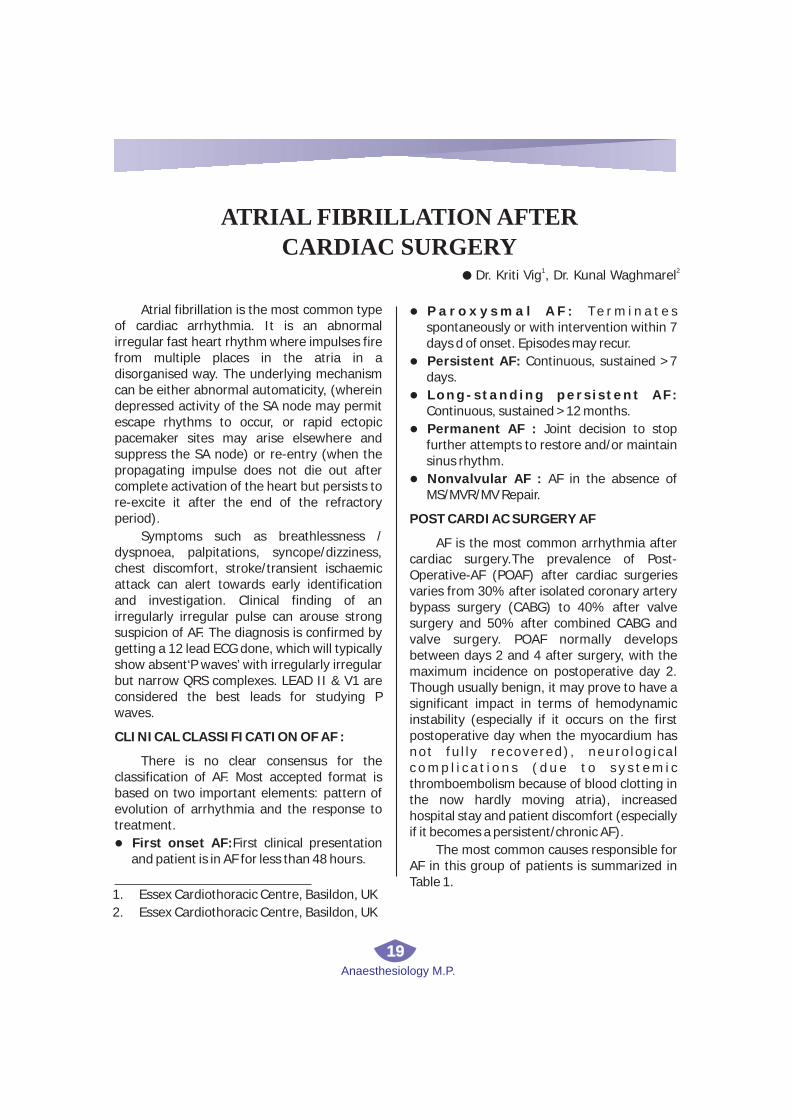
Atrial fibrillation is the most common type of cardiac arrhythmia. It is an abnormal irregular fast heart rhythm where impulses fire from multiple places in the atria in a disorganised way. The underlying mechanism can be either abnormal automaticity, (wherein depressed activity of the SA node may permit escape rhythms to occur, or rapid ectopic pacemaker sites may arise elsewhere and suppress the SA node) or re-entry (when the propagating impulse does not die out after complete activation of the heart but persists to re-excite it after the end of the refractory period).
Symptoms such as breathlessness / dyspnoea, palpitations, syncope/dizziness, chest discomfort, stroke/transient ischaemic attack can alert towards early identification and investigation. Clinical finding of an irregularly irregular pulse can arouse strong suspicion of AF. The diagnosis is confirmed by getting a 12 lead ECG done, which will typically show absent‘P waves’ with irregularly irregular but narrow QRS complexes. LEAD II & V1 are considered the best leads for studying P waves.
CLINICAL CLASSIFICATION OF AF :
There is no clear consensus for the classification of AF. Most accepted format is based on two important elements: pattern of evolution of arrhythmia and the response to treatment.
First onset AF:First clinical presentation and patient is in AF for less than 48 hours.
l
l
l
l
l
l
P a r o x y s m a l A F : Te r m i n a t e s spontaneously or with intervention within 7 days d of onset. Episodes may recur.
Persistent AF: Continuous, sustained >7 days.
Long-standing pers istent AF: Continuous, sustained >12 months.
Permanent AF : Joint decision to stop further attempts to restore and/or maintain sinus rhythm.
Nonvalvular AF : AF in the absence of MS/MVR/MV Repair.
POST CARDIAC SURGERY AF
AF is the most common arrhythmia after cardiac surgery.The prevalence of Post-Operative-AF (POAF) after cardiac surgeries varies from 30% after isolated coronary artery bypass surgery (CABG) to 40% after valve surgery and 50% after combined CABG and valve surgery. POAF normally develops between days 2 and 4 after surgery, with the maximum incidence on postoperative day 2. Though usually benign, it may prove to have a significant impact in terms of hemodynamic instability (especially if it occurs on the first postoperative day when the myocardium has not fu l l y recovered) , neuro log ica l c o m p l i c a t i o n s ( d u e t o s y s t e m i c thromboembolism because of blood clotting in the now hardly moving atria), increased hospital stay and patient discomfort (especially if it becomes a persistent/chronic AF).
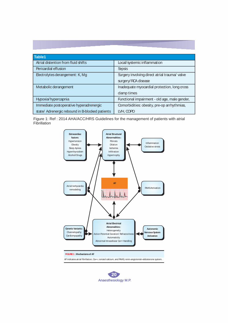
The most common causes responsible for AF in this group of patients is summarized in Table 1.
ATRIAL FIBRILLATION AFTER
CARDIAC SURGERY1 2
l Dr. Kriti Vig , Dr. Kunal Waghmarel
1. Essex Cardiothoracic Centre, Basildon, UK
2. Essex Cardiothoracic Centre, Basildon, UK
Anaesthesiology M.P.
20
Atrial distention from fluid shifts
Pericardial effusion
Electrolytes derangement: K, Mg
Metabolic derangement
Hypoxia/hypercapnia
Immediate postoperative hyperadrenergic
state/ Adrenergic rebound in B-blocked patients
Local/systemic inflammation
Sepsis
Surgery involving direct atrial trauma/ valve
surgery/RCA disease
Inadequate myocardial protection, long cross
clamp times
Functional impairment - old age, male gender,
Comorbidities: obesity, pre-op arrhythmias,
LVH, COPD
Table1
Extracardiac
factors:
Hypertension
Obesity
Sleep Apnea
Hyperthyroidism
Alcohol/Drugs
Atrial Structural
Abnormalities :
Fibrosis
Dilation
Ischemia
Infiltration
Hypertrophy
Inflammation
Oxidative stress
Atrial techycardia
remodelingRAAS Activation
Genetic Variants :
Channelopathy
Cardiomyopathy
Atrial Electrical
Abnomalities :
Heterogeneity
Action Potential Duration/ Refractoriness
Automaticity
Abnormal Intracellular Ca++ Handling
Autonomic
Nervous System
Activation
AF
FIGURE 1 : Mechanisms of AF
AF indicates atrial fibrillation; Ca++, ionized calcium; and RAAS, renin-angiotensin-aldosterone system.
Figure 1: Ref : 2014 AHA/ACC/HRS Guidelines for the management of patients with atrial Fibrillation

Anaesthesiology M.P.
21
We can better understand the factors that could lead to AF if we imagine the atria to be like an electric mesh. Any distention causing thinning of atrial fibres, any deposition or remodeling causing thickening of the fibres, any compression on them or any ectopic foci within them will potentially cause them to fibrillate.
Although, cardioplegia administered through the coronary circulation effectively arrests ventricular mechanical and electrical activity, the atrial septum remains significantly warmer than the ventricle (and usually retains electrical activity). Persistence of atrial electrical activity during bypass is associated with postoperative atrial arrhythmias.
Distension / Stretch Thickening/Remodeling
Infiltration / Deposition Multiple Foci

Anaesthesiology M.P.
22
PREVENTION
POAF per se is a multifactorial problem. Hence, there is no definitive preventive measure for the same. Prophylactic therapy, however can reduce the risk of AF by approximately 50%.
Low-dose b-blockers starting within 12–24 hours of surgery is the most commonly used strategy, e.g. Metoprolol 25–50 mg bid or Atenolol 25 mg qds.
Prophylactic amiodarone has shown to have a significant preventive effect if commenced well in time. Some recommended regimens are: starting 6 days preoperatively with 10 mg/kg/d & continuing the same for 1 week postoperatively; or 200 mg tid for 5 days before surgery followed by 400 mg bid x 4–6 days after surgery (if surgery is scheduled in more than 5 days) or 400 mg qid x 1 day before surgery followed by 600 mg bid on the day of surgery, continuing with 400 mg bid x 4–6 days after surgery (if surgery scheduled within 1–5 days).
Magnesium sulphate given 2 g IV after CPB and on first postoperative morning is especially useful in conjunction with b-blockers. Magnesium is a cofactor for the myocardial cell membrane enzyme Na-K adenosine triphosphatase, which regulates transmembrane sodium and potassium gradients. Its deficiency may predispose to arrhythmias by altering membrane potential and repolarization via its effect on this enzyme. Magnesium repletion is inexpensive and safe and should be considered in all patients undergoing cardiac surgery.
Dual-site pacing helps by causing more uniform electrical activation of the atria, by s u p p r e s s i n g PA C s , e l i m i n a t i n g compensatory pauses after PACs, and reducing the dispersion of refractoriness.
TREATMENT
The goals of treatment are: acute
l
l
l
l
hemodynamic stabilisation in an unstable patient, identification & treatment of reversible causes/risk factors, alleviation of symptoms, reduction in the risk of stroke and prevention of complications like heart failure.
The longer the duration of AF, the more difficult it is to restore sinus rhythm and prevent recurrences. This is due to electrical and structural remodelling of atrial tissue resulting in shortening of effective refractory periods, thereby maintaining and increasing the duration of AF. It is very important to determine whether the pat ient i s hemodynamically stable. As, 50% of these patients revert spontaneously, the primary aim should be to control the ventricular rate to prevent the increase in myocardial workload. The underlying cause should be identified and treated.
RATE CONTROL is primarily achieved by beta antagonists or non-dihydropyridine Calcium channel blockers. Latter is more useful in patients with poor LV function. Digoxin can be added to any of the two categories of drugs, for rate control in chronic settings. Amiodarone is only indicated if the above therapies fail to control rate. It has no negative inotropic effects. However, the rapidity and degree of slowing is less than with the b-blockers and CCBs. Hence it is reserved for rate control only if ventricular response is not too rapid, LV is poor or b-b lockers and CCBs are contraindicated. It should not be used for long-term rate control. A resting HR of <80 bpm should be aimed but a lenient target of HR<110 bpm can be set if the LV is good and the patient asymptomatic.
RHYTHM CONTROL:
Rhythm control is indicated in patients who fail rate-control strategy, are unstable hemodynamically or are likely to deteriorate e.g. patients with poor ventricular function where the cardiac output is significantly dependent on the atrial contribution. Moreover, for postoperative AF following
l
Anaesthesiology M.P.
23
cardiothoracic surgery, rhythm-control strategy should be offered as the initial m a n a g e m e n t o p t i o n , u n l e s s contraindicated. (NICE guidelines)
Cardioversion to sinus rhythm can be achieved electrically or pharmacologically. Electrical method is preferred if the AF has persisted for more than 48 hours or if there is life threatening instability. It is also the method of choice for WPW & other pre-excitation syndromes causing AF, where amiodarone, b blockers and CCBs are contraindicated.
Sinus rhythm can be restored in a significant proportion of patients with success rates varying between 65% and 90%. Electrical cardioversion is done by giving a synchronised shock starting from 50-100 Joules DC on biphasic or 200J monophasic defibrillator, increasing the energy with successive shocks as required. The defibrillation threshold will have individual variation based on the transthoracic impedance. In patients with implanted devices such as permanent pacemaker or internal defibrillator, the device must be interrogated immediately before and after cardioversion to assess any malfunction. The paddles used for cardioversion should be placed as far as possible from the implanted device, preferably in the anterior-posterior position. Patients with underlying conduction defects are at risk of developing profound arrhythmias following cardioversion. These patients are identified by having a slow ventricular response to AF in the absence of rate-reducing medications and facilities for temporary external or endocardial pacing must be made available prior to attempting cardioversion. Electrical cardioversion can also lead to transient ST segment elevation with a rise in blood concentrations of cardiac troponins and CK-MB, even without cardiac damage. The rate of relapse after
l
l
DCC is high unless anti-arrhythmic drug therapy to maintain sinus rhythm is given concomitantly. Hence it is advised to continue amiodarone therapy 4 weeks before till 12 months after elective cardioversion, to maintain normal sinus rhythm
Pharmacological management of AF
Amiodarone is the drug of choice for acute rhythm control while b blockers should be continued long term to sustain the sinus rhythm, once achieved. Other drugs like flecainide, propafenone, dofetilide can be used as in the absence of structural or ischemic heart disease. They are popular as “pill-in-the-pocket” in addition to a beta blocker or CCB to terminate AF outside the hospital (if demonstrated to be safe in a supervised setting).
Vernakalant is a new drug under research. It is an atrial-selective potassium and sodium channel blocking agent and has shown to convert about 50% of patients with new onset postoperative AF to sinus rhythm in approximately 10 minutes. It is used in a dose of 3 mg/kg infused over 10 minutes with a subsequent 2 mg/kg infusion over 10 minutes if AF is still present after 15 minutes. It can be commenced if atrial fibrillation 7 days duration in non-surgery patients or 3 days duration in post-cardiac surgery patients. As it can depress myocardium, it is contraindicated in patients with MI or ACS within the last 30 days.
Non-pharmacological management of AF (Table -2)
ROLE OF ANTICOAGULATION IN AF
As the atria fibrillate, despite excessive electrical activity there is hardly any mechanical activity. This results in sluggish blood flow in atria predisposing to clot formation. It usually takes around 48 hours for
l
l

RHYTHM CONTROL
Device therapy
? Atrial pacing (single or
multisite)
? Atrial defibrillators (stand-
alone or with pacemaker
function)
Ablation therapy
? Operative (Maze
procedure,
Pulmonary vein isolation,
His bundle ablation)
? Percutaneous
transcatheter techniques
(pulmonary vein
isolation,
radiofrequency ablation)
RATE CONTROL
? Transcatheter AV junctional
ablation and permanent
? Pacemaker implantation
? Radiofrequency transcatheter
AV junction modification
STROKE PREVENTION
? Percutaneous left atrial
appendage transcatheter
occlusion (PLAATO)
Anaesthesiology M.P.
24
this to start. Hence any patient who has stayed in AF for more than 48 hours, anticoagulation is crucial for prevention of stroke resulting from systemic thromboembolism. The risk of embolism increases manifold with electrical cardioversion. Thus, anticoagulation must be given 3 weeks before continued till 4 weeks after elective cardioversion in patients with duration of AF>48 hrs or in those with high risk of stroke. However, in life threatening situations, emergency cardioversion must not be delayed, and it should again be followed by 4 weeks of anticoagulation. It is suggested to perform a transoesophageal echo examination to rule out any thrombus in the LA before performing cardioversion in a patient with unknown or suboptimal anticoagulation status.
Warfarin is the anticoagulant of choice in
mechanical heart valves or in patients with moderate to severe mitral valve disease. Heparin is used for bridging with warfarin in certain situations. In patients on warfarin, INR monitoring is recommended weekly and then monthly once INR is stable. Others drugs like rivoraxaban, apixaban and dabigatran are preferably given in non valvular AF as they are safer and have lesser side effects. However, they are contraindicated in patients with renal disease.
W h e n c o n s i d e r i n g l o n g t e r m anticoagulation, a balance should be struck between the risk of stroke versus that of bleeding. CHA2DS2VASc and HASBLED are the widely-used scores to assess the two risks respectively.
Non-pharmacological management of AF
Table 2

Anaesthesiology M.P.
25
Figure 3
Figure 4
+1 Point
+2 Point
+1 Point
+1 Point
+1 Point
+1 Point
+1 Point
+2 Point
CHADSVAS
2
2
Congestive Heart Failure
Hypertension
Age > 75
Diabetes
Stroke/TIA History
Vascular Disease
Age 65-74
Sex (Female)
Risk Factors
0
1
2
3
4
5
6
7
8
9
0%
1.3%
2.2%
3.2%
4.0%
6.7%
9.8%
9.6%
6.7%
15.2%
Stroke Risk Per Year
SCORE % RATE PER YEAR
Reference : European Heart Rhythm Association. Guidelines for the management of atrial fibrillation : the Task Force for
The Management of Atrial Fibrillation of the European Society of Cardiology (ESC). Eur Heart J. 2010;31(19):2369-2429.
HAS-BLED SCORE
Hypertension - Uncontrolled, >160 mmHg systolic
Alcohol use - >8 drinks/week
Age > 65 years
Stroke history
Bleeding - prior major or predisposition to
Labile INR - Unstable/high INRs, time in therapeutic range <60%
End organ disease
? Renal - Dialysis, transplant, Cr >2.26 mg/dL or >200 µmol/L
? Liver - Cirrhosis or bilirubin >2x normal with AST/ALT/AP >3x normal
Drugs predisposing to bleeding - Antiplatelet agents, NSAIDs
1 point for each of above; If score 0-1 : low risk, 2 = moderate risk , 3 or more - high risk of bleeding

Anaesthesiology M.P.
26
Our experience :
In a small study conducted in our centre, we tried to correlate the occurrence of postoperative AF with various predisposing factors and preventive measures. We found the following factors to have a definite contribution towards occurrence of POAF:age > 70 years, insufficient i n t r a o p e ra t i v e myo c a r d i a l protection in terms of cardioplegia volume and bypass time, surgery involving the valves, poor LV preoperatively, comorbidities COPD and HTN. Amongst drugs given preoperatively for prevention, amiodarone significantly reduced the incidence of POAF, while B-blockers or statins didn’t have any preventive effect. However, when statins were given postoperatively they could significantly reduce the incidence of POAF.
In the patients studied, AF occurred most commonly on day 4 after surgery, and was mostly asymptomatic, though leading to heart failure in a small fraction of the patients. Occurrence of AF prolonged the length of stay in ITU as well as overall hospital length of stay thus increasing the financial burden of health services in addition to causing patient discomfort.
REFERENCES
1. Journal of the American College of Cardiology; Vol 64, No.21, 2014; “2014 AHA/ACC/HRS Guidel ine for the Management of Patients with Atrial Fibrillation: Executive summary”
2. The New England Journal of Medicine, May 2016, Vol 374, No.20; “Rate control versus Rhythm control for Atrial Fibrillation after cardiac surgery”
3. NICE guidelines for Atrial fibrillation management, June 2014
4. British Medical Journal, May 2017, Vol 103, No.10; “Integrated care of patients with atrial fibrillation: the 2016 ESC atrial fibrillation guidelines”
5. Manual of Perioperative Care in Adult Cardiac Surgery, 5th edition, Robert M. Bojar
6. Audit on Post cardiac surgery Atrial Fibrillation 2014; Dr S.Mukherjee, Dr G.Namjoshi, Dr K. Waghmare; Essex cardiothoracic centre
7. Continuing Education in Anaesthesia, Critical Care & Pain; Vol 6, No.6, 2006; Atrial Fibrillation, A.Bajpayee, E. Rowland
8. Annals of Internal Medicine, Dec 2001 Volume 135 ; Atrial Fibrillation after Cardiac Surgery, W H Maisel, J D Rawn, W G Stevenson

Anaesthesiology M.P.
27
evaluation of this child revealed harsh breath sounds and was treated with intravenous antibiotics and nebulization continued prior to surgery. Inj. Glycopyrrolate 0.05 mg was given through IM route half an hour before surgery. Difficult airway cart appropriate for age was prepared. Baseline vitals were recorded with noninvasive monitors like Electrocardiogram, Pulse oximeter, and noninvasive blood pressure. Clearly, IV access would be difficult in this child due to limb deformity and repeated hospital admissions. Preoxygenation followed by inhalation induction was stared with O2and sevoflurane in slow incremental doses with simultaneous intravenous 24 G I.V. cannula secured on opposite limb in the second attempt.Inj Midazolam 0.5 mg, inj Hydrocortisone 20mg, Inj ondensetron 1mg were given intravenously. Airway was secured with 2 No. Laryngeal mask airway ( LMA). Brachial block through Supraclavicular route was given in left upper limb by blind technique with 4 ml of Inj Bupivacaine0.25%. Anesthesia was maintained with O2 and sevoflurane with patient on spontaneous venti lation. Continuous intra-operative monitoring showed stable vitals. Duration of surgery was 45 minutes. At the end of surgery, LMA was removed when patient was wide awake with stable hemodynamics.
Apert Syndrome was first described by the French pediatrician Eugene Apert in 1906. It is a rare autosomal dominant disease with an
DISCUSSION
ANESTHETIC MANAGEMENT IN A
PATIENT OF APERT SYNDROME1 2 3
l Dr. Ruchi Tandon , Dr. Emendr Wahnel Dr. Abhay Raj Yadavl
INTRODUCTION-
CASE REPORT-
Apert syndrome is a form of acro-cephalo-syndactyly, a congenital disorder characterized by malformations of the skull, face, hands and feet. In embryology, the hands and feet have selective cells that die, called selective cell death or apoptosis, causing separation of the digits. In case of acrocephalosyndactyly, selective cell death does not occur and skin, and rarely bone between the fingers and toes fuses.Difficulty related to airway management is a major concern. One should also be aware of other complications and difficulties like bronchospasm, wheezing, and even difficulty in intravenous access. We report our experience in the anesthetic management of case of Apert syndrome, referred to us for syndactyly release.
A 4-year-old male child, a diagnosed case of Apert syndrome, born of full-term delivery, was referred to our Institution for bilateral hand syndactyly release. This child had mid facial hypoplasia involving maxillary and zygomatic bones with orbital proptosis, down sloping palpebral fissure, hypertelorism, and all limbs showed fusion of multiple digits. He had history of repeated episodes of respiratory tract infection and incomplete and inadequate response when spoken to which was suggestive of delayed milestones for his age. No relevant family history was noted. A thorough systemic examination showed no other associated anomalies. Preoperative
1. Associate Professor, GM.C. Bhopal
3. III Year RMO, G.M.C. Bhopal
2. II Year RMO, G.M.C. Bhopal

Anaesthesiology M.P.
28
1incidence of around 1 per 160 000 live births . Genetically, the defect is in chromosome 10 affecting fibroblast growth factor receptor 2 gene. This mainly affects digits and cranium causing fusion of multiple digits of both upper and lower limbs and premature fusion of cranial sutures. These children also have other associated anomalies like cardiac defects, polycystic kidneys, and pyloric stenosis.
These children require a number of di fferent operat ive procedures and anesthesiologists face multiple problems while dealing with them, airway management being a major one. Airway dimorphism carries a risk of difficult mask ventilation and intubation due
2,3to mid face hypoplasia . Children with Apert syndrome have profuse secretions with inability to clear these that may cause increase in airway irritability, higher incidence of
bronchospasm, and repeated respiratory tract 4infection .Craniofacial anomalies are often
associated with airway obstruction, especially during sleep, and can cause obstructive sleep apnea, and therefore are prone to becoming
5obstructed on induction and emergence . Preoperative evaluation and optimization is important in these patients. Airway adjutants like appropriate size airway, LMA, endotracheal tubes, and emergency tracheostomy kit have
6to be kept ready . Use of regional anesthesia techniques as an adjuvant to general anesthesia help to decrease the requirement of drugs during intra- and post-operative periods. This reduces the incidence of complications associated with airway handling. Regional anesthesia itself could be difficult in these patients due to anatomical variations in shoulder joint and related structures.

Anaesthesiology M.P.
29
Ultrasonography (USG)-guided regional anesthesia techniques are of help to overcome
7this problem .
Thorough screening for associated anomalies like cardiac defects, polycystic kidneys and pyloric stenosis should be done
8preoperatively .
Another challenge for the anesthetist is intravenous access. Limb deformity and multiple operative procedures make intravenous access more difficult in these patients. Placing an IV cannula is very difficult with the child fully awake and struggling. It is preferable to place the IV cannula after the child is sedated using inhalational agents like sevoflurane. For short procedures, e.g., change of dressings or computed tomography
3(CT) scans, intramuscular route can be used .
Dealing with a case of Apert syndrome is very challenging for anesthetist. So, thorough preoperative evaluation, a proper anesthesia plan as well as a backup for difficult airway management is very important in these patients.
1.Cohen MM, Jr, Kreiborg S. New indirect method for estimating the birth prevalence of the Apert’s syndrome. Int J Oral Maxillofac Surg. 1992;21:107–9. [PubMed].
CONCLUSION
REFERENCES
2.Cruz Pardos P, CebrianPazos J. Management of the airway in an infant with Apert syndrome. Rev EspAnestesiolReanim. 2000;47:97–8. [PubMed].
3.Barnett S, Moloney C, Bingham R. Perioperative complications in children with Apert Syndrome: A review of 509 anesthetics. PediatrAnesth. 2011;21:72–7. [PubMed].
4. Cohen MM, Jr, Kreiborg S. Upper and lower airway compromise in the Apert syndrome. Am J Med Genet.1992;44:90–3. [PubMed]
5. Perkins JA, Sie KC, Milczuk H, Richardson MA. Airway management in children with craniofacial anomalies.Cleft Palate Craniofac J. 1997;34:135–40. [PubMed].
6.Atalay C, Dogan N, Yuksek S, Erdem A. Anesthesia and airway management in two cases of apert syndrome: Case reports. E A J M . 2 0 0 8 ; 4 0 : 9 1 – 3 . [ P M C f r e e article][PubMed].
7. Chan VW, Perlas A, McCartney CJ, Brull R, Xu D, Abbas S. Ultrasound guidance improves success rate of axillary brachial plexus block. Can J Anaesth. 2007;54:176–82. [PubMed]
8.Cohen MM, Jr, Kreiborg S. Visceral anomalies in the Apert syndrome. Am J Med Genet.1993;45:758–60. [PubMed].
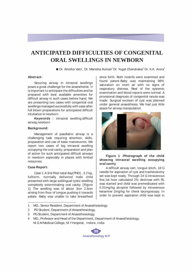
Anaesthesiology M.P.
30
Abstract:
Securing airway in intraoral swellings poses a great challenge for the anaesthetist. It is important to anticipate the difficulties and be prepared with best available amenities for difficult airway in such cases before hand. We are presenting two cases with congenital oral swellings managed successfully with ease after full blown preparations for anticipated difficult intubation in newborn.
Keywords : intraoral swelling,difficult airway,newborn
Background:
Management of paediatric airway is a challenging task requiring attention, skills, preparation and use of basic manoeuvres. We report two cases of big intraoral swelling occupying the oral cavity, preparation and plan of action for such anticipated difficult airways in newborn especially in places with limited resources.
Case Report:
Case 1:A 3rd-Post natal day(PND) , 3.2 kg, fullterm, normally delivered male child presented with large sublingual cystic swelling completely exterminating oral cavity. [Figure 1] The swelling was of about 3cm 2.6cm arising from floor of tongue pushing it towards palate. Baby was unable to take breastfeed
since birth. Both nostrils were examined and found patent.Baby was maintaining 98% saturation on room air with no signs of respiratory distress. Rest of the systemic examination and blood reports were normal. A provisional diagnosis of congenital ranula was made. Surgical excision of cyst was planned under general anaesthesia. We had just little space for airway manipulation.
Figure 1 :Photograph of the child showing intraoral swelling occupying oral cavity
A difficult airway cart, tongue stitch, 18 G needle for aspiration of cyst and tracheostomy set was kept ready. Through 24 G intravenous line,1st hour calculated 2% dextrose with RL was started and child was premedicated with 0.01mg/kg atropine followed by intravenous ketamine 2mg/kg for check laryngoscopy. In order to prevent aspiration child was kept in
ANTICIPATED DIFFICULTIES OF CONGENITAL
ORAL SWELLINGS IN NEWBORN1 2 3 4
l Dr. Amisha Vats , Dr. Manisha Kumari Dr. Yugal Chandrakar Dr. K.K. Arora
1 MD., Senior Resident, Department of Anaesthesiology
2 PG Student, Department of Anaesthesiology,
3 PG Student, Department of Anaesthesiology,
4 MD., Professor and Head of the Department, Department of Anaesthesiology,
M.G.M Medical College, M.Y Hospital, Indore, India

Anaesthesiology M.P.
31
left lateral position.Child was preoxygenated and conf i rmed for bag and mask ventilation.Check laryngoscopy in supine position was difficult so in lateral position, direct laryngoscopy was done to visualize the vocal cords under ketamine sedation and inhalat ional anaesthesia (oxygen & sevoflurane).In checkscopy we could visualize the vocal cords without any external manipulation of larynx and so we proceeded with deepening of plane with gradually increasing concentration of sevoflurane to 6%, patient was intubated with 3.0 mm portex uncuffed tracheal tube using 0 size miller’s straight blade. The tube was fixed after confirming bilateral equal air entry and tracing of capnography and atracurium 0.3 mg/kg was given. Throat packing was done to prevent aspiration of secretions during skeletonization of cyst. Anesthesia was maintained with sevoflurane in oxygen and nitrous oxide. 2 µg/kg fentanyl was given for analgesia and dexamethasone 0.8 mg/kg intravenous was given to prevent airway edema.Boluses of Ringer’s lactate were given to replace the intraoperative blood loss during dissection of mass.Peroperatively, heart rate and oxygen saturation was attentively observed.
Surgical excision of swelling with lining epithelium was done successfully taking care not to injure the surrounding structures [ Figure 2]. Anticipating postoperative edema of tongue, airway patency was maintained using a tongue stitch fixed to the chin.Oozing was controlled using local hemostatic agent feracrylum soaked gauge compression. Neuromuscular blockade was reversed (0.05mg/kg neostigmine and 0.01mg/kg glycopyrrolate) and extubated in lateral position after thorough suctioning of oral cavity and removing throat pack only after adequate breathing efforts and muscle tone. Postoperatively, baby was crying ,moving limbs actively, maintaining saturation on room air and haemodynamically stable. Patient was closely observed for next 24 hours, and patient
was discharged on 2nd post operative day.
Figure 2 : Intraoperative view of oral swelling
Case 2 :A 1-PND , fullterm, normally delivered neonate weighing 3.0 kg having a large mass occupying the oral cavity with no signs of respiratory discomfort was scheduled for excision and biopsy. The primary diagnosis made was congenital epulis(a benign tumour of oral cavity).[Figure3] Preoperative assessment and investigations were all within normal limits and with no other associated congenital anomalies. On examination, the mass was around 2 cm in length and 4cm in breadth, mobile, smooth surface and firm consistency with no clear pedicle. It was taken for excision under general anaesthesia. NBM status was confirmed.After monitors being attached a 22 gauge intravenous line was obtained and intravenous fluid 2% dextrose in Ringer’s lactate was started to make up for the deficits.Inj. atropine 0.01mg/kg was given as premedication. Before inducing, check laryngoscopy was done with No. 0 Miller’s straight blade under ketamine 2.5mg/kg, we were able to visualize epiglottis and posterior part of the cords. After preoxygenation and confirming ability to ventilate with correct sized mask, inhalation induction of anaesthesia was accomplished using sevoflurane 2%+ oxygen and intubating dose of succinylcholine (2.5mg/kg) . Neonate was manually mask

Anaesthesiology M.P.
32
ventilated with JRC for 45seconds. We were able to successfully intubate the neonate with a 3.5 mm uncuffed endotracheal tube in first attempt using Miller’s No 0 straight blade. [Figure4]Throat packing was done. Lignocaine 2% with adrenaline (5mg/kg) was infiltrated on the base of the mass to reduce blood loss and pain relief. Inj fentanyl 1.0µg/kg,Inj. Dexamethasone 0.8mg/kg and paracetamol suppository 40mg/kg were given.The maintenance of anaesthesia was done with oxygen, nitrous oxide, and sevoflurane to achieve a MAC of one and Inj. Atracurium 0.3 mg/kg. No event of desaturation and bradycardia was witnessed. After completion of surgery the trachea was extubated after throat pack removal and clearing the oral cavity in lateral position while the baby was fully awake after reversal of muscle relaxant with neost igmine (0.05mg/kg) and glycopyrrolate (0.01 mg/kg ). Postextubation, child was active, maintaining saturation on room air. Breast feed was allowed same day and the postoperative period was uneventful throughout, thus patient was discharged subsequently.
Figure 3: Congenital Epulis
Figure 4 : Confirmed secure airway
Figure 5 : En Mass dissection
Discussion :
Neonates with big mass occupying oral cavity and protruding through mouth often prove to be difficult candidates for airway management as these large intraoral cysts in the newborn may cause airway obstruction
1and swallowing difficulties. Congenital epulis is a rare lesion of newborn also known as Neumann’s tumor, which is benign in nature. The appearance of the lesion is more common in maxillary alveolar ridge than mandibular
2with predominance in females than in males. Whereas ranulas are a mucus extravasation cyst originating from the sublingual gland on

Anaesthesiology M.P.
33
the floor of the mouth as a result of ductal obstruction and mucus collection.Surgical excision is very effective and curative. Anaesthesia for excision of epulis could be either general or local. Some cases were reported in the literatures where epulis was
3,4excised successfully under local anesthesia. Other reports stated that epulis was excised
5under general anaesthesia. In the present case report, epulis was excised under general anaesthesia. Prognosis of these oral swellings is generally very good with a very low incidence of relapse. Planning the surgical excision and achieving it requires a team approach.
Preoperative xray and ultrasonography of the neck aid in identification of factors leading to difficulty in establishing artificial airway during intubation but could not be done in our setup because of feasibility and economic issues. The key step in anaesthetic management of such patients is the provision of safe and secure airway to avoid hypoxia, as hypoxia results in grave mishaps.It is thus mandatory to keep the difficult intubation cart ready and ENT surgeons team ready as backup
6for emergency tracheostomy. Induction of anaesthesia can result in the ‘cannot ventilate, cannot intubate’ situation or complete loss of the airway tone hence it is preferable to maintain spontaneous ventilation until trachea is successfully intubated or bag and mask ventilation is well established before giving short acting muscle relaxant. In such patients, an awake intubation is often the primary approach of airway management under sedation and adequate application of local anaesthetics to the airway butawake intubation is a tough job and traumatic in neonates and infants and requires expertise with the procedure. Better alternative is to keep the infant anaesthetised but breathing spontaneously on 100% oxygen using an inhalational agent such as sevoflurane. Another important issue during the ongoing surgery is the possibility of accidental extubation of the endotracheal tube thereby
tight fixation of tube and holding tube properly is very crucial as a part of airway management.Rupture of swelling and aspiration of its contents during laryngoscopy should be kept in mind. Gentle handling of the swelling and thorough throat packing specially when using uncuffed tube and placement of nasogastric tube should minimize such complications.Decision of timing of extubation and postoperative airway edema is always a concern as extubation in this situation would make ventilation and intubation difficult. Supraglottic edema can be prevented and treated using dexamethasone and nebulized
7 racemic epinephrine with moist oxygen.Lateral positioning is known to decrease upper airway obstruction in awake as well as anesthetized individuals. Turning the child into lateral position allows the tongue to fall out of
8,9the mouth and clears airway obstruction. For these reasons it is commonly employed during recovery from anaesthesia.
In some of the published case reports the similar surgical excision was performed at 7th
10,11day, at 2 months respectively. In these babies aspiration of the cyst was followed by nasotracheal intubation, but in our case report cyst aspiration was avoided as it could alter the anatomy of cyst and can even lead to aspiration of contents into unsecured airway. Orotracheal intubation was achieved quite easily as sometimes the “recognized difficult airway” proves not to be much difficult —much to our relief !!!!
Conclusion :
The success in management of a congenital oral swellings largely depends upon a detailed and proper preoperative evaluation with emphasis on airway assessment as well as formulation of an appropriate plan for intubation. Equally important is to not hurry extubation considering the potential for postoperative airway-related complications.
Acknowledgement :

Anaesthesiology M.P.
34
We would like to thank the parents for providing consent to use their babies photograph in this article.
Footnote :
Source of Support: Nil
Conflict of Interest: None declared
References :
1. J S M Daniels congenital sublingual dermoid cyst in a neonate a case report Ghana Medical Journal vol 38, no: 1, March 2004
2. Dash JK, Sahoo PK, Das SN. Congenital granular cell lesion “congenital epulis”-report of a case. Indian Soc Pedod Prev Dent. 2004;22:63–7
3. Merrett SJ, Crawford PJ. Congenital epulis of the newborn: a case report. Int J Paediatric Dent 2003; 13:127-9.
4. Khalid Khattala et al. A case report of a huge congenital granular cell epulis operated under local anesthesia.The Pan African Medical Journal. 2012;12:105-7
5. Mukhopadhyay M, Mohanta PK, Mukhopadhyay B. Congenital epulis-a case report.J Indian Med Assoc 2004; 102:222.
6. Kumar N, Bindra A, Kumar N, Yadav N, Sharma S. Anesthetic concerns in a huge congenital sublingual swelling obscuring airway access. Saudi Journal of Anaesthes ia. 2015;9(2):202-203. doi:10.4103/1658-354X.152888.
7. Kamal G, Gupta A, Bathla S, Prasad N. Cystic hygroma and anaesthetic i m p l i c a t i o n . I n d i a n J A n a e s t h . 2011;55:89–90.
8. Litman RS, Wake N, Chan Lai ML, McDonough J M, Sanghun Sin MS, Mahboubi S, Arens R. Effect of lateral positioning on upper airway size and morphology in sedated children. Anesthesiology. 2005;103:484–8.
9. Walker RWM, Ellwood J. The Management of difficult intubation in children. Pediatric Anesthesia. 2009;19(Suppl 1):77–87.
10. Sublingual epidermoid cyst in a neonate Fadekemi Olufunmilayo Oginni, Taoreed Oladejo, Ramat Oyebunmi Braimah, Anthony Taiwo Adenekan Annals of maxillofacial surgery Jan-June 2014 page no 96.
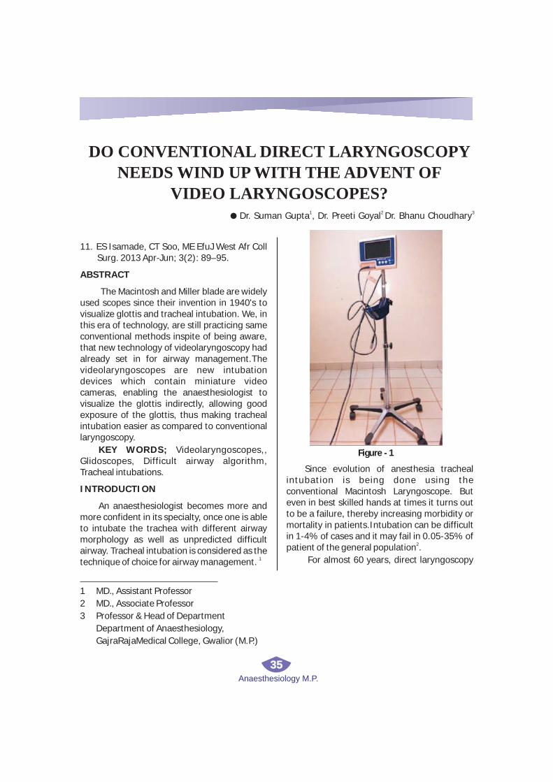
Anaesthesiology M.P.
35
11. ES Isamade, CT Soo, ME EfuJ West Afr Coll Surg. 2013 Apr-Jun; 3(2): 89–95.
ABSTRACT
INTRODUCTION
The Macintosh and Miller blade are widely used scopes since their invention in 1940's to visualize glottis and tracheal intubation. We, in this era of technology, are still practicing same conventional methods inspite of being aware, that new technology of videolaryngoscopy had already set in for airway management.The videolaryngoscopes are new intubation devices which contain miniature video cameras, enabling the anaesthesiologist to visualize the glottis indirectly, allowing good exposure of the glottis, thus making tracheal intubation easier as compared to conventional laryngoscopy.
KEY WORDS; Videolaryngoscopes,, Glidoscopes, Difficult airway algorithm, Tracheal intubations.
An anaesthesiologist becomes more and more confident in its specialty, once one is able to intubate the trachea with different airway morphology as well as unpredicted difficult airway. Tracheal intubation is considered as the
1technique of choice for airway management.
Since evolution of anesthesia tracheal intubation is being done using the conventional Macintosh Laryngoscope. But even in best skilled hands at times it turns out to be a failure, thereby increasing morbidity or mortality in patients.Intubation can be difficult in 1-4% of cases and it may fail in 0.05-35% of
2patient of the general population .
For almost 60 years, direct laryngoscopy
DO CONVENTIONAL DIRECT LARYNGOSCOPY
NEEDS WIND UP WITH THE ADVENT OF
VIDEO LARYNGOSCOPES?1 2 3
l Dr. Suman Gupta , Dr. Preeti Goyal Dr. Bhanu Choudhary
1 MD., Assistant Professor
2
3 Professor & Head of Department
Department of Anaesthesiology,
GajraRajaMedical College, Gwalior (M.P.)
MD., Associate Professor
Figure - 1

Anaesthesiology M.P.
36
was the sole method used by anesthesiologist, 3to insert tracheal tube into the trachea. .The
search for bigger and better angle of view of glottis during difficult intubations led to the
3development of video assisted devices. A varied types of video laryngoscopes are now available to facilitate trachaeal intubation.
So far flexible bronchoscopes were considered as Gold standard, but it needs experience and long learning curve. With the emergence of video loryngo scopes as a leading method of obtaining an improved view of the glottis opening, tracheal intubation has became easier as compared to conventional
4laryngoscopy. .
From the initial direct techniques, laryngoscopy advanced through an era of
F l e x i b l e f i b e r - o p t i c v i d e o laryngoscopes,followed by the more highly developed transition scopes.The first generation of video loryngo scopes employed technology used in rigid f iberoptic
5laryngoscopes. eg,Bullard scope, Wuscope, Upsherscope.
To overcome the difficulties met with first generation of 2001, a new type of videolaryngoscopes arrived in the shape of Glidoscopes,Trueview scopes offering camera on blade technology as shown in Figure-1 The Glidescope used a high resolution digital camera placed at the tip of an improved Macintosh Laryngoscopes blade attached to a high- definition screen results in better view of
4glottis Subsequently other types of videolaryngoscopes were then developed, all have been shown to improve the view of the
6vocal cords
To understand the technique of videolaryngoscopy we need to get well acquainted with Greenland's new model of two phases of laryngoscopy, i.e
1. Staticphase (posterior complex)
2.Dynamic phase (anterior complex)
DISCUSSION
Static phase is controlled by the posterior anatomical structures of neck, i.e, positioning in sniffing is governed by the flexion of lower cervical spine and extension of occipito- atlanto -axial complex, which brings in line alignment of Laryngeal axis(LA),Pharyngeal axis (PA),Oral axis (OA) to have proper view of glottis and facilitate intubation as shown in Figure 2.
Dynamic phase controls the anterior anatomical structures of the neck as shown in Figure 3.
During the dynamic phase, the mandible is drawn forward and bulk of submandibular tissue is compressed anterolaterally to provide a line of vision of glottis.The hyoid is also moved anteriorly and downward. Considering

Anaesthesiology M.P.
37
this, it is clear that only positioning is not .7enough to intubate via direct laryngoscopy
Videolaryngoscopes can be classified based on optical technology, blade shape, and guiding channels as:
1. Non channeled Videolaryngoscopes (Glidoscope, McGrath, VMAC, C MAC)
2. Channeled videolaryngoscopes (pentax AWS, King vision)
3. Optical-Airtraq
4. Fibroptic stylets- Bonefils
BENEFITS OF VIDEOLARYNGOSCOPY
1. In contrast to fiberoptic bronchoscope,it is relatively inexpensive and easy to handle. And better suited for tracheal intubation of patients with pharyngeal secretions or
8bleeding
2 Not only make intubation easier with easy learning curve but offer the possibility to record intubation procedure and stored in patients file as 'digital airway footprint' can
3be seen before next anesthetic procedure .
3. Visualization of videos of previous patients tracheal intubation should become as standard as regarding a patients
3laboratory results.
4. It should be done in all intubations and recorded in AIMS [Anaesthes ia
3Information Management System].
5. Does not require compulsory alignment of buccopharyngeal axis of the laryngoscope
2as in direct laryngoscopy
6. The glottis view by classic direct visualization offers a 10-degree view as compared to a much wider 60-degree view projected by the lens on the video
9laryngoscopes as shown in Figure 4.
7. It reduces the time required for intubation 1as well as the risk of dental trauma.
8. Kramer et al. showed that in patients with an anticipated difficult airway, awake nasal intubation using a C-MAC_ D-BLADE videolaryngoscope yielded the same
success rate as using a fibreoptic bronchoscope, but in a shorter intubation
10time
9. Gordon et al, used the Glidescopes to inspect airway with minimal stimulation
11following facial reconstructive surgery.
10. Videolaryngoscope has been used to 12check the cords after thyroid surgery.
11. Videoaryngoscopes can be used to displace excess airway tissue and may provide superior views of the morbidly obese patients larynx, and shown to be
13,14useful for awake tracheal intubations.
12 .It has found a place in the algorithm of diff icult intubation according to
15international recommendation as well as national AIDAA (All India Difficult Airway Association) algorithms, as shown in
16Figure 4.
13. The video laryngoscope provides an improved view of the glottic opening. Numerous studies have shown that there was an improvement of at least one Cormack-Lehane grade using video laryngoscopy as compared with standard direct laryngoscopy.9
Constraints of Direct laryngoscopy
1.Failure rate is about1.5%-8.5% despite experience, adequate positioning and mouth
17opening.
2. A need for Optimal line of sight for
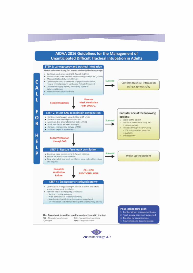
Anaesthesiology M.P.
38
Anaesthesiology M.P.
39
glottic visualization and greater cervical spine 18movement during laryngoscopy.
3.Lack of visual feedback for other health 18care providers.
Need to change from earlier to new concept of videolaryngoscopes, change from three axis alignment to anterior and posterior complex . In coming per iod V ideo l a ryngoscopes may rep lace d i rec t laryngoscopes as smart phones have replaced standard cell phone. So why not to get along with smart way of intubation with videolaryngoscopes which is easier and more advanced.
CONCLUSION
REFERENCES
1. Alain TI, Flavien K, Drissa BS, Idriss T, S e r g e I , N a z i n i g o u b a O . Videolaryngoscope is not a luxury,but a necessity. Anaesth Pain & Intensive Care 2015; 19(4):502-504
2. Benumof JL. Management of the difficult adult airway. With special emphasis on awake tracheal intubation. Anesthesiology 1991;75:1087–110.
3. C. Zaouter, J. Calderon, T. M. Hemmerling Videolaryngoscopy as a new standard of careBJA: British Journal of Anaesthesia, Volume 114, Issue 2, 1 February 2 0 1 5 , P a g e s 1 8 1 – 1 8 3 , h t t p s : //doi.org/10.1093/bja/aeu266
4. Cooper RM, Pacey JA, Bishop MJ, McCluskey SA. Early clinical experience w i th a new v ideo la ryngoscope (GlideScope) in 728 patients, Can J Anaesth , 2005, vol. 52 (191-8)
5. Cooper R, Law J, Hung O, Murphy MF, Law JA. , Rigid and Semi-Rigid Fiberoptic and Video- Laryngoscopy and Intubation, Management of the Difficult and Failed Airway , 2007 New YorkMcGraw Hill Medicals met
6. Pott LM, Murray WB. Review of video
laryngoscopy and rigid fiberoptic laryngoscopy, Curr Opin Anaesthesiol , 2008, vol. 21 ( 750-8)
7. K.B .Greenland.A proposed model of direct l a r y n g o s c o p y a n d t r a c h e a l intubation,Anaesthesia;2008(.63)156-161
8. A.R. Moore, T.Schricker ,O.Court. Awake videolaryngoscopy-assisted tracheal intubation of the morbidly obese. Anaesthesia 2012.67,232-235.
9. Ben H. Boedeker, W. Bosseau Murray and Thomas A. NicholasIntubation using the Video LaryngoscopeAmerican Medical Journal; 2012 :3 (2): 67-74,
10. Kramer A, M€uller D, Pf€ortner R, Mohr C , G r o e b e n H . F i b r e o p t i c v s videolaryngoscopic (C-MAC_ D-BLADE) nasal awakeintubation under local anaesthesia.Anaesthesia 2015; 70: 400–6.
11. Gordon JK, Rodney G, Ball DR. Extendedroles for videolaryngoscopy. Anaesthesia2014; 69: 793.
12. Sandhu R, Higgs A. Extendedroles for videolaryngoscopy. Anaesthesia2014; 69: 1287-1279.
13. Doyle DJ. Awake intubation using the GlideScope_ videolaryngoscope: initial experience in four cases. Canadian Journal of Anesthesia 2004; 51: 520–1.
14. Andersen LH, Rovsing L, Olsen KS. GlideScope videolaryngoscope vs. Macintosh direct laryngoscope for intubation of morbidlyobese patients: a r a n d o m i s e d t r i a l . A c t a AnaesthesiologicaScandinavica 2011; 55: 1090–7.
15. Apfelbaum JL, , Hagberg CA, Caplan RA, Blitt CD, Connis RT, Nickinovich DG, et al. American Society of Anesthesiologists Task Force on Management of the Difficult A i rway. Prac t i ce gu ide l ines fo r management of the difficult airway: an updated report by the American Society of

Anaesthesiology M.P.
40
Anesthesiologists Task Force on Management of the Difficult Airway. Anesthesiology. 2013 Feb;118(2):251-70.
16. Myatra SN, Shah A, Kundra P, Patwa A, Ramkumar V, Divatia JV, et al. All India Difficult Airway Association 2016 guidelines for the management of unanticipated difficult tracheal intubation in adults. Indian J Anaesth 2016;60:885-98.
17. Crosby ET,Cooper RM, Douglas MJ,Doyle DJ etal.The unanticipated difficult airway with recommendations for management, Can J Anaesth 1998;45:757-776.
18. Aziz MF, Dillman D, Fu R, Brambrink AM. Comparative effectiveness of the C-MAC video laryngoscope versus direct laryngoscopy in the setting of the predicted difficult airway. Anesthesiology 2012;116:629-36.