Embed Size (px)
Citation preview

Megaloblastic anemia
Dr Mohamid Afroz KhanModerator-Dr Namrata Shetty
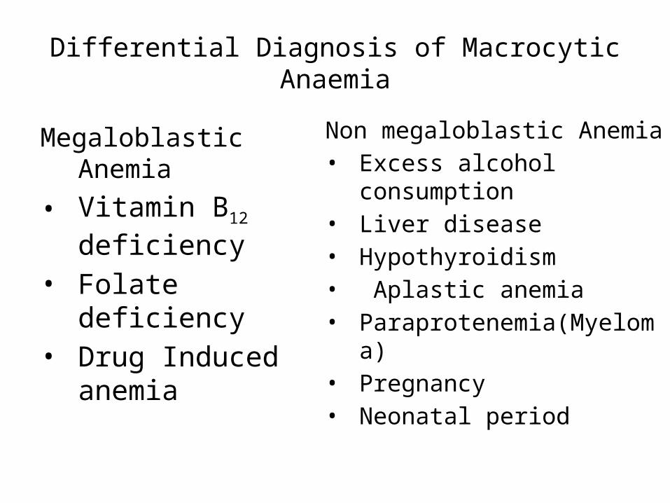
Differential Diagnosis of MacrocyticAnaemia
Megaloblastic Anemia• Vitamin B12
deficiency• Folate deficiency• Drug Induced anemia
Non megaloblastic Anemia• Excess alcohol consumption• Liver disease• Hypothyroidism• Aplastic anemia• Paraprotenemia(Myeloma)• Pregnancy• Neonatal period
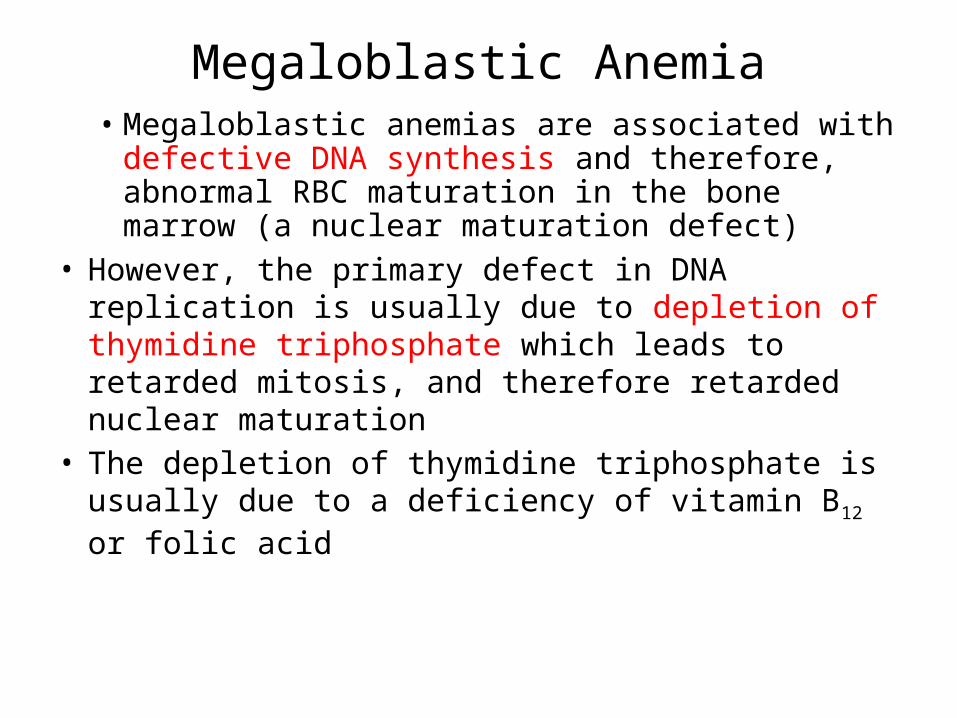
Megaloblastic Anemia• Megaloblastic anemias are associated with
defective DNA synthesis and therefore, abnormal RBC maturation in the bone marrow (a nuclear maturation defect)
• However, the primary defect in DNA replication is usually due to depletion of thymidine triphosphate which leads to retarded mitosis, and therefore retarded nuclear maturation
• The depletion of thymidine triphosphate is usually due to a deficiency of vitamin B12 or folic acid
• RNA synthesis is less impeded than is DNA synthesis hence cytoplasmic maturation and growth continues accounting for enlargement of the cells
• Increase in total erythropoiesis that may be up to three times normal
• Decreased rate of appearance of iron in the Hb of circulating erythrocytes and reticulocytopenia indicate ineffective erythropoiesis
• Increased destruction of defective erythroid precursors in the marrow, survival of circulating erythrocytes is short, indicating hemolysis


Absorption and metabolism of vitamin B12
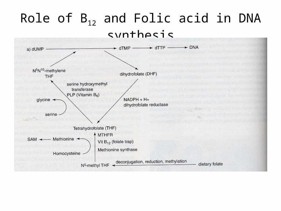
Role of B12 and Folic acid in DNA synthesis

Causes of Cobalamine Deficiency1. Reduced intake2. Malabsorption – Addisonian pernicious anemia,
Gastrectomy, pancreatic dysfunction, Tropical sprue, Zollinger Ellison syndrome
3. Food cobalamine malabsorption- Atrophic gastritis with achlorhydria
4. Abnormal transport protein- Tco I/II deficiency5. Inborn error of cobalamine metabolism6. Acquired drug effects

Causes of Folate Deficiency1. Reduced Intake2. Malabsorption- coeliac disease, tropical spure, small
bowel resection, malabsorption syndrome3. Drug Effect- Sulfa drugs, MTX, OCP, anticonvulsants4. Increased folate turnover- pregnancy progressive
fall, breastfeeding, skin disease(psoriasis & exfoliation), haemodylasis, PNH, haemoglobinopathy, autoimmune haemolytic anemia

MORPHOLOGY• Certain morphologic features are common to all forms of
megaloblastic anemia
Bone Marrow1. The bone marrow is markedly hypercellular and contains
numerous megaloblastic erythroid progenitors• Megaloblasts are larger than normal erythroid
progenitors (normoblasts) and have delicate, finely reticulated nuclear chromatin (indicative of nuclear immaturity)
• As megaloblasts differentiate and acquire Hb, the nucleus retains its finely distributed chromatin and fails to undergo the chromatin clumping typical of normoblasts

Bone marrow is hypercellular with cellularity > 95%
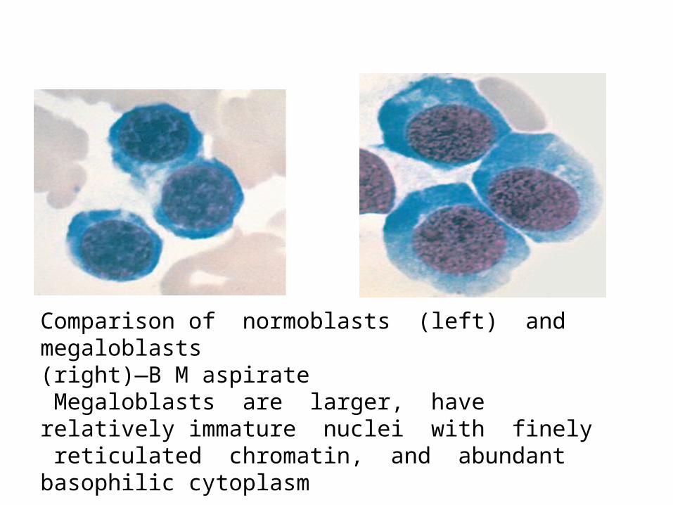
Comparison of normoblasts (left) and megaloblasts (right)—B M aspirate Megaloblasts are larger, have relatively immature nuclei with finely reticulated chromatin, and abundant basophilic cytoplasm

Megaloblasts – Bone Marrow
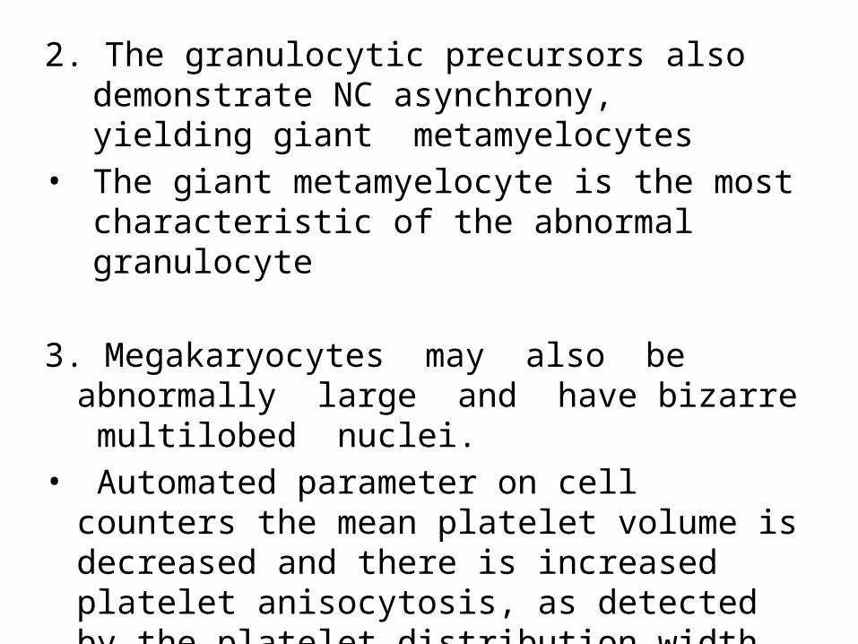
2. The granulocytic precursors also demonstrate NC asynchrony, yielding giant metamyelocytes
• The giant metamyelocyte is the most characteristic of the abnormal granulocyte
3. Megakaryocytes may also be abnormally large and have bizarre multilobed nuclei.
• Automated parameter on cell counters the mean platelet volume is decreased and there is increased platelet anisocytosis, as detected by the platelet distribution width (PDW)
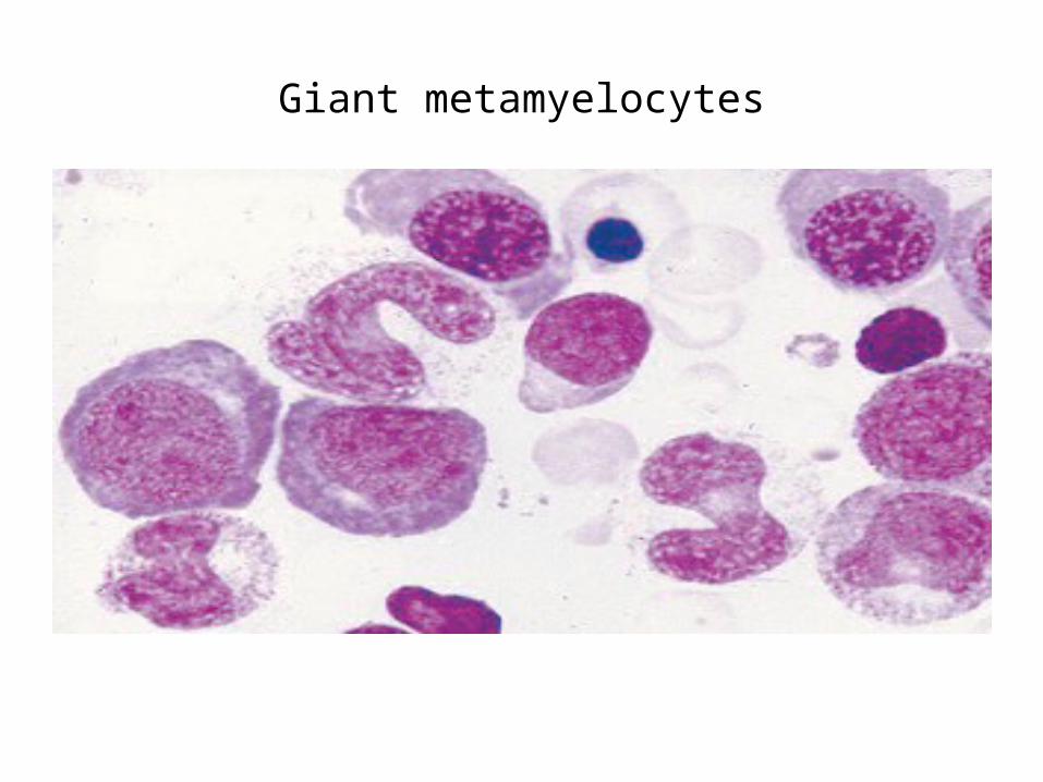
Giant metamyelocytes
Periphral Blood Smears• Macrocytic anemias associated with magaloblastosis
differs from nonmegaloblastic macrocytic anemia in that macro-ovalocytes and giant hypersegmented neutrophils in blood
• Pancytopenia is the rule• Anemia is macrocytic with an elevated MCV and
extreme degree of anisocytosis and poikilocytosis• Microcytes are common
• Basophilic stippling, multiple Howell-Jolly bodies, nucleated red cells with karyorrhexis and cabots ring may be seen
• Leucopenia is present• Thrombocytopenia usually seen, rarely sufficiently
severe to be responsible for bleeding• Neurologic symptoms may be present in the absence
of anemia• Reticulocytopenia, increased S.Fe and elevated S.LDH

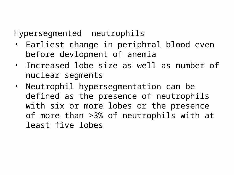
Hypersegmented neutrophils• Earliest change in periphral blood even before
devlopment of anemia• Increased lobe size as well as number of nuclear
segments• Neutrophil hypersegmentation can be defined as
the presence of neutrophils with six or more lobes or the presence of more than >3% of neutrophils with at least five lobes

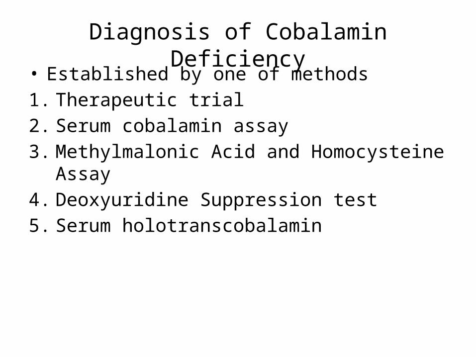
Diagnosis of Cobalamin Deficiency• Established by one of methods1. Therapeutic trial2. Serum cobalamin assay3. Methylmalonic Acid and Homocysteine Assay4. Deoxyuridine Suppression test5. Serum holotranscobalamin

Therapeutic trial• With the patient on a diet low in cobalamin and
folate• Parenteral physiologic dose of cobalamin (10 µg/day)
is given• Optimal hematologic response indicates deficiency and
consists of reticulocytosis beginning on the third or fourth day, reaching a peak on the seventh day
• Erythropoiesis becomes normoblastic by 2 days, and leukopoiesis becomes normal by 12 to 14 days.
• Within a week, leukocyte and platelet counts have returned to normal, and the Hb concentration begins to rise

Serum Cobalamin Assay• Reference values are 200–900 ng/L• In megaloblastic anemia due to cobalamin
deficiency, serum cobalamin is usually less than 100 ng/L
• Individuals with folate deficiency and mild cobalamin deficiency have borderline values between 100 and 200 ng/L as in pregnancy
Microbiological assay (Euglena gracilis), Radioisotopic dilution chemiluminescence assays
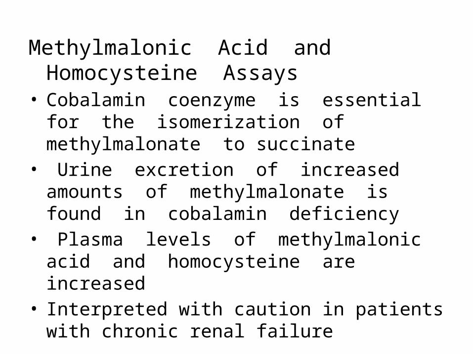
Methylmalonic Acid and Homocysteine Assays• Cobalamin coenzyme is essential for the
isomerization of methylmalonate to succinate• Urine excretion of increased amounts of
methylmalonate is found in cobalamin deficiency• Plasma levels of methylmalonic acid and
homocysteine are increased• Interpreted with caution in patients with chronic
renal failure

Deoxyuridine Suppression Test• Measures the ability of marrow cells in vitro to
utilize deoxyuridine in DNA synthesis• Normally, in marrow cells, the major source of
thymidine for DNA is de novo synthesis from deoxyuridine, which requires intact cobalamin and folate enzymes
• Tritium-labeled thymidine (3H-Tdr), normal<10% incarporated to DNA
• An abnormal deoxyuridine suppression test indicates cobalamin or folate deficiency
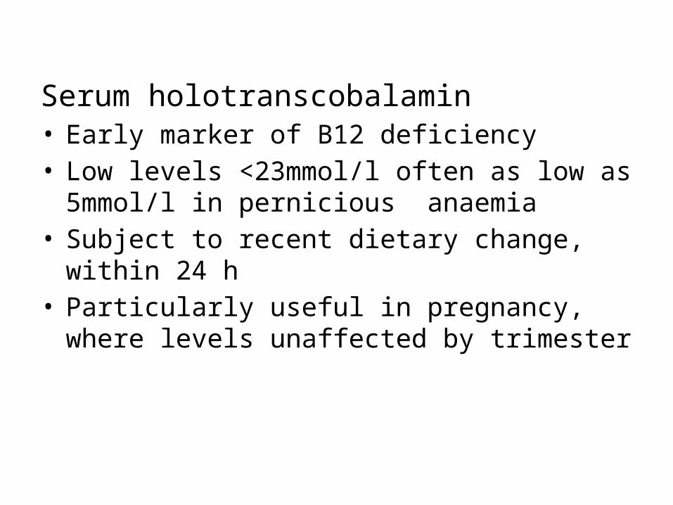
Serum holotranscobalamin • Early marker of B12 deficiency• Low levels <23mmol/l often as low as 5mmol/l in
pernicious anaemia• Subject to recent dietary change, within 24 h• Particularly useful in pregnancy, where levels
unaffected by trimester

Detecting the Cause of Cobalamin Deficiency• Clinical history is useful in suggesting whether cobalamin
or folate deficiency is the cause of megaloblastic anemia• Clinical associations of pernicious anemia include a
family history of PA in one third of patients• certain endocrine deficiencies -thyroid disease,
diabetes mellitus, hypothyroidism, and Addison’s disease
• Immune disorders (immune thrombocytopenic purpura, autoimmune hemolytic anemia, and acquired hypogammaglobulinemia

Diagnosis of Folate DeficiencySerum and Red Cell Folate• Microbiological assay employing Lactobacillus casei
is reliable method for definitive diagnosis• Radioisotopic and chemiluminescence methods
widely used due to rapidity and greater convenience
• In cobalamin deficiency, serum folate is decreased in 10% of cases, increased in 20%, and normal in the remainder

• Unlike serum folate (entirely 5-methyltetrahydrofolate), red cell folates are a heterogeneous mixture of different forms with varying polyglutamate chain lengths
• The red cell folate is a better test of body folate stores and is decreased in megaloblastic anemia due to folate deficiency
Urinary Formiminoglutamic Acid• Useful in megaloblastic anemia due to antifolate drugs

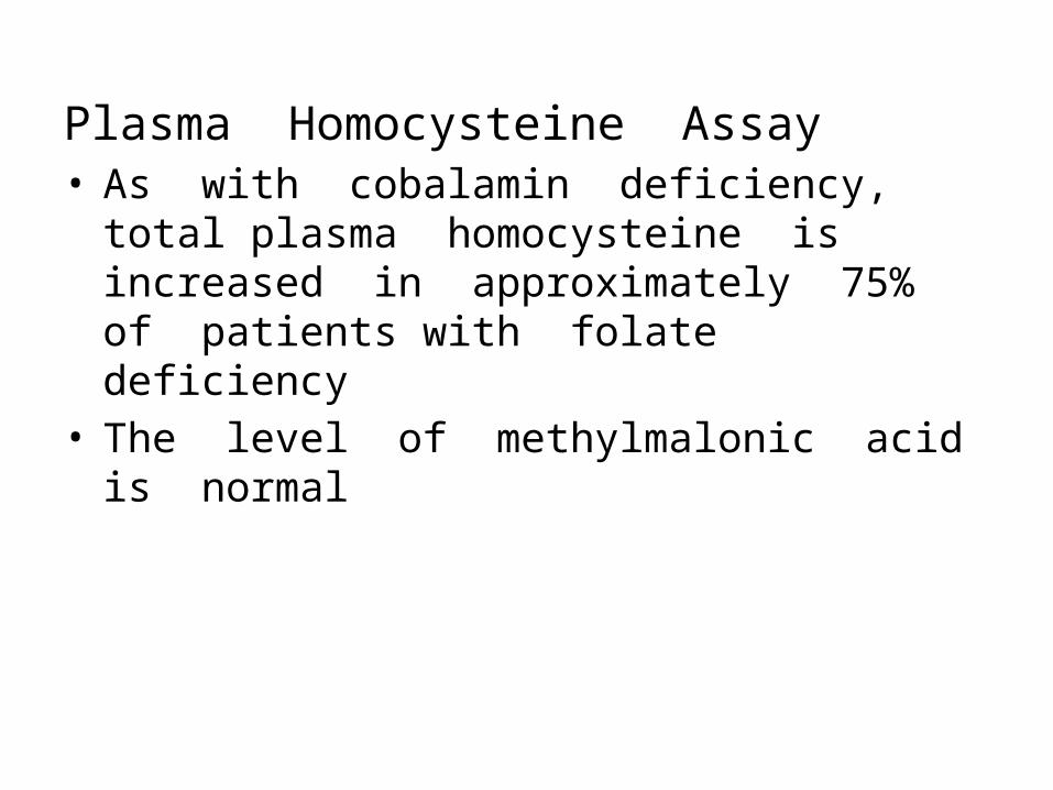
Plasma Homocysteine Assay• As with cobalamin deficiency, total plasma
homocysteine is increased in approximately 75% of patients with folate deficiency
• The level of methylmalonic acid is normal

Pernicious Anemia• Most common cause of cobalamin deficiency• caused by failure of the gastric mucosa to secrete intrinsic
factor• Abnormality is genetically determined & manifested late in life
>40 years
Immune Abnormalities1. Anti–parietal cell antibodies2. Anti–intrinsic factor antibodies -Two types• Blocking” antibodies, which block the binding of cobalamin to IF• Binding” antibodies, which bind to the cobalamin–IF
complex and prevent the complex from binding to receptors in the ileum


Schilling Test• Determine whether IF is lacking• Measures radioactivity in a 24-hour sample of urine• Two hours after oral administration of 0.5–2.0 µg of
radioactive cobalamin, a large “flushing” dose of nonlabeled cobalamin is given parenterally
• Normal individuals will excrete more than 7% of a 1-µg dose of ingested cobalamin in the urine in 24 hours
• Patients lacking IF will excrete less


• If excretion is low, the test must be repeated using the same procedure, except that hog IF is given orally, along with labeled cobalamin
• If 24-hour excretion is normal, the low value in the first part was due to IF deficiency
• If excretion remains abnormal in the second part of the procedure, an explanation for malabsorption of cobalamin on the basis of intestinal disease must be sought.
• The test may be repeated after 7–10 days of antibiotic administration if bacterial overgrowth is suspected, and pancreatic extracts may be added to investigate the possibility of pancreatic dysfunction

Morphology• Changes in BM and Blood are similar to all
megaloblastic anemia• In addition it leads to1. Atrophic glossitis2. Intestinalization of gastric mucosa3. In CNS principal alteration in spinal cord causing
degeneration of myelin in dorsal and lateral tract
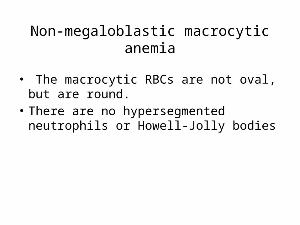
Non-megaloblastic macrocytic anemia
• The macrocytic RBCs are not oval, but are round.
• There are no hypersegmented neutrophils or Howell-Jolly bodies
Liver Disease• Liver disease associated with alcoholism may
lead to folate-deficient megaloblastic anemia • Because of the grossly inadequate diet of the
alcoholic, and because the liver is the major site for folate storage and metabolism
• With adequate dietary folic acid intake, however, the anemia that is found with liver disease is macrocytic and normoblastic—not megaloblastic
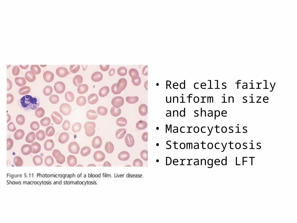
• Red cells fairly uniform in size and shape
• Macrocytosis• Stomatocytosis• Derranged LFT
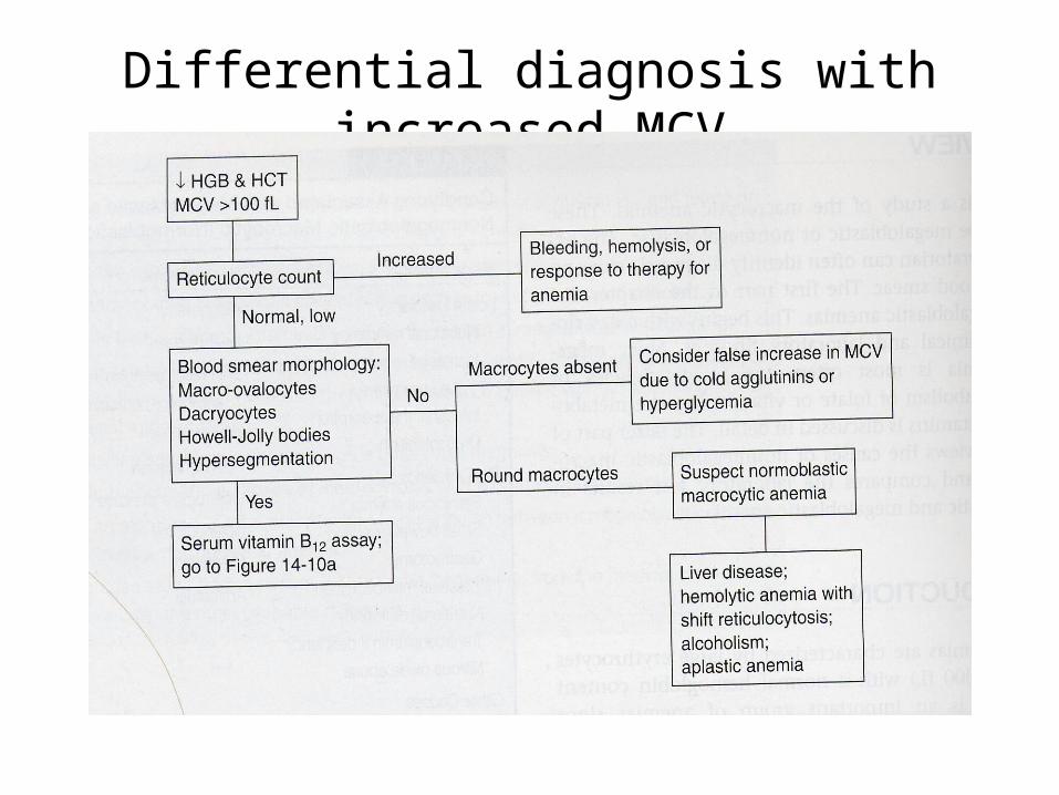
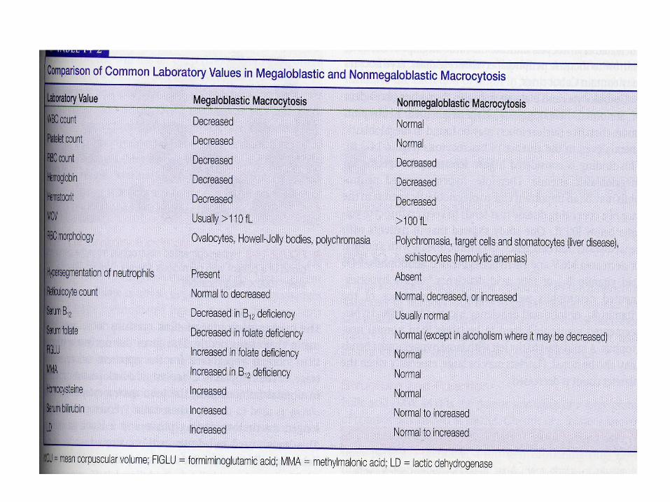
Differential diagnosis with increased MCV


Thank You