Embed Size (px)
Citation preview
Functional Anatomy Functional Anatomy and Mechanism of and Mechanism of
Knee InjuryKnee Injury
Dr. Hytham NafadyDr. Hytham Nafady
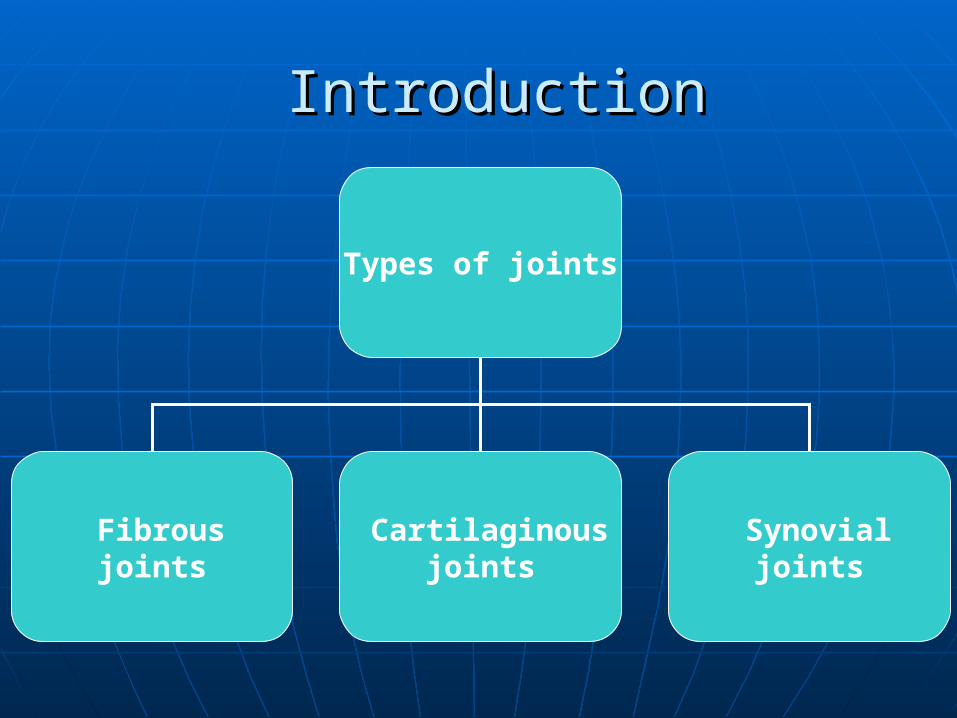
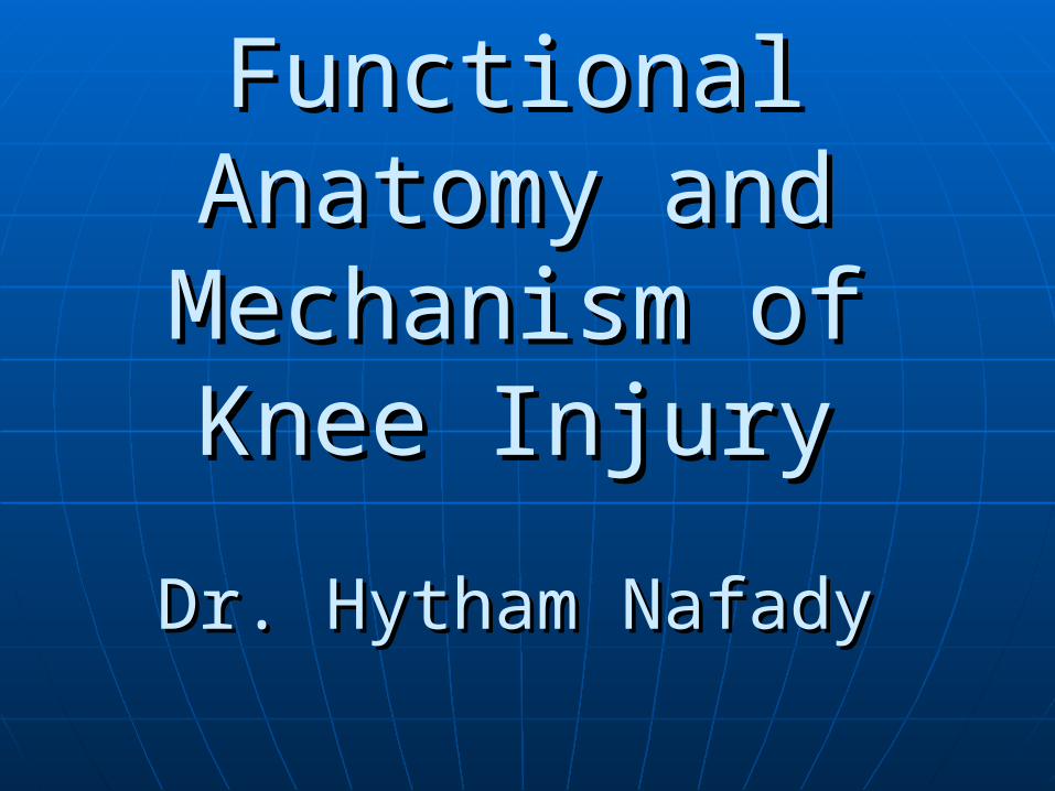
IntroductionIntroduction
Types of joints
Fibrous joints
Cartilaginous joints
Synovial joints
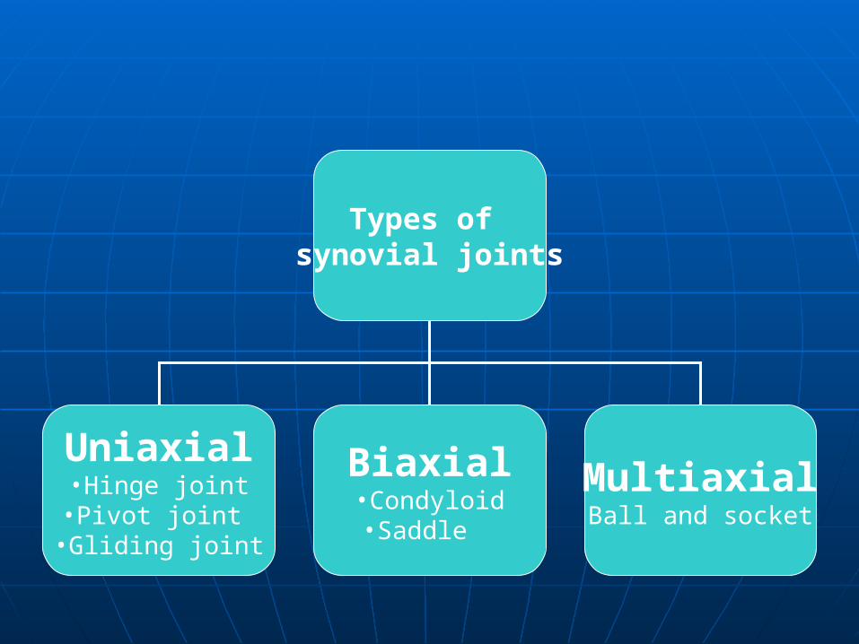
Types of synovial joints
Uniaxial•Hinge joint•Pivot joint
•Gliding joint
Biaxial•Condyloid•Saddle
MultiaxialBall and socket
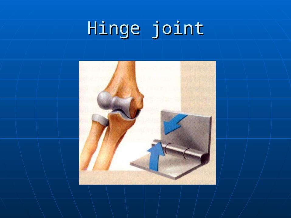
Hinge jointHinge joint
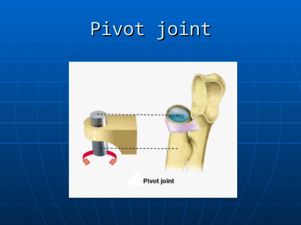
Pivot jointPivot joint
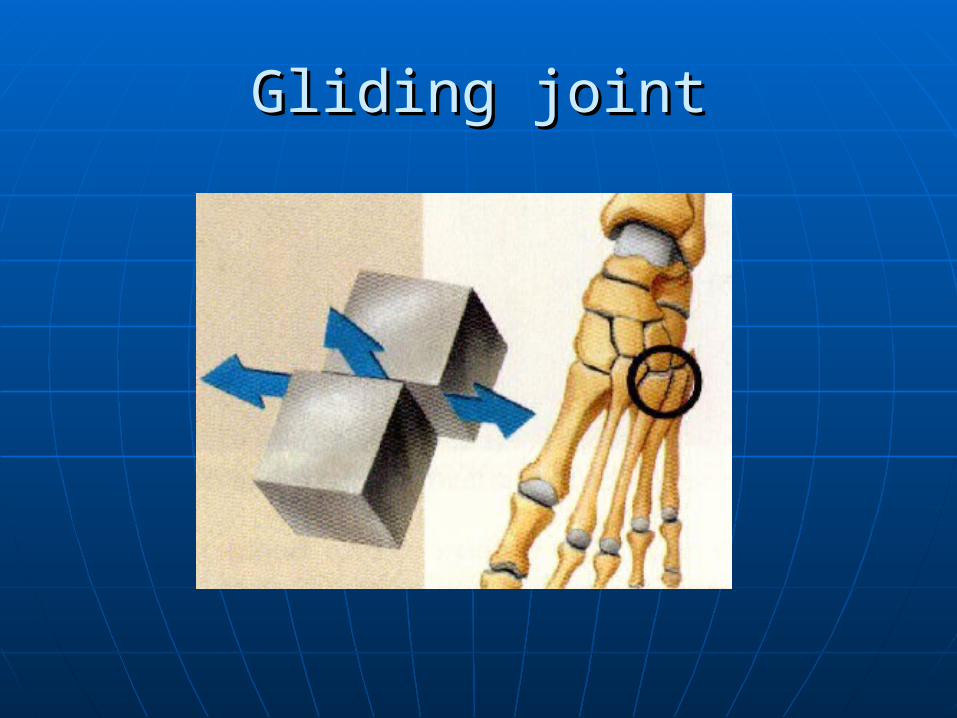
Gliding jointGliding joint
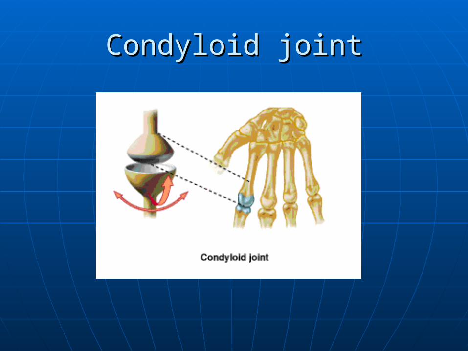
Condyloid jointCondyloid joint
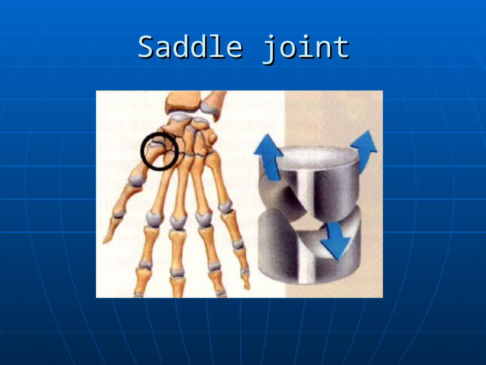
Saddle jointSaddle joint
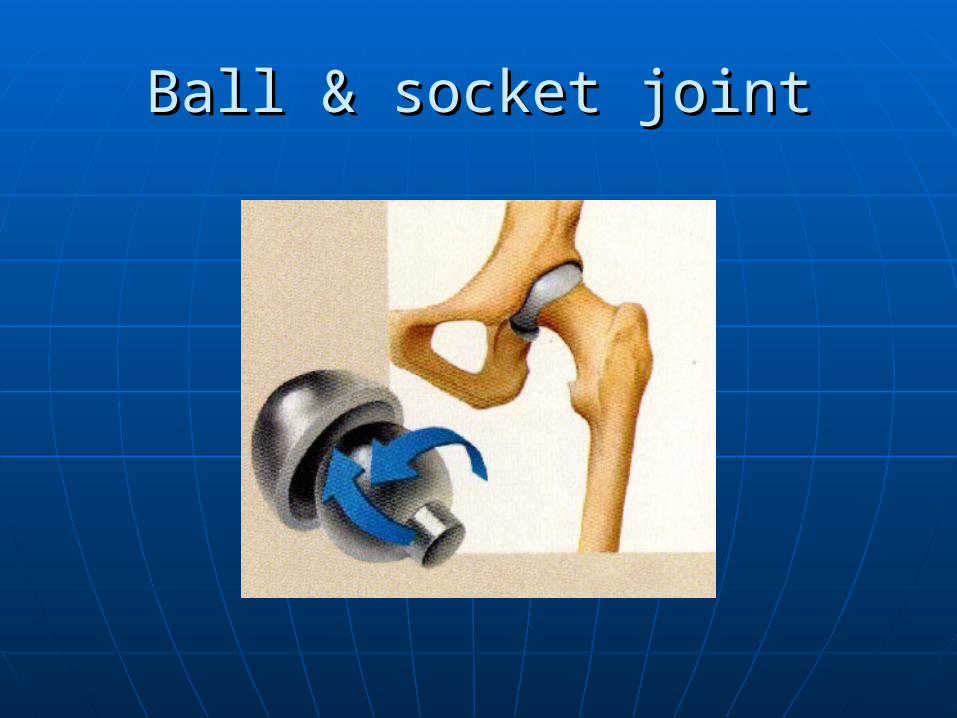
Ball & socket jointBall & socket joint
Functional anatomy of the kneeFunctional anatomy of the knee
The articular bones of the knee have a The articular bones of the knee have a little contribution to the stability of little contribution to the stability of the joint.the joint.
Stability of the knee joint depends Stability of the knee joint depends mainly on its supporting soft tissue mainly on its supporting soft tissue structures.structures.
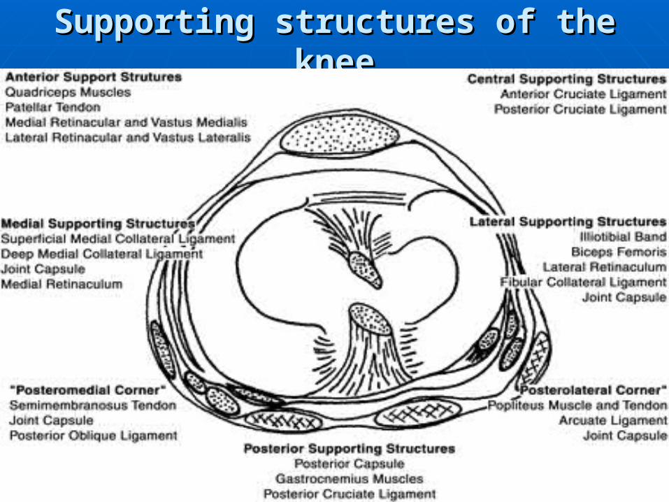
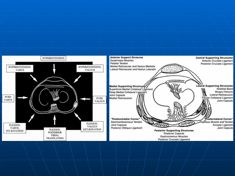
Supporting structures of the kneeSupporting structures of the knee
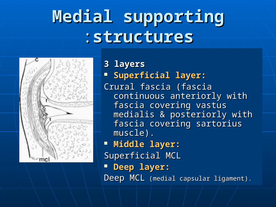
Medial supporting structuresMedial supporting structures::
3 layers 3 layers Superficial layer:Superficial layer:Crural fascia (fascia continuous Crural fascia (fascia continuous
anteriorly with fascia covering anteriorly with fascia covering vastus medialis & posteriorly vastus medialis & posteriorly with fascia covering sartorius with fascia covering sartorius muscle). muscle).
Middle layer:Middle layer:Superficial MCLSuperficial MCL Deep layer:Deep layer:Deep MCLDeep MCL (medial capsular ligament). (medial capsular ligament).
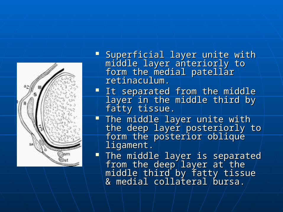
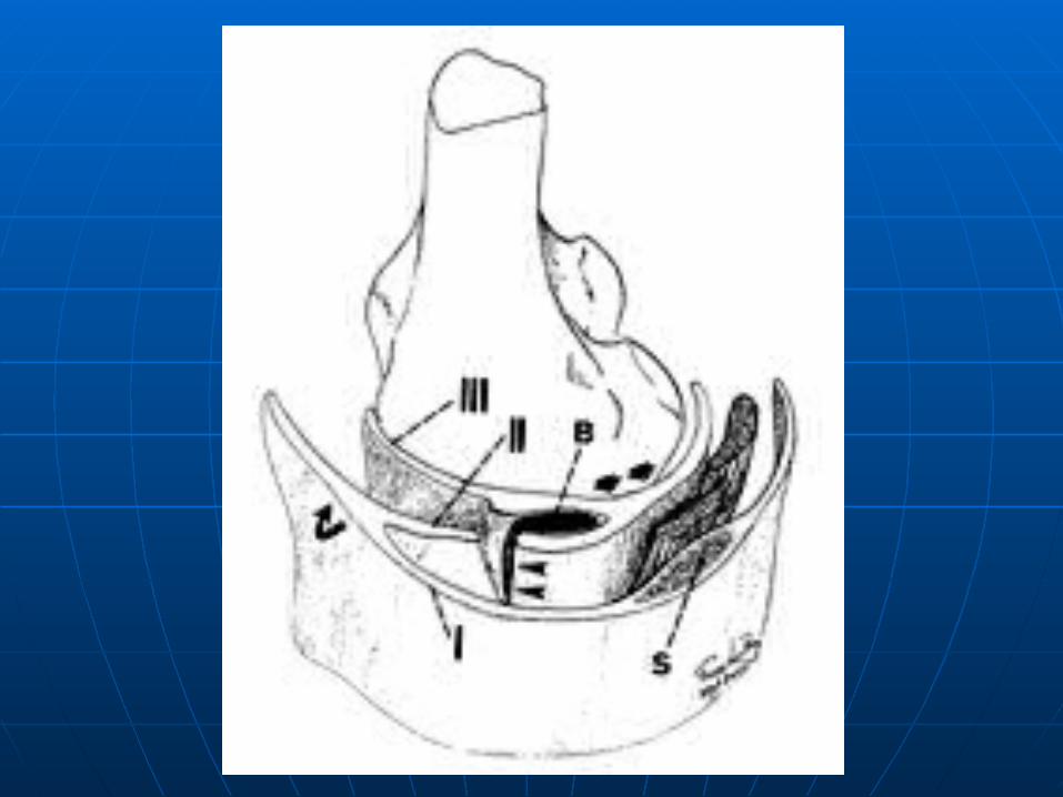
Superficial layer unite with Superficial layer unite with middle layer anteriorly to form middle layer anteriorly to form the medial patellar retinaculum.the medial patellar retinaculum.
It separated from the middle It separated from the middle layer in the middle third by fatty layer in the middle third by fatty tissue.tissue.
The middle layer unite with the The middle layer unite with the deep layer posteriorly to form the deep layer posteriorly to form the posterior oblique ligament.posterior oblique ligament.
The middle layer is separated The middle layer is separated from the deep layer at the middle from the deep layer at the middle third by fatty tissue & medial third by fatty tissue & medial collateral bursa.collateral bursa.
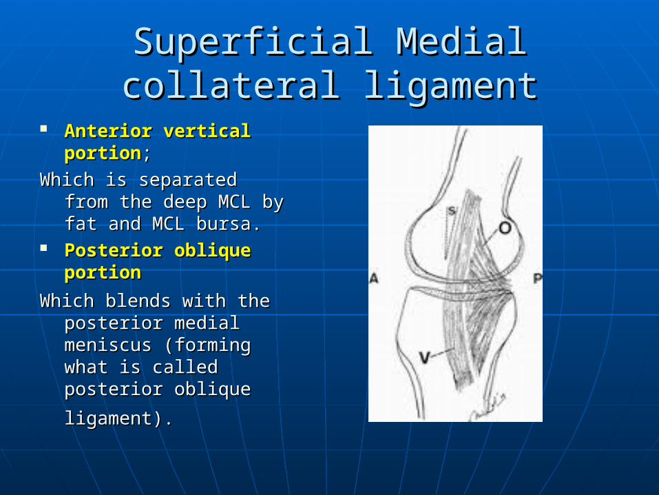
Superficial Medial collateral Superficial Medial collateral ligamentligament
Anterior vertical Anterior vertical portionportion;;
Which is separated from Which is separated from the deep MCL by fat the deep MCL by fat and MCL bursa.and MCL bursa.
Posterior oblique Posterior oblique portionportion
Which blends with the Which blends with the posterior medial posterior medial meniscus (forming meniscus (forming what is called posterior what is called posterior
oblique ligament).oblique ligament).
Deep medial collateral ligamentDeep medial collateral ligament
Menisco-tibial attachment.Menisco-tibial attachment. Menisco-femoral attachment.Menisco-femoral attachment.
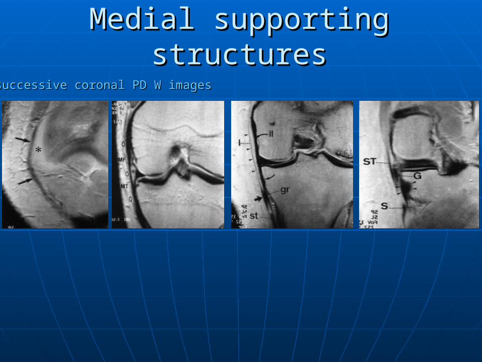
Medial supporting structuresMedial supporting structures
successive coronal PD W imagessuccessive coronal PD W images
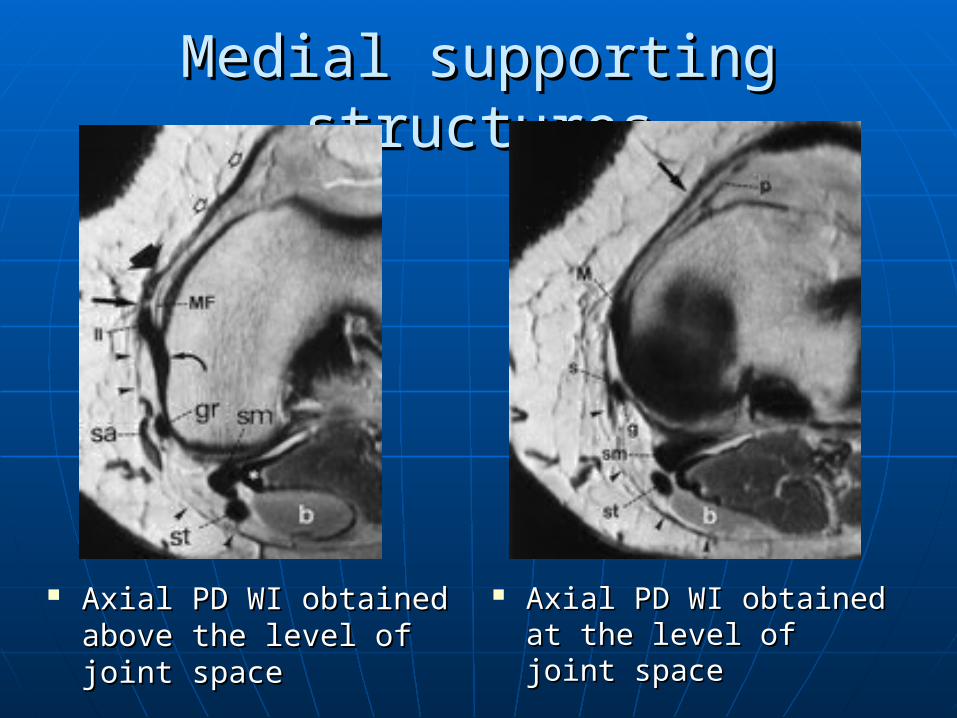
Medial supporting structuresMedial supporting structures
Axial PD WI obtained Axial PD WI obtained above the level of joint above the level of joint spacespace
Axial PD WI obtained Axial PD WI obtained at the level of joint at the level of joint spacespace
Posteromedial supporting Posteromedial supporting structuresstructures
Semimembranosis muscle & tendon.Semimembranosis muscle & tendon. Medial head of gastrocnemiusMedial head of gastrocnemius Posterior oblique ligament Posterior oblique ligament
(obliquely oriented portion of MCL).(obliquely oriented portion of MCL). Joint capsuleJoint capsule
Postero-medial supporting Postero-medial supporting structuresstructures
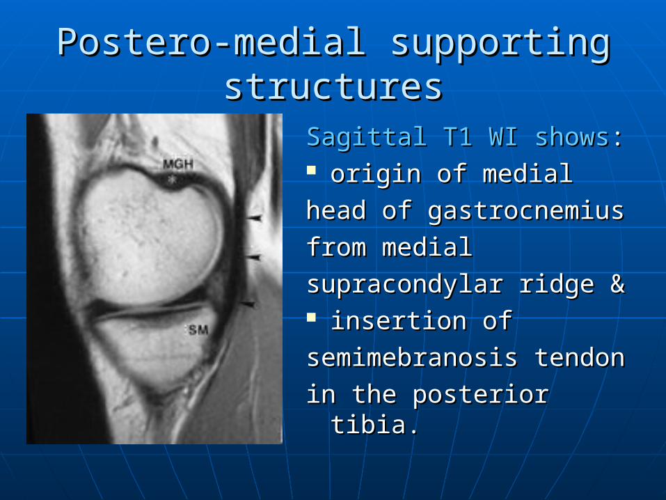
Sagittal T1 WI showsSagittal T1 WI shows: : origin of medialorigin of medial
head of gastrocnemiushead of gastrocnemius
from medialfrom medial
supracondylar ridge & supracondylar ridge & insertion ofinsertion of
semimebranosis tendonsemimebranosis tendon
in the posterior tibia.in the posterior tibia.
Lateral supporting structuresLateral supporting structures::
3 layers:3 layers:Superficial layer:Superficial layer: iliotibial tract & its anterior expansion.iliotibial tract & its anterior expansion. Biceps tendon & its posterior expansion.Biceps tendon & its posterior expansion.Middle layer:Middle layer: Lateral patellar retinaculum which is complete anteriorly Lateral patellar retinaculum which is complete anteriorly
and represented posteriorly by 2 patellofemoral ligaments.and represented posteriorly by 2 patellofemoral ligaments.Deep layer:Deep layer:Superficial lamina:Superficial lamina: LCL LCL And terminates at the fabellofibular ligament.And terminates at the fabellofibular ligament.Deep lamina:Deep lamina: The coronay ligament (with its meniscotibial & mensico-The coronay ligament (with its meniscotibial & mensico-
femoral attachment). femoral attachment). And terminates at the arcuate ligament.And terminates at the arcuate ligament.
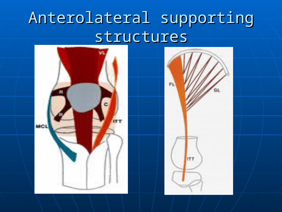
Anterolateral supporting structuresAnterolateral supporting structures
Iliotibial tract. (inserted in Gerdy’s Iliotibial tract. (inserted in Gerdy’s tubercle).tubercle).
Superior and inferior Lateral patellar Superior and inferior Lateral patellar retinacula.retinacula.
Vastus lateralis muscle.Vastus lateralis muscle. Joint capsule.Joint capsule.
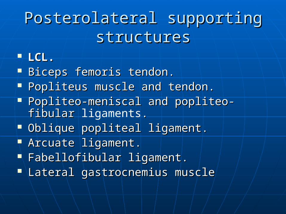
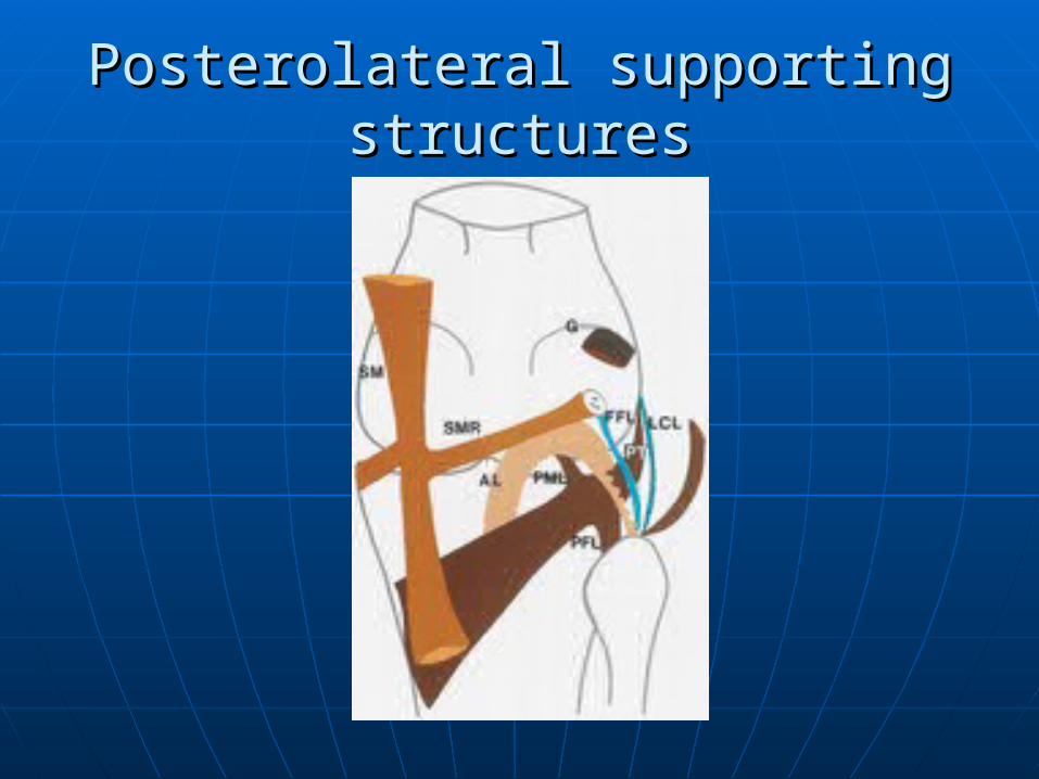
Posterolateral supporting structuresPosterolateral supporting structures
LCL.LCL. Biceps femoris tendon.Biceps femoris tendon. Popliteus muscle and tendon. Popliteus muscle and tendon. Popliteo-meniscal and popliteo-fibular Popliteo-meniscal and popliteo-fibular
ligaments.. Oblique popliteal ligament. Oblique popliteal ligament. Arcuate ligament.Arcuate ligament. Fabellofibular ligament.Fabellofibular ligament. Lateral gastrocnemius muscle Lateral gastrocnemius muscle
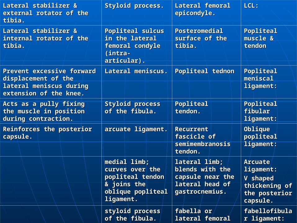
LCL:LCL:Lateral femoral Lateral femoral epicondyle.epicondyle.
Styloid process.Styloid process.Lateral stabilizer & Lateral stabilizer & external rotator of the external rotator of the tibia.tibia.
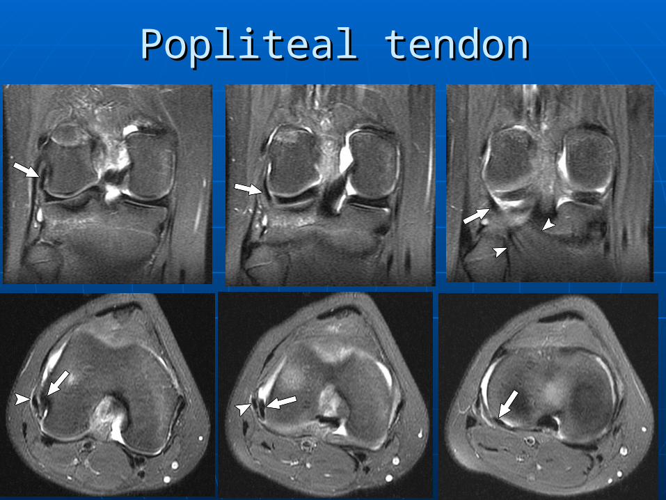
Popliteal Popliteal muscle & muscle & tendontendon
Posteromedial Posteromedial surface of the surface of the tibia.tibia.
Popliteal sulcus in Popliteal sulcus in the lateral the lateral femoral condyle femoral condyle (intra-articular).(intra-articular).
Lateral stabilizer & Lateral stabilizer & internal rotator of the internal rotator of the tibia.tibia.
Popliteal Popliteal meniscal meniscal ligament:ligament:
Popliteal tednonPopliteal tednonLateral meniscus.Lateral meniscus.Prevent excessive forward Prevent excessive forward displacement of the lateral displacement of the lateral meniscus during extension meniscus during extension of the knee.of the knee.
Popliteal Popliteal fibular fibular ligament:ligament:
Popliteal tendon.Popliteal tendon.Styloid process of Styloid process of the fibula.the fibula.
Acts as a pully fixing the Acts as a pully fixing the muscle in position during muscle in position during contraction.contraction.
Oblique Oblique popliteal popliteal ligament: ligament:
Recurrent Recurrent fascicle of fascicle of semimembranosisemimembranosis tendon.s tendon.
arcuate ligament.arcuate ligament.Reinforces the posterior Reinforces the posterior capsule.capsule.
Arcuate Arcuate ligament:ligament:
V shaped V shaped thickening of thickening of the posterior the posterior capsule.capsule.
lateral limb; lateral limb; blends with the blends with the capsule near the capsule near the lateral head of lateral head of gastrocnemius. gastrocnemius.
medial limb; medial limb; curves over the curves over the popliteal tendon popliteal tendon & joins the & joins the oblique popliteal oblique popliteal ligament.ligament.
fabellofibular fabellofibular ligament:ligament:
fabella or lateral fabella or lateral femoral condyle.femoral condyle.
styloid process of styloid process of the fibula.the fibula.
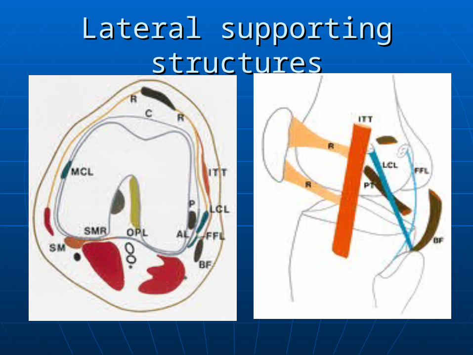
Lateral supporting structuresLateral supporting structures
Anterolateral supporting structuresAnterolateral supporting structures
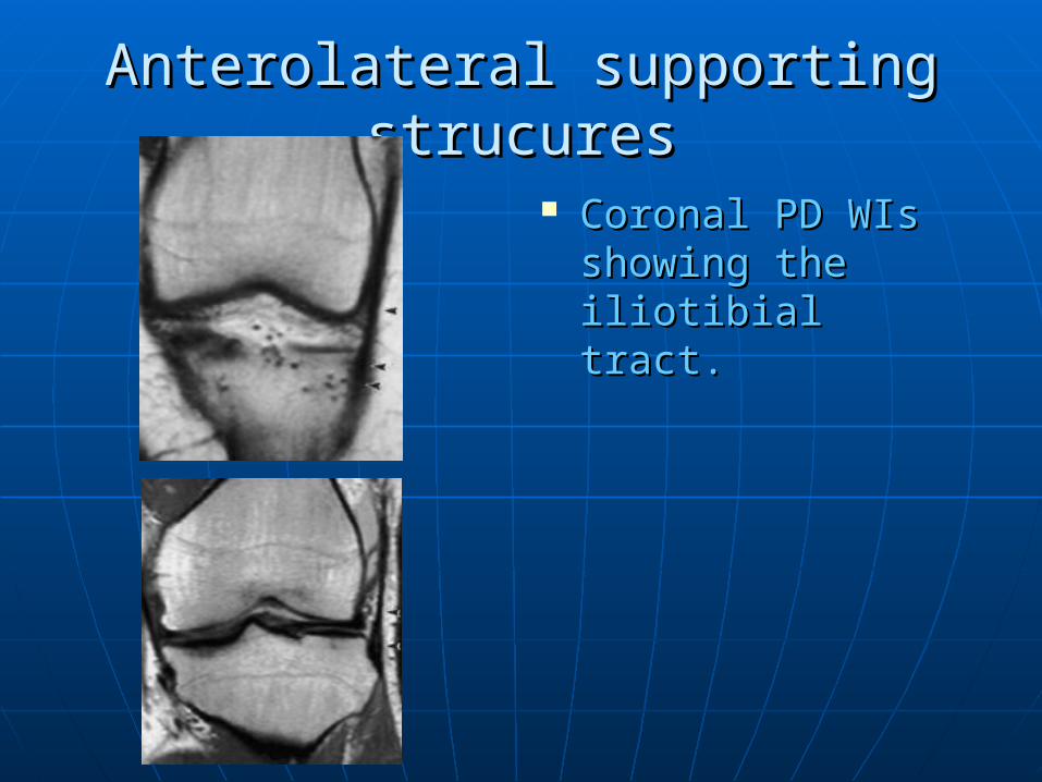
Anterolateral supporting strucuresAnterolateral supporting strucures
Coronal PD WIs Coronal PD WIs showing the showing the iliotibial tract.iliotibial tract.
Posterolateral supporting structuresPosterolateral supporting structures
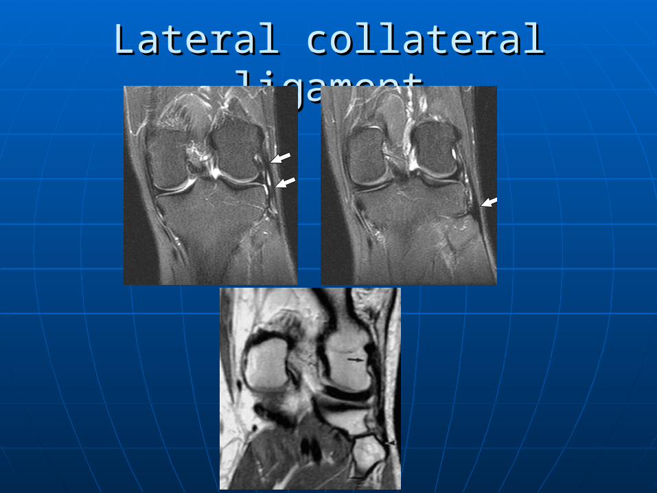
Lateral collateral ligamentLateral collateral ligament
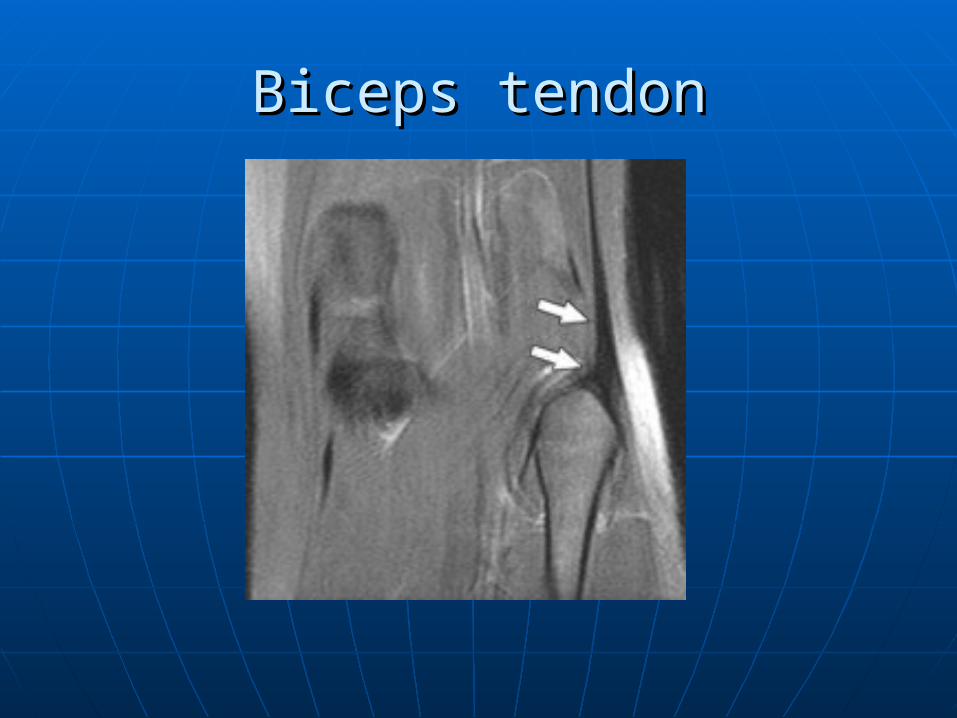
Biceps tendonBiceps tendon
Popliteal tendonPopliteal tendon
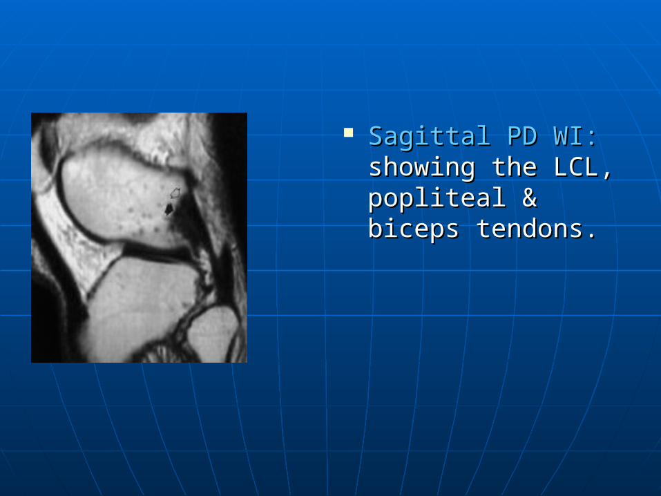
Sagittal PD WI:Sagittal PD WI: showing the LCL, showing the LCL, popliteal & biceps popliteal & biceps tendons.tendons.
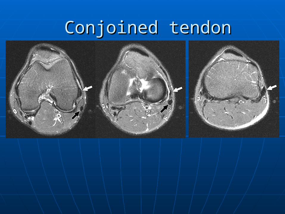
Conjoined tendonConjoined tendon
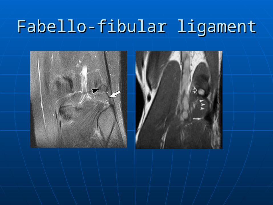
Fabello-fibular ligamentFabello-fibular ligament
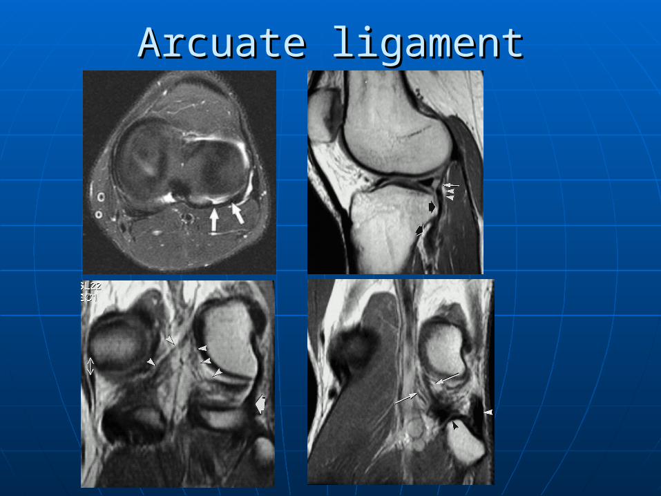
Arcuate ligamentArcuate ligament
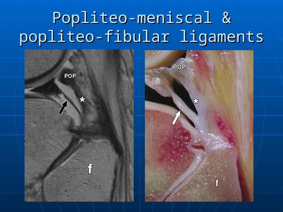
Popliteo-meniscal & popliteo-fibular Popliteo-meniscal & popliteo-fibular ligamentsligaments
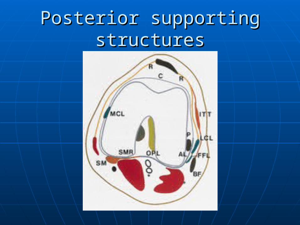
Posterior supporting structuresPosterior supporting structures
Gastrocnemius muscle and tendons.Gastrocnemius muscle and tendons. Posterior joint capsule reinforced by Posterior joint capsule reinforced by
the oblique popliteal ligament.the oblique popliteal ligament. PCL.PCL.
Posterior supporting structuresPosterior supporting structures
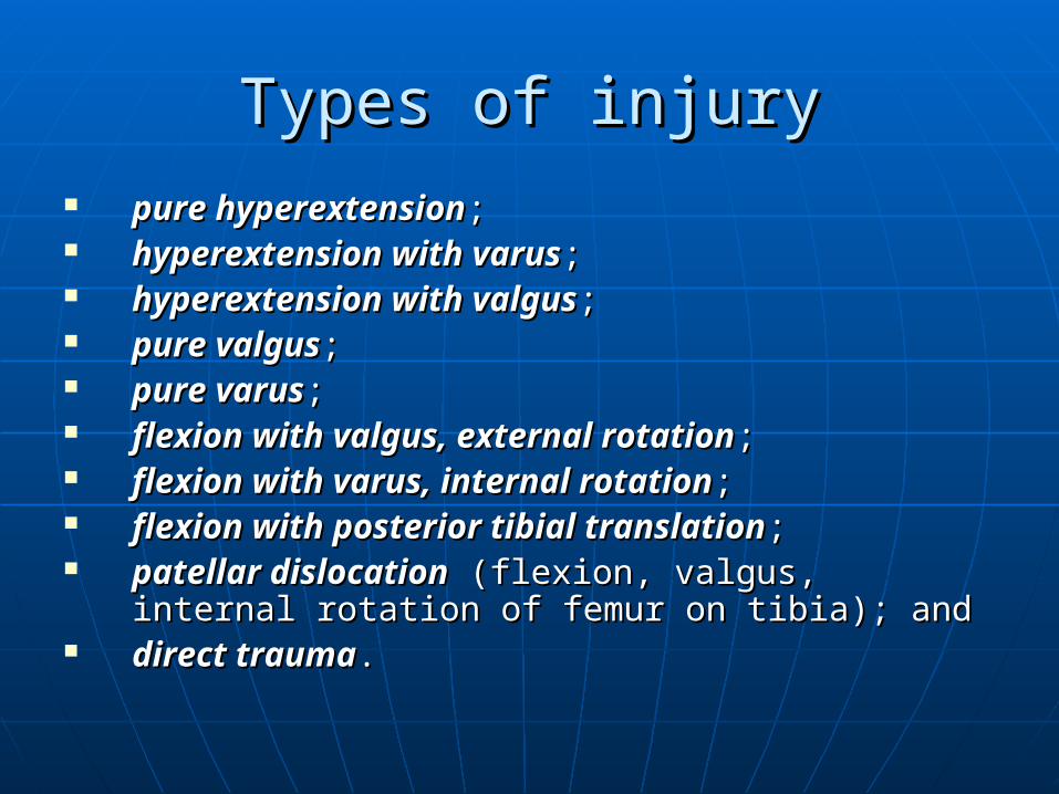
Types of injuryTypes of injury pure hyperextensionpure hyperextension; ; hyperextension with varushyperextension with varus; ; hyperextension with valgushyperextension with valgus; ; pure valguspure valgus; ; pure varuspure varus; ; flexion with valgus, external rotationflexion with valgus, external rotation; ; flexion with varus, internal rotationflexion with varus, internal rotation; ; flexion with posterior tibial translationflexion with posterior tibial translation; ; patellar dislocationpatellar dislocation (flexion, valgus, internal (flexion, valgus, internal
rotation of femur on tibia); and rotation of femur on tibia); and direct traumadirect trauma. .
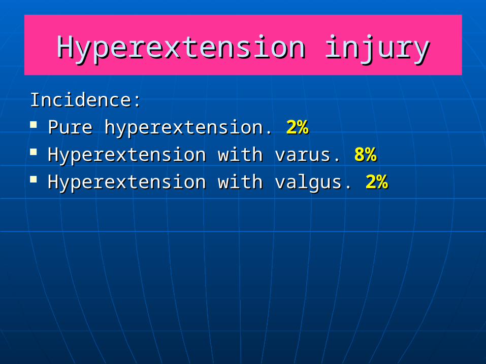
Hyperextension injuryHyperextension injury
Incidence:Incidence: Pure hyperextension. Pure hyperextension. 2%2% Hyperextension with varus. Hyperextension with varus. 8%8% Hyperextension with valgus. Hyperextension with valgus. 2%2%
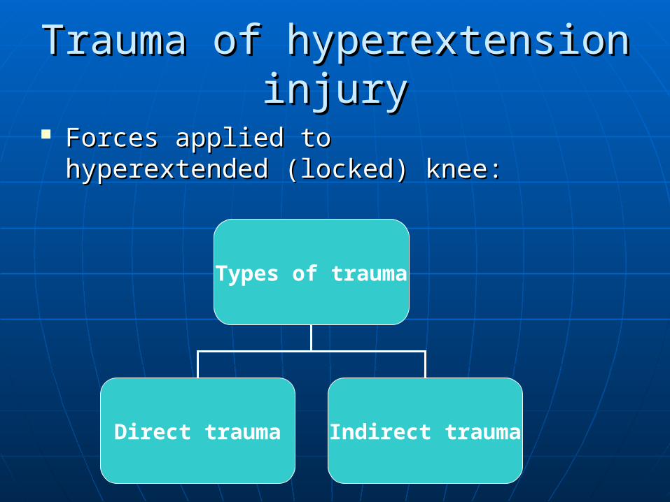
Trauma of hyperextension injuryTrauma of hyperextension injury
Forces applied to hyperextended Forces applied to hyperextended (locked) knee:(locked) knee:
Types of trauma
Direct trauma Indirect trauma
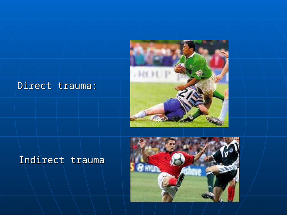
Indirect traumaIndirect trauma
Direct trauma:Direct trauma:
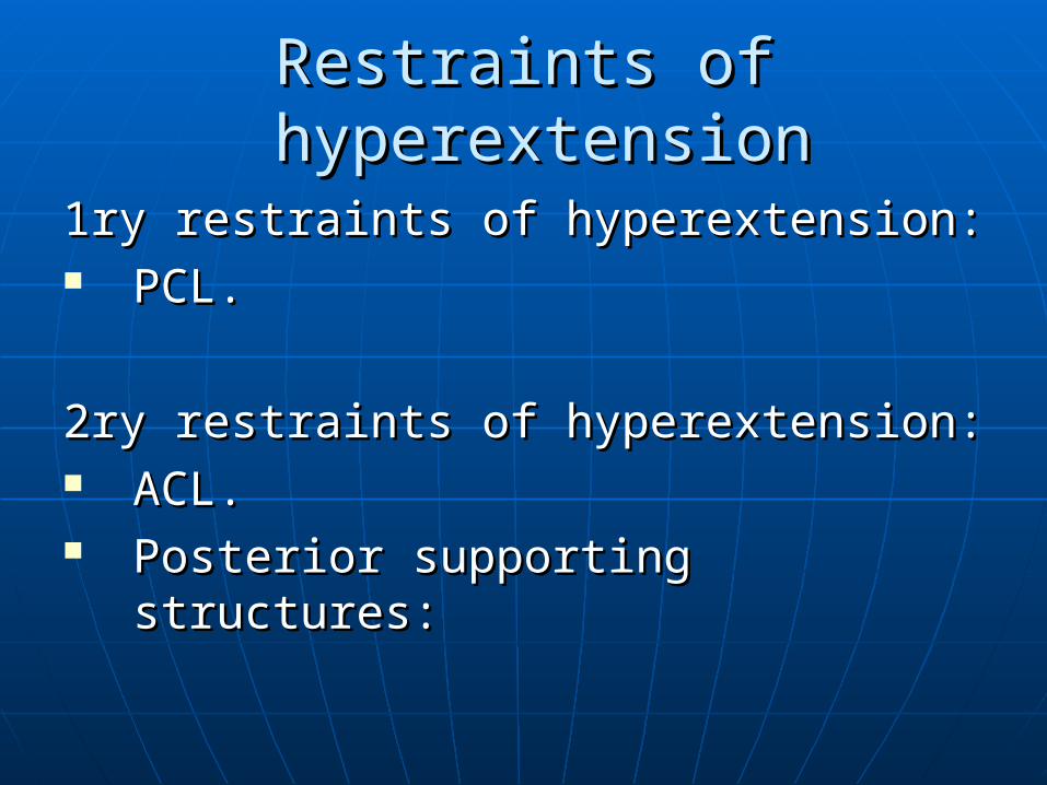
Restraints of hyperextensionRestraints of hyperextension
1ry restraints of hyperextension:1ry restraints of hyperextension: PCL.PCL.
2ry restraints of hyperextension:2ry restraints of hyperextension: ACL.ACL. Posterior supporting structures:Posterior supporting structures:
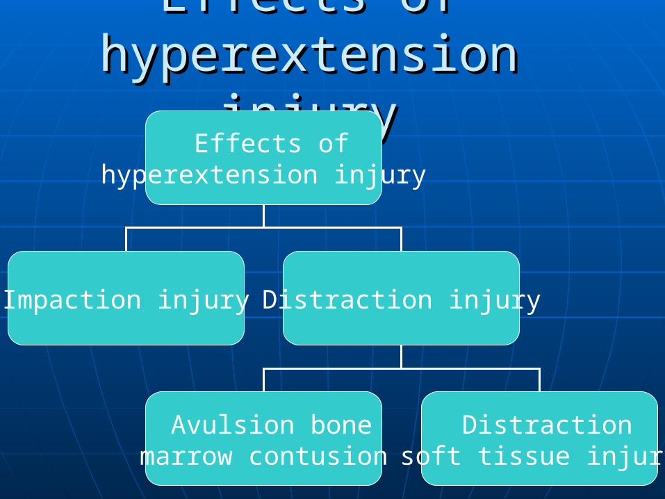
Effects of Effects of hyperextension injuryhyperextension injury
Effects of hyperextension injury
Impaction injury Distraction injury
Avulsion bone marrow contusion
Distraction soft tissue injury
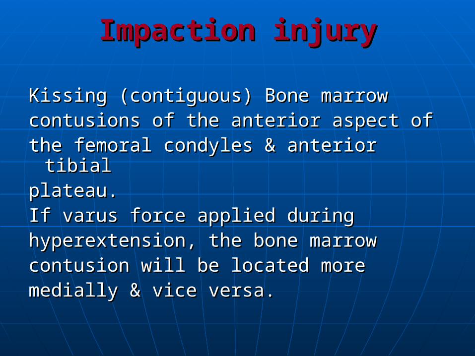
Impaction injuryImpaction injury
Kissing (contiguous) Bone marrowKissing (contiguous) Bone marrowcontusions of the anterior aspect ofcontusions of the anterior aspect ofthe femoral condyles & anterior tibialthe femoral condyles & anterior tibialplateau.plateau.If varus force applied duringIf varus force applied duringhyperextension, the bone marrowhyperextension, the bone marrowcontusion will be located morecontusion will be located moremedially & vice versa. medially & vice versa.
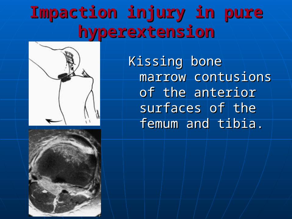
Impaction injury in pure Impaction injury in pure hyperextensionhyperextension
Kissing bone marrow Kissing bone marrow contusions of the contusions of the anterior surfaces of anterior surfaces of the femum and the femum and tibia.tibia.
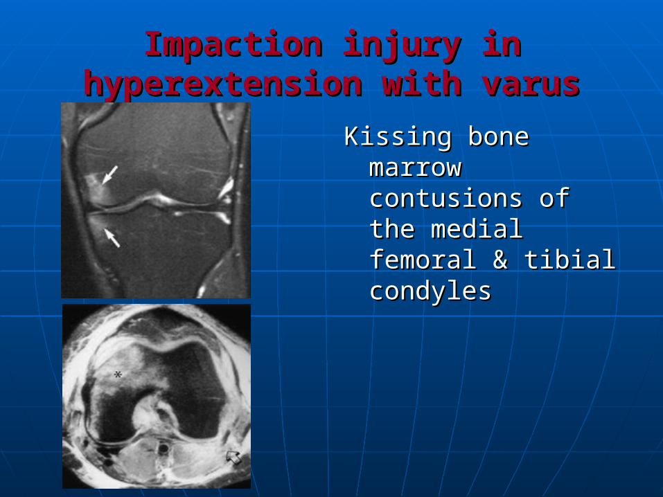
Impaction injury in hyperextension Impaction injury in hyperextension with varuswith varus
Kissing bone marrow Kissing bone marrow contusions of the contusions of the medial femoral & medial femoral & tibial condylestibial condyles
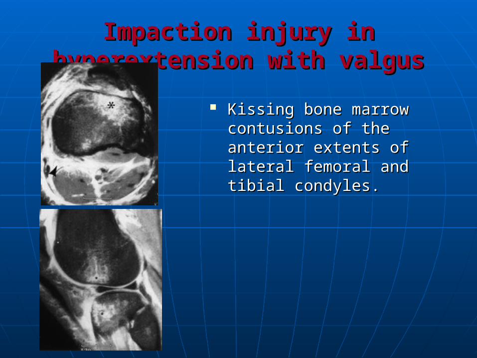
Impaction injury in hyperextension Impaction injury in hyperextension with valguswith valgus
Kissing bone marrow Kissing bone marrow contusions of the contusions of the anterior extents of anterior extents of lateral femoral and lateral femoral and tibial condyles.tibial condyles.
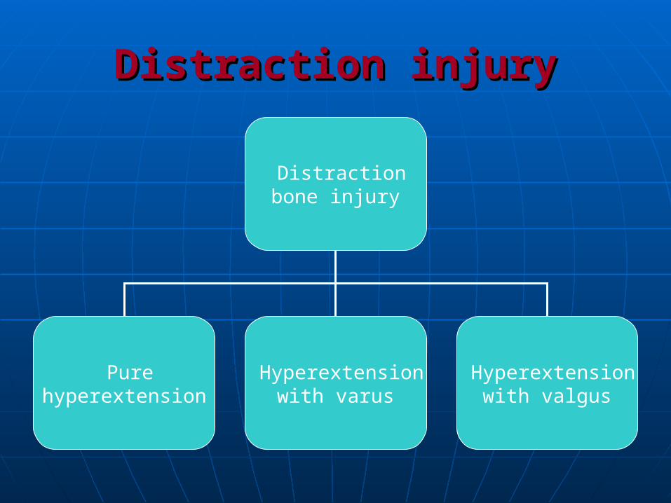
Distraction injuryDistraction injury
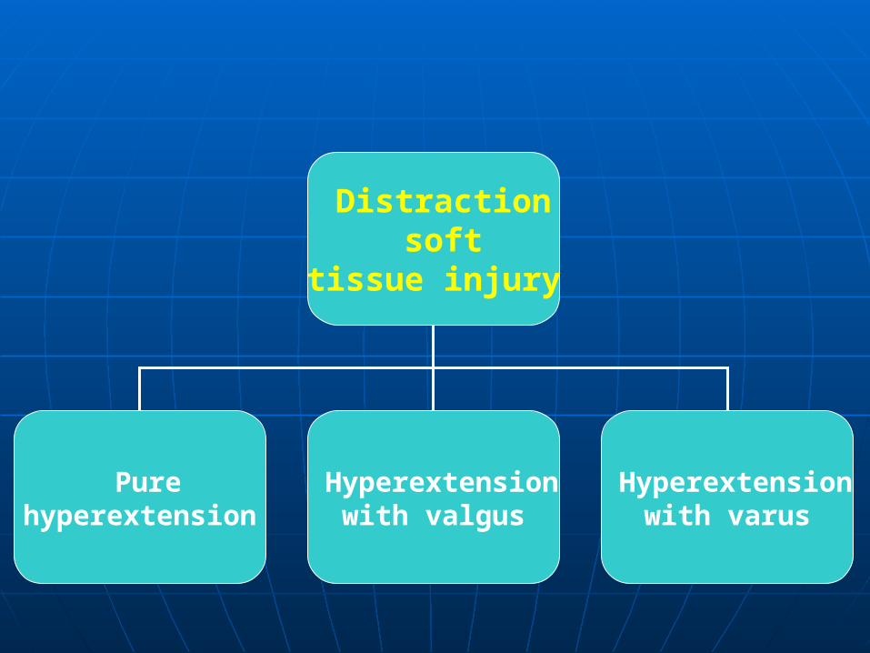
Distraction bone injury
Pure hyperextension
Hyperextension with varus
Hyperextension with valgus

Distraction bone injury Distraction bone injury in pure hyperextensionin pure hyperextension
Avulsion bone marrow contusionAvulsion bone marrow contusion of of the posterior tibial plateau.the posterior tibial plateau.
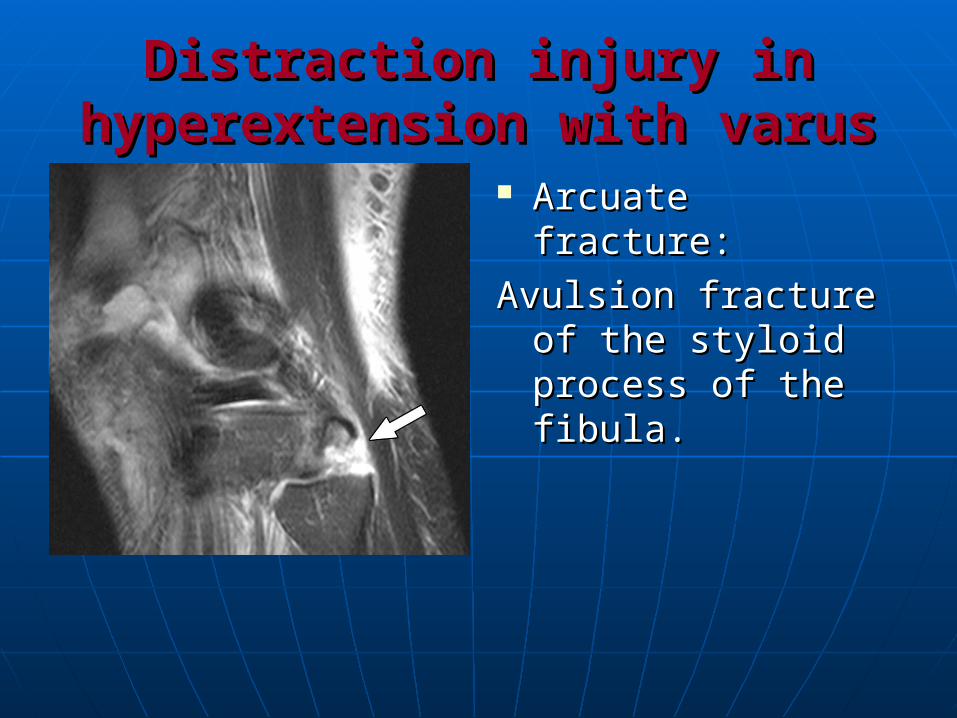
Distraction injury in Distraction injury in hyperextension with varushyperextension with varus
Arcuate fracture:Arcuate fracture:
Avulsion fracture of Avulsion fracture of the styloid process the styloid process of the fibula.of the fibula.
Distraction injury in Distraction injury in hyperextension with valgushyperextension with valgus
Avulsion bone marrow contusion of Avulsion bone marrow contusion of the posteromedial tibial plateau.the posteromedial tibial plateau.
Distraction soft
tissue injury
Pure hyperextension
Hyperextension with valgus
Hyperextension with varus
Distraction soft tissue injuryDistraction soft tissue injury(pure hyperextension)(pure hyperextension)
ACL tear.ACL tear.
PCL tear.PCL tear.
Injury of the posterior supporting Injury of the posterior supporting structures:structures:
(posterior capsule disruption or injury (posterior capsule disruption or injury to gastrocnemius muscle).to gastrocnemius muscle).
Injury to the popliteal vesselsInjury to the popliteal vessels
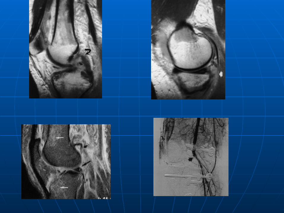
Distraction soft tissue injuryDistraction soft tissue injury(hyperextension with varus)(hyperextension with varus)
ACL injury.ACL injury. PCL injury.PCL injury. Posterior capsule disruption.Posterior capsule disruption. Injury of the posterolateral Injury of the posterolateral
supporting structures (posterolateral supporting structures (posterolateral corner syndrome).corner syndrome).
Posterolateral corner syndromePosterolateral corner syndrome
Unrecognized injuries to the Unrecognized injuries to the posterolateral corner is an important posterolateral corner is an important factor in factor in
postsurgical failure after cruciate postsurgical failure after cruciate ligament reconstruction and ligament reconstruction and
in chronic instability and in chronic instability and degenerative changes after knee degenerative changes after knee traumatrauma
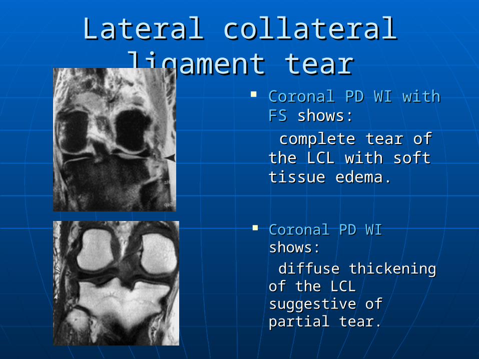
Lateral collateral ligament tearLateral collateral ligament tear
Coronal PD WICoronal PD WI shows: shows:
diffuse thickening of diffuse thickening of the LCL suggestive of the LCL suggestive of partial tear.partial tear.
Coronal PD WI with FSCoronal PD WI with FS shows: shows:
complete tear of the complete tear of the LCL with soft tissue LCL with soft tissue edema.edema.
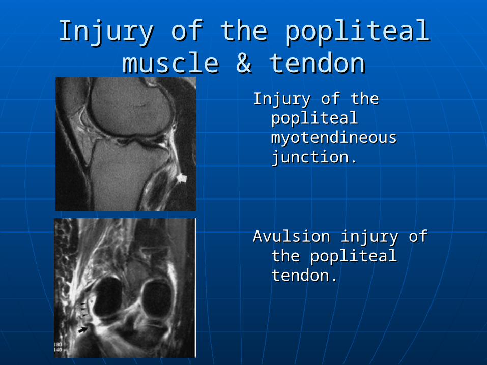
Injury of the popliteal muscle & Injury of the popliteal muscle & tendontendon
Injury of the popliteal Injury of the popliteal myotendineous myotendineous junction.junction.
Avulsion injury of the Avulsion injury of the popliteal tendon.popliteal tendon.
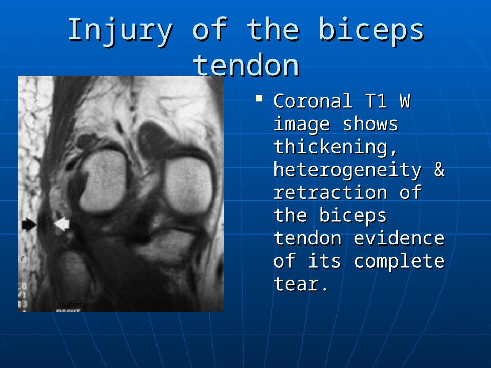
Injury of the biceps tendonInjury of the biceps tendon
Coronal T1 W Coronal T1 W image shows image shows thickening, thickening, heterogeneity & heterogeneity & retraction of the retraction of the biceps tendon biceps tendon evidence of its evidence of its complete tear.complete tear.
Distraction soft tissue injuryDistraction soft tissue injury (hyperextension with valgus)(hyperextension with valgus)
Posteromedial instability:Posteromedial instability: Injury of semimembranosis tendon.Injury of semimembranosis tendon. Injury of the medial head of Injury of the medial head of
gastrocnemius.gastrocnemius.
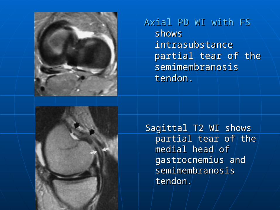
Axial PD WI with FSAxial PD WI with FS shows intrasubstance shows intrasubstance partial tear of the partial tear of the semimembranosis semimembranosis tendon.tendon.
Sagittal T2 WI shows Sagittal T2 WI shows partial tear of the partial tear of the medial head of medial head of gastrocnemius and gastrocnemius and semimembranosis semimembranosis tendon.tendon.
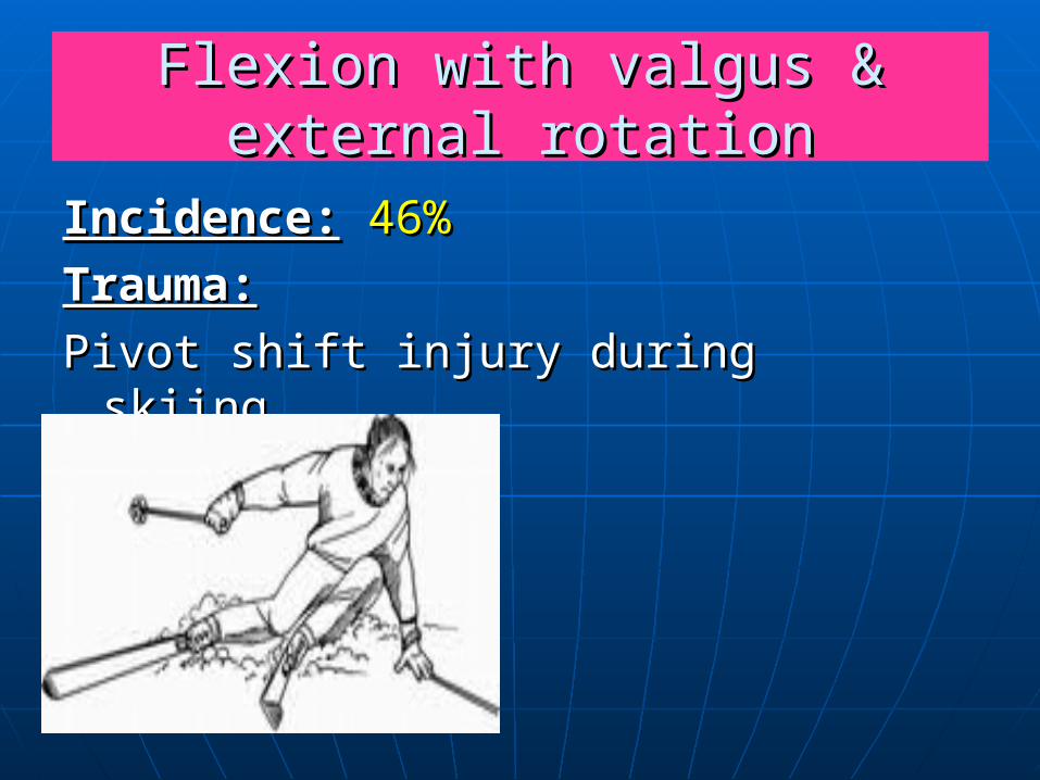
Flexion with valgus & external Flexion with valgus & external rotationrotation
Incidence:Incidence: 46%46%
Trauma:Trauma:
Pivot shift injury during skiing .Pivot shift injury during skiing .
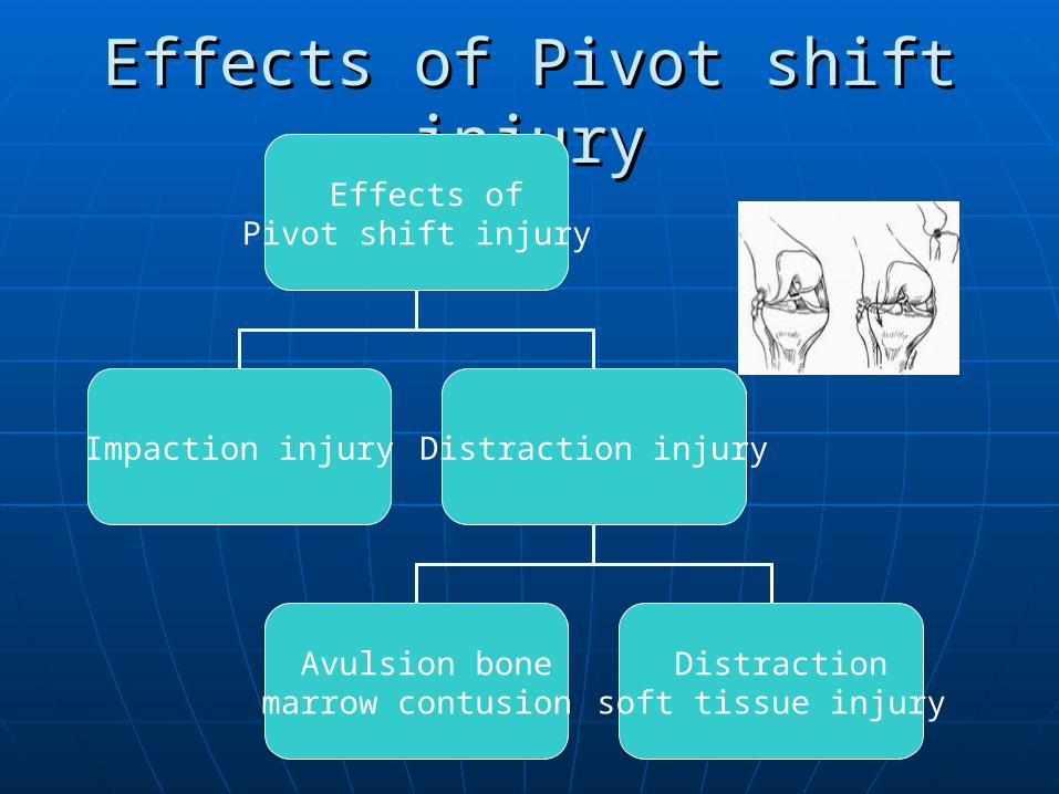
Effects of Pivot shift injuryEffects of Pivot shift injuryEffects of
Pivot shift injury
Impaction injury Distraction injury
Avulsion bone marrow contusion
Distraction soft tissue injury
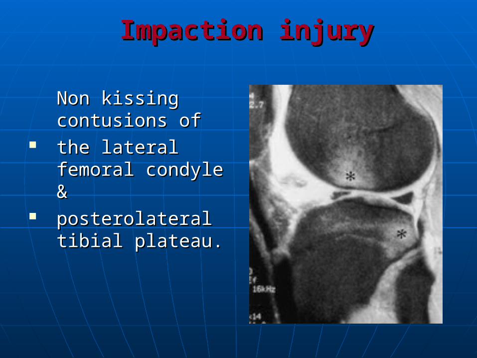
Impaction injuryImpaction injury
Non kissing Non kissing contusions of contusions of
the lateral femoral the lateral femoral condyle & condyle &
posterolateral posterolateral tibial plateau.tibial plateau.
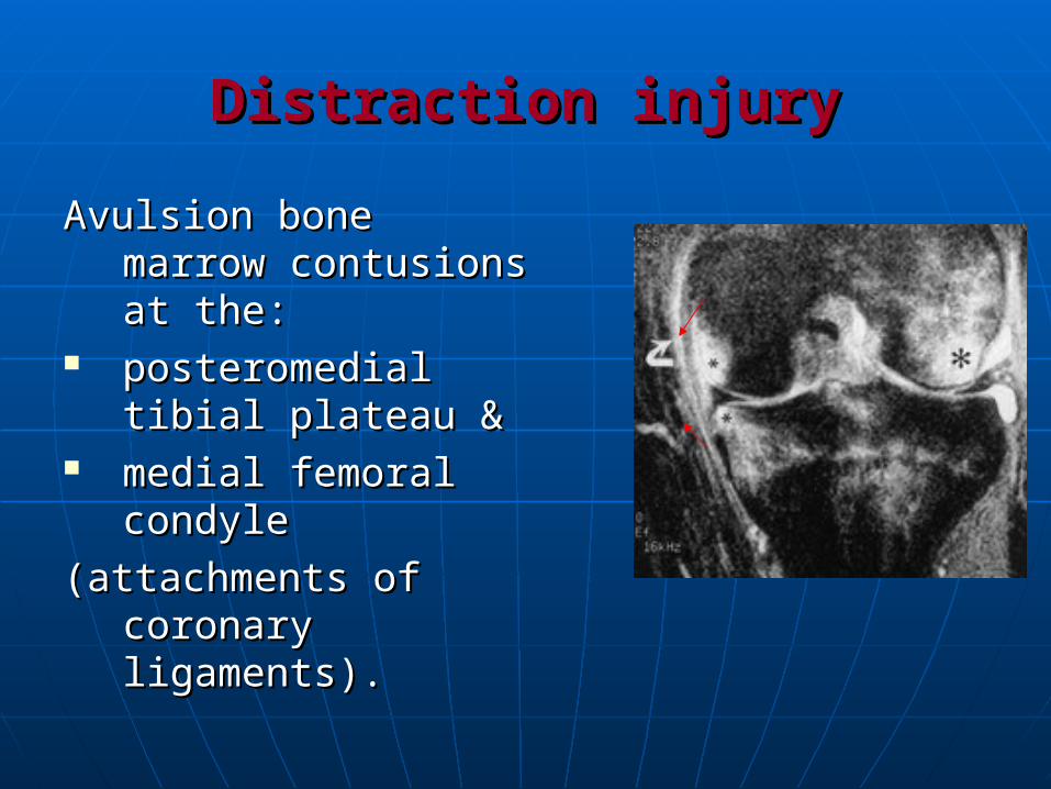
Distraction injuryDistraction injury
Avulsion bone marrow Avulsion bone marrow contusions at the: contusions at the:
posteromedial posteromedial tibial plateau & tibial plateau &
medial femoral medial femoral condylecondyle
(attachments of (attachments of coronary coronary ligaments).ligaments).
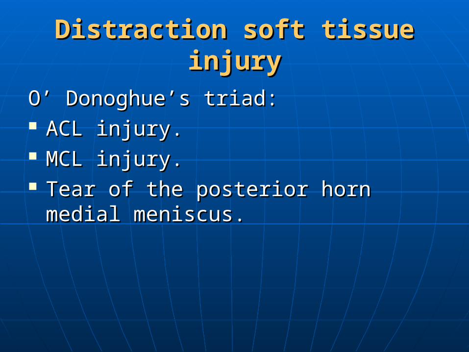
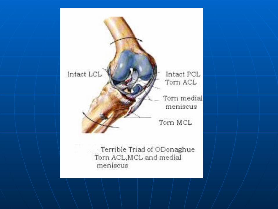
Distraction soft tissue injuryDistraction soft tissue injury
O’ Donoghue’s triad:O’ Donoghue’s triad: ACL injury.ACL injury. MCL injury.MCL injury. Tear of the posterior horn medial Tear of the posterior horn medial
meniscus.meniscus.
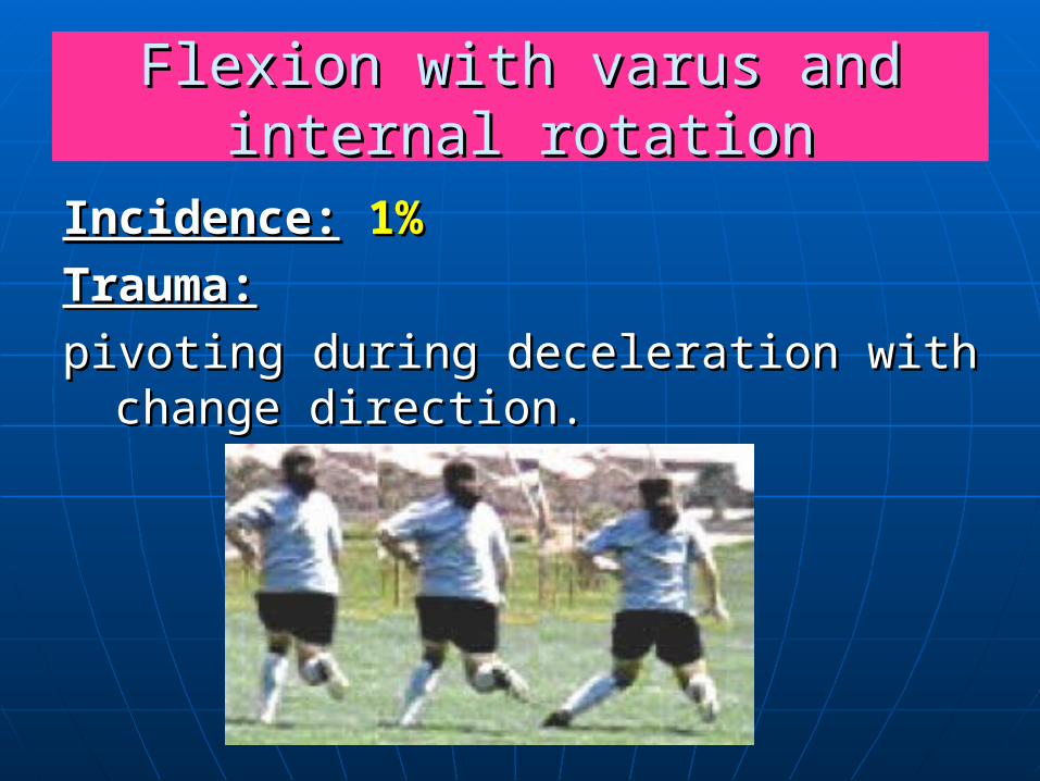
Flexion with varus and internal Flexion with varus and internal rotationrotation
Incidence:Incidence: 1%1%
Trauma:Trauma:
pivoting during deceleration with pivoting during deceleration with change direction.change direction.
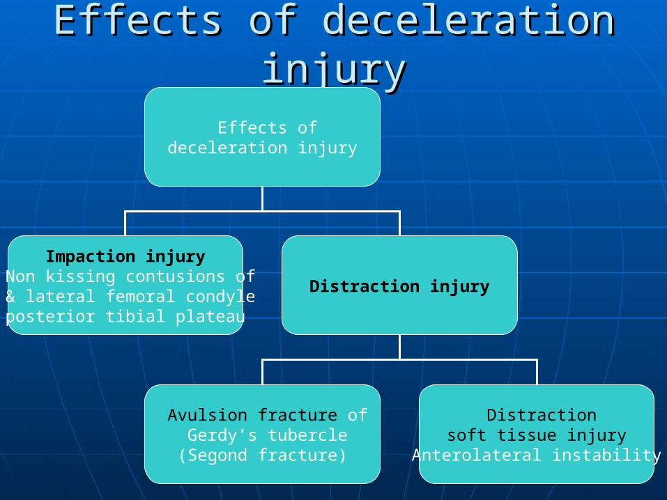
Effects of deceleration injuryEffects of deceleration injury
Effects of deceleration injury
Impaction injuryNon kissing contusions of
lateral femoral condyle & posterior tibial plateau
Distraction injury
Avulsion fracture of Gerdy’s tubercle
)Segond fracture(
Distraction soft tissue injury
Anterolateral instability
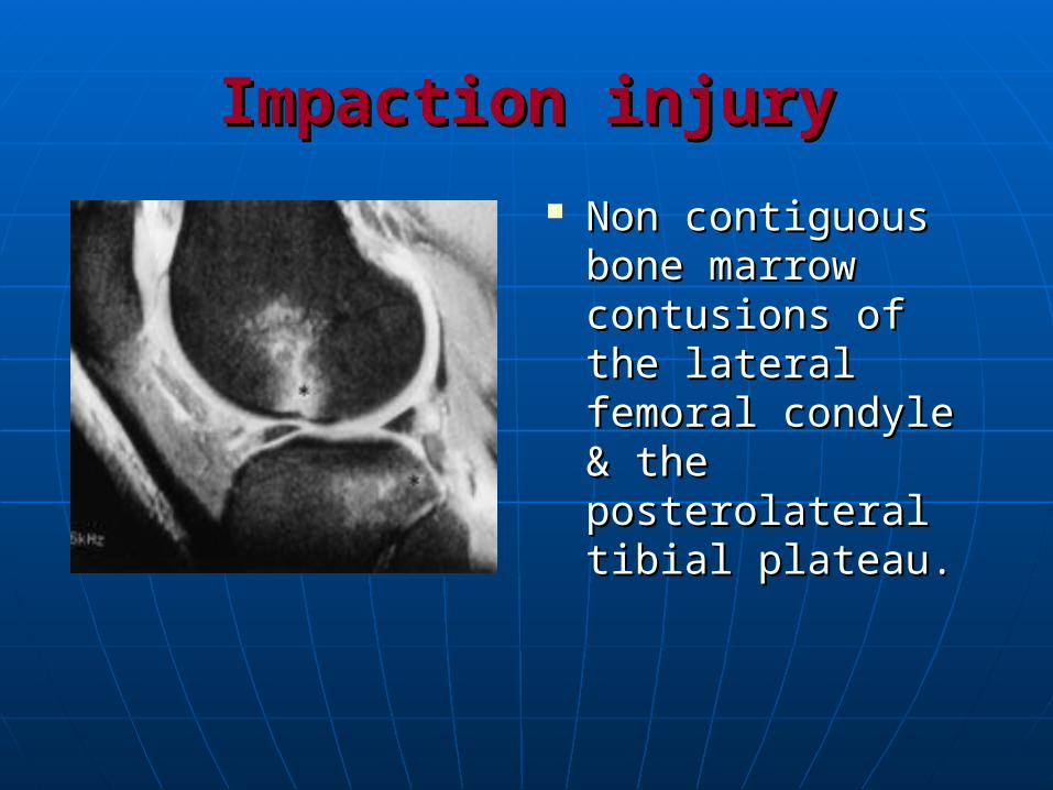
Impaction injuryImpaction injury
Non contiguous Non contiguous bone marrow bone marrow contusions of the contusions of the lateral femoral lateral femoral condyle & the condyle & the posterolateral tibial posterolateral tibial plateau.plateau.
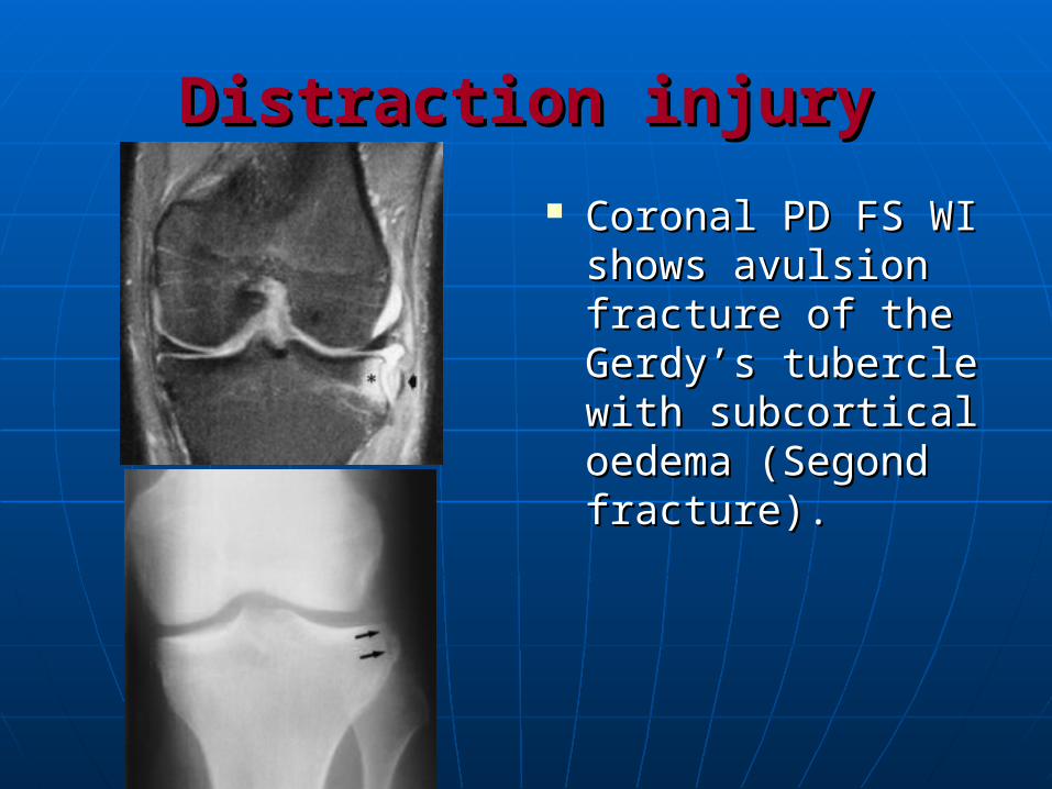
Distraction injuryDistraction injury
Coronal PD FS WI Coronal PD FS WI shows avulsion shows avulsion fracture of the fracture of the Gerdy’s tubercle Gerdy’s tubercle with subcortical with subcortical oedema (Segond oedema (Segond fracture).fracture).
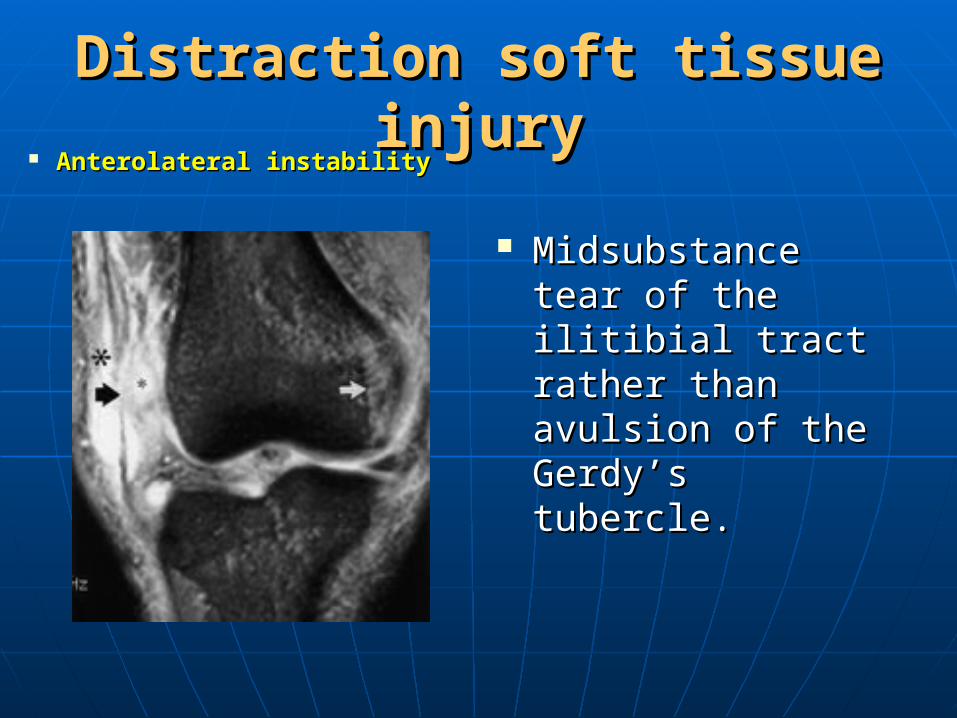
Distraction soft tissue injuryDistraction soft tissue injury Anterolateral instabilityAnterolateral instability
Midsubstance tear Midsubstance tear of the ilitibial tract of the ilitibial tract rather than rather than avulsion of the avulsion of the Gerdy’s tubercle.Gerdy’s tubercle.
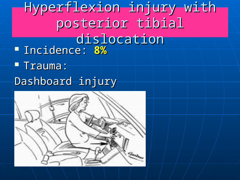
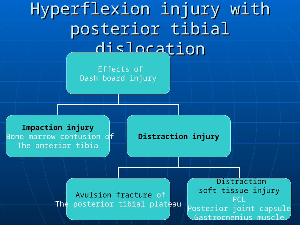
Hyperflexion injury with posterior Hyperflexion injury with posterior tibial dislocationtibial dislocation
Incidence: Incidence: 8%8% Trauma:Trauma:
Dashboard injuryDashboard injury
Hyperflexion injury with posterior Hyperflexion injury with posterior tibial dislocationtibial dislocation
Effects of Dash board injury
Impaction injuryBone marrow contusion of
The anterior tibiaDistraction injury
Avulsion fracture of The posterior tibial plateau
Distraction soft tissue injury
PCLPosterior joint capsuleGastrocnemius muscle
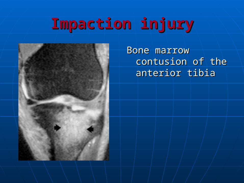
Impaction injuryImpaction injury
Bone marrow Bone marrow contusion of the contusion of the anterior tibiaanterior tibia
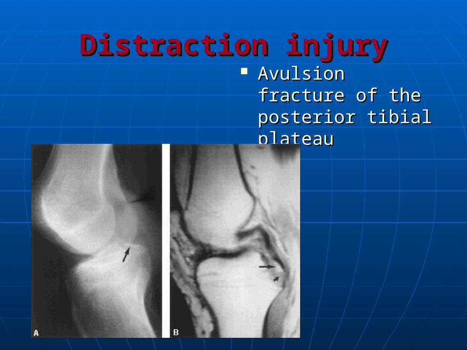
Distraction injuryDistraction injury Avulsion fracture of Avulsion fracture of
the posterior tibial the posterior tibial plateauplateau
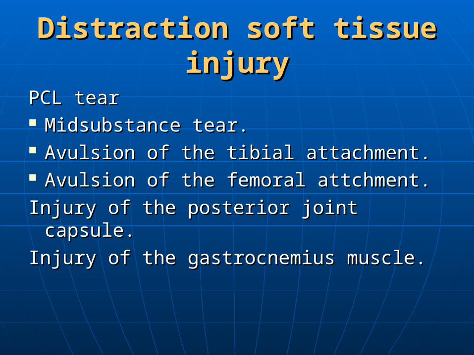
Distraction soft tissue injuryDistraction soft tissue injury
PCL tearPCL tear Midsubstance tear.Midsubstance tear. Avulsion of the tibial attachment.Avulsion of the tibial attachment. Avulsion of the femoral attchment.Avulsion of the femoral attchment.
Injury of the posterior joint capsule.Injury of the posterior joint capsule.
Injury of the gastrocnemius muscle.Injury of the gastrocnemius muscle.
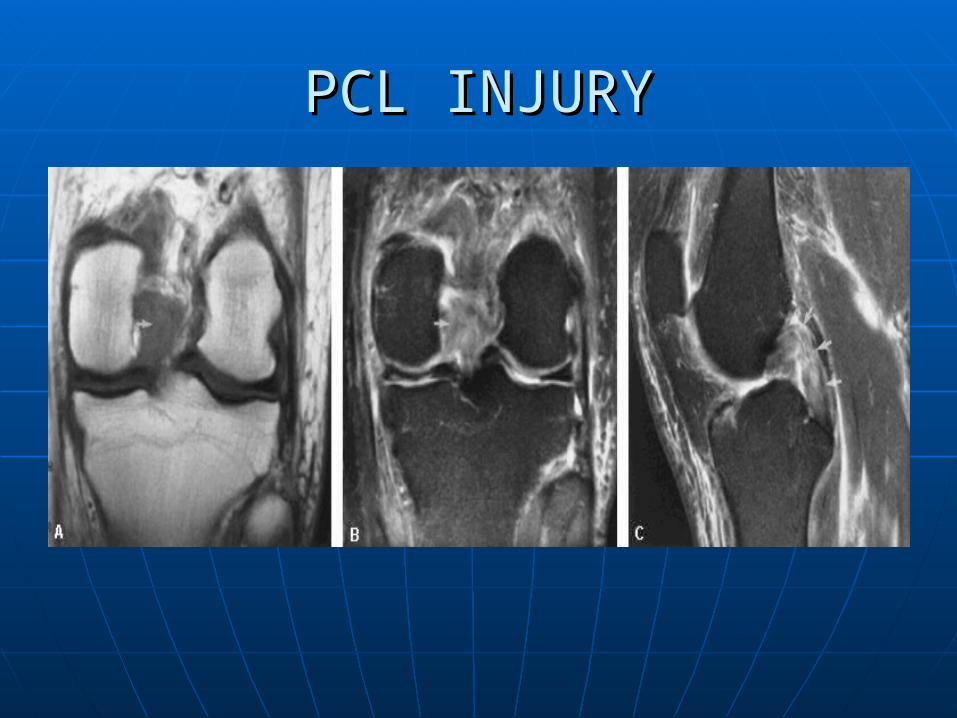
PCL INJURYPCL INJURY
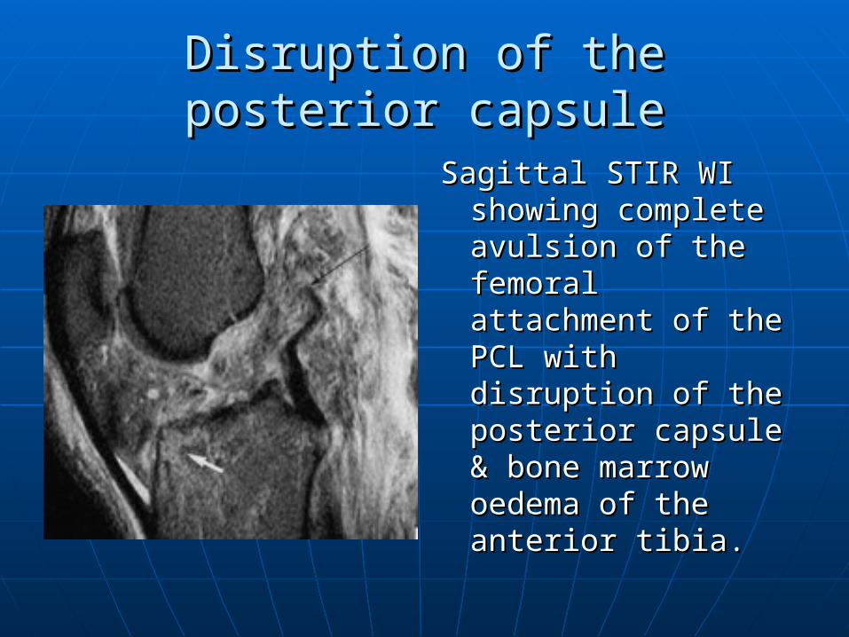
Disruption of the posterior capsuleDisruption of the posterior capsule
Sagittal STIR WI Sagittal STIR WI showing complete showing complete avulsion of the avulsion of the femoral femoral attachment of the attachment of the PCL with disruption PCL with disruption of the posterior of the posterior capsule & bone capsule & bone marrow oedema of marrow oedema of the anterior tibia.the anterior tibia.
Injury of the gastrocnemius muscleInjury of the gastrocnemius muscle
Grade I sprain:Grade I sprain:Interstitial oedema (giving feathery Interstitial oedema (giving feathery
appearance on STIR & T2 images).appearance on STIR & T2 images).Intramuscular hematoma.Intramuscular hematoma.Grade II:Grade II:Partial tear without retraction.Partial tear without retraction.Grade III:Grade III:Complete tear with retraction.Complete tear with retraction.
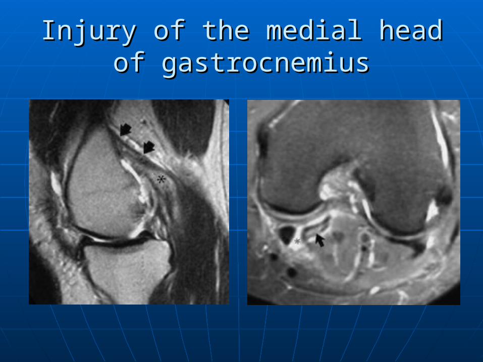
Injury of the medial head of Injury of the medial head of gastrocnemiusgastrocnemius
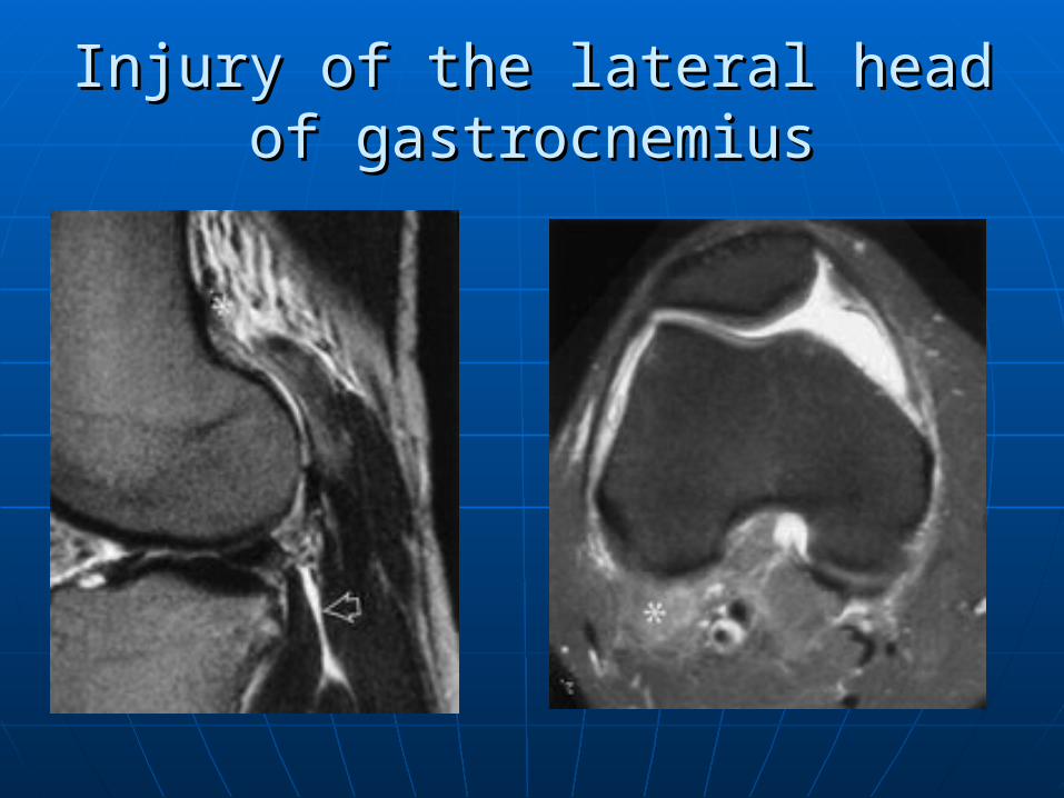
Injury of the lateral head of Injury of the lateral head of gastrocnemiusgastrocnemius
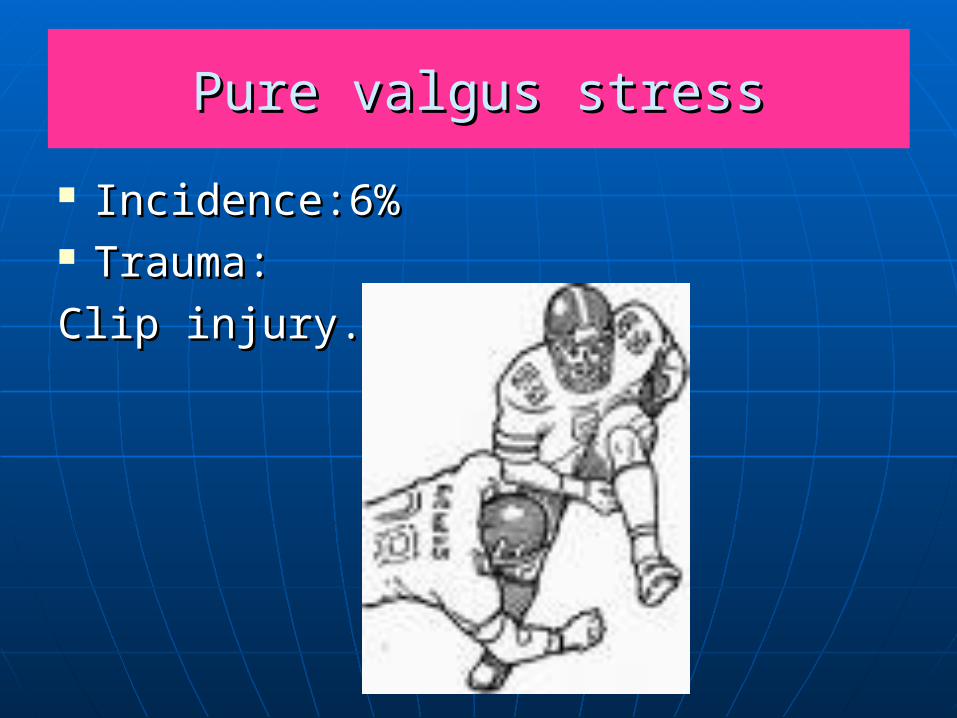
Pure valgus stressPure valgus stress
Incidence:6%Incidence:6% Trauma:Trauma:
Clip injury.Clip injury.
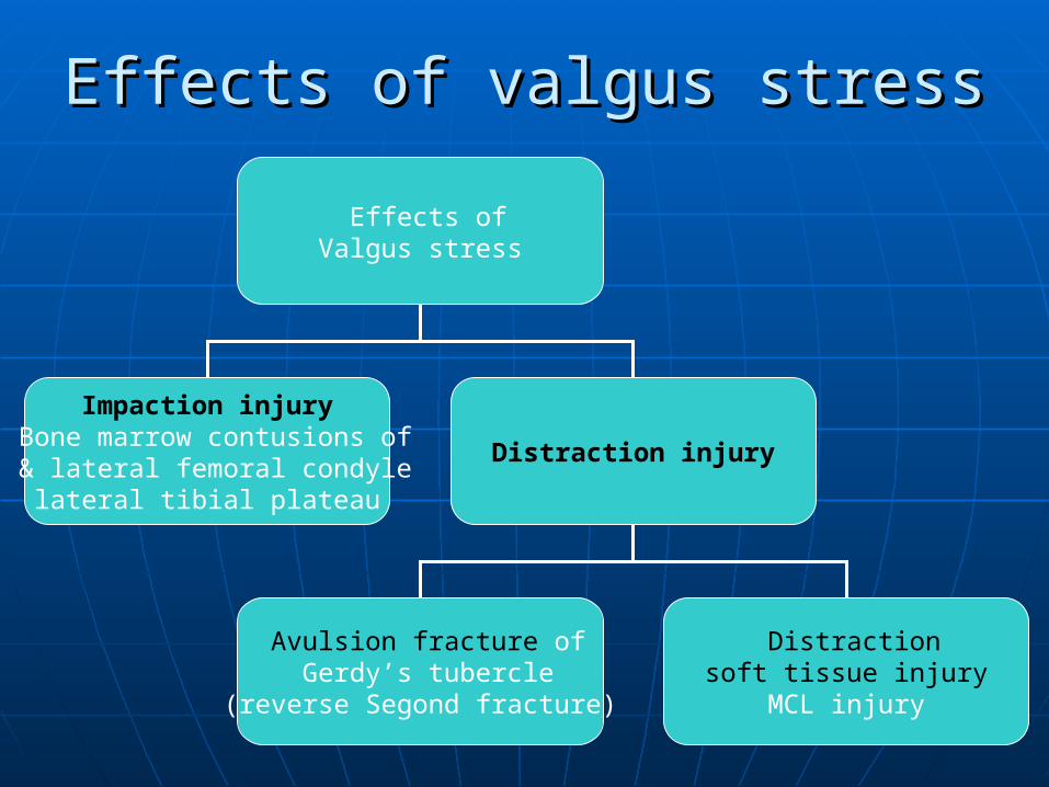
Effects of valgus stressEffects of valgus stress
Effects of Valgus stress
Impaction injuryBone marrow contusions of
lateral femoral condyle & lateral tibial plateau
Distraction injury
Avulsion fracture of Gerdy’s tubercle
(reverse Segond fracture)
Distraction soft tissue injury
MCL injury
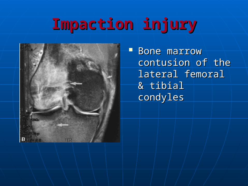
Impaction injuryImpaction injury
Bone marrow Bone marrow contusion of the contusion of the lateral femoral & lateral femoral & tibial condylestibial condyles

Distraction injuryDistraction injury
Avulsion bone Avulsion bone marrow contusion marrow contusion or fracture at the or fracture at the tibial attachment of tibial attachment of the MCL (reversed the MCL (reversed Segond fracture).Segond fracture).
Distraction soft tissue injuryDistraction soft tissue injury
injury of the medial collateral injury of the medial collateral ligament:ligament:
Grade I:Grade I:
Grade II:Grade II:
Grade III:Grade III:
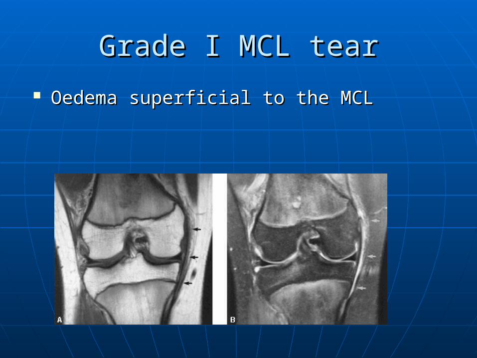
Grade I MCL tearGrade I MCL tear
Oedema superficial to the MCLOedema superficial to the MCL
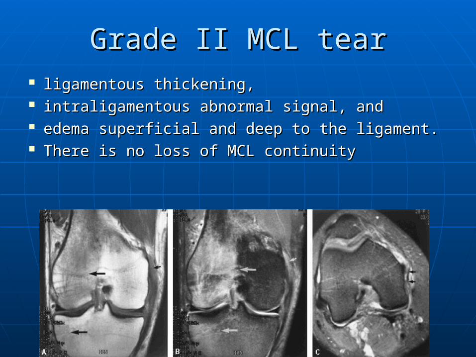
Grade II MCL tearGrade II MCL tear ligamentous thickening, ligamentous thickening, intraligamentous abnormal signal, and intraligamentous abnormal signal, and edema superficial and deep to the ligament. edema superficial and deep to the ligament. There is no loss of MCL continuityThere is no loss of MCL continuity
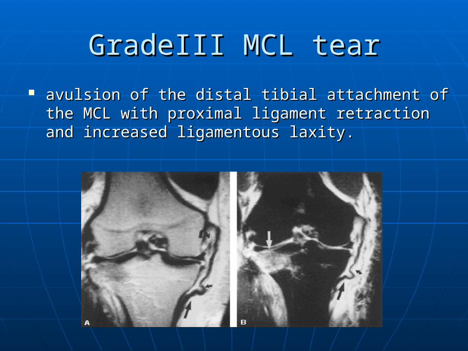
GradeIII MCL tearGradeIII MCL tear
avulsion of the distal tibial attachment of the MCL avulsion of the distal tibial attachment of the MCL with proximal ligament retraction and increased with proximal ligament retraction and increased ligamentous laxity.ligamentous laxity.
Pure varus stressPure varus stress
Incidence: Incidence: 1 %1 %
Trauma:Trauma:
It is a rare pattern of injury as valgus It is a rare pattern of injury as valgus stress is usually assoicated with stress is usually assoicated with flexion and internal rotation.flexion and internal rotation.
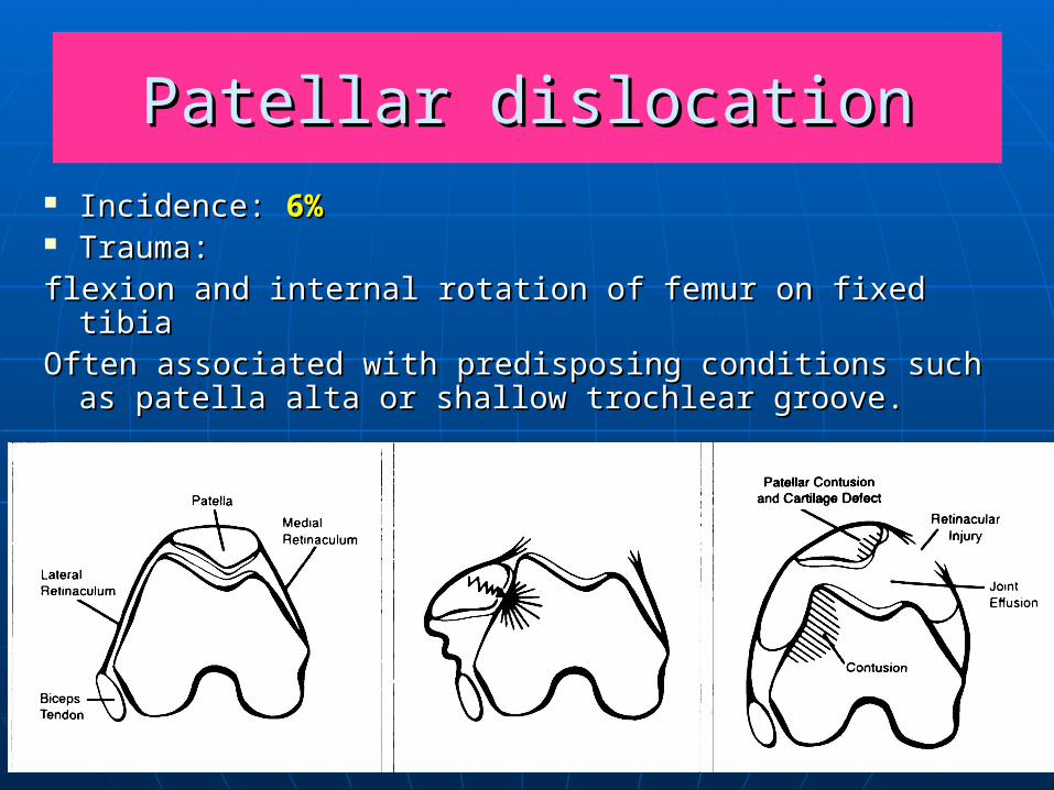
Patellar dislocationPatellar dislocation Incidence: Incidence: 6%6% Trauma:Trauma:flexion and internal rotation of femur on fixed tibiaflexion and internal rotation of femur on fixed tibiaOften associated with predisposing conditions such Often associated with predisposing conditions such
as patella alta or shallow trochlear groove.as patella alta or shallow trochlear groove.
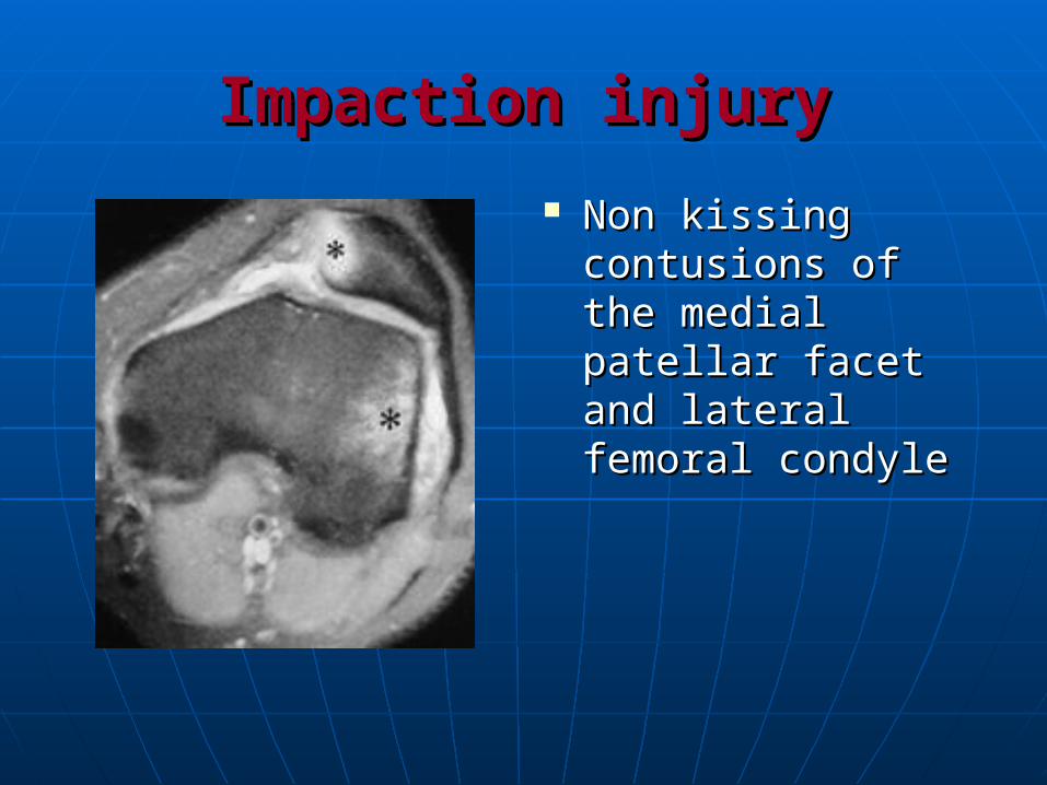
Impaction injuryImpaction injury
Non kissing Non kissing contusions of the contusions of the medial patellar medial patellar facet and lateral facet and lateral femoral condylefemoral condyle
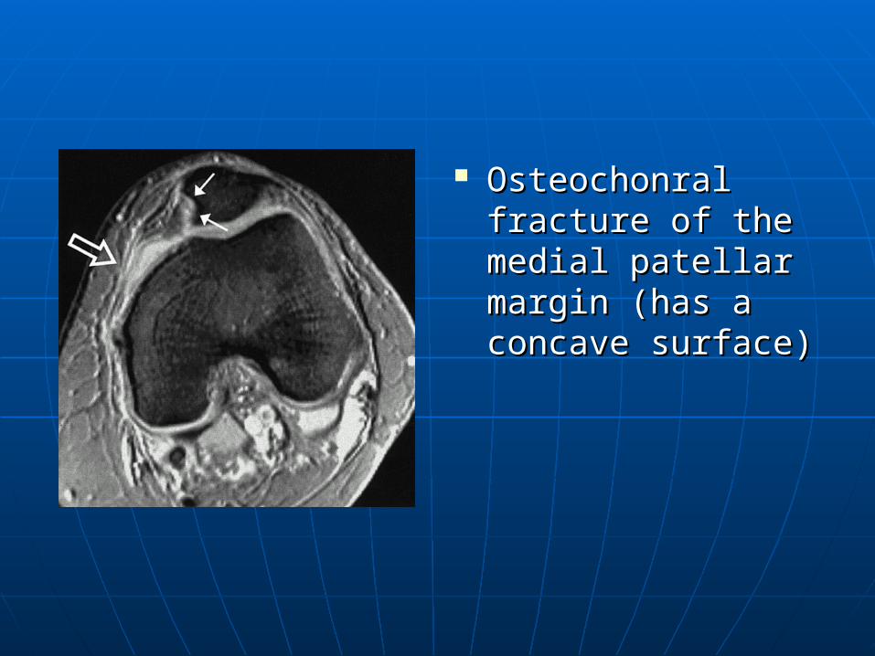
Osteochonral Osteochonral fracture of the fracture of the medial patellar medial patellar margin (has a margin (has a concave surface)concave surface)
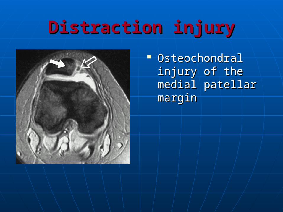
Distraction injuryDistraction injury
Osteochondral Osteochondral injury of the medial injury of the medial patellar marginpatellar margin
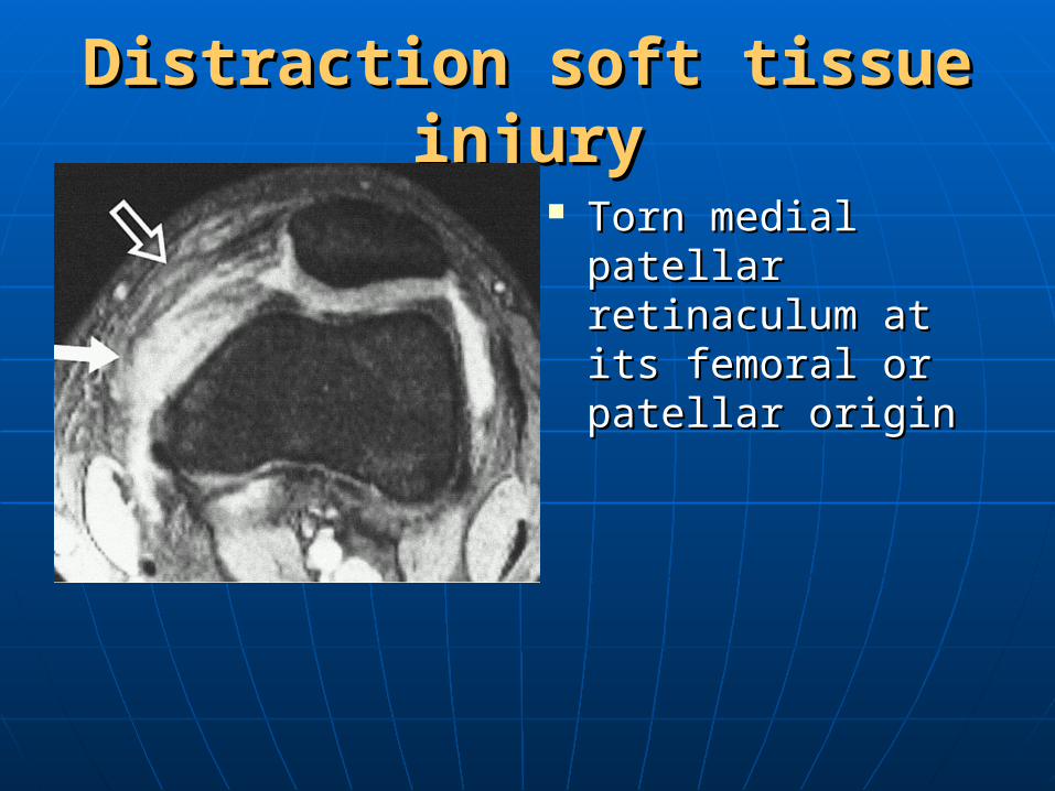
Distraction soft tissue injuryDistraction soft tissue injury
Torn medial Torn medial patellar patellar retinaculum at its retinaculum at its femoral or patellar femoral or patellar originorigin
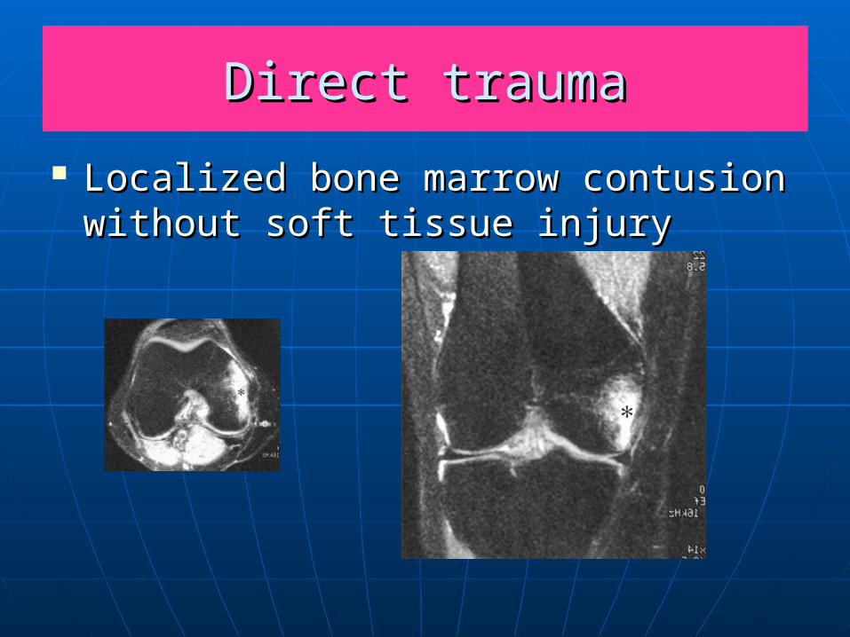
Direct traumaDirect trauma
Localized bone marrow contusion Localized bone marrow contusion without soft tissue injurywithout soft tissue injury