Embed Size (px)
Citation preview

http://fiehnlab.ucdavis.edu/teaching/folder.2007-08-20.0671728135/
Fri Nov 02 Assay of enzyme activities reading list
Mo Nov 05 Mass spectrometry: fundamentals
Wed Nov 07 Mass spectrometry: quantification and identification
Fri Nov 09 Primary metabolism: overview and integration
Mo Nov 12 Veteran's Day
Wed Nov 14 Homework discussion I
Fri Nov 16 Animal models for studying metabolic networks
Mo Nov 19 Regulation of glycogen breakdown
Wed Nov 21 Inborn errors of glycogen metabolism
Fri Nov 23 Thanksgiving
Mo Nov 26 Metabolic networks in humans: from KO to SNP variants
Wed Nov 28 Homework discussion II
Fri Nov 30 Flux analysis, stoichiometry and elementary modes
Mo Dec 03 (Bio)chemical databases (Guest lecturer Dr. Tobias Kind)
Wed Dec 05 Tools for modeling metabolism (Guest lecturer Dr. Tobias Kind)
Fri Dec 07 Homework discussion III

Enzyme Assays

(1) Development of an assay
A useful enzyme assay must meet four criteria:
(a) absolute specificity
(b) high sensitivity
(c) high precision & accuracy
(d) convenience

(A) Absolute specificityMost enzyme assays monitor disappearance of a substrate or
appearance of a product
Ensure that only one enzyme activity is contributing to the
monitored effect!
e.g. PEPCK
PEP + CO2 + GDP ⇋ OAA + GTP
Ensure
absence of PEPcarboxylase
PEP + HCO3- → OAA + Pi
absence of pyruvate kinase
PEP + ADP → pyruvate + ATP
absence of PEPcarboxytransphosphorylase
PEP + CO2 + Pi ⇋ OAA + PPi
Study cofactor requirements and product identification under a variety of conditions / scientific papers.
Examples as above are found for almost any enzyme. Be aware of possible reactions that may contribute to a given product accumulation or substrate utilization!

(B) High sensitivity
e.g. for purification, specific activities of most enzymes are very low.
Therefore, the assay must be highly sensitive.

(C) High precision
The accuracy and precision of an enzyme assay usually depend on the underlying chemical basis of techniques that are used.
For example, if an assay is carried out in buffer of the wrong pH, the observed rates will not accurately reflect the rate of enzymatically produced products

Six major characteristics of a
protein solutionSix major characteristics of a protein solution warrant
consideration
1. pH
2. Degree of oxidation
3. Heavy metal contamination
4. Medium polarity
5. Protease contamination
6. temperature

pH
pH values yielding the highest reaction rates are not always those at which the enzyme is most stable. It is advisable to determine the pH optima for enzyme assay and stability separately.
For protein purifications: Buffer must have an appropriate pKa and not adversely affect the protein(s) of interest. Buffer capacity may be higher for tissues with large vacuoles such as plants and fungi.

Degree of oxidationMost proteins contain free SH groups. One or more of
these groups may participate in substrate binding and therefore are quite reactive.
Upon oxidation, SH turn form intra- or inter-molecular S-S bonds, which usually result in loss of enzyme activity.
A wide variety of compounds are available to prevent disulfide bond formation: 2-mercaptoethanol, cysteine, reduced glutathione, and thioglycolate. These compounds are added to protein solutions at concentration ranging from 10-4 to 5 10-3 M (excess because equilibrium are near unity).
Dithiothreitol is advantageous (lower amounts needed) because of formation of stable six-ring.
Antioxidants against quinones (e.g. protein isolation from plants) by polyvinylpyrrolidone.
Heavy metal contamination
SH groups may react with heavy metal ions such as Pb, Fe, Cu stemming from buffers, ion exchange resins or even the water in which solutions are prepared.
If trace amounts of heavy metals continue to be a problem, EDTA (ethylenediaminetetraacetic acid) may be included in the buffer solutions at a concentration of 1 to 3 10-4M. The compounds chelates most, if not all, deleterious metal ions.

Protease or nuclease
contaminationDuring cell breakage, proteases and nucleases are
liberated.
PMSF (phenylmethylsulfonyl fluoride):
a commonly used protease inhibitor

Temperature
Not all proteins are most stable at 0 °C, e.g. Pyruvate carboxylase is cold sensitive and may be stabilized only at 25 °C.
Freezing and thawing of some protein solutions is quite harmful. If this is observed, addition of glycerol or small amounts of dimethyl sulfoxide to the preparation before freezing may be of help.
Storage conditions must be determined by trial and error for each protein.

More on ‘keeping proteins for
enzyme assays’Proteins requiring a more hydrophobic environment may be
successfully maintained in solutions whose polarity has been decreased using sucrose, glycerol, and in more drastic cases, dimethyl sulfoxide or dimethylformamide. Appropriate concentrations must usually be determined by trial and error but concentrations of 1 to 10% (v/v) are not uncommon.
A few proteins, on the other hand, require a polar medium with high ionic strength to maintain full activity. For these infrequent occasions, KCl, NaCl, NH4Cl, or (NH4)2SO4
may be used to raise the ionic strength of the solution.

Protein purification for testing novel
enzymes: series of isolation and
concentration procedures
Major techniques for the isolation and concentration of proteins :differential solubility, ion exchange chromatography, absorption chromatography, molecular sieve techniques, affinity chromatography, electrophoresis.
Which technique will be successful? ….trial and error.

Coupled enzyme assays
• If neither the substrates nor products of an enzyme-
catalyzed reaction absorb light at an appropriate
wavelength,the enzyme can be assayed by linking to
another enzyme-catalyzed reaction that does involve
a change in absorbance.
• The second enzyme must be in excess,so that the
rate-limiting step in the linked assay is the action of
the first enzyme.
Most enzyme assays monitor disappearance of a substrate or
appearance of a product…
So, how to measure?

Coupled enzyme assays• Most useful, most frequent
• Not at all foolproof!

Errors and artifacts
in coupled enzyme assays

Mg2+
A little reminder on Glycolysis
stage 1: phosphofructokinase activates for C-C cleavage

A little reminder on GlycolysisGo` and G in heart muscle

In (mammals), Phosphofructokinase (PFK) is a 340 kd tetramer, which enables it to respond
allosterically to changes in the concentrations of the substrates fructose 6-phosphate and ATP
In addition to the substrate-binding sites, there are multiple regulatory sites on the enzyme, including
additional binding sites for ATP
A little reminder on Glycolysis
Allosteric sites in PFK
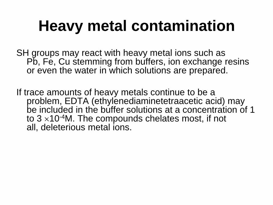
High ATP levels will change the kinetics of PFK from an asymptotic curve to a
sigmoidal one:
The sigmoidal curve reflects the reduced need for glycolysis at high energy levels
in the cell
This base ATP-dependent curve of PFK can then be further modulated by the
concentration of fructose 2,6-bisphosphate
A little reminder on Glycolysis/Gluconeogenesis
High ATP levels inhibit PFK activity

A little reminder on glycolysis
….and gluconeogenesis

Fructose 2,6-Bisphosphate is an Activator of PFK
Fructose 2,6-bisphosphate (F-2,6-BP) is a second allosteric effector of PFK
It functions as an activator that overrides the inhibitory effect of ATP:

F-2,6-BP Levels are Controlled by a Bifunctional Enzyme
The concentration of Fructose 2,6-Bisphosphate (F-2,6-BP) in cells is determined by a
bifunctional enzyme, phosphofructokinase 2 / fructose bisphosphatase2 ((PFK2/FBPase2),
to provide an additional level of control for PFK activity
F2,6-BP is formed by phosphorylation of fructose 6-phosphate in a reaction catalyzed by
PFK2
The resulting phosphoryl group on the C-2 can then be removed by the phosphatase
FBPase2

Reminder of gluconeogenesis by glucagon/cAMP cascade
plus allosteric activation of PFK by Fructose-2,6-bisphosphate

cited 557xcited 157x
cited 82x
cited 19x
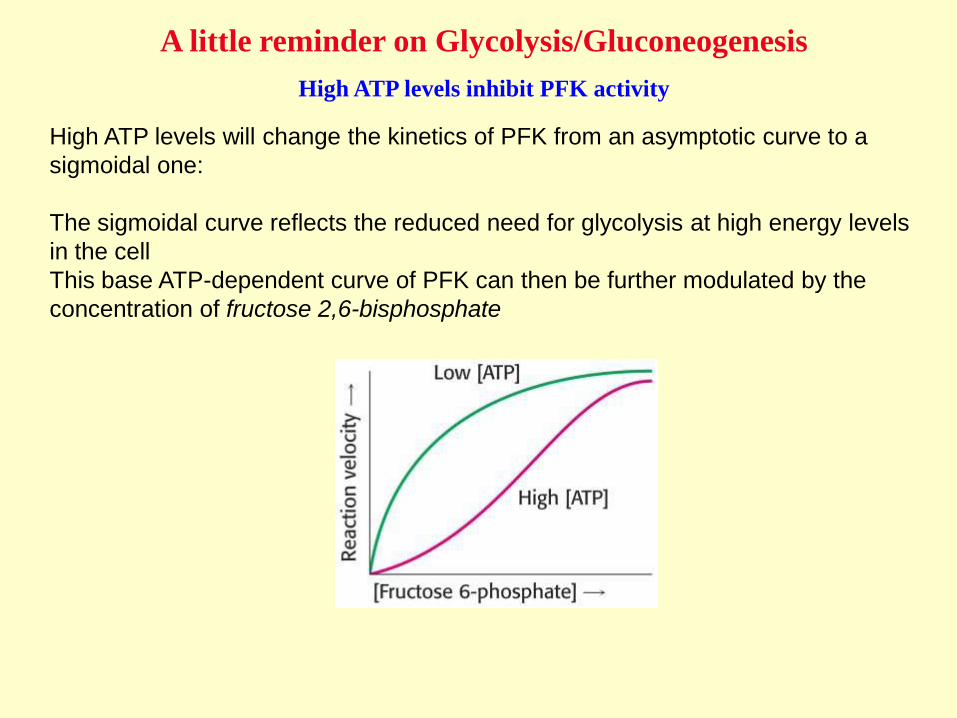
“F6P may contain
~ 0.001% F2,6BP”
…ATP was contaminated by 0.3% PPi,
and PPi is an activator of PFK…
PFP has
…imidodiphosphate is
contaminated by 2% PPi and is
actually inhibiting PFP.
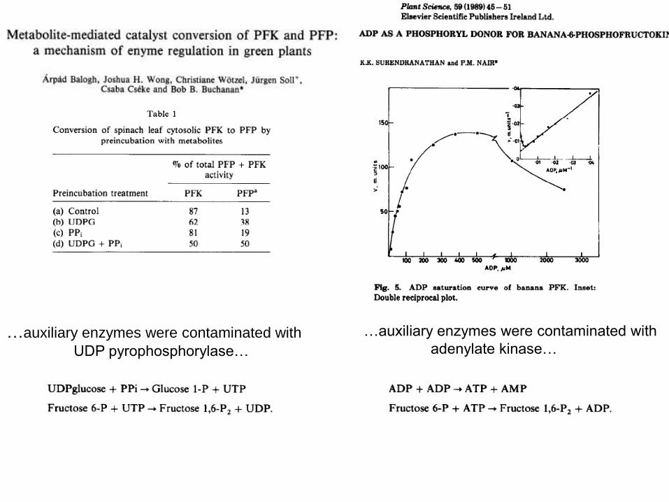
…auxiliary enzymes were contaminated with
UDP pyrophosphorylase…
…auxiliary enzymes were contaminated with
adenylate kinase…

Errors and
artifacts
in coupled
enzyme assays

Errors and artifacts
in coupled enzyme assays
Strategy:• Optimize your assay.
(1) pH (2) substrate concentrations should not be too large (3) conc. of coupled enzymes
should be not too large (4) vary buffers and counter ions. Compromise between „your‟ enzyme
and the requirements for the coupled enzymes. (5) Consider isozymes.
• Consider particularities of „your‟ enzyme and coupled enzymes.
• Question anomalous response in changing [E] or unusual kinetics (bursts, lag times)
• Use substrates from different vendors
• Check that reaction does not stop before depletion of limiting substrate/cofactor

If one coupled enzyme assay is difficult to control…
…23 assays must be easy !?

Robotized multi-enzyme assay
Measurement of „enzymome‟ not possible
• Group subsets of enzymes in modules that share common detection method.
• Cycling assays used. (pseudo zero order, rate depending on [metabolite]
• In combination with stopped assay, some 10^4fold more sensitive.

Cycling assay?

Dye- or fluorescent labels
Classic substrates Novel substrates

Real-time labels

In vivo assay FRET(fluorsc. resonance energy transfer)
Wolf Frommer
Carnegie

Red color indicates low internal glucose levels, green color shows high internal glucose
concentrations. Ratio red/green over time.
HepG2 cells expressing glucose-sensitive FRET
nanosensor in the cytosol. Addition of 5 mM glucose

Further reading


![ECE1001 Optoelectronics JBai Nov02 2010[2]](https://img.pdfslide.us/doc/110x75/563dbba4550346aa9aaef295/ece1001-optoelectronics-jbai-nov02-20102.jpg)