Embed Size (px)
Citation preview

LACTIC
ACIDOSIS-AN UPDATE
Dr W A P S R Weerarathna
Regisrtar-WD 10/02-THJ

OBJECTIVES…
Introduction
Pathophysiology
Hyperlactataemia/Lactic acidosis
Effects on cellular function
Causes of lactic acidosis
Diagnosis
Treatment including future therapies
Monitoring/goals of therapy &prognosis
Summary
References

INTRODUCTION
Most common cause of metabolic acidosis in hospitalized patients.
Lactic acidosis results from the accumulation of
lactate and protons in the body fluids and is often
associated with poor clinical outcomes
Associated with elevated anion gap and plasma [lactate] > 4mEq/L
Result of both overproduction and underuse of lactate.
Normal plasma [lactate] is 0.5 to 2.2 mEq/L (varies acc. Labs)

PATHOPHYSIOLOGY
The body tissues produce ~ 1500 mmol of lactate
each day (15 to 30 mmol/kg per day)-mainly in
skeletal muscles
Metabolized mainly by the liver (Cori cycle)
All tissues can produce lactate under anaerobic
conditions
In normal state production= elimination, causing
internal acid-base balance!

LACTATE PRODUCTION-ANAEROBIC
GLYCOLYSIS

LACTATE ELIMINATION-CORI CYCLE

HYPERLACTATAEMIA
Lactate production exceeds lactate consumption.
Addition of a number of protons equivalent to
the number of excess lactate ions
Coexisting acidemia contributes to decreased
lactate removal by the liver
Severe hypoxia and acidemia can convert the liver
into a net lactate-producing organ.

In the hyperdynamic stage of sepsis, epinephrine-
dependent stimulation of the β2-adrenoceptor
augments the glycolytic flux both directly and
through enhancement of the sarcolemmal Na+,K+-
ATPase.
Severe asthma (especially with overuse of β2-
adrenergic agonists), extensive trauma, cardiogenic
or hemorrhagic shock, and pheochromocytoma,
can cause hyperlactatemia through this
mechanism.

Drugs that impair oxidative phosphorylation, such
as antiretroviral agents and propofol, can augment
lactic acid production and on rare occasions cause
severe lactic acidosis.
The liver accounts for up to 70% of wholebody
lactate clearance.
Chronic liver disease exacerbates hyperlactatemia
due to sepsis or other disorders.

Hyperlactatemia is common in acute fulminant liver
disease, reflecting both reduced clearance and
increased production of lactate by the liver, and is
an important prognostic factor.

EFFECTS ON CELLULAR FUNCTION
The cellular dysfunction in hyperlactatemia is
complex.
Tissue hypoxia, if present, is a major factor.
If the cellular milieu is also severely acidic, cellular
dysfunction is likely to be exacerbated.
Decrease cardiac contractility, cardiac output, blood
pressure, and tissue perfusion.
Sensitize the myocardium to cardiac arrhythmias;
and can attenuate the cardiovascular
responsiveness to catecholamines.

Whether hyperlactatemia itself has an effect on
cellular function remains unclear.
In vitro studies suggest that lactate can depress
cardiac contractility.

CAUSES OF LACTIC ACIDOSIS
Divided into disorders associated with tissue
hypoxia (type A) and disorders in which tissue
hypoxia is absent (type B).
Evidence of tissue hypoxia can be subtle, and
hyperlactatemia can be of both hypoxic and
nonhypoxic origin.
Cardiogenic or hypovolemic shock, severe heart
failure, severe trauma, and sepsis are the most
common causes of lactic acidosis.


DIAGNOSIS
Evidence of severe cardiopulmonary disease,
SIRS, sepsis, severe trauma, or volume depletion
offers important clues.
An elevated serum AG, particularly a value higher
than 30 mmol per liter, can provide supportive
evidence.
The increase in (ΔAG) can mirror the blood lactate
level, but a close relationship might not always be
found.
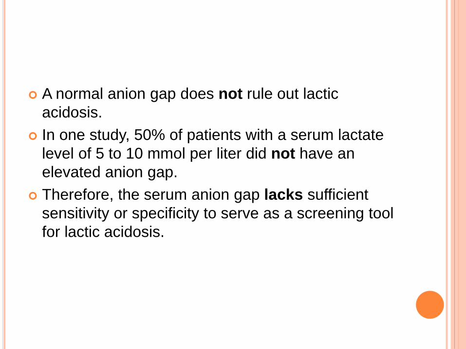
A normal anion gap does not rule out lactic
acidosis.
In one study, 50% of patients with a serum lactate
level of 5 to 10 mmol per liter did not have an
elevated anion gap.
Therefore, the serum anion gap lacks sufficient
sensitivity or specificity to serve as a screening tool
for lactic acidosis.

An elevated blood lactate level is essential for
confirmation of the diagnosis.
The lower limit of the normal range for the blood
lactate level, 0.5 mmol per liter, is consistent
among clinical laboratories, but the upper limit can
vary substantially, from as low as 1.0 mmol per
liter to as high as 2.2 mmol per liter
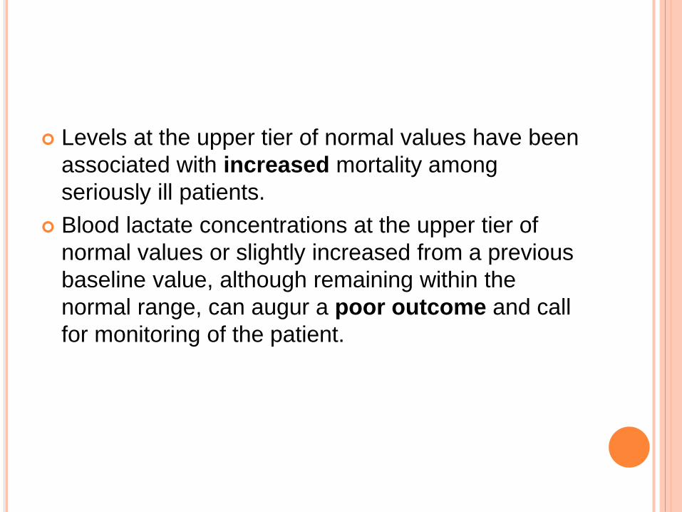
Levels at the upper tier of normal values have been
associated with increased mortality among
seriously ill patients.
Blood lactate concentrations at the upper tier of
normal values or slightly increased from a previous
baseline value, although remaining within the
normal range, can augur a poor outcome and call
for monitoring of the patient.

A serum osmolal gap of more than 20 mOsm per
kilogram of water has been reported in some cases,
probably reflecting the release of osmotically active
solute from ischemic tissues.

TREATMENT- SUPPORTING THE CIRCULATION
AND VENTILATION
Restoring tissue perfusion after hemodynamic
compromise is essential.
Vasopressors and inotropic agents should be
administered as needed.
Acidemia blunts the response to catecholamines,
thereby increasing the required dose.
Crystalloid and colloid solutions are both effective in
restoring tissue perfusion in patients with sepsis or
hypovolemia.
However, reports of AKI, bleeding, and increased
mortality in association with hydroxyethyl starch
synthetic colloid solutions provide evidence against
their use.
If a colloid solution is indicated, albumin should be
used.

Crystalloids containing bicarbonate or its precursors
(balanced salt solutions), such as Ringer’s
solution with lactate and Plasma-Lyte (Baxter
International) with acetate and gluconate, will not
cause non–anion gap metabolic acidosis and may
reduce the risk of AKI, but they can occasionally
cause metabolic alkalosis

Citrate-containing solutions can lead to the
generation of microthrombi.
Randomized, controlled studies are needed to
determine the most effective and safe crystalloid!
Red-cell transfusions should be administered to
maintain the hemoglobin concentration at a level
above 7 g per deciliter.

An adequate Po2 should be maintained by ensuring
an appropriate inspired oxygen concentration, with
endotracheal intubation and mechanical ventilation
as needed.
Invasive ventilation may also be required to prevent
hypercapnia, particularly if acidemia persists or
worsens.
IMPROVING THE MICROCIRCULATION
Abnormalities of the microcirculation, if persistent,
can augur clinical deterioration and death.
Dobutamine, acetylcholine, and nitroglycerin, have
been shown to improve microvascular perfusion
independently of systemic hemodynamics, to
reduce hyperlactatemia, and even to improve the
outcome.

INITIATING CAUSE-SPECIFIC MEASURES
Resuscitative efforts should be complemented by measures targeting the cause or causes of lactic acidosis.
Treatment of sepsis with the appropriate antibiotic agents;
Management of arrhythmias,
Resynchronization therapy, and left-ventricular assist devices for advanced heart failure;
Coronary intervention for acute myo- cardial infarction;
Surgery for trauma, tissue ischemia, or toxic megacolon; dialysis for removal of toxins or drugs;
Discontinuation of certain drugs; and
Reduction of tumor mass for cancer.

BASE ADMINISTRATION
Some clinicians recommend therapy with
intravenous sodium bicarbonate for severe
acidemia (blood pH, <7.2)
However, the value of bicarbonate therapy in
reducing mortality or improving hemodynamics
remains unproven! Due to-
1.intracellular acidification due to the accumulation
of carbon dioxide after bicarbonate infusion and
2.a pH-dependent decrease in levels of ionized
calcium, a modulator of cardiac contractility

Using dialysis to provide bicarbonate can prevent
a decrease in ionized calcium, prevent volume
overload and hyperosmolality (potential
complications of bicarbonate infusion), and remove
substances associated with lactic acidosis, such as
metformin.
Continuous dialysis is often favored over
intermittent dialysis because it delivers bicarbonate
at a lower rate and is associated with fewer
adverse events in patients with hemodynamic
instability.

Other buffers have been developed to minimize
carbon dioxide generation, including THAM (tris-
hydroxymethyl aminomethane) and Carbicarb (a
1:1 mixture of sodium carbonate and sodium
bicarbonate).
Only THAM is currently available for clinical use!

POTENTIAL FUTURE THERAPIES
The sodium–hydrogen (Na+–H+) exchanger NHE1
is activated during lactic acidosis, leading to
deleterious sodium and calcium overload in the
heart; its inhibition reduces cellular injury.
NHE1 inhibitors attenuated the lactic acidosis and
hypotension, improved myocardial performance
and tissue oxygen delivery, enabled resuscitation,
and reduced mortality in experimental models of
lactic acidosis.
The export of lactate and protons in the tumor
microenvironment has emerged as a critical
regulator of cancer development, maintenance, and
metastasis.
Inhibitors of LDH and MCT lactate transporters
are being investigated as promising cancer
therapies.

MONITORING OF PATIENTS, GOALS OF THERAPY,
AND PROGNOSIS
Close assessment of hemodynamic, oxygenation,
and acid–base status is paramount.
Measurement of the blood lactate level remains the
cornerstone of monitoring for lactic acidosis.
Although a single elevated blood lactate level often
predicts an adverse outcome, sustained
hyperlactatemia is associated with even worse
prognoses.

Transient hyperlactatemia does not necessarily
predict a poor clinical outcome.
An interval of 2 to 6 hours has been suggested for
repeat lactate measurements.
There is a dose–response relationship between
lactate levels and mortality: the higher the level,
the greater the risk of death.

Changes in levels of blood lactate have been used
to guide therapy.
Evidence that in seriously ill patients even lactate
levels at the upper end of the normal range are
associated with poor clinical outcomes argues for
the normalization of blood lactate as a primary
goal of therapy.

In patients with severe circulatory compromise,
central venous blood more accurately reflects the
acid–base status of tissues than does arterial or
peripheral venous blood.


SUMMARY
• lactic acidosis accompanies low-flow states or sepsis, mortality rates increase sharply.
• This review summarizes our current understanding of the pathophysiological aspects of lactic acidosis, as well as the approaches to its diagnosis and management.

REFERENCES
Review Article: Disorders of Fluids and Electrolytes
Jeffrey A. Kraut, M.D., and Nicolaos E. Madias, M.D.
N Engl J Med
Volume 371(24):2309-2319
December 11, 2014
Lactic Acidosis

THANK YOU!


![Biosensors based on electrochemical lactate detection … · clearance by liver and kidney, the accumulated concentration of lactic acid results in lactic acidosis [8]. ... monitored](https://img.pdfslide.us/doc/110x75/5b6d03227f8b9aed178ca8cd/biosensors-based-on-electrochemical-lactate-detection-clearance-by-liver-and.jpg)