Embed Size (px)
Citation preview
1
Sh
yam
Ku
ma
r Mish
ra
Shyam Kumar MishraAssistant Professor, Institute of Medicine
Laboratory diagnosis of LoWEr rEsPiratory traCt infECtions inCLuding tubErCuLosis

RESPIRATORY TRACT: ANATOMICAL STRUCTURE
It is utmost important to be familiar with the anatomic structure of the thoracic cavity, so that specimens collected from various sites in the lower respiratory tract are appropriately processed by the laboratory.
2
Sh
yam
Ku
ma
r Mish
ra

ANATOMY OF RESPIRATORT TRACT
3
Sh
yam
Ku
ma
r Mish
ra

INITIATION………
Aspiration of colonizing flora into the alveoli Inhalation of aerosols Hematologic seeding of the lung from a distant
focus
4
Sh
yam
Ku
ma
r Mish
ra

When paediatric patients with cystic fibrosis (CF) have respiratory infections (exacerbations), the trigger is most commonly related to an increased growth of bacteria that are chronically present in the airway.
However, in adult patients with COPD, the cause of respiratory exacerbations is usually related to the acquisition of new strains of bacteria, rather than an increase in the number of bacteria present when clinically stable.
5
Sh
yam
Ku
ma
r Mish
ra

1- Bronchitis.A- Acute:
Usually caused by viruses.
In infants & preschool children is Bordetella pertussis.
B- Chronic:
It is defined by clinical symptoms in which excessive mucus production leads to coughing up sputum on most days during at least 3 consecutive months for more than 2 successive years.
2- Bronchiolitis. It is the inflammation of smaller diameter bronchiolar epithelial surfaces.
It is an acute viral LRTI that primarily occurs during the first 2 years of life.
Diseases of the Lower Respiratory Tract
6
Sh
yam
Ku
ma
r Mish
ra

2- Pneumonia Community-Acquired Pneumonia:
patients are believed to have acquired infection outside the hospital setting.
The etiology of acute pneumonias is strongly dependent on age. More than 80% of pneumonias in infants & children are caused by viruses, whereas 10%-20% of pneumonias in adults are viral.
Hospital-Acquired Pneumonia
It is the leading cause of death among patients with nosocomial infections (as high as 50% mortality among patients in intensive care units).
Diseases of the Lower Respiratory Tract
7
Sh
yam
Ku
ma
r Mish
ra

RESPIRATORY TRACT PATHOGENS
Definitive Respiratory Tract Pathogens Haemophilus influenzae Streptococcus pneumoniae Mycobacterium tuberculosis Mycoplasma pneumoniae Chlamydia trachomatis Chlamydophila pneumoniae Bordetella pertussis Legionella spp. Pneumocystis jiroveci Nocardia spp. Histoplasma capsulatum Coccidioides immitis Cryptococcus neoformans Blastomyces dermatitidis Viruses ( Respiratory syncytal viruse, human metapneumovirus,
adenoviruses, enteroviruses, Hantavirus, herpes simplex virus, influenza and parainfluenza viruses, Rhinoviruses, SARS etc.)
8
Sh
yam
Ku
ma
r Mish
ra

RESPIRATORY TRACT PATHOGENS
Rare Respiratory Tract Pathogens Francisella tularensis Bacillus anthracis Yersinia pestis Burkholderia pseudomallei Coxiella burnetti Chlamydophila psittaci Brucella spp. Pasteurella multocida Klebsiella rhinoscleromatis Varicella-zooster viruse (VZV) parasites
9
Sh
yam
Ku
ma
r Mish
ra

10
Sh
yam
Ku
ma
r Mish
ra
PATHOGENESIS
Host Factors Nasal hairs Convoluted passage Mucus lining Secretary IgA Nonspecific antibacterial substances Cilia Reflexes such as coughing, sneezing & swallowing.
Microorganism Factors Adherence Colonization Fimbriae Toxins
11
Sh
yam
Ku
ma
r Mish
ra

LABORATORY DIAGNOSIS
Specimen collection and TransportA. Sputum1. Expectorated Food should not have been ingested for 1-2 hours before
expectoration. Mouth should be rinsed with saline or water just before
expectoration. The patient should be standing, if possible or sitting upright in bed. He or she should take deep breath to full the lungs, and empty then in one
breath, coughing as hard and as deeply as possible. Sputum brought up should be spit into screw capped container. Visually inspect the specimen. Tighten the cap of the container and send immediately to lab
12
Sh
yam
Ku
ma
r Mish
ra

Sputum of less than 2ml should not be processed unless obviously purulent
Only 1 sputum per 24hr submitted
SPUTUM COLLECTIOND
r.T.V
.Ra
o M
D
13

TRANSPORTATION OF SPUTUM
Transportation in <2 hr is recommended with refrigeration if delays anticipated.
Handle all samples using universal precautions.
Perform Gram stain and plant specimen as soon as possible
14

2. Induced Assisted by respiratory therapy technician. Using postural drainage & thoracic percussion to
stimulate production of acceptable sputum. Aerosol induced specimen: Breathing of aerosolized droplets of a solution
containing 15% NaCl and 10% glycerin for approximately 10 min. or until a strong cough reflex is initiated.
No pre-screening required (Although these specimen appear watery resembling saliva but they contain material directly from alveolar spaces ).
15
Sh
yam
Ku
ma
r Mish
ra

SPECIMEN COLLECTION AND TRANSPORT
3. Gastric aspirate: For isolation of Acid-Fast bacilli. Inability to produce sputum. Nasogastric tube is inserted into the stomach and contests are
withdrawn. The relative resistance of mycobacteria to acidity allows them to
remain viable for short period. Acidity of content is neutralized.
B. Endotracheal or Tracheostomy suctions specimens. Collected in Lukens trap. Tracheostomy aspirates should be treated as sputum.
16
Sh
yam
Ku
ma
r Mish
ra

ENDOTRACHEAL OR TRACHEOSTOMY SUCTIONS SPECIMENS
Fig: Collection of sputum Fig: Lukens Trap
17
Sh
yam
Ku
ma
r Mish
ra

SPECIMEN COLLECTION AND TRANSPORT
C. Bronchoscopy Fibreoptic devices Bronchial washings Bronchial aspirates Broncoalveolar lavage BAL (100-300ml NS is infused). Protected bronchial brushing samples.
Broncoalveolar lavage (BAL) 100-300ml NS is infused. Estimated that: 1 million alveoli are sampled during this process. Significant correlation : “Between”
Acute bacterial pneumonia and >103-104 bacterial colonies per mililitres of BAL fluid.
18
Sh
yam
Ku
ma
r Mish
ra

BRONCHOALVEOLAR LAVAGE (BAL)
SPECIMEN ACCEPTABILITY
Microscopic examination of Gram-stained smear Acceptable
<1% of cells present are squamous epithelial cells
Unacceptable>1% of cells present are squamous epithelial cells
Thorpe JE et. al. 1987. Bronchoalveolar lavage for diagnosing acute bacterial pneumonia. J. Infect. Dis. 155:855-861
19

SPECIMEN COLLECTION AND TRANSPORT Mini-BAL: Bedside, Non-bronchoscopic. Using metras catheter. 20ml or less saline is infused.
Protected bronchial brushing: Can collect 0.001 to 0.01 ml of material. Material can be suspended in 1 ml of broth dilution with vigorous
vortexing. Inoculate into culture media using 0.001 ml calibrated inoculating
loop. Correlation to infection: ≥ 1000 CFU/ml of broth diluent. ≥10^6 CFU/ml of original specimen. Have been considered to correlate with infection.
20
Sh
yam
Ku
ma
r Mish
ra

SPECIMEN COLLECTION AND TRANSPORT
D. Transtracheal Aspirates(TTA): Reduces the likelihood of contamination by upper respiratory
tract flora. Inserting small plastic catheter into the trachea via a needle
previously inserted through the skin and cricothyroid membrane.
E. Other invasive procedure: Thoracentesis (In pleural empyema). Open lung biopsy.
F. Blood culture: Only 20% of patients requiring hospitalization due to pnuemonia
are Blood culture positive.21
Sh
yam
Ku
ma
r Mish
ra

CRITERIA FOR REJECTING SAMPLES
22
Mismatch of information on the label and the request
Inappropriate transport temperatureExcessive delay in transportation Inappropriate transport medium
specimen received in a fixativedry specimensample with questionable relevance
Insufficient quantityLeakage

SPECIMEN PROCESSING
The purulent material which contains most of the relevant pathogens, is usually embeded in clear mucoid secretion.
If homogenised: Every drop & loopful of it will contains some of pathogen. Suitable for quantitative examinations.
Dithiothreitol or Buffered Pancreatin Mix and incubate equal volumes of the sputum and a solution of
dithiothreitol or buffered pancreatin.
23
Sh
yam
Ku
ma
r Mish
ra

SPECIMEN PROCESSING
I. Dithiothreitol Mix rapidly on a vortex mixer for 15 seconds and stand for 15
minutes at ambient temperature. Or mix gently and continuously on a machine that tilts to & fro
placed for 30 minutes in an incubator at 37ºC.
I. Pancreatin Incubate for 30 min. at 37ºC with continuous or occasional
shaking.
24
Sh
yam
Ku
ma
r Mish
ra
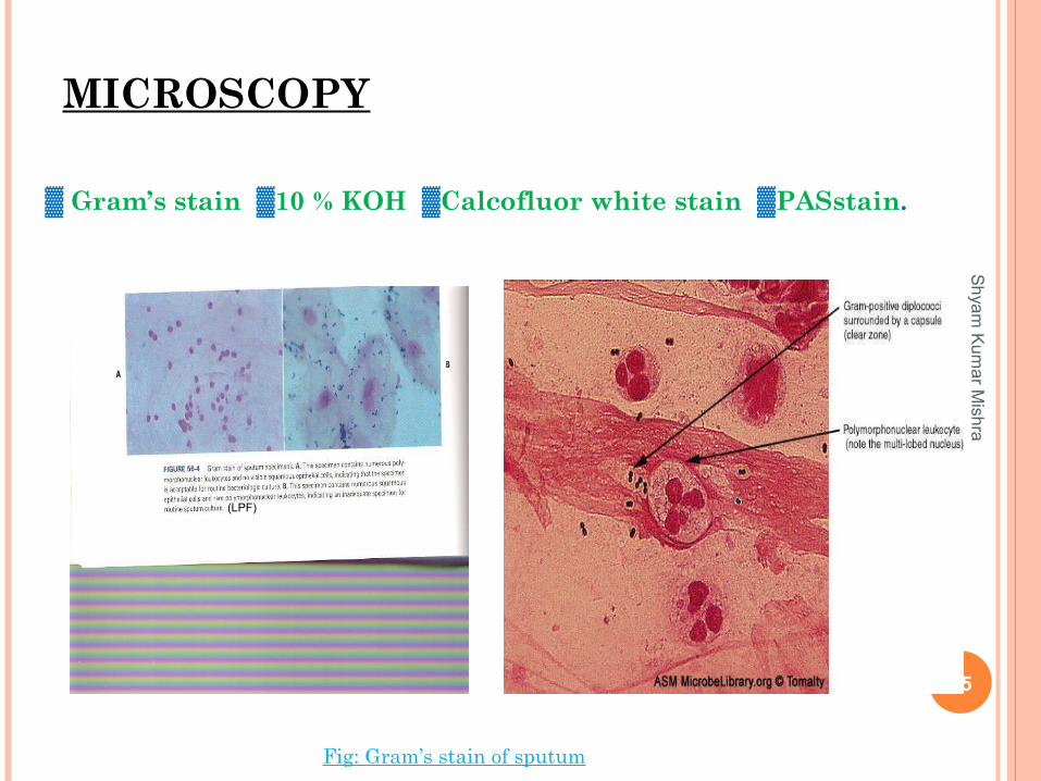
MICROSCOPY
▓ Gram’s stain ▓10 % KOH ▓Calcofluor white stain ▓PASstain.
Fig: Gram’s stain of sputum
25
Sh
yam
Ku
ma
r Mish
ra
(LPF)
MICROSCOPY
Screening by Gram’s stain Acceptable criteria: <10 epithelial cells /LPF. >25 WBCs/LPF ( Except in leucopenic patients).
Reasonable rejection criteria: >10 epithelial cells /LPF.
Rejection criteria for ETA: >10 epithelial cells /LPF. Or no organism seen under oil immersion.
In Legionella pneumophila Should not be subjected to screening by gram’s stain since sputum may
be scant and watery, with few or no host cells. 26
Sh
yam
Ku
ma
r Mish
ra
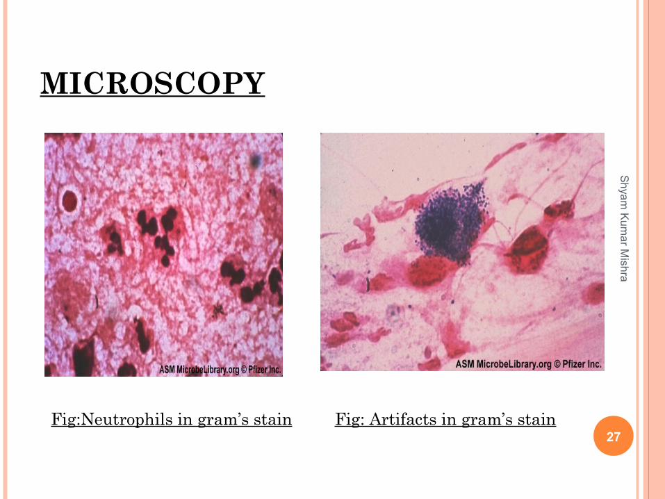
MICROSCOPY
27Fig:Neutrophils in gram’s stain Fig: Artifacts in gram’s stain
Sh
yam
Ku
ma
r Mish
ra
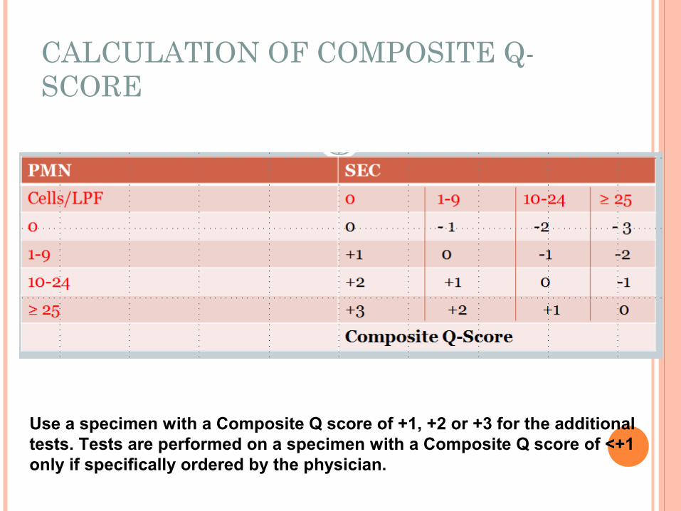
CALCULATION OF COMPOSITE Q-SCORE
Use a specimen with a Composite Q score of +1, +2 or +3 for the additional tests. Tests are performed on a specimen with a Composite Q score of <+1 only if specifically ordered by the physician.
28

29
Sh
yam
Ku
ma
r Mish
ra

30

31
Sh
yam
Ku
ma
r Mish
ra

INOCULATION:
Inoculated onto MA and BA at 37ºC. CA at 37ºC in increased carbondioxide condition. 5ug optochin (Ethyl hydrocupriene dihydrocloride to screen
for S. pneumoniae. 5-20 Unit (10U) Bacitracin for H. influenzae. Only specimens obtained by percutaneous aspiration
(including transtracheal aspiration) and by protected bronchial brush are suitable for anaerobic culture.
Buffered charcoal-yeast extract (BCYE) agar for Legionella spp.
PC 0r OFPBL agars for B. cepacia.
32
Sh
yam
Ku
ma
r Mish
ra

PROCESSING SPECIMENS FOR CULTURE
Process specimens in biological safety cabinet, as aerosol can result in laboratory-squired respiratory infections.
Process all specimens as rapidly as possible, especially specimen from emergency department, and inpatients. Select the most purulent or most blood-tinged portion of the specimen. Significant growth above the cutoff should be reported; however if more than one pathogen is isolated than it is suggestive of oropharyngeal contamination and clinical correlation should be done before reporting the samples.
33

34
Sh
yam
Ku
ma
r Mish
ra

CONTAMINATION WITH ORAL FLORA INTERFERES
RESULTS Because of contaminating oral flora ,sputum
specimens, specimens obtained by bronchial washing, and lavage tracheostomy, or endotracheal tube aspirates are not inoculated to enriched broth or incubated anaerobically. Only specimens obtained by percutaneous aspiration (including trans tracheal aspiration )and by protected bronchial brush are suitable for anaerobic culture: he latter must be done quantitatively for proper interpretation.
35

IDENTIFICATION OF MICRORGANISM
Identification of microorganism is based on the gram’s staining, colonial characters, biochemical tests and specific anti-sera if required.
36
Sh
yam
Ku
ma
r Mish
ra

OTHER SPECIMENS
Urine for antigen detection
Blood for serology
37
Sh
yam
Ku
ma
r Mish
ra

38
Sh
yam
Ku
ma
r Mish
ra

OPTOCHIN SENSITIVITY TEST

“DRAUGHTSMAN” APPEARANCE
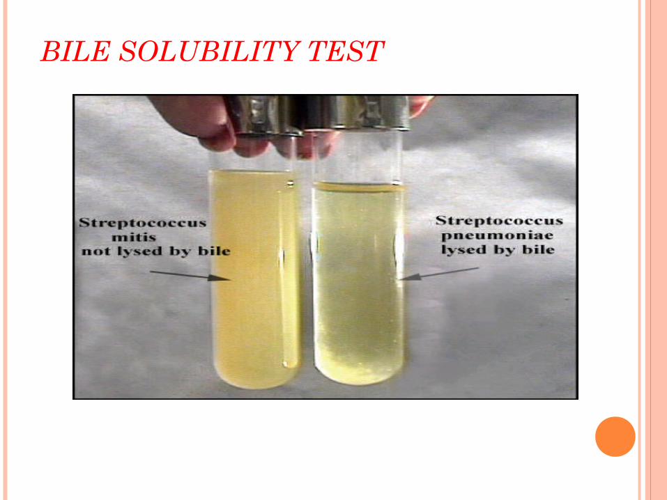
BILE SOLUBILITY TEST

LABORATORY DIAGONOSIS
COLONY PROCEDURE /Rapid presumptive test-This method works well on large or mucoid colonies-Select a well-isolated single colony from a blood or chocolate agar plate.-Circle the colony on the bottom of the Petri dish to locate- Place one drop of 2% sodium deoxycholate directly on the colony. -Incubate at 37°Cfor up to 30 minutes. Do not invert the plate. The lid may be left slightly ajar to aid evaporation- When the reagent has dried examine the area for lysis or disintegration of the original colony
Positive result: Colony lysed or disintegrated Negative result: No change

QUELLUNG REACTION (NEUFELD QUELLUNG RXN)
Word origin: German quellung for "swelling" quellung phenomenon
Increase in opacity and visibility of the capsule of capsulated organisms exposed to specific agglutinating anticapsular antibodies
Apparent swelling of the capsule upon binding of homologous antibody due to change in refractive index
reagent - antipneumoccal rabbit sera (omniserum)is used against 90 sero- types of S.pnuemoniae from State Serum Institute, DK2300,Copenhagen, Denmark)

SEROTYPING
different serotypes causes different diseases• Serotyping can be done by:
- Quellung reaction(described by Neufeld in 1902), classical method Agglutination of the pneumococci with type specific antiserum Capillary Precipitation of SSS with type specific antiserum Coagglutination dot blot assay counterimmuno-electrophoresis (CIEP)
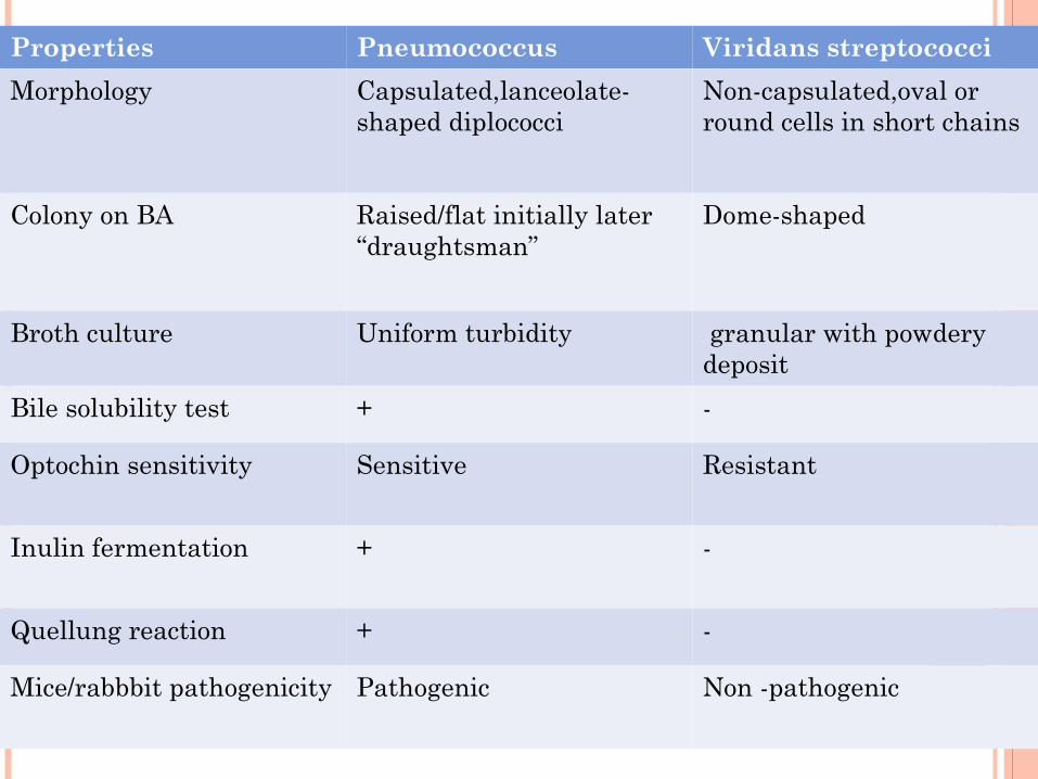
Properties Pneumococcus Viridans streptococci
Morphology Capsulated,lanceolate-shaped diplococci
Non-capsulated,oval or round cells in short chains
Colony on BA Raised/flat initially later “draughtsman”
Dome-shaped
Broth culture Uniform turbidity granular with powdery deposit
Bile solubility test + -
Optochin sensitivity Sensitive Resistant
Inulin fermentation + -
Quellung reaction + -
Mice/rabbbit pathogenicity Pathogenic Non -pathogenic

NUCLEIC ACID TECHNIQUE
Polymerase chain reaction Samples : blood, serum, CSF and respiratory samples,
body fluids Gene used as primers
The pneumolysin gene, pneumococcal surface adhesin protein (PsaA) gene, cell wall autolysin (LytA) protein gene
DNA/DNA hybridization
CASE STUDY…….
A 16-month-old boy was admitted with fever, lethargy and trouble breathing. A diagnosis of pneumonia was made by physical examination. The child had recently been to Panama and at that time was treated with ceftriaxone for cough and fever. His fever continued, despite treatment. On admission, he was given erythromycin therapy. Thacheal aspirate and blood cultures were obtained, but the respiratory specimen contained numerous epithelial cells and yielded normal respiratory flora on culture. A pleural aspirate and blood cultures were positive with Streptococcus pneumoniae, which was reisistant to erythromycin and penicillin and intermediate in susceptibility to ceftriaxone. The patient was given high doses of ceftriaxone and vancomycin and responded to therapy. 47
Sh
yam
Ku
ma
r Mish
ra

QUESTIONS..??? What criteria are used in laboratories to reject sputum and
tracheal aspirates for culture?
If greater than 10 squamous epithelial cells per low-power field are seen in a gram stain but the smear also has numerous white blood cells (greater than 25/LPF), should the specimen be rejected for culture?
In cases of pneumococcal pneumonia, what percentage of blood and sputum cultures is positive for S. pneumoniae?
The organism was reported as resistant to penicillin and intermediate in susceptibility to third-generation cephalosporins . How does the laboratory perform testing for this organism?
48
Sh
yam
Ku
ma
r Mish
ra

SPECIAL CONSIDERATION TO PULMONARY TUBERCULOSIS
Recovery of mycobacteria from clinical samples is more time consuming procedure than for normal pathogenic bacteria, requiring as it does the following steps:
1. Homogenization2. Centrifugation3. Neutralization4. Inoculation5. Incubation for at least 6wks
49
Sh
yam
Ku
ma
r Mish
ra
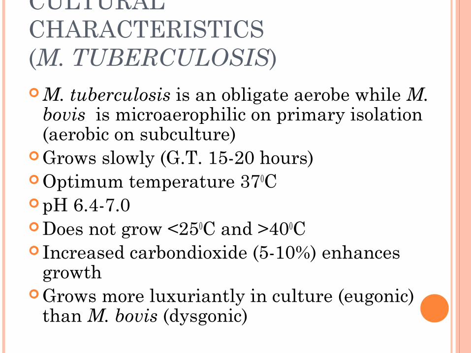
CULTURAL CHARACTERISTICS (M. TUBERCULOSIS) M. tuberculosis is an obligate aerobe while M.
bovis is microaerophilic on primary isolation (aerobic on subculture)
Grows slowly (G.T. 15-20 hours) Optimum temperature 370C pH 6.4-7.0 Does not grow <250C and >400C Increased carbondioxide (5-10%) enhances
growth Grows more luxuriantly in culture (eugonic)
than M. bovis (dysgonic)

Addition of 0.5% glycerol improves the growth of M. tuberculosis but has no effect on or even impair the growth of M. bovis.
Sodium pyruvate helps the growth of both types
Human tubercle bacilli do not grow in presence of p-nitrobenzoic acid (500 mg/l), unlike other slow growing nonchromogens.
Highly susceptible even to traces of toxic substance like fatty acid in culture media which can be neutralized by serum albumin or charcoal.

CULTURE MEDIA Solid media:
Egg containing – Lowenstein-Jensen mediaPetragnani mediaDorset egg mediaOgawa media
Blood containing – Tarshis media Serum containing – Loeffler’s serum slope Potato containing – Pawlowsky’s media
Liquid media: Dubo’s media Middlebrook’s media 7H9 Proskauer and Beck’s media Sula’s and Sauton’s media Kirchner media

To prevent overgrowth by contaminants, Cocktail of antibiotics
Polymyxin BAmphotericin BNalidixic acidTrimethoprimAzlocillin
PANTA
Polymyxin B Amphotericin BCarbenicillinTrimethoprim
PACT

COLONY CHARACTERS Solid media:
M. tuberculosis: Colonies appear after 2-3 weeks.
Colonies first appear as small, dry, friable, rough and granular, creamy white (Rough, tough, buff)
Typical colonies have a flat irregular margin and look like a cauliflower in the center.
M. bovis: Colonies appear after 3-6 weeks
Colonies are smooth, translucent and pyramidal

QUANTIFICATION SCALE FOR MYCOBACTERIAL GROWTH ON AGAR PLATES (ATS, 2000)
No. of colonies seen Quantity reported
No colonies seen
<50 colonies
50-100 colonies
100-200 colonies
200-500 colonies (almost confluence)
>500 colonies (confluence)
Negative
Report actual number seen
1 +
2 +
3 +
4+

Liquid media:Bacilli grow either on surface as pellicle or as floccules
throughout the medium due to hydrophobic nature of their cell wall lipid.
Generally, virulent strains often grow as twisted rope like colonies called serpentine cords??????
Diffuse bacterial growth is obtained by adding a detergent Tween 80.

Lowenstein-Jensen media Fresh whole eggs Defined salts Glycerol Potato flour Asparagine Malachite green

Middlebrook 7H10Defined saltsVitaminsCofactorsGlycerolOleic acidAlbuminDextroseCatalaseMalachite green

Ogawa media Potassium dihydrogen phosphate Sodium glutamate Glycerine 2% malachite green Distilled water Whole egg homogenate

Niacin test- Niacin test is positive with human type and negative with bovine type of bacilli.
Aryl sulphatase test- Aryl sulphatase test is positive with atypical mycobacteria.
Neutral red test- Neutral red test is positive with virulent strains of tubercle bacilli while avirulent strains are negative.
Catalase-peroxidase test- Atypical mycobacterial strains - strongly catalase
positive Tubercle bacilli - only weakly positive for catalase Tubercle bacilli - peroxidase positive Atypical mycobacteria – peroxidase negative Catalase and peroxidase activity are lost when tubercle
bacilli become INH resistant.
60
Sh
yam
Ku
ma
r Mish
ra

Amidase test- Amidase test differentiates mycobacteria by its ability to split amides. A useful pattern is provided by testing five amides, acetamide, benzamide, carbamide, nicotinamide and pyrazinamide.
Nitrate reduction test- M. tuberculosis + M. bovis -
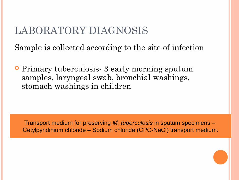
LABORATORY DIAGNOSIS
Sample is collected according to the site of infection
Primary tuberculosis- 3 early morning sputum samples, laryngeal swab, bronchial washings, stomach washings in children
Transport medium for preserving M. tuberculosis in sputum specimens – Cetylpyridinium chloride – Sodium chloride (CPC-NaCl) transport medium.

Conventional techniques Sputum microscopy
Acid fast stains: Ziehl Neelsen stain Kinyoun’s modification of ZN stain Auramine-Rhodamine stain
Sputum cultureMantoux skin test

Direct methodsSmear CulturePhage based assaysPhenotypic methods- lipid analysisMolecular methods- Probes, PCR
Indirect methodsSerology- ELISA, Latex agglutinationELISPOT AssaysDemonstration of biological products
ADA estimation Tuberculostearic acid test Bromide partition test

SPUTUM SMEAR EXAMINATION
Most widely applicable, most reliable ( WHO )
Minimum 3 samples (spot- early morning-spot)
Sensitivity- 50-70%

DISADVANTAGES OF SMEAR EXAMINATION
Lower limit of detection - 104 bacilli/ml
Sample
Cannot determine viability
Cannot identify species
False positive/ False negative result

INTERNATIONAL UNION AGAINST TUBERCULOSIS (IUAT)
If definite pink bacilli are not seen – AFB not found
If 1 or 2 bacilli in the entire field – Doubtful, repeat for another sample
If 1-9 AFB/100 fields – AFB found (exact number)
10-100 AFB/ 100 fields – AFB found (+)
1-10 AFB/ field – AFB found (++)
> 10 AFB/ field – AFB found (+++)

DIGESTION, CONCENTRATION AND CULTURAL METHODS Homogenization – To release the mycobacteria
from the body fluid or tissue in which they are contained
Decontamination – To kill the contaminants or else they will overgrow the medium

1. Petroff’s methodEqual vol. of Sputum and 4% NaOH370C – 30 mins with occasional shakingCentrifugation 3000g – 20-30minsDiscard the supernatantNeutralize the deposit with 8% HCl in presence of
phenol red indicatorCentrifugationDeposit
Inoculated on to the culture media Smear preparation Animal inoculation Molecular methods

2. Modified Petroff’s method Sputum + Double vol. of 4% NaOH R.T. – 15mins with occasional shaking Centrifugation 3000g – 15mins Discard the supernatant 15 ml saline or water + sediment Centrifugation 3000g – 15mins Deposit – inoculated on to the culture media

3. N-acetyl-L-Cysteine-NaOH method
4. Zephiran (Benzalkonium chloride)-TSP method

ANIMAL INOCULATION Intramuscular inoculation of 0.5 ml bacterial suspension into the
thigh of 12 week old guinea pig The tuberculin test becomes positive after 3-4 weeks and there
will be progressive loss of weight of the animal The animal is autopsied after 6wks or 12 wks. Results:
Caseous lesion at the site of inoculation Enlarged inguinal LN Tubercules in the lungs Splenomegaly
The identity of the bacteria is confirmed by demonstration of AFB from the lesions to exclude the Y. pseudotuberculosis, Brucella as their lesions macroscopically simulate tubercles.
ROOT INDEX OF VIRULENCE
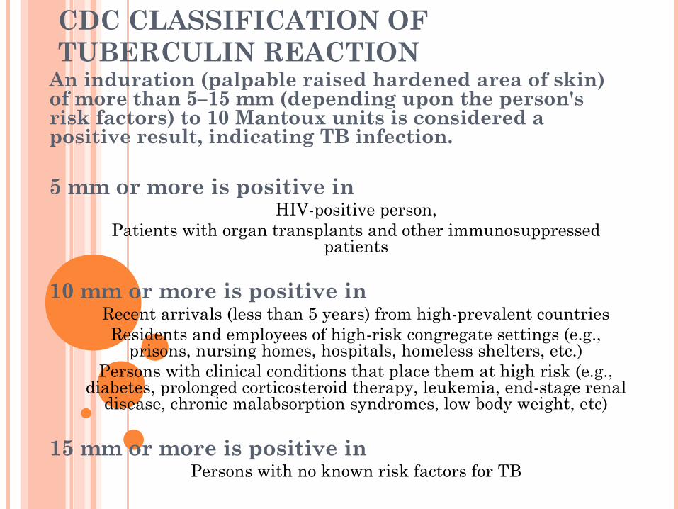
CDC CLASSIFICATION OF TUBERCULIN REACTION
An induration (palpable raised hardened area of skin) of more than 5–15 mm (depending upon the person's risk factors) to 10 Mantoux units is considered a positive result, indicating TB infection.
5 mm or more is positive in HIV-positive person,
Patients with organ transplants and other immunosuppressed patients
10 mm or more is positive in Recent arrivals (less than 5 years) from high-prevalent countriesResidents and employees of high-risk congregate settings (e.g.,
prisons, nursing homes, hospitals, homeless shelters, etc.)Persons with clinical conditions that place them at high risk (e.g.,
diabetes, prolonged corticosteroid therapy, leukemia, end-stage renal disease, chronic malabsorption syndromes, low body weight, etc)
15 mm or more is positive in Persons with no known risk factors for TB

Recent advances Serological diagnosis
IgM and IgG antibody to mycobacterial antigens (ELISA, LA) IgG and IgA antibody against the mycobacterial antigen A60 in
patient with EPT
Radiometric BACTEC 460 TB method
MGIT 960 Mycobacterial detection system
Chemical detection of biological compounds Adenosine deaminase Tuberculostearic acid test Bromide partition test HPLC
Mycobacteriophage typing
Genotyping methods

BACTEC Organisms multiply in the broth and metabolize
14C – containing palmitic acid substrate producing radioactively labelled 14CO2 in the atmosphere that collects above the broth in the bottle.
The BACTEC instrument withdraws the 14CO2 – containing atmosphere and measures the amount of radioactivity present which is converted proportionally to a quantitative growth index (GI).

BACTEC MGIT-960

-It is based on a glass tube containing a modified Middlebrook 7H9 broth enriched with OADC and PANTA antibiotic mixture.
-A fluorescent compound is embedded at the bottom of tube.
- The fluorescent compound does not fluoresce in the presence of oxygen, but it fluoresces following depletion of oxygen by actively respiring organisms as a result of mycobacterial growth.
Mycobacterial growth indicator tube (MGIT) system

CULTURE METHODS
Modern method Conventional
Detection 14 days 28 days
Susceptibility 3 wks 8 wks
Sensitivity 95% 84%
Contamination 4% 5%

LYSIS-CENTRIFUGATION BLOODCULTURE SYSTEM
- The recovery of mycobacterium from peripheral blood and bone marrow samples may be improved by releasing the intracellular mycobacterial cells into the blood culture broth, increasing the rate and reducing the time of recovery.
- In the lysis- centrifugation blood culture method, blood is put into a tube containing an anticoagulant and a lysing agent to effect rupture of both erythrocytes and neutrophils.
- Following centrifugation of the tube, the sediment is inoculated into the appropriate culture media.
- This method has increased both the yield and shortened the time of recovery of mycobacteria from blood cultures.

LIPID ANALYSIS
Mycolic acid profiles
HPLC- Confirmation of isolate , directly
from clinical specimen

OTHER TESTS Immunomagnetic separation of M. tuberculosis
(Murtagh et al 1995)
Magnetic polystrene microspheres coated with antibody to mycobacteria are added to the specimen and mixed together
The beads and bound organisms are collected by using a magnet and detected by microscopic examination with the use of conventional method.
It improves mycobacterial detection level by 2-3 times more than conventional method.
Used for CSF, Sputum.

Mycodot antibody test Purified lipoarabinomannan is used as an antigen
and it detects antimycobacterial antibody levels likely to be found in those with active diseases
Specificity 97-98% Sensitivity 70-75%

URINARY ANTIGEN DETECTION
LAM antigen
83
Sh
yam
Ku
ma
r Mish
ra

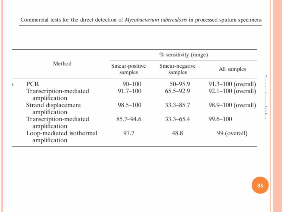
MOLECULAR DIAGNOSIS OF M. TUBERCULOSIS
Nucleic acid probes
Amplification techniques
PCR assay
Transcription mediated Assay
Strand displacement amplification
Q β replicase gene amplification
Spoligotyping
85
Sh
yam
Ku
ma
r Mish
ra

DNA MICROARRAY

NUCLEIC ACID PROBES
DNA probes M. tuberculosis complex, M. avium
Not sensitive enough (104 organisms)
Ribosomal RNA based probes Target r- RNA
M. tuberculosis , M. avium, M. gordonae
Chemiluminiscent system

PCR ASSAYS
Target
IS 6110 (10-20 times repeated in genome) 25% of Indian population lack IS 6110
PCR ( ROCHE AMPLICOR)
Target gene 16S-rRNA (584 bp)
Reproducible, sensitive, specific
Can detect 1-10 organisms

DRAWBACKS OF PCR ASSAY
Dead bacilli are detected.
False Positive due to contamination.
No prognostic value.

DEMONSTRATION OF BIOLOGICAL PRODUCTS
Adenosine Deaminase Assay
Lymphocytic enzyme - Increase in TB
Surrogate marker for extrapulmonary
tuberculosis in Pleural, pericardial, CSF
Sensitivity & specificity > 90%
Limitations - False +
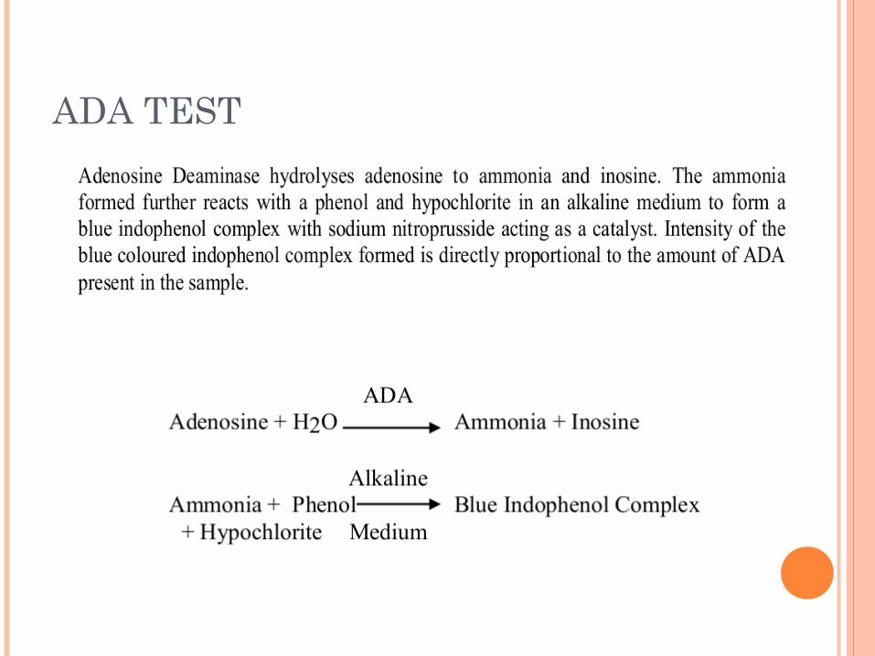
ADA TEST

FAST PLAQUE ASSAY

93
Sh
yam
Ku
ma
r Mish
ra