Embed Size (px)
Citation preview
INFLAMMATION - PART 1
Student: Dr. Mohamid Afroz KhanDepartment of Pathology
Inflammation is a protective response involving host cells, blood vessels, and proteins and other mediators that is intended to eliminate the initial cause of cell injury, as well as the necrotic cells and tissues resulting from the original insult, and to initiate the process of repair.
• Although inflammation helps clear infections and other noxious stimuli and initiates repair, the inflammatory reaction and the subsequent repair process can themselves cause considerable harm.
• The cells and molecules of host defense, including leukocytes and plasma proteins, normally circulate in the blood, and the goal of the inflammatory reaction is to bring them to the site of infection or tissue damage.
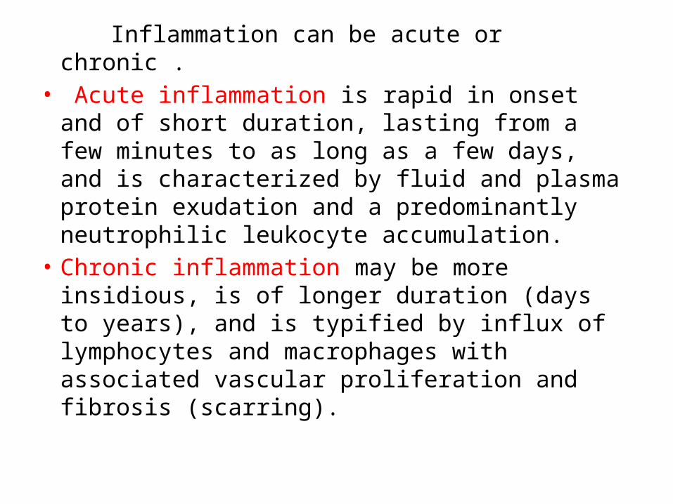
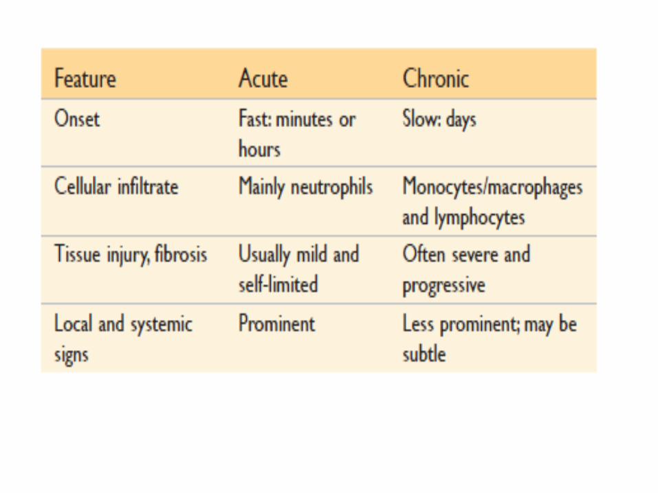
Inflammation can be acute or chronic .• Acute inflammation is rapid in onset and of
short duration, lasting from a few minutes to as long as a few days, and is characterized by fluid and plasma protein exudation and a predominantly neutrophilic leukocyte accumulation.
• Chronic inflammation may be more insidious, is of longer duration (days to years), and is typified by influx of lymphocytes and macrophages with associated vascular proliferation and fibrosis (scarring).
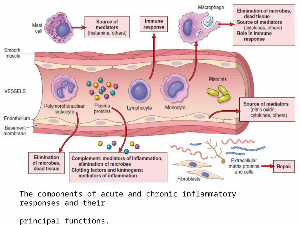
The components of acute and chronic inflammatory responses and their principal functions.
• Inflammation is induced by chemical mediators that are produced by host cells in response to injurious stimuli.
• When a microbe enters a tissue or the tissue is injured, the presence of the infection or damage is sensed by resident cells, mainly macrophages, but also dendritic cells, mast cells, and other cell types. These cells secrete molecules (cytokines and other mediators) that induce and regulate the subsequent inflammatory response.

• The Roman writer Celsus in 1st century A.D. named the famous 4 cardinal signs of inflammation as:
• Rubor(redness)• Tumor(swelling)• Calor(heat)• Dolor(pain)• To these, fifth sign functio laesa (loss of
function) was later added by Virchow.
Inflammation is normally controlled and self-limited.
The mediators and cells are activated only in response to the injurious stimulus and are short-lived, and they are degraded or become inactive as the injurious agent is eliminated.
ACUTE INFLAMMATION• The acute inflammatory response rapidly
delivers leukocytes and plasma proteins to sites of injury.
• Acute inflammation has two major components-
• Vascular events • Cellular events
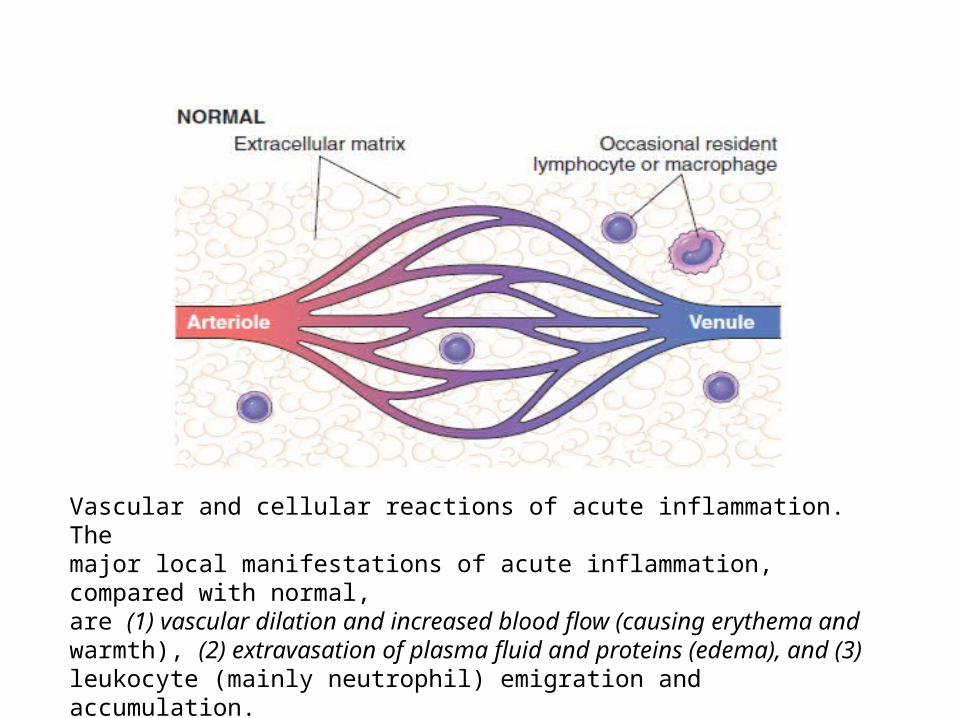
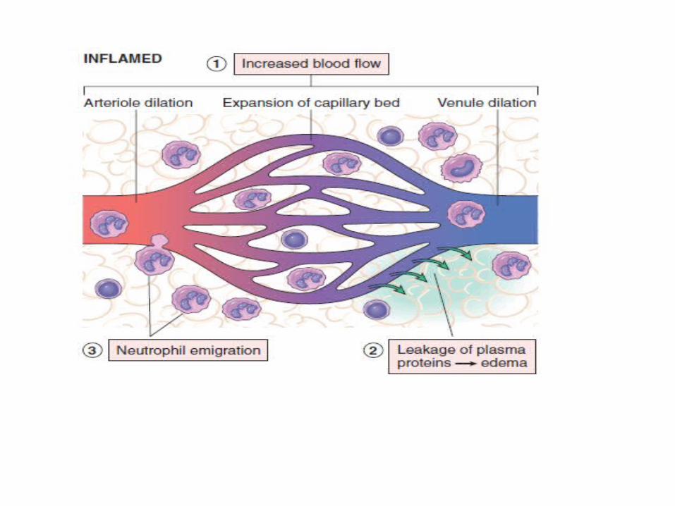
Vascular and cellular reactions of acute inflammation. Themajor local manifestations of acute inflammation, compared with normal,are (1) vascular dilation and increased blood flow (causing erythema andwarmth), (2) extravasation of plasma fluid and proteins (edema), and (3) leukocyte (mainly neutrophil) emigration and accumulation.
Acute inflammatory reactions may be triggered by a variety of stimuli:
• Infective agents like bacteria, viruses and their toxins, fungi, parasites.
• Immunological agents like cell mediated and antigen-antibody reactions.
• Chemical agents like organic and inorganic poisons.
• Inert materials such as foreign bodies.• Tissue necrosis (from any cause), including
ischemia (as in a myocardial infarct) and physical and chemical injury
VASCULAR EVENTS Alteration in the microvasculature (arterioles,
capillaries and venules) is the earliest response to tissue injury. These include-
• Hemodynamic changes• Changes in vascular permeability.
Hemodyanamic changes:• The earliest features of inflammatory response
result from changes in the vascular flow and calibre of small blood vessels in the injured tissue.
• 1)Transient vasoconstriction: immediate vascular response of arterioles.
• 2)Persistent progressive vasodilatation: mainly
arterioles, but to a lesser extent involves venules and capillaries. This vascular expansion is the cause of the redness (erythema) and warmth characteristic of acute inflammation.
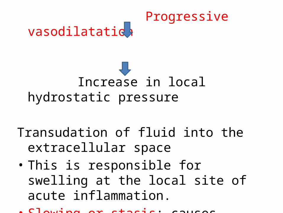
Progressive vasodilatation
Increase in local hydrostatic pressure
Transudation of fluid into the extracellular space• This is responsible for swelling at the local site
of acute inflammation.• Slowing or stasis: causes increased
concentration of red cells in microcirculation and thus, raised blood viscosity.
• Leucocytic margination: or peripheral orientation of leucocytes (mainly neutrophils) along the vascular endothelium.
• The leucocytes stick to the vascular endothelium, and then move and migrate through the gaps between the endothelial cells into the extravascular space.
• This process is known as emigration.
• The features of haemodyamic changes in inflammation are best demonstrated by the Lewis experiment. Lewis induced the changes in the skin of inner aspect of forearm by firm stroking with a blunt point.
• The reaction so elicited is known as triple response or red line response consisting of following:
• Red line appears within a few seconds following stroking and is due to local vasodilatation of capillaries and venules.
• Flare is the bright reddish appearance or flush surrounding the red line and results from vasodilatation of the adjacent arterioles.
• Wheal is the swelling or edema of the surrounding skin occurring due to transudation of fluid into the extravascular space.
Altered vascular permeability: • Pathogenesis: in and around the inflammed
tissue, there is accumulation of oedema fluid in the interstitial compartment which comes from blood plasma by its escape through the endothelial wall of the peripheral vascular bed.
• In the initial stage, the escape of fluid is due to vasodilatation and consequent elevation in hydrostatic pressure. This is transudate in nature.But subsequently, the characteristic inflammatory edema,exudate, appears by increased vascular permeability of microcirculation.
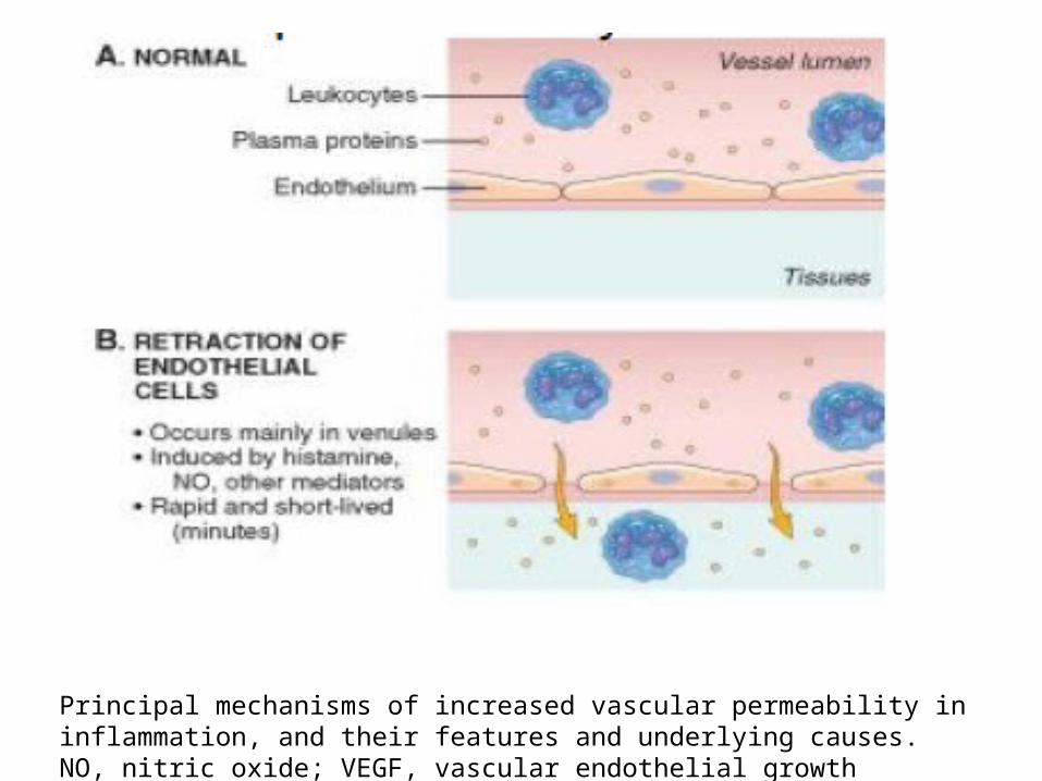
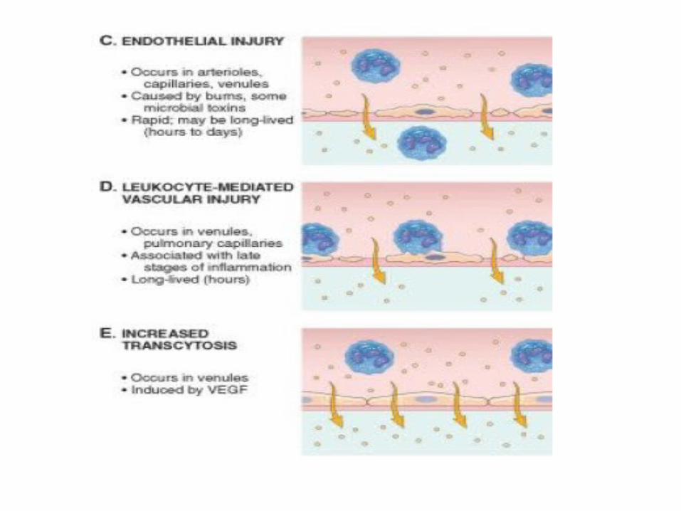
Principal mechanisms of increased vascular permeability in inflammation, and their features and underlying causes. NO, nitric oxide; VEGF, vascular endothelial growth factor.
• The appearance of inflammatory edema due to increased vascular permeability of microvascular bed is explained on the basis of Starling’s hypothesis.
• In normal circumstances the fluid balance is maintained by two opposing sets of forces.
• Forces that cause outward movement of fluid from microcirculation are intravascular hydrostatic pressure and colloid osmotic pressure of interstitial fluid.
• Forces that cause inward movement of interstitial fluid into circulation are intravascular colloid osmotic pressure and hydrostatic pressure of interstitial fluid.
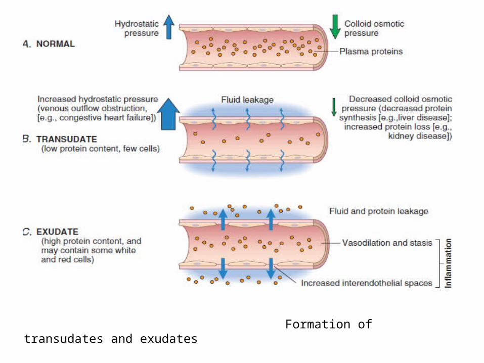
Formation of transudates and exudates
Mechanisms of Increased Vascular Permeability
1. Contraction of endothelial cells: Most common mechanism of increased leakiness that affects venules exclusively while capillaries and arterioles remain unaffected.
• Mediated by release of histamin, bradykanin and chemical mediators.
• Begins immediately after injury, reversible, short duration.
• Eg. Of such immediate transient leakage is mild thermal injury of skin of forearm.
2. Retraction of endothelial cells: • Structural reorganization of the cytoskeleton of
endothelial cells that causes reversible retraction at the inter cellular junctions
• Mediated by cytokines such as IL-1 and TNF-α.• Takes 4-6 hours after injury and lasts for 2-4 hrs
or more (somewhat delayed and prolonged leakage)
• Eg. Invitro experimental work.
3. Direct injury to endothelial cells: • Direct injury to the endothelium causes cell
necrosis and appearance of physical gaps at the site of detached endothelial cells.
• Affects all levels of microvasculatures• May appear immediately after injury and last for
several hours or days (immediate sustained leakage), or may occur after a delay of 2-12hrs and last for hrs or days (delayed prolonged leakage)
• Eg. Severe bacterial infections, following moderate thermal and radiation injury.
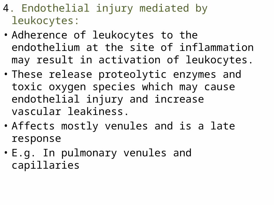
4. Endothelial injury mediated by leukocytes:• Adherence of leukocytes to the endothelium at
the site of inflammation may result in activation of leukocytes.
• These release proteolytic enzymes and toxic oxygen species which may cause endothelial injury and increase vascular leakiness.
• Affects mostly venules and is a late response• E.g. In pulmonary venules and capillaries
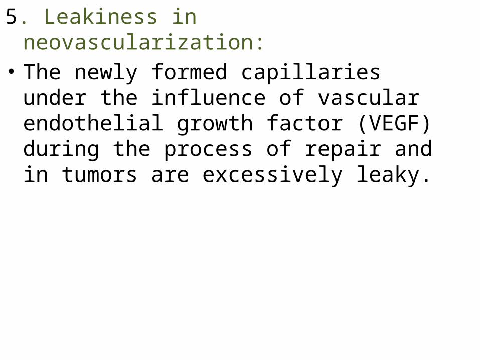
5. Leakiness in neovascularization:• The newly formed capillaries under the influence
of vascular endothelial growth factor (VEGF) during the process of repair and in tumors are excessively leaky.
CELLULAR EVENTS
• Consists of two processess:1. Exudation of leucocytes2. Phagocytosis
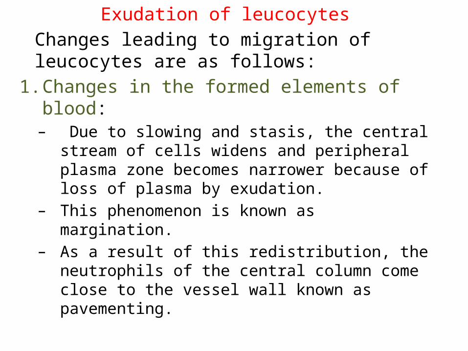
Exudation of leucocytesChanges leading to migration of leucocytes are as follows:
1. Changes in the formed elements of blood:– Due to slowing and stasis, the central stream of
cells widens and peripheral plasma zone becomes narrower because of loss of plasma by exudation.
– This phenomenon is known as margination.– As a result of this redistribution, the neutrophils
of the central column come close to the vessel wall known as pavementing.
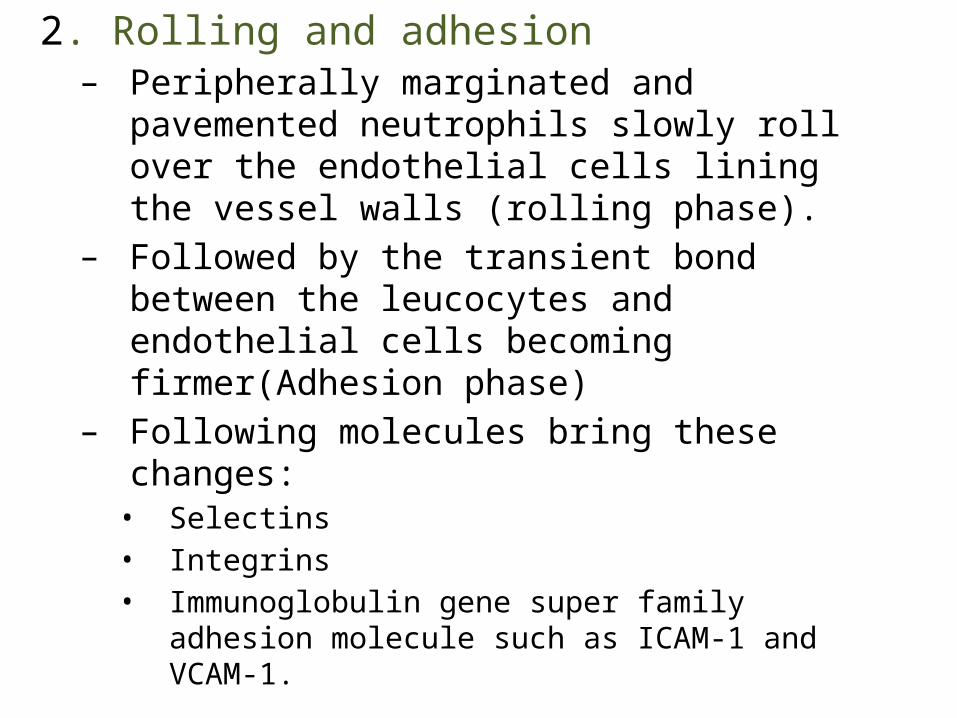
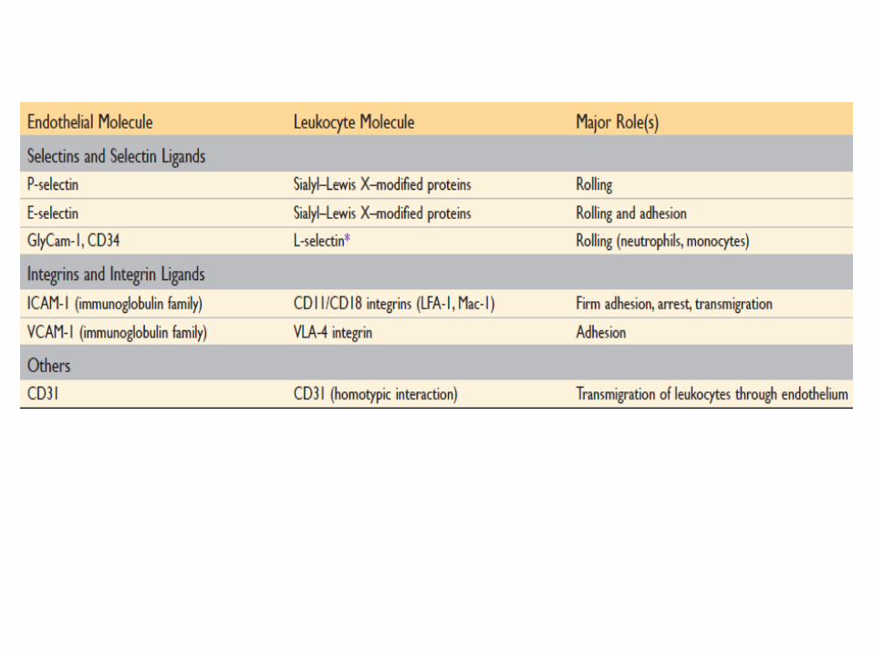
2. Rolling and adhesion– Peripherally marginated and pavemented
neutrophils slowly roll over the endothelial cells lining the vessel walls (rolling phase).
– Followed by the transient bond between the leucocytes and endothelial cells becoming firmer(Adhesion phase)
– Following molecules bring these changes:• Selectins• Integrins• Immunoglobulin gene super family adhesion molecule
such as ICAM-1 and VCAM-1.
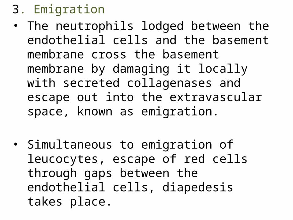
3. Emigration• The neutrophils lodged between the
endothelial cells and the basement membrane cross the basement membrane by damaging it locally with secreted collagenases and escape out into the extravascular space, known as emigration.
• Simultaneous to emigration of leucocytes, escape of red cells through gaps between the endothelial cells, diapedesis takes place.
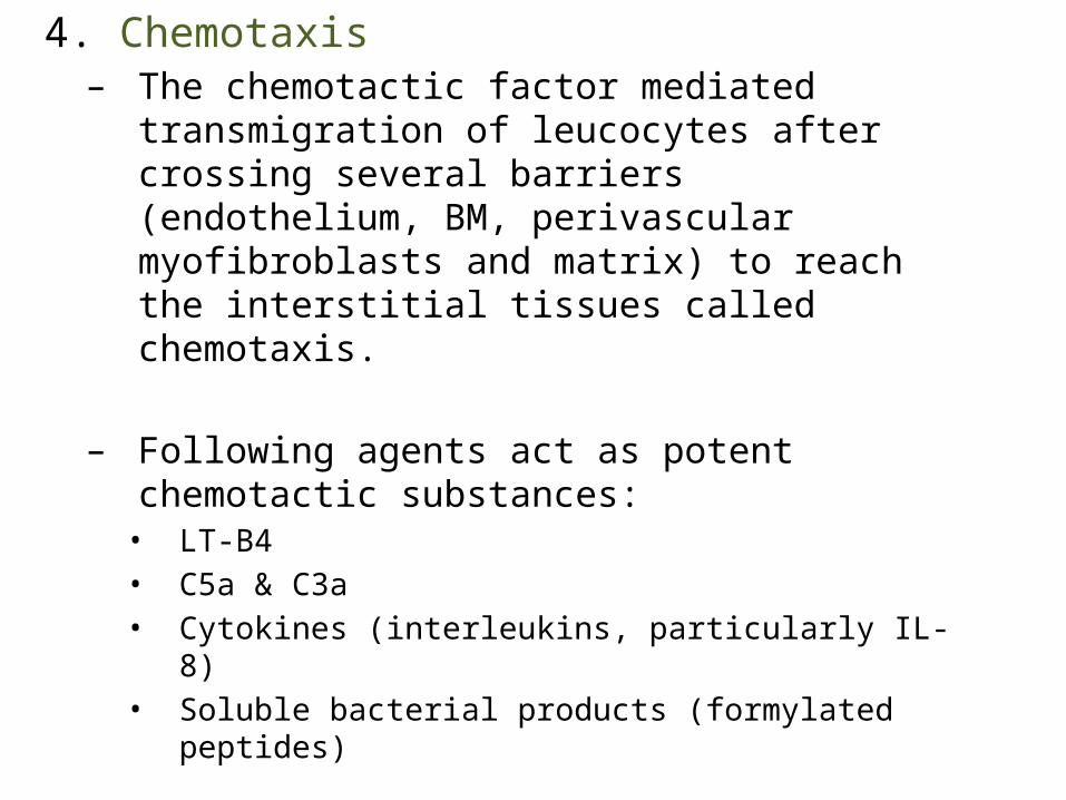
4. Chemotaxis– The chemotactic factor mediated transmigration
of leucocytes after crossing several barriers (endothelium, BM, perivascular myofibroblasts and matrix) to reach the interstitial tissues called chemotaxis.
– Following agents act as potent chemotactic substances:• LT-B4• C5a & C3a• Cytokines (interleukins, particularly IL-8)• Soluble bacterial products (formylated peptides)
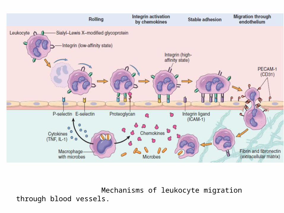
Mechanisms of leukocyte migration through blood vessels.
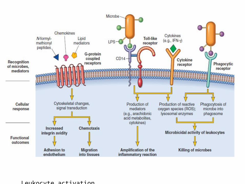
Leukocyte activation
PhagocytosisDefined as the process of engulfment of solid particulate material by the cells (Cell-eating). The cells performing this function are called phagocytes.
• Two main types of phagocytic cells-– PMNs called microphages– Circulating monocytes and fixed tissue
mononuclear phagocytes called macrophages• Release several proteolytic enzymes-
lysozyme, protease, collagenase, elastase, lipase, proteinase, gelatinase and acid hydrolases.
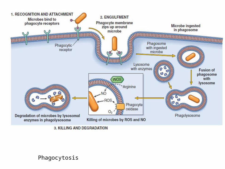
Phagocytosis
The microbe undergoes the process of phagocytosis by polymorphs and macrophages involves 3 steps:
1. RECOGNITION AND ATTACHMENT
• Initiated by the expression of the surface receptors on macrophages which recognize microorganisms: mannose receptors and scavenger receptors.
• IgG opsonins• C3b opsonins• Lectin
2. ENGULFMENT • The opsonised particle bound to the surface
of phagocyte is ready to be engulfed• Formation of cytoplasmic pseudopods
around the particle due to activation of acting filaments beneath cell wall and enveloping in it a phagocytic vacuole.
• The phagosome fuses with one or more lysosomes of the cell and form bigger vacuole called phagolysosome.
3. KILLING AND DEGRADATION• The microorganisms after being killed by anti
bacterial substances are degraded by hydrolytic enzymes
• Disposal of microorganism can proceed by following mechanisms:– Intracellular– Extracellular
Intracellular Mechanisms:1. Oxidative bactericidal mechanism by oxygen
free radicals: – By production of reactive oxygen metabolites (O’2 ,
H2O2, OH’, HOCl, HOI, HOBr)– A phase of increased oxygen
consumption(respiratory burst) by activated phagocytic leucocytes requires the essential presence of NADPH oxidase
– Carried out either via enzyme MPO present in azurophillic granules or neutrophils and monocytes or independent of enzyme MPO.
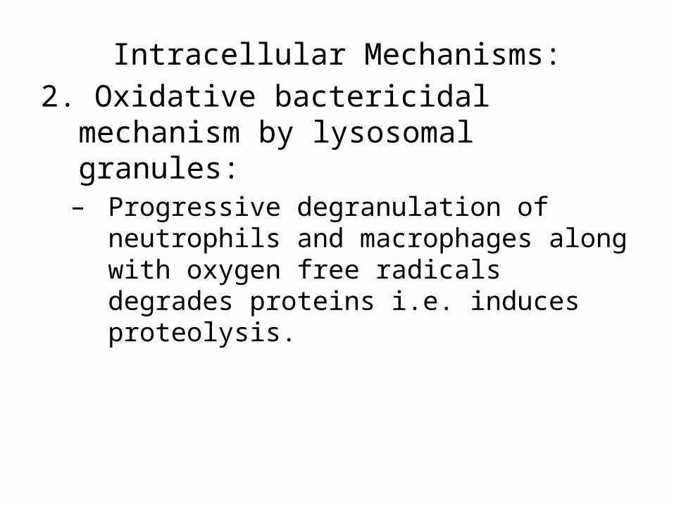
Intracellular Mechanisms:2. Oxidative bactericidal mechanism by
lysosomal granules:– Progressive degranulation of neutrophils and
macrophages along with oxygen free radicals degrades proteins i.e. induces proteolysis.
Intracellular Mechanisms:3. Non oxidative bactericidal mechanism:– Granules– Nitric Oxide
Extracellular Mechanisms:1. Granules2. Immune Mechanisms
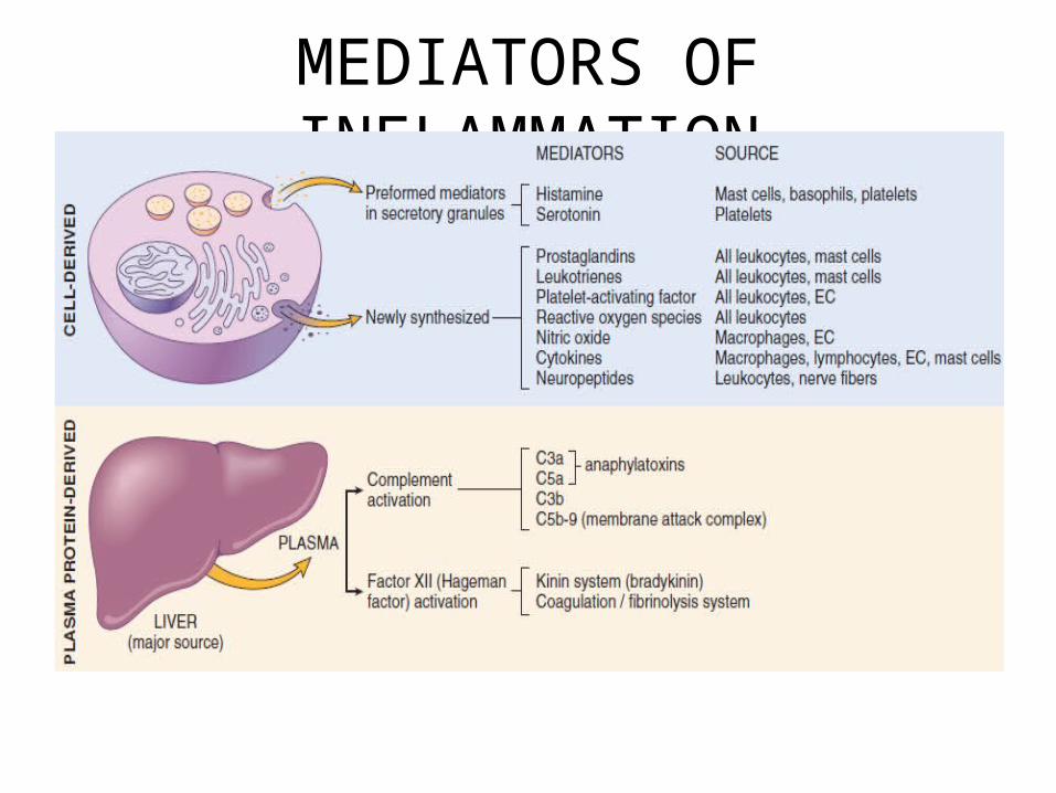
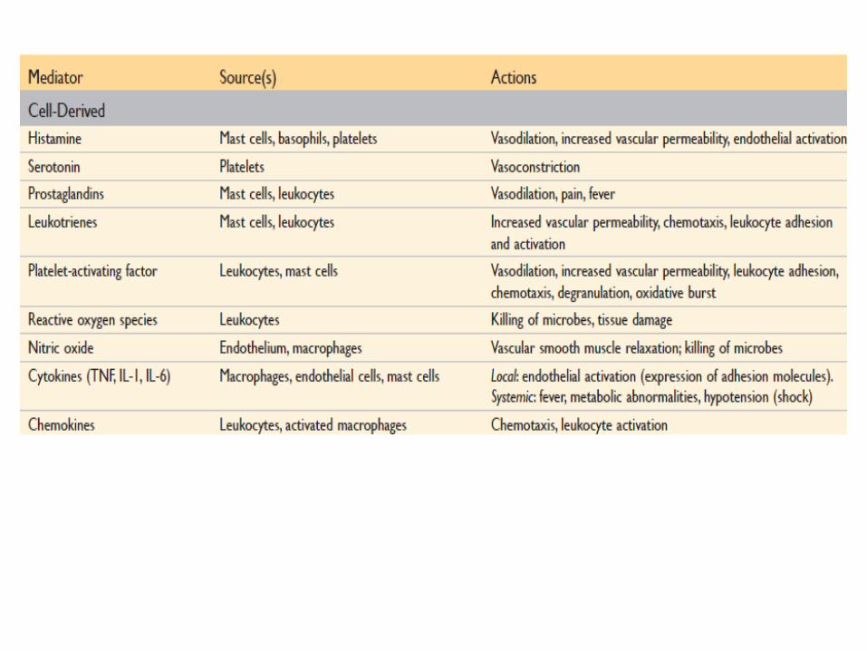
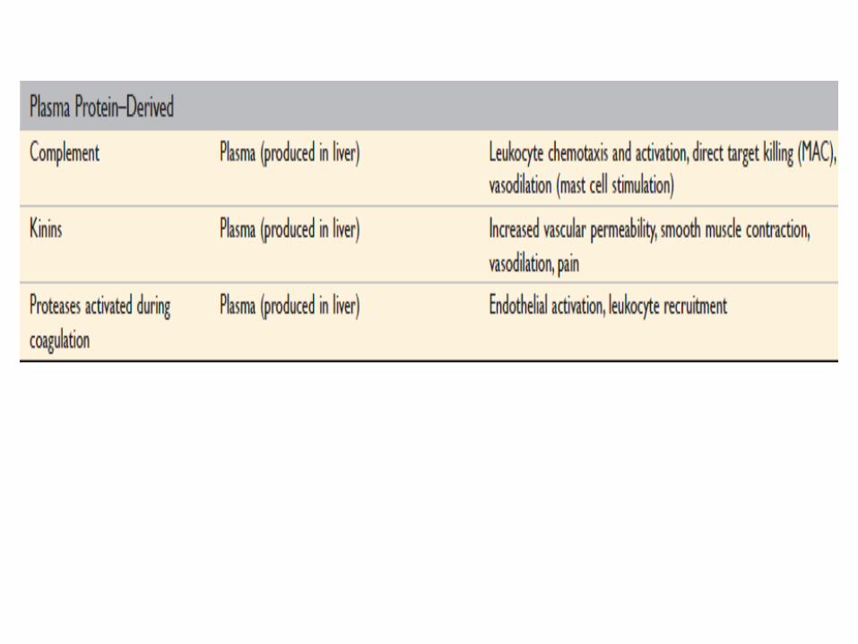
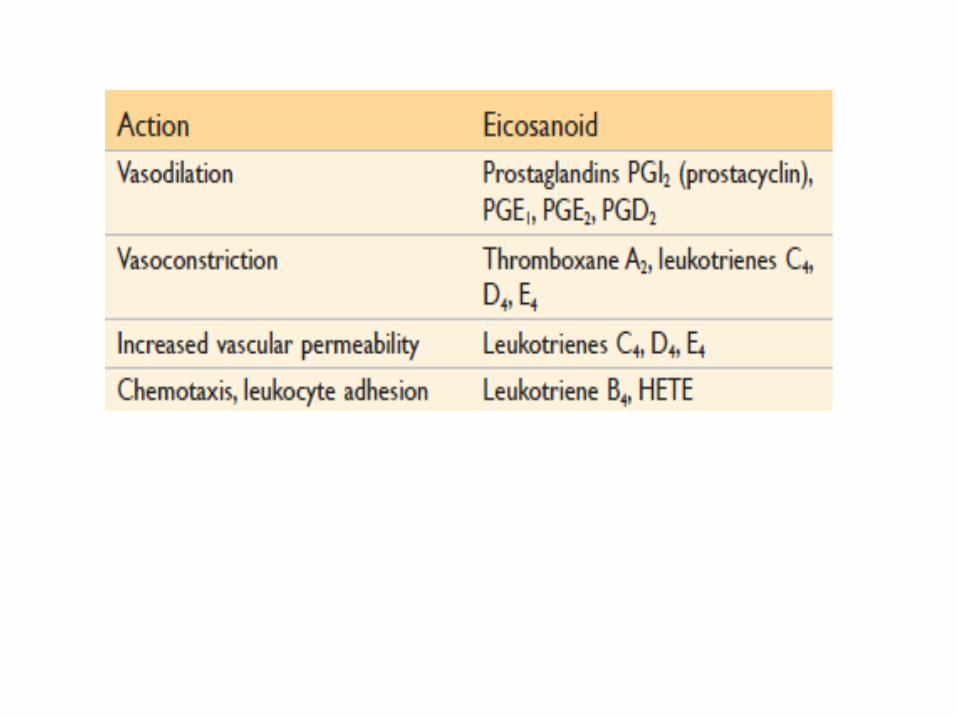
MEDIATORS OF INFLAMMATION
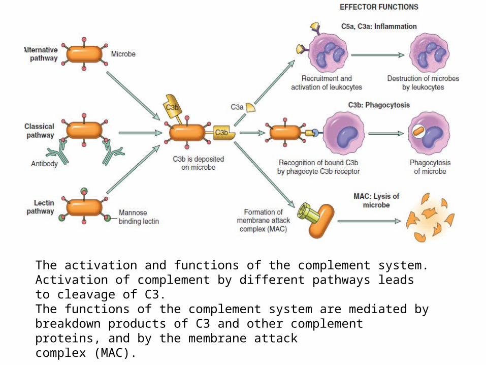
The activation and functions of the complement system. Activation of complement by different pathways leads to cleavage of C3.The functions of the complement system are mediated by breakdown products of C3 and other complement proteins, and by the membrane attackcomplex (MAC).
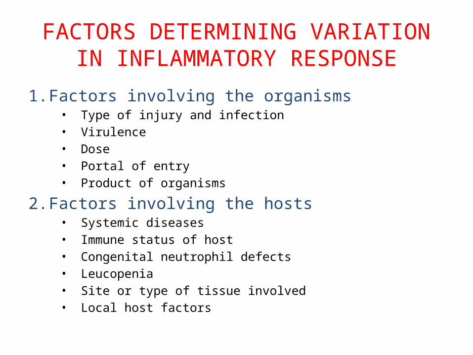
FACTORS DETERMINING VARIATION IN INFLAMMATORY RESPONSE
1. Factors involving the organisms• Type of injury and infection• Virulence• Dose• Portal of entry• Product of organisms
2. Factors involving the hosts• Systemic diseases• Immune status of host• Congenital neutrophil defects• Leucopenia• Site or type of tissue involved• Local host factors
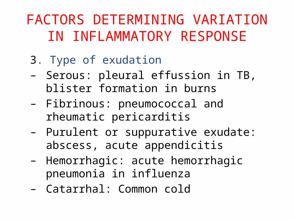
FACTORS DETERMINING VARIATION IN INFLAMMATORY RESPONSE
3. Type of exudation– Serous: pleural effussion in TB, blister formation
in burns– Fibrinous: pneumococcal and rheumatic
pericarditis– Purulent or suppurative exudate: abscess, acute
appendicitis– Hemorrhagic: acute hemorrhagic pneumonia in
influenza– Catarrhal: Common cold
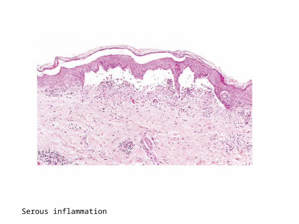
Serous inflammation
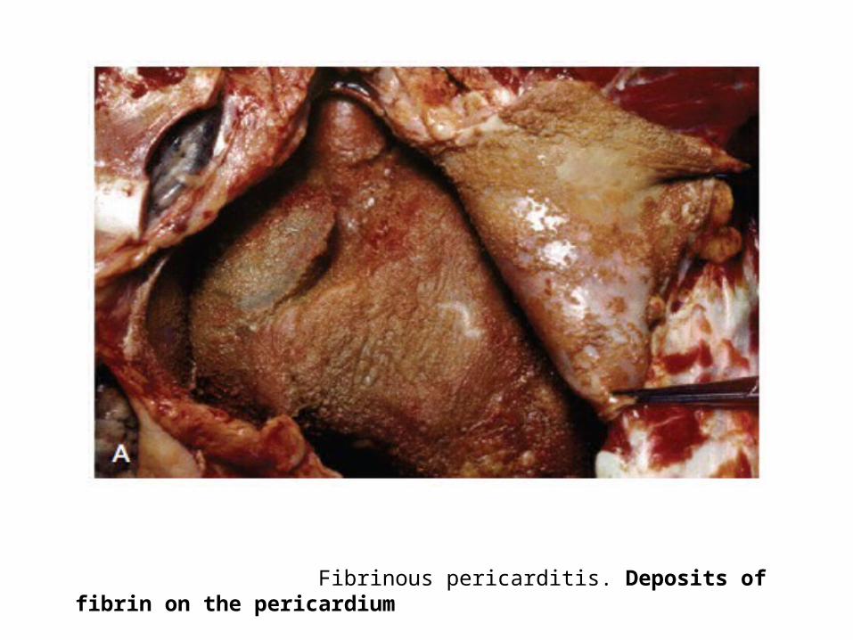
Fibrinous pericarditis. Deposits of fibrin on the pericardium
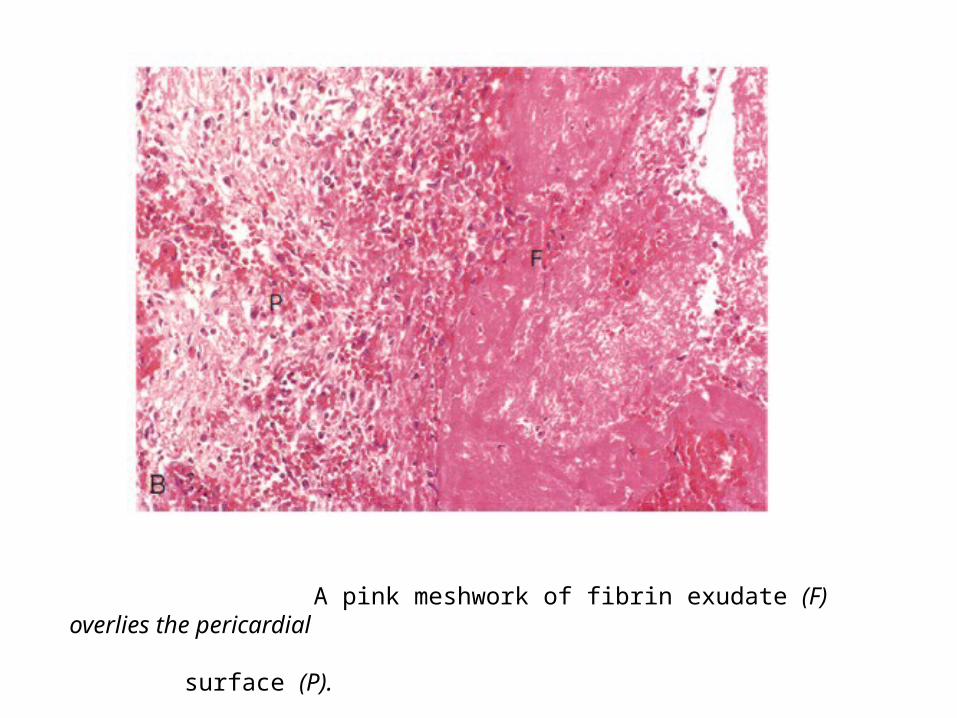
A pink meshwork of fibrin exudate (F) overlies the pericardial surface (P).
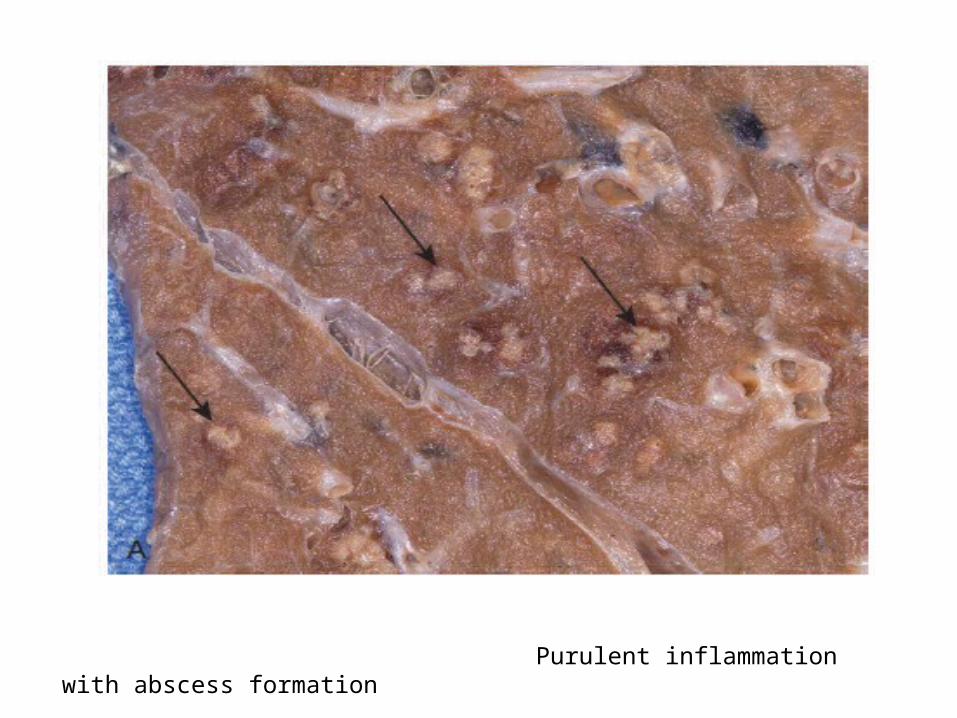
Purulent inflammation with abscess formation
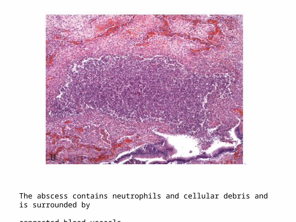
The abscess contains neutrophils and cellular debris and is surrounded by congested blood vessels
MORPHOLOGY OF ACUTE INFLAMMATION
1. Pseudomembranous inflammation: It is inflammatory response of mucous surface(oral, respiratory, bowl) to toxins of diphtheria or irritant gases
2. Ulcer: Are local defects on the surface of an organ produced by inflammation- common sites are stomach, duodenum, intestinal ulcers in typhoid fever, intestinal TB, bacillary and amoebic dysentery, ulcers of legs due to varicose veins.
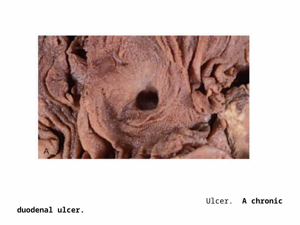
Ulcer. A chronic duodenal ulcer.
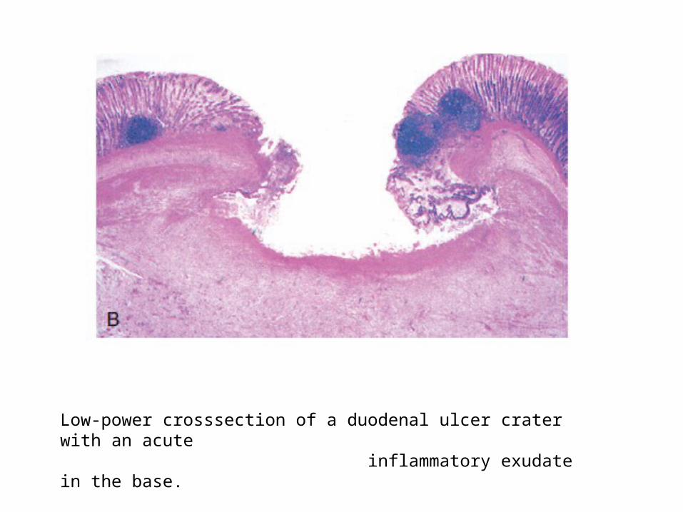
Low-power crosssection of a duodenal ulcer crater with an acute inflammatory exudate in the base.
3. Suppuration (abscess formation):• When acute bacterial infection is accompanied by
intense neutrophilic infiltrate in the inflamed tissue, it results in tissue necrosis.
• A cavity is formed called an abscess and contains purulent exudate or pus and the process of abscess formation is known as suppuration.
• E.g. boil or furruncle • Carbuncle
4. Cellulitis:Diffuse inflammation of soft tissues resulting from spreading effects of substances like hyluronidase released by some bacteria
5. Bacterial Infection of the blood:– Bacteraemia: Presence of small number of
bacteria in the blood which do not multiply significantly. E.g. infection with S typhi, E coli, S viridans
– Septicaemia: Presence of rapidly multiplying, highly pathogenic bacteria in the blood. E.g. pyogenic cocci, bacilli of plague
– Pyaemia: Is the dissemination of small septic thrombi in the blood which cause their effects at the site where they are lodged. This can result in pyaemic abscesses or septic infarcts
SYSTEMIC EFFECT OF ACUTE INFLAMMATION
1. Fever2. Leucocytosis3. Lymphangitis-lymphadenitis4. Shock
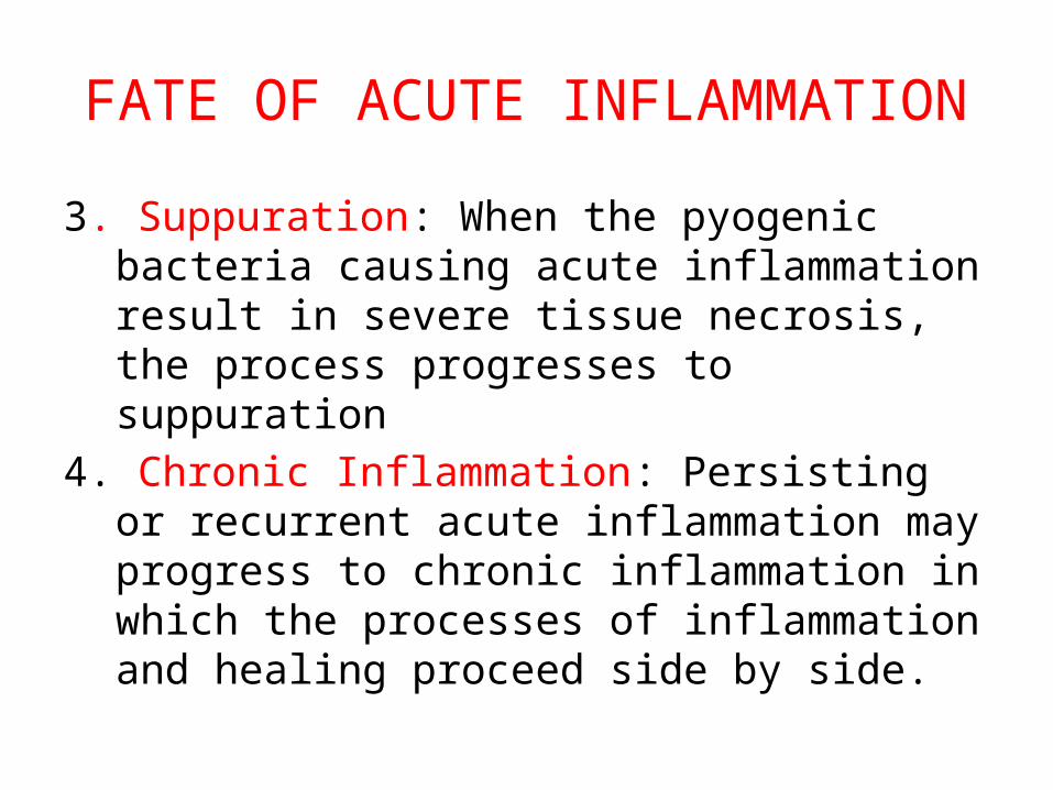
FATE OF ACUTE INFLAMMATION
• Resolution• Healing• Suppuration• Chronic Inflammation
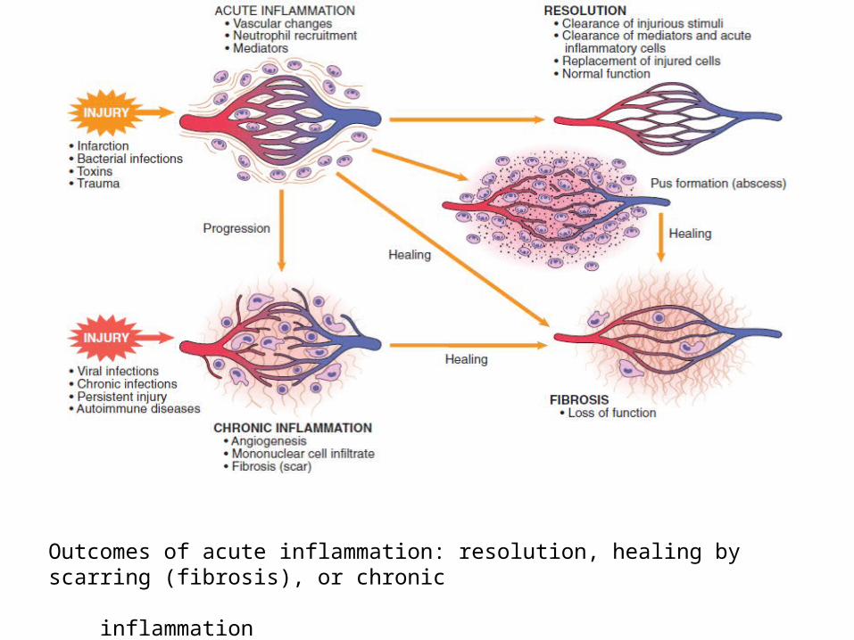
Outcomes of acute inflammation: resolution, healing by scarring (fibrosis), or chronic inflammation
FATE OF ACUTE INFLAMMATION
1. Resolution: means complete return to normal tissue following acute inflammation. E.g. resolution in lobar pneumonia
2. Healing: Healing by fibrosis takes place when the tissue destruction in acute inflammation is extensive so that there is no tissue regeneration
FATE OF ACUTE INFLAMMATION
3. Suppuration: When the pyogenic bacteria causing acute inflammation result in severe tissue necrosis, the process progresses to suppuration
4. Chronic Inflammation: Persisting or recurrent acute inflammation may progress to chronic inflammation in which the processes of inflammation and healing proceed side by side.

THANK YOU


![Untitled-1 [] · MAK PARK SQUARE is a luxurious apartment complex and the design revolves around providing cross ventilation to ... MAK AJMERA STONE PARK MAK LIFESTYLE MAK MARK STATUS](https://img.pdfslide.us/doc/110x75/5f22e60bcd225029067a7748/untitled-1-mak-park-square-is-a-luxurious-apartment-complex-and-the-design-revolves.jpg)