Embed Size (px)
Citation preview

Hypoxic Ischemic Encephalopathy with a focus on recent advances
Moderator : Dr.B.P.Kalra
Presenter: Dr.Varun Mamgain

Scope Of The Problem• occurs in up to 6 /1000 live term births
• a major cause of neurodevelopmental disability, with one-quarter of survivors sustaining permanent neurological deficits
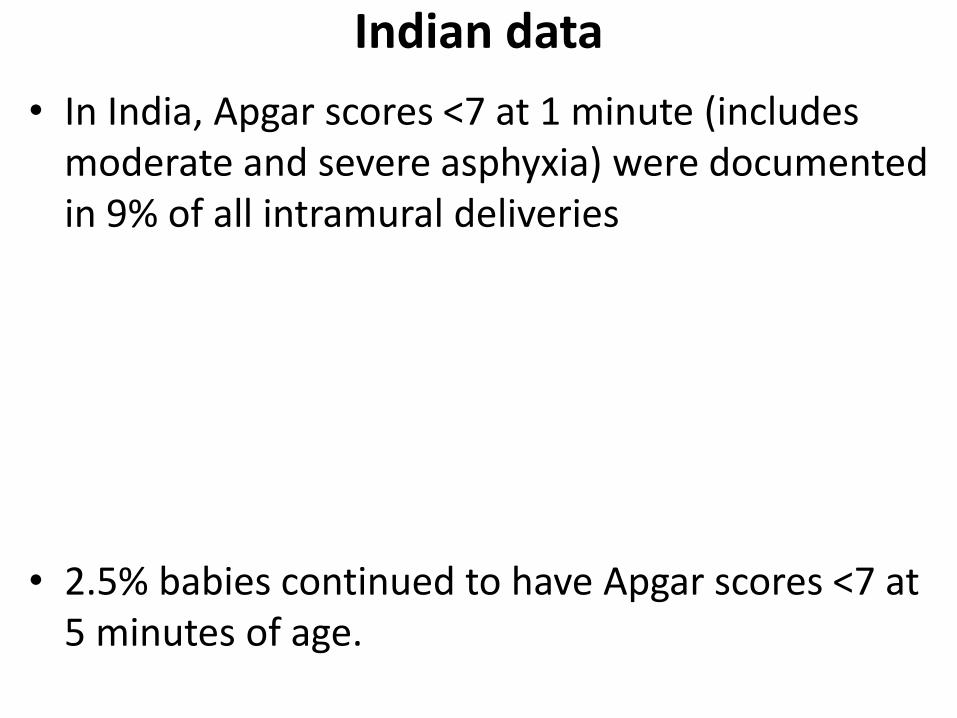
Indian data
• In India, Apgar scores <7 at 1 minute (includes moderate and severe asphyxia) were documented in 9% of all intramural deliveries
• 2.5% babies continued to have Apgar scores <7 at 5 minutes of age.

• Manifestations of HIE seen in approx 1.5%

DefinitionsHypoxia or Anoxia: A partial (hypoxia) or complete
(anoxia) lack of oxygen
Hypoxemia - A partial (hypoxia) or complete (anoxia)
lack of oxygen in the brain or blood
Asphyxia: The state in which placental or pulmonary
gas exchange is compromised or ceases altogether
Ischemia: The reduction or cessation of blood flow to
an organ which compromises both oxygen and
substrate delivery to the tissue

• Perinatal Asphyxia refers to a condition during the first and second stage of labor in which impaired gas exchange leads to fetal hypoxemia and hypercarbia.
• identified by fetal acidosis as measured in umbilical arterial blood.
• the most widely accepted definition of fetalacidosis is a pH<7.0, even with this degree of acidosis, the likelihood of brain injury is low.

• Perinatal Asphxia is a combination of Hypoxia, Hypercarbia, Metabolic acidosis
• According to American Academy of Pediatrics, perinatal asphyxia is described as
• Cord umbilical PH < 7 with base deficit of <10 mEq/l
• Neonatal neurologic manifestation suggestive of HIE
• Evidences of multi organ failure (CVS ,Renal etc)

WHO Definition
• a “failure to initiate and sustain breathing at birth”
• WHO/NNF – Apgar 0-3 at 1 minute – severe asphyxia,4-7 – moderate asphyxia
• In Community Settings NNF defines – asphyxia as absence of cry at 1 minute, severe asphyxia as absent or inadequate breathing at five minutes

The National Neonatal Perinatal Database (NNPD), 2000
• defined moderate asphyxia as slow gasping breathing or an Apgar score of 4-6 at 1 minute of age
• Severe asphyxia was defined as no breathing or an Apgar score of 0-3 at 1 minute of age.

• Perinatal/neonatal depression is a clinical, descriptive term that pertains to the condition of the infant on physical examination in the immediate postnatal period (i.e., in the first hour after birth).
• clinical features
– depressed mental status,
– muscle hypotonia,
– disturbances in spontaneous respiration and cardiovascular function.
– Term makes no association with the prenatal or later postnatal condition, physical exam, laboratory tests, imaging studies, or electroencephalograms (EEGs).

After the first hour• Neonatal encephalopathy is a clinical and not an
etiologic term that describes an abnormal neurobehavioral state consisting of decreased level of consciousness and usually other signs of brain stem and/or motor dysfunction.
– does not imply a specific etiology, nor does it imply irreversible neurologic injury as it may be caused by such reversible conditions as maternal medications or hypoglycemia.

• Hypoxic-ischemic encephalopathy (HIE) describes encephalopathy as defined before, with objective data to support a hypoxic-ischemic mechanism as the underlying cause for the encephalopathy.
• Hypoxic-ischemic (HI) brain injury refers to neuropathology attributable to hypoxia and/or ischemia as evidenced by – biochemical (such as serum creatine k inase brain bound [CK-
BB]),
– electrophysiologic (EEG),
– neuroimaging (head ultrasonography [HUS], magnetic resonance imaging [MRI], computed tomog-raphy [CT]),
– pathologic (postmortem) abnormalities.

ETIOLOGY• In term newborns, asphyxia can occur in the
antepartum or intrapartum period
• result of impaired gas exchange across the placenta– inadequate provision of oxygen (O2)
– removal of carbon dioxide (CO2) and hydrogen (H2) from the fetus.
• In the postpartum period, usually secondary to pulmonary, cardiovascular, or neurologic abnormalities

Factors that increase the risk of perinatal asphyxia
• Impairment of maternal oxygenation
• Decreased blood flow from mother to placenta
• Decreased blood flow from placenta to fetus
• Impaired gas exchange across the placenta or at the fetal tissue level
• Increased fetal O2 requirement

Etiologies• may be multiple and include the following:
• Preconceptual– Advanced maternal age
– IDDM
– Thyroid disease
– Fertility Treatments
• Maternal factors:– hypertension (acute or chronic),
– hypotension,
– infection (including chorioamnionitis),
– hypoxia from pulmonary or cardiac disorders, d iabetes, maternal vascular disease, and in utero exposure to cocaine

• Placental factors: – abnormal placentation,
– abruption,
– infarction,
– fibrosis
• Uterine rupture
• Umbilical cord accidents: – prolapse,
– entanglement,
– true knot,
– compression
• Abnormalities of umbilical vessels
• Instrumentation
• Stat C-Section

• Fetal factors:
– anemia,
– infection,
– cardiomyopathy,
– hydrops,
– severe cardiac/ circulatory insufficiency
• Neonatal factors:
– cyanotic congenital heart disease,
– persistent pulmonary hyper-tension of the newborn(PPHN),
– cardiomyopathy,
– neonatal cardio-genic and/or septic shock

Pathophysiology
• Hypoxia-ischemia causes a number of physiologic and biochemical alterations
• The adverse consequences of cerebral ischemia include deprivation of energy substrates and oxygen, and an inability to clear accumulated, potentially toxic metabolites.

• Cerebral Blood Flow and Energy Metabolism
• Excitotoxicity
• Oxidative Stress
• Inflammation
• Apoptosis

Cerebral Blood Flow and Energy Metabolism
• Disruption of cerebrovascular autoregulation - important factor in the pathophysiology of neonatal hypoxic-ischemic brain injury.
• widely accepted that preterm infants have a “pressure-passive” cerebral circulation; however,
• term infants may remain at risk for impairment of cerebrovascular autoregulation and susceptibility to cerebral ischemia with fluctuations in systemic blood pressure.


• With brief asphyxia, there is
– a transient increase, followed by a decrease in heart rate (HR)
– mild elevation in blood pressure (BP)
– an increase in central venous pressure (CVP)
– and essentially no change in cardiac output (CO)
• Accompanied by a redistribution of CO with an increased proportion going to the brain, heart, and adrenal glands (diving reflex).

• other basic physiologic mechanisms may contribute to impaired autoregulation-
• Increased expression of inducible and neuronal isoforms of nitric oxide synthase (iNOS and nNOS), as well as endothelial NOS, may narrow the autoregulatory window,
• downregulation of prostaglandin receptors in response to high circulating prostaglandin levels may blunt the prostaglandin-mediated vasoconstrictive response to hypertension and thereby contribute to inappropriately increased cerebral blood flow

• With prolonged asphyxia, there can be a loss of pressure autoregulation and/or CO2 vasoreactivity.
• This, in turn, may lead to further disturbances in cerebral perfusion, particularly when there is cardiovascular involvement with hypotension and/or decreased cardiac output

• An inadequate supply of glucose or alternate substrates plays a pivotal role in hypoxic-ischemic neuronal cell death.
• Although overall metabolic demands are lower in the neonatal than in the adult brain, during periods of rapid brain growth, particularly the perinatal period, metabolic needs rise.

• Brain development is associated with a transition from the ability to use glucose and ketones as energy substrates in the neonate to an absolute requirement for glucose in the adult.
• The immature brain can use lactate as an alternate fuel source to some degree, and the deleterious effects of lactate accumulation after hypoxia-ischemia therefore may be attenuated in the neonate compared with the adult.

• However, normal maturation is characterized by limitations in glucose transport capacity and increased use of these alternative fuels such as lactate.
• The inability to transport glucose across the blood–brain barrier threatens cerebral glucose utilization.
• These factors illustrate the importance of understanding the use of glucose, lactate, and ketones in the newborn brain under normal and pathologic conditions
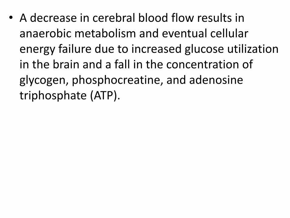
• A decrease in cerebral blood flow results in anaerobic metabolism and eventual cellular energy failure due to increased glucose utilization in the brain and a fall in the concentration of glycogen, phosphocreatine, and adenosine triphosphate (ATP).


• Cellular dysfunction occurs as a result of diminished oxidative phosphorylation and ATP production.
• This energy failure impairs ion pump function, causing accumulation of intracellular Na , Cl , H2O, and Ca2 ; extracellular K ; and excitatory neurotransmitters (e.g., glutamate)

Excitotoxicity• Glutamate can activate a variety of excitatory amino
acid receptors
• Excitatory amino acid neurotransmission plays a pivotal role in brain development and in learning and memory.
• substantial body of data has emerged over the past 30 years documenting the fact that overactivation of excitatory amino acid receptors (i.e., excitotoxicity) contributes to neurodegeneration in a broad range of acute and chronic neurologic disorders

• Two closely linked mechanisms contribute to ischemia-induced increases in synaptic glutamate:
– increased efflux from presynaptic nerve terminals
– impaired reuptake by glia and neurons

• initial increase in efflux is mediated by a calcium dependent process through activation of voltage-dependent calcium channels
• later, calcium-independent efflux is thought to be mediated primarily by functional reversal of glutamate transporters.

• Removal of glutamate from the synaptic cleft depends primarily on energy-dependent glutamate transporters, which are predominantly glial
• Any pathophysiologic process that depletes energy supply (e.g., hypoxia-ischemia, hypoglycemia, prolonged seizures) disrupt these mechanisms and result in increased synaptic glutamate accumulation

• The NMDA receptor is relatively overexpressed in the developing brain compared with the adult brain
• in postnatal day 6–14 rats (which approximates to the term human neonate), the NMDA receptor is expressed at 150–200 % of adult levels.
• In humans, receptor expression is significantly higher at term than in the adult

• The predominating combination of subunits in the perinatal period seems to favor a more prolonged and pronounced calcium influx.
• In the setting of hypoxia-ischemia, NMDA receptor overactivation leads to – massive sodium and water influx– cell swelling– elevated intracellular calcium and its associated mitochondrial
dysfunction, – increased nitric oxide production, – increased phospholipid turnover – accumulation of potentially toxic free fatty acids,– cell death by apoptotic or necrotic mechanisms.
• However, ischemia and energy failure also result in cationinflux by non-NMDA-mediated mechanisms.


Oxidative Stress• Oxidative stress describes the alterations in
cellular milieu that result from an increase in free radical production as a result of oxidative metabolism under pathologic conditions
• consequence of mitochondrial dysfunction is an accumulation of superoxide,

• Excitotoxicity causes energy depletion, mitochondrial dysfunction, and cytosolic calcium accumulation, the generation of free radicals, such as superoxide, nitric oxide derivatives, and the highly reactive hydroxyl radical.
• With reoxygenation, mitochondrial oxidative phosphorylation is overwhelmed and reactive oxygen species accumulate
• Intrinsic antioxidant defenses are depleted, and free radicals directly damage multiple cellular constituents (lipids, DNA, protein) and can activate pro-apoptotic pathways.

• Contributing factors include a high polyunsaturated fatty acid content, high level of lipid peroxidation (particularly in response to hypoxic stress), immaturity of antioxidant defenseenzymes, and high free iron concentrations, compared with the adult brain

• Nitric oxide metabolism provides critical link between excitotoxicity and oxidative injury in the hypoxic ischemic injured brain
• Hypoxic-ischemic increases in nitric oxide production have multiple potential beneficial and detrimental effects.
• Nitric oxide regulates vascular tone, influences inflammatory responses to injury, and directly modulates NMDA receptor function

• Early endothelial NO is protective by maintaining blood flow, but early neuronal NO and late inducible NO are neurotoxic by promoting cell death
Inflammation• Cytokines that have been strongly implicated as
mediators of brain inflammation in neonates include interleukin (IL)-1b, tumor necrosis factor (TNF)a, IL-6,and membrane co-factor protein-1
• After an asphyxial episode, there are many potential sources of plasma cytokines,– injured endothelium
– acutely injured organs, e.g brain by means of a disrupted blood–brain barrier
Apoptosis
• Apoptosis is critical for normal brain development, but it is also an important component of injury following neonatal hypoxia-ischemia and stroke
• Immediate neuronal death (necrosis) can occur due to intracellular osmotic overload of Na and Ca2 , from ion pump failure or excitatory neu-rotransmitters acting on inotropic receptors (such as the N-methyl-D-aspartate (NMDA) receptor.

• Delayed neuronal death (apoptosis) occurs secondary to uncontrolled activation of enzymes and second messenger systems within the cell
– Ca2+-dependent lipases, proteases, and caspases);
– perturbation of mitochondrial respiratory electron chain transport;
– generation of free radicals and leukotri-enes;
– generation of nitric oxide (NO) through NO synthase; and depletion of energy stores.

• The pattern of injury after hypoxia-ischemia can be explained in part on the basis of this metabolic demand;
• brain regions most susceptible to hypoxic-ischemic injury in the term infant
• subcortical gray matter structures such as the basal ganglia and thalamus are the same regions that are most vulnerable to mitochondrial toxins.

Neurological Patterns of HIE• Premature
– Selective subcortical neuronal necrosis
– Periventricular leukomalacia
– Focal/Multifocal ischemic necrosis
– Periventricular hemorrhage/infarction
• Term
– Selective Subcortical Neuronal necrosis
– Status Marmoratusof basal ganglia and thalamus
– Parasagittal cerebral injury
– Focal/Multifocal Ischemic cerebral necrosis

DIAGNOSIS• Assessment
• Low Apgar scores and need for resuscitation in the delivery room are common but nonspecific findings
• Many features of the Apgar score relate to cardiovascular integrity and not neurologic dysfunction resulting from asphyxia.

• the differential diagnosis for a term new-born with an Apgar score <3 for >10 minutes includes
– depression from maternal anesthesia or analgesia
– trauma
– Infection
– cardiac or pulmonary disorders
– Neuromuscular
– other central nervous system disorders or malformations
– If the Apgar score is >6 by 5 minutes, perinatal asphyxia is not likely.

Umbilical cord or first blood gas determination.
• The specific blood gas criteria that define asphyxia causing brain damage are uncertain
• the pH and base deficit on the cord or first blood gas is helpful
• In the randomized clinical trials of hypothermia for neonatal HIE, severe acidosis was defined as pH<7.0 or base deficit <16 mmol/L

Clinical Suspicion• Suspect HIE in encephalopathic newborns with a
history of fetal and neonatal distress and laboratory evidence of asphyxia.
• Diagnosis not be overlooked in scenarios such as
– meconium aspiration,
– pulmonary hypertension,
– birth trauma
– fetal–maternal hemorrhage

• Consider Asphyxia/HIE if -
– 1. Prolonged (>1 hour) antenatal acidosis
– 2. Fetal HR <60 beats/minute
– 3. Apgar score <=3 at >=10 minutes
– 4. Need for positive pressure ventilation for >1 minute or first cry delayed >5 minutes
– 5. Seizures within 12 to 24 hours of birth
– 6. Burst suppression or suppressed background pattern on EEG or amplitude-integrated EEG (aEEG)

Neurologic Signs• The clinical spectrum of HIE is described as mild,
moderate, or severe (Sarnat stages of HIE).
• EEG is useful
• Encephalopathy.
– must have depressed consciousness by definition, whether mild, moderate, or severe.
– An initial period of well-being or mild HIE may be followed by sudden deterioration, suggesting ongoingbrain cell dysfunction, injury, and death; during this period, seizure intensity might increase.


Levene Staging
Feature Mild Moderate Severe
Consciousness Irritability Lethargy Comatose
Tone Hypotonia Marked Hypotonia
Severe Hypotonia
Seizures No Yes Prolonged
Sucking/Respiration
Poor Suck Unable to Suck Unable to sustain spontaneousrespiration

TOPOGRAPHY OF BRAIN INJURY IN TERM INFANTS WITH HYPOXIC-
ISCHEMIC ENCEPHALOPATHY AND CLINICAL CORRELATES
AREA OF INJURY LOCATION OF INJURYCLINICAL
CORRELATE(S)LONG-TERM SEQUELA(E)
Selective neuronal necrosis
Entire neuroaxis, deep cortical area, brainstem and pentocubicular
Stupor or comaSeizuresHypotoniaOculomotor abnormalitiesSuck/swallow abnormalities
Cognitive delayCerebral palsyDystoniaSeizure disorderAtaxiaBulbar and pseudobulbar palsy
Parasagittal injury
Cortex and subcortical white matterParasagittal regions, especially posterior
Proximal limb weaknessUpper extremities affected more than lower extremities
Spastic quadriparesisCognitive delayVisual and auditory processing difficulty

Focal ischemic necrosis
Cortex and subcortical white matterVascular injury (usually middle cerebral artery distribution)
Unilateral findingsSeizures common and typically focal
HemiparesisSeizuresCognitive delays
Periventricular injuryInjury to motor tracts, especially lower extremity
Bilateral and symmetric weakness in lower extremitiesMore common in preterm infants
Spastic diplegia

• Mild encephalopathy can consist of an apparent hyperalert or jittery state, but the newborn does not respond appropriately to stimuli, and thus consciousness is abnormal.
• Moderate and severe encephalopathies are characterized by more impaired responses to stimuli such as light, touch, or even noxious stimuli.
• The background pattern detected by EEG or aEEGis useful for determining the severity of encephalopathy.

• Brain stem and cranial nerve abnormalities manifest as abnormal or absent brain stem reflexes Pupillary/corneal/oculocephalic/cough/gag
• abnormal eye movements dysconjugate gaze/ gaze preference/ocular bobbing/absence of visual fixation or blink to light
• Newborns may show facial weakness (usually symmetric) and have a weak or absent suck and swallow with poor feeding.
• They can have apnea or abnormal respiratory patterns.

• Motor abnormalities. With greater severity of encephalopathy, there is generally greater hypotonia
• Weakness
• abnormal posture with lack of flexor tone, which is usually symmetric

• Asymmetry in the amount of movement and posture is a subtle sign of hemiparesis, but it may be the only focal feature of the examination
• Patients with borderzone parasagittal injury (ulegyria) tend to have proximal greater than distal weakness and upper extremity more than lower extremity weakness (man-in-the-barrel).

• A unilateral, focal infarct, especially one involving the middle cerebral artery, causes contralateral hemiparesis and focal seizures.
• Patients with selective neuronal necrosis may have severe hypotonia, stupor, and coma.

• Neonates with severe bilateral infarcts may have quadriparesis.
• Moro and tonic neck reflexes do not habituate, reflecting the lack of cortical modulation, which attenuates the response after repeated trials or sustained stimulus.

• Newborns with diencephalic lesions cannot regulate their temperature and have problems with sleep-wake cycles.
• The long-term sequelae of focal or multifocal cerebral necrosis include spastic hemiparesis and quadriparesis (eg, bilateral hemiparesis), cognitive deficits, and seizures.
• Persistence of tonic neck reflex posture is a sign of cortical dysfunction.

• With severe HIE, primitive reflexes such as the Moro or grasp reflex may be diminished
• Over days to weeks, the initial hypotonia may evolve into spasticity and hyperreflexia if there is significant HI brain injury
• if a newborn shows significant hypertonia within the first day or so after birth, the HI insult may have occurred earlier in the antepartum and have already resulted in established HI brain injury.

• infants with damage to the corticospinal tract may have sustained ankle clonus.
• the initial motor manifestation will be flaccid hypotonia with spasticity later developing.
• Increased active neck and trunk extensor tone are predictors of quadriparesis.
• sign of spasticity that can develop relatively early is scissoring, where the previously abducted legs extend, become rigid, and have extreme hip adduction such that they cross with stimulation or crying.
• Seizures occur in up to 50% of newborns with HIE, and usually start within 24 hours after the HI insult.
• Seizures indicate that the severity of encephalopathy is moderate or severe, not mild.

• Seizures may be subtle, tonic, or clonic.
• difficult to differentiate seizures from jitteriness or clonus, although the latter two are usually suppressible with firm hold of the affected limb(s).

• Subtle manifestations of neonatal seizures are confirmed on EEG and include apnea; tonic eye deviation; sustained eye opening; slow, rhythmic, tongue thrusting; and boxing, bicycling, and swimming movements

• Being subclinical, EEG remains the gold standard for diagnosing neonatal seizures, particularly in HIE.
• Seizures may compromise ventilation and oxygenation, especially in newborns who are not receiving mechanical ventilation

• Mizrahi and Kellaway suggested the name brainstem release phenomena because tonic posturing and some subtle seizure like motor automatisms are probably the result of primitive brainstem and spinal motor patterns liberated because the lack of inhibition from damaged forebrain structures.
• However, this tonic posturing is not a seizure and, thus, treatment with antiepileptics does not have benefit unless the infant is having other semiology consistent with seizures.

Increased intracranial pressure (ICP)
• resulting from diffuse cerebral edema in HIE often reflects extensive cerebral necrosis rather than swelling of intact cells and indicates a poor prognosis.
• Treatment to reduce ICP does not affect outcome.

Multiorgan Dysfunction
• In a minority of cases (<15%), the brain may be the only organ exhibiting dysfunction following asphyxia
• The kidney is the most common organ to be affected
– acute tubular necrosis with oliguria
– water and electrolyte imbalances
• Cardiac dysfunction
– caused by transient myocardial ischemia.
– In severely asphyxiated newborns, dysfunction more commonly affects the right ventricle.
– reduced myocardial contractility
– severe hypotension
– passive cardiac dilatation
– tricuspid regurgitation
– A fixed HR may raise suspicion of severe brain stem injury.
• Gastrointestinal
– bowel ischemia
– necrotizng enterocolitis

• Hematologic effects – disseminated intravascular coagulation – damage to blood vessels– poor production of clotting factors due to liver dysfunction– poor production of platelets by the bone marrow.
• Liver dysfunction – manifested by isolated elevation of hepatocellular
enzymes– DIC– inadequate glycogen stores with resultant hypoglycemia– altered metabolism
• Pulmonary effects include – PPHN– pulmonary hemorrhage– pulmonary edema due to cardiac dysfunction– meconium aspiration.

• Severely depressed respiratory and cardiac functions and signs of brainstem compression suggest a life-threatening rupture of the vein of Galen (ie, great cerebral vein) with a hematoma in the posterior cranial fossa.

Assessment Tools in HIE• Amplitude-integrated EEG (aEEG)
– Most useful in infants who have moderate to severe encephalopathy• Marginally abnormal or normal aEEG is very
reassuring of good outcome• Severely abnormal aEEG in infants with moderate
HIE raises the probability of death or severe disability from 25% to 75%
• Evoked Potentials– Brainstem auditory evoked potentials, visual evoked
potentials and somatosensory evoked potentials can be used in full-term infants with HIE
– More sensitive and specific than aEEG alone

Neuroimaging– Cranial ultrasound:
• Not the best in assessing abnormalities in term infants. Echogenicity develops gradually over days
• most useful for detection of PVL.• Less useful in assessing edema, subtle midline shift
& posterior fossa hemorrhage & ventricular compression
– CT: Less sensitive than MRI for detecting changes in the central gray nuclei
– MRI: Most appropriate technique and is able to show different patterns of injury. Presence of signal abnormality in the internal capsule later in the first week has a very high predictive value for neurodevelopmental outcome

ACID – BASE MEASUREMENTS
Assessed by umbilical artery pH measurement
are correlated with neonatal seizures & death
when pH < 7.04.
Low umbilical – pH may be due to sepsis.
Association of low pH & long term outcome is
weak.

CARDIAC EVALUATION
Cardiac troponin (CTNI) & troponin T (CTnT).
Cardiac regulatory proteins and are markers of
myocardial damage.
Elevated levels in asphyxia.
Serum creative kinase myocardial bound:-
(CK-MB) fraction of > 5% to 10% may indicate
myocardial injury

Brain Injury CK-BB
infants within 12 hrs of the insult.
No specific relation with long term neurodevelopmental outcome.
CK-BB also expressed in placenta, lungs, GIT and kidneys.
Renal EvaluationBUN and serum creatinine may be elevated in
perinatal asphyxia in 2-4 days after the insult.
Urine levels of ß2- microglobulin is used as an indicator of proximal tubular dysfunction.

TREATMENT
• Perinatal management of high-risk pregnancies
– Fetal HR and rhythm abnormalities may provide supporting evidence of asphyxia, especially if accompanied by presence of thick meconium.
–Measurement of fetal scalp pH is a better determinant of fetal oxygenation than PO2.
• With intermittent hypoxia-ischemia, PO2 may improve transiently whereas the pH progressively falls.

• Close monitoring of progress of labor with awareness of other signs of in utero stress is important.
• The presence of a constellation of abnormal findings may indicate the need to mobilize the perinatal team for a newborn that could require immediate intervention.

Postnatal management
• Ventilation. CO2 should be maintained in the normal range. – Hypercapnia can cause cerebral acidosis and cerebral
vasodilation. – Excessive hypocapnia (CO2 <25 mm Hg) may decrease CBF
• Oxygenation. Oxygen levels should be maintained in the normal range,– although poor peripheral perfusion may limit the
accuracy of continuous non-invasive monitoring– Hypoxemia should be treated with supplemental O2
and/or ventilation. – Hyperoxia may cause decreased CBF or exacerbate free
radical damage.
Respiratory
• Maintain physiologic metabolic state
• Hypocalcemia
• Hypoglycemia
• Hyperglycemiabrain lactate, damage to cellular integrity, cerebral edema, or further disturbance in vascular autoregulation.
Metabolic
• Temperature. Passive cooling by turning off warming lights is an effective way to initiate therapeutic hypothermia as soon as possible after the HI insult.
– Hyperthermia should always be avoided.

Cardiovascular
• Fluid restriction may aid in minimizing cerebral edemaalthough the effect of uid restriction on long-term outcome in newborns who are not in renal failure is not known.
• Judicious fluid managementfluid overload and inadequate circulating volume to be avoided.– Fluid overload in asphyxiated newborns
• SIADH secretion hy-ponatremia and hypo-osmolarity in combination with low urine output and inappropriately concentrated urine (elevated urine specific gravity, osmolarity, and Na+).
• ATN

• PerfusionCardiovascular stability and adequate mean systemic arterial BP are to be maintained
• Dopamine,Milnirone
Control of seizures.• start within 12 hours of birth, increase in
frequency, and then usually resolve within days, although may persist in severe cases.
• can be extremely difficult to control and may not be possible to eliminate completely with currently available anticon-vulsants.
• not yet been proven that improved seizure control results in improved neurologic outcome

• Metabolic perturbations such as hypoglycemia, hypocalcemia, and hyponatremia that may cause or exacerbate seizure activity and should be corrected.

• Phenobarbital is the initial drug of choice
• ii. Phenytoin may be added when seizures are not controlled by phe-nobarbital.
– fosphenytoin can be used
Acute anticonvulsant management

• iii. Benzodiazepines are considered third-line drugs
• iv. Levetiracetam has been used recently because of its availability in IV form and relative safety and efficacy for various types of childhood epilepsy.

Long-term anticonvulsant management
• Anticonvulsants can be weaned when the clinical exam and EEG indicate that the newborn is no longer h aving seizures.
• If a newborn is receiving more than one anticonvulsant, weaning should be in the reverse order of initiation, with phenobarbital being weaned last.

• controversy regarding when phenobarbital should be discontinued, with some favoring discontinuation shortly before discharge and some favoring continued treatment for 1 to 6 months or more
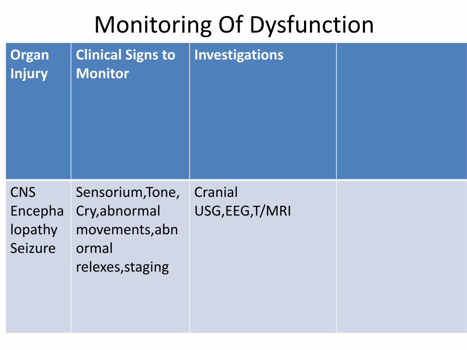
Monitoring Of DysfunctionOrgan Injury
Clinical Signs to Monitor
Investigations
CNSEncephalopathySeizure
Sensorium,Tone,Cry,abnormalmovements,abnormalrelexes,staging
CranialUSG,EEG,T/MRI

RenalARFMyoglobinuria
Uop,Hematria,edema,weight
Serum Na,K,BUN,Cr,SpGr,R/M
GIIleusNEC
Abdmonialdistension,bowelsounds,delayedmeconium passage,gastricaspirates
Abdomnal X-Ray when clinical signs are present
CVSMyocardialIschemiaPapillary Muscle Necrosis
BP,CFT,HR,Murmur,crepts,hepatomegaly,weight gain
ECG,CxR,ECHO

PulmonaryMeconium AspirationPPHN
RR,Apnea,Chestretraction,Cyanosis,O2(FiO2,SaO2)
CxRECHO

HematologicalDIC
GI Aspirates,PulmonaryHemorrhages,Petechiae,Prolonged bleed from puncture sites
BT,CT,Pltcout,PT/INR,aPTT
MetabolicHypogycemiaHyponatremia(SIADH)HypocalcemiaMetabolicAcidosis
RBSCalciumNaABG

PROGNOSIS
A. Overall mortality is 10% to 30%.
Neurodevelopment sequelae 15% to 45%.
B. Risk of CP in survivors of perinatal asphyxia is 5% to
10% compared to 0.2% in general population.
Most CP is not related to perinatal asphyxia & most perinatal
asphyxia does not cams CP.
Only 3%-13% of infants with CP have evidence of intrapartum
asphyxia

C. According to Sarnat staging.
a) Stage 1 – 90% to 1=0% (N) neurologic outcome < 10% mortality.
b) Stage - 2 – 20% to 37% die or have abnormal neurodevelopmental outcomes.
Infants who exhibit stage 2 sign for > 7 days have poorer outcomes.

c) Stage 3 HIE-50% to 89% die and all survivors have major neurodevelopment.
d) Prognosis is good if an infant does not progress to &/or remain in stage 3 & if total duration of stage 2<5 days.
e) Term baby not breast feeding by Day 10 of life
f) Seizures on Day 1 requiring multiple drugs to control

Outcomes
• depend on the pattern and severity of the brain injury
• involve motor, visual, and cognitive functions
• follow-up of these newborns should include assessment of motor function, vision and hearing, cognition,behavior, and quality of life, through infancy and childhood

• pattern of neurodevelopmental deficits follows an overt neonatal encephalopathy, often in the context of a critical illness
• is most commonly associated with the watershed pattern of injury and white matter damage, rather than the basal nuclei-predominant pattern of injury.

• (ACOG) Task Force on Neonatal encephalopathy concluded that an acute intrapartum event could result in cerebral palsy of the spastic quadriplegic or dyskinetic type, but could not account for isolated cognitive deficits

Motor Functions
• the risk of cerebral palsy or severe disability may involve more than one third of affected newborns
• More in those with severe encephalopathy
• Spastic quadriparesis is the most common type of CP

Vision and Hearing• Injury to the posterior visual pathway, including the
primary visual cortex, results in “cortical visual impairment”
• Injuries to the basal nuclei may also affect acuity, visual fields, or stereopsis (depth perception)
• SNHL, likely secondary to brainstem injury, is also seen following neonatal encephalopathy affecting 18 percent of survivors of moderate encephalopathy without cerebral palsy

Cognition• cognitive deficits are seen in 30–50 percent of
childhood survivors
• Cognitive deficits, such as those in language and memory, may be seen,even when IQ scores are normal.
Brain Imaging and Outcome• basal nuclei pattern of injury and abnormal signal
intensity in the posterior limb of the internal capsule are both predictive of severely impaired motor and cognitive outcomes
• watershed pattern is associated with cognitive impairments that are not necessarily accompanied by major motor deficits
– may only be evident after 2 years of age

Investigations For Prognosis
• EEG – background burst suppression,low voltage or electrocerebral silence poor prognosis
• CT/MRI – Diffuse decrease in density on CT scan at 2-4 weeks of life indicates poor prognosis
• Early MRI – basal ganglia and thalamic enhancment poor prognosis

Recent advances


Therapeutic Cooling• Extensive experimental data suggest that mild
hypothermia (3-4°C below baseline temperature) applied within a few hours (no later than 6 h) of injury is neuroprotective.
• The neuroprotective mechanisms are not completely understood. Possible mechanisms include
– reduced metabolic rate and energy depletion;
– decreased excitatory transmitter release;
– reduced alterations in ion flux;
– reduced apoptosis due to hypoxic-ischemic encephalopathy
– reduced vascular permeability, edema, and disruptions of blood-brain barrier functions.

• The clinical efficacy of therapeutic hypothermia in neonates with moderate-to-severe hypoxic-ischemic encephalopathy has been evaluated in 7 randomized controlled trials.

• Criteria from the larger trials (NICHD, CoolCap, and TOBY) are summarized as follows:
• Near-term infants born at 36 weeks' gestation or more with birth weight of 1800-2000 g or more, younger than 6 hours at admission
• Evidence of acute event around the time of birth
– Apgar score of 5 or less at 10 minutes after birth
– severe acidosis, defined as pH level of less than 7 or base deficit of 16 mmol/L or less (cord blood or any blood gas obtained within 1 h of birth)
– continued need for resuscitation at 10 minutes after birth
– Evidence of moderate to severe encephalopathy at birth

• clinical studies have been reassuring thus far regarding safety and applicability of hypothermia therapy
• Many theoretical concerns surround hypothermia and its side effects, – coagulation defects
– leukocyte malfunctions
– pulmonary hypertension
– worsening of metabolic acidosis
– abnormalities of cardiac rhythm, especially during rewarming.

• Although many components of its implementation remain to be optimized, hypothermia therapy is increasingly offered to infants with moderate-to-severe hypoxic-ischemic encephalopathy.
•

Erythropoietin (Epogen)• Epogen receptors are present in the developing
human embryo
– higher levels of Epogen in cerebral spinal fluid have been correlated with improved neurodevelopmental outcomes.
• may benefit infants with HIE through protection from neuronal apoptosis, neural regeneration, decreased inflammation, and decreased susceptibility to glutamate toxicity.

• Term infants with HIE treated with Epogen show decreased seizure activity, improved EEG results, and enhanced neurologic outcome.
• Although additional clinical trials are needed, Epogen appears to be effective in the treatment of infants with HIE if administered within 48 hours after delivery.

• potential adverse reactions associated with Epogen including hypertension, clotting abnormalities, seizures, and polycythemia, though have not been seen in neonates
• 1 study reported an increased incidence of retinopathy of prematurity in premature infants treated with Epogen

Magnesium sulfate (MgSO4
• Magnesium sulfate is an N-methyl-D-aspartate receptor antagonist.
• N-methyl-D-aspartate is a receptor for glutamate, an amino acid important in cell proliferation, differential, and survival in the developing brain.

• Conflicting data exist regarding the effectiveness of MgSO4 as a neuroprotective agent.
• Prenatal administration of MgSO4 to mothers at risk for preterm delivery is associated with reduced incidence of cerebral palsy at 3 years and improved neurodevelopmental outcomes
• When MgSO4 was administered postnatally to term infants with HIE, there was no improvement in their amplitude-integrated EEG, and when administered in large doses, MgSO4 can cause profound hypotension.[26]

Allopurinol• Allopurinol is an antioxidant that inhibits
formation of the free radicals that play such a significant role in the cellular damage associated with HIE.
• when term infants with HIE received allopurinol within 3 hours after birth, there was less free radical formation.

• Administration of allopurinol to mothers whose pregnancies are complicated by fetal hypoxia, has been associated with improved cord gases

Stem Cells• Stem cell transplantation may minimize the
effect of HIE by replacing damaged cells, promoting cell regeneration, inhibiting inflammation, and releasing trophic factors that heal and improve cell survival
• Efficacy of stem cell transplantation, however, appears dependent on timing of implantation, and this therapeutic window is presently unknown.

THANK YOU


• Neurologic Findings• Cranial nerves• Lack of reflex activity mediated by the cranial nerves can indicate brainstem dysfunction.• Full-term infants should blink and sustain eye closure in response to a sustained light stimulus. Repeated
flashes of light should produce habituation (eg, attenuated blinking) after 3-4 stimuli. Virtually all full-term newborns can track a ball of red wool, and the movement of stripes of at least one eighth of an inch or bigger can elicit opticokinetic nystagmus. Objects and pictures with round contours and facial appearances also make good targets for tracking in the newborn. Tracking is possible in infants with complete destruction of the occipital cortex by virtue of a subcortical pulvinar-collicular system. Retinal hemorrhagesare commonly observed in the neonate after vaginal delivery and can result in decreased pupil response. Destruction of the occipital cortex will also not affect pupillary response, because the responsible pathways leave the optic nerve and travel to the Edinger-Westphal nucleus, which sends back axons via the bilateral oculomotor nerves (consensual pupillary reflex).
• Neurologic examination may be difficult in the small and frail premature infant, but weakness of the lower extremities sometimes reflects the neuropathologic substrate of periventricular leukomalacia. Over time, the patient with periventricular white-matter lesions develops spastic diplegia affecting the lower extremities more than the upper extremities.
• Blinking to light starts at 26 weeks’ gestational age, sustained eye closure to light is seen around 32 weeks, and 90% of newborns track a ball of red wool by 34 weeks. Opticokinetic reflexes can be seen at 36 weeks. The pupil starts reacting to light around 30 weeks, but the light reflex is not consistently assessable until the gestational age of 32-35 weeks. Pupillary reflexes are reliably present at term. Extraocular movements can be elicited by performing the doll's-eye maneuver at 25 weeks’ gestation and by performing caloric stimulation at 30 weeks’ gestation.
• In infants aged 32-34 weeks’ gestation, suck and swallow are reasonably coordinated with breathing, but the actions are not perfected until after term.
• Patients with mild HIE-NE often have mydriasis. Progression of the disease may produce miosis (even in the dark) responsive to light, and in severe cases (stage 3 of Sarnat classification), the pupils are small or midpositioned and poorly reactive to light, reflecting sympathetic or parasympathetic dysfunction.
• The lack of pupillary, eye movement, corneal, gag, and cough reflexes may reflect damage to the brainstem, where the cranial-nerve nuclei are located. Decreased respiratory drive or apnea can be from lesions of the respiratory center, which overlap with vagal nuclei (ambiguous and solitaire) or medullary reticular formation. Ventilatory disturbances in HIE may manifest as periodic breathing apnea (similar to Cheyne-Stokes respiration) or just decreased respiratory drive

• Motor function
• Begin the motor examination of an infant with suspected HIE-NE by qualitatively and quantitatively observing his or her posture and spontaneous movements. Asymmetry in the amount of movement and posture is a subtle sign of hemiparesis, but it may be the only focal feature of the examination. Slight stimulation (eg, gently touching the patient) can increase motor activity in the term neonate and may be helpful in demonstrating asymmetrical hemiparesis.
• Eliciting the Moro reflex may be an excessive stimulus and mask a subtle asymmetry in limb movement. Asymmetry in the Moro reflex is seen in peripheral lesions (eg, those due to brachial plexus injury).
• Total absence or paucity of spontaneous movements, especially if associated with no reaction to painful stimuli and generalized hypotonia, indicates brainstem dysfunction or severe, diffuse, or multifocal cortical damage.
• Specific patterns of motor weakness indicate cerebral injury patterns. Patients with borderzone parasagittal injury (ulegyria) tend to have proximal greater than distal weakness and upper extremity more than lower extremity weakness (man-in-the-barrel). A unilateral, focal infarct, especially one involving the middle cerebral artery, causes contralateral hemiparesis and focal seizures. Patients with selective neuronal necrosis may have severe hypotonia, stupor, and coma.
• Motor examination of a newborn with large unilateral lesions may reveal mild hemiparesis and seizures in as many as 80%. The seizures are often partial (focal) and contralateral to the cortical lesion. Neonates with severe bilateral infarcts may have quadriparesis. Moro and tonic neck reflexes do not habituate, reflecting the lack of cortical modulation, which attenuates the response after repeated trials or sustained stimulus. Newborns with diencephalic lesions cannot regulate their temperature and have problems with sleep-wake cycles. The long-term sequelae of focal or multifocal cerebral necrosis include spastic hemiparesis and quadriparesis (eg, bilateral hemiparesis), cognitive deficits, and seizures.
• Foot-ankle dorsiflexion or triple flexion (eg, foot-ankle dorsiflexion, knee and hip flexion) after plantar stimulation reflects only an intact spinal cord and sensory and motor nerves. Extensor movements (eg, arm elevation above the level of the shoulders) are more sophisticated motor actions than the dorsiflexion or triple flexion and require some cortical function.
• A tonic neck reflex is performed by turning the patient's head to one side. The patient demonstrates arm and leg extension on the side to which the head is turned and flexion on the opposite side (fencer's posture). The tonic neck reflex posture should go away after several seconds, and its persistence is a sign of cortical dysfunction.
• Spasticity is a velocity-dependent increase in tone that is generally most prominent with limb extension in muscle groups with antigravitational action (arm flexion, plantar extension). This sign can be seen over time in infants with corticospinal tract damage caused by a hypoxic-ischemic insult. In the neonatal period, spasticity is commonly noted first and is most prominent in the distal parts of the extremities. All fingers are flexed with the thumb under the second to fifth fingers, a pattern commonly referred to as cortical thumbs. Fewer than 5-10 beats of ankle clonus may be present in healthy neonates, but infants with damage to the corticospinal tract may have sustained ankle clonus. However, the initial motor manifestation will be flaccid hypotonia with spasticity later developing.
• When assessing muscle tone, the state of arousal and prematurity must be taken into account. In the acute phase, tone is decreased in a generalized fashion affecting trunk and extremities. The flexor tone in the limbs is best assessed in term infants by showing a discrepancy in the scoring system between Dubowitz neurologic examination and morphologic examination. The infant looks like a “rag doll” when supported by a hand under the chest (vertical suspension). Head lag is demonstrated by traction of the hands in a supine position. The infant folds around the examiner's hand when lifted prone with a hand supporting the chest (horizontal suspension).
• Hip abduction may be seen with increased tone and even with decerebrate posturing (frog-leg posture). Another manifestation of CNS dysfunction in the neonatal period is increased axial extensor tone with arching of the back and neck extension or opisthotonus. Many infants simultaneously have decreased axial flexor tone (eg, major head lag on arm traction maneuver) and increased axial extensor tone. In many cases, limb and axial hypotonia are present for several months before increased axial extensor tone or limb spasticity can be detected. Increased active neck and trunk extensor tone are predictors of quadriparesis. Another sign of spasticity that can develop relatively early is scissoring, where the previously abducted legs extend, become rigid, and have extreme hip adduction such that they cross with stimulation or crying.

• . • Seizures• HIE is often reported to be the most frequent cause of neonatal seizures. They usually occur 12-24 hours after birth and are difficult to control with
anticonvulsants. Large, unilateral infarcts occur with neonatal seizures in as many as 80% of patients. Seizures are often partial (focal) and contralateral to the cortical lesion. About two thirds of newborns with cerebral venous infarcts have seizures. Those with multiple or diffuse lesions and cerebral venous infarcts often have multifocal or migratory seizures. Seizures are observed during physical examination and may confirm the diagnosis. Observation often reveals clonic rhythmic contractions. When holding the limb affected by clonic seizures, the examiner's hand shakes or feels limb movement. Limb flexion or extension does not suppress the clonic activity, as it does in jitteriness and clonus. Newborn infants cannot have generalized seizures due to immaturity of the neuronal pathways connecting the 2 halves of the brain.
• Tonic, unilateral, or focal seizures consistently have an EEG signature. In the seizures, unilateral arm and leg posturing is often accompanied by ipsilateral trunk flexion. Generalized tonic posturing (eg, extension of the upper and lower extremities or extension of the legs and flexion of the arms) is related to an EEG seizure in 15% of affected neonates.
• Tonic seizures can be seen in neonates with local anesthetic intoxication. Although generalized tonic posturing is infrequently associated with electrical seizures, it is not a benign sign. Of neonates with tonic posturing and an abnormal EEG background, 13% have normal development.
• Subtle seizures may be a part of the HIE-NE picture. Subtle manifestations of neonatal seizures are confirmed on EEG and include apnea; tonic eye deviation; sustained eye opening; slow, rhythmic, tongue thrusting; and boxing, bicycling, and swimming movements. Most still accept that some subtle seizures may be correlated with EEG results. However, publications since the late 1980s have shown that seizures are not as frequent as previously thought and that they are unusual in patients close to term. Several other patterns of subtle neonatal seizures are described without EEG confirmation. The lack of an EEG signature does not exclude CNS pathology because neonates with HIE often have motor automatisms without EEG seizures. Management is controversial, but treatment is not usually beneficial unless more overt seizure activity is noted.[31]
• Seizures may be difficult to clinically diagnose in the premature neonate. Subtle seizures associated with ictal EEG changes are not rare in premature infants. The subtle patterns of neonatal seizures in the premature infant include sustained eye opening, oral-buccal-lingual movements (smacking, drooling, chewing), pedaling movements, grimacing, and autonomic manifestations.[#sarnat]