Embed Size (px)
Citation preview
Hypertensive Disorders in Pregnancy
Mohammad Sohail Khan
Tasbeeh ur Rehman
Ayub Medical College
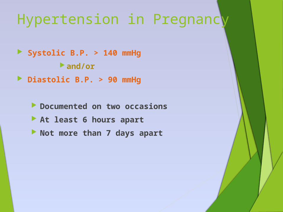
Hypertension in Pregnancy
Systolic B.P. > 140 mmHg
and/or
Diastolic B.P. > 90 mmHg
Documented on two occasions
At least 6 hours apart
Not more than 7 days apart
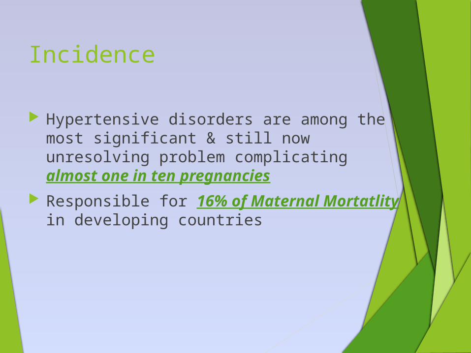
Incidence
Hypertensive disorders are among the most significant & still now unresolving problem complicating almost one in ten pregnancies
Responsible for 16% of Maternal Mortatlity in developing countries
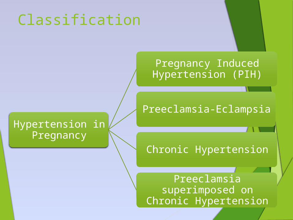
Classification
Hypertension in Pregnancy
Pregnancy Induced Hypertension (PIH)
Preeclamsia-Eclampsia
Chronic Hypertension
Preeclamsia superimposed on Chronic Hypertension
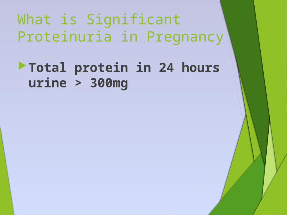
What is Significant Proteinuria in Pregnancy
Total protein in 24 hours urine > 300mg
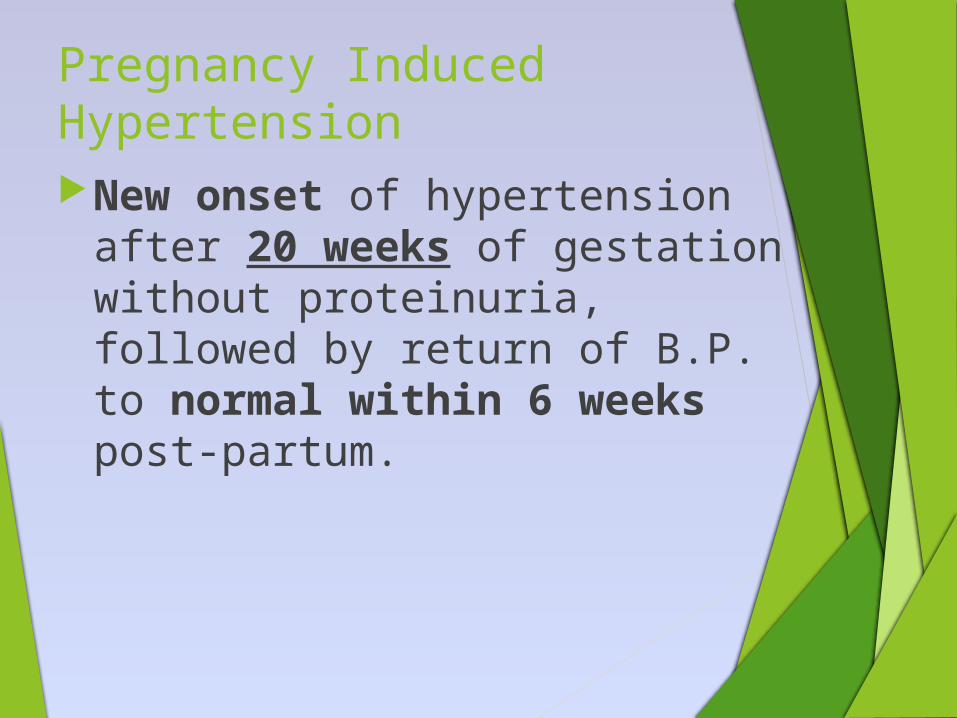
Pregnancy Induced HypertensionNew onset of hypertension after
20 weeks of gestation without proteinuria, followed by return of B.P. to normal within 6 weeks post-partum.
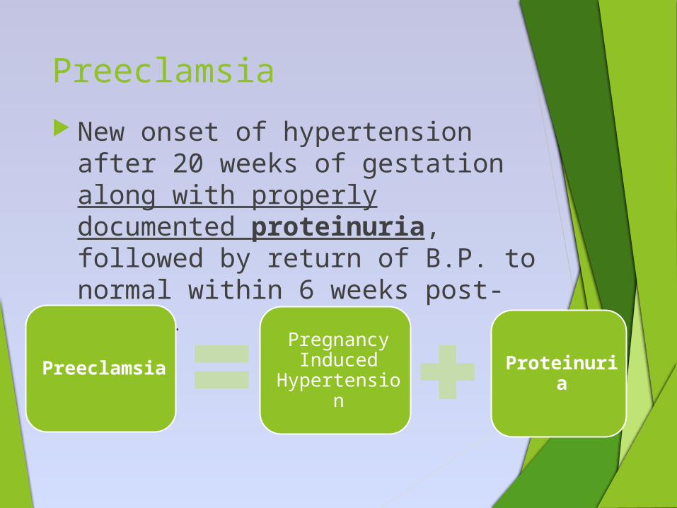
Preeclamsia
New onset of hypertension after 20 weeks of gestation along with properly documented proteinuria, followed by return of B.P. to normal within 6 weeks post-partum.
PreeclamsiaPregnancy Induced
HypertensionProteinuri
a
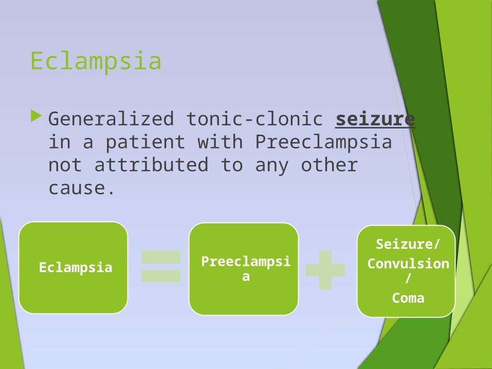
Eclampsia
Generalized tonic-clonic seizure in a patient with Preeclampsia not attributed to any other cause.
Eclampsia Preeclampsia
Seizure/Convulsion
/Coma
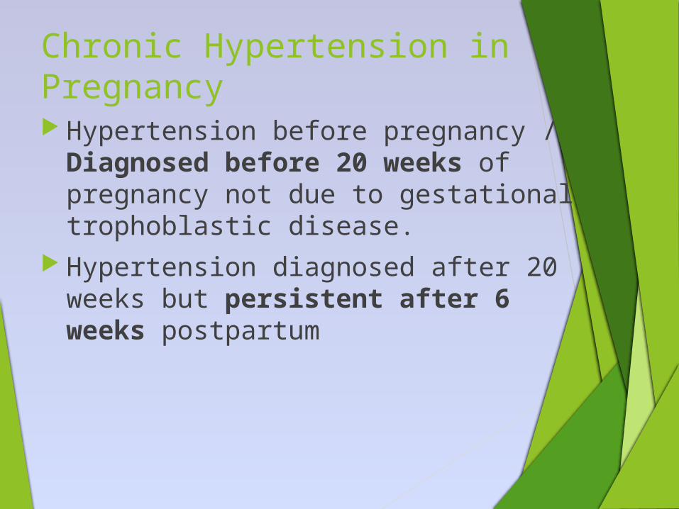
Chronic Hypertension in Pregnancy Hypertension before pregnancy /
Diagnosed before 20 weeks of pregnancy not due to gestational trophoblastic disease.
Hypertension diagnosed after 20 weeks but persistent after 6 weeks postpartum
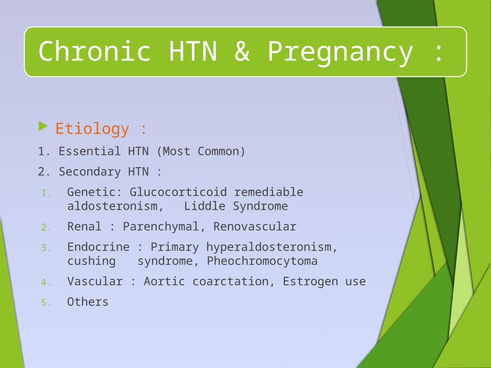
Chronic HTN & Pregnancy :
Etiology :1. Essential HTN (Most Common)
2. Secondary HTN :
1. Genetic: Glucocorticoid remediable aldosteronism, Liddle Syndrome
2. Renal : Parenchymal, Renovascular
3. Endocrine : Primary hyperaldosteronism, cushing syndrome,
Pheochromocytoma
4. Vascular : Aortic coarctation, Estrogen use
5. Others
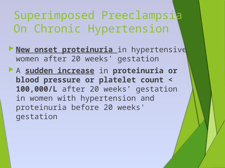
Superimposed Preeclampsia On Chronic Hypertension
New onset proteinuria in hypertensive women after 20 weeks' gestation
A sudden increase in proteinuria or blood pressure or platelet count < 100,000/L after 20 weeks’ gestation in women with hypertension and proteinuria before 20 weeks' gestation
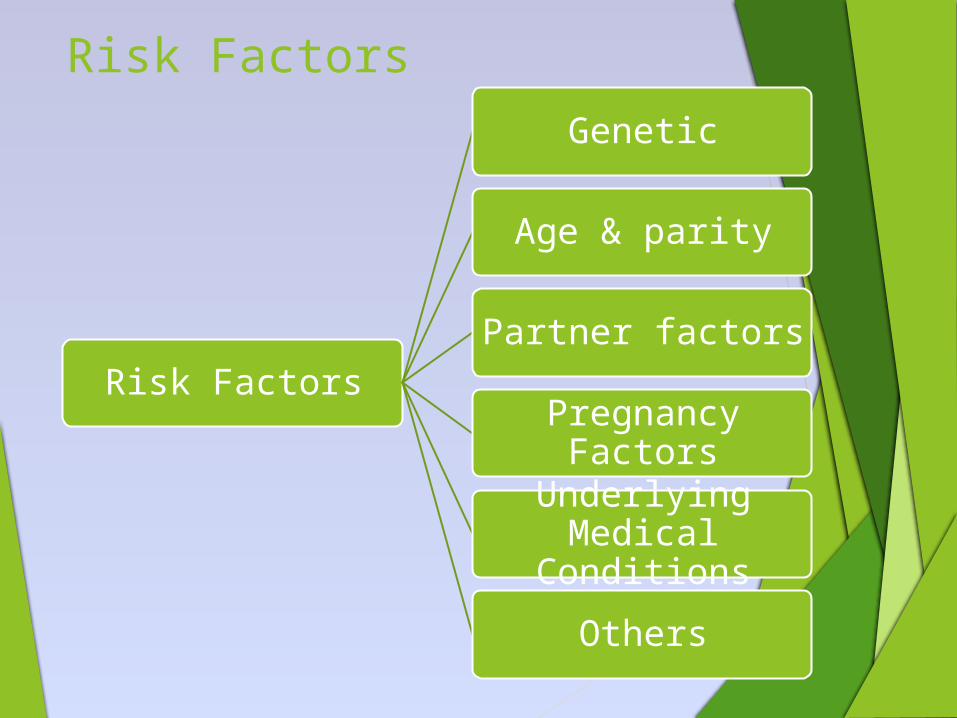
Risk Factors
Genetic
Age & parity
Partner factors
Pregnancy Factors
Underlying Medical
Conditions
Others
Risk Factors
Risk Factors: Cont.
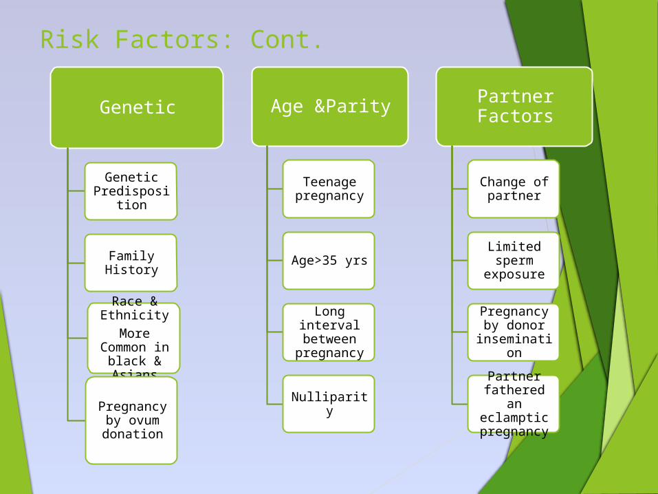
Genetic
Genetic Predispositio
n
Family History
Race & Ethnicity
More Common in
black & Asians
Pregnancy by ovum donation
Age &Parity
Teenage pregnancy
Age>35 yrs
Long interval between
pregnancy
Nulliparity
Partner Factors
Change of partner
Limited sperm
exposure
Pregnancy by donor
insemination
Partner fathered an eclamptic pregnancy
Risk Factors: Cont.
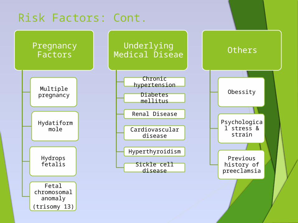
Pregnancy Factors
Multiple pregnancy
Hydatiform mole
Hydrops fetalis
Fetal chromosomal
anomaly(trisomy 13)
Underlying Medical Diseae
Chronic hypertension
Diabetes mellitus
Renal Disease
Cardiovascular disease
Hyperthyroidism
Sickle cell disease
Others
Obessity
Psychological stress & strain
Previous history of
preeclamsia

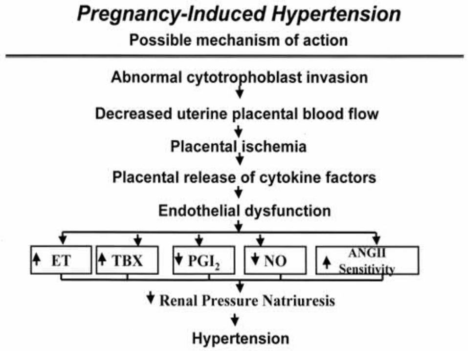
PATHOPHYSIOLOGY

2 stage model for preeclampsia
Stage 2Maternal syndrome
(HTN, proteinuria,Endothelial dysfunction)
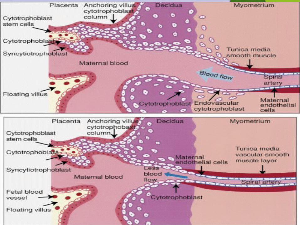
Stage1Reduced placental
implantation???
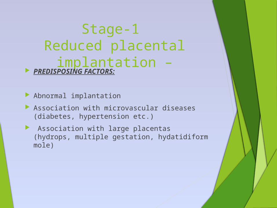
Stage-1 Reduced placental
implantation – PREDISPOSING FACTORS:
Abnormal implantation
Association with microvascular diseases (diabetes, hypertension etc.)
Association with large placentas (hydrops, multiple gestation, hydatidiform mole)
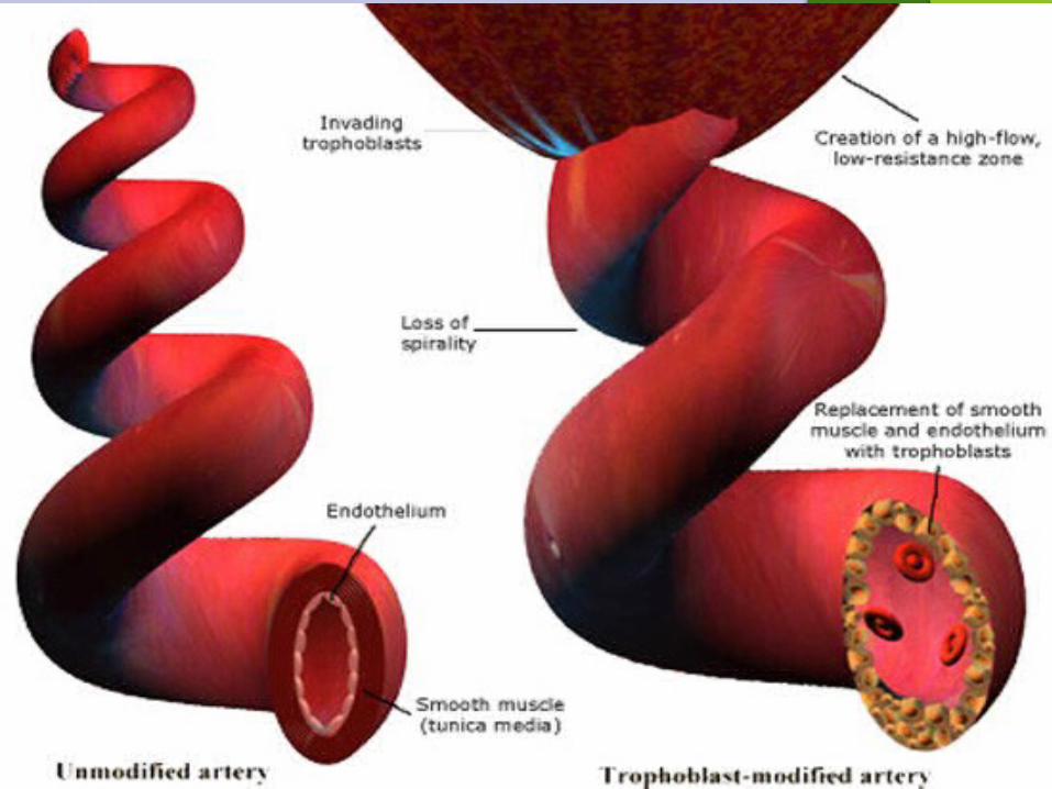
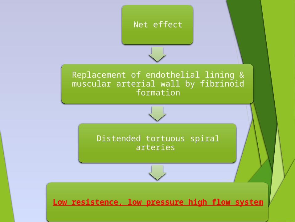
Net effect
Replacement of endothelial lining & muscular arterial wall by fibrinoid formation
Distended tortuous spiral arteries
Low resistence, low pressure high flow system
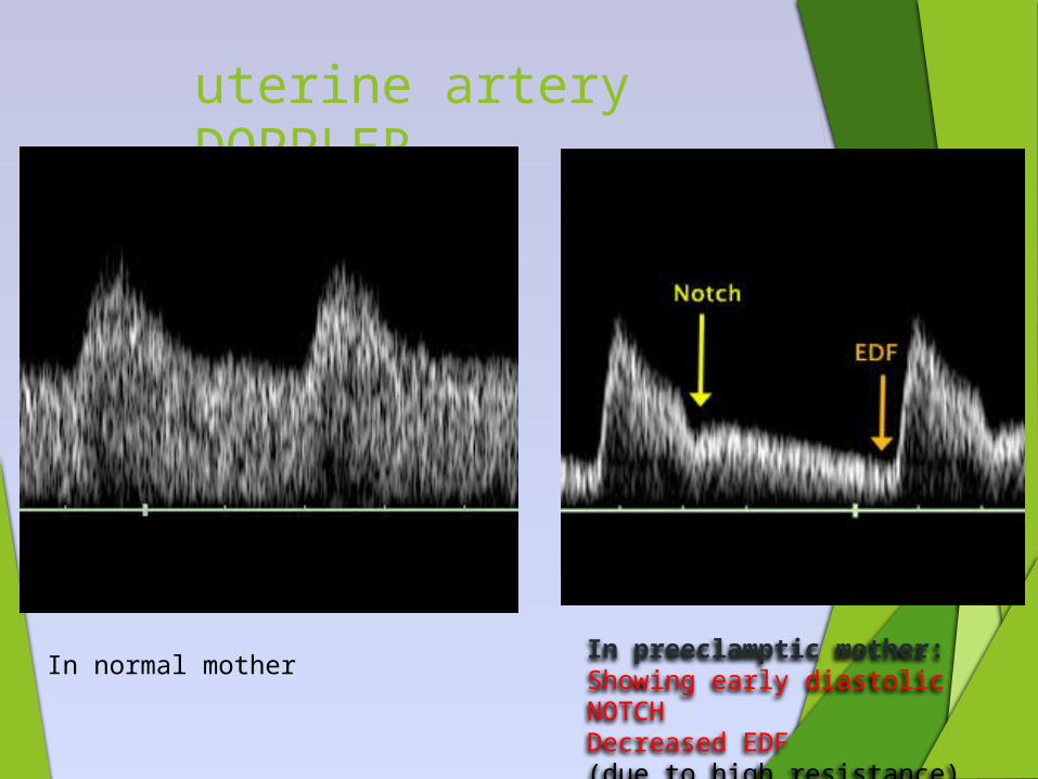
uterine artery DOPPLER
In preeclamptic mother:Showing early diastolic NOTCHDecreased EDF(due to high resistance)
In normal mother
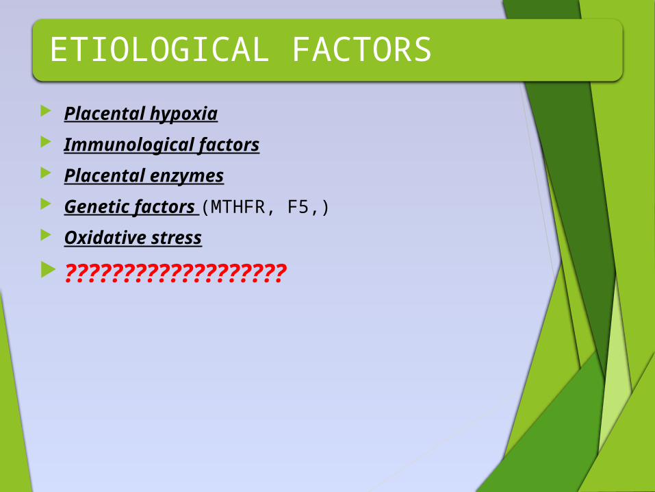
ETIOLOGICAL FACTORS
Placental hypoxia
Immunological factors
Placental enzymes
Genetic factors (MTHFR, F5,)
Oxidative stress
???????????????????
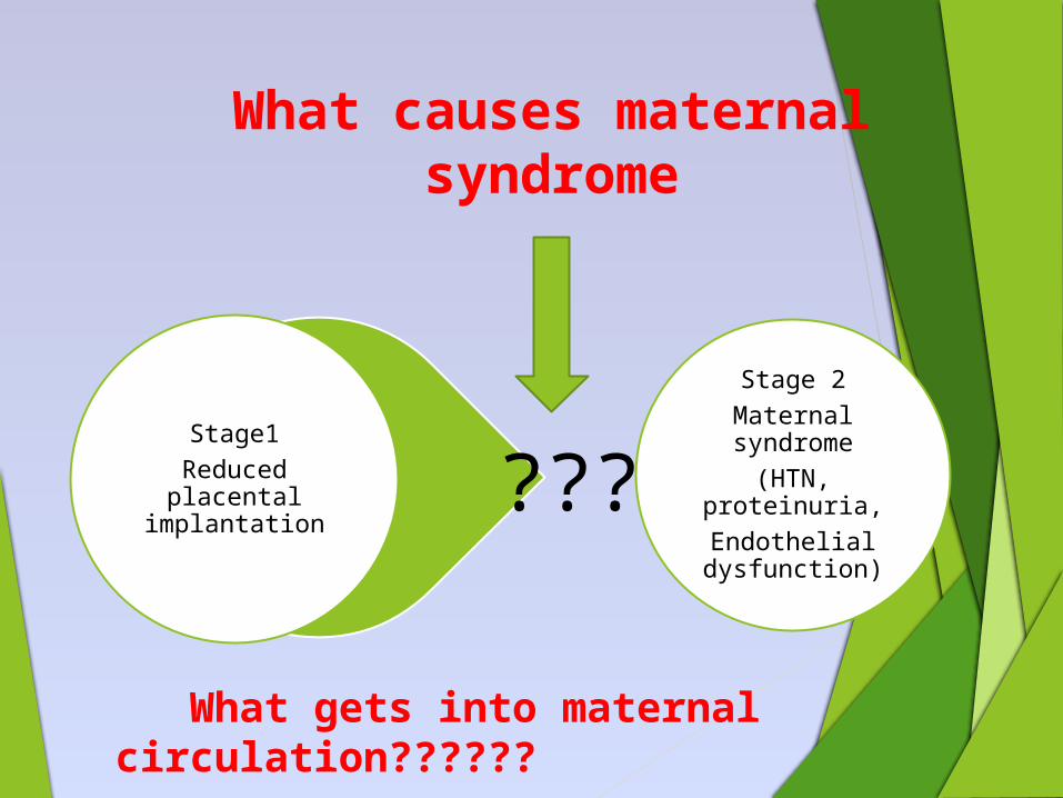
What causes maternal syndrome
Stage 2Maternal syndrome
(HTN, proteinuria,Endothelial dysfunction)
Stage1Reduced placental
implantation???
What gets into maternal circulation??????
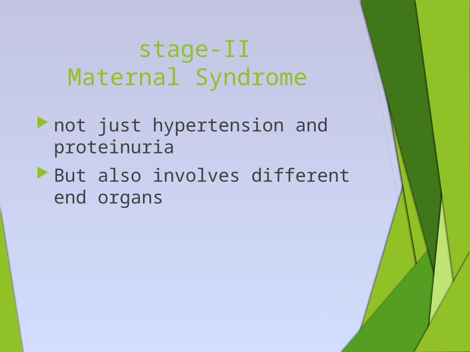
stage-IIMaternal Syndrome
not just hypertension and proteinuria
But also involves different end organs
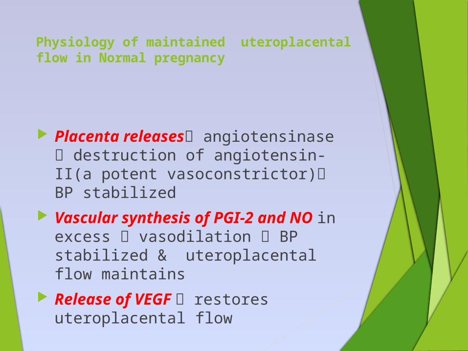
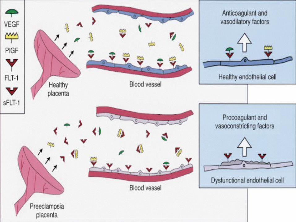
Physiology of maintained uteroplacental flow in Normal pregnancy
Placenta releases angiotensinase destruction of angiotensin-II(a potent vasoconstrictor) BP stabilized
Vascular synthesis of PGI-2 and NO in excess vasodilation BP stabilized & uteroplacental flow maintains
Release of VEGF restores uteroplacental flow
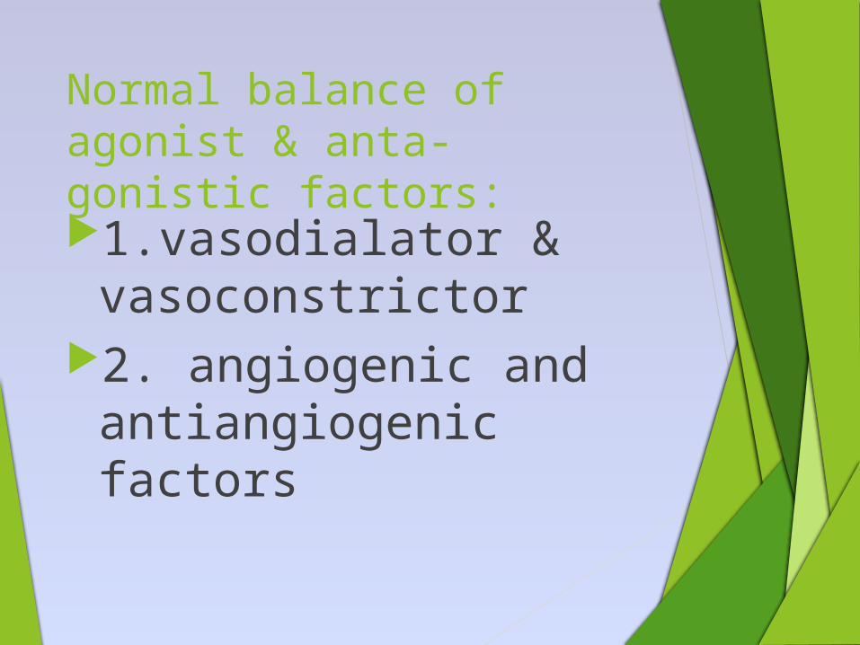
Normal balance of agonist & anta-gonistic factors:
1.vasodialator & vasoconstrictor
2. angiogenic and antiangiogenic factors
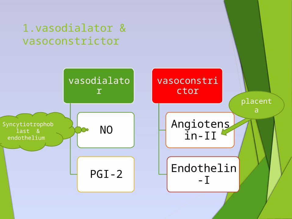
1.vasodialator & vasoconstrictor
vasodialator
NO
PGI-2
vasoconstrictor
Angiotensin-II
Endothelin-I
placenta
Syncytiotrophoblast &
endothelium
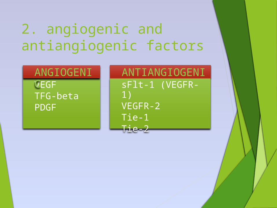
2. angiogenic and antiangiogenic factors
VEGFTFG-betaPDGF
ANGIOGENIC
ANTIANGIOGENICsFlt-1 (VEGFR-1)VEGFR-2Tie-1Tie-2

Pathophysiology for different organ damage
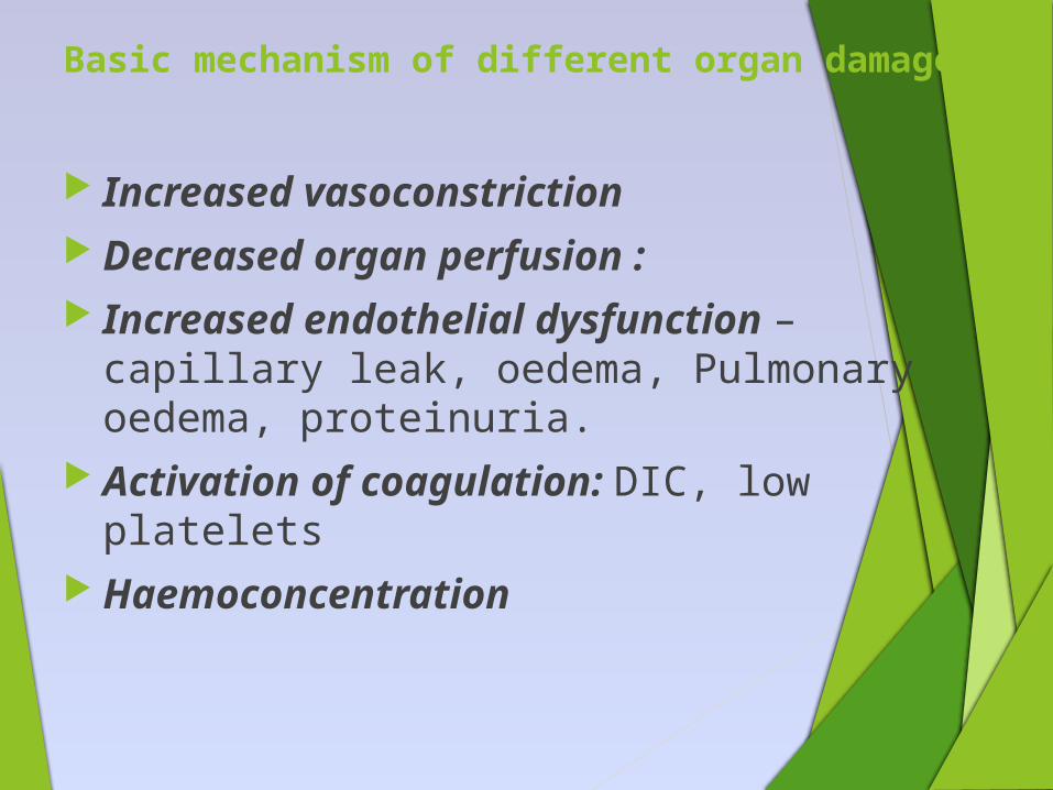
Basic mechanism of different organ damage:
Increased vasoconstriction Decreased organ perfusion : Increased endothelial dysfunction –
capillary leak, oedema, Pulmonary oedema, proteinuria.
Activation of coagulation: DIC, low platelets
Haemoconcentration
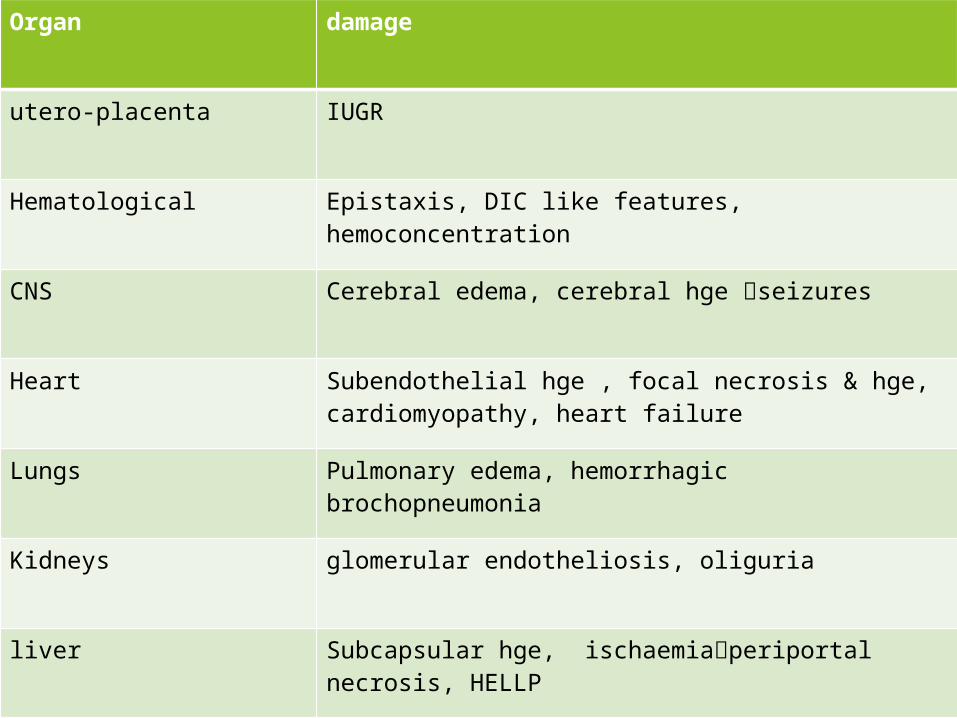
Organ damage
utero-placenta IUGR
Hematological Epistaxis, DIC like features, hemoconcentration
CNS Cerebral edema, cerebral hge seizures
Heart Subendothelial hge , focal necrosis & hge, cardiomyopathy, heart failure
Lungs Pulmonary edema, hemorrhagic brochopneumonia
Kidneys glomerular endotheliosis, oliguria
liver Subcapsular hge, ischaemiaperiportal necrosis, HELLP
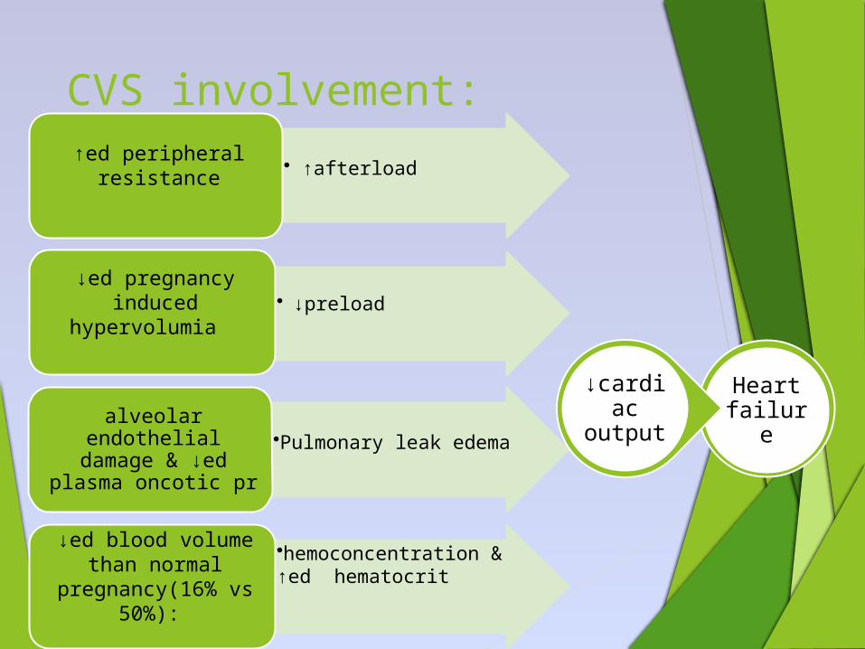
CVS involvement:
• ↑afterload↑ed peripheral
resistance
• ↓preload ↓ed pregnancy
induced hypervolumia
•Pulmonary leak edemaalveolar endothelial
damage & ↓ed plasma oncotic pr
•hemoconcentration & ↑ed hematocrit
↓ed blood volume than normal
pregnancy(16% vs 50%):
Heart failure
↓cardiac output
Hematological system
Thrombocytopenia & other PL abnormality:
• ↑ed PL activation & degranulation,
• ↓ed life span. • Corelates well
wth disease severity.
Intravascular hemolysis
• endothelial damage & altered fluidity of erythrocyte membrane d/t change in serum lipid content → ↑ed LDH, spherocytosis, reticulocytosis
• microangiopathic hemolysis
↑ed coagulation & fibrinolysis
• Feature like DIC• Release of
thromboplastin• ↓fibrinogen• AT-III• plasminogen
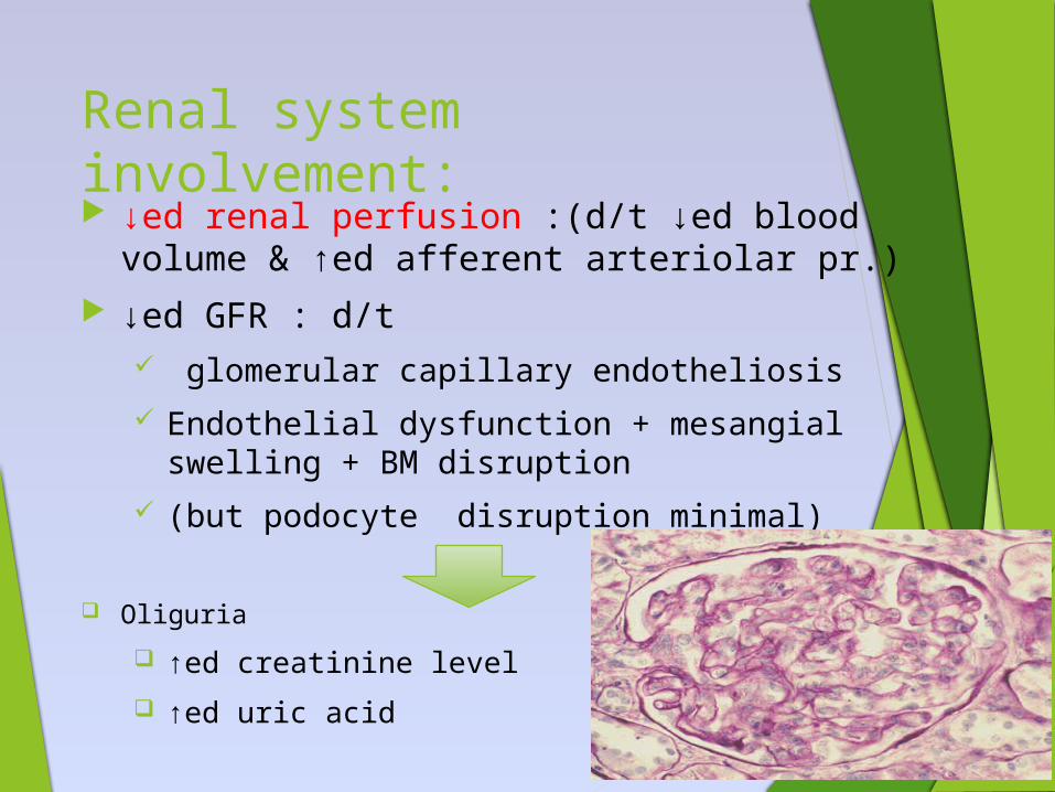
Renal system involvement: ↓ed renal perfusion :(d/t ↓ed blood volume &
↑ed afferent arteriolar pr.) ↓ed GFR : d/t
glomerular capillary endotheliosis Endothelial dysfunction + mesangial swelling +
BM disruption (but podocyte disruption minimal)
Oliguria
↑ed creatinine level
↑ed uric acid
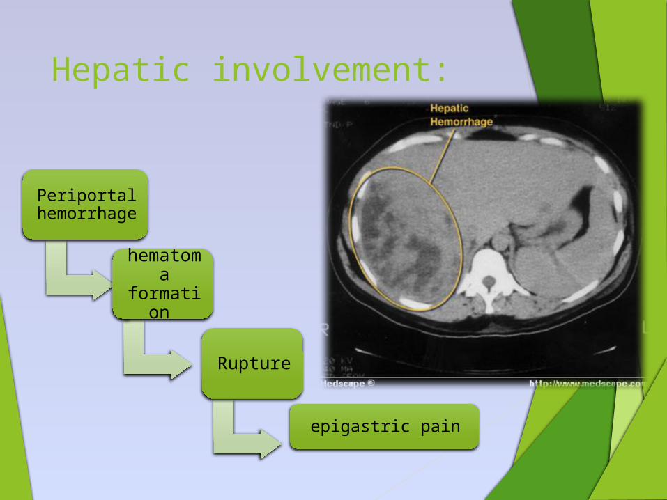
Hepatic involvement:
Periportal hemorrhage
hematoma
formation
Rupture
epigastric pain
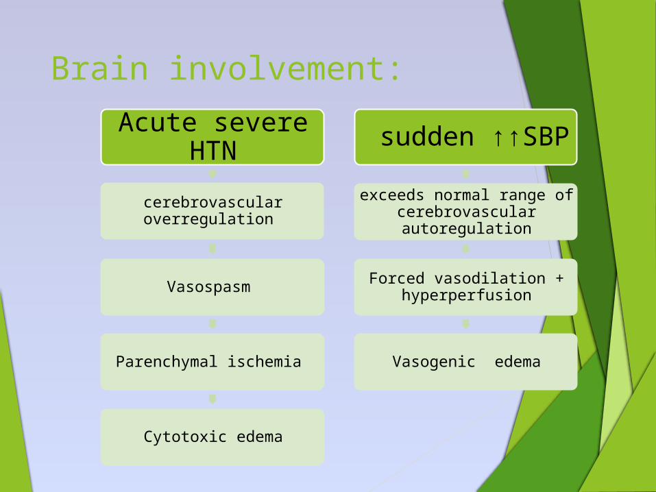
Brain involvement:
Acute severe HTN
cerebrovascular overregulation
Vasospasm
Parenchymal ischemia
Cytotoxic edema
sudden ↑↑SBP
exceeds normal range of cerebrovascular autoregulation
Forced vasodilation + hyperperfusion
Vasogenic edema
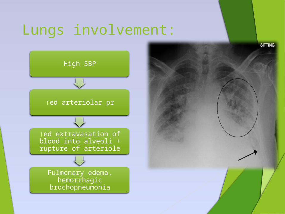
Lungs involvement:
High SBP
↑ed arteriolar pr
↑ed extravasation of blood into alveoli + rupture of arteriole
Pulmonary edema, hemorrhagic
brochopneumonia
Diagnosisof HDP
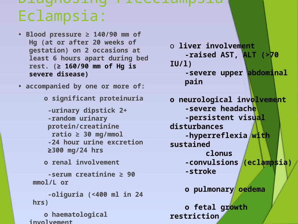
Diagnosing Preeclampsia-Eclampsia:• Blood pressure ≥ 140/90 mm of
Hg (at or after 20 weeks of gestation) on 2 occasions at least 6 hours apart during bed rest. (≥ 160/90 mm of Hg is severe disease)
• accompanied by one or more of:
o significant proteinuria
-urinary dipstick 2+-random urinary protein/creatinine ratio ≥ 30 mg/mmol-24 hour urine excretion ≥300 mg/24 hrs
o renal involvement
-serum creatinine ≥ 90 mmol/L or
-oliguria (<400 ml in 24 hrs)
o haematological involvement
-platelet count<1 lakh
o liver involvement -raised AST, ALT (>70
IU/l) -severe upper abdominal pain
o neurological involvement -severe headache -persistent visual
disturbances-hyperreflexia with
sustained clonus
-convulsions (eclampsia) -stroke
o pulmonary oedema o fetal growth restriction
o placental abruption
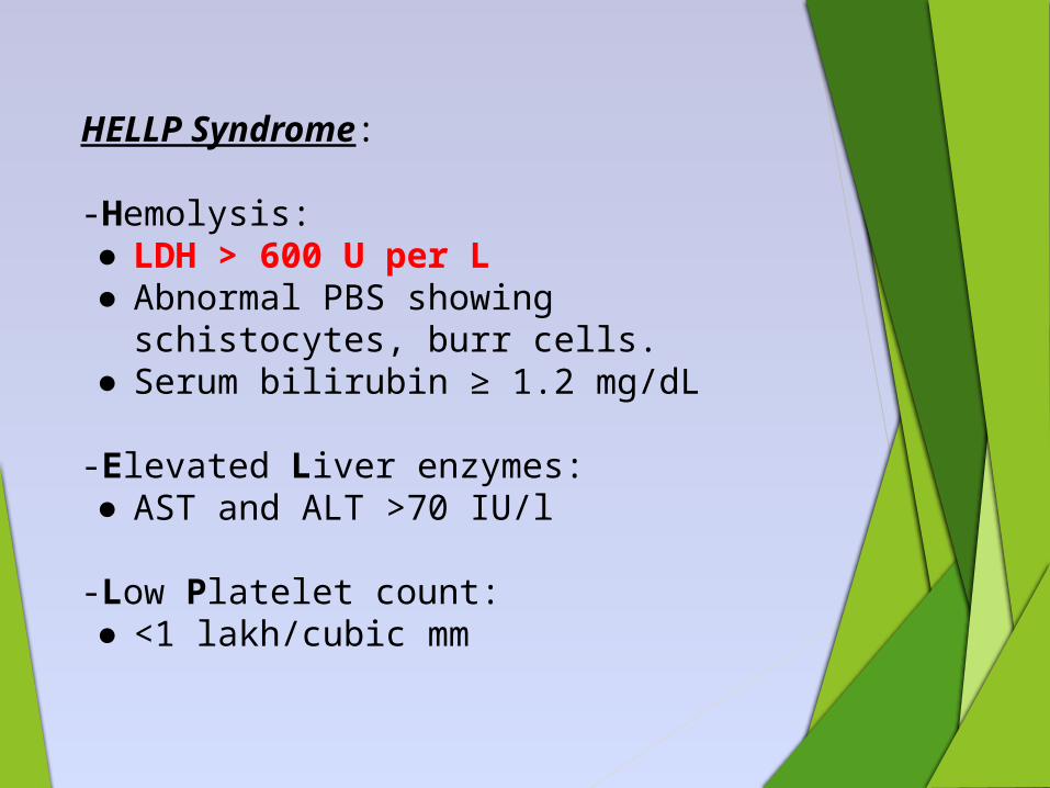
HELLP Syndrome: -Hemolysis:● LDH > 600 U per L● Abnormal PBS showing
schistocytes, burr cells.● Serum bilirubin ≥ 1.2 mg/dL
-Elevated Liver enzymes:● AST and ALT >70 IU/l
-Low Platelet count:● <1 lakh/cubic mm
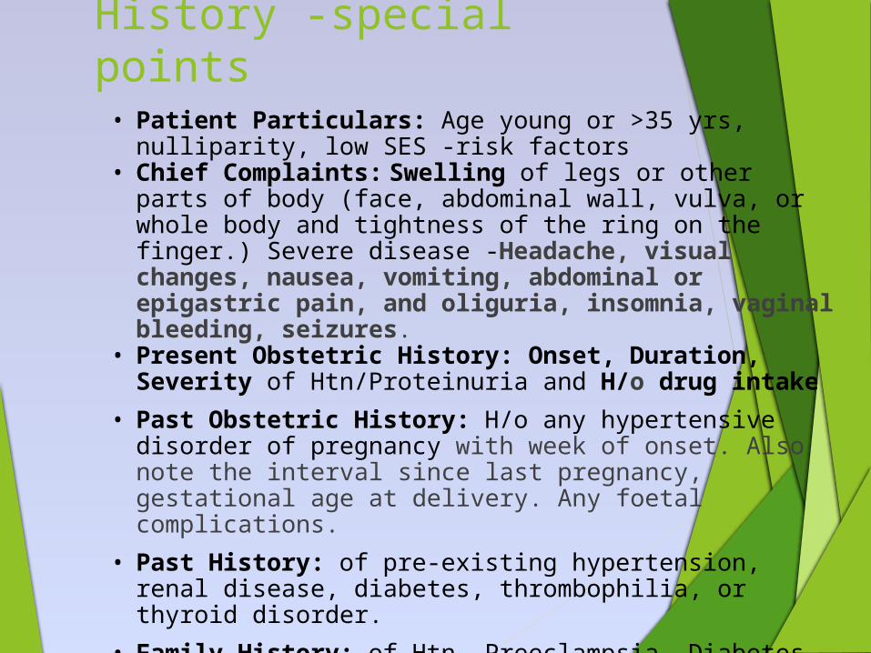
History -special points• Patient Particulars: Age young or >35 yrs, nulliparity,
low SES -risk factors• Chief Complaints: Swelling of legs or other parts of
body (face, abdominal wall, vulva, or whole body and tightness of the ring on the finger.) Severe disease -Headache, visual changes, nausea, vomiting, abdominal or epigastric pain, and oliguria, insomnia, vaginal bleeding, seizures.
• Present Obstetric History: Onset, Duration, Severity of Htn/Proteinuria and H/o drug intake
• Past Obstetric History: H/o any hypertensive disorder of pregnancy with week of onset. Also note the interval since last pregnancy, gestational age at delivery. Any foetal complications.
• Past History: of pre-existing hypertension, renal disease, diabetes, thrombophilia, or thyroid disorder.
• Family History: of Htn, Preeclampsia, Diabetes, CVD
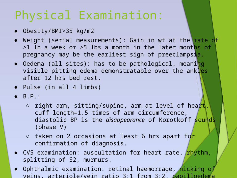
Physical Examination:● Obesity/BMI>35 kg/m2
● Weight (serial measurements): Gain in wt at the rate of >1 lb a week or >5 lbs a month in the later months of pregnancy may be the earliest sign of preeclampsia.
● Oedema (all sites): has to be pathological, meaning visible pitting edema demonstratable over the ankles after 12 hrs bed rest.
● Pulse (in all 4 limbs)
● B.P.:
○ right arm, sitting/supine, arm at level of heart, cuff length=1.5 times of arm circumference, diastolic BP is the disappearance of Korotkoff sounds (phase V)
○ taken on 2 occasions at least 6 hrs apart for confirmation of diagnosis.
● CVS examination: auscultation for heart rate, rhythm, splitting of S2, murmurs.
● Ophthalmic examination: retinal haemorrage, nicking of veins, arteriole/vein ratio 3:1 from 3:2, papilloedema
● Deep tendon reflexes: hyperreflexia/presence of clonus
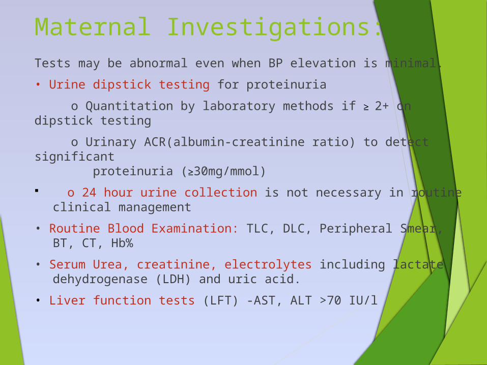
Maternal Investigations:Tests may be abnormal even when BP elevation is minimal.
• Urine dipstick testing for proteinuria
o Quantitation by laboratory methods if ≥ 2+ on dipstick testing
o Urinary ACR(albumin-creatinine ratio) to detect significant proteinuria (≥30mg/mmol)
o 24 hour urine collection is not necessary in routine clinical management
• Routine Blood Examination: TLC, DLC, Peripheral Smear, BT, CT, Hb%
• Serum Urea, creatinine, electrolytes including lactate dehydrogenase (LDH) and uric acid.
• Liver function tests (LFT) -AST, ALT >70 IU/l
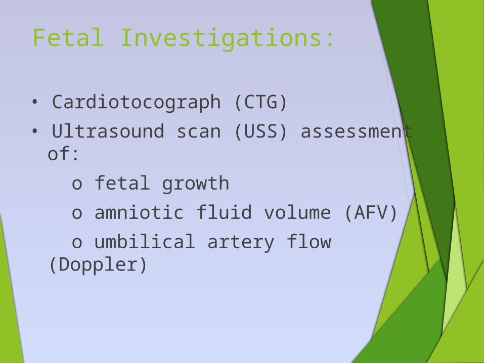
Fetal Investigations:
• Cardiotocograph (CTG)
• Ultrasound scan (USS) assessment of:
o fetal growth
o amniotic fluid volume (AFV)
o umbilical artery flow (Doppler)
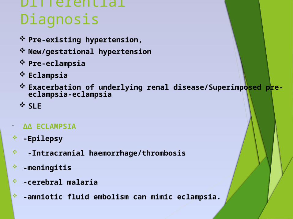
Differential Diagnosis Pre-existing hypertension, New/gestational hypertension Pre-eclampsia Eclampsia Exacerbation of underlying renal disease/Superimposed pre-
eclampsia-eclampsia SLE
ΔΔ ECLAMPSIA -Epilepsy
-Intracranial haemorrhage/thrombosis
-meningitis
-cerebral malaria
-amniotic fluid embolism can mimic eclampsia.
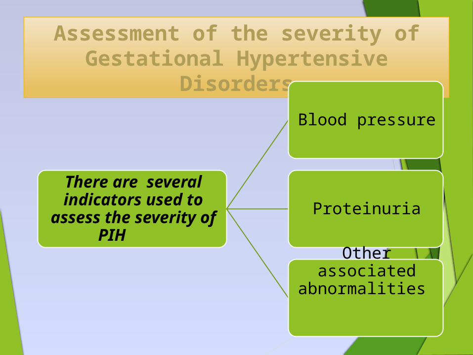
Assessment of the severity of Gestational Hypertensive
Disorders
There are several indicators used to assess the severity
of PIH
Blood pressure
Proteinuria
Other associated abnormalities
N.B: Grades of proteinuria (in g/L): Trace=0.1, 1+=0.3, 2+=1, 3+=3, 4+=10
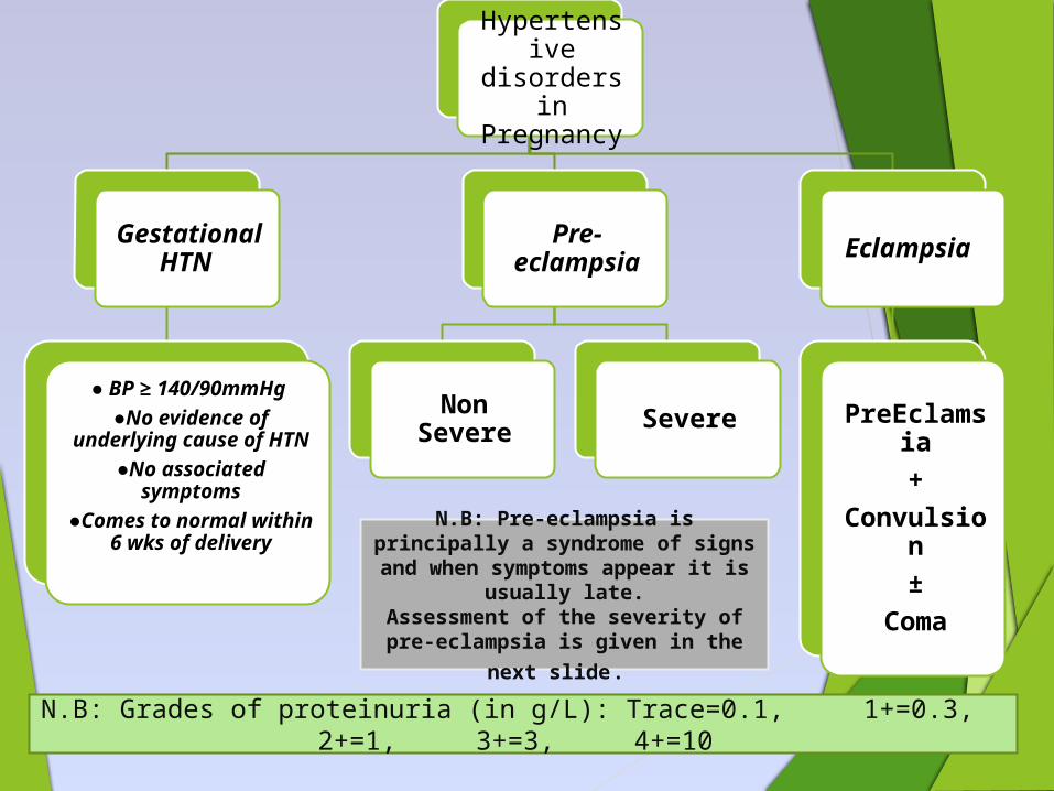
Hypertensive disorders
in Pregnancy
Gestational HTN
● BP ≥ 140/90mmHg ●No evidence of
underlying cause of HTN
●No associated symptoms
●Comes to normal within 6 wks of
delivery
Pre-eclampsia
Non Severe Severe
Eclampsia
PreEclamsia+
Convulsion±
Coma
N.B: Pre-eclampsia is principally a syndrome of signs and when symptoms appear it is usually
late.Assessment of the severity of pre-eclampsia is given in the
next slide.
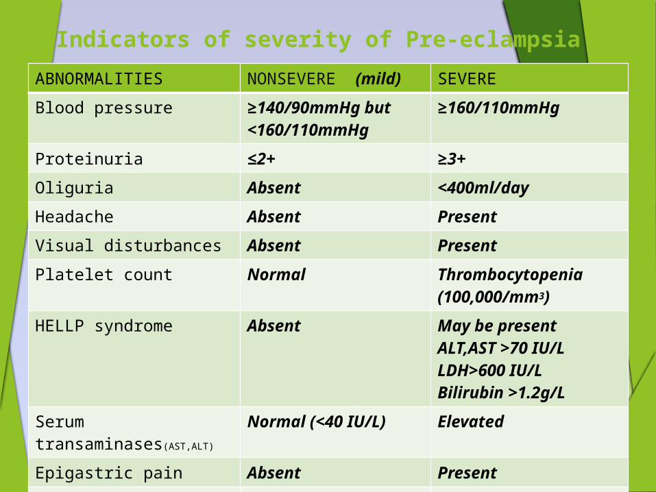
Indicators of severity of Pre-eclampsia
ABNORMALITIES NONSEVERE (mild) SEVERE
Blood pressure ≥140/90mmHg but <160/110mmHg
≥160/110mmHg
Proteinuria ≤2+ ≥3+
Oliguria Absent <400ml/day
Headache Absent Present
Visual disturbances Absent Present
Platelet count Normal Thrombocytopenia (100,000/mm3)
HELLP syndrome Absent May be presentALT,AST >70 IU/LLDH>600 IU/LBilirubin >1.2g/L
Serum transaminases(AST,ALT)
Normal (<40 IU/L) Elevated
Epigastric pain Absent Present
Fetal growth restriction Absent Obvious
Pulmonary oedema Absent present
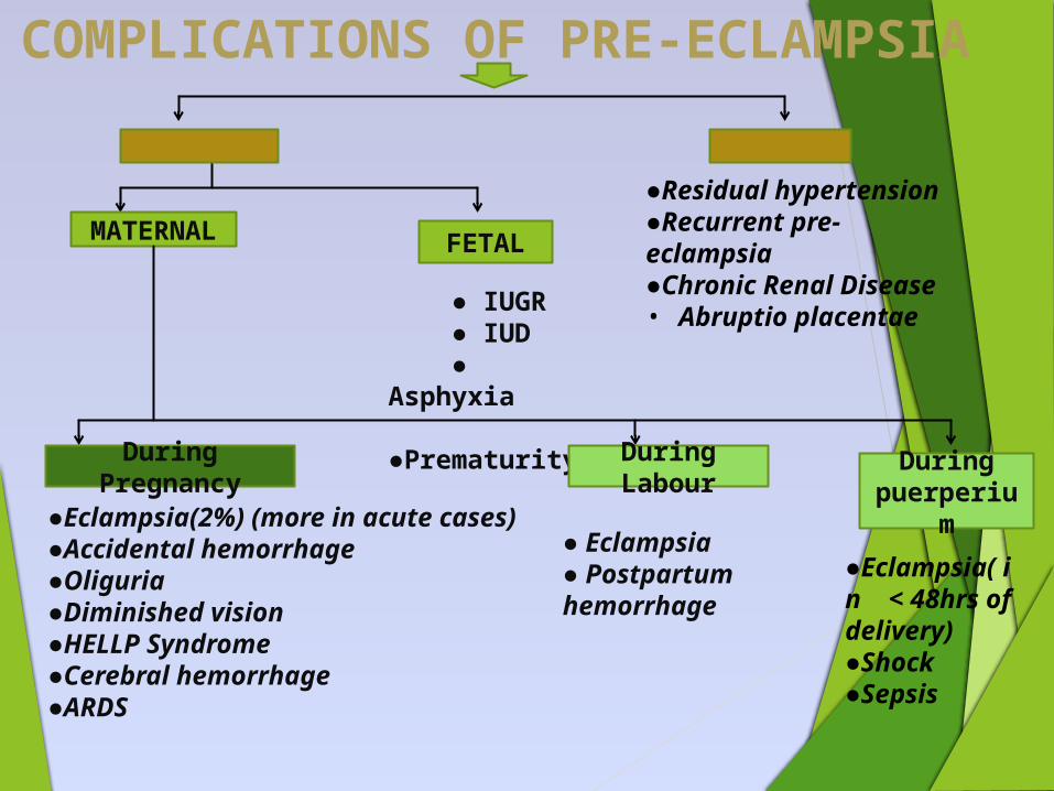
COMPLICATIONS OF PRE-ECLAMPSIA
IMMEDIATE
REMOTE
MATERNAL FETAL
● IUGR ● IUD ● Asphyxia ●Prematurity
During Pregnancy
During Labour
During puerperiu
m●Eclampsia(2%) (more in acute cases)●Accidental hemorrhage●Oliguria●Diminished vision●HELLP Syndrome●Cerebral hemorrhage●ARDS
● Eclampsia● Postpartum hemorrhage
●Eclampsia( in < 48hrs of delivery)●Shock ●Sepsis
●Residual hypertension●Recurrent pre-eclampsia●Chronic Renal Disease• Abruptio
placentae
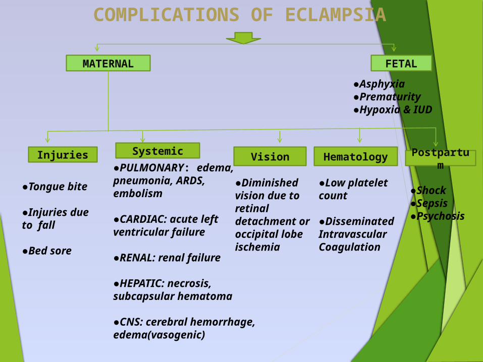
COMPLICATIONS OF ECLAMPSIA
MATERNAL FETAL
●Asphyxia●Prematurity●Hypoxia & IUD
Injuries Systemic
●Tongue bite
●Injuries due to fall
●Bed sore
●PULMONARY: edema, pneumonia, ARDS, embolism
●CARDIAC: acute left ventricular failure
●RENAL: renal failure
●HEPATIC: necrosis, subcapsular hematoma
●CNS: cerebral hemorrhage, edema(vasogenic)
Vision
●Diminished vision due to retinal detachment or occipital lobe ischemia
Hematology
●Low platelet count
●Disseminated Intravascular Coagulation
Postpartum
●Shock●Sepsis●Psychosis
HELLP Syndrome
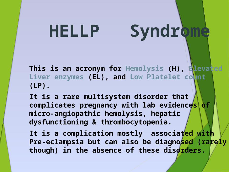
This is an acronym for Hemolysis (H), Elevated Liver enzymes (EL), and Low Platelet count (LP).
It is a rare multisystem disorder that complicates pregnancy with lab evidences of micro-angiopathic hemolysis, hepatic dysfunctioning & thrombocytopenia.
It is a complication mostly associated with Pre-eclampsia but can also be diagnosed (rarely though) in the absence of these disorders.
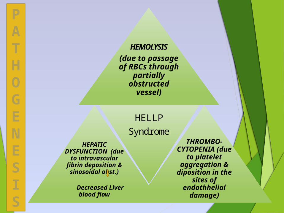
HEMOLYSIS(due to
passage of RBCs through
partially obstructed
vessel)
s)HEPATIC DYSFUNCTION
(due to intravascular
fibrin deposition & sinosoidal
obst.)
Decreased Liver
blood flow
HELLPSyndrome
THROMBO-CYTOPENIA
(due to platelet aggregation & diposition in the sites of
endothhelial damage)
PATHOGENESIS
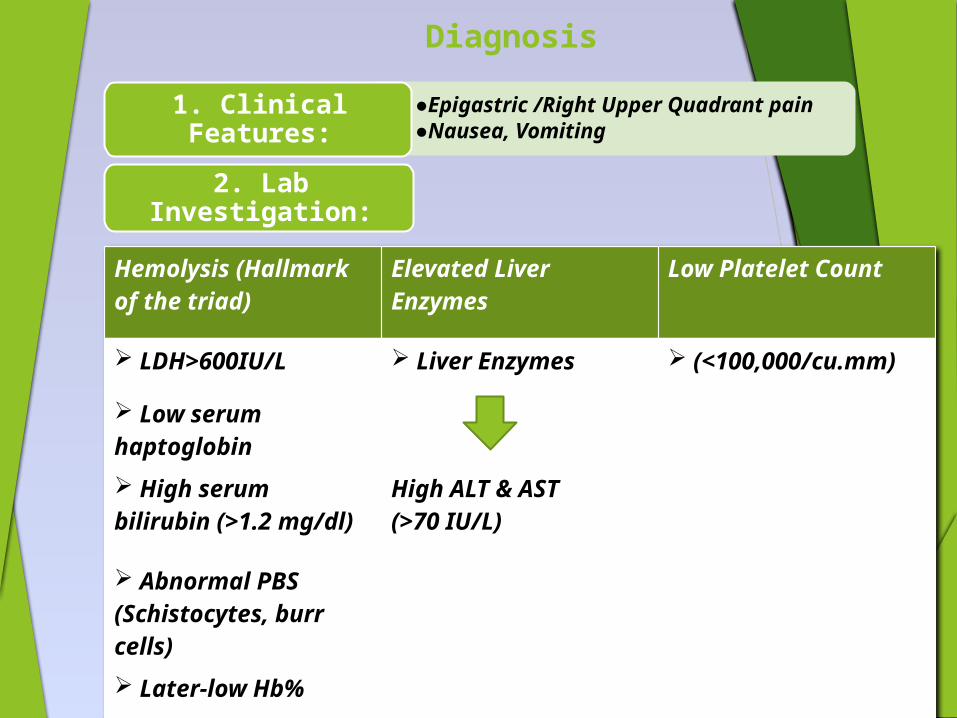
Diagnosis
Hemolysis (Hallmark of the triad)
Elevated Liver Enzymes
Low Platelet Count
LDH>600IU/L Liver Enzymes (<100,000/cu.mm)
Low serum haptoglobin
High serum bilirubin (>1.2 mg/dl)
High ALT & AST(>70 IU/L)
Abnormal PBS (Schistocytes, burr cells) Later-low Hb%
• ●Epigastric /Right Upper Quadrant pain
• ●Nausea, Vomiting
1. Clinical Features:
2. Lab Investigation:

Treatment
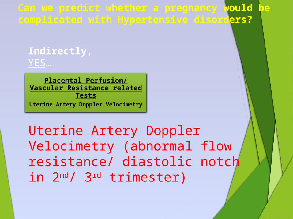
Can we predict whether a pregnancy would be complicated with Hypertensive disorders?
Indirectly, YES…
Placental Perfusion/ Vascular Resistance related
TestsUterine Artery Doppler Velocimetry
Uterine Artery Doppler Velocimetry (abnormal flow resistance/ diastolic notch in 2nd/ 3rd trimester)
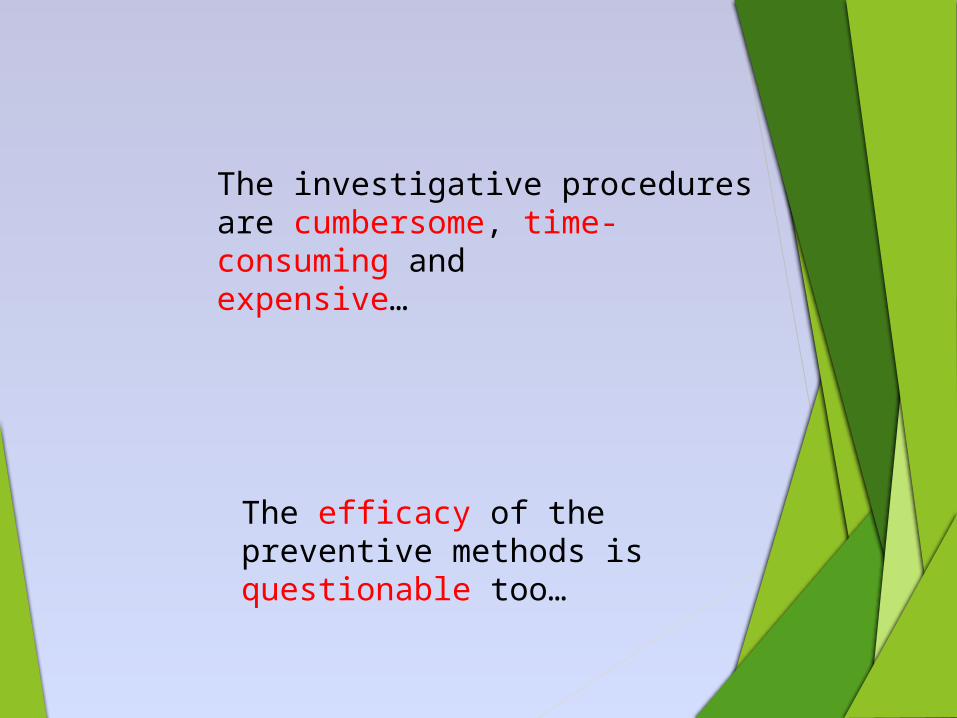
The efficacy of the preventive methods is questionable too…
The investigative procedures are cumbersome, time-consuming and expensive…
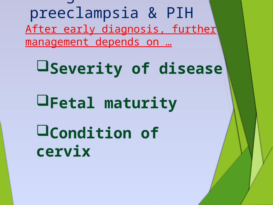
Management of preeclampsia & PIH
After early diagnosis, further management depends on …
Severity of disease
Fetal maturity
Condition of cervix

Treatment proper
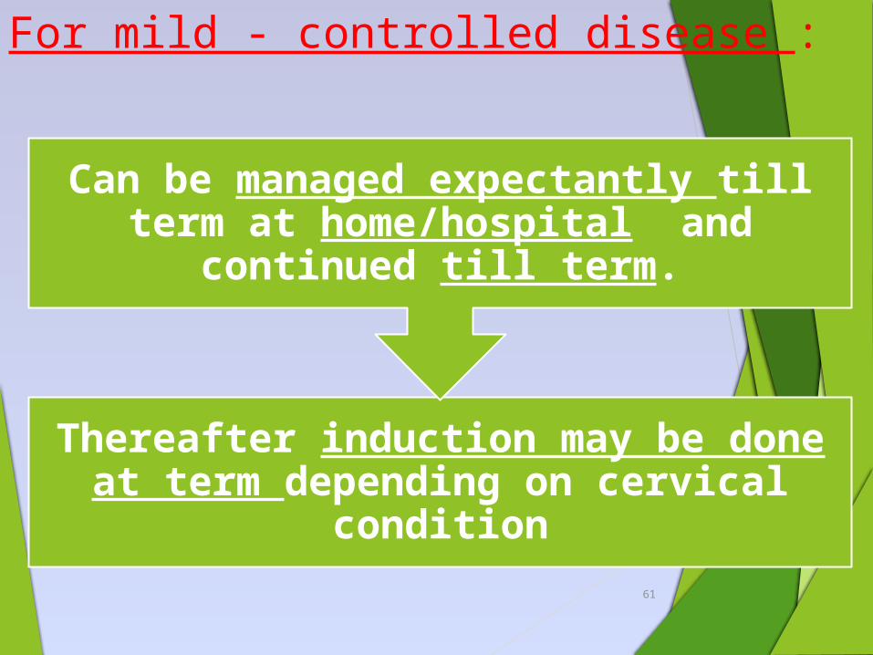
For mild - controlled disease :
Thereafter induction may be done at term depending on cervical
condition
Can be managed expectantly till term at home/hospital and
continued till term.
61
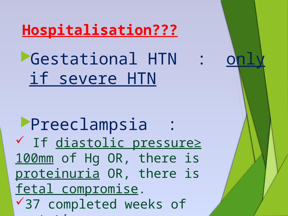
Hospitalisation???
Gestational HTN : only if severe HTN
Preeclampsia : If diastolic pressure≥ 100mm of Hg OR, there is proteinuria OR, there is fetal compromise.37 completed weeks of gestation.
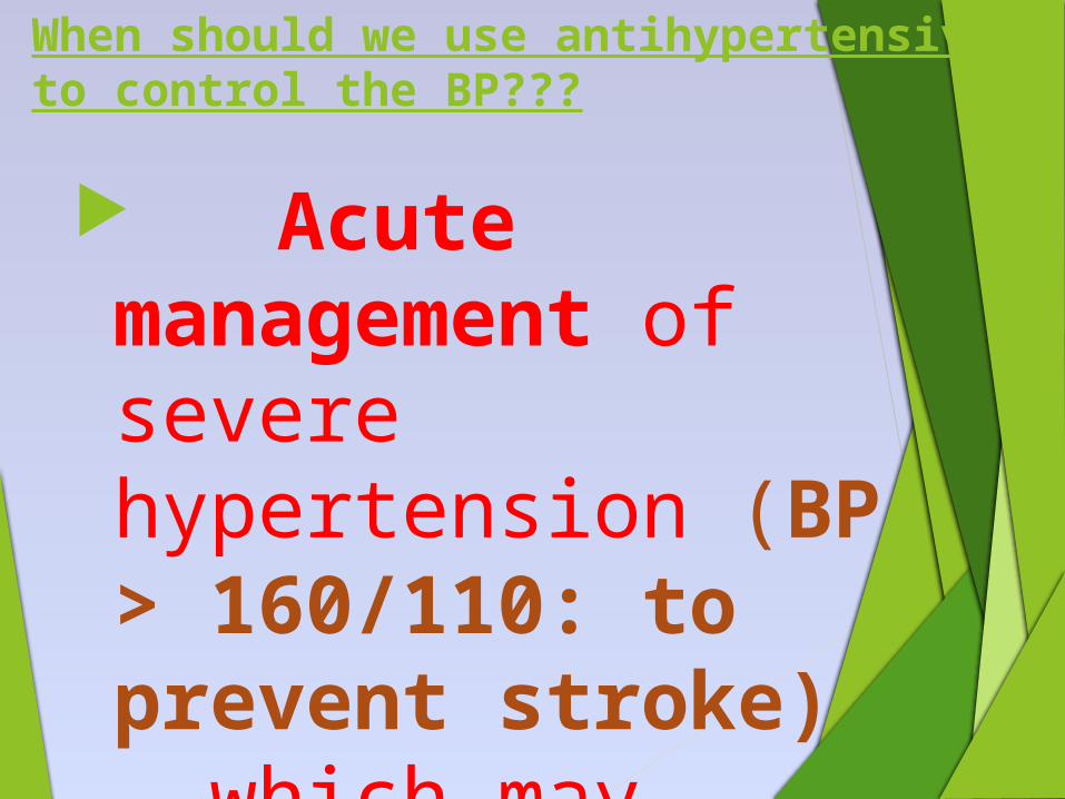
When should we use antihypertensive to control the BP???
Acute management of severe hypertension (BP > 160/110: to prevent stroke) which may require parenteral therapy.
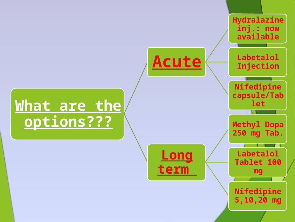
What are the options???
Acute
Hydralazine inj.: now available
Labetalol Injection
Nifedipine capsule/Tabl
et
Long term
Methyl Dopa 250 mg Tab.
Labetalol Tablet 100
mg
Nifedipine 5,10,20 mg
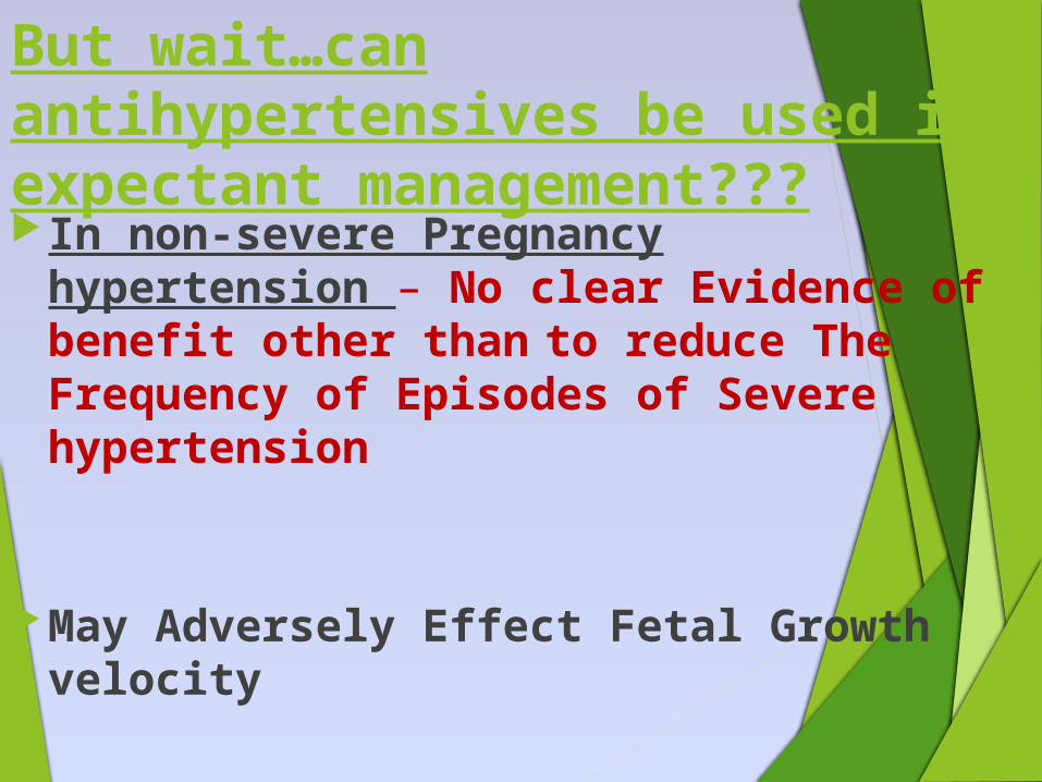
But wait…can antihypertensives be used in expectant management???In non-severe Pregnancy
hypertension – No clear Evidence of benefit other than to reduce The Frequency of Episodes of Severe hypertension
May Adversely Effect Fetal Growth velocity
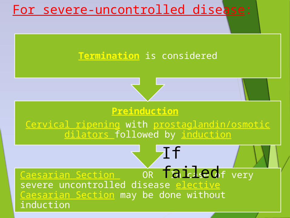
For severe-uncontrolled disease:
Caesarian Section OR In case of very severe uncontrolled disease elective Caesarian Section may be done without induction
Preinduction Cervical ripening with prostaglandin/osmotic dilators
followed by induction
Termination is considered
66
If failed
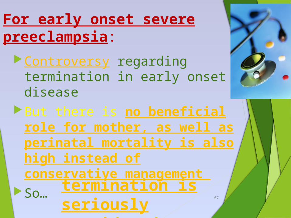
For early onset severe preeclampsia:
Controversy regarding termination in early onset disease
But there is no beneficial role for mother, as well as perinatal mortality is also high instead of conservative management
So… 67
termination is seriously considered
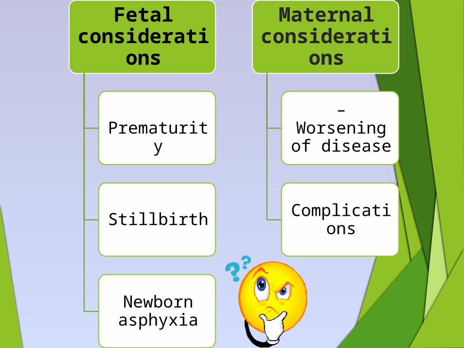
Fetal considerati
ons
Prematurity
Stillbirth
Newborn asphyxia
Maternal considerati
ons
– Worsening of disease
Complications
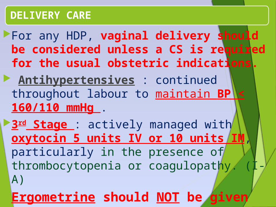
DELIVERY CARE
For any HDP, vaginal delivery should be considered unless a CS is required for the usual obstetric indications.
Antihypertensives : continued throughout labour to maintain BP < 160/110 mmHg .
3rd Stage : actively managed with oxytocin 5 units IV or 10 units IM, particularly in the presence of thrombocytopenia or coagulopathy. (I-A)
Ergometrine should NOT be given
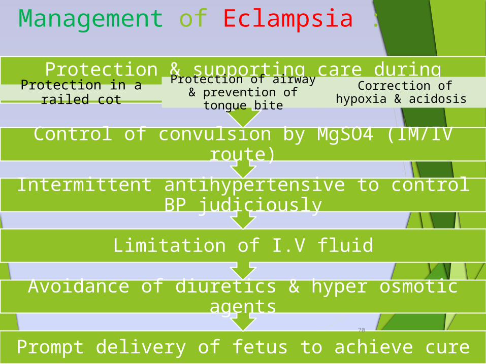
Management of Eclampsia :
Prompt delivery of fetus to achieve cure
Avoidance of diuretics & hyper osmotic agents
Limitation of I.V fluid
Intermittent antihypertensive to control BP judiciously
Control of convulsion by MgSO4 (IM/IV route)
Protection & supporting care during convulsionProtection in a railed cot
Protection of airway & prevention of tongue
bite
Correction of hypoxia & acidosis
70
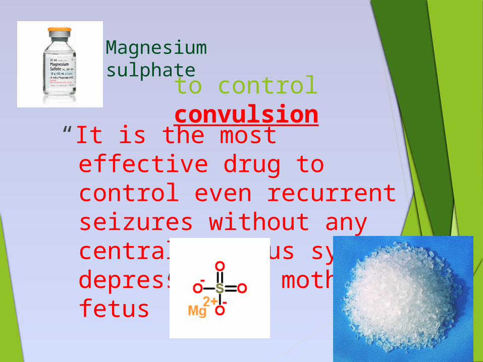
to control convulsion
“It is the most effective drug to control even recurrent seizures without any central nervous system depression to mother & fetus”
71
Magnesium sulphate
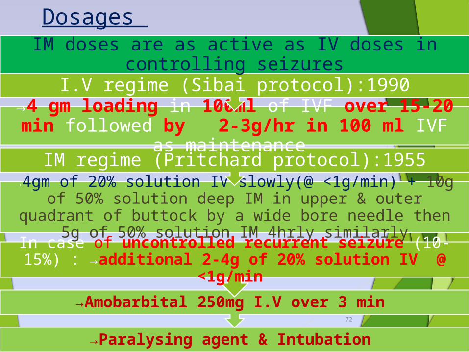
Dosages
→Paralysing agent & Intubation
→Amobarbital 250mg I.V over 3 min
In case of uncontrolled recurrent seizure (10-15%) : →additional 2-4g of 20% solution IV @ <1g/min
→4gm of 20% solution IV slowly(@ <1g/min) + 10g of 50% solution deep IM in upper & outer quadrant of
buttock by a wide bore needle then 5g of 50% solution IM 4hrly similarly
IM regime (Pritchard protocol):1955
→4 gm loading in 100ml of IVF over 15-20 min followed by 2-3g/hr in 100 ml IVF as
maintenance
I.V regime (Sibai protocol):1990
IM doses are as active as IV doses in controlling seizures
72
S o m e mo re a b o u t Ma g n e s iu m
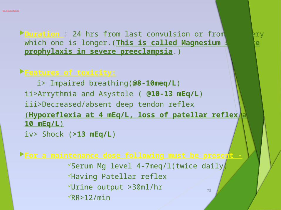
Duration : 24 hrs from last convulsion or from delivery which one is longer.(This is called Magnesium sulphate prophylaxis in severe preeclampsia.)
Features of toxicity:
i> Impaired breathing(@8-10meq/L)
ii>Arrythmia and Asystole ( @10-13 mEq/L)
iii>Decreased/absent deep tendon reflex
(Hyporeflexia at 4 mEq/L, loss of patellar reflex at 7-10 mEq/L)
iv> Shock (>13 mEq/L)
For a maintenance dose following must be present - Serum Mg level 4-7meq/l(twice daily) Having Patellar reflex Urine output >30ml/hr RR>12/min
73
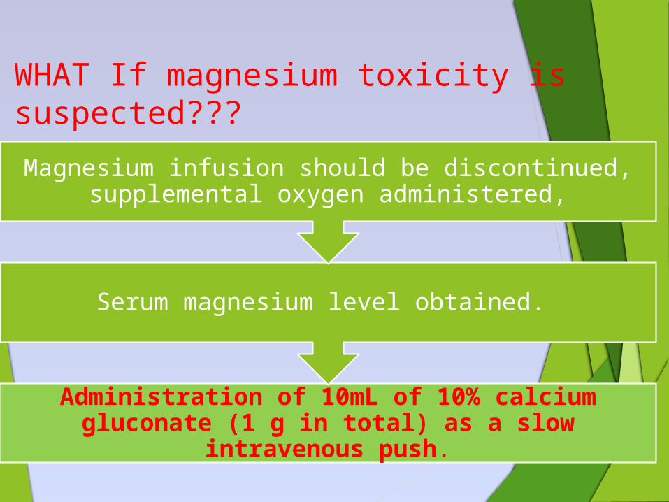
WHAT If magnesium toxicity is suspected???
Administration of 10mL of 10% calcium gluconate (1 g in total) as a slow intravenous
push.
Serum magnesium level obtained.
Magnesium infusion should be discontinued, supplemental oxygen administered,

Thank You!!!