Embed Size (px)
Citation preview
Histology of Cardiac
Muscle
1
BY: Jehad Abdullah
Introduction
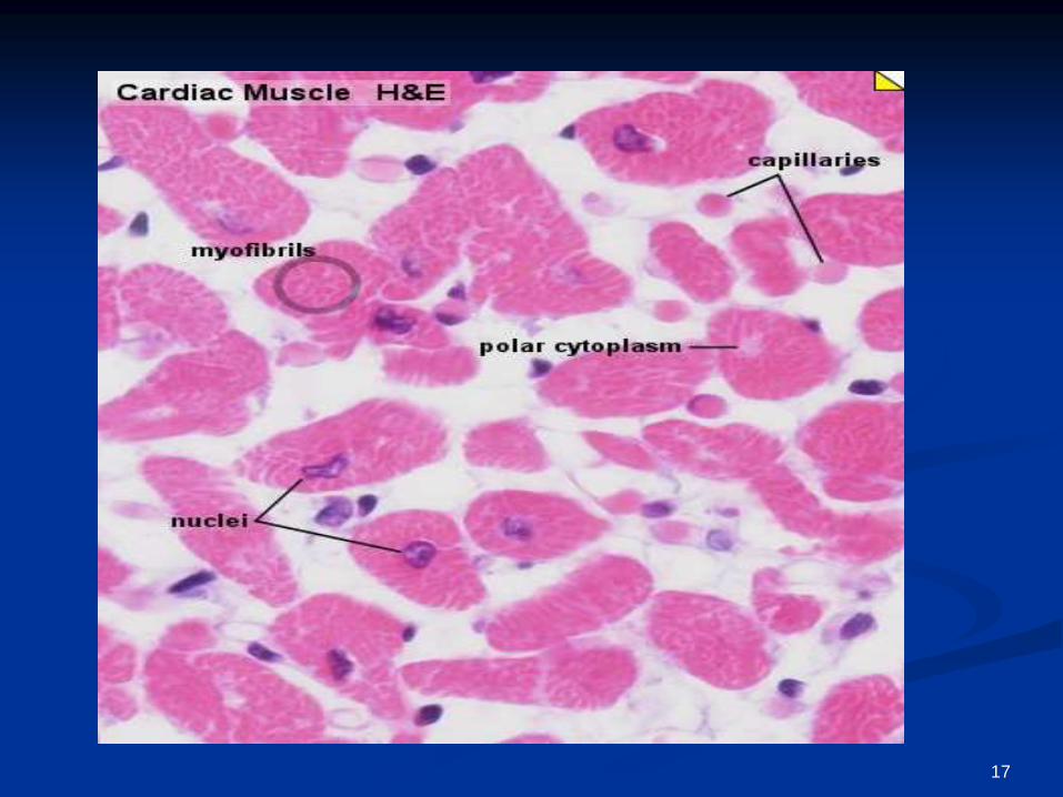
Cardiac muscle, the myocardium, consists of cross-
striated muscle cells, cardiomyocytes, with one
centrally placed nucleus.
Nuclei are oval, rather pale and located centrally in the
muscle cell which is 10 - 15 µm wide.
Cardiac muscle cells excitation is mediated by
rythmically active modified cardiac muscle cells.
Cardiac muscle is innervated by the autonomic
nervous system (involuntary), which adjusts the force
generated by the muscle cells and the frequency of the
heart beat.
Cardiac muscle does not contain cells equivalent to
the satellite cells of skeletal muscle
2
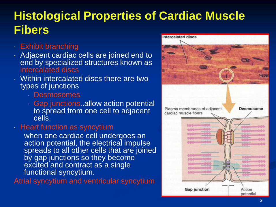
Exhibit branching Adjacent cardiac cells are joined end to
end by specialized structures known as intercalated discs
Within intercalated discs there are two types of junctions
Desmosomes Gap junctions..allow action potential
to spread from one cell to adjacent cells.
Heart function as syncytiumwhen one cardiac cell undergoes an action potential, the electrical impulse spreads to all other cells that are joined by gap junctions so they become excited and contract as a single functional syncytium.
Atrial syncytium and ventricular syncytium
Histological Properties of Cardiac Muscle
Fibers
3
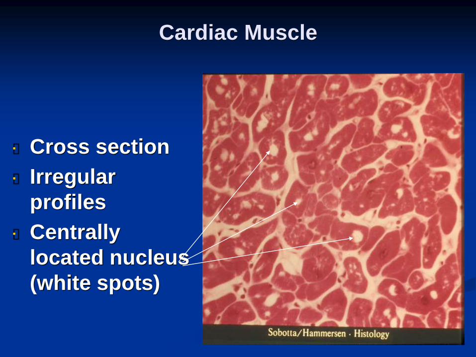
Cardiac Muscle
Cross section
Irregular
profiles
Centrally
located nucleus
(white spots)
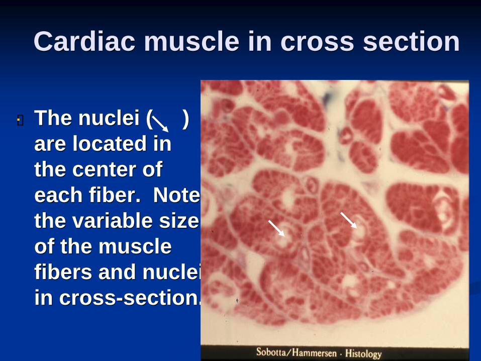
Cardiac muscle in cross section
The nuclei ( )
are located in
the center of
each fiber. Note
the variable size
of the muscle
fibers and nuclei
in cross-section.
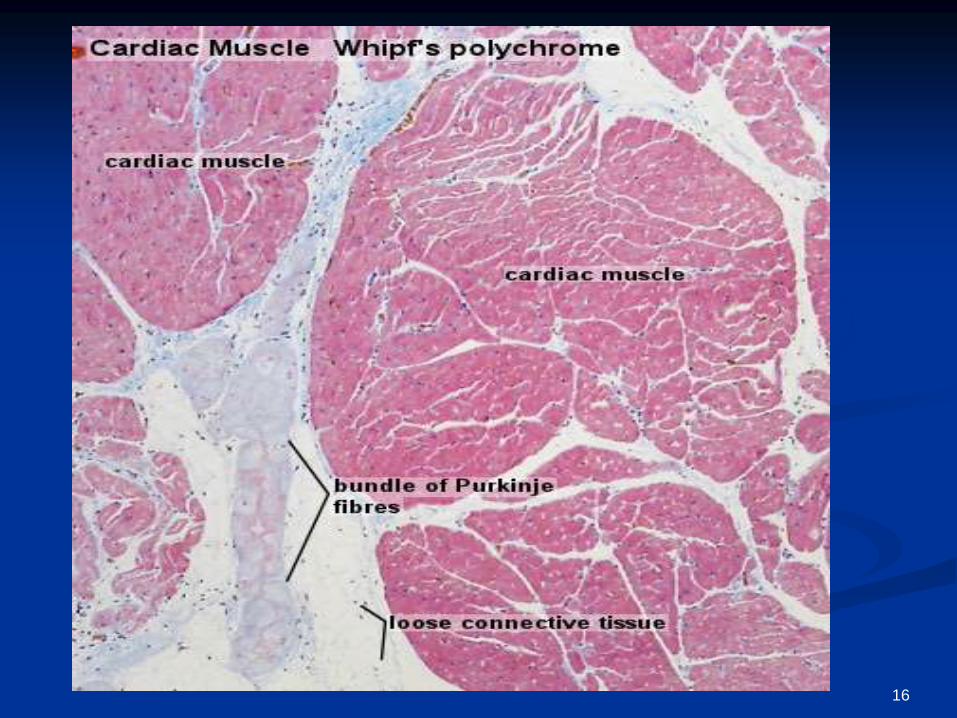
Purkinje Fibres:-
modified cardiac muscle cells. Compared to
ordinary cardiac muscle cells:
contain large amounts of glycogen.
fewer myofibrils.
thicker cells.
extend from the atrioventricular node, pierces the
fibrous body, divides into left and right bundles,
and travels, beneath the endocardium, towards
the apex of the heart.
conduct stimuli faster than ordinary cardiac
muscle cells (2-3 m/s vs. 0.6 m/s).
discovered in 1839 by Jan Evangelista Purkyně)
6

Intercalated Discs
seen in longitudinal sections.
connect the individual muscle cells.
permit the conduction of electrical impulses
between the cells.
7
Junctional Components
Fascia adherens – major portion of
transverse component. Anchoring sites for
actin, and connect to the closest
sarcomere.
- The contractile unit of a skeletal
muscle fiber. Sarcomeres are divided into b
ands of filaments made of actin or myosin.
During muscle contraction, the filaments sli
de over each other to cause shortening of t
he sarcomere.8
cont,
Macula adherens – (desmosomes)
transverse and lateral components. Bind
individual myocytes to one another. stop
separation during contraction by binding
intermediate filaments, joining the cells
together. Macula adherens junctions are
also called desmosomes.
9
Cont,
Gap junctions - lateral component. Allow
action potentials to spread between cardiac
cells by passage of ions between cells,
producing depolarization of the heart
muscle. Allows muscle to act as syncytium.
10
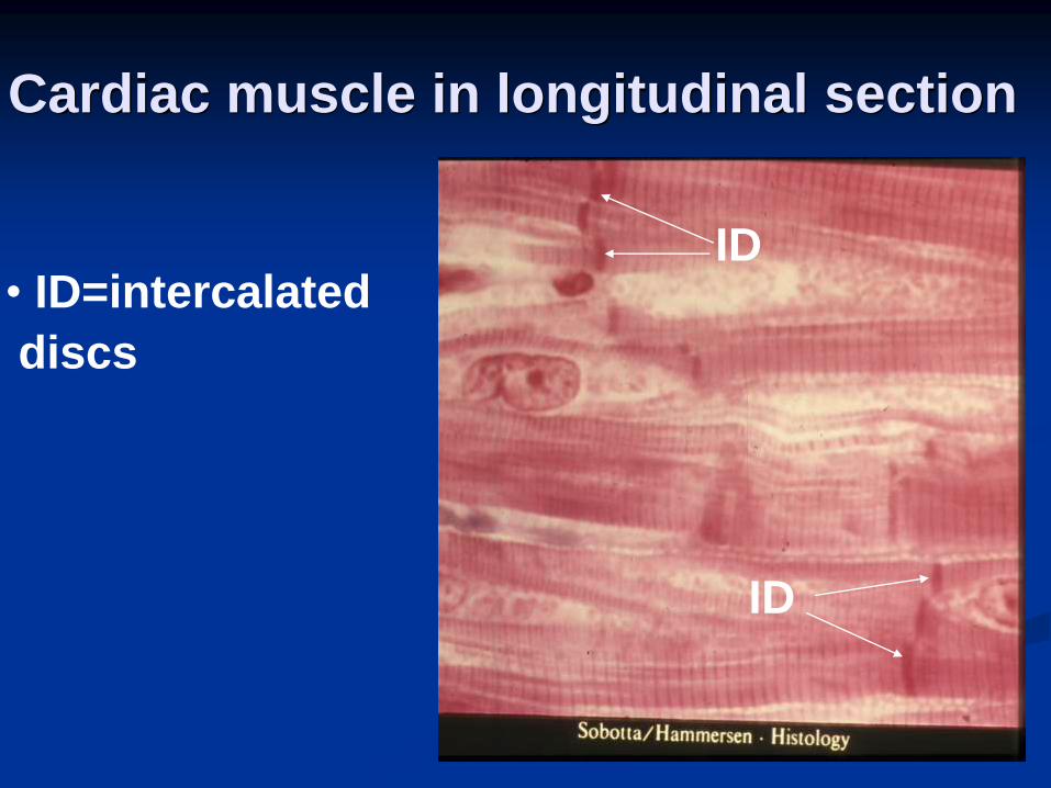
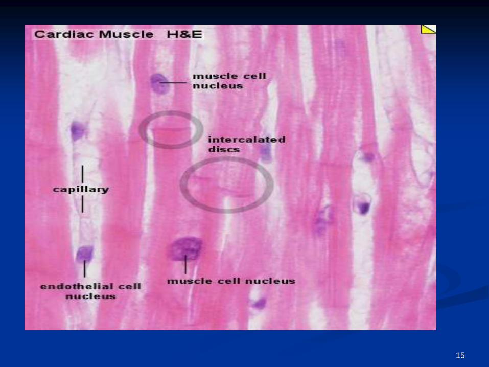
Cardiac muscle in longitudinal section
• ID=intercalated
discs
ID
ID
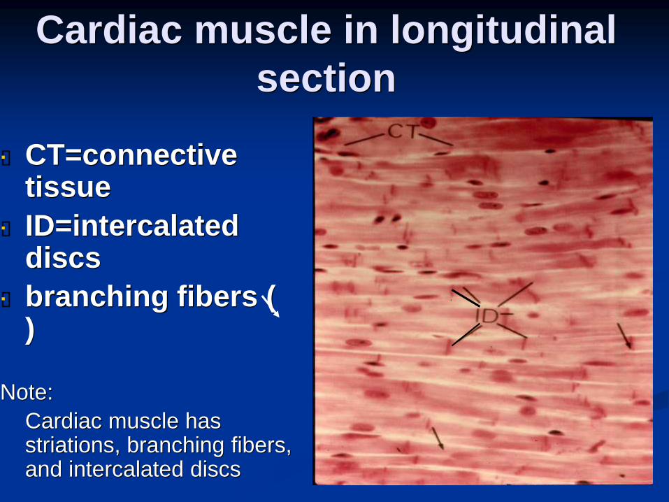
Cardiac muscle in longitudinal
section
CT=connective tissue
ID=intercalated discs
branching fibers ( )
Note:
Cardiac muscle has striations, branching fibers, and intercalated discs
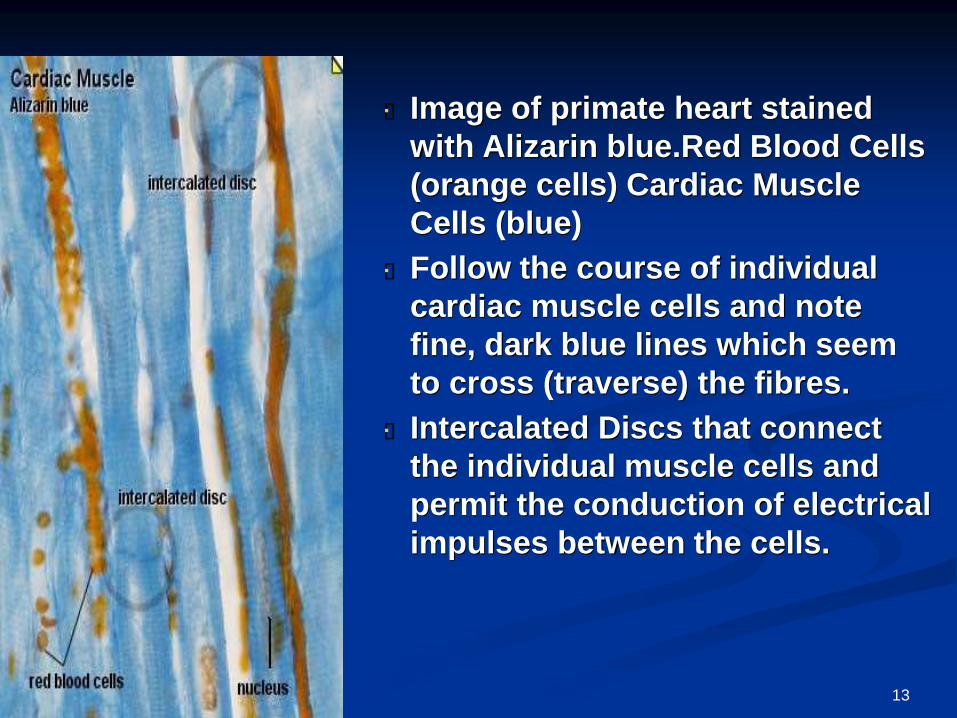
Image of primate heart stained
with Alizarin blue.Red Blood Cells
(orange cells) Cardiac Muscle
Cells (blue)
Follow the course of individual
cardiac muscle cells and note
fine, dark blue lines which seem
to cross (traverse) the fibres.
Intercalated Discs that connect
the individual muscle cells and
permit the conduction of electrical
impulses between the cells.
13
14
15
16
17