Embed Size (px)
Citation preview

Moderator : Dr. M. S. Somannavar
Presenter : Jay prakash sah
Genetic code
Salient features
Mutation
Types of mutation
Mutagens
Clinical correlations

Crick, Brenner et al. experiment - Demonstratethat codons consist of three DNA bases
Marshall Nirenberg and Heinrich J. Matthaei in1961 reported the first three letter code words foreach amino acid
H. Gobind Khorana- Genetic code dictionary andprevious results confirmed

Dictionary that identifies the correspondencebetween a sequence of nucleotide bases and asequence of amino acids
Codon- triplet sequence of nucleotides on themRNA ( A G,C & U)
64 codon

Triplet codons
Non-overlapping
Non-punctuated
Degenerate
Unambiguous
Universal
Wobbling phenomenon
Terminator codon
Initiator codon

Consecutive sequence of three bases onmRNA
Ex:
UUU -Phenylalanine


AUACGAGUC
A U A C G A G U C
1 2 3
overlapping code
A U A C G A G U C

Consecutive or continuous


A particular codon always codes for the sameamino acid
Ex:
UGG - Tryptophan

Same codons are used to code for the same amino acid in all the living organisms.

I


Amber(UAA),Ochre(UAG) & Opal codons(UGA)
UGA - Selenocysteine(Sec)-21(Glutathione peroxidase)
UAG - Pyrolysine-22(Methyl transferase)
(Methanosarcina barkeri)
UGA & UAA- Glutamine( paramecium)
UGA – Trp (mycoplasma)
Garret

AUG - codon for methionine.
In few proteins,
GUG

Change in nucleotide sequence of DNA
Out of every 106 cell divisions-1 mutation occurs
May occur in somatic cells ( aren’t passedoffspring)
May occur in gametes (eggs & sperm) and passedto offspring


Substitution of one base pair by another .
Transition :
purine to purine
Transversions :
purine to pyrimidine

satyanarayana

Mutations: SubstitutionsSubstitution mutation
GGTCACCTCACGCCA
↓
CCAGUGGAGUGCGGU
↓
Pro-Arg-Glu-Cys-Gly
Normal gene
GGTCTCCTCACGCCA
↓
CCAGAGGAGUGCGGU
Codons
↓
Pro-Glu-Glu-Cys-Gly
Amino acids

Mutation Codon Change to DNA
sense strand
Change in
Amino Acid
S (sickle cell
anaemia)
6 GAG to GUG Glu to Val
C (cooley’s
syndrome)
6 GAG to AAG Glu to Lys
GSan Jose 7 GAG to GGG Glu to Gly
E 26 GAG to AAG Glu to Lys
MSaskatoon 63 CAT to TAT His to Tyr
MMilwauki 67 GTG to GAG Val to Glu
OArabia 121 GAA to GTA Glu to Val

One or more base pairs are inserted in or deleted from the DNA .


I . Single base additions ,
Normal gene
GGTCTCCTCACGCCA
↓
CCAGAGGAGUGCGGU
Codons
↓
Pro-Glu-Glu-Cys-Gly
Amino acids
Addition mutation
GGTGCTCCTCACGCCA
↓
CCACGAGGAGUGCGGU
↓
Pro-Arg-Gly-Val-Arg

II. Trinucleotide expansion
Huntingtons’s chorea
CAG repeated 30 to 300 times
III. Duplication.
Ex: Duchene Muscular Dystrophy

i) Large gene deletion
Ex: Alpha-thalassemia
i) Deletion of codon
Ex: Cystic fibrosis
i) Deletion of a single base

Consequences of point mutation
Normal gene
GGTCTCCTCACGCCA
↓
CCAGAGGAGUGCGGU
Codons
↓
Pro-Glu-Glu-Cys-Gly
Amino acids
Substitution mutation
GGTCTTCTCACGCCA
↓
CCAGAAGAGUGCGGU
↓
Pro-Glu-Glu-Cys-Gly
1. SILENT MUTATION

A. Acceptable
Eg: Normal Hemoglobin A molecule ,
67th amino acid in beta chain
GUU(Val)
GCU(Ala)
Hb sydney (functionally normal)

Eg: HbS or sickle cell Haemoglobin
beta chain - 6th position
GAG(glutamine)
GUG( Valine).
Hbs leads to sickle cell anemia.
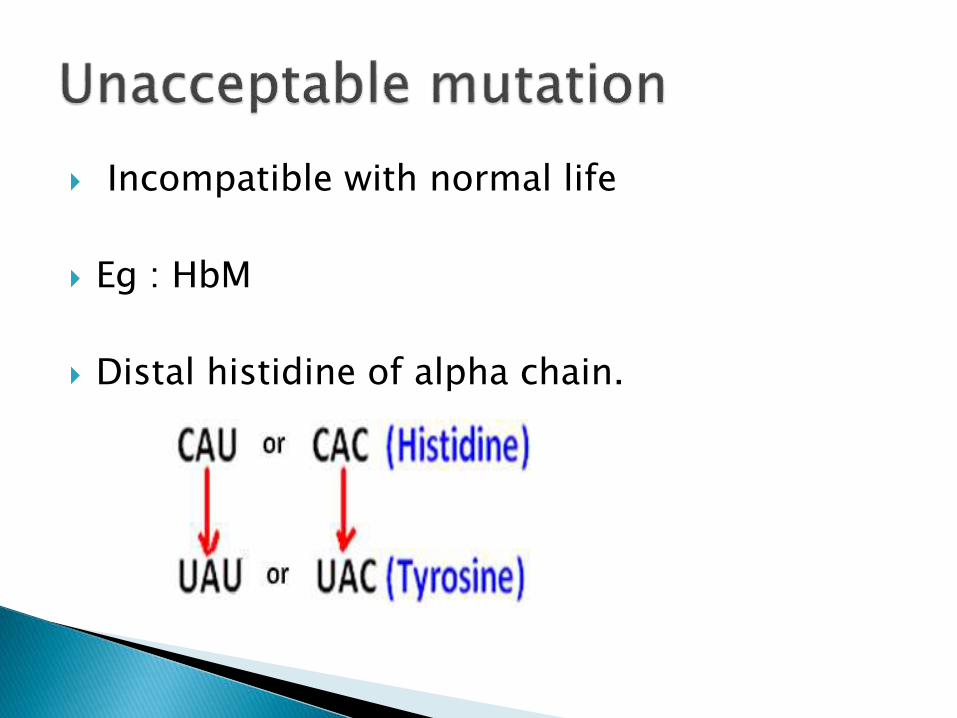
Incompatible with normal life
Eg : HbM
Distal histidine of alpha chain.

leads to premature termination of the protein
Eg: Thalassemia
Codon 17 of the β-chain

Non-SenceNormal gene
GGTCTCCTCACGCCA
↓
CCAGAGGAGUGCGGU
Codons
↓
Pro-Glu-Glu-Cys-Gly
Amino acids
Substitution mutation
GGTCTCCTCACTCCA
↓
CCAGAAGAGUGAGGU
↓
Pro-Glu-Glu-STOP


Insertion or deletion of base in a gene results
in an altered reading frame of the mRNA
A ‘garbled’ protein - produced.

Normal mRNA AUG UCU UGC AAA……..
Normal Protein Met Ser Cys Lys …….
Deleted U mRNA AUG CUU GCA AA…..
garbled Protein Met Leu Ala ……..

1. physical agents
i) UV light
ii) Ionising radiation e.g. X-ray.
iii) visible light
iv) Heat
2. chemical agents
i) 5-Bromouracil
ii) 2-Aminopurine
iii) Nitrous acid
iv) Acridine dyes.

Spontaneous tautomeric shifts in the basescontribute to replication errors
Ex: Thymine (keto form) shifts to enol form ,which pairs with guanine


Double strand DNA breaks
It penetrates the whole body –
cause both somatic and Germ line mutations

Mutagenic component of sunlight
Can not penetrate beyond the outer layer ofthe skin and - unable cause germ linemutations.
only causes sunburn and skin cancer mainlythrough the formation of pyrimidine dimers


1. Base analog
Bromouracil (structural analog of Thymine)
Enzyme of nucleotide synthesis and DNA synthesistreat Bromouracil as thymine and incorporate itinto DNA , where it pairs with adenine


Attach alkyl groups to nitrogen or oxygen atomsin the bases.
Ex:
Methyl bromide ( used as grain fumigent)
Ethylene oxide (used for sterilization of surgical instruments)


Planar fused ring structures –
Insert themselves between the stacked DNA bases.

Salmonella Typhimurium (his-) are selected
Mutagenesis – indicated by his+ phenotype.
The compound to be tested is mixed withbacteria and introduced into histidine deficientmedium.
Reverse mutation.
Number of colonies -proportional to quantity ofmutagen.

Ethyl methanasulphonate
Spontaneous revertantcolonies

1.Single strand conformation polymorphism(SSCP technique)
2.Heteroduplex Analysis
3.Conformation sensitive gel electrophoresis
4.Protein truncation test(PTT)
5.Denaturing HPLC

CLINICAL CORRELATION

HbS
Sickle cell disease:
Due to missense mutation.
Changes from A to U
GAA or GAG (Glu)
GUA or GUG (Val)


Hemoglobin C disease:
Due to missense mutation.
Changes from G to A
GAA or GAG (Glu)
AAA or AAG (Lys)

In Hb Mckees Rocks
145 ( beta chain)
UAU or UAC (Tyrosine)
UAA or UAG(terminator codon )
Shortening of the beta chain from its normal 146residue to 144 residues
cause overproduction of red blood cells

>36
>200
100-1000


Encoding genes on chromosome 16 (shortarm)
Each cell has 4 copies of the alpha globingene
◦ Loss of ONE gene silent carrier◦ Loss of TWO genes thalassemia minor (trait)◦ Loss of THREE genes Hemoglobin H
◦ Loss of FOUR genes Hemoglobin Barts

Encoding genes on chromosome 11 (short arm)
Each cell contains 2 copies of beta globin gene
“Loss” of ONE gene thalassemia minor (trait)
“Loss” of BOTH gene Thalassemia major

Hb α-codon(142)
Amino acid(142)
α-globin length(residues)
A UAA 141
Contant spring CAA Glutamine 172
Icaria AAA Lysine 172
Seal Rock GAA Glutamate 172
Koya Dora UCA Serine 172

Deletion of phe residue at position 508 inCFTR Gene (chromosome 7)
Causes improper folding of protein
Defective chloride transport ( pancreas,lungtestis & sweat glands)


Harper’s Review of Biochemistry
Lehniger’s principle of Biochemistry
Lippincott’s Illustrated Review of Biochemistry
Text Book of Biochemistry with clinical correlations- Devlin TM
Text Book of Biochemistry by Vasudevan
Text book of biochemistry, satyanarayana
Principle of biochemistry, William H. simmons.

zxcvbnmasfghhjlkjhgfddssaqwertyiopplkjhgfdsazxcvbnmmlkjhgdssaaaqwwerlkj