Embed Size (px)
Citation preview

Fluids & Electrolytes
Abdelrahman Al-daqqaAbdelrahman Al-daqqa
An-najah UniversityAn-najah University

BODY FLUIDS
Total Body Water
Fifty to seventy percent of total body weight. Greater in lean individuals because fat contains little water,
average 60%. Greatest percentage in newborns 70%, then decreases with
age to around 50%. Example: Average 70-kg male would be 42 L water since
1 L of water = 1 kg. Made up of two compartments—ICF and ECF.

Intracellular Fluid (ICF(
Mostly in skeletal muscle mass, thus slightly lower in females (50%) than males (60%).
Cell wall separates the ICF from the ECF and acts as a semipermeable membrane.

Extracellular Fluid (ECF(
Made up of plasma and interstitial (extravascular) fluid.
Capillary membrane separates plasma and interstitial fluid and acts as a semipermeable membrane .


NORMAL F LUID AND E LECTROLYTE EXCHANGEWater Movement Between ICF and ECF
Water flows freely between the three compartments, shifting compartments to maintain osmotic equilibrium between them .
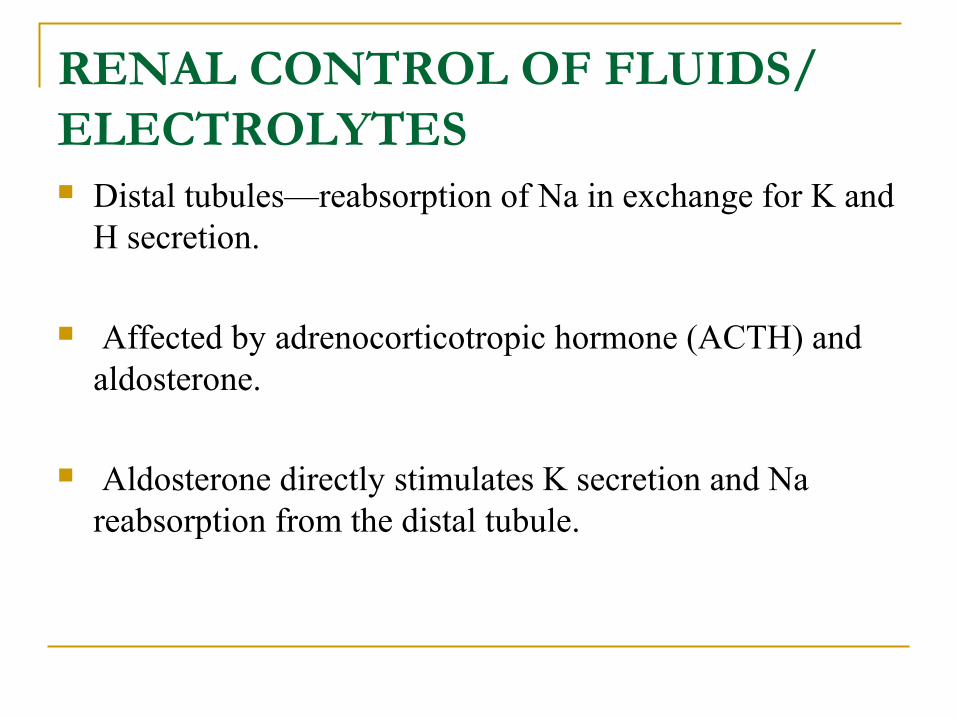
RENAL CONTROL OF FLUIDS/ ELECTROLYTES Distal tubules—reabsorption of Na in exchange for K and
H secretion.
Affected by adrenocorticotropic hormone (ACTH) and aldosterone.
Aldosterone directly stimulates K secretion and Na reabsorption from the distal tubule.

Volume Deficit (Dehydration(

Volume Deficit (Dehydration(
Most common fluid disorder.CAUSES :1- Losses that Mimic ECF Hemorrhage. Loss of gastrointestinal (GI) fluid—vomiting, nasogastric (NG)
suction, diarrhea, fistular drainage. Postoperative fluid sequestration (third spacing): Intestinal obstruction. Intra-abdominal and retroperitoneal inflammation (e.g., pancreatitis, peritonitis). Systemic inflammatory response syndrome (SIRS), burns, sepsis,
pancreatitis.

Volume Deficit (Dehydration(
CAUSES :
2- Losses that Are Principally Water Fever. Osmotic diuresis. Diabetes insipidus. Prolonged water deprivation. Inadequate input during procedure.

SIGNS AND SYMPTOMS
Central nervous system (CNS) and cardiovascular (CV) signs occur early with acute loss.
CV signs are secondary to a decrease in plasma volume.
Tissue signs may be absent until the deficit has existed for 24 hours.
Tissue signs may be diffi cult to assess in the elderly patient or patient with recent weight loss.
SIGNS AND SYMPTOMS
Body temperature varies with environment—cool room may mask fever.
After partial correction of volume deficit, the temperature will generally rise to the appropriate level.
Severe volume depletion depresses all body systems and interferes with the clinical evaluation of the patient.
SIGNS AND SYMPTOMS
Volume depleted patient with severe sepsis from peritonitis may be afebrile and have normal white blood count (WBC), complain of little pain, and have unremarkable findings on abdominal exam. This may change dramatically when the ECF is restored.

History items important for evaluating fluid deficits include:
Weight change, intake (quantity and composition), output, general medical status.
Degree of dehydration dependent on acute loss of body weight and is assessed clinically:
1. Mild—3% for adults, 5% for kids2. Moderate—6% for adults, 10% for kids3. Severe—9% for adults, 15% for kids

VOLUME EXC E SS
CAUSES
1-Isotonic Iatrogenic—intravascular overload of IV fl uids with
electrolytes. Increased ECF without equilibration with ICF especially
postoperative or trauma when the hormonal responses to stress are to decrease Naand water excretion by kidney.
Often secondary to renal insuffi ciency, cirrhosis, or CHF.

2-Hypotonic
Inappropriate NaCl-poor solution as a replacement for GI losses (most common).
Third spacing. Increased antidiuretic hormone (ADH) with
surgical stress, inappropriate ADH (SIADH).

3-HypertonicMost common cause: excessive Na load without adequate water intake:
Water moves out of the cells because of increased ECF osmolarity.
Causes an increase in intravascular and interstitial fluid.
Worse when renal tubular excretion of water and/or Na is poor.
Can also be caused by rapid infusion of nonelectrolyte osmotically active solutes such as glucose and mannitol.

SIGNS AND SYMPTOMS

ONGOING FLUID LOSS
Besides normal maintenance loss, there may be other ongoing losses.
Rule of thumb: Replace one half of the “usual” ongoing losses along with the assumed maintenance and the rehydration replacement fluid.
Electrolyte content of the ongoing loss can be either assumed based on serum electrolyte values or can be determined by direct electrolyte measurement of the fluid

ONGOING FLUID LOSS
CAUSES
Fever: Each °C above 37°C adds 2.0 to 2.5 mL/kg/day of insensible water loss.
Loss of body fluids: From vomit, NG suction, fistulas.

ONGOING FLUID LOSS
Third-space losses:
Adults—approximately 1 L of third-space fluid intra-abdominally for each quadrant of the abdomen that is traumatized, inflamed, or operated on.
Kids—approximately one fourth of calculated maintenance fluid per 24-hour period is sequestered for each quadrant of the abdomen that is traumatized, inflamed, or operated on.

ONGOING FLUID LOSS
Burns: Osmotic diuresis: Secondary to urea, mannitol, or glucose. Urine electrolytes should be checked to determine the
appropriate replacement fluid, if one is necessary.

ASSESSING VOLUME STATUS
Vital Signs
Early signs of hypovolemia: Tachycardia, decreased pulse pressure, orthostatic blood pressure (BP).
BP is not persistently lowered until 20–30% of circulating volume is lost.

ASSESSING VOLUME STATUS
History and Physical Exam in Hypervolemia
Hx: weight gain, recent myocardial infarction (MI), shortness of breath, orthopnea.
PE: Jugular venous distention (JVD), rales, S3, pitting edema, ascites.

ASSESSING VOLUME STATUS
History and Physical Exam in Hypovolemia
Hx: Weight loss, vomiting, diarrhea, burns.
PE: Flat neck veins, poor tissue turgor, dry mucous membranes, cool extremities, slow capillary refill.

ASSESSING VOLUME STATUS
Input, Output, Weight Daily weight is one of the best methods for assessing
volume status.
Urine Output (UO) Normal UO: 0.5 cc/kg/hr for adults, 1 cc/kg/hr for kids. Low UO: Hypovolemia, renal failure, low fl ow states. High UO: Hypervolemia, diabetes insipidus, osmotic
diuresis, postobstructive diuresis.
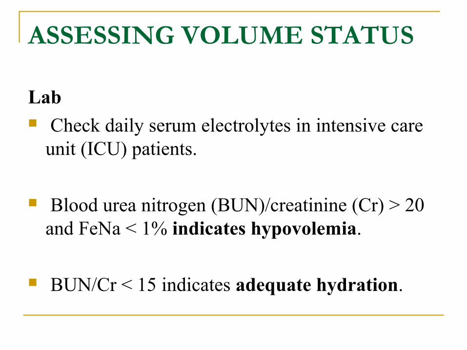
ASSESSING VOLUME STATUS
Lab Check daily serum electrolytes in intensive care
unit (ICU) patients.
Blood urea nitrogen (BUN)/creatinine (Cr) > 20 and FeNa < 1% indicates hypovolemia.
BUN/Cr < 15 indicates adequate hydration.

SODIUM BALANCE

Hyponatremia
DEFINITION : [Na] < 130 mEq/L.
First steps in hyponatremia: Determine volume status clinically, then determine plasma osmolality!
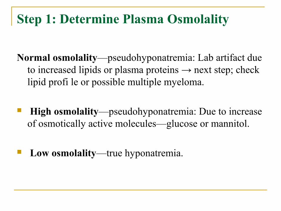
Step 1: Determine Plasma Osmolality
Normal osmolality—pseudohyponatremia: Lab artifact due to increased lipids or plasma proteins → next step; check lipid profi le or possible multiple myeloma.
High osmolality—pseudohyponatremia: Due to increase of osmotically active molecules—glucose or mannitol.
Low osmolality—true hyponatremia.

Step 2: Assess Volume Status Hypovolemia Euvolemia Hypervolemia
HYPONATREMIA WITH HIGH PLASMA OSMOLALITY (PSEUDOHYPONATREMIA)
CAUSES
Hyperglycemia, either physiologic or due to rapid infusion of glucose or mannitol will cause increased osmotic pressure that shifts fluid from the ICF to the ECF. The total body sodium in this case is normal but has become diluted due to the fluid shift.
The expected Na concentration can be calculated as follows: For every 100 mg/dL that glucose is increased over 100 mg/dL, the Na concentration falls 1.6 mEq/L. Remember “sweet 16.”
For example, a patient with a glucose concentration of 500 mg/dL is expected to have a hyponatremia of around 133.6 mEq/L (4 × 1.6 = 6.4, 140 – 6.4 = 133.6).

HYPONATREMIA WITH HYPOTONICITY (TRUE HYPONATREMIA(
True hyponatremia reflects excess ingestion of water that overwhelms the kidneys (either normal or diseased) or due to increased ADH.
Hyponatremia is not due to increased excretion of sodium.

Hypovolemia (dehydration)
Renal cause: Diuretics. Extrarenal cause: Vomiting, diarrhea, burns, pancreatitis. Differentiate using urine Na: Urine Na < 20 mEq/L indicates expected
renal retention in the face of hypovolemia, suspect an extrarenal cause. Urine Na > 20 mEq/L indicates a renal cause.
Hypervolemia
May be from CHF, cirrhosis, or nephrotic syndrome. Increased thirst and vasopressin. Edematous state.

Euvolemia
SIADH: Most common cause of normovolemic hyponatremia.
Increased vasopressin release from posterior pituitary or ectopic source causes decreased renal free water excretion.

Signs and symptoms: Hypo-osmotic hyponatremia (hyponatremia with
hypotonicity). Inappropriately concentrated urine (urine
osmolality > 100 mOsm/ kg).
Normal renal, adrenal, and thyroid function.
Causes: Neuropsychiatric disorders, malignancies (especially lung), and
head trauma. Glucocorticoid defi ciency (Addison’s disease)—cortisol deficiency
causes hypersecretion of vasopressin.
Hypothyroidism—causes decreased CO and glomerular filtration rate (GFR), which leads to increased vasopressin secretion.
Primary polydipsia—usually seen in psychiatric patients who compulsively drink massive volumes of water.

SIGNS AND SYMPTOMS OF (TRUE) HYPONATREMIA Signs: Decreased reflexes, respiratory depression, seizures, coma .
Symptoms: Nausea/vomiting, headache, lethargy, muscle cramps. Hypovolemic hyponatremia: Give 0.9% NaCl. Na repletion with saline
isotonic to the patient, in order to avoid rapid changes in ICF volume. Major complication from rapid correction of chronic hyponatremia is
central pontine myelinolysis. Hypervolemic hyponatremia: Correct underlying disorder—CHF, liver or renal failure. Euvolemic hyponatremia: Raise plasma Na (lower ICF volume)—
restrict water intake.

Hypernatremia
DEFINITION : [Na] > 145 mEq/L.
Hypernatremia is always associated with hyperosmolarity. (Note that in the plasma osmolality equation, Na is the major factor).

Hypernatremia
CAUSES Loss of water (dehydration!): Diabetes insipidus, diuretics, sweating,
GI loss, burns, fistulas.
Gain of sodium due to excess mineralocorticoid activity: Primary hyperaldosteronism, Cushing’s, renal artery stenosis (hyperreninism), congenital adrenal hyperplasia (will cause concomitant hypokalemia).
If thirst mechanism is intact and water is available, hypernatremia will not persist. Suspect hypernatremia in the young, elderly, and patients with altered mental status who may not have access to water.

Hypernatremia
SYMPTOMS
Thirst. Restlessness, weakness, delirium. Hypotension and tachycardia. Decreased saliva and tears. Red, swollen tongue. Oliguria.