Embed Size (px)
Citation preview

4/15/2015
1

FIBROMATOSIS
Dr. Muhammad Umar Nisar

What is fibromatosis?
Fibromatosis is a condition where fibrous
overgrowths of dermal and subcutaneous
connective tissue develop tumours called
fibromas. These fibromas are usually benign
(non-cancerous).
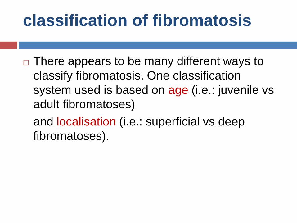
classification of fibromatosis
There appears to be many different ways to
classify fibromatosis. One classification
system used is based on age (i.e.: juvenile vs
adult fibromatoses)
and localisation (i.e.: superficial vs deep
fibromatoses).

• Congenital generalised fibromatosis (infantile myofibromatosis)
• Aponeurotic fibroma
• Aggressive infantile fibromatosis
• Infantile digital fibromatosis
• Fibromatosis colli
• Dermatofibrosis lenticularis
Juvenile

• Superficial (fascial) fibromatoses
• Palmar (Dupuytren contracture) and plantar (Ledderhose disease) fibromatosis
• Penile fibromatosis (Peyronie disease)
• Knuckle pads
• Dermatofibroma
• Nodular fasciitis
• Elastofibroma
• Fibrous papule of the face
• Deep (musculoaponeurotic) fibromatoses`
• Desmoid tumours (aggressive fibromatoses)
• Extraabdominal fibromatosis
• Abdominal fibromatosis
• Intraabdominal fibromatosis (e.g. pelvic fibromatosis)
Adult

features of fibromatosis
• Slow growing tumour
• Small size
• Arise from fascia or aponeurosis
• Less aggressive
Superficial fibromatosis
• Rapidly growing tumour
• Usually reach large size
• Often involve deeper structures (muscles of the trunk and extremities)
Deep fibromatoses

Whilst most fibromatoses are benign tumours
and do not metastasise (spread to other parts
of the body), the desmoid tumours although
they do not metastasise like malignant cancers
can be locally aggressive. They can grow
quickly into large tumours that can obstruct
vital structures such as major blood vessels,
nerves and organs.

Causes
The cause of fibromatosis remains unclear
In some types of fibromatosis such as desmoid
tumours it is thought that the condition may be
related to:
Trauma
Hormonal factors
Estrogens
Genetic association: Trisomy 8
Deletion of 5q

Causes cont...
Superficial fibromatoses such as palmar,
plantar and penile fibromatosis have
sometimes been linked to certain diseases
such as
Diabetes
Liver disease
Hypertension
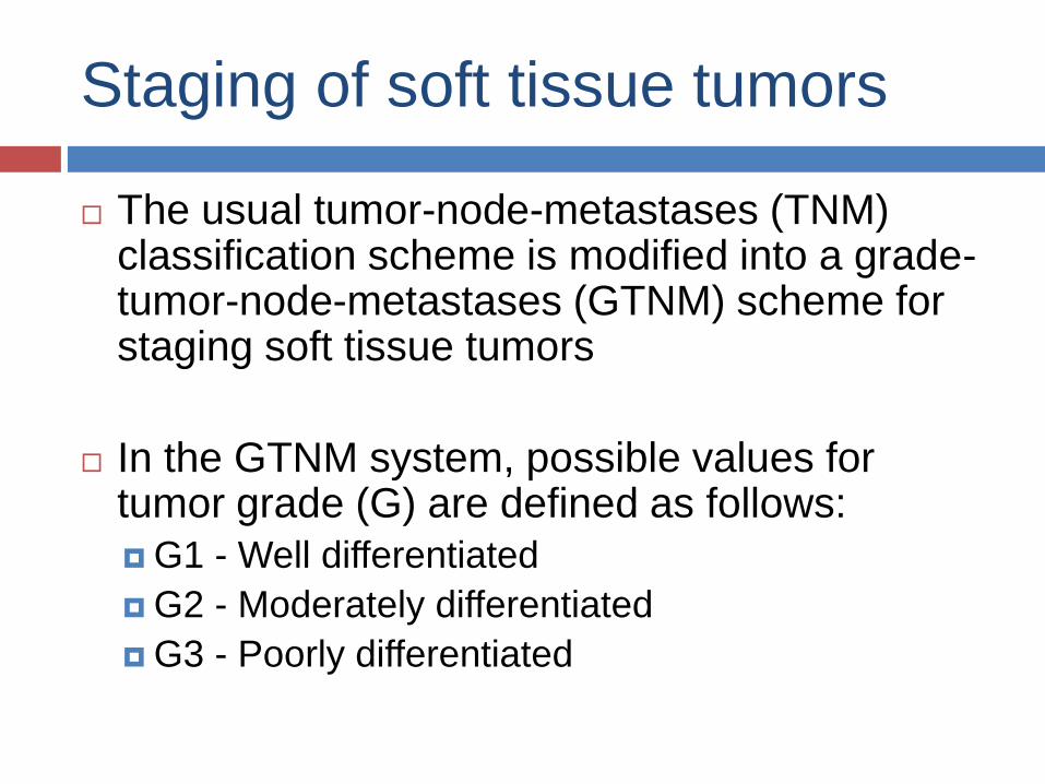
Staging of soft tissue tumors
The usual tumor-node-metastases (TNM) classification scheme is modified into a grade-tumor-node-metastases (GTNM) scheme for staging soft tissue tumors
In the GTNM system, possible values for tumor grade (G) are defined as follows:
G1 - Well differentiated
G2 - Moderately differentiated
G3 - Poorly differentiated

Possible values for primary tumor (T) are
defined as follows:
T1 - Tumor less than 5 cm in greatest diameter
T2 - Tumor more than 5 cm in greatest diameter

Possible values for regional lymph node
involvement (N) are defined as follows:
N0 - No known metastasis to lymph nodes
N1 - Verified metastasis to lymph nodes

Possible values for distant metastasis are
defined as follows:
M0 - No known distant metastasis
M1 - Known distant metastasis

GTNM Classification and Stage
Grouping of Soft Tissue Tumors
Stage Groupings Tumor Grade Primary TumorRegional Lymph
Node InvolvementDistant Metastasis
Stage IA G1 T1 N0 M0
Stage IB G1 T2 N0 M0
Stage II A G2 T1 N0 M0
Stage IIB G2 T2 N0 M0
Stage IIIA G3 T1 N0 M0
Stage IIIB G3 T2 N0 M0
Stage IVA Any G Any T N1 M0
Stage IVB Any G Any T Any N M1

DEEP
(MUSCULOAPONEUROTIC)
FIBROMATOSES
Desmoid Tumor

Background
Desmoid tumors are tumors that arise from cells called fibroblasts.
Fibroblasts are found throughout our body and their main function is to provide structural support and protection to the vital organs
They also play a critical role in wound healing.
When fibroblast cells undergo mutations they can become cancerous and become desmoid tumors
The term desmoid, coined by Muller in 1838, is derived from the Greek word desmos, which means tendonlike.

Epidemiology
Frequency
International
Overall, desmoid tumors are reported to account for 0.03% of all neoplasms.When present in patients with familial polyposis of the colon, the prevalence of desmoid tumors is as high as 13%.
Mortality/Morbidity
Despite their benign histologic appearance and negligible metastatic potential, the tendency of desmoid tumors to cause local infiltration is significant in terms of
(1) deformity, morbidity, and mortality resulting from pressure effects
(2) potential obstruction of vital structures and organs.
Sex
Twice as common in females than in males
Age
more common in persons aged 10-40 years

Pathophysiology
most commonly arise from the rectus abdominis muscle in postpartum women and in scars due to abdominal surgery, they may arise in any skeletal muscle.
Desmoid tumors tend to infiltrate adjacent muscle bundles, frequently entrapping them and causing their degeneration.
The myofibroblast is the cell considered to be responsible for the development of desmoid tumors
May be familial (associated with Gardner's syndrome/FAP syndrome, or familial desmoid syndrome, or related to trauma.

Clinical Presentation
History
Lump that can arise in any skeletal muscle,
they most commonly develop in the anterior
abdominal wall and shoulder girdle.
A history of trauma (often surgical) to the site of
the desmoid tumor is elicited in 1 in 4 cases.
Implant-associated breast desmoid tumors
may occur
Clusters of cases in families without evidence
of any associated syndromes have also been
reported.

Physical Examination:
Peripheral desmoid tumors
Peripheral desmoid tumors are firm, smooth, and mobile.
They often adhere to surrounding structures. The overlying skin is usually unaffected.
Intra-abdominal and extra-abdominal desmoid tumors
Intra-abdominal desmoid tumors remain asymptomatic until their growth and infiltration cause visceral compression. Symptoms of intestinal, vascular, ureteric, or neural involvement may be the initial manifestations.
Breast desmoid tumors
Desmoid tumors account for 0.2% of primary breast tumors, developing from muscular fasciae and aponeuroses.
Desmoid tumors may mimic breast cancer.

Skeletal distribution of extra-
abdominal desmoid tumours.

Differential diagnosis
Fibrosarcoma: atypia or mitotic figures present
GIST: strong CD117+, CD34+
Idiopathic retroperitoneal fibrosis inflammatory, strangles the ureters
Leiomyoma: bright pink cytoplasm of smooth muscle, desmin+
Low grade fibromyxoid sarcoma: heavily collagenized stroma with abrupt transition to myxoid areas, often epithelioid areas or poorly formed but large collagen rosettes; beta catenin negative
Neurofibroma: no myofibroblasts, S100+
Schwannoma: palisading Schwann cells, usually minimal collagen, S100+
Sclerosing omentitis: grows like panniculitis, beta catenin negative

Work up
Laboratory Studies
Immunostaining with
vimentin, alpha
smooth muscle
actin, muscle actin,
and desmin are
helpful in
distinguishing the
tumors in the
differential diagnosis
of desmoid tumors

Imaging Studies
CT and MRI are used for the diagnosis and follow-up of desmoid tumors. They can help determine the extent of the tumor and its relationship to nearby structures, especially prior to surgical removal

Biopsy of the tumor:
A fine-needle aspiration
biopsy specimen may
be considered.
Fine-needle aspirates of desmoid tumors showing coherent clusters of uniform spindle cells with abundant cytoplasm and oval-to-elongated nuclei with evenly distributed chromatin

Electron microscopy:
Fibroblastic and
myofibroblastic
features, including
intrareticular collagen
fibers, thin filament
bundles, cytoplasmic
dense bodies

Colonoscopy :
indicated to
investigate for the
presence of Gardner
syndrome
Multiple large intestinal polyp in a patient with gardner syndrome

Treatment & ManagementSurgery
Aggressive, wide surgical resection with complete excision is the
treatment of choice.
In selected patients, radical resection with intraoperative margin
evaluation by frozen sections followed by immediate reconstruction
of the defect may be a safe and effective procedure providing
definitive cure yet minimizing functional limitations.
Resection of Tumor

In those patients who refuse surgery or are not
surgical candidates, the following options may
be considered.
Treatment Options for Nonsurgical Patients
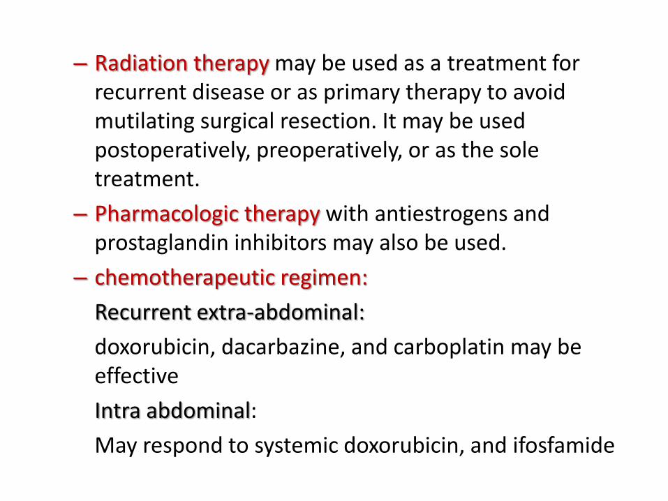
– Radiation therapy may be used as a treatment for recurrent disease or as primary therapy to avoid mutilating surgical resection. It may be used postoperatively, preoperatively, or as the sole treatment.
– Pharmacologic therapy with antiestrogens and prostaglandin inhibitors may also be used.
– chemotherapeutic regimen:
Recurrent extra-abdominal:
doxorubicin, dacarbazine, and carboplatin may be effective
Intra abdominal:
May respond to systemic doxorubicin, and ifosfamide

Follow up
Further Outpatient Care
After surgery, MRI may be useful for monitoring
desmoid tumor recurrence
follow-up visits are every three months for two
years, and then in 6 to 12 month intervals.

Prognosis
Local desmoid tumor recurrence rates are reported to be as
high as 70%.
A positive surgical margin is a significant risk factor for
recurrence
Five-year survival rates of such patients with stage I, II, III,
and IV intra-abdominal desmoid tumors were found to be
95%, 100%, 89%, and 76%, respectively.
The 5-year survival rate of stage IV patients with severe
pain/narcotic dependency, tumor size larger than 10 cm, and
need for total parenteral nutrition was only 53%.

Take home message
There should be proper follow up of the
patients to reduce the misery of the patients as
well as the cost of treatment.
There should be a multidisciplinary approach
in the management of agressive fibromatosis.
Whenever there is a doubt histopathologist
himself/herself should proceed for
immunohistochemistry instead of waiting for
surgeon’s advice..

References
Book: Textbook of Dermatology. Ed Rook A, Wilkinson DS, Ebling FJB, Champion RH, Burton JL. Fourth edition. Blackwell Scientific Publications.
Wu C, Amini-Nik S, Nadesan P, Stanford WL, Alman BA. Aggressive fibromatosis (desmoid tumor) is derived from mesenchymal progenitor cells. Cancer Res. Oct 1 2010;70(19):7690-8. [Medline].
Brueckl WM, Ballhausen WG, Fortsch T, et al. Genetic testing for germline mutations of the APC gene in patients with apparently sporadic desmoid tumors but a family history of colorectal carcinoma. Dis Colon Rectum. Jun 2005;48(6):1275-81. [Medline].
Sturt NJ, Gallagher MC, Bassett P, et al. Evidence for genetic predisposition to desmoid tumours in familial adenomatous polyposis independent of the germline APC mutation. Gut. Dec 2004;53(12):1832-6.[Medline].
Caspari R, Olschwang S, Friedl W, et al. Familial adenomatous polyposis: desmoid tumours and lack of ophthalmic lesions (CHRPE) associated with APC mutations beyond codon 1444. Hum Mol Genet. Mar 1995;4(3):337-40. [Medline].
Wang WL, Nero C, Pappo A, Dina Lev DL, Lazar AJ, Lopez-Terrada DH. CTNNB1 Genotyping and APC Screening in Pediatric Desmoid Tumors: A Proposed Alogrithm. Pediatr Dev Pathol. Feb 28 2012;[Medline].
Mullen JT, Delaney TF, Rosenberg AE, Le L, Iafrate AJ, Kobayashi W, et al. ß-Catenin Mutation Status and Outcomes in Sporadic Desmoid Tumors. Oncologist. Aug 19 2013;[Medline].
Shields CJ, Winter DC, Kirwan WO, Redmond HP. Desmoid tumours. Eur J Surg Oncol. Dec 2001;27(8):701-6. [Medline].
Klemmer S, Pascoe L, DeCosse J. Occurrence of desmoids in patients with familial adenomatous polyposis of the colon. Am J Med Genet. Oct 1987;28(2):385-92. [Medline].
Lee JC, Thomas JM, Phillips S, Fisher C, Moskovic E. Aggressive fibromatosis: MRI features with pathologic correlation. AJR Am J Roentgenol. Jan 2006;186(1):247-54. [Medline].
Raynham WH, Louw JH. Desmoid tumours in familial polyposis of the colon. S Afr J Surg. Jul-Sep 1971;9(3):133-40. [Medline].
Gaches C, Burke J. Desmoid tumour (fibroma of the abdominal wall) occurring in siblings. Br J Surg. Jul 1971;58(7):495-8. [Medline].
Lopez R, Kemalyan N, Moseley HS, Dennis D, Vetto RM. Problems in diagnosis and management of desmoid tumors. Am J Surg. May 1990;159(5):450-3. [Medline].
Jeong WS, Oh TS, Sim HB, Eom JS. Desmoid tumor following augmentation mammoplasty with silicone implants. Arch Plast Surg. Jul 2013;40(4):470-2. [Medline]. [Full Text].