Embed Size (px)
Citation preview
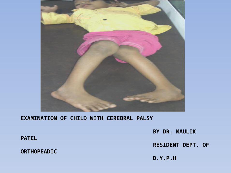
EXAMINATION OF CHILD WITH CEREBRAL PALSY
BY DR. MAULIK PATEL RESIDENT DEPT. OF ORTHOPEADIC D.Y.P.H
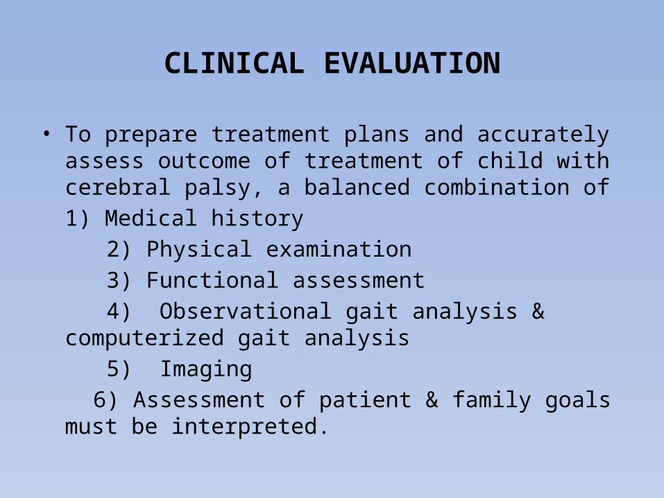
CLINICAL EVALUATION
• To prepare treatment plans and accurately assess outcome of treatment of child with cerebral palsy, a balanced combination of 1) Medical history
2) Physical examination 3) Functional assessment 4) Observational gait analysis & computerized gait analysis 5) Imaging 6) Assessment of patient & family goals must be interpreted.
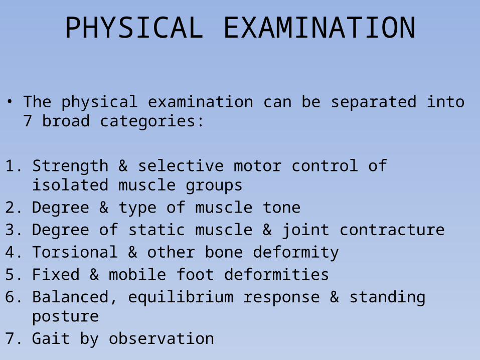
PHYSICAL EXAMINATION
• The physical examination can be separated into 7 broad categories:
1. Strength & selective motor control of isolated muscle groups2. Degree & type of muscle tone3. Degree of static muscle & joint contracture4. Torsional & other bone deformity5. Fixed & mobile foot deformities6. Balanced, equilibrium response & standing posture7. Gait by observation
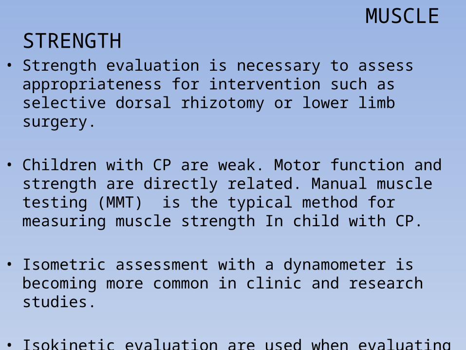
MUSCLE STRENGTH• Strength evaluation is necessary to assess appropriateness for
intervention such as selective dorsal rhizotomy or lower limb surgery.
• Children with CP are weak. Motor function and strength are directly related. Manual muscle testing (MMT) is the typical method for measuring muscle strength In child with CP.
• Isometric assessment with a dynamometer is becoming more common in clinic and research studies.
• Isokinetic evaluation are used when evaluating strength throught the range of motion (ROM). This assessment used to measure torque generated through an arc of movement.
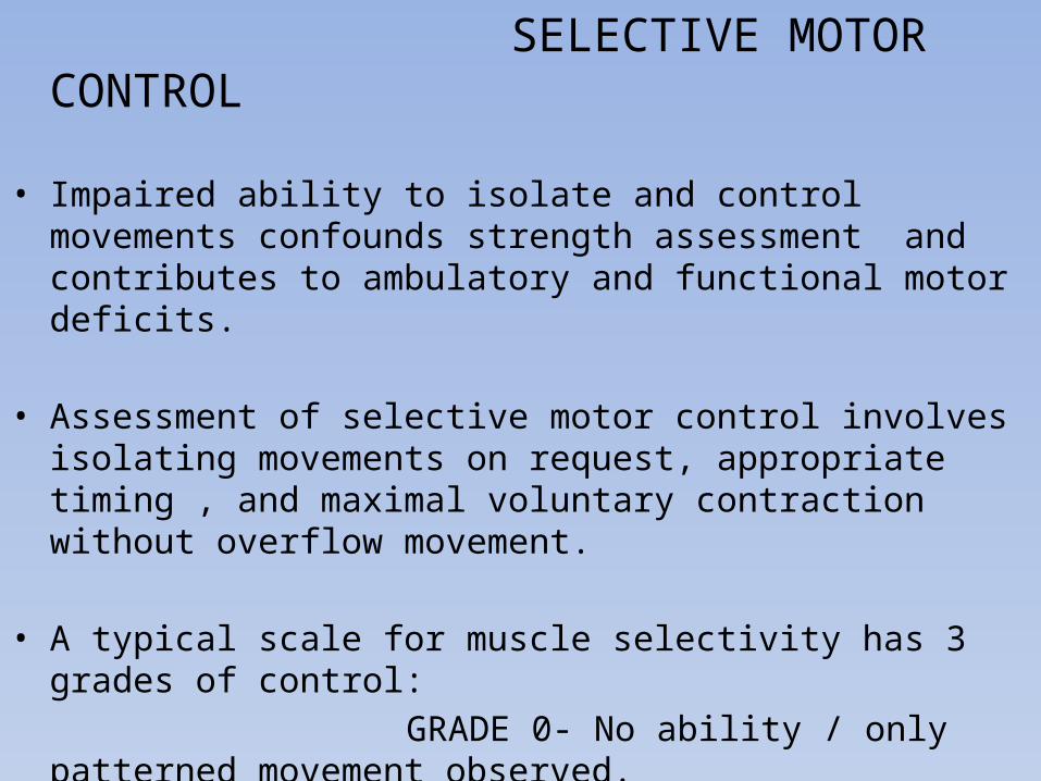
SELECTIVE MOTOR CONTROL
• Impaired ability to isolate and control movements confounds strength assessment and contributes to ambulatory and functional motor deficits.
• Assessment of selective motor control involves isolating movements on request, appropriate timing , and maximal voluntary contraction without overflow movement.
• A typical scale for muscle selectivity has 3 grades of control: GRADE 0- No ability / only patterned movement observed. 1- Partially isolated movements observed. 2- Completely isolated movements observed.
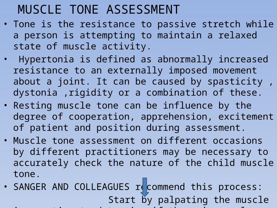
MUSCLE TONE ASSESSMENT • Tone is the resistance to passive stretch while a person is attempting
to maintain a relaxed state of muscle activity.• Hypertonia is defined as abnormally increased resistance to an
externally imposed movement about a joint. It can be caused by spasticity , dystonia ,rigidity or a combination of these.
• Resting muscle tone can be influence by the degree of cooperation, apprehension, excitement of patient and position during assessment.
• Muscle tone assessment on different occasions by different practitioners may be necessary to accurately check the nature of the child muscle tone.
• SANGER AND COLLEAGUES recommend this process: Start by palpating the muscle in question to determine if
there is muscle contracture at rest.
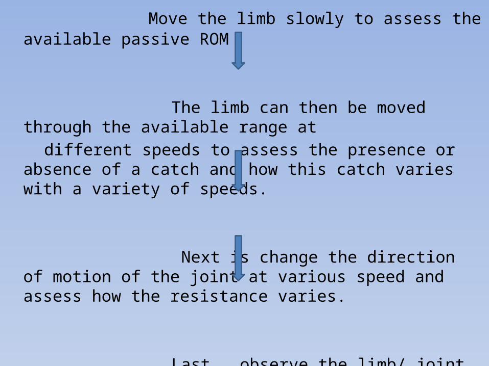
Move the limb slowly to assess the available passive ROM
The limb can then be moved through the available range at different speeds to assess the presence or absence of a catch and how
this catch varies with a variety of speeds.
Next is change the direction of motion of the joint at various speed and assess how the resistance varies.
Last , observe the limb/ joint while asking the patient to move the same joint on contralateral side.
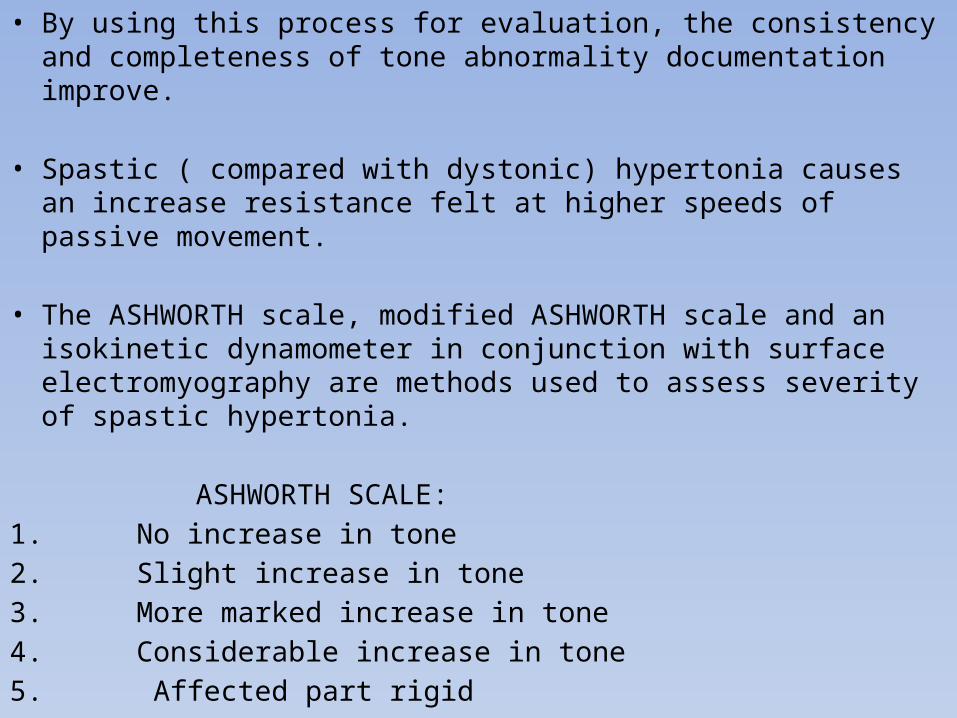
• By using this process for evaluation, the consistency and completeness of tone abnormality documentation improve.
• Spastic ( compared with dystonic) hypertonia causes an increase resistance felt at higher speeds of passive movement.
• The ASHWORTH scale, modified ASHWORTH scale and an isokinetic dynamometer in conjunction with surface electromyography are methods used to assess severity of spastic hypertonia.
ASHWORTH SCALE:1. No increase in tone2. Slight increase in tone3. More marked increase in tone4. Considerable increase in tone5. Affected part rigid
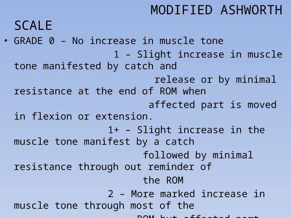
MODIFIED ASHWORTH SCALE• GRADE 0 – No increase in muscle tone 1 – Slight increase in muscle tone manifested by catch and release or by minimal resistance at the end of ROM when affected part is moved in flexion or extension. 1+ – Slight increase in the muscle tone manifest by a catch followed by minimal resistance through out reminder of the ROM 2 – More marked increase in muscle tone through most of the ROM but affected part easily moved. 3 – Considerable increase in muscle tone, passive movement difficult. 4 – Affected part rigid in flexion or extension.
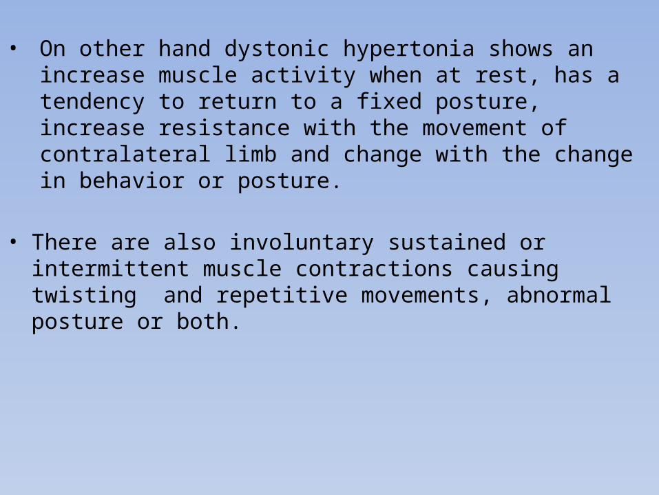
• On other hand dystonic hypertonia shows an increase muscle activity when at rest, has a tendency to return to a fixed posture, increase resistance with the movement of contralateral limb and change with the change in behavior or posture.
• There are also involuntary sustained or intermittent muscle contractions causing twisting and repetitive movements, abnormal posture or both.
• ROM AND CONTRACTURE:• Variation in ROM measurement between observer is common and
frustrating.• Differentiation between static and dynamic deformity may be difficult
in nonanesthetized patient. However static examination of muscle length provides some insight in to whether contracture are static or dynamic.
• Comparison of joint ROM with slow and rapid stretch can be useful in evaluation of spasticity.
• Dynamic contracture disappears under G.A. thus the ROM examination under G.A. can be used to help decide whether to inject botulinum toxin for spasticity in muscle or perform surgery to lengthen a contracture of the tendon.
• Differentiation between contracted biarticular and monoarticular muscle is important.
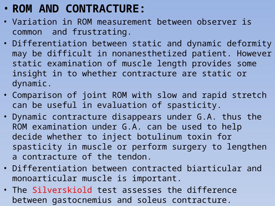
• The Silverskiold test assesses the difference between gastocnemius and soleus contracture.
The Silverskiold test: a) this test differentiates tightness of gastrocnemius and soleus. In this test the knee is flexed to 90, hind foot is positioned in varus, and maximally dorsiflexed. B) As the knee extented , if ankle moves towards plantarflexion, contracture of gastrocnemius present.

• The DUNCAN-ELY test differentiates between contracture of the monoarticular vasti and the biarticular rectus femoris.
• Perry and colleagues have shown that when these test are performed with electromyography, both mono articular and bi articular muscle crossing the joint contract.
• For example, in nonanesthetized person The DUNCAN-ELY test induce contraction of not only the rectus femoris but also iliopsoas and SILVERSKIOLD test induce contraction of both gastrcnemius and soleus. But under G.A. the biarticular muscle tests reliably differentiate the location of contraction. So this should routinely include as part of pre surgical examination under G.A.
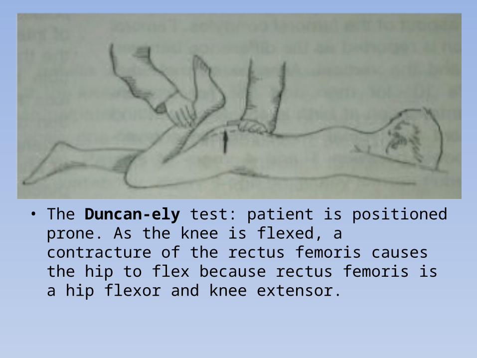
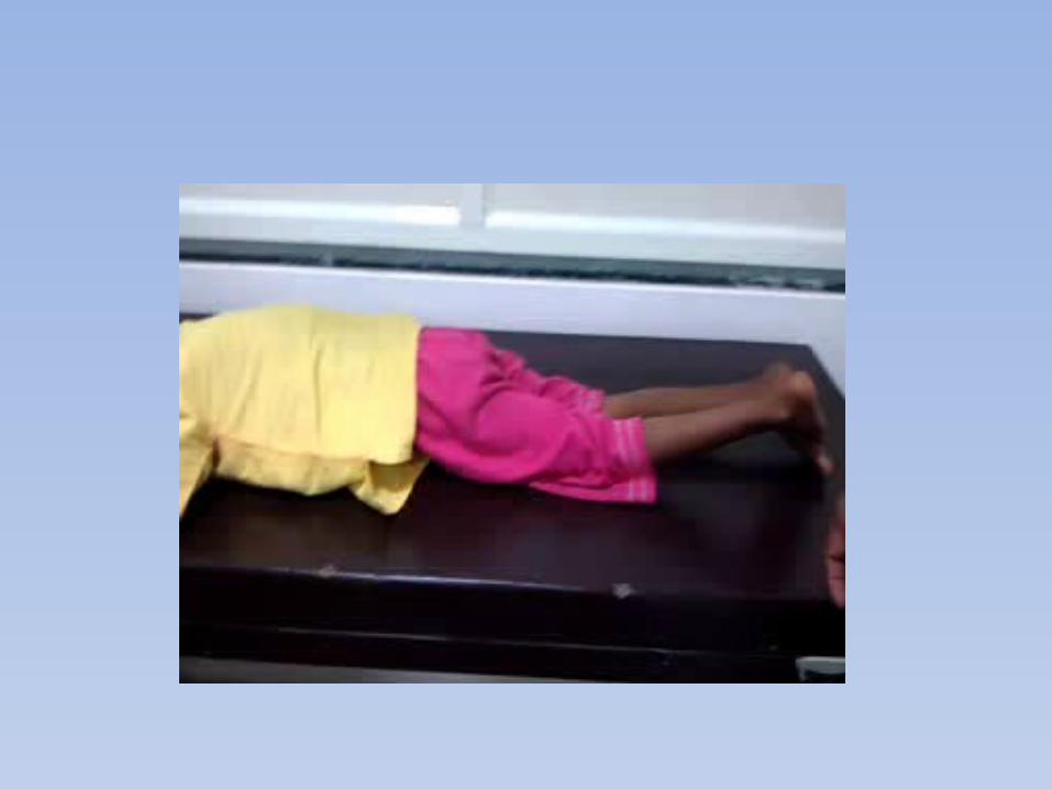
• The Duncan-ely test: patient is positioned prone. As the knee is flexed, a contracture of the rectus femoris causes the hip to flex because rectus femoris is a hip flexor and knee extensor.
HIP• The Thomas test is used to measure the degree of hip flexor
tightness.
• It is preformed with patient supine position and pelvis held in such that the ASIS and PSIS are aligned vertically.
• Defining the pelvis position consistently rather than using the flattened the lordosis method improves reliability.
• Because of the origin and insertion points, the causes of limited hip abduction ROM can be distinguished by measuring hip abduction in various position of hip and knee with the patient in supine.
The one joint adductors ( adductor longus, brevis , magnus) are isolated with the knee flexed. In this position, the gracilis is relaxed.
(cont..)
• With the knee in full extension the length of 2 joint gracilis in a position of maximum stretch.
• If the hip abduction is more limited when the knee is extended compare with the knee flexed, contracture of gracilis is the cause.
KNEE• In child with CP capsular contracture causes knee flexion contracture.
It is must to differentiate between true knee joint contracture and hamstring contracture.
• Knee joint contracture is identify if knee extension is limited with hip extension (to relax hams) and ankle relax in position of equinus (to relax gastrocnemius) .
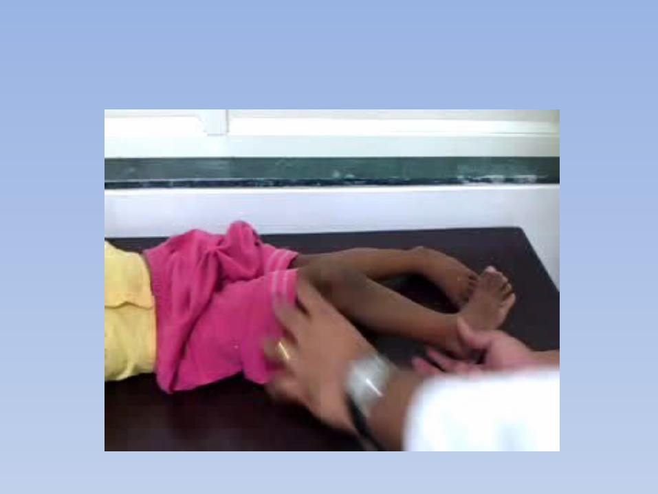
• Hamstring contracture is identified if knee extension is limited when the hip is flexed 90 (popliteal angle). normal values for popliteal angle are age and gender dependent, with boys tighter than girls and both tighter with increase with age, mainly at adolescent growth spurt.
• In the patient normal resting supine position, hip contracture causes lumbar lordosis and ant. Pelvic tilting that shift the origin of hamstrings on the ischial tuberosity proximally. The contralateral hip in full extension, where as the ipsilateral hip is flexed to 90. The measurement of degree lacking from full extension is recorded as UNILATERAL POPLITEAL ANGLE. Normal popliteal angle of 5 to 18 year is 0 to 49 with mean of 26.
• The bilateral popliteal angle measurement is performed with contralateral hip flexed until ASIS and PSIS aligned vertically.
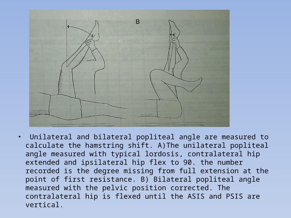
• Unilateral and bilateral popliteal angle are measured to calculate the hamstring shift. A)The unilateral popliteal angle measured with typical lordosis, contralateral hip extended and ipsilateral hip flex to 90. the number recorded is the degree missing from full extension at the point of first resistance. B) Bilateral popliteal angle measured with the pelvic position corrected. The contralateral hip is flexed until the ASIS and PSIS are vertical.
• A significant smaller popliteal angle with pelvis position corrected is referred as a HAMSTRING SHIFT.
• The value popliteal angle with a neutral pelvis is a measure of true hams contracture and the value with the lordosis presents the functional hams contracture. The difference between this two represents the degree of HAMSTRING SHIFT.
• Hamstring contracture is frequently implicated as a causes of crouch gait. However increased ant. Pelvic tilt is common in crouch gait caused by CP and this produce hamstring shift. In this situation, hamstring length may be normal or long, and hams lengthening surgery weakens hip extension and exacerbates the excessive hamstring length.
• Because of difficulty in establishing dynamic hamstring length on physical examination, static hamstring length from supine physical examination should supplemented by estimation of hamstring length obtained from gait analysis before consideration of hamstring lengthening surgery.
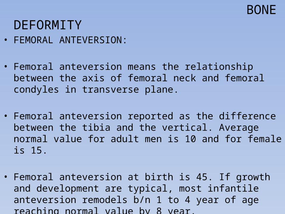
BONE DEFORMITY• FEMORAL ANTEVERSION:
• Femoral anteversion means the relationship between the axis of femoral neck and femoral condyles in transverse plane.
• Femoral anteversion reported as the difference between the tibia and the vertical. Average normal value for adult men is 10 and for female is 15.
• Femoral anteversion at birth is 45. If growth and development are typical, most infantile anteversion remodels b/n 1 to 4 year of age reaching normal value by 8 year.
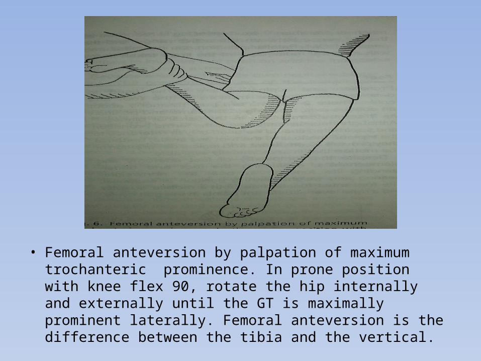
• Femoral anteversion by palpation of maximum trochanteric prominence. In prone position with knee flex 90, rotate the hip internally and externally until the GT is maximally prominent laterally. Femoral anteversion is the difference between the tibia and the vertical.
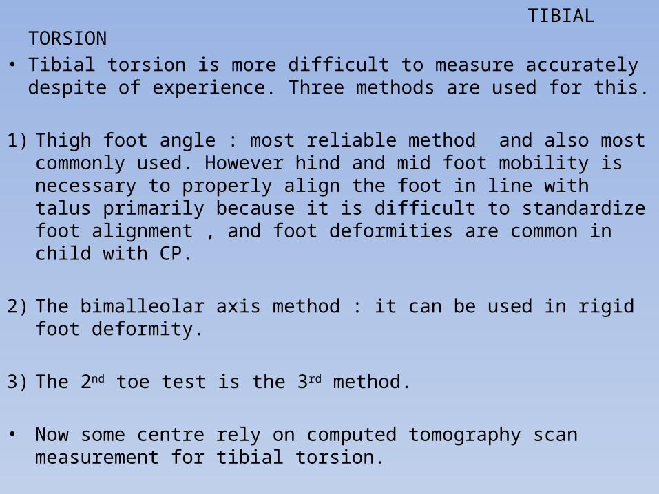
TIBIAL TORSION• Tibial torsion is more difficult to measure accurately despite of
experience. Three methods are used for this.
1) Thigh foot angle : most reliable method and also most commonly used. However hind and mid foot mobility is necessary to properly align the foot in line with talus primarily because it is difficult to standardize foot alignment , and foot deformities are common in child with CP.
2) The bimalleolar axis method : it can be used in rigid foot deformity.
3) The 2nd toe test is the 3rd method.
• Now some centre rely on computed tomography scan measurement for tibial torsion.
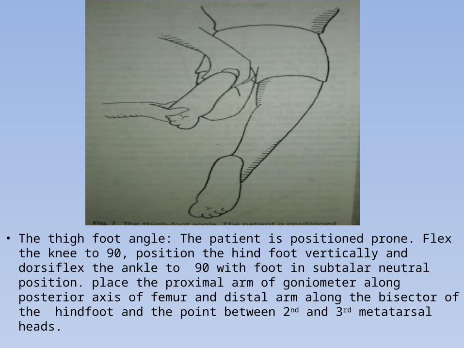
• The thigh foot angle: The patient is positioned prone. Flex the knee to 90, position the hind foot vertically and dorsiflex the ankle to 90 with foot in subtalar neutral position. place the proximal arm of goniometer along posterior axis of femur and distal arm along the bisector of the hindfoot and the point between 2nd and 3rd metatarsal heads.
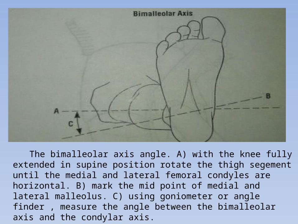
The bimalleolar axis angle. A) with the knee fully extended in supine position rotate the thigh segement until the medial and lateral femoral condyles are horizontal. B) mark the mid point of medial and lateral malleolus. C) using goniometer or angle finder , measure the angle between the bimalleolar axis and the condylar axis.
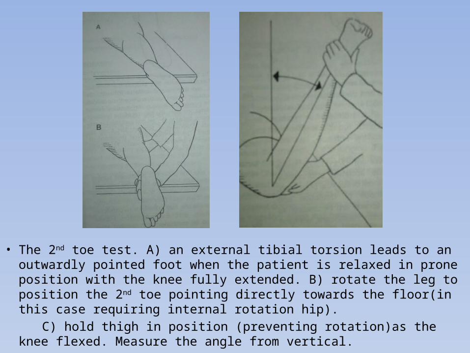
• The 2nd toe test. A) an external tibial torsion leads to an outwardly pointed foot when the patient is relaxed in prone position with the knee fully extended. B) rotate the leg to position the 2nd toe pointing directly towards the floor(in this case requiring internal rotation hip).
C) hold thigh in position (preventing rotation)as the knee flexed. Measure the angle from vertical.
PATELLA ALTA • Patella Alta is common in child with CP and probably due to chronic
excessive knee extensor forces of rectus femoris spasticity and crouch gait. These same forces may lead to inferior pole sleeve avulsion
fractures.
• To screen patella Alta, patient is positioned supine with knee extended. The top of the patella is then palpated. The superior pole of patella is typically one finger width proximal to adductor tubercle.
• Patella alta may contribute to patellofemoral instability, pain,
subluxation and terminal knee extensor dysfunction (quadriceps insufficiency) which is measured by extensor lag.
• Extensor lag: child supine with the leg flexed at the knee over the end of table. Then child is asked to actively extend the knee as much as possible. The extensor lag is difference b/n the active range and passive ROM.
FOOT • Pronation and supination are terms used to describe the tri planar
motion in the foot and ankle. These 2 are pure rotation movement around an oblique axis.
• Despite the complexity of foot anatomy and bio mechanics, evaluating the foot and understanding its function in both non-weight bearing and weight-bearing position is essential.
• Correctly identifying structural abnormality in non-weight bearing position and compensation that occur as a result of this abnormality in weight bearing is essential to determine intervention to improve foot position and function of entire lower extremity.
• Because every foot has its own neutral subtalarjoint (STJ) position, the use of non weight bearing STJ neutral position provides consistency in positioning the foot in order to assess and identify patient specific structural abnormalities and their resultant compensation In weight bearing.
• STJN is defined as the position from which the STJ can maximally pronated and supinated and there for position from which STJ can function optimally. STJN is found through palpation at the articulation b/n the head of talus and the navicular. Congruency of the talonavicular joint is the position of foot at which neither medial nor the lateral head of talus protudes and examiner feels symmetry of the navicular on the head of talus. From this starting point, the patient rear foot and fore foot relation are evaluated.
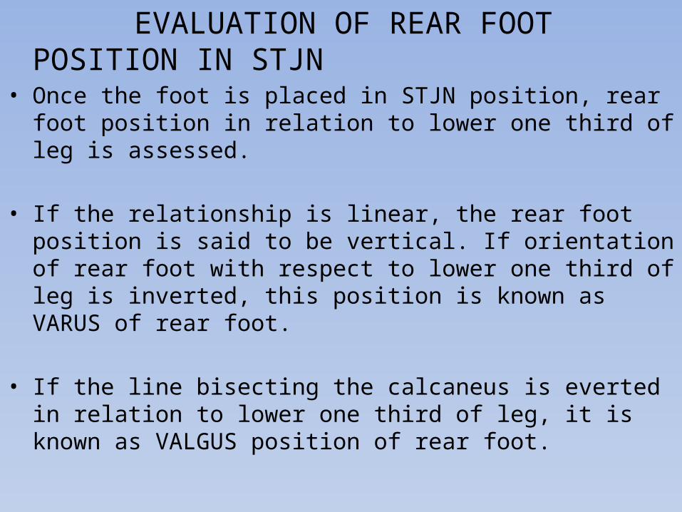
EVALUATION OF REAR FOOT POSITION IN STJN• Once the foot is placed in STJN position, rear foot position in relation
to lower one third of leg is assessed.
• If the relationship is linear, the rear foot position is said to be vertical. If orientation of rear foot with respect to lower one third of leg is inverted, this position is known as VARUS of rear foot.
• If the line bisecting the calcaneus is everted in relation to lower one third of leg, it is known as VALGUS position of rear foot.
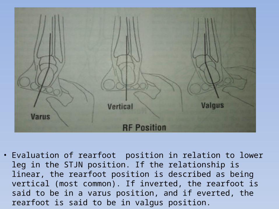
• Evaluation of rearfoot position in relation to lower leg in the STJN position. If the relationship is linear, the rearfoot position is described as being vertical (most common). If inverted, the rearfoot is said to be in a varus position, and if everted, the rearfoot is said to be in valgus position.
EVALUATION OF FOREFOOT POSITION IN STJN• Evaluation in 3 planes 1) FRONTAL 2) SAGITTAL 3) TRANSVERSE
• While maintaining STJN , forefoot position in the frontal plane can be describe by assessing the angle b/n a line that is perpendicular to the bisection of the posterior calcaneus and the plane of the heads of metatarsals.
• In this position, if the plane of the metatarsal heads is in the same plane as the line that is perpendicular to bisection of calcaneus, the fore foot position is called as NEUTRAL.
• If the plane of fore foot in relation to rear foot shows shows the medial side of foot to be higher than lateral side (forefoot inverted) this position is described as fore foot VARUS deformity.
• If the opposite is seen ( lateral border is higher than medial border) this position is called as fore foot VALGUS deformity.
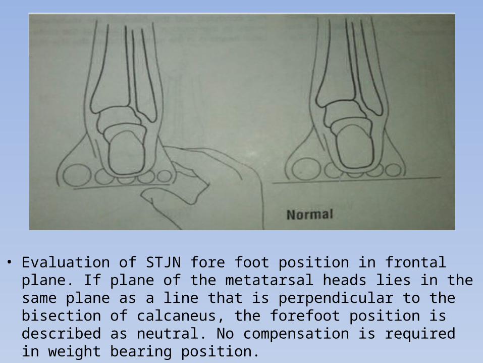
• Evaluation of STJN fore foot position in frontal plane. If plane of the metatarsal heads lies in the same plane as a line that is perpendicular to the bisection of calcaneus, the forefoot position is described as neutral. No compensation is required in weight bearing position.
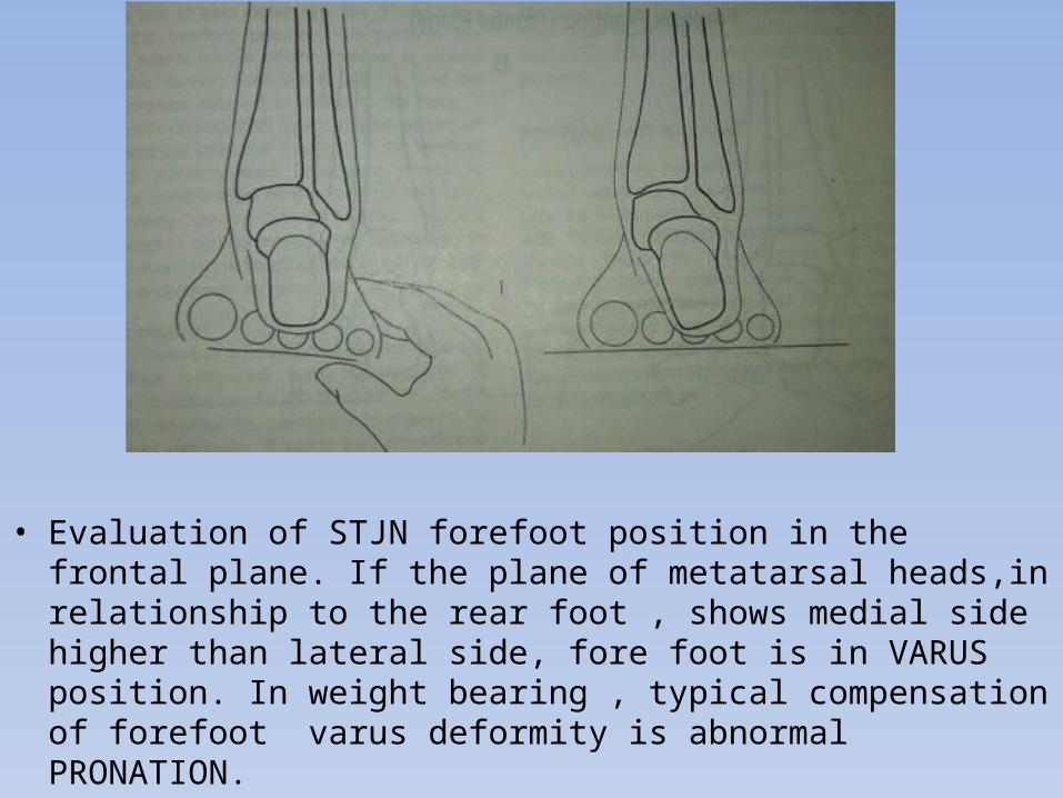
• Evaluation of STJN forefoot position in the frontal plane. If the plane of metatarsal heads,in relationship to the rear foot , shows medial side higher than lateral side, fore foot is in VARUS position. In weight bearing , typical compensation of forefoot varus deformity is abnormal PRONATION.
• The relation of the forefoot to the rear foot must also be assessed in the sagittal plane.
• If the examiner visualizes a plane representing a ground surface applied to the planter surface of the calcaneus, the planter surface of the metatarsal heads should lie on this plane. If the plane of metatarsals sits below that of calcaneus, the forefoot describe as planter flexed in relation with rear foot and referred as forefoot EQUINUS deformity.
• In the transverse plane, the typical relationship b/n forefoot and rearfoot requires the forefoot to be have same longitudinal direction as the rear foot.
• Deviation of fore foot in the transverse plane towards the mid line are referred to ADDUCTION and away from mid line as ABDUCTION.
COMPENSATION• COMPENSATION is a change in the structural alignment or position of
foot to neutralize the effect of an abnormal force, resulting in a deviation in structural alignment or position of another part.
• FORE FOOT VARUS: To compensate this deformity during gait, this foot shows abnormal amount of pronation during mid stance , because when the medial calcaneus condyle has reached the ground in mid stance, the fore foot rather than being in contact with floor, shows orientation where the medial border of foot is elevated from the ground. To assist with the medial border reaching the ground, the STJN continues to pronate…..and this pronation leads to
Eversion of calcaneus Abduction of fore foot Lowering of medial longitudinal arch
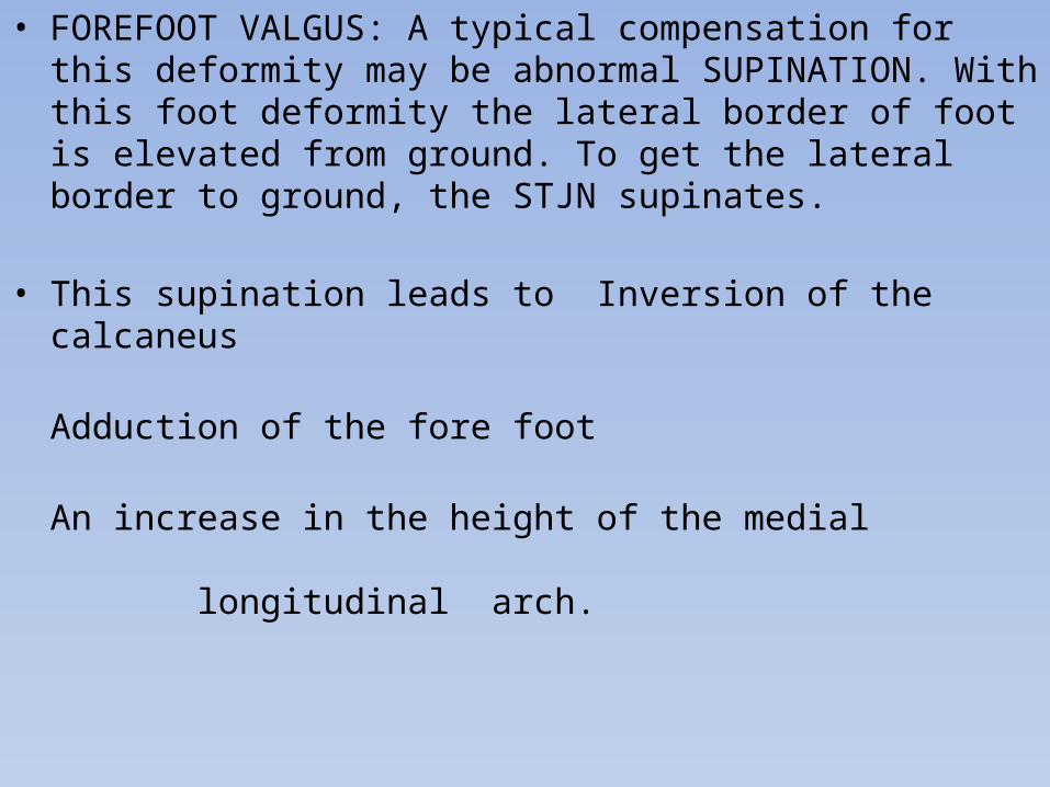
• FOREFOOT VALGUS: A typical compensation for this deformity may be abnormal SUPINATION. With this foot deformity the lateral border of foot is elevated from ground. To get the lateral border to ground, the STJN supinates.
• This supination leads to Inversion of the calcaneus Adduction of the fore foot An increase in the height of the medial
longitudinal arch.
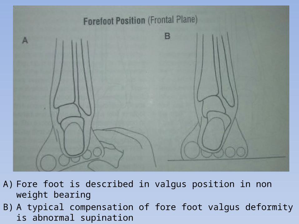
A) Fore foot is described in valgus position in non weight bearingB) A typical compensation of fore foot valgus deformity is abnormal
supination
LEG LENGTH• Good assessment of limb length inequality can by complicated by
scoliosis, hip subluxation, pelvic obliquity, unilateral contracture of hip adductors or abductors, or knee flexion contracture.
• In the absence of an asymmetric hip or knee contracture, limb length can be measured clinically in supine using inferior border of ASIS and distal aspect of the medial malleolus.
• Radiographic assessment is necessary if too many compounding factors are present.
POSTURE AND BALANCE
• Assessment of posterior, anterior, medial and lateral equilibrium responses should not be neglected when planning treatment.
• Many children with CP have delayed or deficient post equilibrium responses.
• Assessment of posture including trunk, pelvis and lower extremity posture in static standing and during walking often gives insight to areas of weakness and poor motor control.
GAIT ANALYSIS• Gait analysis consist of observing the patient with or without use of
formal gait analysis equipment. Now a days computerized gait analysis is very much in use.
• It is still an essential component for diagnosis. Clinical observation is done by repeatedly watching the child walk from sides, back and front. Attention should be paid at pelvis, hip, knee, ankle and foot.
• Beginning with the feet , here several thing to be noted: 1) Is the foot neutral or is it in varus or valgus position? 2) Is the ankle in neutral position or in equinus? 3) what portion of foot contacts the floor first? 4) Is the arch maintain? 5) At which point in the cycle does any deviation in the foot occur?
• At the knee following should be noted: 1) what is the position of knee at initial contact? 2) does knee hyper extended or extension controlled? 3) does the knee comes in full extension at any point of stance? 4) what is the maximum knee flexion in swing? 5) is there varus or valgus motion during loading?• Following thing consider in general: 1) is the pelvic position normal or it is ant. Or post.? 2) is pelvic mal rotation or obliquity present? 3) are the abnormal motions present? 4) how are the arm moving during gait? Are they moving
symmetrically and reciprocally or are they postured? 5) does the child elevates his arms to assist with balance?

THANK YOU