Embed Size (px)
Citation preview
1
Epidemiology of Malocclusion
Presented by:
Dr. preyas joshi
Second year post-graduate student
Deptt. Of Public Health Dentistry
Rajasthan Dental College
Guided by:
Dr. Girish R. Shavi
Head of the Deptt.
Public Health Dentistry
Rajasthan Dental College
2
CONTENTS• Definition:
Orthodontics
Occlusion
Malocclusion
• Classification of Malocclusion
• Etiology of Malocclusion – Classifications
• Etiology of Malocclusion – General Factors
• Etiology of Malocclusion – Local Factors
• Epidemiology and Public Health Aspects of Malocclusion
• Factors affecting Receipt of Orthodontic Treatment
• Bibliography
3
DEFINITIONS
• The definition of orthodontics proposed by the American board of orthodontics (ABO)
and later adopted by the American Association of Orthodontists is:
“Orthodontics is that specific area of the dental profession that has as its responsibility
the study and supervision of the growth and development of the dentition and its related
anatomical structures from birth to dental maturity, including all preventive and
corrective procedures of dental irregularities requiring the repositioning of teeth by
functional and mechanical means to establish normal occlusion and pleasing facial
contours.” 1
• Occlusion is defined as a manner in which the upper and lower teeth intercuspate
between each other in all mandibular positions and movements. It is a result of
neuromuscular control of the components of the mastication systems namely: teeth,
periodontal structures, maxilla and mandibular, temporomandibular joints and their
associated muscles and ligaments (Ash & Ram fjord, 1982).
4
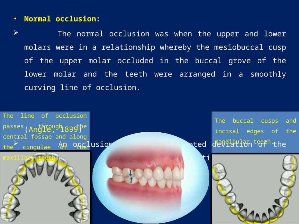
• Normal occlusion:
The normal occlusion was when the upper and lower molars were in a relationship
whereby the mesiobuccal cusp of the upper molar occluded in the buccal grove of the
lower molar and the teeth were arranged in a smoothly curving line of occlusion.
(Angle, 1899)
An occlusion within the accepted deviation of the ideal and did not constitute
aesthetic or functional problems. (Houston et al. 1992)
The line of occlusion passes
through the central fossae and
along the cingulae of the maxillary
teeth.
The buccal cusps and incisal
edges of the mandibular teeth.
5
• Malocclusion can be defined as:
a) Improper relations of apposing teeth when the jaws are in contact.
(Dorland's Medical Dictionary for Health Consumers, 2007)
b) Faulty contact between the upper and lower teeth when the jaw is closed.
(The American Heritage Medical Dictionary, 2007)
c) A deviation in intramaxillary and/or intermaxillary relations of teeth that presents a hazard to
the individual's oral health. Often associated with other orofacial deformities.
(Mosby's Dental Dictionary, 2nd edition, 2008)
d) The World Health Organization (1987), had included malocclusion under the heading of
Handicapping Dento Facial Anomaly, defined as an anomaly which causes disfigurement or
which impedes function, and requiring treatment “if the disfigurement or functional defect
was likely to be an obstacle to the patient’s physical or emotional well-being”.
e) Malocclusion is an appreciable deviation from the ideal occlusion that may be considered
aesthetically unsatisfactory (Houston, et al., 1992) thus implying a condition of imbalance in
the relative sizes and position of teeth, facial bones and soft tissues (lips, cheek, and tongue).
6
MALOCCLUSION 1
The advantages of classifying malocclusion is that it helps in,
a) Diagnosis and planning treatment for the patient.
b) Visualizing and understanding the problem associated with that malocclusion.
c) Communicating the problem.
d) Easy comparison of the various malocclusions.
Depending upon which part of the oral and maxillofacial unit is at fault malocclusions can
be broadly divided into three types –
e) Individual tooth malpositions.
f) Malrelation of the dental arches or dentoalveolar segments.
g) Skeletal malrelationships.
These three can exist individually in a patient or in combination involving each other.
7
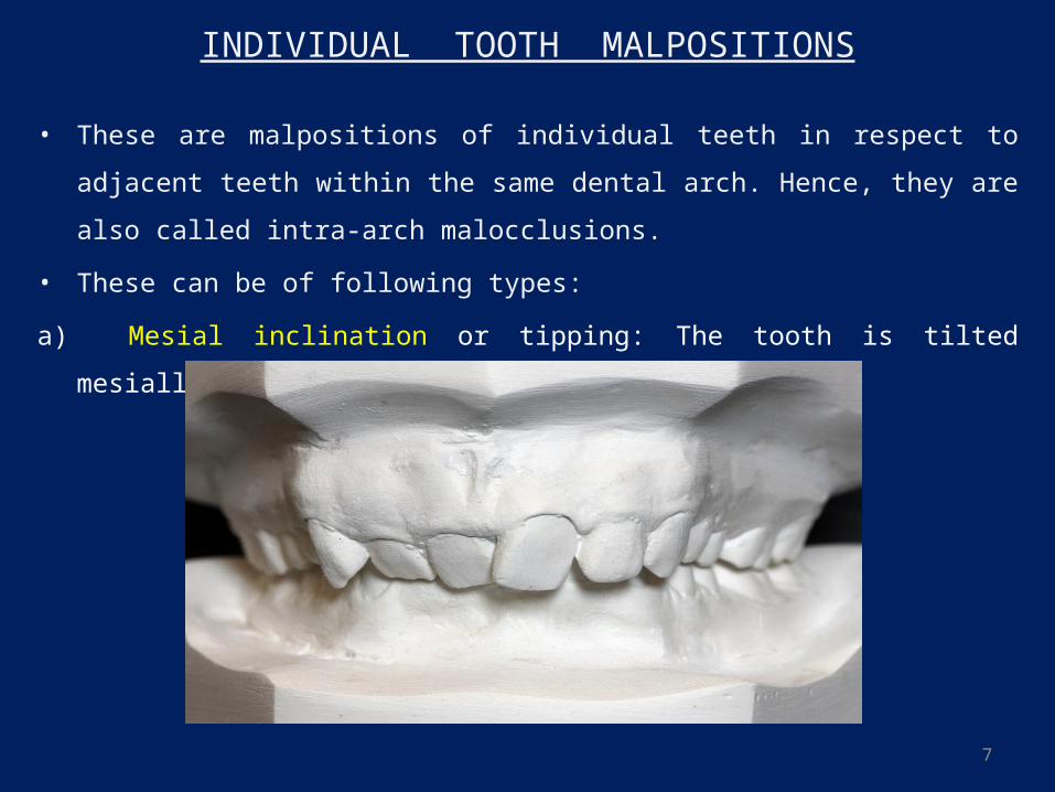
INDIVIDUAL TOOTH MALPOSITIONS
• These are malpositions of individual teeth in respect to adjacent teeth within the same
dental arch. Hence, they are also called intra-arch malocclusions.
• These can be of following types:
a) Mesial inclination or tipping: The tooth is tilted mesially, i.e. the crown is mesial to the
root.
8
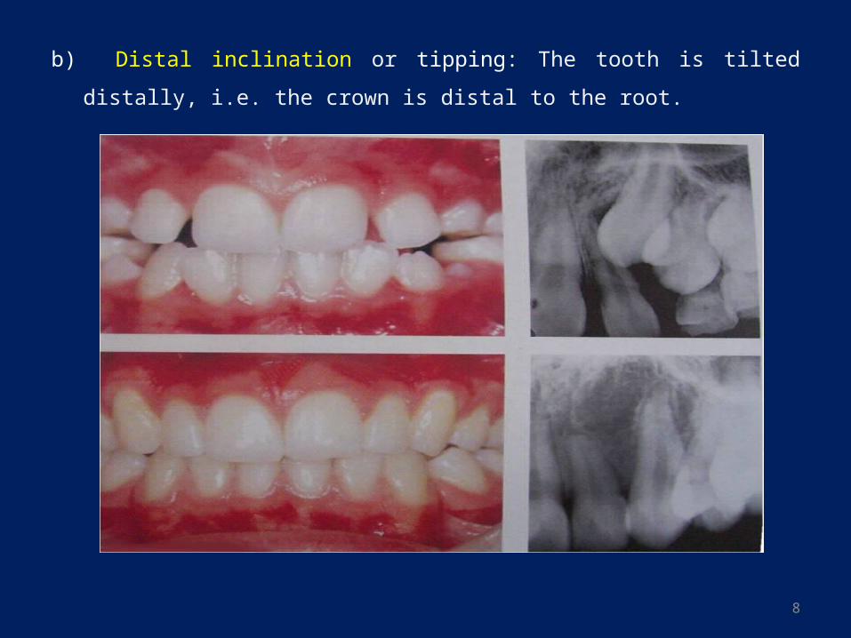
b) Distal inclination or tipping: The tooth is tilted distally, i.e. the crown is distal to the
root.
9
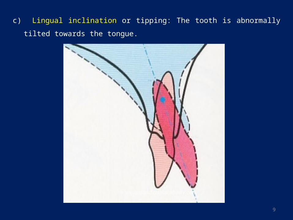
c) Lingual inclination or tipping: The tooth is abnormally tilted towards the tongue.
10
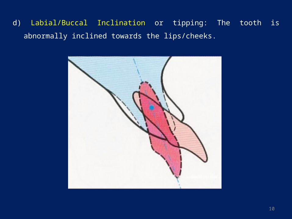
d) Labial/Buccal Inclination or tipping: The tooth is abnormally inclined towards the
lips/cheeks.
11
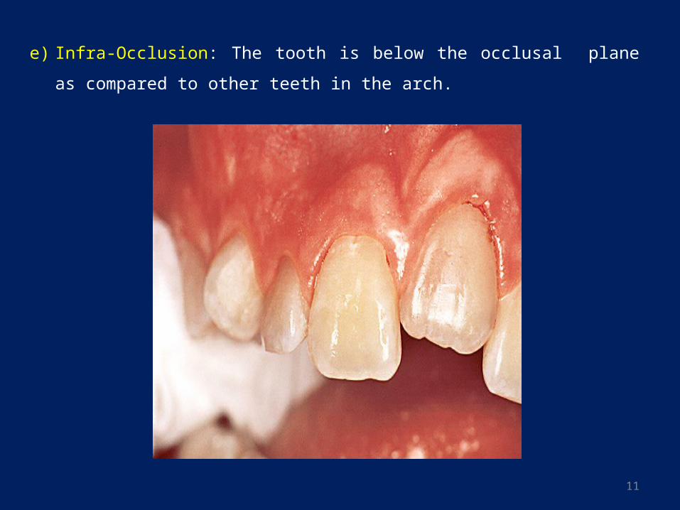
e) Infra-Occlusion: The tooth is below the occlusal plane as compared to other teeth in
the arch.
12
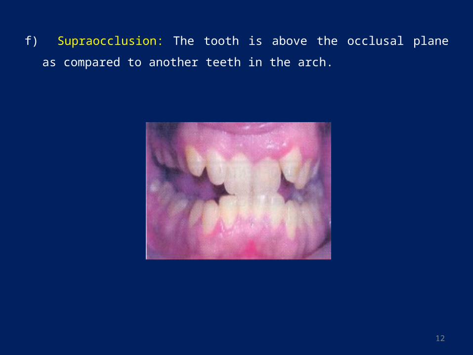
f) Supraocclusion: The tooth is above the occlusal plane as compared to another teeth in
the arch.
13
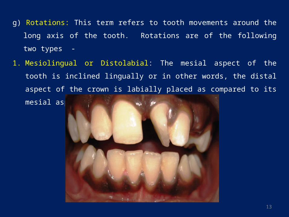
g) Rotations: This term refers to tooth movements around the long axis of the tooth.
Rotations are of the following two types -
1. Mesiolingual or Distolabial: The mesial aspect of the tooth is inclined lingually or in
other words, the distal aspect of the crown is labially placed as compared to its mesial
aspect.
14
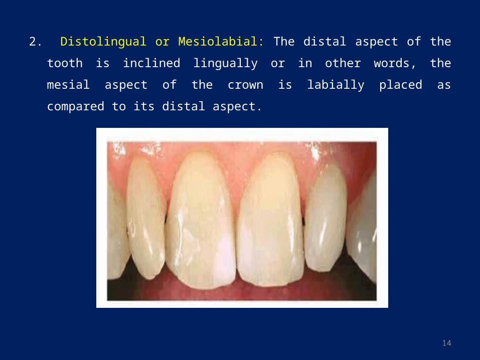
2. Distolingual or Mesiolabial: The distal aspect of the tooth is inclined lingually or in
other words, the mesial aspect of the crown is labially placed as compared to its
distal aspect.
15
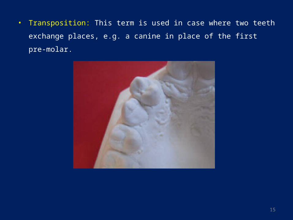
• Transposition: This term is used in case where two teeth exchange places, e.g. a
canine in place of the first pre-molar.
16
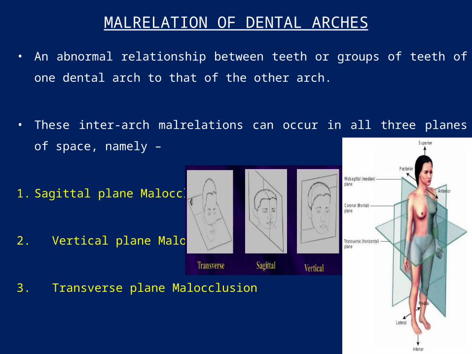
MALRELATION OF DENTAL ARCHES
• An abnormal relationship between teeth or groups of teeth of one dental arch to that of the
other arch.
• These inter-arch malrelations can occur in all three planes of space, namely –
1. Sagittal plane Malocclusion
2. Vertical plane Malocclusion
3. Transverse plane Malocclusion
17
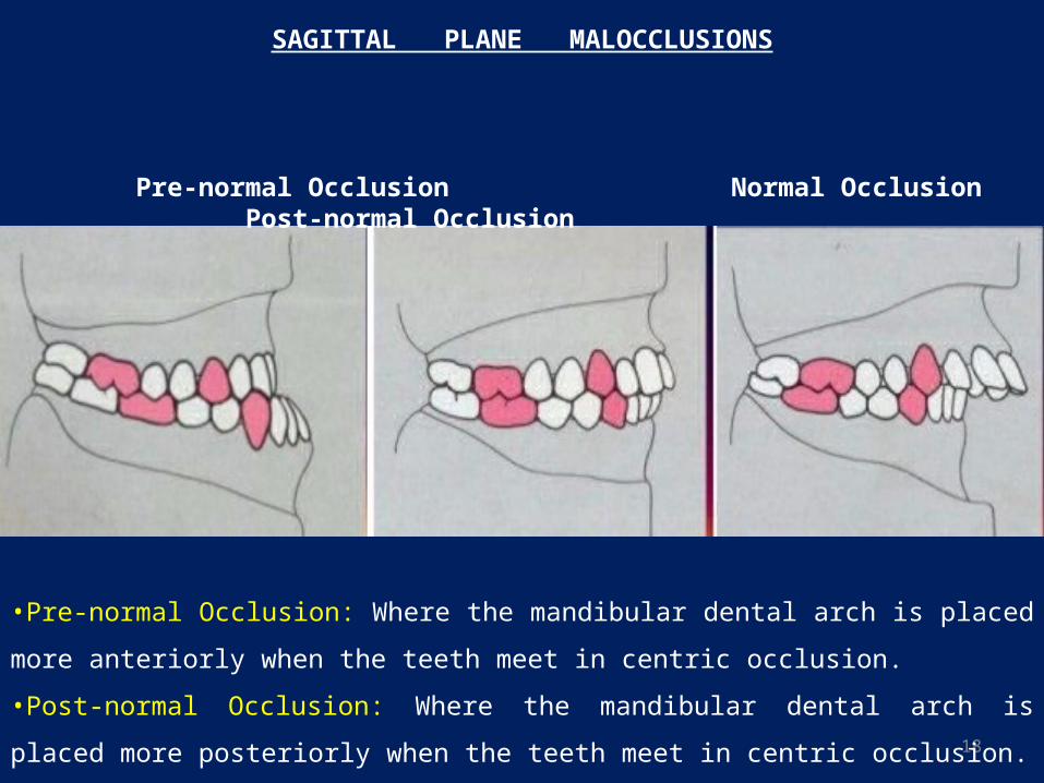
18
Pre-normal Occlusion Normal Occlusion Post-normal Occlusion
•Pre-normal Occlusion: Where the mandibular dental arch is placed more anteriorly when the
teeth meet in centric occlusion.
•Post-normal Occlusion: Where the mandibular dental arch is placed more posteriorly when the
teeth meet in centric occlusion.
SAGITTAL PLANE MALOCCLUSIONS
19
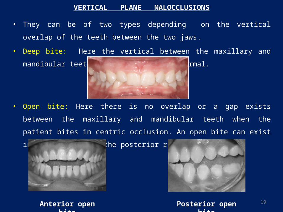
VERTICAL PLANE MALOCCLUSIONS
• They can be of two types depending on the vertical overlap of the teeth between the
two jaws.
• Deep bite: Here the vertical between the maxillary and mandibular teeth is in excess
of the normal.
• Open bite: Here there is no overlap or a gap exists between the maxillary and
mandibular teeth when the patient bites in centric occlusion. An open bite can exist in
the anterior or the posterior region.
Anterior open bite Posterior open bite
20
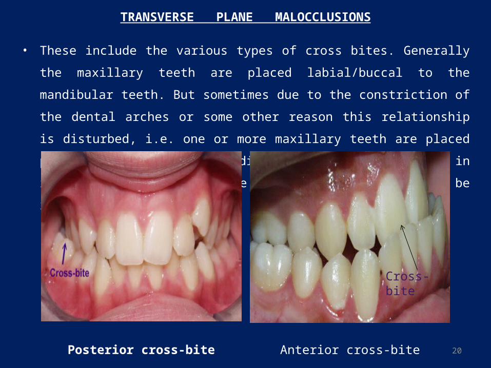
TRANSVERSE PLANE MALOCCLUSIONS
• These include the various types of cross bites. Generally the maxillary teeth are placed
labial/buccal to the mandibular teeth. But sometimes due to the constriction of the
dental arches or some other reason this relationship is disturbed, i.e. one or more
maxillary teeth are placed palatal/lingual to the mandibular teeth. These differ in
intensity, position and the number of teeth that may be involved.
Posterior cross-bite
Cross-bite
Anterior cross-bite
21
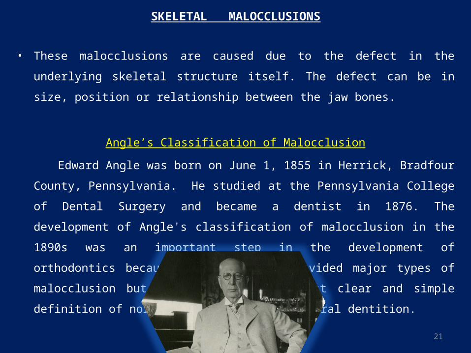
SKELETAL MALOCCLUSIONS
• These malocclusions are caused due to the defect in the underlying skeletal structure
itself. The defect can be in size, position or relationship between the jaw bones.
Angle’s Classification of Malocclusion
Edward Angle was born on June 1, 1855 in Herrick, Bradfour County, Pennsylvania. He
studied at the Pennsylvania College of Dental Surgery and became a dentist in 1876.
The development of Angle's classification of malocclusion in the 1890s was an
important step in the development of orthodontics because it not only subdivided major
types of malocclusion but also included the first clear and simple definition of normal
occlusion in the natural dentition.
22
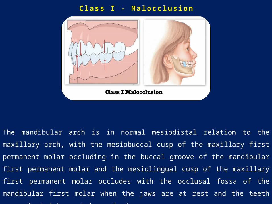
C l a s s I - M a l o c c l u s i o n
The mandibular arch is in normal mesiodistal relation to the maxillary arch, with the mesiobuccal
cusp of the maxillary first permanent molar occluding in the buccal groove of the mandibular
first permanent molar and the mesiolingual cusp of the maxillary first permanent molar occludes
with the occlusal fossa of the mandibular first molar when the jaws are at rest and the teeth
approximated in centric occlusion.
23
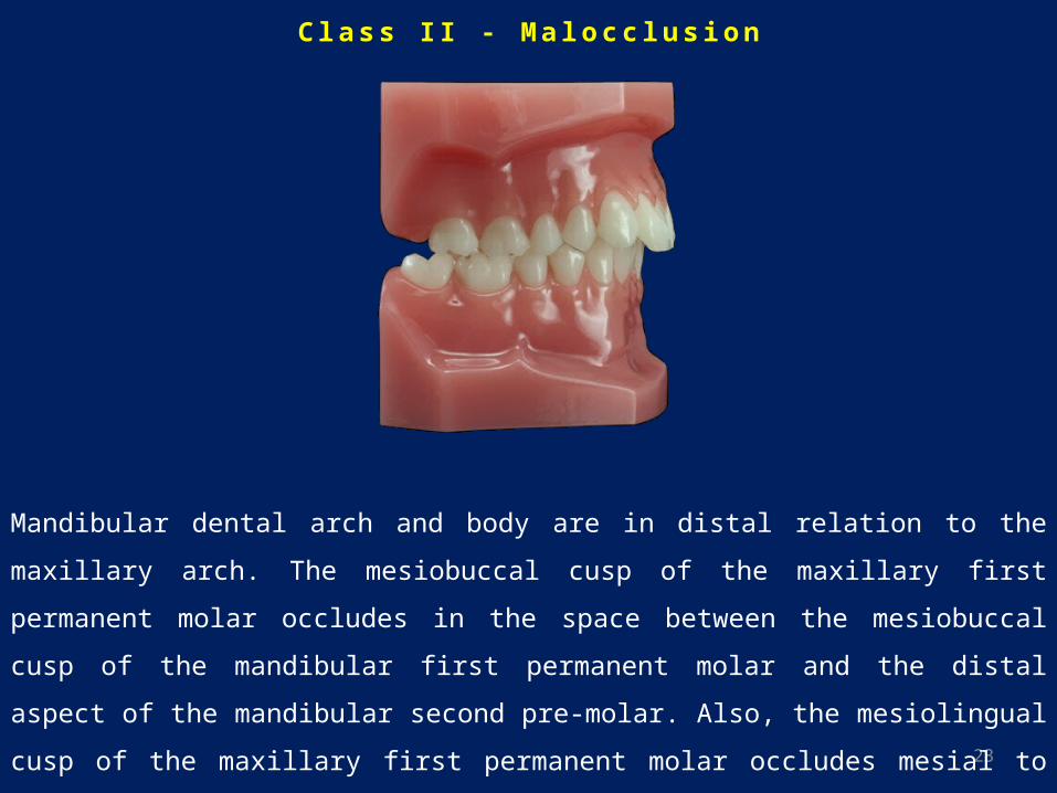
C l a s s I I - M a l o c c l u s i o n
Mandibular dental arch and body are in distal relation to the maxillary arch. The mesiobuccal
cusp of the maxillary first permanent molar occludes in the space between the mesiobuccal cusp
of the mandibular first permanent molar and the distal aspect of the mandibular second pre-
molar. Also, the mesiolingual cusp of the maxillary first permanent molar occludes mesial to the
mesio-lingual cusp of the mandibular first permanent molar.
24
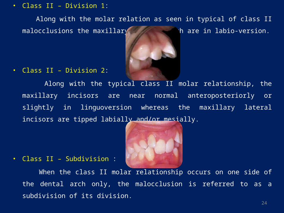
• Class II – Division 1:
Along with the molar relation as seen in typical of class II malocclusions the maxillary
incisor teeth are in labio-version.
• Class II – Division 2:
Along with the typical class II molar relationship, the maxillary incisors are near
normal anteroposteriorly or slightly in linguoversion whereas the maxillary lateral
incisors are tipped labially and/or mesially.
• Class II – Subdivision :
When the class II molar relationship occurs on one side of the dental arch only, the
malocclusion is referred to as a subdivision of its division.
25
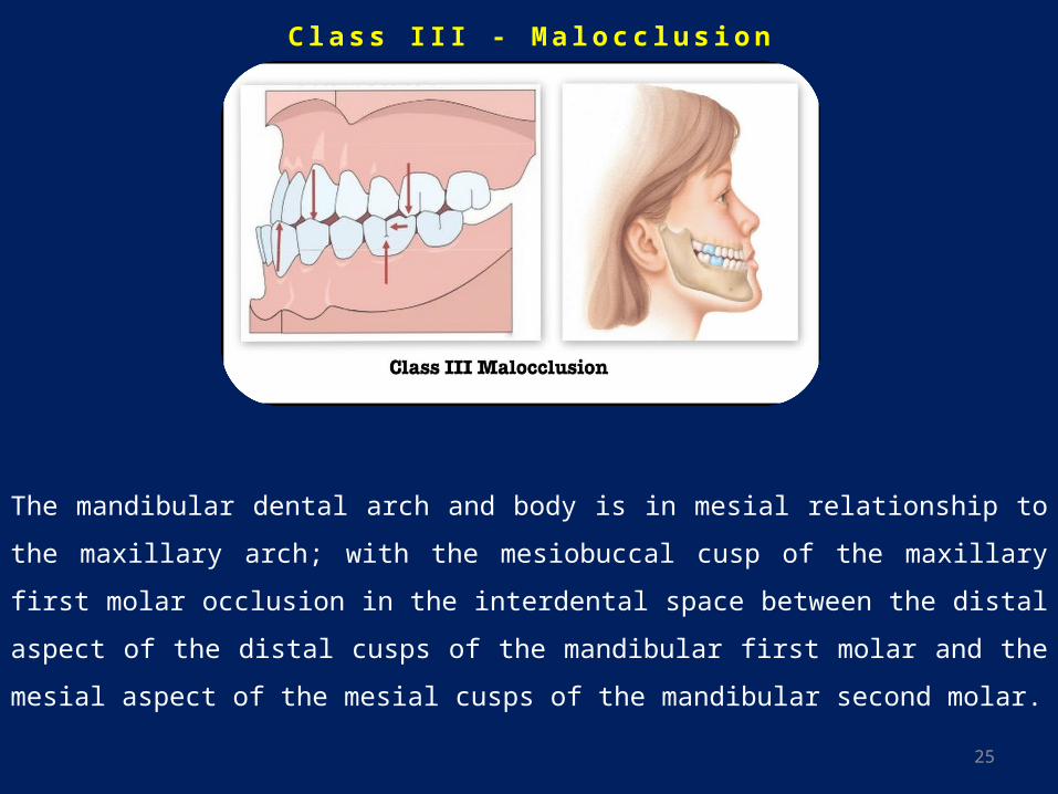
C l a s s I I I - M a l o c c l u s i o n
The mandibular dental arch and body is in mesial relationship to the maxillary arch; with the
mesiobuccal cusp of the maxillary first molar occlusion in the interdental space between the
distal aspect of the distal cusps of the mandibular first molar and the mesial aspect of the mesial
cusps of the mandibular second molar.
26
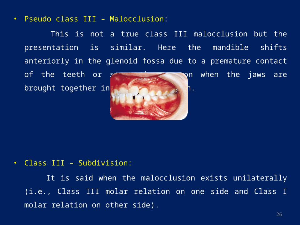
• Pseudo class III – Malocclusion:
This is not a true class III malocclusion but the presentation is similar. Here the
mandible shifts anteriorly in the glenoid fossa due to a premature contact of the teeth
or some other reason when the jaws are brought together in centric occlusion.
• Class III – Subdivision:
It is said when the malocclusion exists unilaterally (i.e., Class III molar relation on one
side and Class I molar relation on other side).
27
Drawbacks of Angle’s classification
• Angle presumed the first permanent molars as fixed points within the jaws, which
definitely is not so.
• Angle depended exclusively on the first molars. Hence, the classification is not possible
if the first molars are missing or in deciduous dentition.
• Malocclusions are considered only in the anteroposterior plane. Malocclusion in the
transverse and vertical planes are not considered.
• Individual tooth malocclusions have not been considered.
• There is no differentiation between skeletal and dental malocclusions.
• Etiology of the malocclusions has not been elaborated upon.
28
Etiology of Malocclusion - Classifications
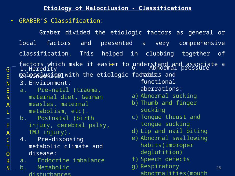
• GRABER’S Classification:
Graber divided the etiologic factors as general or local factors and presented a very
comprehensive classification. This helped in clubbing together of factors which make
it easier to understand and associate a malocclusion with the etiologic factors.
1. Heredity 2. Congenital3. Environment:a. Pre-natal (trauma, maternal diet,
German measles, maternal metabolism, etc).
b. Postnatal (birth injury, cerebral palsy, TMJ injury).
4. Pre-disposing metabolic climate and disease:
a. Endocrine imbalanceb. Metabolic disturbancesc. Infectious diseases5. Dietary problems (nutritional
deficiency)
6. Abnormal pressure habits and functional aberrations:
a) Abnormal suckingb) Thumb and finger suckingc) Tongue thrust and tongue suckingd) Lip and nail bitinge) Abnormal swallowing
habits(improper deglutition)f) Speech defectsg) Respiratory abnormalities(mouth
breathing, etc).h) Tonsils and adenoidsi) Psychogenic tics and bruxism7. Posture8. Trauma and accidents.
GENERAL
FACTORS
29
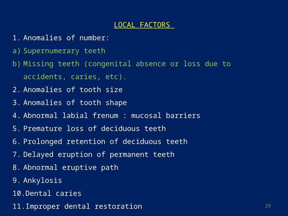
LOCAL FACTORS
1. Anomalies of number:
a) Supernumerary teeth
b) Missing teeth (congenital absence or loss due to accidents, caries, etc).
2. Anomalies of tooth size
3. Anomalies of tooth shape
4. Abnormal labial frenum : mucosal barriers
5. Premature loss of deciduous teeth
6. Prolonged retention of deciduous teeth
7. Delayed eruption of permanent teeth
8. Abnormal eruptive path
9. Ankylosis
10. Dental caries
11. Improper dental restoration
30
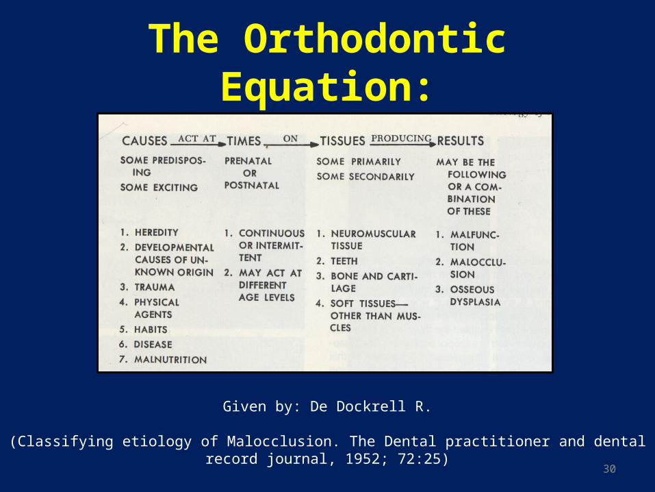
The Orthodontic Equation:
Given by: De Dockrell R.
(Classifying etiology of Malocclusion. The Dental practitioner and dental record journal, 1952; 72:25)
31
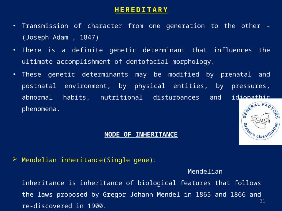
H E R E D I TA R Y
• Transmission of character from one generation to the other – (Joseph Adam , 1847)
• There is a definite genetic determinant that influences the ultimate accomplishment of
dentofacial morphology.
• These genetic determinants may be modified by prenatal and postnatal environment,
by physical entities, by pressures, abnormal habits, nutritional disturbances and
idiopathic phenomena.
MODE OF INHERITANCE
Mendelian inheritance(Single gene):
Mendelian inheritance is inheritance of biological features that follows the laws
proposed by Gregor Johann Mendel in 1865 and 1866 and re-discovered in 1900.
32
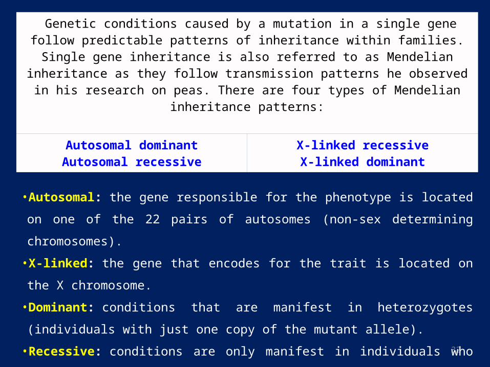
Genetic conditions caused by a mutation in a single gene follow predictable patterns of inheritance within families. Single gene inheritance is also referred to as Mendelian
inheritance as they follow transmission patterns he observed in his research on peas. There are four types of Mendelian inheritance patterns:
Autosomal dominantAutosomal recessive X-linked recessiveX-linked dominant
•Autosomal: the gene responsible for the phenotype is located on one of the 22 pairs of
autosomes (non-sex determining chromosomes).
•X-linked: the gene that encodes for the trait is located on the X chromosome.
•Dominant: conditions that are manifest in heterozygotes (individuals with just one copy
of the mutant allele).
•Recessive: conditions are only manifest in individuals who have two copies of the mutant
allele (are homozygous).
33
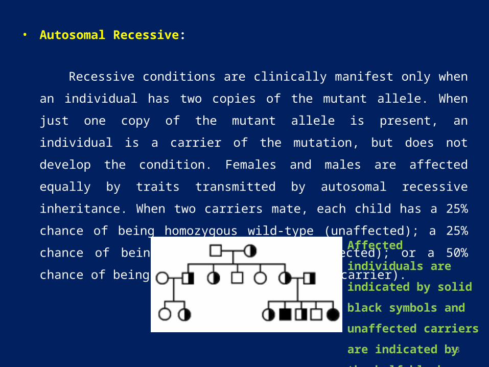
• Autosomal Recessive:
Recessive conditions are clinically manifest only when an individual has two copies of
the mutant allele. When just one copy of the mutant allele is present, an individual is a
carrier of the mutation, but does not develop the condition. Females and males are
affected equally by traits transmitted by autosomal recessive inheritance. When two
carriers mate, each child has a 25% chance of being homozygous wild-type
(unaffected); a 25% chance of being homozygous mutant (affected); or a 50% chance
of being heterozygous (unaffected carrier).
Affected individuals are
indicated by solid black
symbols and unaffected
carriers are indicated by
the half black symbols.
34
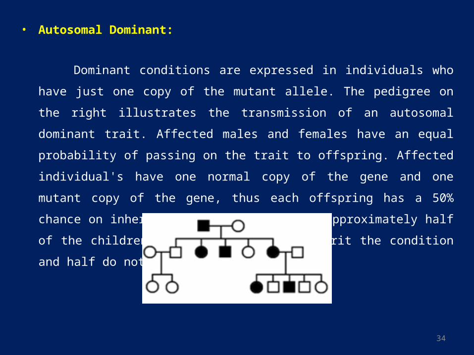
• Autosomal Dominant:
Dominant conditions are expressed in individuals who have just one copy of the
mutant allele. The pedigree on the right illustrates the transmission of an autosomal
dominant trait. Affected males and females have an equal probability of passing on the
trait to offspring. Affected individual's have one normal copy of the gene and one
mutant copy of the gene, thus each offspring has a 50% chance on inheriting the
mutant allele. Approximately half of the children of affected parents inherit the
condition and half do not.
35
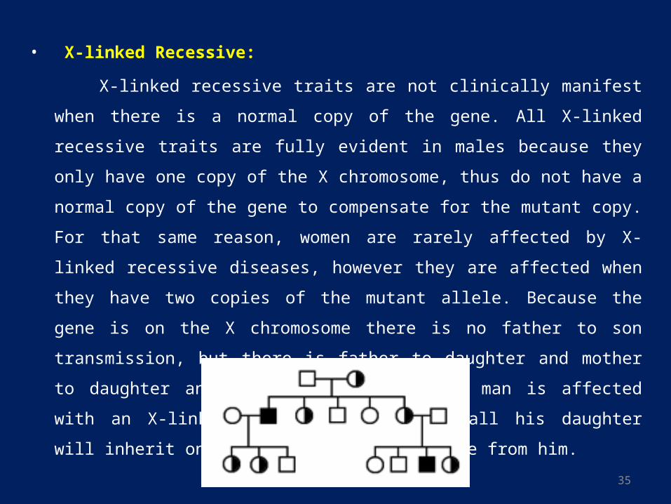
• X-linked Recessive:
X-linked recessive traits are not clinically manifest when there is a normal copy of the
gene. All X-linked recessive traits are fully evident in males because they only have
one copy of the X chromosome, thus do not have a normal copy of the gene to
compensate for the mutant copy. For that same reason, women are rarely affected by
X-linked recessive diseases, however they are affected when they have two copies of
the mutant allele. Because the gene is on the X chromosome there is no father to son
transmission, but there is father to daughter and mother to daughter and son
transmission. If a man is affected with an X-linked recessive condition, all his daughter
will inherit one copy of the mutant allele from him.
36
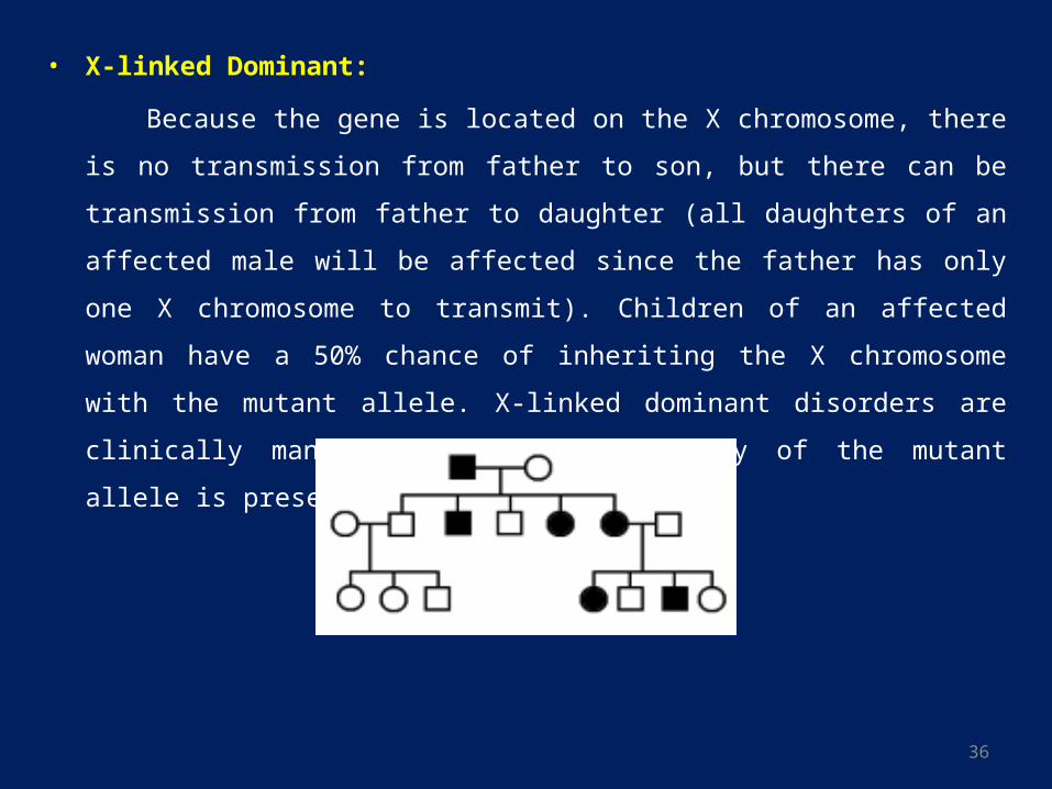
• X-linked Dominant:
Because the gene is located on the X chromosome, there is no transmission from father
to son, but there can be transmission from father to daughter (all daughters of an
affected male will be affected since the father has only one X chromosome to
transmit). Children of an affected woman have a 50% chance of inheriting the X
chromosome with the mutant allele. X-linked dominant disorders are clinically
manifest when only one copy of the mutant allele is present.
37
M U LT I FA C T O R I A L I N H E R I TA N C E
• Most diseases have multifactorial inheritance patterns. As the name implies,
multifactorial conditions are not caused by a single gene, but rather are a result of
interplay between genetic factors and environmental factors. Diseases with
multifactorial inheritance are not genetically determined, but rather a genetic mutation
may predispose an individual to a disease.
• Other genetic and environmental factors contribute to whether or not the disease
develops.
• Numerous genetic alterations may predispose individuals to the same disease (genetic
heterogeneity). For instance coronary heart disease risk factors include high blood
pressure, diabetes, and hyperlipidemia. All of those risk factors have their own genetic
and environmental components. Thus multifactorial inheritance is far more complex
than Mendelian inheritance and is more difficult to trace through pedigrees.
38
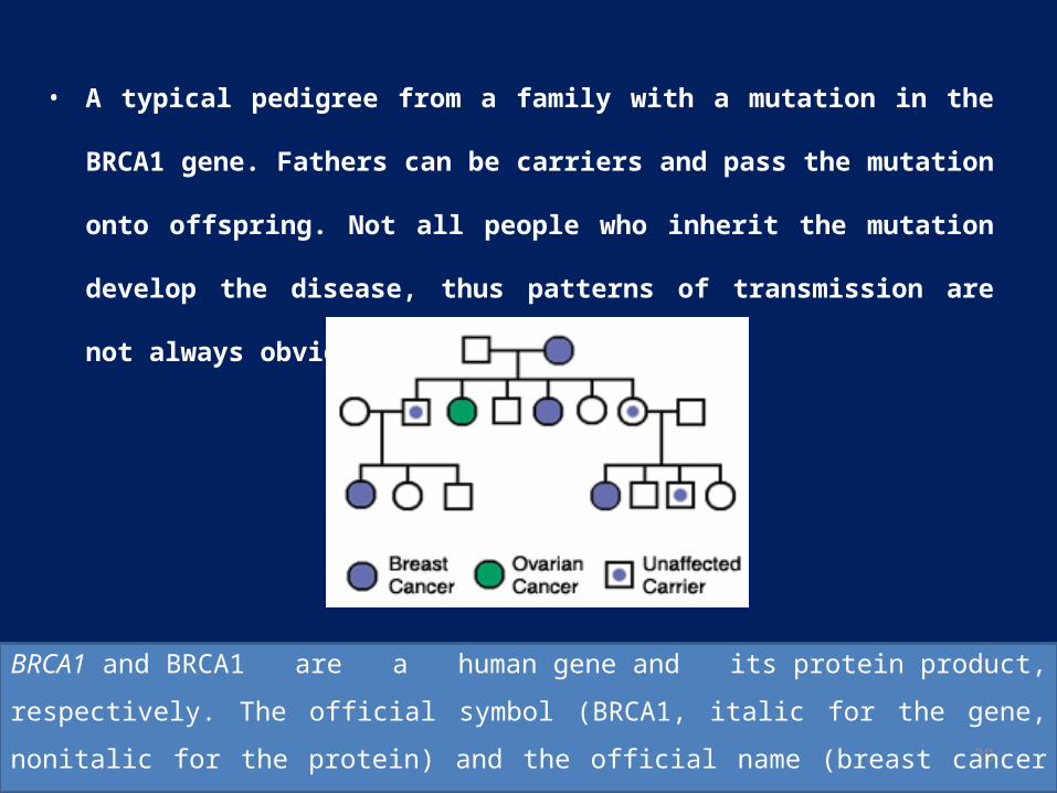
• A typical pedigree from a family with a mutation in the BRCA1 gene. Fathers
can be carriers and pass the mutation onto offspring. Not all people who inherit
the mutation develop the disease, thus patterns of transmission are not always
obvious.
BRCA1 and BRCA1 are a human gene and its protein product, respectively. The official symbol
(BRCA1, italic for the gene, nonitalic for the protein) and the official name (breast cancer 1,
early onset) are maintained by the HUGO Gene Nomenclature Committee
39
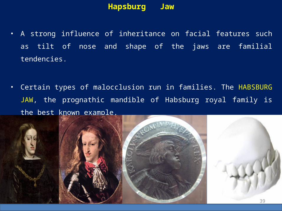
• A strong influence of inheritance on facial features such as tilt of nose and shape of the
jaws are familial tendencies.
• Certain types of malocclusion run in families. The HABSBURG JAW, the prognathic
mandible of Habsburg royal family is the best known example.
Hapsburg Jaw

40
• The most prominent case of mandibular prognathism is that of Charles II of Spain,
who had prognathism so pronounced he could neither speak clearly nor chew as a
result of generations of politically motivated inbreeding.

41
• Hapsburg Jaw, A very diminished feature, on the current King of Spain, Juan Carlos-I
42
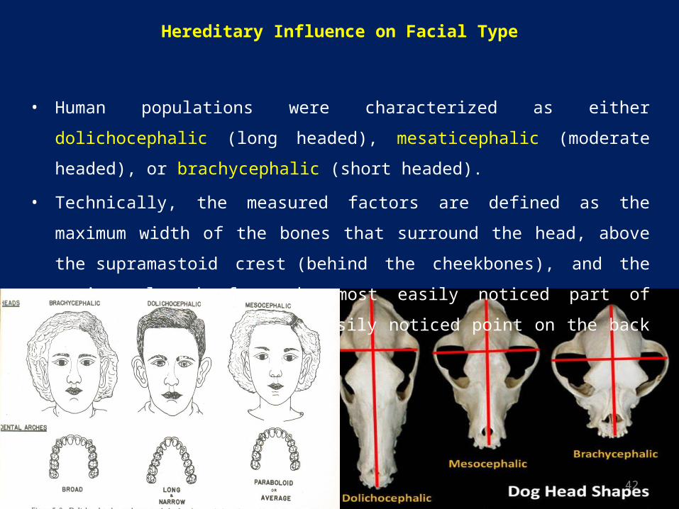
Hereditary Influence on Facial Type
• Human populations were characterized as either dolichocephalic (long headed),
mesaticephalic (moderate headed), or brachycephalic (short headed).
• Technically, the measured factors are defined as the maximum width of the bones that
surround the head, above the supramastoid crest (behind the cheekbones), and the
maximum length from the most easily noticed part of the glabella to the most easily
noticed point on the back part of the head.
43
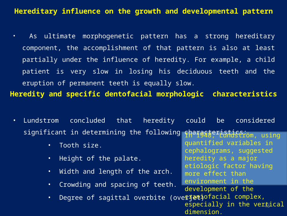
Hereditary influence on the growth and developmental pattern
• As ultimate morphogenetic pattern has a strong hereditary component, the
accomplishment of that pattern is also at least partially under the influence of heredity.
For example, a child patient is very slow in losing his deciduous teeth and the eruption
of permanent teeth is equally slow.
• Lundstrom concluded that heredity could be considered significant in determining the
following characteristics:
• Tooth size.
• Height of the palate.
• Width and length of the arch.
• Crowding and spacing of teeth.
• Degree of sagittal overbite (overjet)
Heredity and specific dentofacial morphologic characteristics
In 1948, Lundström, using quantified variables in cephalograms, suggested heredity as a major etiologic factor having more effect than environment in the development of the craniofacial complex, especially in the vertical dimension.
44
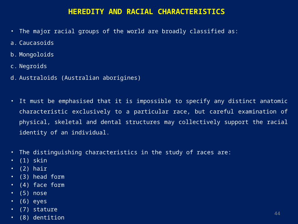
HEREDITY AND RACIAL CHARACTERISTICS
• The major racial groups of the world are broadly classified as:
a. Caucasoids
b. Mongoloids
c. Negroids
d. Australoids (Australian aborigines)
• It must be emphasised that it is impossible to specify any distinct anatomic characteristic exclusively to
a particular race, but careful examination of physical, skeletal and dental structures may collectively
support the racial identity of an individual.
• The distinguishing characteristics in the study of races are:• (1) skin• (2) hair• (3) head form• (4) face form• (5) nose• (6) eyes• (7) stature• (8) dentition
45
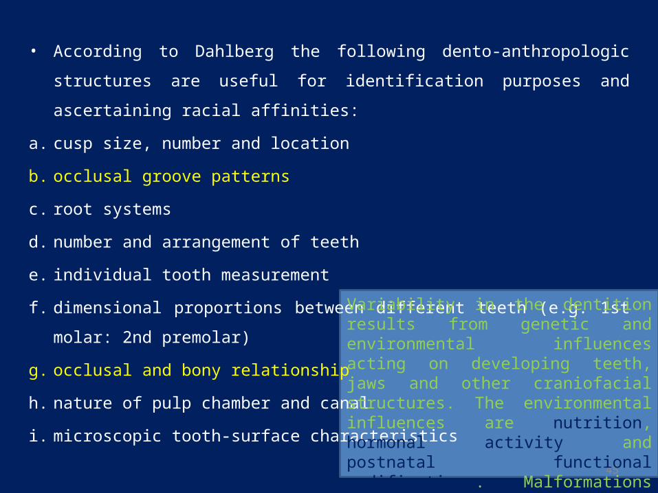
• According to Dahlberg the following dento-anthropologic structures are useful for
identification purposes and ascertaining racial affinities:
a. cusp size, number and location
b. occlusal groove patterns
c. root systems
d. number and arrangement of teeth
e. individual tooth measurement
f. dimensional proportions between different teeth (e.g. 1st molar: 2nd premolar)
g. occlusal and bony relationship
h. nature of pulp chamber and canal
i. microscopic tooth-surface characteristics
Variability in the dentition results from genetic and environmental influences acting on developing teeth, jaws and other craniofacial structures. The environmental influences are nutrition, hormonal activity and postnatal functional modifications. Malformations may arise if growth in the jaws and other craniofacial elements is not coordinated.
46
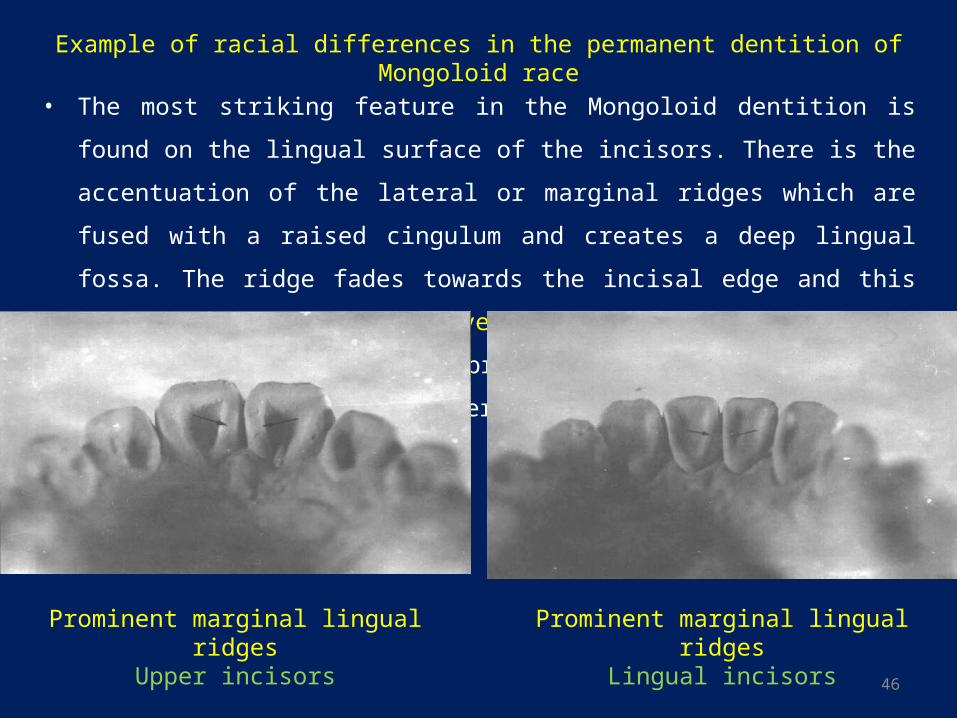
• The most striking feature in the Mongoloid dentition is found on the lingual surface of
the incisors. There is the accentuation of the lateral or marginal ridges which are fused
with a raised cingulum and creates a deep lingual fossa. The ridge fades towards the
incisal edge and this gives the tooth a 'shovel' or ‘scoop’ shape. This condition is found
in approximately 90% of Mongoloids inclusive of Eskimos and American Indians
Example of racial differences in the permanent dentition of Mongoloid race
Prominent marginal lingual ridgesUpper incisors
Prominent marginal lingual ridgesLingual incisors
47
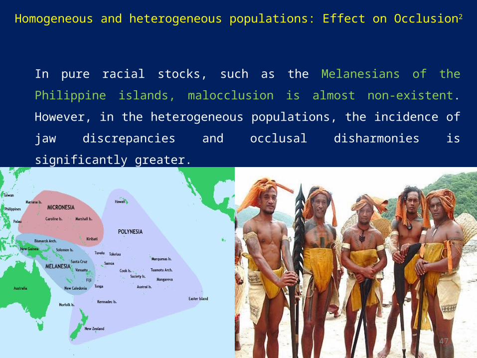
Homogeneous and heterogeneous populations: Effect on Occlusion2
In pure racial stocks, such as the Melanesians of the Philippine islands, malocclusion is
almost non-existent. However, in the heterogeneous populations, the incidence of jaw
discrepancies and occlusal disharmonies is significantly greater.
48
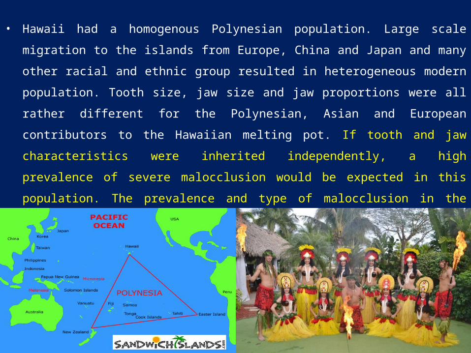
• Hawaii had a homogenous Polynesian population. Large scale migration to the islands from
Europe, China and Japan and many other racial and ethnic group resulted in heterogeneous
modern population. Tooth size, jaw size and jaw proportions were all rather different for the
Polynesian, Asian and European contributors to the Hawaiian melting pot. If tooth and jaw
characteristics were inherited independently, a high prevalence of severe malocclusion would
be expected in this population. The prevalence and type of malocclusion in the current
Hawaiian population, though greater than the prevalence of malocclusion in the original
population , do not support this concept. The effect of interracial crosses appear to be additive
than multiplicative.
49
• It is logical to assume that heredity plays a part in the following conditions:
1. Congenital deformities
2. Facial asymmetries
3. Macrognathia and micrognathia
4. Macrodontia and microdontia
5. Oligodontia and anodontia
6. Tooth shape variations (peg-shaped lateral incisors, carabelli’s cusps, mamelons etc)
7. Cleft palate and harelip
8. Frenum diastemas
9. Deep overbite
10. Growing and rotation of teeth
11. Mandibular retrusion
12. Mandibular prognathism
50
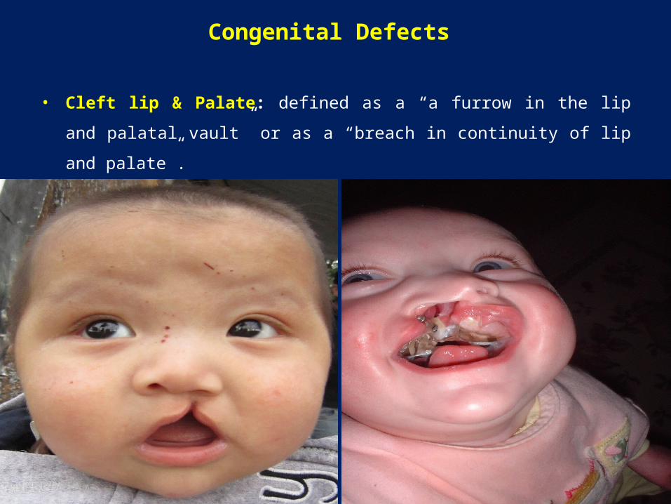
Congenital Defects
• Cleft lip & Palate: defined as a “a furrow in the lip and palatal vault” or as a
“breach in continuity of lip and palate”.
51
• Etiology:
Clefts usually have a strong genetic relationship.
About 1/3 or 1/2 of all cleft palate children have a familiar history of this
deformity.
According to Bhatia the possible modes of transmission are either by a single
mutant gene producing a large effect, or by a number of gene (polygenic
inheritance) each producing a small effect together, create this condition.
According to Fogh-Andersen slightly less than 40 % of the cleft lip cases with or
without cleft palate are genetic in origin where as slightly less than 20% of the
isolated cleft palate cases appear to be genetically derived.
52
• Environment: Teratogens, radiation & dietary deficiency
A. Teratogens are:
a) Aspirin – cleft lip and palate
b) Cigarette smoke (hypoxia) – cleft lip and palate
c) Dilantin – cleft lip and palate
d) Valium- cleft lip and palat
e) Rubella virus
B. Radiations such as X-rays, gamma rays are capable of producing clefts in fetus
during pregnancy.
C. Dietary deficiency such as folic acid deficiency can produce clefts.
53
• Multifactorial Etiology:
Recent studies have shown that the etiology of cleft lip and palate cannot be
attributed solely to either genetic or environmental factors. It seems to involve
more than one factor.
Multi-factorial inheritance theory implies that many contributory risk genes interact
with one another and the environment, resulting in a defect in the developing fetus.
Unless a person is genetically susceptible, the environment factors may not by
themselves cause clefts.
54
Incidence:
• Unilateral cleft accounts for nearly 80% of all cleft seen.
• While bilateral clefts account for remaining 20%.
• Among the unilateral clefts, clefts involving the left side are more common.
• Male patients show a higher incidence of cleft lip with and without palate.
• Female patients suffer from isolated cleft palate more.
• Cleft lip & palate are common congenital malformations.
• The reported incidence of clefts of the lip and palate from 1 in 500 to 1 in 2500 live births depending on
geographic origin, racial and ethnic backgrounds and socioeconomic status.
• Asian populations have the highest frequencies, often at 1 in 500 or higher, with Caucasian populations
intermediate, and African-derived populations the lowest at 1 in 2500.
• In the USA, one child in every 700 live births is afflicted.
55
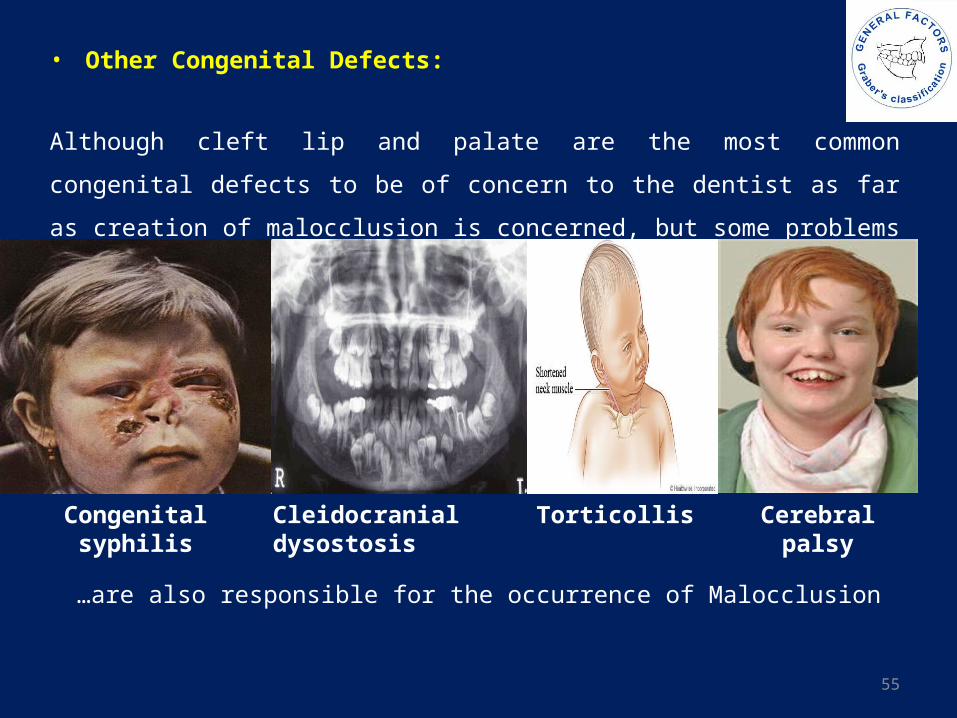
• Other Congenital Defects:
Although cleft lip and palate are the most common congenital defects to be of concern
to the dentist as far as creation of malocclusion is concerned, but some problems such
as:
Cerebral palsyTorticollis Cleidocranial dysostosisCongenital syphilis
…are also responsible for the occurrence of Malocclusion
56
Cerebral palsy
• Cerebral palsy is considered a neurological
disorder caused by a non-progressive brain
injury or malformation that occurs while the
child’s brain is under development. Though
cerebral palsy can be defined, having cerebral
palsy does not define the person that has the
condition. cerebral palsy is a blanket term
commonly referred to as “CP” and described
by loss or impairment of motor function. The
brain damage is caused by brain injury or
abnormal development of the brain that
occurs while a child’s brain is still developing
- before birth, during birth, or immediately
after birth.
Cerebral palsy is…
Non-life-threatening: Most children with cerebral palsy are expected to live well into adulthood.
Incurable: Treatment and therapy help manage effects on the body.
Non-progressive: one-time brain injury and will not produce further degeneration of the brain.
Permanent: cerebral palsy itself will not change for better or worse during a person’s lifetime.
Not contagious nor communicable.
Manageable: T/t, therapy, surgery, medications & asst. technology can help maximize independence.
Chronic: An individual diagnosed with cerebral palsy will have the condition for their entire life.
57
To r t i c o l l i s / W r y n e c k / L o x i a
• Torticollis is the common term for various conditions of head and neck dystonia*, which display specific
variations in head movements (phasic components) characterized by the direction of movement.
• Torticollis results in a fixed or dynamic posturing of the head and neck in tilt, rotation, and flexion.
• Spasms of the sternocleidomastoid, trapezius, and other neck muscles, usually more prominent on one
side than the other, cause turning or tipping of the head.
• Current studies demonstrates a strong association between asymmetric class III malocclusion, torticollis,
and cranial base asymmetry. Undiagnosed torticollis is a likely etiology for otherwise idiopathic cranial
base asymmetry and cranial base asymmetry in turn causes facial asymmetry and malocclusion.3
*a neurological movement disorder, in which sustained muscle contractions cause twisting and repetitive
movements or abnormal postures.
58
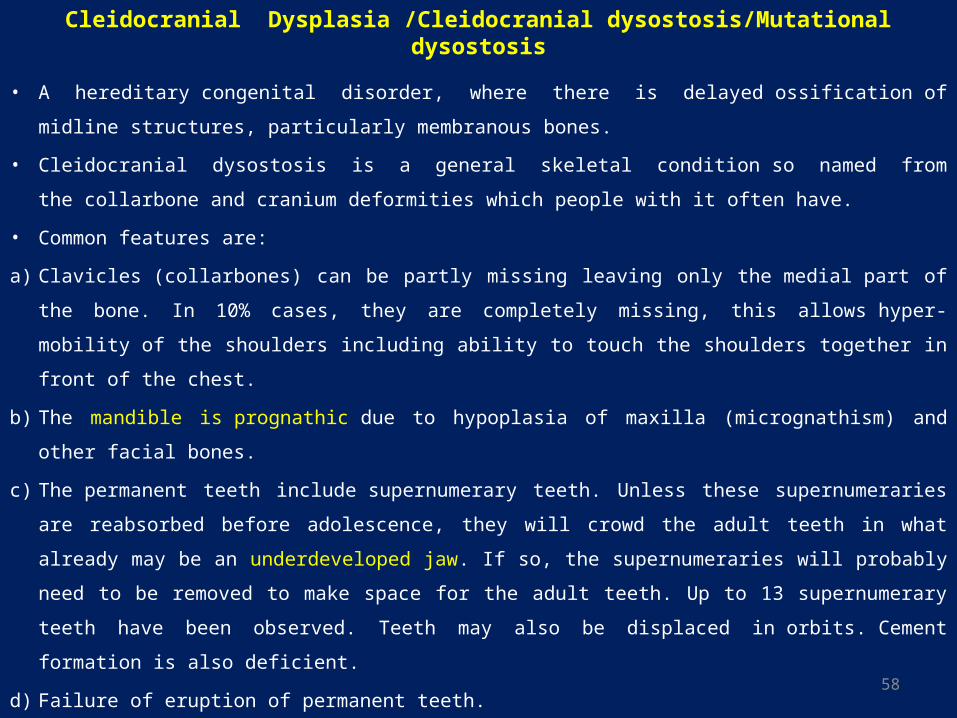
Cleidocranial Dysplasia /Cleidocranial dysostosis/Mutational dysostosis
• A hereditary congenital disorder, where there is delayed ossification of midline structures,
particularly membranous bones.
• Cleidocranial dysostosis is a general skeletal condition so named from the collarbone
and cranium deformities which people with it often have.
• Common features are:
a) Clavicles (collarbones) can be partly missing leaving only the medial part of the bone. In 10%
cases, they are completely missing, this allows hyper-mobility of the shoulders including ability
to touch the shoulders together in front of the chest.
b) The mandible is prognathic due to hypoplasia of maxilla (micrognathism) and other facial bones.
c) The permanent teeth include supernumerary teeth. Unless these supernumeraries are reabsorbed
before adolescence, they will crowd the adult teeth in what already may be an underdeveloped
jaw. If so, the supernumeraries will probably need to be removed to make space for the adult
teeth. Up to 13 supernumerary teeth have been observed. Teeth may also be displaced
in orbits. Cement formation is also deficient.
d) Failure of eruption of permanent teeth.
59
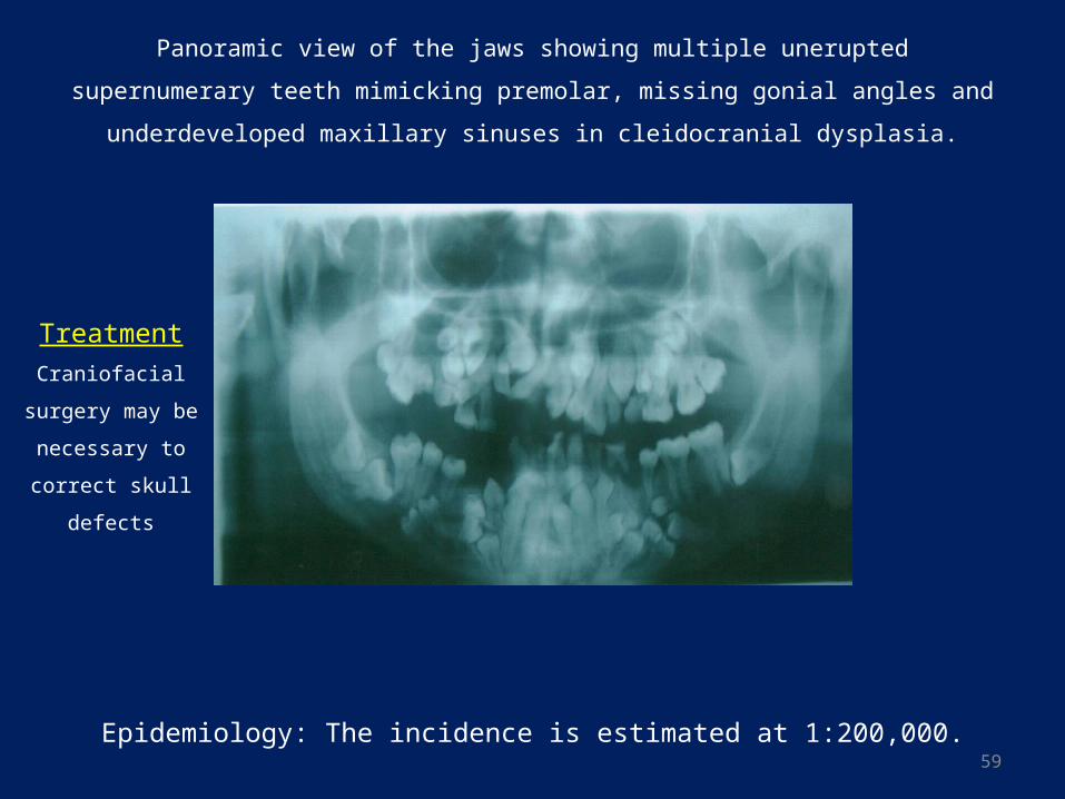
Panoramic view of the jaws showing multiple unerupted supernumerary teeth
mimicking premolar, missing gonial angles and underdeveloped maxillary sinuses in
cleidocranial dysplasia.
Epidemiology: The incidence is estimated at 1:200,000.
Treatment
Craniofacial surgery
may be necessary to
correct skull defects
60
Congenital syphilis
• Congenital syphilis is syphilis present in utero and at birth, and occurs when a child is
born to a mother with syphilis. It is a severe, disabling, and often life-threatening
infection seen in infants. A pregnant mother who has syphilis can spread the disease
through the placenta to the unborn infant.
• Congenital syphilis is caused by the bacterium Treponema pallidum, which is passed
from mother to child during fetal development or at birth. Nearly half of all children
infected with syphilis while they are in the womb die shortly before or after birth.
• Possible Complications:
a. Blindness
b. Deafness
c. Deformity of the face
d. Nervous system problems
• Treatment: Penicillin is used to treat all forms of syphilis.
61
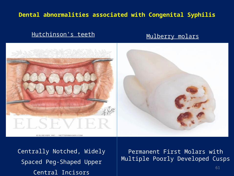
Dental abnormalities associated with Congenital Syphilis
Hutchinson's teeth
Centrally Notched, Widely Spaced Peg-
Shaped Upper Central Incisors
Permanent First Molars with Multiple Poorly Developed Cusps
Mulberry molars
62
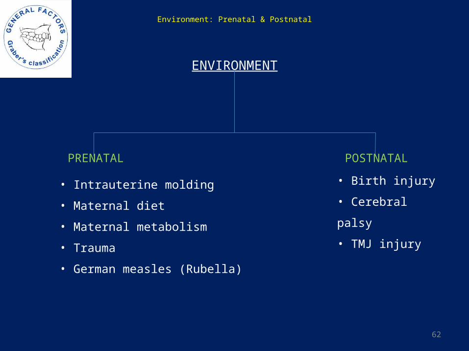
Environment: Prenatal & Postnatal
ENVIRONMENT
PRENATAL POSTNATAL
• Intrauterine molding
• Maternal diet
• Maternal metabolism
• Trauma
• German measles (Rubella)
• Birth injury
• Cerebral palsy
• TMJ injury
63
Prenatal Influence
• The role of prenatal influences on malocclusion is probably very small.
• Intrauterine molding pressure against the developing face prenatally can lead to distortion of
rapidly growing areas. Eg: On rare occasions an arm is pressed across the face in uterus, resulting
in severe maxillary deficiency at birth.
• Pierre Robin syndrome: a congenital condition of facial abnormalities in humans.
Cause(s): one theory is that, at some time during the stage of the formation of the bones of the
fetus, the tip of the jaw (mandible) becomes 'stuck' in the point where each of the collar bones
(clavicle) meet (the sternum), effectively preventing the jaw bones from growing. It is thought
that, at about 12 to 14 weeks gestation, when the fetus begins to move, the movement of the head
causes the jaw to "pop out' of the collar bones. From this time on, the jaw of the fetus grows as it
would normally, with the result that, when born, the jaw of the baby is much smaller
(micrognathia) than it would have been with normal development, although it does continue to
grow at a normal rate until the child reaches maturity.
64
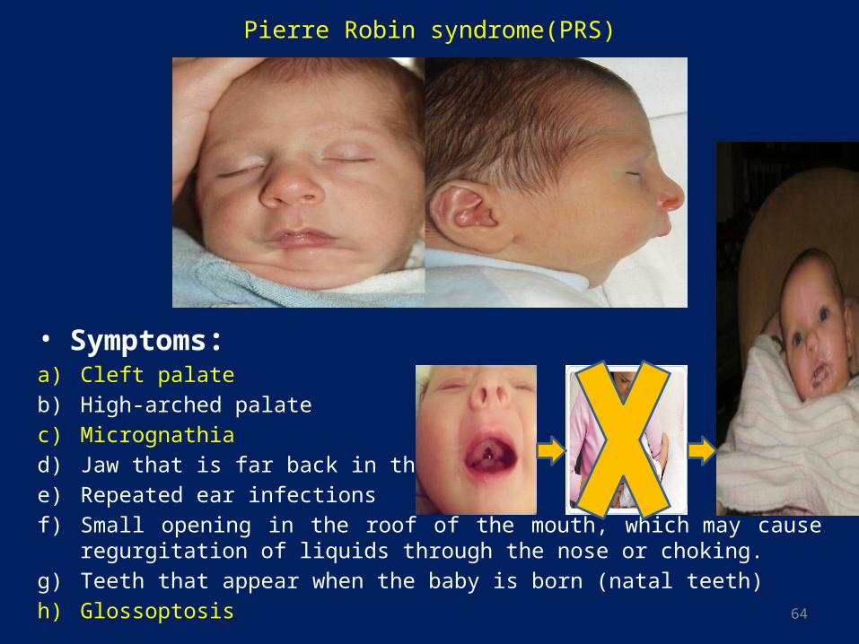
Pierre Robin syndrome(PRS)
• Symptoms:a) Cleft palate
b) High-arched palate
c) Micrognathia
d) Jaw that is far back in the throat
e) Repeated ear infections
f) Small opening in the roof of the mouth, which may cause regurgitation of liquids through the nose or choking.
g) Teeth that appear when the baby is born (natal teeth)
h) Glossoptosis
65
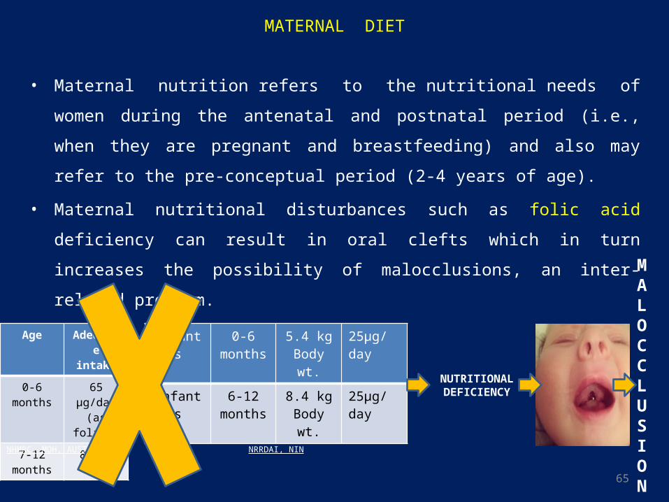
MATERNAL DIET
• Maternal nutrition refers to the nutritional needs of women during the antenatal and
postnatal period (i.e., when they are pregnant and breastfeeding) and also may refer to
the pre-conceptual period (2-4 years of age).
• Maternal nutritional disturbances such as folic acid deficiency can result in oral clefts
which in turn increases the possibility of malocclusions, an inter-related problem.
Infants 0-6 months
5.4 kg Body wt.
25µg/day
Infants 6-12 months
8.4 kg Body wt.
25µg/day
Age Adequate intake
0-6 months 65 µg/day (as folate)
7-12 months
80µg/day
NRRDAI, NINNHMRC, MOH, AUSTRALIA
NUTRITIONAL DEFICIENCY
MALOCCLUSION
66
• Teratogens are substances or environmental agents which cause the development of
abnormal cell masses during fetal growth, resulting in physical defects in the fetus. The
time of exposure is important concept for teratogen, as certain stages of embryonic &
fetal development are more vulnerable than others. In general, the embryonic stage (first
trimester) is more vulnerable than the fetal period (second & third trimester).
• Examples of teratogens include certain chemicals, medications, and infections or other
diseases in the mother.
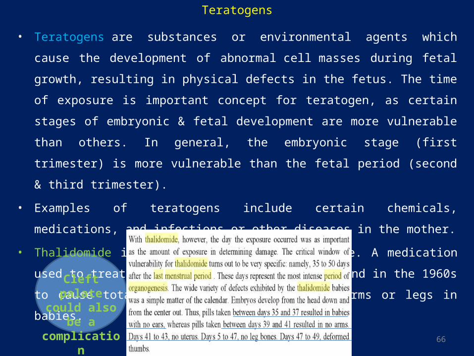
• Thalidomide is one notable & classic example. A medication used to treat morning
sickness, which was found in the 1960s to cause total or partial absence of the arms or
legs in babies.
Cleft palate could also be a complication
Teratogens
67
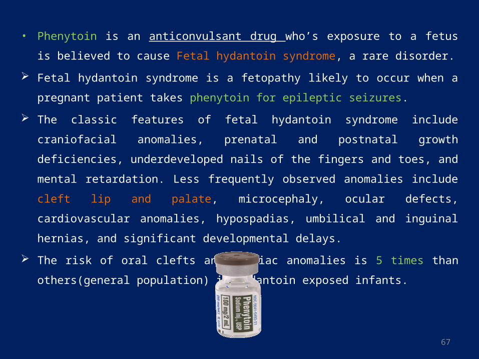
• Phenytoin is an anticonvulsant drug who’s exposure to a fetus is believed to cause
Fetal hydantoin syndrome, a rare disorder.
Fetal hydantoin syndrome is a fetopathy likely to occur when a pregnant patient takes
phenytoin for epileptic seizures.
The classic features of fetal hydantoin syndrome include craniofacial anomalies,
prenatal and postnatal growth deficiencies, underdeveloped nails of the fingers and
toes, and mental retardation. Less frequently observed anomalies include cleft lip and
palate, microcephaly, ocular defects, cardiovascular anomalies, hypospadias, umbilical
and inguinal hernias, and significant developmental delays.
The risk of oral clefts and cardiac anomalies is 5 times than others(general population)
in hydantoin exposed infants.
68
• Folic acid antagonists, as a group, increase the risk of certain birth defects.
Multiple studies have evaluated the role of folic acid in the occurrence and recurrence of
orofacial clefts.
Folate antagonist are chemotherapeutic agents used in many neoplastic, autoimmune,
and inflammatory disorders. The first suggestions that folic acid antagonists were
teratogenic in humans were based on reports of failed terminations in mothers given
aminopterin in the first trimester. Newborns who survived after aminopterin exposure
were noted for years to have defects of the neural tube, skull, or limbs. There is now a
well-defined syndrome of congenital anomalies associated with the use of aminopterin.
The Fetal aminopterin syndrome consists of cranial dysostosis, hypertelorism, anomalies
of the external ears, micrognathia, limb anomalies, and cleft palate. The use of
aminopterin has now fallen out of favor.4

69
• Trimethoprim: Women who use a dihydrofolate reductase inhibitor(Trimethoprim,
bacteriostatic antibiotic used mainly in the prevention and treatment of urinary tract
infections) during the period when it could have an effect on the development of the
embryo are at increased risk of having an infant with a cardiovascular defect or an oral
cleft.
• Isotretinoin(13-cis-retinoic acid): It is a synthetic Vitamin-A derivative prescribed for
severe cystic acne. Rarely, it is also used to prevent certain skin cancers (squamous-cell
carcinoma). A pattern of anomalies termed Retinoic acid embryopathy has been
associated with isotretinoin exposure in pregnancy. The clinical features include
craniofacial anomalies, micrognathia, flat nasal bridge, cleft lip& palate).
• Carbamazepine has been assigned to pregnancy category D by the FDA. Carbamazepine
can cause fetal harm when administered to a pregnant woman. Epidemiological data
suggest that there may be an association between the use of carbamazepine during
pregnancy and congenital malformations, including spina bifida.
70
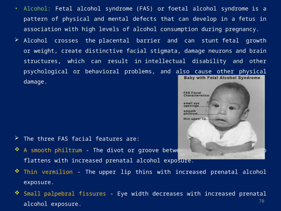
• Alcohol: Fetal alcohol syndrome (FAS) or foetal alcohol syndrome is a pattern of physical
and mental defects that can develop in a fetus in association with high levels of alcohol
consumption during pregnancy.
Alcohol crosses the placental barrier and can stunt fetal growth or weight, create
distinctive facial stigmata, damage neurons and brain structures, which can result
in intellectual disability and other psychological or behavioral problems, and also cause
other physical damage.
The three FAS facial features are:
A smooth philtrum - The divot or groove between the nose and upper lip flattens with
increased prenatal alcohol exposure.
Thin vermilion - The upper lip thins with increased prenatal alcohol exposure.
Small palpebral fissures - Eye width decreases with increased prenatal alcohol exposure.
71
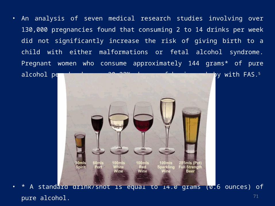
• An analysis of seven medical research studies involving over 130,000 pregnancies
found that consuming 2 to 14 drinks per week did not significantly increase the risk of
giving birth to a child with either malformations or fetal alcohol syndrome. Pregnant
women who consume approximately 144 grams* of pure alcohol per day have a 30–
33% chance of having a baby with FAS.5
• * A standard drink/shot is equal to 14.0 grams (0.6 ounces) of pure alcohol.
72
Trauma
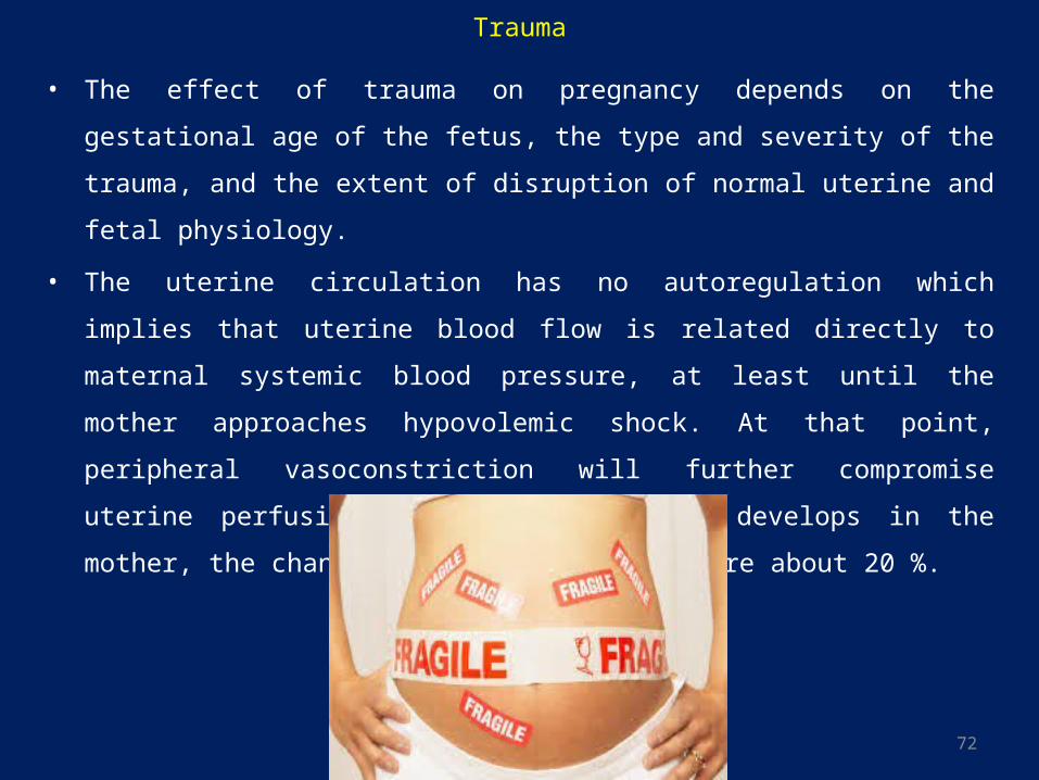
• The effect of trauma on pregnancy depends on the gestational age of the fetus, the type
and severity of the trauma, and the extent of disruption of normal uterine and fetal
physiology.
• The uterine circulation has no autoregulation which implies that uterine blood flow is
related directly to maternal systemic blood pressure, at least until the mother
approaches hypovolemic shock. At that point, peripheral vasoconstriction will further
compromise uterine perfusion. Once obvious shock develops in the mother, the
chances of saving the fetus are about 20 %.
73
German measles (Rubella)
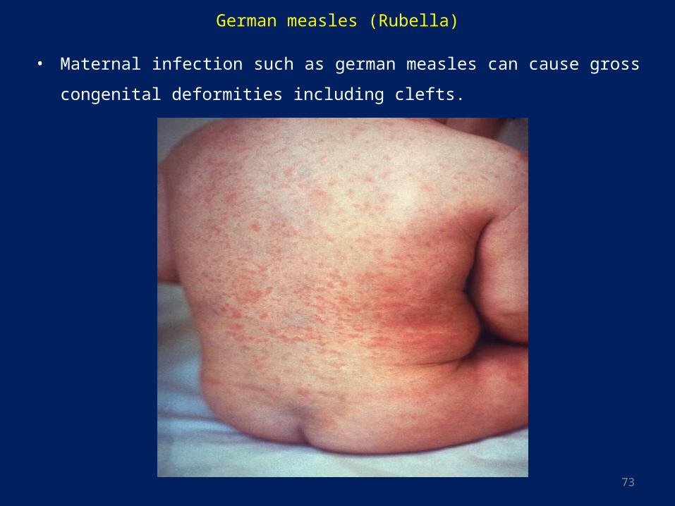
• Maternal infection such as german measles can cause gross congenital deformities
including clefts.
74
POSTNATAL INFLUENCE
Birth Injury
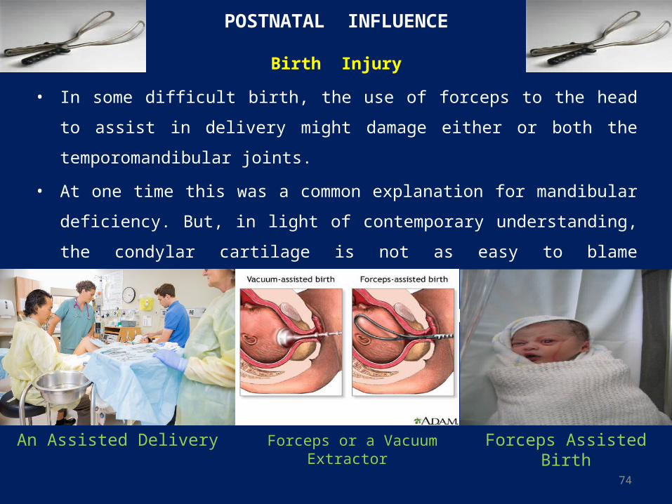
• In some difficult birth, the use of forceps to the head to assist in delivery might
damage either or both the temporomandibular joints.
• At one time this was a common explanation for mandibular deficiency. But, in light
of contemporary understanding, the condylar cartilage is not as easy to blame
underdevelopment of the mandible. So, injury to the mandible during a traumatic
delivery appears to be rare and unusual cause of facial deformity.
Forceps or a Vacuum ExtractorAn Assisted Delivery Forceps Assisted Birth
75
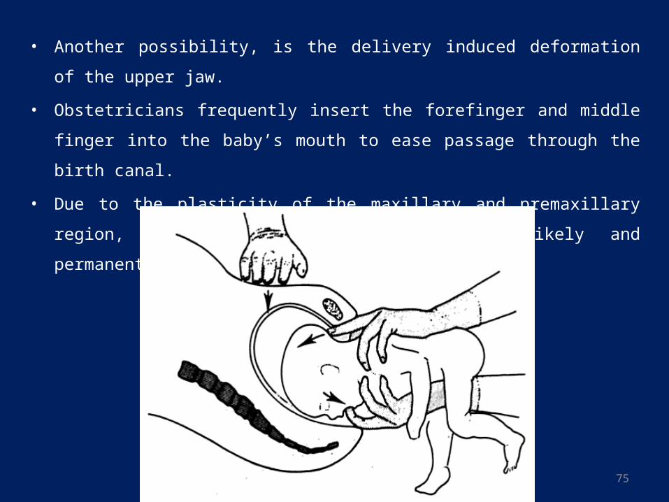
• Another possibility, is the delivery induced deformation of the upper jaw.
• Obstetricians frequently insert the forefinger and middle finger into the baby’s mouth
to ease passage through the birth canal.
• Due to the plasticity of the maxillary and premaxillary region, temporary deformation
is quite likely and permanent damage may result.
76
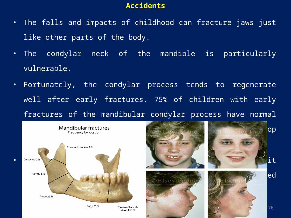
Accidents
• The falls and impacts of childhood can fracture jaws just like other parts of the body.
• The condylar neck of the mandible is particularly vulnerable.
• Fortunately, the condylar process tends to regenerate well after early fractures. 75% of
children with early fractures of the mandibular condylar process have normal
mandibular growth, therefore do not develop malocclusions.
• When a problem does arise following condylar fracture, it usually is asymmetric
growth, with the previously injured side lagging behind.
77
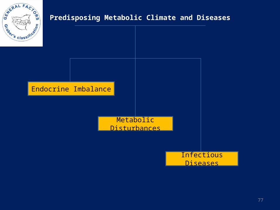
Predisposing Metabolic Climate and Diseases
Endocrine Imbalance
Metabolic Disturbances
Infectious Diseases
78
ENDOCRINE DISTURBANCES
PITUITARY PROBLEMS
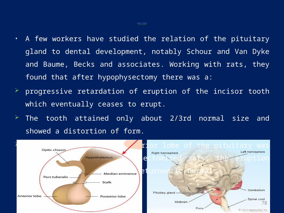
• A few workers have studied the relation of the pituitary gland to dental development,
notably Schour and Van Dyke and Baume, Becks and associates. Working with rats,
they found that after hypophysectomy there was a:
progressive retardation of eruption of the incisor tooth which eventually ceases to
erupt.
The tooth attained only about 2/3rd normal size and showed a distortion of form.
When an extract of the anterior lobe of the pituitary was injected into the
hypophysectomized rats, the eruption rate of the incisor tooth returned to normal.
79
• Baume and his associates injected thyroxin into hypophysectomized animals, either
alone or with purified growth hormones. Their findings led them to the following
explanations:
The pituitary gland influence eruption not only with its thyrotropin but also with its
growth hormones.
The effect of thyroxin on dental growth and development are different from those
of the pituitary growth hormone.
Thyroxin is the factor which stimulates the eruption movements and tooth size but
it has little influence on alveolar growth.
Growth hormones on the other hand spur dental as well as alveolar growth.
80
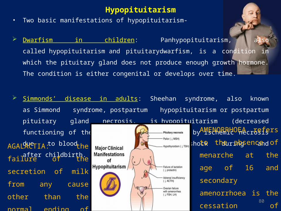
Hypopituitarism• Two basic manifestations of hypopituitarism-
Dwarfism in children: Panhypopituitarism, also called hypopituitarism and pituitarydwarfism, is a
condition in which the pituitary gland does not produce enough growth hormone. The condition is
either congenital or develops over time.
Simmonds’ disease in adults: Sheehan syndrome, also known as Simmond syndrome, postpartum
hypopituitarism or postpartum pituitary gland necrosis, is hypopituitarism (decreased functioning
of the pituitary gland), caused by ischemic necrosis due to blood loss and hypovolemic shock
during and after childbirth.
AMENORRHOEA refers
to the absence of menarche
at the age of 16 and
secondary amenorrhoea is
the cessation of menses for
at least 6 months in already
cycling women.
AGALACTIA: the failure of
the secretion of milk from
any cause other than the
normal ending of the
lactation period
81
82
83
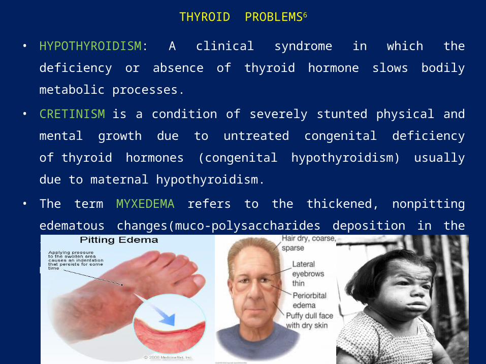
THYROID PROBLEMS6
• HYPOTHYROIDISM: A clinical syndrome in which the deficiency or absence of
thyroid hormone slows bodily metabolic processes.
• CRETINISM is a condition of severely stunted physical and mental growth due to
untreated congenital deficiency of thyroid hormones (congenital hypothyroidism)
usually due to maternal hypothyroidism.
• The term MYXEDEMA refers to the thickened, nonpitting edematous changes(muco-
polysaccharides deposition in the layer of the skin) to the soft tissues of patients in a
markedly hypothyroid state.
84
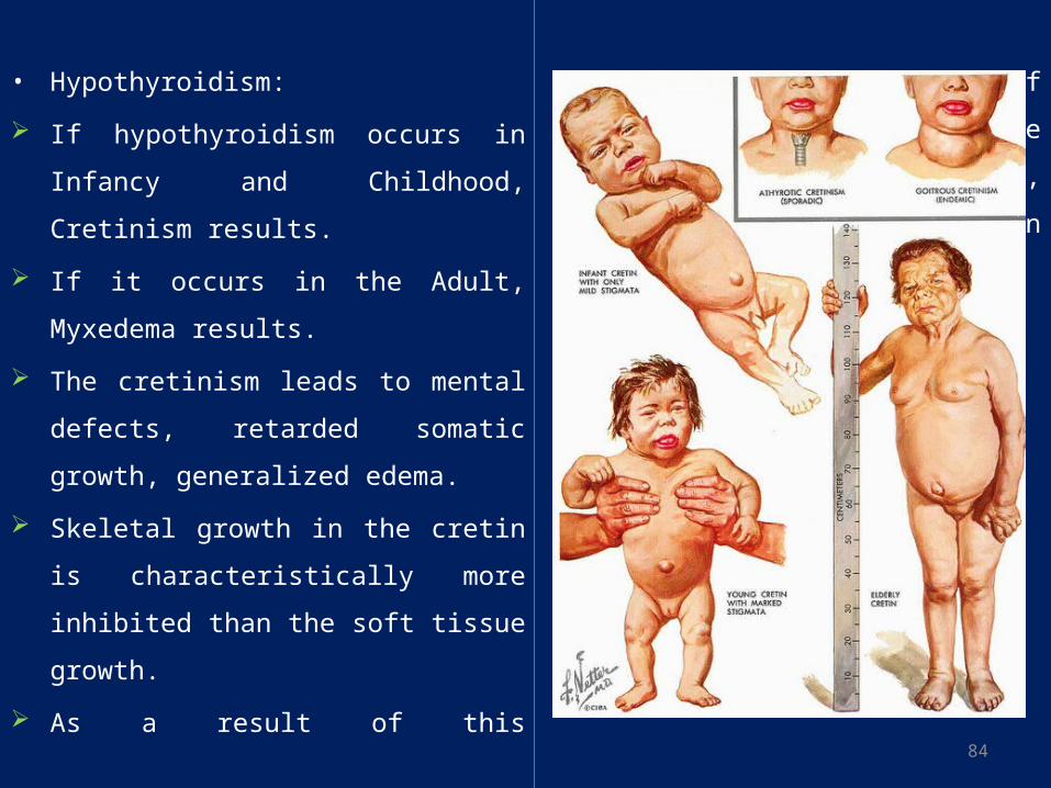
• Hypothyroidism:
If hypothyroidism occurs in Infancy and
Childhood, Cretinism results.
If it occurs in the Adult, Myxedema results.
The cretinism leads to mental defects,
retarded somatic growth, generalized edema.
Skeletal growth in the cretin is
characteristically more inhibited than the
soft tissue growth.
As a result of this disproportionate rate of
growth, the soft tissues are likely to enlarge
excessively, giving the appearance of an
obese and short child.
85
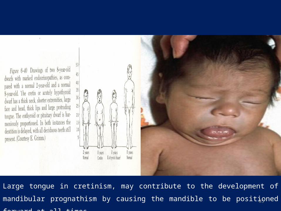
Large tongue in cretinism, may contribute to the development of mandibular prognathism by
causing the mandible to be positioned forward at all times
86
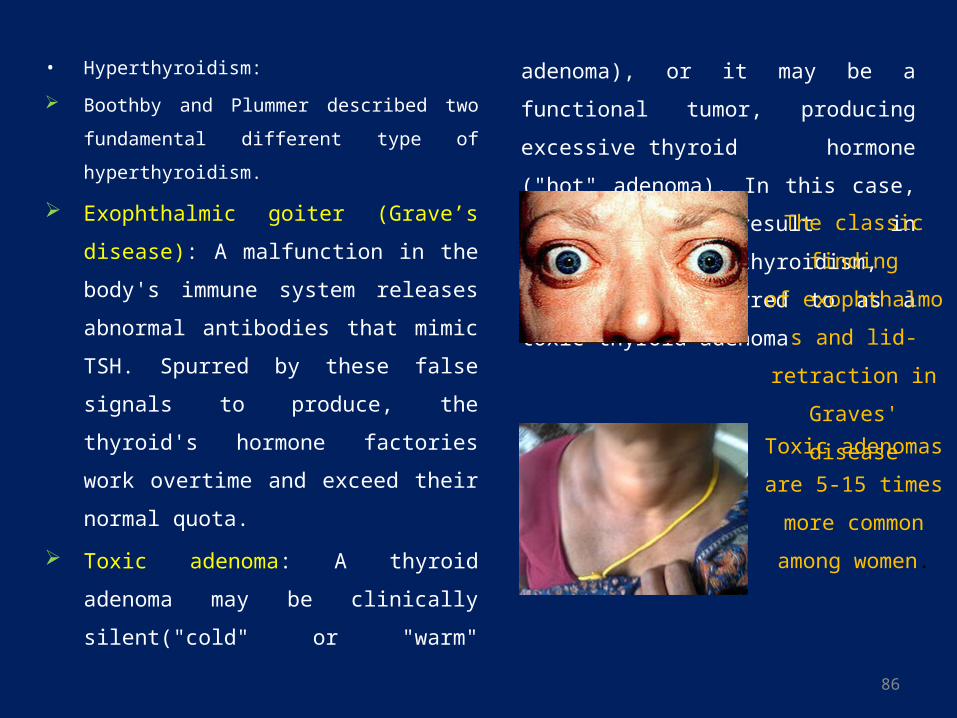
• Hyperthyroidism:
Boothby and Plummer described two fundamental
different type of hyperthyroidism.
Exophthalmic goiter (Grave’s disease): A
malfunction in the body's immune system
releases abnormal antibodies that mimic TSH.
Spurred by these false signals to produce, the
thyroid's hormone factories work overtime
and exceed their normal quota.
Toxic adenoma: A thyroid adenoma may be
clinically silent("cold" or "warm" adenoma),
or it may be a functional tumor, producing
excessive thyroid hormone ("hot" adenoma).
In this case, it may result in
symptomatic hyperthyroidism, and may be
referred to as a toxic thyroid adenoma
The classic finding
of exophthalmos and
lid-retraction in
Graves' disease
Toxic adenomas are
5-15 times more
common among
women.
87
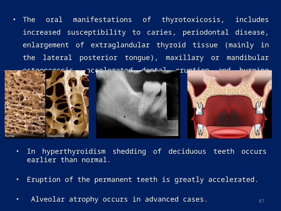
• The oral manifestations of thyrotoxicosis, includes increased susceptibility to caries,
periodontal disease, enlargement of extraglandular thyroid tissue (mainly in the lateral
posterior tongue), maxillary or mandibular osteoporosis, accelerated dental eruption
and burning mouth syndrome.
• In hyperthyroidism shedding of deciduous teeth occurs earlier than normal.
• Eruption of the permanent teeth is greatly accelerated.
• Alveolar atrophy occurs in advanced cases.

88
PRIMARY HYPERPARATHYROIDISM
• Increased activity is usually due to an adenoma of one or more of the four parathyroid
glands.
• Almost all patients with hyperparathyroidism have skeletal lesions, some of which
may occur in the skull or jaws.
• The skeletal disturbances in hyperparathyroidism vary from vague to
roentgenographically characteristic lesions and even gross clinical evidence of bone
lesions.
• Occasionally the first sign of the disease may be a giant cell tumor or a cyst of the jaw.
• Loss of phosphorus and calcium in this disturbance results in a generalized
osteoporosis.
• Malocclusion caused by sudden drifting with definite spacing of teeth.
89
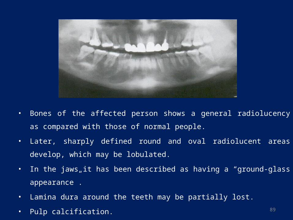
• Bones of the affected person shows a general radiolucency as compared with those of
normal people.
• Later, sharply defined round and oval radiolucent areas develop, which may be
lobulated.
• In the jaws it has been described as having a “ground-glass appearance”.
• Lamina dura around the teeth may be partially lost.
• Pulp calcification.
90
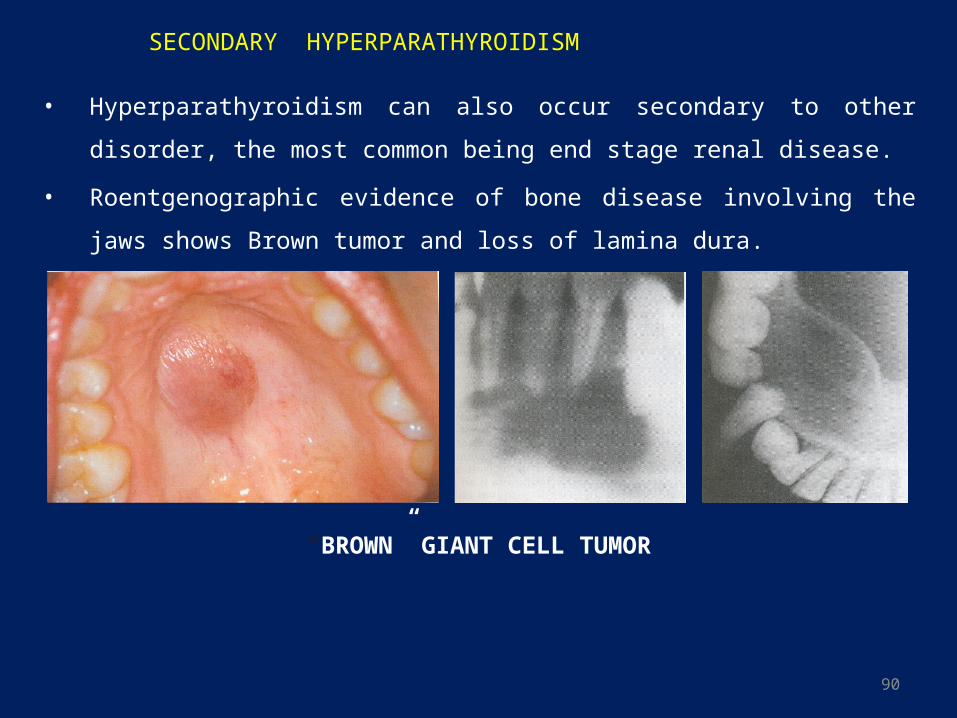
SECONDARY HYPERPARATHYROIDISM
• Hyperparathyroidism can also occur secondary to other disorder, the most common
being end stage renal disease.
• Roentgenographic evidence of bone disease involving the jaws shows Brown tumor
and loss of lamina dura.
“BROWN” GIANT CELL TUMOR
91
SEX HORMONES7
• The two major female sex hormones: estrogen and progesterone influence the growth
of oral epithelium.
• They also dilate the blood vessels in the underlining tissue and increase their
permeability.
• Endogenous sex steroid hormones can influence the periodontium at different life
times such as puberty, menstruation, pregnancy, menopause and postmenopause.
• Clinical changes in the periodontal tissues during menstruation: Bleeding and swollen gingiva: Progesterone - increased permeability of the
microvasculature - increases folate metabolism - stimulates the production of prostaglandins - enhances the chemotaxis of polymorphonuclear leukocytes (PMNL)
An increase in gingival exudate: A peak level of exudate is detected just before
ovulation, coinciding with the highest levels of these hormones.
A minor increase in tooth mobility: During the luteal phase of the cycle, when
progesterone reaches its highest concentration, intraoral recurrent aphthous ulcers,
herpes labialis lesions and candida infections may also occur in women.
92
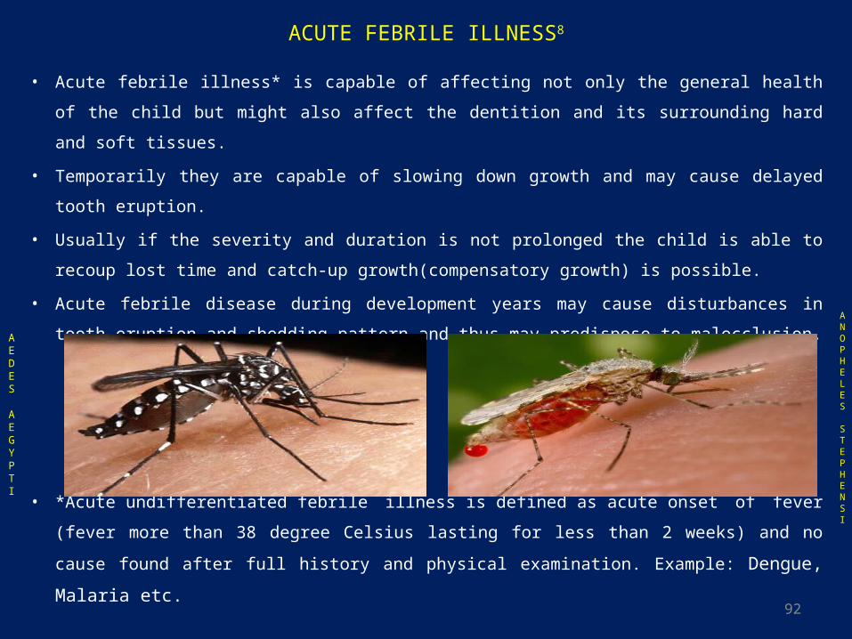
ACUTE FEBRILE ILLNESS8
• Acute febrile illness* is capable of affecting not only the general health of the child but
might also affect the dentition and its surrounding hard and soft tissues.
• Temporarily they are capable of slowing down growth and may cause delayed tooth
eruption.
• Usually if the severity and duration is not prolonged the child is able to recoup lost time
and catch-up growth(compensatory growth) is possible.
• Acute febrile disease during development years may cause disturbances in tooth eruption
and shedding pattern and thus may predispose to malocclusion.
• *Acute undifferentiated febrile illness is defined as acute onset of fever (fever more than
38 degree Celsius lasting for less than 2 weeks) and no cause found after full history and
physical examination. Example: Dengue, Malaria etc.
AEDES
AEGYPTI
ANOPHELES
STEPHENSI
93
OSTEOMYELITIS
• Osteomyelitis of the mandibular condyle – necrosis/complete destruction of the
mandibular condyle – Malocclusion.
• Teeth in the affected region may demonstrate increased mobility even leading to
malocclusion and show decreased or loss of sensitivity.9
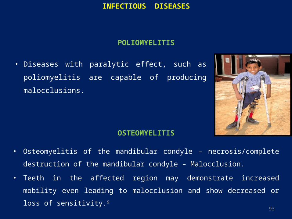
INFECTIOUS DISEASES
• Diseases with paralytic effect, such as poliomyelitis are
capable of producing malocclusions.
POLIOMYELITIS
94
DIETARY PROBLEMS10
• A cross-sectional study with probabilistic sampling design was used. 2,060 students
aged 12 to 15 years enrolled in schools in the northeast of Brazil were evaluated.
Crowding was defined according to World Health Organization (WHO) as
misalignment of teeth due to lack of space for them to erupt in the correct position.
Nutritional status was evaluated by means of body mass index and height-for-age,
using the WHO’s reference curves.
• It was found that malnutrition is related to crowding in permanent dentition among
mouth-breathing adolescents.
• However, further studies are needed to increase the consistency of these findings and
improve understanding of the subject.
95
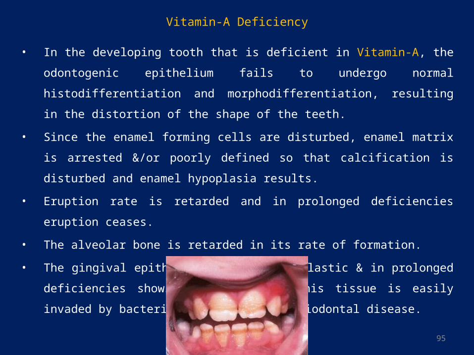
• In the developing tooth that is deficient in Vitamin-A, the odontogenic epithelium
fails to undergo normal histodifferentiation and morphodifferentiation, resulting in
the distortion of the shape of the teeth.
• Since the enamel forming cells are disturbed, enamel matrix is arrested &/or poorly
defined so that calcification is disturbed and enamel hypoplasia results.
• Eruption rate is retarded and in prolonged deficiencies eruption ceases.
• The alveolar bone is retarded in its rate of formation.
• The gingival epithelium becomes hyperplastic & in prolonged deficiencies shows
keratinization. This tissue is easily invaded by bacteria that may cause periodontal
disease.
Vitamin-A Deficiency
96
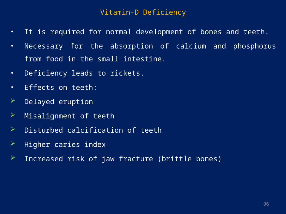
Vitamin-D Deficiency
• It is required for normal development of bones and teeth.
• Necessary for the absorption of calcium and phosphorus from food in the small
intestine.
• Deficiency leads to rickets.
• Effects on teeth:
Delayed eruption
Misalignment of teeth
Disturbed calcification of teeth
Higher caries index
Increased risk of jaw fracture (brittle bones)
97
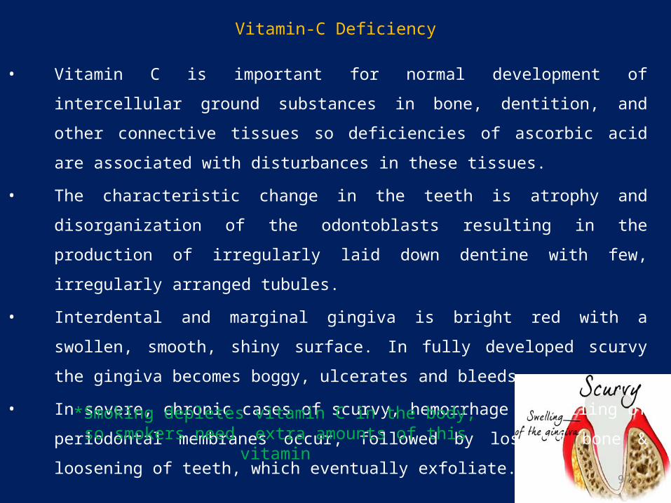
Vitamin-C Deficiency
• Vitamin C is important for normal development of intercellular ground substances in
bone, dentition, and other connective tissues so deficiencies of ascorbic acid are
associated with disturbances in these tissues.
• The characteristic change in the teeth is atrophy and disorganization of the odontoblasts
resulting in the production of irregularly laid down dentine with few, irregularly
arranged tubules.
• Interdental and marginal gingiva is bright red with a swollen, smooth, shiny surface. In
fully developed scurvy the gingiva becomes boggy, ulcerates and bleeds
• In severe, chronic cases of scurvy, hemorrhage & swelling of periodontal membranes
occur, followed by loss of bone & loosening of teeth, which eventually exfoliate.
*Smoking depletes vitamin C in the body, so smokers need extra amounts of this vitamin

98
Various studies which have been conducted, dictate that malnutrition and protein energy malnutrition affect the dentition. The resultant defects include the effects on the tooth eruption patterns, enamel hypoplasia, dental caries prevalence and periodontal ligament. They also have other effects on the oral cavity, like inflammation of the lining of the oral cavity and the tongue and oral ulcers. (Aparna Sheetal et al. Malnutrition and its Oral Outcome – A Review).11
99
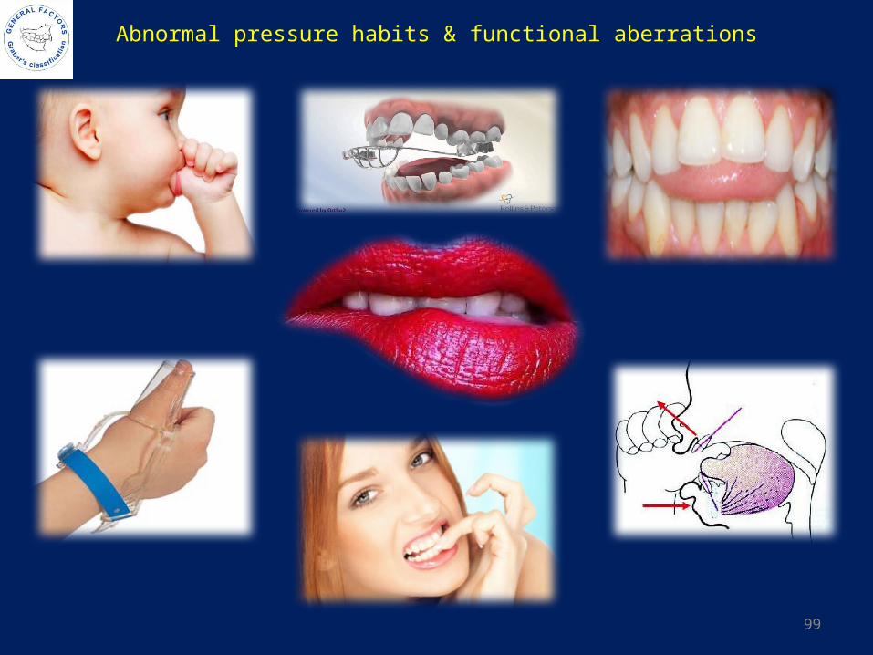
Abnormal pressure habits & functional aberrations

100
DEFINITIONS
• Johnson (1938): A habit is an inclination or aptitude for some action acquired by
frequent repetition and showing itself in increased facility to performance and
reduced power of resistance.
• Maslow (1949): A habit is a formed reaction that is resistant to change, whether
useful or harmful, depending upon the degree to which it interferes with the
child’s physical, emotional and social functions.
• Dorland (1957): Habit can be defined as a fixed or constant practice established by
frequent repetition.
• Buttersworth (1961): A frequent or constant practice or acquired tendency, which
has been fixed by frequent repetition.
• Finn (1972): A habit is an act, which is socially unacceptable.
• Mathewson ( 1982): Oral habits are learned patterns of muscular contractions.
101
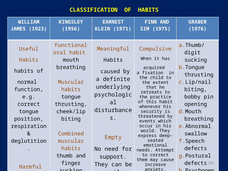
CLASSIFICATION OF HABITS
WILLIAM JAMES (1923)
KINGSLEY (1956)
EARNEST KLEIN (1971)
FINN AND SIM (1975)
GRABER (1976)
Useful Habits
habits of normal function, e.g. correct tongue
position, respiration & deglutition.
Harmful Habits
habits which exert
pressures/stresses against teeth
and dental arches and also mouth breathing, lip biting & lip
sucking.
Functional oral habit
mouth breathing
Muscular habitstongue thrusting, cheek/lip biting
Combined muscular habits
thumb and finger sucking
Postural habitschin propping, face leaning on hand, abnormal
pillowing
Meaningful
Habits caused bya definite
underlying psychological disturbances.
Empty
No need for support. They can be easily
treated by reminder
appliances.
Compulsive
When it has
acquired a fixation in the
child to the extent that he retreats to the practice of this habit
whenever his security is
threatened by events which occur in his
world. They express deep-seated
emotional needs. Attempt to correct them may cause increase anxiety.
Non-compulsive
Habits which are easily added or
dropped from the child’s behavior
pattern as he matures.
a. Thumb/digit sucking
b. Tongue thrusting
c. Lip/nail biting, bobby pin opening
d. Mouth breathing
e. Abnormal swallow
f. Speech defects
g. Postural defects
h. Psychogenic habits – bruxism
i. Defective occlusal habits
102
THUMB AND FINGER SUCKING
• Definition:
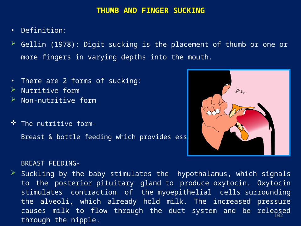
Gellin (1978): Digit sucking is the placement of thumb or one or more fingers in varying
depths into the mouth.
• There are 2 forms of sucking: Nutritive form Non-nutritive form
The nutritive form-
Breast & bottle feeding which provides essential nutrients.
BREAST FEEDING-
Suckling by the baby stimulates the hypothalamus, which signals to the posterior pituitary gland to produce oxytocin. Oxytocin stimulates contraction of the myoepithelial cells surrounding the alveoli, which already hold milk. The increased pressure causes milk to flow through the duct system and be released through the nipple.
103
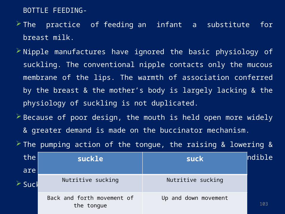
BOTTLE FEEDING-
The practice of feeding an infant a substitute for breast milk.
Nipple manufactures have ignored the basic physiology of suckling. The conventional
nipple contacts only the mucous membrane of the lips. The warmth of association
conferred by the breast & the mother’s body is largely lacking & the physiology of
suckling is not duplicated.
Because of poor design, the mouth is held open more widely & greater demand is
made on the buccinator mechanism.
The pumping action of the tongue, the raising & lowering & the rhythmic backward &
forward movement of the mandible are reduced.
Suckling becomes sucking.
suckle suck
Nutritive sucking Nutritive sucking
Back and forth movement of the tongue Up and down movement
Weaker lip seal Stronger lip seal
104
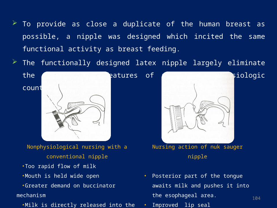
To provide as close a duplicate of the human breast as possible, a nipple was designed
which incited the same functional activity as breast feeding.
The functionally designed latex nipple largely eliminate the objectionable features of
previous non-physiologic counterparts.
Nonphysiological nursing with a
conventional nipple
•Too rapid flow of milk
•Mouth is held wide open
•Greater demand on buccinator mechanism
•Milk is directly released into the digestive tract
Reducing the period of predigestion.
Nursing action of nuk sauger nipple
• Posterior part of the tongue awaits milk and
pushes it into the esophageal area.
• Improved lip seal
• Nipple is drawn upward and backward
towards the palate.

105
The non-nutritive form-
Children who neither receive unrestricted breast feeding nor have access to a pacifier
may satisfy their need with habits like thumb sucking which ensures a feeling of
warmth & sense of security but may be detrimental to their dentofacial development.
• Nearly all infants engage in some sort of habitual non nutritive sucking- sucking of the
thumb, finger or similarly shaped objects.
• Vast majority of infants do so during from 6 months to 2 years or later.
• After the eruption of the primary molars during the second year, drinking from a cup
replaces drinking from a bottle or continued nursing from the mother’s breast, the
number of children who engage in non nutritive sucking then diminishes.
• Some fetuses have been reported to suck their thumbs in utero.12
106
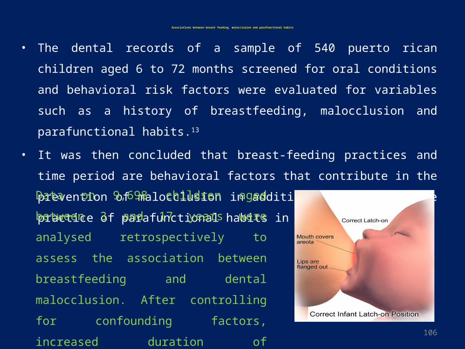
Associations between breast feeding, malocclusion and parafunctional habits
• The dental records of a sample of 540 puerto rican children aged 6 to 72 months
screened for oral conditions and behavioral risk factors were evaluated for variables
such as a history of breastfeeding, malocclusion and parafunctional habits.13
• It was then concluded that breast-feeding practices and time period are behavioral
factors that contribute in the prevention of malocclusion in addition to decreasing the
practice of parafunctional habits in preschool children.
Data on 9,698 children aged between 3 and 17
years were analysed retrospectively to assess the
association between breastfeeding and dental
malocclusion. After controlling for confounding
factors, increased duration of breastfeeding was
associated with a decline in the prevalence of
malocclusion. (http://www.unicef.org.uk)14
107
• Warren JJ et al. (JADA. 2001) concluded that continuous nonnutritive sucking habits
of 48 months or longer produced the greatest changes in dental arch and occlusal
characteristics, children with shorter sucking durations also had detectable differences
from those with minimal habit durations.15
• Mônica VH et al. (Eur J Ortho. 2008) conducted a longitudinal study to assess the
relationship between non-nutritive sucking habits and the presence of anterior open
bites (AOBs) and posterior crossbites and their association with facial morphology.
They concluded that the Non-nutritive sucking habits are risk factors for the
occurrence of an AOB and a posterior crossbite. AOBs were associated with the
presence of continuing sucking habits, whereas the same was not true for posterior
crossbites. Also it was found that Self-correction of an AOB was associated with the
cessation of sucking habits.16
108
• Common in infants 23% to 46% of children aged 1 to 4years
(Infante,1976;larson & dahin,1985.)
• Most of the children discontinue the habit by 3‐4 years of age.
(Friman & Schmitt,1989;Traisman & Traisman,1958)
• Continues after 4 yrs ‐ greater risk for dental malocclusion (Friman&Schmit1987)
digital deformities(Reid&Price,1984) speech difficulties(Luke&Howard,1983)
• Severity depending on frequency, duration, Intensity
109
Thumb and Finger-sucking Habits: the long-term effects
1. Flared and spaced maxillary incisors(proclination) and lingually positioned
lower/mandibular incisors(retroclination).
• The labially posed upper permanent incisors are particularly vulnerable to
accidental fractures.
• Afzelius-Alm A et al. (Swed Dent J. 2004;28) however concluded that the majority
of children with prolonged thumb-sucking have proclined lower incisors rather than
retroclined lower incisors.(contrary to popular belief ).17
• In the group with retroclined lower incisors the angle between the thumb and the
lower incisors was significantly smaller and the thickness of the lower lip
significantly thinner than in the group with proclined incisors. A higher frequency
of early loss of deciduous molars was also observed in the group with retroclined
incisors.
110
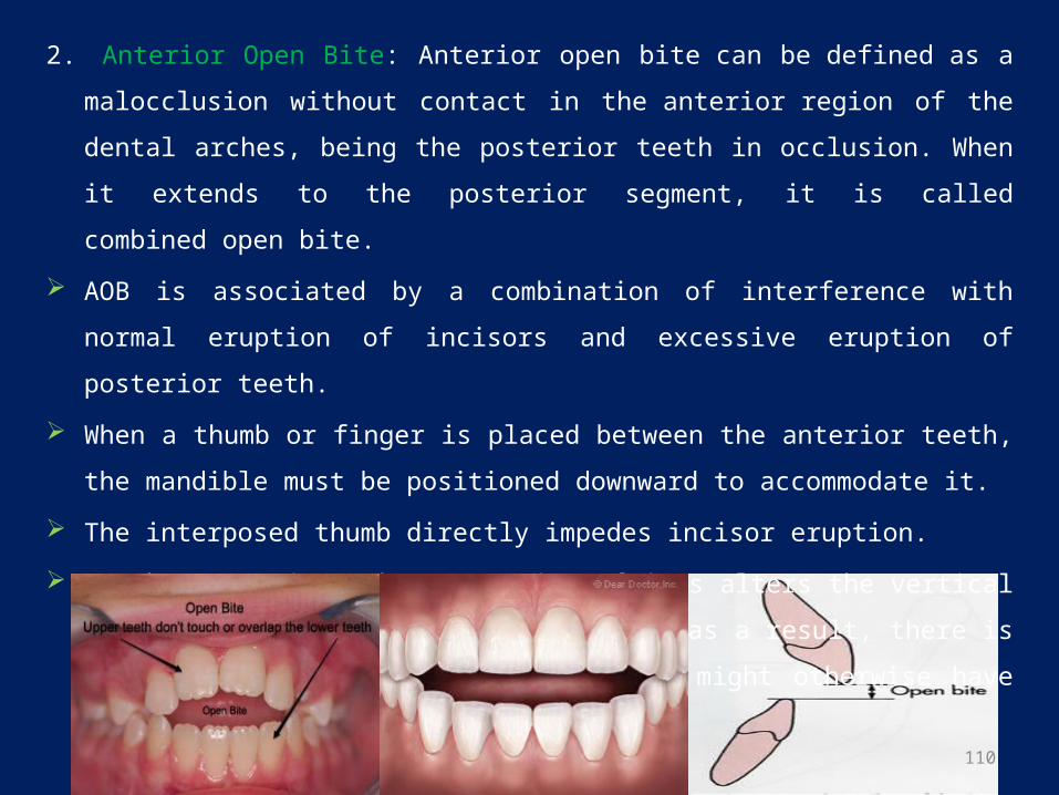
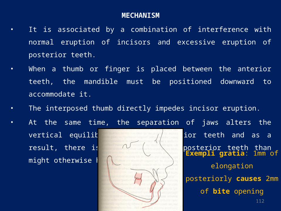
2. Anterior Open Bite: Anterior open bite can be defined as a malocclusion without
contact in the anterior region of the dental arches, being the posterior teeth in
occlusion. When it extends to the posterior segment, it is called combined open bite.
AOB is associated by a combination of interference with normal eruption of incisors
and excessive eruption of posterior teeth.
When a thumb or finger is placed between the anterior teeth, the mandible must be
positioned downward to accommodate it.
The interposed thumb directly impedes incisor eruption.
At the same time, the separation of jaws alters the vertical equilibrium on the posterior
teeth and as a result, there is more eruption of posterior teeth than might otherwise
have occurred.18
111
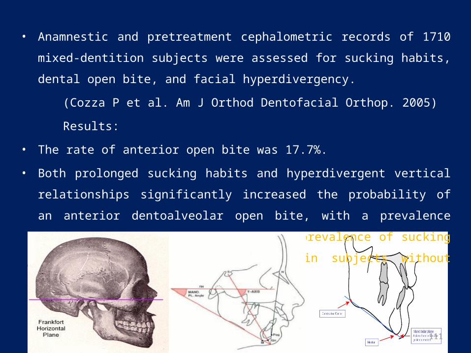
• Anamnestic and pretreatment cephalometric records of 1710 mixed-dentition subjects
were assessed for sucking habits, dental open bite, and facial hyperdivergency.
(Cozza P et al. Am J Orthod Dentofacial Orthop. 2005)
Results:
• The rate of anterior open bite was 17.7%.
• Both prolonged sucking habits and hyperdivergent vertical relationships significantly
increased the probability of an anterior dentoalveolar open bite, with a prevalence rate
of 36.3%. This was 4 times the prevalence of sucking habits and facial
hyperdivergency in subjects without anterior open bite (9.1%).19
112
MECHANISM
• It is associated by a combination of interference with normal eruption of incisors and
excessive eruption of posterior teeth.
• When a thumb or finger is placed between the anterior teeth, the mandible must be
positioned downward to accommodate it.
• The interposed thumb directly impedes incisor eruption.
• At the same time, the separation of jaws alters the vertical equilibrium on the
posterior teeth and as a result, there is more eruption of posterior teeth than might
otherwise have occurred.
Exempli gratia: 1mm of
elongation
posteriorly causes 2mm
of bite opening
113
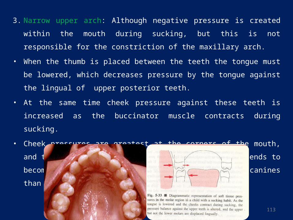
3. Narrow upper arch: Although negative pressure is created within the mouth during
sucking, but this is not responsible for the constriction of the maxillary arch.
• When the thumb is placed between the teeth the tongue must be lowered, which
decreases pressure by the tongue against the lingual of upper posterior teeth.
• At the same time cheek pressure against these teeth is increased as the buccinator
muscle contracts during sucking.
• Cheek pressures are greatest at the corners of the mouth, and this probably explains
why the maxillary arch tends to become V-shaped, with more constriction across the
canines than the molars.
114
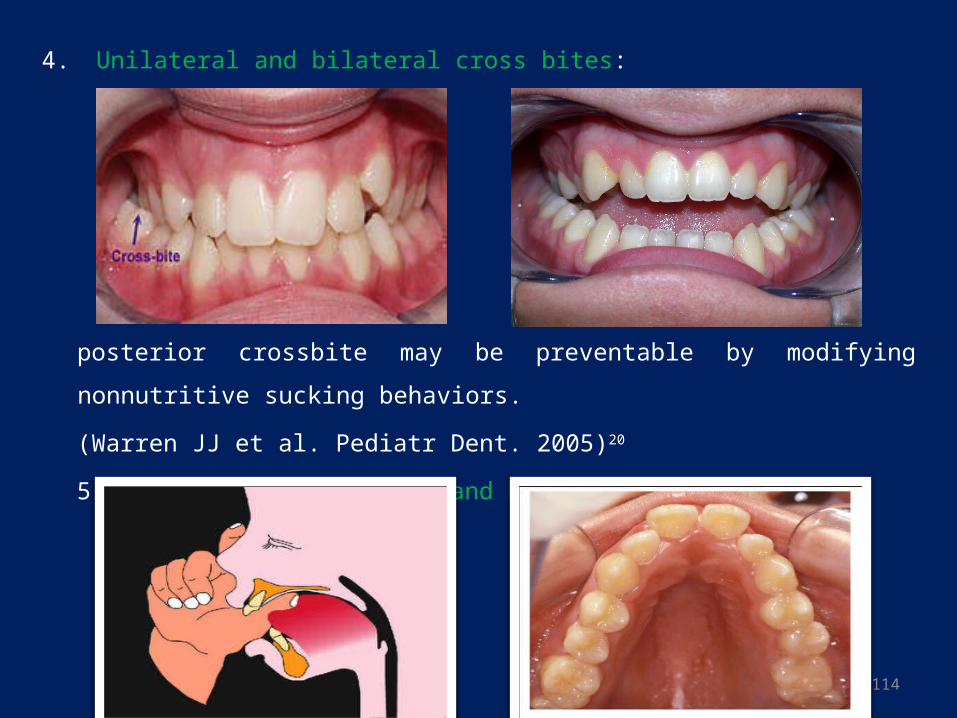
4. Unilateral and bilateral cross bites:
posterior crossbite may be preventable by modifying nonnutritive sucking behaviors.
(Warren JJ et al. Pediatr Dent. 2005)20
5. A narrower nasal floor and high palatal vault:
115
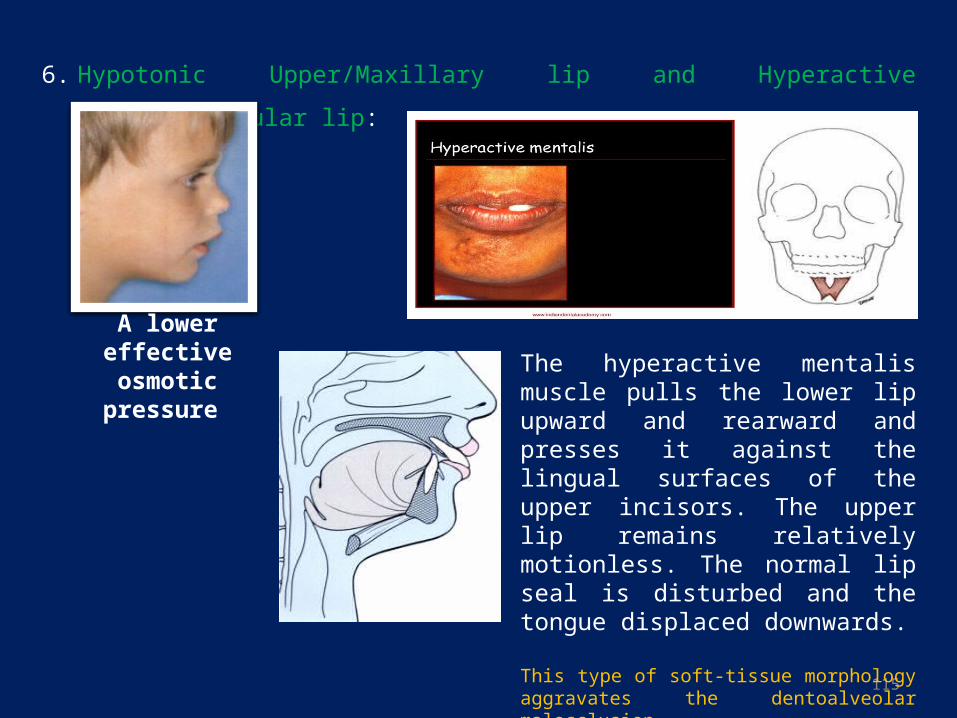
6. Hypotonic Upper/Maxillary lip and Hyperactive Lower/Mandibular lip:
The hyperactive mentalis muscle pulls the lower lip upward and rearward and presses it against the lingual surfaces of the upper incisors. The upper lip remains relatively motionless. The normal lip seal is disturbed and the tongue displaced downwards.
This type of soft-tissue morphology aggravates the dentoalveolar malocclusion.
A lower effective osmotic pressure
116
TONGUE THRUSTING HABIT
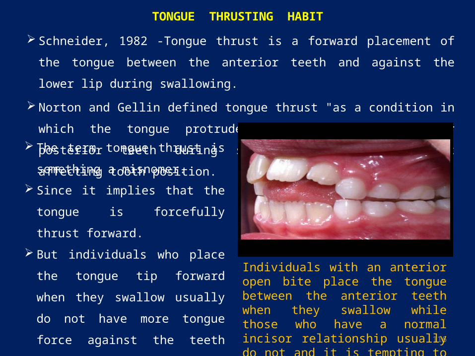
Schneider, 1982 ‐Tongue thrust is a forward placement of the tongue between the
anterior teeth and against the lower lip during swallowing.
Norton and Gellin defined tongue thrust "as a condition in which the tongue protrudes
between the anterior or posterior teeth during swallowing with or without affecting
tooth position.
The term tongue thrust is something a
misnomer.
Since it implies that the tongue is
forcefully thrust forward.
But individuals who place the tongue
tip forward when they swallow usually
do not have more tongue force against
the teeth than those who keep the
tongue tip back.
Individuals with an anterior open bite place the tongue between the anterior teeth when they swallow while those who have a normal incisor relationship usually do not and it is tempting to blame the open bite for this pattern of tongue activity.
117
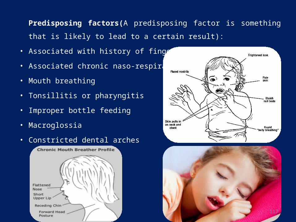
Predisposing factors(A predisposing factor is something that is likely to lead to a
certain result):
• Associated with history of finger sucking
• Associated chronic naso-respiratory distress
• Mouth breathing
• Tonsillitis or pharyngitis
• Improper bottle feeding
• Macroglossia
• Constricted dental arches
118
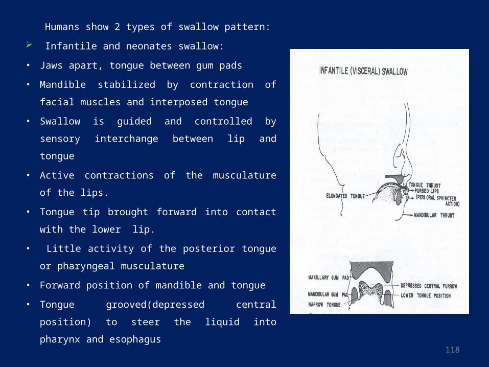
Humans show 2 types of swallow pattern:
Infantile and neonates swallow:
• Jaws apart, tongue between gum pads
• Mandible stabilized by contraction of facial
muscles and interposed tongue
• Swallow is guided and controlled by sensory
interchange between lip and tongue
• Active contractions of the musculature of the lips.
• Tongue tip brought forward into contact with the
lower lip.
• Little activity of the posterior tongue or
pharyngeal musculature
• Forward position of mandible and tongue
• Tongue grooved(depressed central position) to
steer the liquid into pharynx and esophagus
119
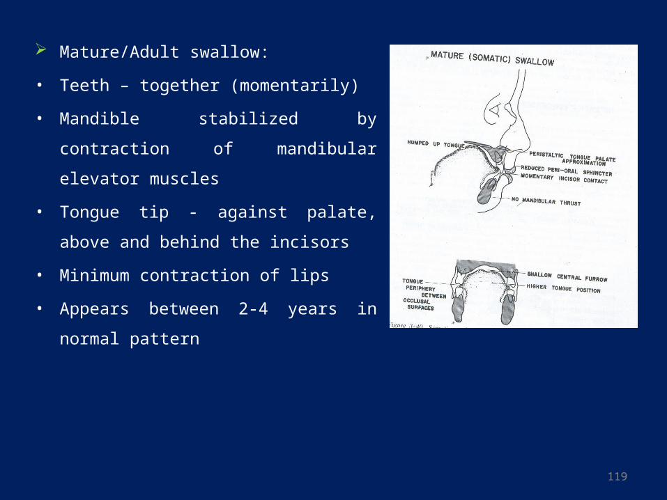
Mature/Adult swallow:
• Teeth – together (momentarily)
• Mandible stabilized by contraction of
mandibular elevator muscles
• Tongue tip - against palate, above and behind
the incisors
• Minimum contraction of lips
• Appears between 2-4 years in normal pattern
120
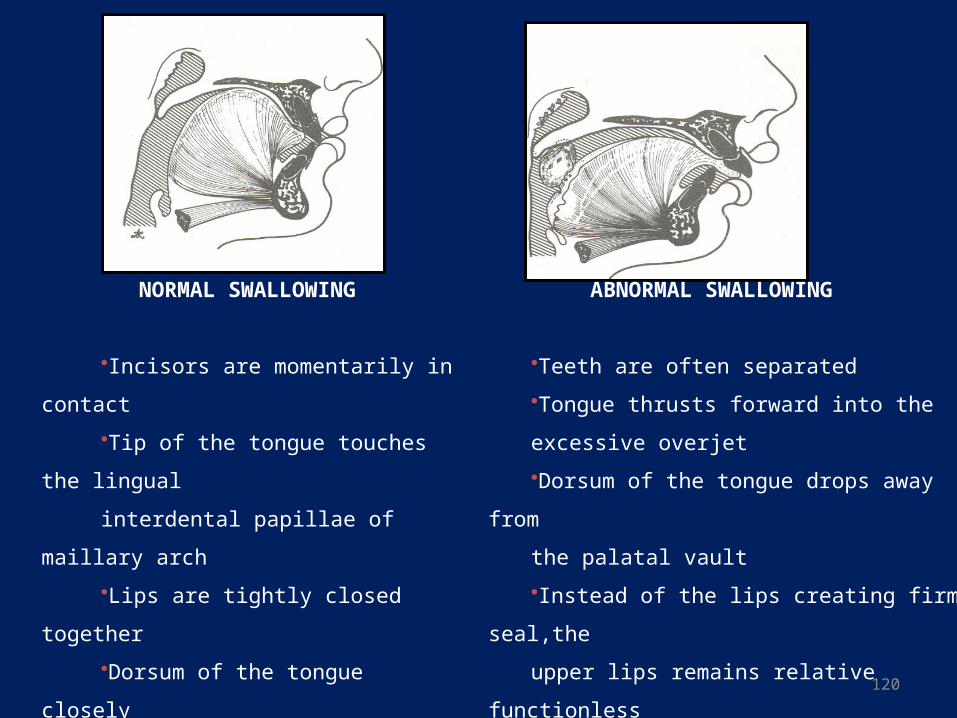
NORMAL SWALLOWING
•Incisors are momentarily in contact
•Tip of the tongue touches the lingual
interdental papillae of maillary arch
•Lips are tightly closed together
•Dorsum of the tongue closely
approximates the palate during
swallowing
ABNORMAL SWALLOWING
•Teeth are often separated
•Tongue thrusts forward into the
excessive overjet
•Dorsum of the tongue drops away from
the palatal vault
•Instead of the lips creating firm seal,the
upper lips remains relative functionless
•Mentalis exerts strong forward and upward
thrust of lower lip against lingual surfaces
of maxillary incisors
121
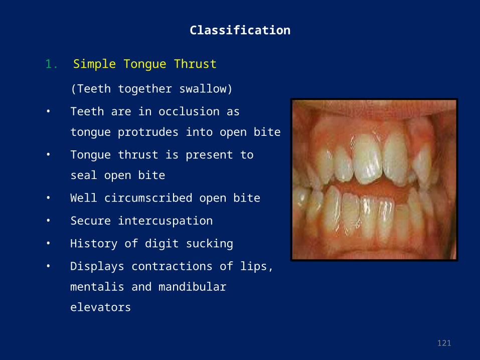
Classification
1. Simple Tongue Thrust
(Teeth together swallow)
• Teeth are in occlusion as tongue protrudes
into open bite
• Tongue thrust is present to seal open bite
• Well circumscribed open bite
• Secure intercuspation
• History of digit sucking
• Displays contractions of lips, mentalis and
mandibular elevators
122
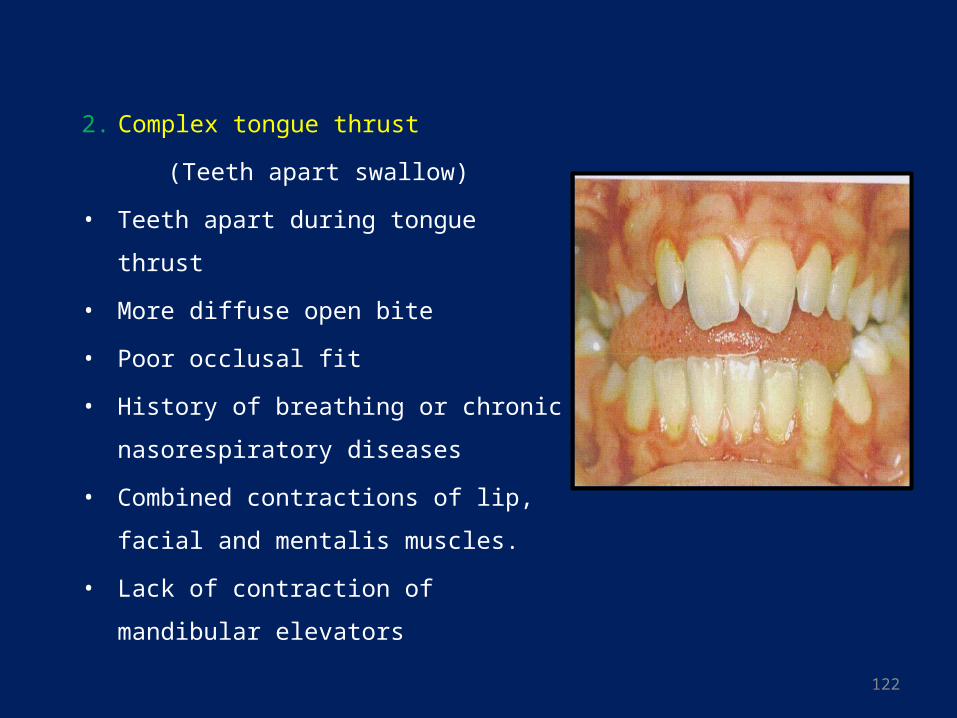
2. Complex tongue thrust
(Teeth apart swallow)
• Teeth apart during tongue thrust
• More diffuse open bite
• Poor occlusal fit
• History of breathing or chronic
nasorespiratory diseases
• Combined contractions of lip, facial and
mentalis muscles.
• Lack of contraction of mandibular elevators
123
• Clinical Feature:
• If the postural position is normal, the tongue thrust swallow has no clinical
significance because tongue thrust swallowing simply has too short a duration to
have an impact on tooth position
• Pressure by the tongue against the teeth during a typical swallow lasts for
approximately 1 second.
• A typical individual swallows about 800 times/day while awake but has only a few
swallows /hour while asleep. The total/day therefore is usually under 1000.
• One thousand seconds of pressure, of course, totals only a few minutes, not nearly
enough to effect the equilibrium.
• On the other hand, if a patient has a altered resting posture of the tongue, the
duration of this pressure, even if very light, could effect tooth position, vertically or
horizontally.
124
LOCAL FACTORS
1. Anomalies of tooth number
2. Anomalies of tooth size
3. Anomalies of tooth shape
4. Abnormal labial frenum
5. Premature loss of deciduous teeth
6. Prolonged retention of deciduous teeth
7. Delayed eruption of permanent teeth
8. Abnormal eruptive path
9. Ankylosis
10. Dental caries
11. Improper dental restoration
125
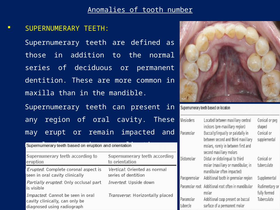
Anomalies of tooth number
SUPERNUMERARY TEETH:
Supernumerary teeth are defined as those in addition
to the normal series of deciduous or permanent
dentition. These are more common in maxilla than in
the mandible.
Supernumerary teeth can present in any region of
oral cavity. These may erupt or remain impacted and
may lead to various complications.
(Abhishek Parolia et al. J Conserv Dent. 2011 )21
126
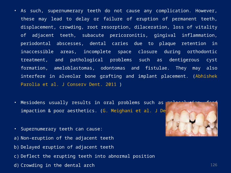
• As such, supernumerary teeth do not cause any complication. However, these may lead to
delay or failure of eruption of permanent teeth, displacement, crowding, root resorption,
dilaceration, loss of vitality of adjacent teeth, subacute pericoronitis, gingival
inflammation, periodontal abscesses, dental caries due to plaque retention in inaccessible
areas, incomplete space closure during orthodontic treatment, and pathological problems
such as dentigerous cyst formation, ameloblastomas, odontomas and fistulae. They may
also interfere in alveolar bone grafting and implant placement. (Abhishek Parolia et al. J
Conserv Dent. 2011 )
• Mesiodens usually results in oral problems such as malocclusion, food impaction & poor
aesthetics. (G. Meighani et al. J Dent Tehran. 2010)22
• Supernumerary teeth can cause:
a) Non-eruption of the adjacent teeth
b) Delayed eruption of adjacent teeth
c) Deflect the erupting teeth into abnormal position
d) Crowding in the dental arch
127
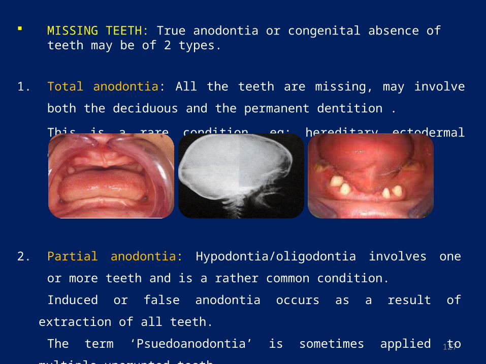
MISSING TEETH: True anodontia or congenital absence of teeth may be of 2 types.
1. Total anodontia: All the teeth are missing, may involve both the deciduous and the
permanent dentition .
This is a rare condition, eg: hereditary ectodermal dysplasia
2. Partial anodontia: Hypodontia/oligodontia involves one or more teeth and is a rather
common condition.
Induced or false anodontia occurs as a result of extraction of all teeth.
The term ‘Psuedoanodontia’ is sometimes applied to multiple unerupted teeth
128
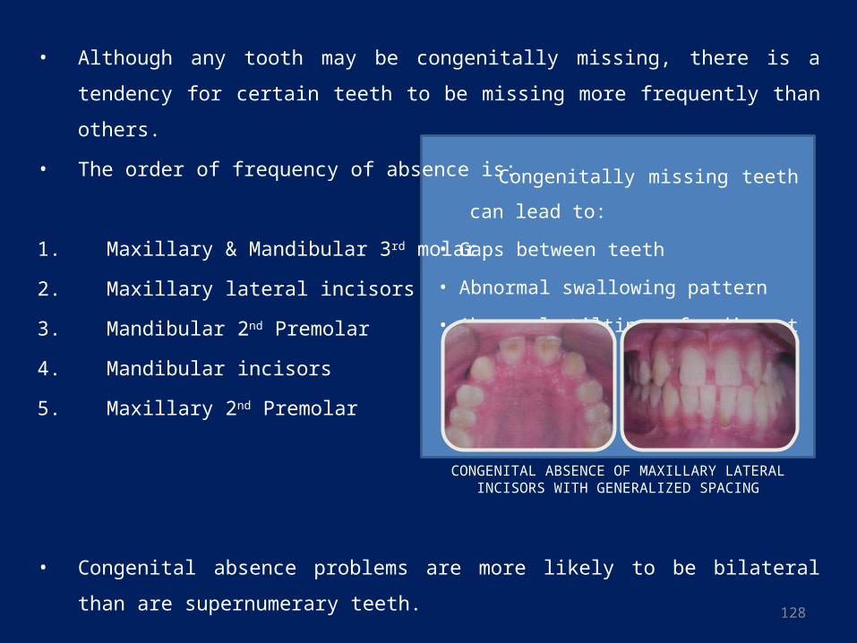
• Although any tooth may be congenitally missing, there is a tendency for certain teeth
to be missing more frequently than others.
• The order of frequency of absence is:
1. Maxillary & Mandibular 3rd molar
2. Maxillary lateral incisors
3. Mandibular 2nd Premolar
4. Mandibular incisors
5. Maxillary 2nd Premolar
• Congenital absence problems are more likely to be bilateral than are supernumerary
teeth.
Congenitally missing teeth can lead to:
• Gaps between teeth
• Abnormal swallowing pattern
• Abnormal tilting of adjacent teeth
CONGENITAL ABSENCE OF MAXILLARY LATERAL INCISORS WITH GENERALIZED SPACING
129
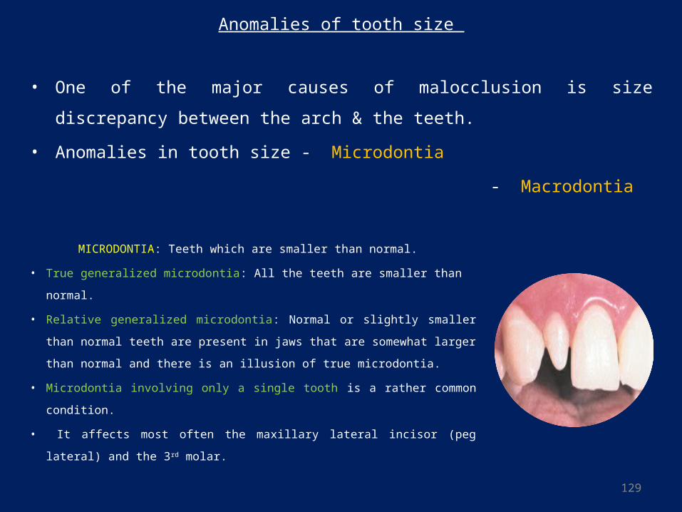
Anomalies of tooth size
• One of the major causes of malocclusion is size discrepancy between the arch & the
teeth.
• Anomalies in tooth size - Microdontia
- Macrodontia
MICRODONTIA: Teeth which are smaller than normal.
• True generalized microdontia: All the teeth are smaller than normal.
• Relative generalized microdontia: Normal or slightly smaller than
normal teeth are present in jaws that are somewhat larger than normal
and there is an illusion of true microdontia.
• Microdontia involving only a single tooth is a rather common condition.
• It affects most often the maxillary lateral incisor (peg lateral) and the 3rd
molar.
130
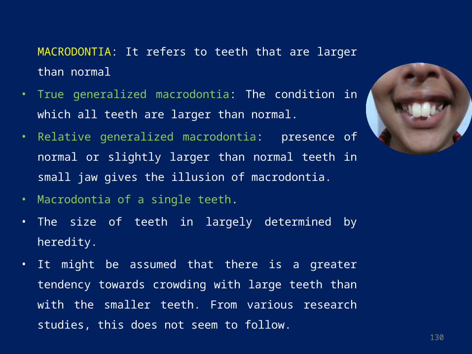
MACRODONTIA: It refers to teeth that are larger than normal
• True generalized macrodontia: The condition in which all teeth are
larger than normal.
• Relative generalized macrodontia: presence of normal or slightly
larger than normal teeth in small jaw gives the illusion of
macrodontia.
• Macrodontia of a single teeth.
• The size of teeth in largely determined by heredity.
• It might be assumed that there is a greater tendency towards
crowding with large teeth than with the smaller teeth. From
various research studies, this does not seem to follow.
131
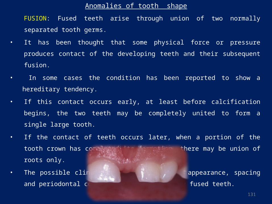
FUSION: Fused teeth arise through union of two normally separated tooth germs.
• It has been thought that some physical force or pressure produces contact of the
developing teeth and their subsequent fusion.
• In some cases the condition has been reported to show a hereditary tendency.
• If this contact occurs early, at least before calcification begins, the two teeth may be
completely united to form a single large tooth.
• If the contact of teeth occurs later, when a portion of the tooth crown has completed
its formation, there may be union of roots only.
• The possible clinical problems related to appearance, spacing and periodontal
conditions brought about by fused teeth.
Anomalies of tooth shape
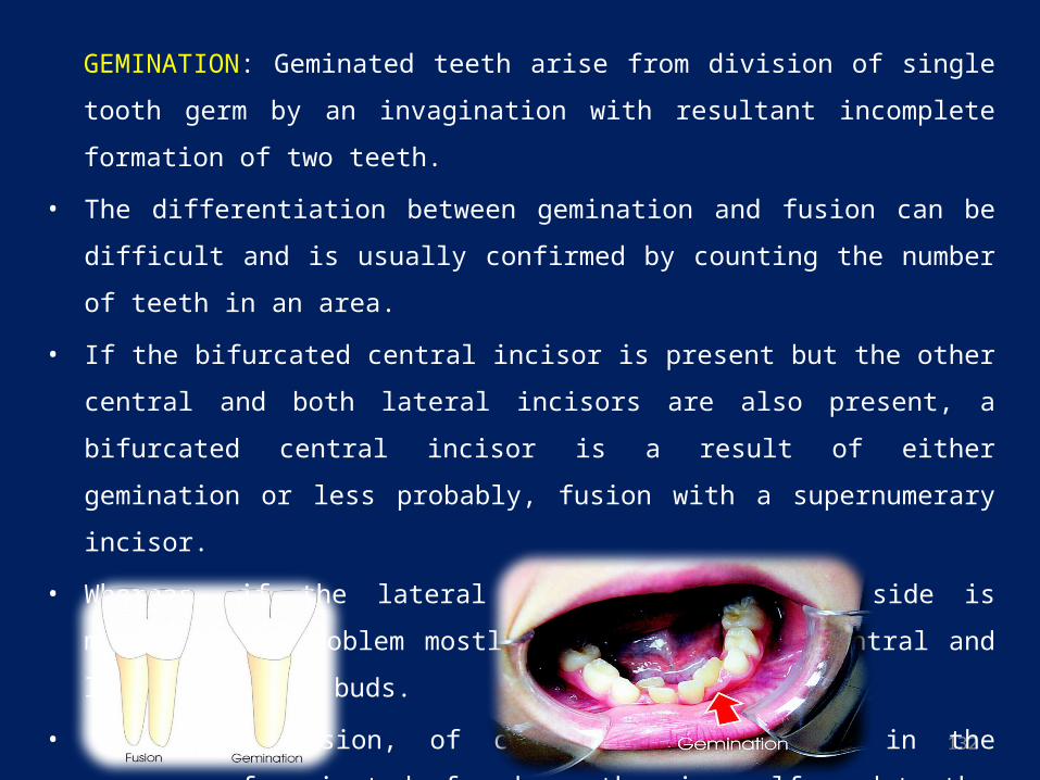
132
GEMINATION: Geminated teeth arise from division of single tooth germ by an
invagination with resultant incomplete formation of two teeth.
• The differentiation between gemination and fusion can be difficult and is usually
confirmed by counting the number of teeth in an area.
• If the bifurcated central incisor is present but the other central and both lateral incisors
are also present, a bifurcated central incisor is a result of either gemination or less
probably, fusion with a supernumerary incisor.
• Whereas, if the lateral incisor on affected side is missing, the problem mostly is fusion
of the central and lateral incisor buds.
• Normal occlusion, of course, is impossible in the presence of geminated, fused or
otherwise malformed teeth.
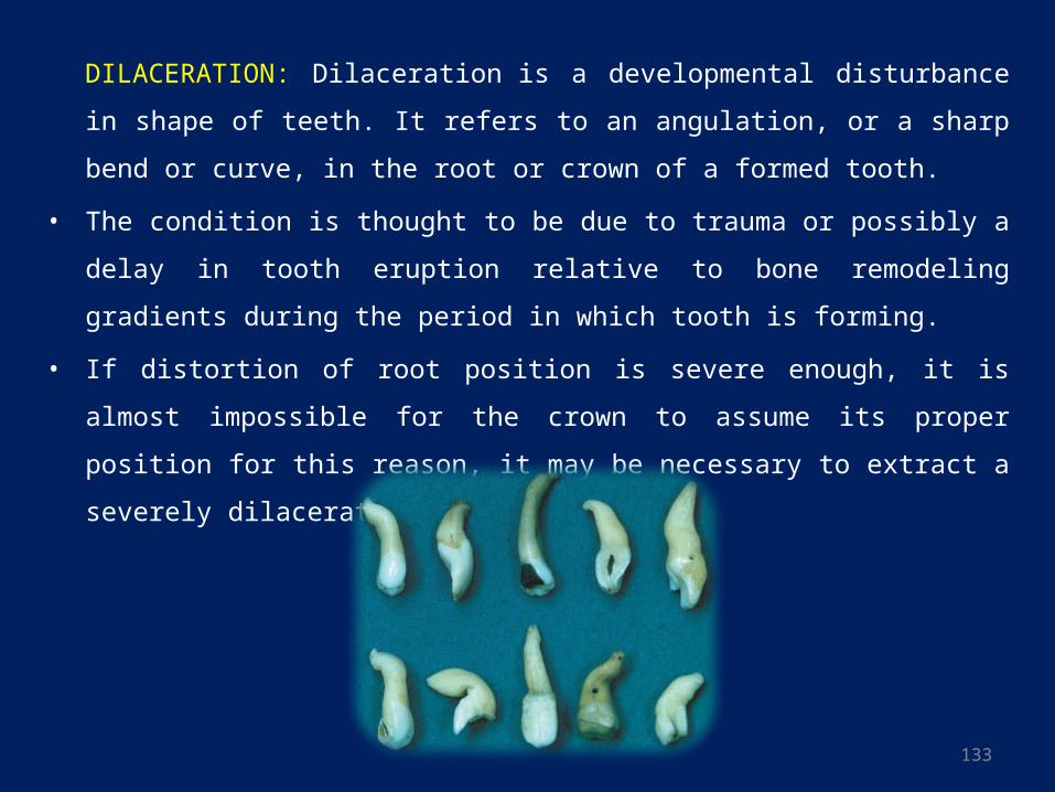
133
DILACERATION: Dilaceration is a developmental disturbance in shape of teeth. It
refers to an angulation, or a sharp bend or curve, in the root or crown of a
formed tooth.
• The condition is thought to be due to trauma or possibly a delay in tooth eruption
relative to bone remodeling gradients during the period in which tooth is forming.
• If distortion of root position is severe enough, it is almost impossible for the crown to
assume its proper position for this reason, it may be necessary to extract a severely
dilacerated tooth.
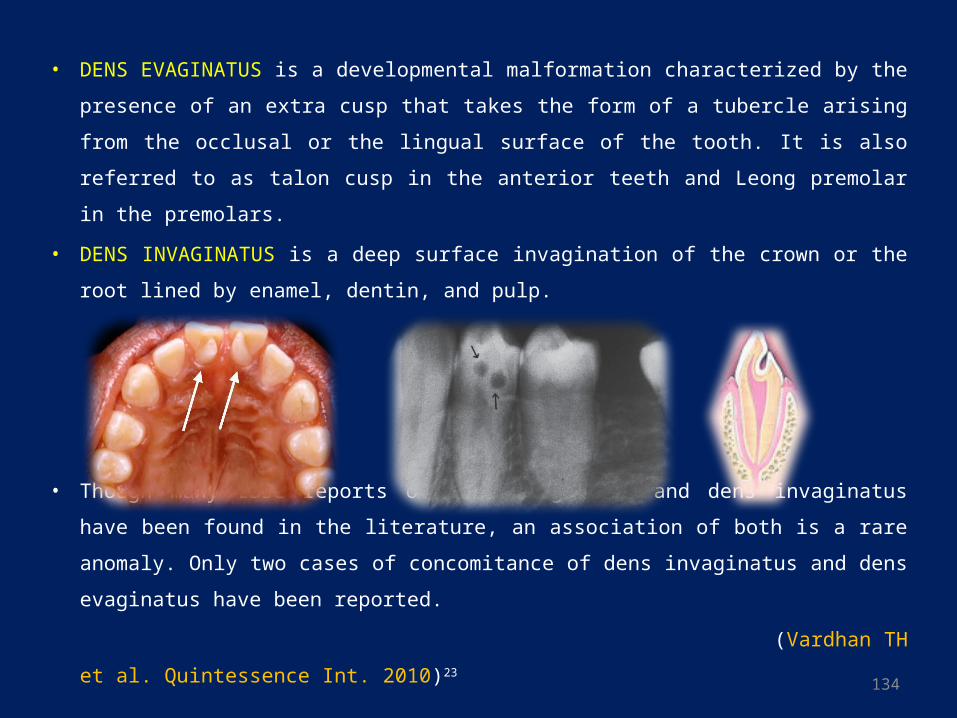
134
• DENS EVAGINATUS is a developmental malformation characterized by the
presence of an extra cusp that takes the form of a tubercle arising from the occlusal
or the lingual surface of the tooth. It is also referred to as talon cusp in the anterior
teeth and Leong premolar in the premolars.
• DENS INVAGINATUS is a deep surface invagination of the crown or the root lined
by enamel, dentin, and pulp.
• Though many case reports of dens evaginatus and dens invaginatus have been found
in the literature, an association of both is a rare anomaly. Only two cases of
concomitance of dens invaginatus and dens evaginatus have been reported.
(Vardhan TH et al. Quintessence Int. 2010)23
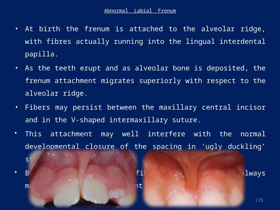
135
Abnormal Labial Frenum
• At birth the frenum is attached to the alveolar ridge, with fibres actually running into
the lingual interdental papilla.
• As the teeth erupt and as alveolar bone is deposited, the frenum attachment migrates
superiorly with respect to the alveolar ridge.
• Fibers may persist between the maxillary central incisor and in the V-shaped
intermaxillary suture.
This attachment may well interfere with the normal developmental closure of the
spacing in ‘ugly duckling’ stage.
But existence of a heavy fibrous frenum does not always mean that spacing is present
136
• Spacing between the maxillary central incisors and the presence of the fibrous tissue
attachment such as the labial frenum provide an excellent “chicken egg” routine for
controversy - which came first?
• The difficulty lies in establishing whether this fibrous attachment is causative or resultant.
• The hereditary component is a major factor in persistant diastemas. Therefore a check of
parents and siblings is advisable whenever a diastema is observed.
• Diastema may be due to other factors any and all of the following list should be eliminated
as possible causative factors: Microdontia, macrognathia, supernumerary teeth (especially
mesiodens), peg laterals, missing lateral incisors, habits such as thumb sucking, tongue
thrust, and midline cyst.
• In many other instances, due to the lack of recognition of habit problems, tooth size
discrepancy, congenitally missing teeth or midline supernumerary teeth, the clipping of the
frenum has done little to close the space.
• It is sufficient to say here that the mere clipping of the frenum attachment will not solve the
diastema problem.
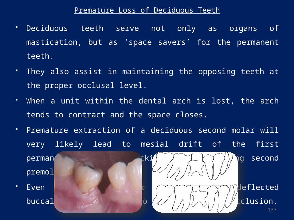
137
Premature Loss of Deciduous Teeth
Deciduous teeth serve not only as organs of mastication, but as ‘space savers’ for
the permanent teeth.
They also assist in maintaining the opposing teeth at the proper occlusal level.
When a unit within the dental arch is lost, the arch tends to contract and the
space closes.
Premature extraction of a deciduous second molar will very likely lead to mesial
drift of the first permanent molar and blocking of the erupting second premolars.
Even when the premolar erupts, it is deflected buccally or lingually into a
position of malocclusion.
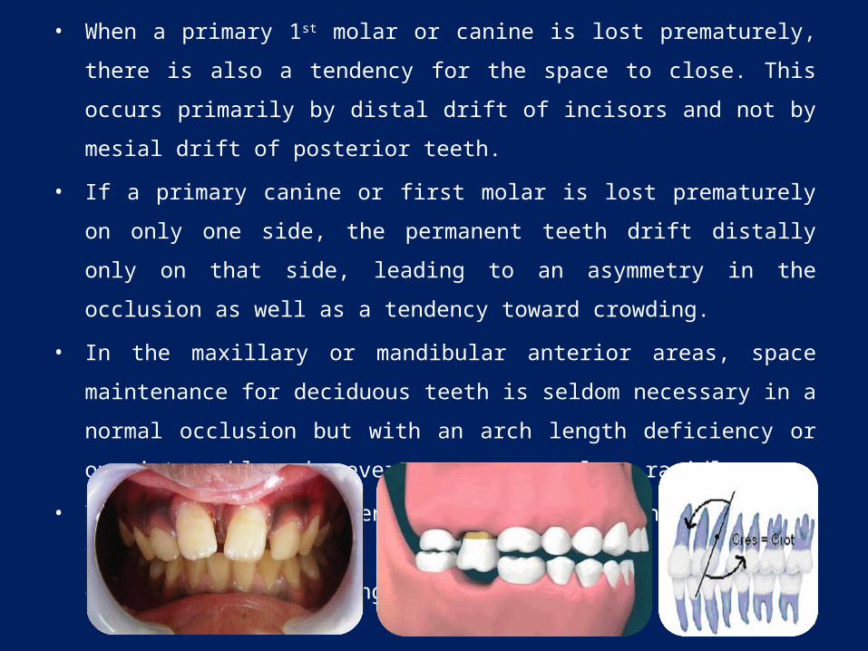
138
• When a primary 1st molar or canine is lost prematurely, there is also a tendency for
the space to close. This occurs primarily by distal drift of incisors and not by mesial
drift of posterior teeth.
• If a primary canine or first molar is lost prematurely on only one side, the
permanent teeth drift distally only on that side, leading to an asymmetry in the
occlusion as well as a tendency toward crowding.
• In the maxillary or mandibular anterior areas, space maintenance for deciduous
teeth is seldom necessary in a normal occlusion but with an arch length deficiency
or overjet problem, however, spaces can close rapidly.
• This results in shortening of arch length on the side of the loss, tipping of the
contiguous teeth and, overeuption of opposing teeth.
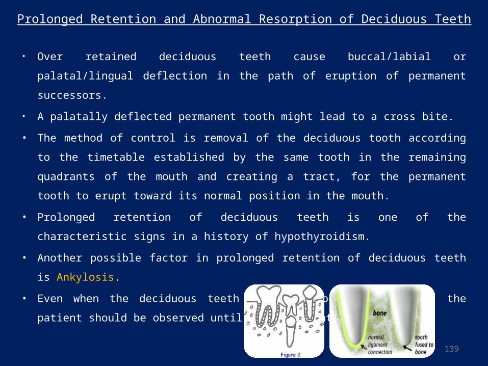
139
Prolonged Retention and Abnormal Resorption of Deciduous Teeth
• Over retained deciduous teeth cause buccal/labial or palatal/lingual deflection in the
path of eruption of permanent successors.
• A palatally deflected permanent tooth might lead to a cross bite.
• The method of control is removal of the deciduous tooth according to the timetable
established by the same tooth in the remaining quadrants of the mouth and creating a
tract, for the permanent tooth to erupt toward its normal position in the mouth.
• Prolonged retention of deciduous teeth is one of the characteristic signs in a history of
hypothyroidism.
• Another possible factor in prolonged retention of deciduous teeth is Ankylosis.
• Even when the deciduous teeth appear to be lost on time, the patient should be
observed until the permanent teeth erupt.
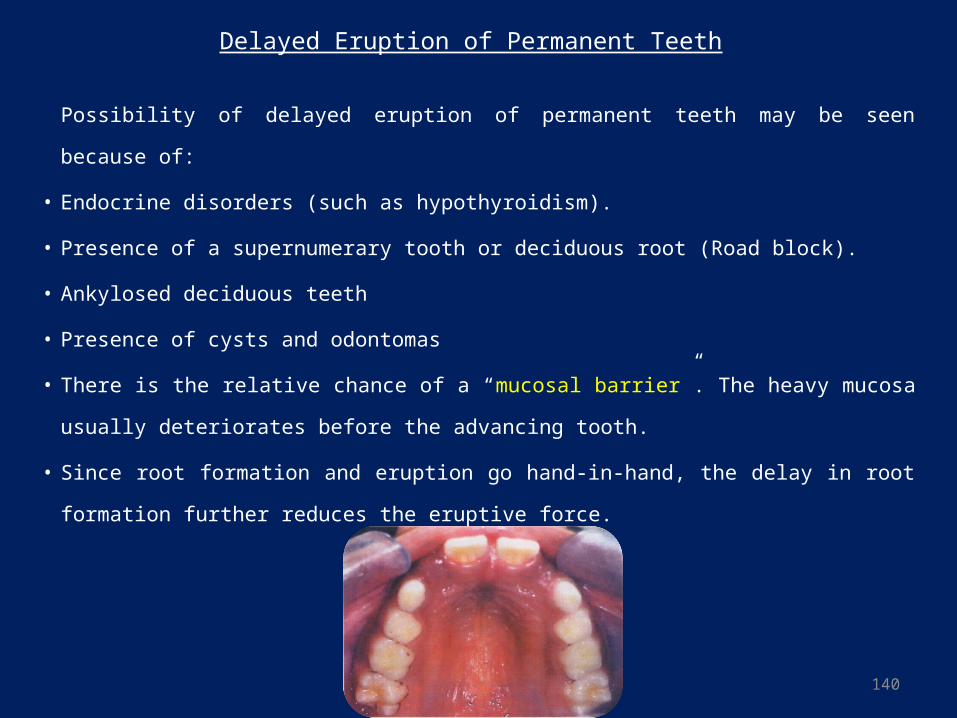
140
Delayed Eruption of Permanent Teeth
Possibility of delayed eruption of permanent teeth may be seen because of:
• Endocrine disorders (such as hypothyroidism).
• Presence of a supernumerary tooth or deciduous root (Road block).
• Ankylosed deciduous teeth
• Presence of cysts and odontomas
• There is the relative chance of a “mucosal barrier”. The heavy mucosa usually
deteriorates before the advancing tooth.
• Since root formation and eruption go hand-in-hand, the delay in root formation further
reduces the eruptive force.
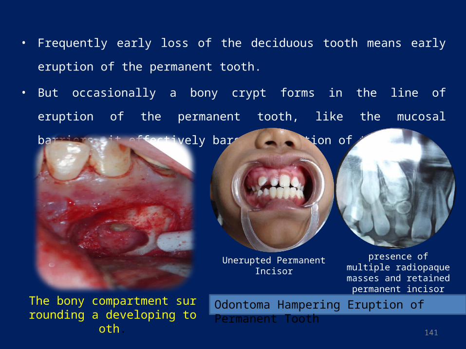
141
• Frequently early loss of the deciduous tooth means early eruption of the permanent
tooth.
• But occasionally a bony crypt forms in the line of eruption of the permanent tooth, like
the mucosal barriers, it effectively bars the eruption of the tooth.
The bony compartment surrounding a developing tooth.
Unerupted Permanent Incisor presence of multiple radiopaque masses and
retained permanent incisor
Odontoma Hampering Eruption of Permanent Tooth
142
Abnormal Eruptive Path
In the case of inadequate space to accommodate all the teeth, deflection of the erupting
tooth may occur because of the presence of:
• A supernumerary tooth.
• Retained deciduous tooth or root fragment.
• Possible bony barrier.
• Ankylosed tooth.
A blow to the tooth: Because of blow to the tooth, deciduous incisor may be driven
into the alveolar process, it may turn the developing successor in an abnormal
direction.
Coronal cysts can also cause abnormal eruptive paths.
Some abnormal eruptive paths are of idiopathic (unknown) origin. A canine or
premolar will erupt buccally, lingually, or transposed, with no apparent cause.
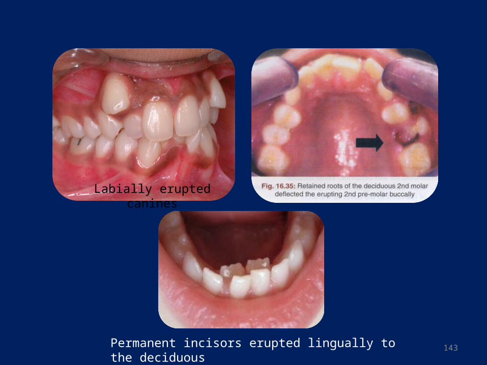
143
Labially erupted canines
Permanent incisors erupted lingually to the deciduous
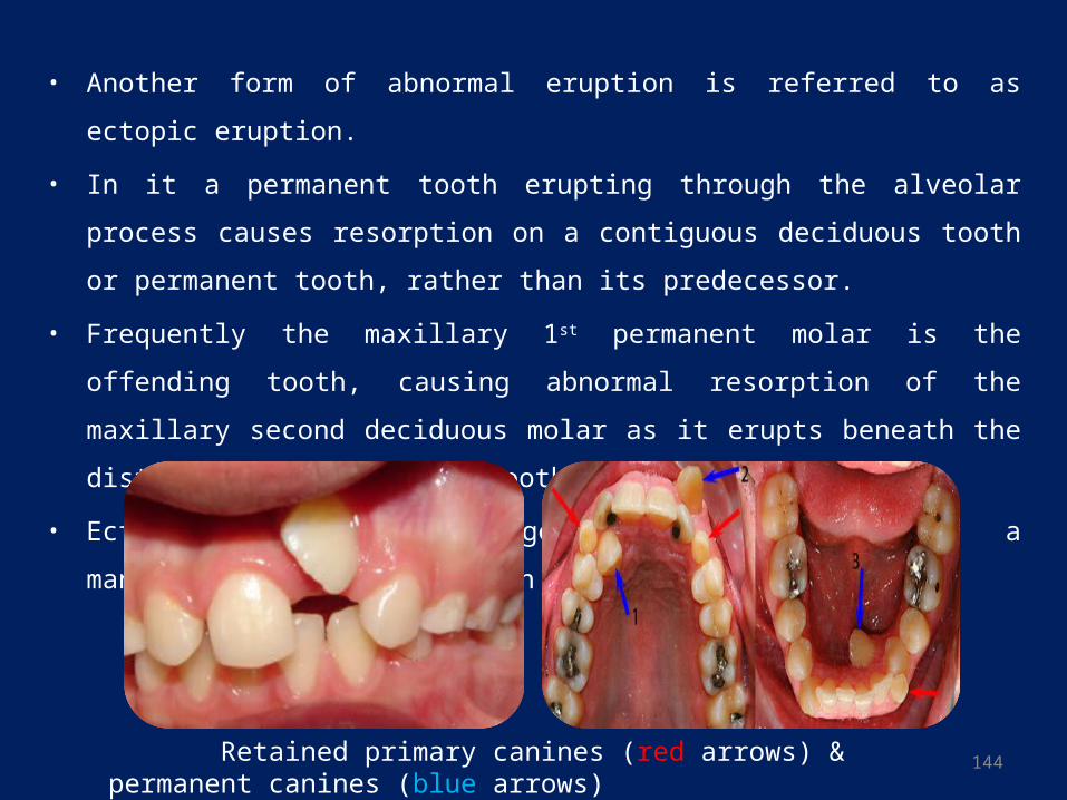
144
• Another form of abnormal eruption is referred to as ectopic eruption.
• In it a permanent tooth erupting through the alveolar process causes resorption on a
contiguous deciduous tooth or permanent tooth, rather than its predecessor.
• Frequently the maxillary 1st permanent molar is the offending tooth, causing abnormal
resorption of the maxillary second deciduous molar as it erupts beneath the distal
convexity of this tooth.
• Ectopic eruption may generally be considered a manifestation of arch length
deficiency.
Retained primary canines (red arrows) & permanent canines (blue arrows)
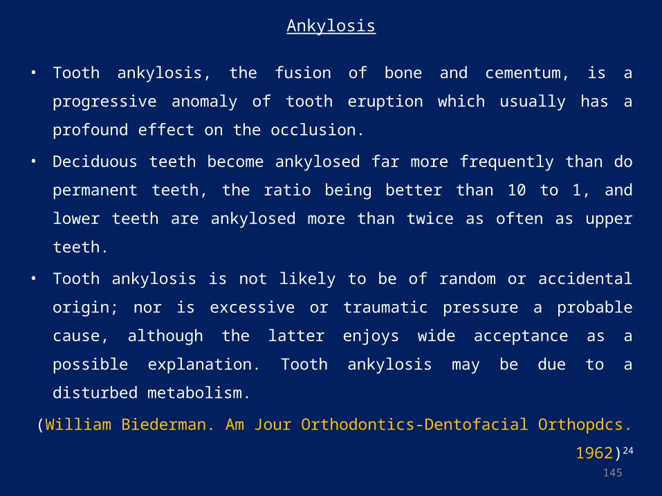
145
Ankylosis
• Tooth ankylosis, the fusion of bone and cementum, is a progressive anomaly of tooth
eruption which usually has a profound effect on the occlusion.
• Deciduous teeth become ankylosed far more frequently than do permanent teeth, the
ratio being better than 10 to 1, and lower teeth are ankylosed more than twice as often
as upper teeth.
• Tooth ankylosis is not likely to be of random or accidental origin; nor is excessive or
traumatic pressure a probable cause, although the latter enjoys wide acceptance as a
possible explanation. Tooth ankylosis may be due to a disturbed metabolism.
(William Biederman. Am Jour Orthodontics-Dentofacial Orthopdcs. 1962)24
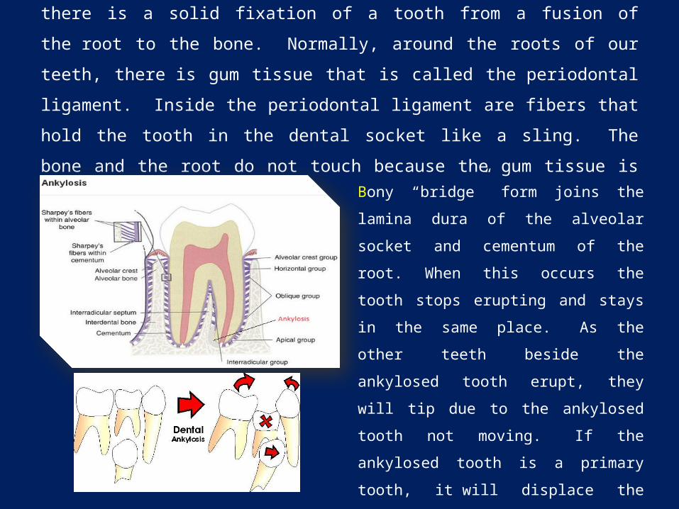
Dental Ankylosis is an abnormal dental condition where there is a solid fixation of a
tooth from a fusion of the root to the bone. Normally, around the roots of our teeth,
there is gum tissue that is called the periodontal ligament. Inside the periodontal
ligament are fibers that hold the tooth in the dental socket like a sling. The bone and
the root do not touch because the gum tissue is between them.
Bony “bridge” form joins the lamina dura of
the alveolar socket and cementum of the root.
When this occurs the tooth stops erupting and
stays in the same place. As the other teeth
beside the ankylosed tooth erupt, they will tip
due to the ankylosed tooth not moving. If the
ankylosed tooth is a primary tooth, it will
displace the permanent tooth that is trying to
erupt into its position. Also, the ankylosis can
cause reduction of the space needed for the
permanent tooth to erupt causing an impaction.
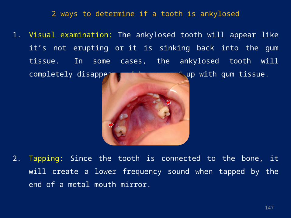
147
2 ways to determine if a tooth is ankylosed
1. Visual examination: The ankylosed tooth will appear like it’s not erupting or it is
sinking back into the gum tissue. In some cases, the ankylosed tooth will completely
disappear and be covered up with gum tissue.
2. Tapping: Since the tooth is connected to the bone, it will create a lower frequency
sound when tapped by the end of a metal mouth mirror.
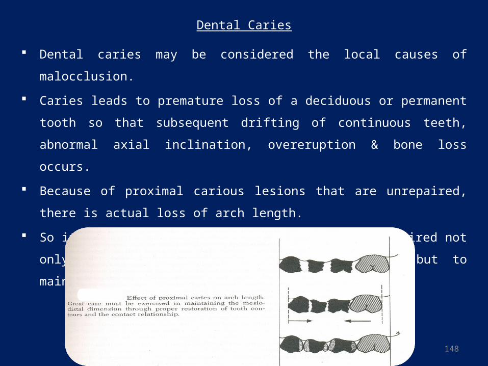
148
Dental Caries
Dental caries may be considered the local causes of malocclusion.
Caries leads to premature loss of a deciduous or permanent tooth so that subsequent
drifting of continuous teeth, abnormal axial inclination, overeruption & bone loss
occurs.
Because of proximal carious lesions that are unrepaired, there is actual loss of arch
length.
So it is basic that carious lesions should be repaired not only to prevent infection and
loss of teeth but to maintain the integrity of the dental arches.
149
Improper Dental restoration
• An increase in arch length through improper restoration of one or more carious
proximal surfaces may result in the creation of broken contacts and/or rotations.
• Poor contacts encourage food packing.
• Lack of anatomic detail in restoration of cuspal area of a tooth can permit elongation
of opposing teeth(supra-eruption) or create functional prematurities.
• Overhanging or poor proximal contacts may predispose to periodontal breakdown
around these teeth.
• Undercontoured proximal restorations result in loss of arch length due to drifting of
adjacent teeth to occupy the space.
• Overcontoured proximal restorations might bulge-into the space to be occupied by a
succedaneous tooth and result in a reduction in this space.
150
Epidemiology and Public Health Aspects of Malocclusion25
• The aim of epidemiologic studies of malocclusion is to describe and analyze the prevalence
and distribution of malocclusion in various populations, the ultimate goal being to identify
etiologic factors.
• A further aim is to contribute to the solution of the public health problems concerning
assessment of need for orthodontic treatment and organization of orthodontic services.
• At an early point it was realized that, due to the complexity of malocclusion, epidemiologic
studies had to be based on some kind of classification. Angle's' classification is the only one
among several typologic classifications which has gained wide ground in the epidemiology of
malocclusion.
• Angle's classification has been misused in epidemiologic studies all over the world.
• Occasionally, it has been emphasized that Angle's classification is not sufficiently differentiated
for epidemiologic purposes, and it has also been pointed out that the individual morphologic
traits of Angle's classes are not all adequately defined. As might be expected, therefore, poor
intra and interexaminer reliability of recording Angle's classes has been demonstrated.
151
• The current major public health problem in this field is to bridge the gap between
recognition of the occurrence of the defined single traits or combinations of traits, and
determination of the need for treatment of these conditions.
• The main purpose of the indices is to interpret malocclusion severity objectively in
terms of treatment priority. Fundamentally, the index scores are based on clinical
estimations of the severity of the various traits. In other words, the scores are assigned
according to clinical concepts of the adverse effects of the traits on facial appearance,
function, and oral health. Thus, the objectivity involved in such interpretations would
seem to be questionable.
• Once the basic needs for caries control in a child population have been met, the
problems of organizing orthodontic care comes into focus. Traditionally, the
responsibility for initiating orthodontic measures and the economic burden of the
treatment have rested mainly with the patients, or rather with their parents. Thus, the
provision of orthodontic treatment has often been determined by the incidental
educational and socio-economic level of the family, instead of the severity of the
patient's malocclusion.
152
Index of Orthodontic Treatment Needs (IOTN)
Index of orthodontic treatment needs attempts to rank maloccluison based on the level of
treatment needed on treatment priority. The index intends to identify people who would most
likely benefit from orthodontic treatment. It has a dental and an aesthetic component wherein
due significance is given to occlusal traits affecting individual dental health and perceived
dental aesthetic impairment.
The 2 Components:
• Dental Health Component(DHC): Based on the Swedish Medical Health Board index (Linder
Aronson, 1974)
Grade 1 = no treatment need (e.g., contact point displacements of less than 1 mm)
Grade 5 = treatment need (e.g., cleft lip/palate or reverse sagittal overjets greater than 9 mm)
• Aesthetic Component(AC): Using a series of 10 color photos the degree of 'dental
attractiveness' is assessed.
Grade 1 = aesthetically optimal dentition
Grade 10 = aesthetically worst imaginable dentition
153
Factors affecting Receipt of Orthodontic Treatment
• Improvements in personal appearance: Strongest motivational factor.
• Self-perception and self-esteem: People with low self-esteem greatly underrate
their dental appearance.
• Gender and age: More girls in their teens receive orthodontic treatment, reflecting a
greater societal emphasis of high physical attractiveness on the females.
• Peer groups: pressure varies according to the culture and societal norms.
• Social class: Uptake of services is more in higher social class.
• General dentist’s attitude: Attitudes, beliefs & awareness of the general dentists
influence orthodontic service utilizations.
• Availability of resources: Availability of trained manpower(dentists, specialist
orthodontists & dental assistants) and orthodontic laboratories influence uptake of
orthodontic services in any area.
154
155
Bibliography
1. Gurkeerat Singh. Textbook of Orthodontics. New Delhi: JAYPEE BROTHERS;2004.
2. Lauc T et al. Effect of inbreeding and endogamy on occlusal traits in human isolates. J Orthod. 2003;30(4):301-8.
3. Yuan JT et al. Asymmetric class III malocclusion: association with cranial base deformation and occult torticollis.
The Journal Of Craniofacial Surgery 2012;23(5): 1421-4.
4. Piggott KD et al. Congenital cardiac defects: a possible association of aminopterin syndrome and in utero
methotrexate exposure? Pediatr Cardiol. 201;32(4):518-20.
5. Guerri, C et al. "Commentary on the recommendations of the Royal College of Obstetricians and Gynaecologists
concerning alcohol consumption in pregnancy". Alcohol and alcoholism (Oxford, Oxfordshire) 34(4): 497–501.
6. Alvin F. Gardner. Differential Oral Diagnosis in Systemic Disease. Bristol: JOHN WRIGHT & SONS LTD;1970
7. GN Güncü et al. Effects of endogenous sex hormones on the periodontium – Review of literature. Australian
Dental Journal 2005;50(3):138-45
8. Basavaraj Subhashchandra Phulari. Orthodontics: Principles and Practice. New Delhi: JAYPEE BROTHERS;2011
9. Marc Baltensperger and Gerold Eyrich. Osteomyelitis of the jaws: definition and classification. In: Robert E.
Marx, Marc Baltensperger, Gerold K. Eyrich. Osteomyelitis of the Jaws. Berlin, Germany: Springer;2010: 28.
10. Erika B. A. F. Thomaz. Is Malnutrition Associated with Crowding in Permanent Dentition? Int J Environ Res
Public Health. 2010;7(9): 3531–3544.
156
11. Aparna Sheetal et al. Malnutrition and its Oral Outcome – A Review. J Clin Diagn Res. 2013;7(1): 178–180.
12. William R. Proffit. Contemporary Orthodontics. United States: Mosby-Year Book; 1993
13. López Del Valle LM et al. Associations between a history of breast feeding, malocclusion and parafunctional
habits in Puerto Rican children. P R Health Sci J. 2006;25(1):31-4.
14. Labbok MH et al. Does breast-feeding protect against malocclusion? An analysis of the 1981 Child Health
Supplement to the National Health Interview Survey. Am J Prev Med. 1987;3(4):227-32.
15. Warren JJ et al. Effects of oral habits' duration on dental characteristics in the primary dentition. J Am Dent
Assoc. 2001;132(12):1685-9
16. Mônica Vilela Heimer et al. Non-nutritive sucking habits, dental malocclusions, and facial morphology in
Brazilian children: a longitudinal study. Eur J Orthod.2008;30(6):580-5.
17. Afzelius-Alm A et al. Factors that influence the proclination or retroclination of the lower incisors in children
with prolonged thumb-sucking habits. Swed Dent J. 2004;28(1):37-45.
18. William R. Proffit. Contemporary Orthodontics. United States: Mosby-Year Book; 1993
19. Cozza P et al. Sucking habits and facial hyperdivergency as risk factors for anterior open bite in the mixed
dentition. Am J Orthod Dentofacial Orthop. 2005;128(4):517-9.
20. Warren JJ et al. Effects of nonnutritive sucking habits on occlusal characteristics in the mixed dentition.
Pediatr Dent. 2005;27(6):445-50.
157
21. Abhishek Parolia et al. Management of supernumerary teeth. J Conserv Dent. 2011; 14(3): 221–224.
22. G. Meighani et al. Diagnosis and Management of Supernumerary (Mesiodens): A Review of the Literature.
J Dent (Tehran). 2010; 7(1): 41–49.
23. Vardhan TH et al. Dens evaginatus and dens invaginatus in all maxillary incisors: report of a case.
Quintessence Int. 2010;41(2):105-7.
24. William Biederman. Etiology and treatment of tooth ankylosis. Am Jour Orthodontics-Dentofacial
Orthopdcs. 1962;48(9):670-684
25. SVEN HELM. Epidemiology and Public Health Aspects of Malocclusion. J Dent Res 1977;56 Spec
No:C27-31.