Embed Size (px)
Citation preview

EnterobacteriaceaeBiochemical reactions
ByDr. Nabil El Aila
Assistant Professor of Molecular MicrobiologyMedical Technology Department
Al -Aqsa University
Dr. Nabil El AilaDiagnostic Microbiology
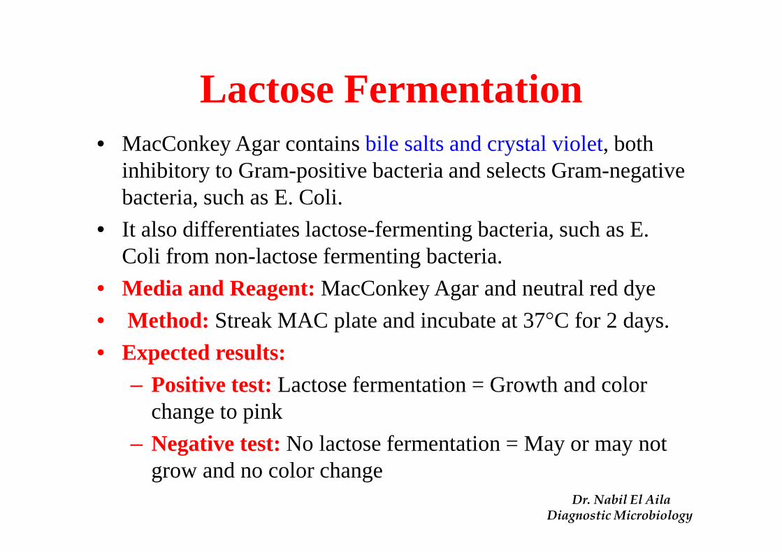
Lactose Fermentation• MacConkey Agar contains bile salts and crystal violet, both
inhibitory to Gram-positive bacteria and selects Gram-negative bacteria, such as E. Coli.
• It also differentiates lactose-fermenting bacteria, such as E. Coli from non-lactose fermenting bacteria.
• Media and Reagent:MacConkey Agar and neutral red dye
• Method: Streak MAC plate and incubate at 37°C for 2 days.
• Expected results: – Positive test:Lactose fermentation = Growth and color
change to pink
– Negative test:No lactose fermentation = May or may not grow and no color change
Dr. Nabil El AilaDiagnostic Microbiology

Results of Lactose Fermentation
Dr. Nabil El AilaDiagnostic Microbiology

IMViC Test
• Indole, Methyl Red, Voges-Prosakaur, Citrate (IMViC ) Tests:– The following four tests comprise a series of
important determinations that are collectively called the IMViC series of reactions
– The IMViC series of reactions allows for the differentiationof the various members of Enterobacteriaceae.
Dr. Nabil El AilaDiagnostic Microbiology

IMViC : Indole test� Principle
� Certain microorganisms can metabolize tryptophan by tryptophanase
� The enzymatic degradation leads to the formation of pyruvic acid, indole and ammonia
� The presence of indole is detected by addition of Kovac's reagent.
Tryptophaneamino acids
TryptophanaseIndole + Pyurvic acid + NH3
Kovac’s Reagent
Red color in upper organic layer`Dr. Nabil El AilaDiagnostic Microbiology

IMViC : Indole test
� Method:
� Inoculate the test organism into tryptophanebroth
� Incubate at 37°C for 24 hours
� After incubation interval, add 1 ml Kovacs reagentwhich contain 4 (p) – dimethylamino
benzaldehyde, shake the tube gently and read
immediately
Dr. Nabil El AilaDiagnostic Microbiology

IMViC: Indole test
� Result:
� A bright pink color in the top layer indicates the presence of indole
� The absence of color means that indole was not produced i.e. indole is negative
� Significance:
� Used in the differentiation of genera and species. e.g. E. coli (+) from Klebsiella (-).
Positive teste.g. E. coli
Negative teste.g. Klebsiella
Dr. Nabil El AilaDiagnostic Microbiology
IMVi C testMethyl Red-Voges Proskauer (MR-VP) Tests
• Different bacteria convert dextrose and glucose to pyruvate
using different metabolic pathways.
• Some of these pathways produce unstable acidic products
which quickly convert to neutral compounds.
• Some organisms use the butylene glycol pathway, which
produces neutral end products, including acetoinand 2,3-
butanediol.
• Other organisms use the mixed acid pathway, which produces
acidic end products such as lactic, acetic, and formic acid. These
acidic end products are stable and will remain acidic.

IMV iC testMethyl Red-Voges Proskauer (MR-VP) Tests
Glucose
Acidic pathway
Mixed acids ���� pH less than 4.4
Methyl Redindicator
Red color
Principle
MR positiveE. coli
Or Neutral pathway
Acety methyl carbinol(ACETOIN)
Barrit’s A (α-naphthol)Barrit;s B (40% KOH)
Pink colorVP positiveKlebsiella

IMViC: Methyl red
Principle:• Methyl red test is used to identify enteric bacteria based on
their pattern of glucose metabolism.
• If they use mixed acid pathway and produce acidic products, then they are called methyl-red-positive.
• If they use butylene glycol pathway and produce neutral end products, then they are called methyl-red-negative
Dr. Nabil El AilaDiagnostic Microbiology

IMViC: Methyl red
• Method:• Inoculate 10ml portion of the MR-VP medium and incubate at
37°C for 2-5 days.
• After incubation, transfer 2.5 ml of inoculate to another tube and add five drops of methyl red.
• Roll between the palms of hands to disperse methyl red.
Dr. Nabil El AilaDiagnostic Microbiology

IMViC: Methyl red• Results:
– Positive test:acids + methyl red = red solution
– Negative test:neutral end products + methyl red =
yellow color
• Significance: This test is used to differentiate
Enterobacteriacaceae species
espciallyE. coli andE. aerogens
Dr. Nabil El AilaDiagnostic Microbiology

IM ViC: VOGES-PROSKAUER TEST
Principle:• It is used to identify enteric bacteria based on their pattern of
glucose metabolism.
• The enterics that produce neutral end-products, such as acetoinare detected by VP test.
• Its presence is used as indicator of 2,3 butylene glycol
Fermentation
• The detection of acetoin in alkaline pH is accomplished by
alpha-Naphthol reagent.
Dr. Nabil El AilaDiagnostic Microbiology

IM ViC: VOGES-PROSKAUER TEST
• Method:• Inoculate medium and incubate at 37°C for 48 hours.
• After incubation, transfer 2.5 ml of inoculate to another tube and add six drops of Barritt’s Reagent A (contains alpha-naphthol) and two drops of Barritt’s Reagent B(contains KOH).
• Gently mix and let it sit for 10-15 minutes to allow time for color development.
Dr. Nabil El AilaDiagnostic Microbiology

IM ViC: VOGES-PROSKAUER TEST
• Results:– Positive test: acetoin + alpha-naphthol + KOH = red color
– Negative test:alpha-naphthol +KOH = copper color
• Significance: This test is used todifferentiate
Enterobacteriacaceaespecies
espciallyE. coli andE. aerogens
Dr. Nabil El AilaDiagnostic Microbiology

Principle:
Citrate Na2CO3
Alkaline,↑pH
Bromothymol blue
Simmone’s Citrate media
CO2 + Na + H2OPyruvate
Positive test
Contains Citrate as a sole of C source
IMVi C: CITRATE TEST
Dr. Nabil El AilaDiagnostic Microbiology

IMVi C: CITRATE TEST
• Principle:• Citrate is an organic molecule that can be utilized by bacteria
that produce the enzyme citrase. • Citrase is produced by some bacteria such as E. aerogenes but
not by others like E. Coli• Method:• Inoculate the test organism onto a slant containing Simmon
Citrate agar.• Simmon’s Citrate Agar contains sodium citrate (carbon
source), & pH indicator—bromthymol blue.• Incubate at 37°C for 24 hours.
Dr. Nabil El AilaDiagnostic Microbiology

IMVi C: CITRATE TEST
• Results:– Positive test:Growth and color changes to blue– Negative test:No growth and color remains green
• Significance:• This test is used to help differentiate
species of the family Enterobacteriaceae.
• It is selective for bacteria that has
the ability to consume citrate as
its sole source of carbon
NegativePositiveDr. Nabil El AilaDiagnostic Microbiology

UreaseTest
• Urea agar contains urea and phenol red• Urease is an enzyme that catalyzes the conversion of urea
to CO2 and NH3• Ammonia combines with water to produce ammonium
hydroxide, a strong base which ↑ pH of the medium. • ↑ in the pH causes phenol red r to turn a deep pink. This is
indicative of a positive reaction for urease
UreaUrease
CO2 + NH3H2O
NH4 OH ↑ in pH
Phenol Red
PinkPositive test
� Streak a urea agartube with the organism
� incubate at 37°C for 24 h
Method
Principle

Urease Test• Result
• Positive test: production of alkaline end products = pinkish red color
• Negative test:No color change
• Significance:
• Differentiatesalmonella and shigellawhich are urease negative fromurease positive Non pathogens.
• Proteus, klebsiella and somecitrobacter species are ureasepositive
• Helicobater pylori is also
Urease positive
Positive testNegative test
Dr. Nabil El AilaDiagnostic Microbiology

Motility Test• Principle:
• Motility Test Media is a semi-solid agar designed to
demonstrate motility by diffusion.
• This is not a biochemical test, but it can distinguish bacteria. It
determines presence of flagella.
• Method:
• Inoculate a semi-solid nutrient medium by stabbing 2 cm
into the center of the medium
• Inoculate at 37C° for 24-48 hours.
Dr. Nabil El AilaDiagnostic Microbiology

Motility Test• Expected results:
– Positive test:Growth spread away from the line of
inoculation = motile
– Negative test:Growth only occurred at the line of
inoculation = Non-motile
• Significance:• This test is used for the differentiation
of microorganisms on the basis of
motility.
Dr. Nabil El AilaDiagnostic Microbiology

H2S Production
• Principle:
• Bacteria use enzyme cysteine desulfuraseto hydrolyze the amino acid cysteine, forming hydrogen sulfide as end-product.
• To test for hydrogen sulfide production, a medium with a sulfur-containing compound and iron salts is inoculated and incubated. If the sulfur is reduced and hydrogen sulfide is produced, it will combine with the iron salt to form a visible black ferric sulfide (FeS) in the tube
• Media and Reagent: SIM tube (sulphide, Indole and Motility) with cysteine and ferrous sulfate (detects H2S)
• Method: Inoculate the media and incubate at 37°C for 24-48 hours.
Dr. Nabil El AilaDiagnostic Microbiology
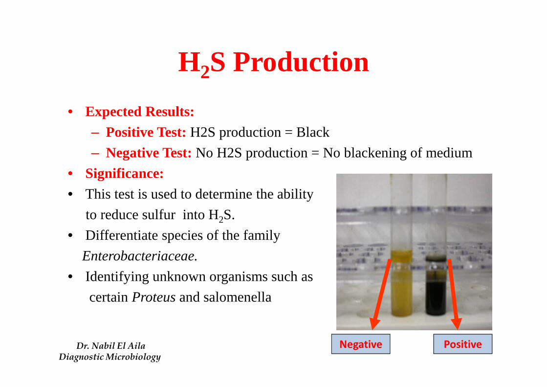
H2S Production
• Expected Results:
– Positive Test:H2S production = Black
– Negative Test:No H2S production = No blackening of medium
• Significance:
• This test is used to determine the ability
to reduce sulfur into H2S.
• Differentiate species of the family
Enterobacteriaceae.
• Identifying unknown organisms such as
certain Proteus and salomenella
Negative PositiveDr. Nabil El AilaDiagnostic Microbiology