Embed Size (px)
Citation preview

DR.MAGDY KHAMES ALY
CRITICAL CARE MEDICINE
ZMH AL BATAYEH
Emergency Red FlagsSafety VS Accuracy
Objectives
Learning about common cognitive errors in emergency
diagnosis .
Identification of specific clinical situation in which
diagnostic errors are most likely to occur, commonly
known as “pitfalls”
List the unique features and (atypical) presentations of common emergency situation.
Identify priorities for initial triage
Teaching Points to be Addressed
What is the usefulness of the clinical
warning criteria (red flags) ?
How do we use the information from the
critical warning criteria?
Is there any special group of patients?
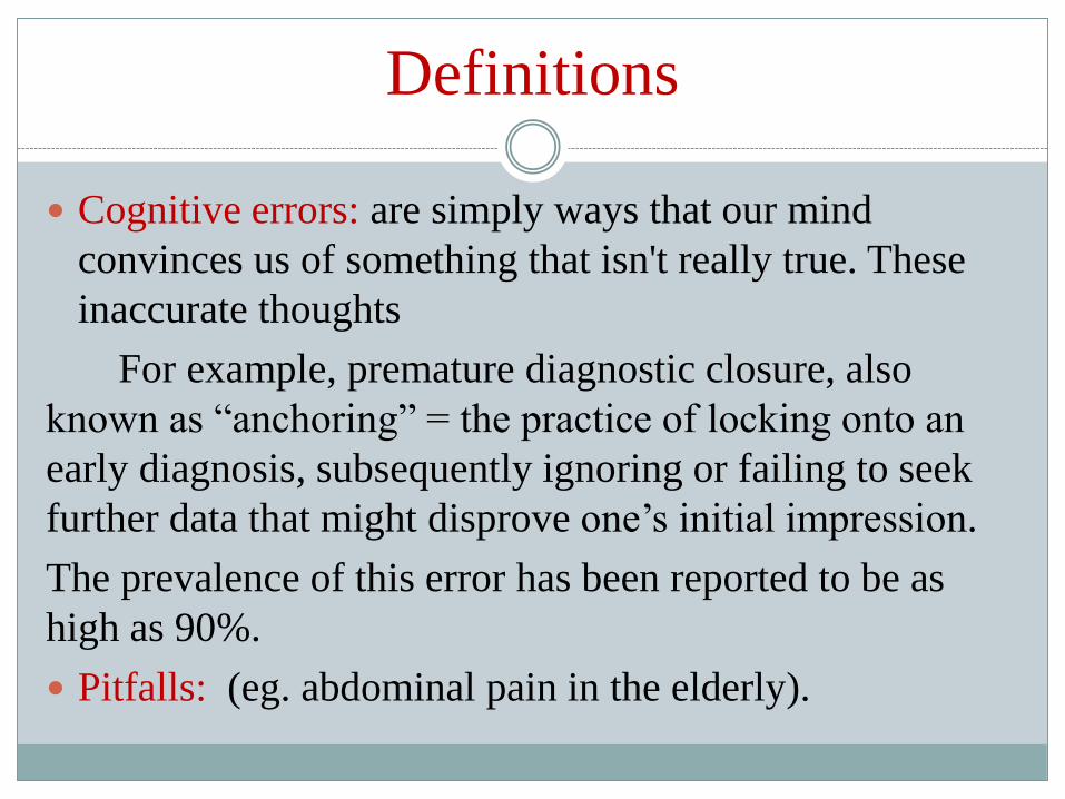
Definitions
Cognitive errors: are simply ways that our mind
convinces us of something that isn't really true. These
inaccurate thoughts
For example, premature diagnostic closure, also
known as “anchoring” = the practice of locking onto an
early diagnosis, subsequently ignoring or failing to seek
further data that might disprove one’s initial impression.
The prevalence of this error has been reported to be as
high as 90%.
Pitfalls: (eg. abdominal pain in the elderly).

Headache and facial pain

Red flags
Systemic upset
Progressive pain, disability and distress
Focal neurological deficit
Weight loss
Facial swelling or rash
Vision disturbance
Hearing loss/tinnitus/vertigo
Unilateral nasal obstruction/discharge
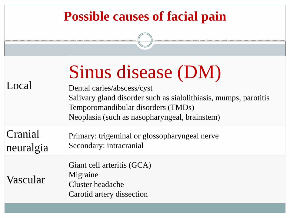
Possible causes of facial pain
LocalSinus disease (DM)Dental caries/abscess/cyst
Salivary gland disorder such as sialolithiasis, mumps, parotitis
Temporomandibular disorders (TMDs)
Neoplasia (such as nasopharyngeal, brainstem)
Cranial
neuralgiaPrimary: trigeminal or glossopharyngeal nerve
Secondary: intracranial
Vascular
Giant cell arteritis (GCA)
Migraine
Cluster headache
Carotid artery dissection
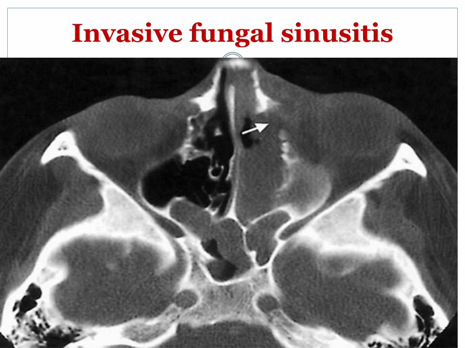
Invasive fungal sinusitis

Acute MI is a Headache
A 42-year-old male, with no history of coronary artery disease or any other risk factor, including hypertension, hyperlipidemia, smoking,or positive family history, presented to a primary health center, complaining for acute onset of severe headache beginning 4 h before. The headache was located mainly frontally and bitemporallyand was constant with no periods of relief, even after taking analgesics. Since the physical examination and electrocardiogram were normal
no further evaluation was requested and the patient was discharged home.
What was wrong?
What was the diagnosis?
Did the patient improve before discharge?

Two hours later, the patient was admitted to the
emergency department complaining of persistent
headache.
On admission, he was conscious, well-orientated
and the vital signs and physical examination were
normal. The electrocardiogram showed a q wave
with mild ST elevation (1.2 mm) and inverted T
wave in the precordial leads V2-V5
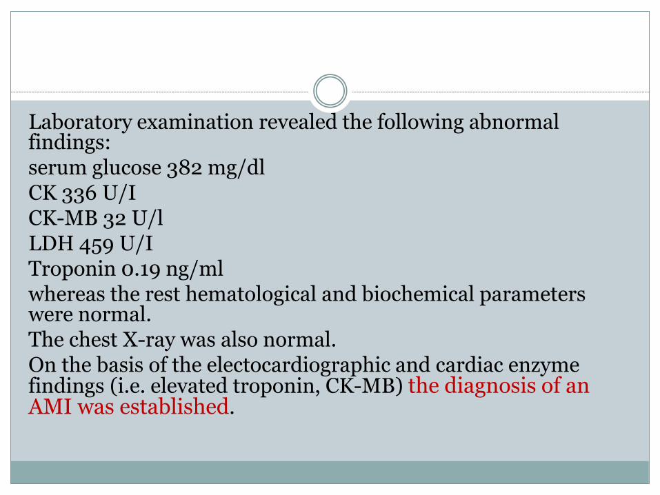
Laboratory examination revealed the following abnormal findings: serum glucose 382 mg/dl CK 336 U/I CK-MB 32 U/l LDH 459 U/I Troponin 0.19 ng/ml whereas the rest hematological and biochemical parameters were normal. The chest X-ray was also normal. On the basis of the electocardiographic and cardiac enzyme findings (i.e. elevated troponin, CK-MB) the diagnosis of an AMI was established.

What is the lesson here?
AMI rarely presents by only Headache but can happen.1
AMI should be excluded in any case presented by pain or discomfort above epigastrium to forehead.
AMI should be excluded in cases have CVD risk factors.2
AMI should be excluded in any new onset or known migraine patient(masked MI)1

A 45 yo female patient known to have migraine headache presented to ER complaining of severe migraine attack since 2 hours, not resolved by usual migraine’s tablet the patient used to use in the same attacks.Examination was done revealed nothing significantCT brain done and normalECG done revealing I, aVL, V5,V6 S-T elevation(lateral MI)Cardiac enzymes was positive Diagnosed as AMI Patient admitted in CCU treated by thrombolytic therapyHeadache disappeared and ECG returned normal
What is the lesson here?
Women with migraine headache at more risk for IHD.2
Migraine treatment(sumatriptan) can cause ACS in patient at risk.

Chest pain
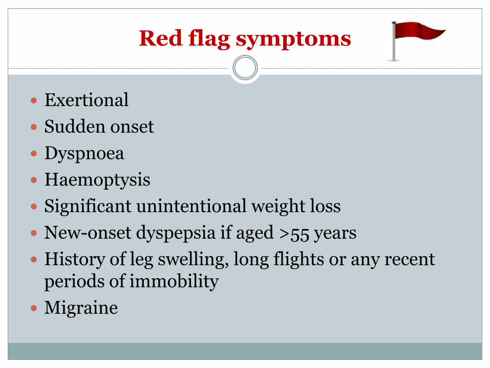
Red flag symptoms
Exertional
Sudden onset
Dyspnoea
Haemoptysis
Significant unintentional weight loss
New-onset dyspepsia if aged >55 years
History of leg swelling, long flights or any recent periods of immobility
Migraine
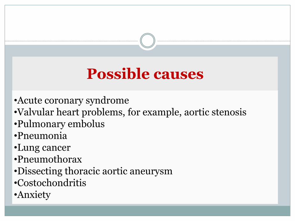
Possible causes
•Acute coronary syndrome•Valvular heart problems, for example, aortic stenosis•Pulmonary embolus•Pneumonia•Lung cancer•Pneumothorax•Dissecting thoracic aortic aneurysm•Costochondritis•Anxiety
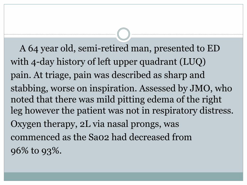
A 64 year old, semi-retired man, presented to ED
with 4-day history of left upper quadrant (LUQ)
pain. At triage, pain was described as sharp and
stabbing, worse on inspiration. Assessed by JMO, who noted that there was mild pitting edema of the right leg however the patient was not in respiratory distress.
Oxygen therapy, 2L via nasal prongs, was
commenced as the Sa02 had decreased from
96% to 93%.

FBC, EUC and coagulation levels were taken.
CXR report stated:
‘focal consolidation as well as mild volume loss (left lower lobe). Acute setting the appearance may be due to pneumonia‘
The JMO and Senior Registrar agreed that a CTPA was warranted, however, after discussion with the Radiology Registrar and then with the ED Staff Specialist, the decision was made to await the pathology results.
Results: elevated Neutrophils (11.2), WCC
(14.6) and CRP (214).

Following discussion amongst the ED team, the
decision was made not to request the CTPA but
to treat as pneumonia.( What is the error here?)
Plan included administration of intravenous antibiotics and monitoring of observations.
Patient was transferred into the HDU, oxygen therapy was removed and Sa02 was 94% on room air.
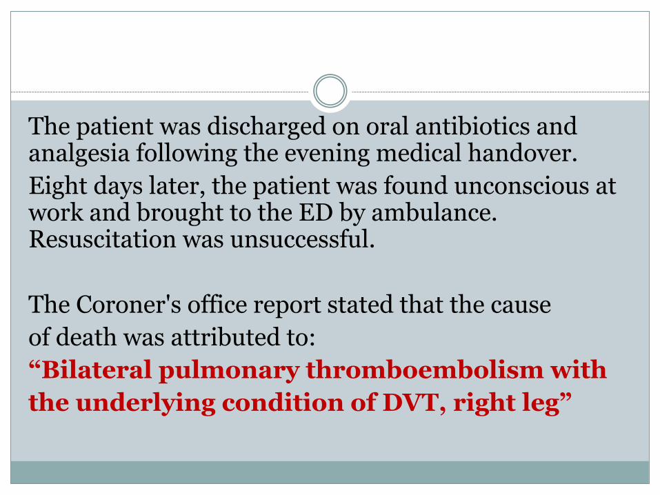
The patient was discharged on oral antibiotics and analgesia following the evening medical handover.
Eight days later, the patient was found unconscious at work and brought to the ED by ambulance. Resuscitation was unsuccessful.
The Coroner's office report stated that the cause
of death was attributed to:
“Bilateral pulmonary thromboembolism with
the underlying condition of DVT, right leg”

PE may present in an atypical manner, with concurrent lower respiratory tract infection.
If you have a high clinical suspicion of a PE, ensure it’s ruled out before committing to an
alternative diagnosis.
A 28 year old fit and well female presented to ED
in the early morning following onset of pain under left lower breast.
Pain reported to be reproducible on movement and with inspiration.
Triage nursing staff recorded patient being on nil
medications. It was later discovered that the
patient was on the OCP.

Vitals:
HR 76/min
BP 95mmHg systolic
RR 18
SaO2 98% RA.
Given paracetamol and ibuprofen, and triage
category 4.

On review, patient’s pain had resolved 30mins after initial
presentation.
ECG performed, which showed sinus tachycardia at HR 110.
Patient seen by ED Registrar in Fast Track area, unmonitored.
Noted that the patient “awoke with sharp left chest pain. Unable to
breathe easily. Never happened before. Denies abnormal physical
activity. No cardiac history. No PE risks.
Pain coming from underneath the breast
radiating to the left side. Much improved since
ibuprofen and paracetamol”.
Vital signs repeated, and were normal.
Diagnosis of musculoskeletal chest pain given and patient discharged home.
Two weeks later, patient presented to ED in
cardiac arrest after losing consciousness whilst
her father drove her to hospital. Following nearly an hour of CPR, patient had return of spontaneous circulation and was transferred to ICU.

CTPA performed, which showed
massive pulmonary embolus with associated hemorrhage and infarction.

If a patient has chest pain, a possible life-threatening differential diagnosis could be a
PE.3
Consider their risk factors and apply clinical decision rules to exclude a
PE, before attributing symptoms to a musculoskeletal aetiology
Abdominal pain

Red flag symptoms
• Sudden onset abdominal pain
• Haematemesis
• Unexplained weight loss
• Change in bowel habit for > 3 weeks
• Unexplained PV bleeding
• Post-coital bleeding
• Shortness of breath
• Dysphagia
• Increased vaginal discharge
Bloodstained vaginal discharge
Pre-syncopal symptoms
Haematuria
Fever
New onset dyspepsia
Persistent unexplained vomiting
Amenorrhoea
Testicular pain
Elderly
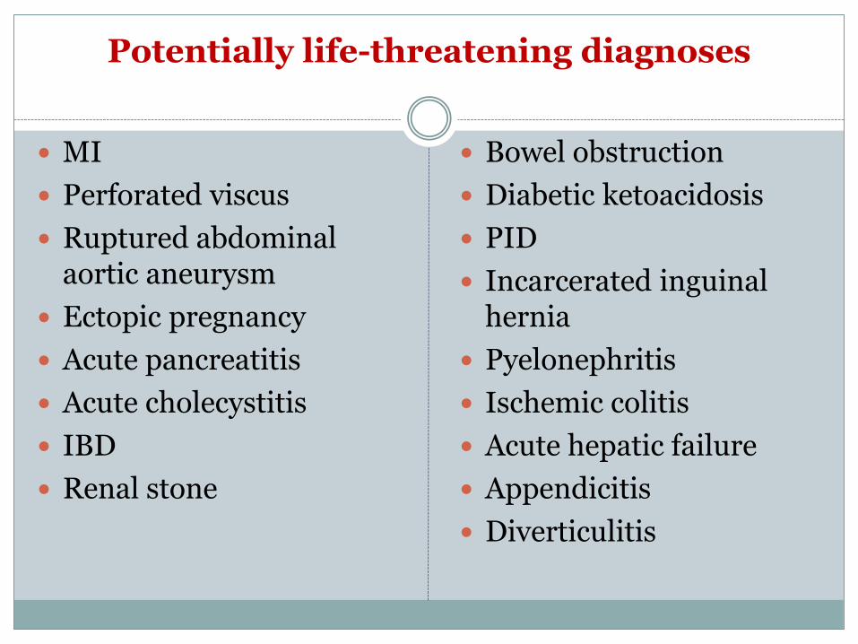
Potentially life-threatening diagnoses
MI
Perforated viscus
Ruptured abdominal aortic aneurysm
Ectopic pregnancy
Acute pancreatitis
Acute cholecystitis
IBD
Renal stone
Bowel obstruction
Diabetic ketoacidosis
PID
Incarcerated inguinal hernia
Pyelonephritis
Ischemic colitis
Acute hepatic failure
Appendicitis
Diverticulitis

HISTORY REPEATs ITSELF?
A 50yo female presented to ED with a three-day history of
abdominal pain and vomiting. Background included
previous appendectomy, similar episodes of self-limiting
abdominal pain for which a cause remained undetermined.
Reported pain usually occurred after eating food from
takeaway outlets.
Patient presented clutching her abdomen. Triage nurse noted
abdominal tenderness / bloating, but soft. Vital signs were
as follows: • BP 165/105mmHg • HR 124/min • RR 36/min

What is going on?
Patient given IV fluids for rehydration and morphine for pain relief. These had good effect, and vital signs improved to be within acceptable parameters.
X-Rays arranged – inconclusive.
Seen by medical officer. Pain described as similar to previous presentations.
Provisional diagnosis: Gastroenteritis/Colitis in keeping with patient’s past history.
Management plan: monitor fluid balance, CT abdomen,
nasogastric tube (NGT) insertion.
Do you agree with the patient’s
provisional diagnosis and
management plan?

Patient declined NGT insertion and her
wishes were respected as there was no active
vomiting. CT delayed and ultimately
postponed until the following day. Patient
remained in the ED overnight whilst waiting
for CT to be performed, during which time
observation frequency is changed from
hourly to fourth hourly.

The next morning, CT abdomen demonstrated
significantly distended fluid filled bowel loops
with collapsed loops of distal and terminal ileum.
On return to ED, patient noted to be diaphoretic
and pale. She vomited and progressed to
cardiorespiratory arrest. CPR commenced with
eventual return of spontaneous circulation.
Following multiple operations and prolonged
period on life support, patient died.
Cause of death: aspiration pneumonitis
secondary to small bowel
obstruction/necrosis caused by stricture.

What is the lesson here?
Any patient who re-presents from any
site of medical care (not just ED) for the
same problem should not be dismissed.
Ask the patient: “Have you seen a doctor or
been to an ED for this problem before?”
If so, and over a short space of time, this is
a RED FLAG.
In their systematic review, LaCalle et al. concluded that:
frequent ED users tend to be sicker than occasional users, and are often sick patients with chronic illness associated with high admission rates and high mortality.
This evidence suggests that patients who present to ED on a frequent basis are a medically vulnerable group.
GUT FEELING
A 76yo male was brought into a local district hospital ED by ambulance at midnight with abdominal pain.
The paramedics report the observations were found to be in normal range except for BP 170/80 and pain score 7/10.
He complained of constipation and abdominal pain for 4 days, described as sharp in nature, but had increased significantly overnight prompting his relatives to call an ambulance.
He was given morphine for analgesia.
Observations were unchanged from time of ambulance assessment.

Within 30mins, patient was reviewed by a medical officer and given a provisional diagnosis of constipation.
Patient was given analgesia and a fleet enema, which resulted in a small bowel motion.
Nil further analgesia was given as ambulance morphine had successfully eased the pain
At 0200, there was discussion between medical staff and the patient and carer regarding patient’s disposition. A plan was to discharge the patient home and have them return later in the morning for further investigation.
At 0230, patient was discharged home into the care of his family.

Patient returned to attend a CT later in the day.
Whilst in the radiology department, patient collapsed at 1130. On arrival of the Rapid Response Team, patient found to have GCS 3.
Patient was given fluid bolus with improvement of GCS to 14 by the time the patient was transferred to ED.
On examination, patient noted to be pale, cold and clammy with a pulsatile abdominal mass palpable in the patient’s epigastricregion.
Patient received further fluid resuscitation and transfusion of four units of blood.
At 1330, patient was transferred to a tertiary facility for consideration of urgent definite management.
During transit in the ambulance, patient suffered a
cardiorespiratory arrest.
With respect to patient and family’s previously discussed
wishes, CPR was not commenced and patient returned to
referring hospital for certification.
Cause of death found to be due to: ruptured AAA.
What is the lesson here?
Elderly abdominal pain patients are more likely to present with vague and nonspecific symptoms while harboring serious disease processes.4
For elderly patients with abdominal pain, it is NOT constipation or gastroenteritis until other serious diagnoses have been actively sought and excluded.4
Any patient over 50 years of age with suspected renal colic should have the diagnosis of ruptured AAA explicitly sought and excluded.

THROWN A CURVEBALL
A 17yo male presented to ED with 30-minute history of sudden onset right iliac fossa pain and associated vomiting.
At triage, observations:
T35C
HR 94/min, regular
BP 128/81
SaO2 97% RA
Abdomen soft with mild guarding.
Given triage category 4.
30mins after presentation, patient did not answer a call from nursing staff.
Officially documented as “Did not wait for treatment” two hours after presentation.

12 hours after initial presentation, patient re-presented with
vomiting, severe right iliac fossa and right testicle pain.
Given triage category 2.
Reviewed by medical officer, ultrasound arranged and Urology
Registrar informed of patient’s arrival.
An ultrasound was completed, demonstrating poor right testicular
blood flow.
Proceeded to OT for scrotal exploration +/- orchidectomy.
In theatre, patient found to have necrotic R testicle with 720 degree
torsion.
Right orchidectomy was performed.
What is the lesson here?
Always examine the scrotum for
testicular torsion in the young
male with abdominal, groin or
penile pain.
Patients with testicular torsion do not always present with
the pathognomic history of “acute excruciating
scrotal pain of relatively short duration”.
Testicular torsion should always be included in differential diagnosis when evaluating lower abdominal pain in young males.
The external genital organs should be examined in every child or adolescent with acute abdominal pain.
Presenting with only abdominal or inguinal pain
is not an uncommon presentation for testicular
torsion, with abdominal pain often preceding and
exceeding scrotal pain. Cass et al. reported that
12.5% of patients with testicular torsion
presented with only abdominal or inguinal pain,
while a 25-year review of testicular torsion cases
in Bristol found that 6% of cases presented with
inguinal pain alone.

Conclusions
Using the clinical warning criteria (red flags) is a must in all medical areas.
In your daily practice the single most important task is to exclude critical conditions.
It is important to trace the Pitfalls in your practice
Safety of your patients much more important than the accuracy of the diagnosis
REFERENCES
1. Amendo MT, Brown BA, Kossow LB, Weinberg FM. Headache as the sole
presentation of acute myocardial infarction in two elderly patients. Am J
Geriatr Cardiol. 2001;10:100–1
2. Auer J, Berent R, Lassnig E, Eber B. Headache as a manifestation of fatal myocardial infarction. Neurol Sci. 2001;22:396–7.
3. Mårten Söderberg; Ulla Hedström; Malgorzata Sjunnesson; Gerd Lärfars; Christina Jorup-Rönström. Initial symptoms in pulmonary embolism differ from those in pneumonia: a retrospective study during seven years. European Journal of Emergency Medicine. 13(4):225-229, AUG 2006.
4. [Guideline] LeFevre ML, U.S. Preventive Services Task Force. Screening for abdominal aortic aneurysm: U.S. Preventive Services Task Force recommendation statement. Ann Intern Med. 2014 Aug 19. 161 (4):281-90.

Thank you

PRETEST & POSTTEST
1. A 40 year old gentleman, diabetic (uncontrolled), who presented with a right cheek pain progressively over a 15-day period. Patient also reports associated headache, blurring of vision in the right eye. Patient had no fever, chills, or nasal discharge. On physical exam he had tenderness over right cheek & blocked right side of the nose His blood sugar was 465 mg/dl upon presentation. CT scan of sinuses and neck was done and showed an aggressive soft tissue lesion in the right maxillary sinus with erosion of the anterior and lateral wall of the maxillary sinus, and erosion of the orbital floor on the right side with invasion of the inferior aspect of the right orbit.
Q what is the Red flag in this case?

A 45 y old female patient with known migraine on treatment presented in ED by acute attack of headache not improved with analgesic what is appropriate next step
1-Discharge the patient with prescribed analgesia.
2-Give her a tramadol 100mg IM to abort the pain then discharge.
3- Admit her under observation and request ECG and cardiac enzymes.