Embed Size (px)
Citation preview

Musculoskeletal
Pediatric Hip Lesions

Mohamed Zaitoun
Assistant Lecturer-Diagnostic Radiology Department , Zagazig University Hospitals
EgyptFINR (Fellowship of Interventional
Neuroradiology)[email protected]


Knowing as much as possible about your enemy precedes successful battle
and learning about the disease process precedes successful management
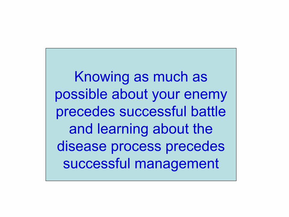
Pediatric Hip Lesions-Three distinct conditions of the hip occur in
children, each of which affects a different age group :
1-Neonates, infants : congenital dislocation of the hip (CDH)
2-School age : Legg-Calve-Perthes (LCP) disease
3-Adolescents : Slipped capital femoral epiphysis (SCFE)
1-Congenital Dislocation of the Hip (CDH) ,Developmental Dysplasia of the Hip (DDH) :
a) Incidence
b) Radiographic features
a) Incidence :
-Results from an abnormal relationship of the femoral head to the acetabulum
-More in females
-More in the left hip, bilateral in 5 %
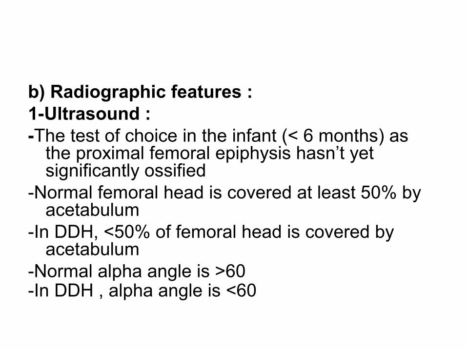
b) Radiographic features :1-Ultrasound :-The test of choice in the infant (< 6 months) as
the proximal femoral epiphysis hasn’t yet significantly ossified
-Normal femoral head is covered at least 50% by acetabulum
-In DDH, <50% of femoral head is covered by acetabulum
-Normal alpha angle is >60-In DDH , alpha angle is <60
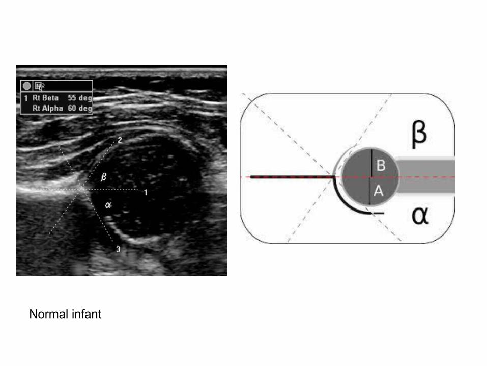
Normal infant
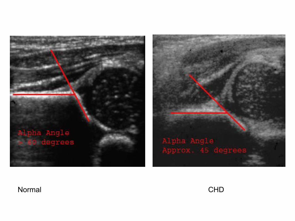
Normal CHD
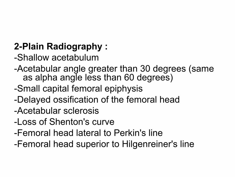
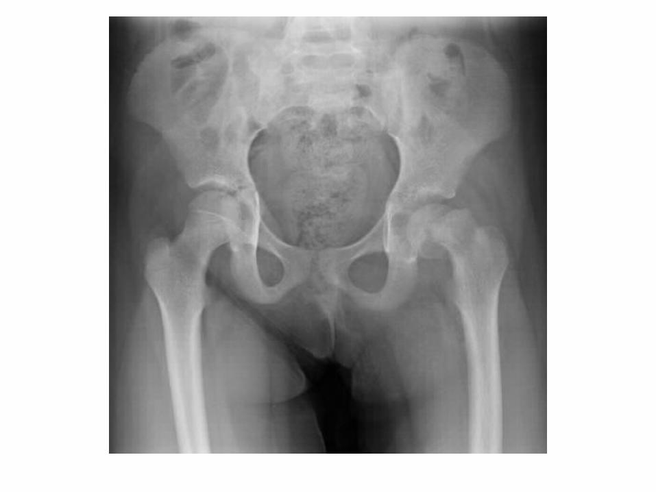
2-Plain Radiography :-Shallow acetabulum-Acetabular angle greater than 30 degrees (same
as alpha angle less than 60 degrees)-Small capital femoral epiphysis-Delayed ossification of the femoral head-Acetabular sclerosis-Loss of Shenton's curve-Femoral head lateral to Perkin's line-Femoral head superior to Hilgenreiner's line
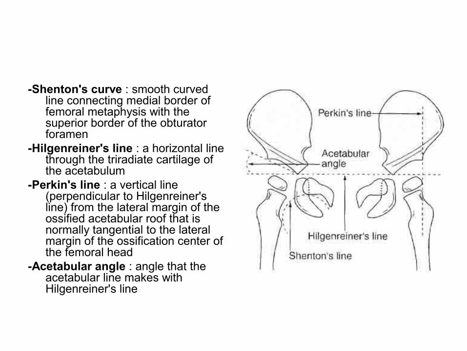
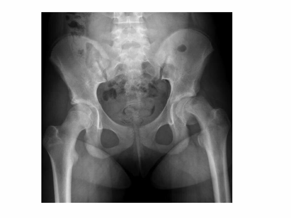
-Shenton's curve : smooth curved line connecting medial border of femoral metaphysis with the superior border of the obturator foramen
-Hilgenreiner's line : a horizontal line through the triradiate cartilage of the acetabulum
-Perkin's line : a vertical line (perpendicular to Hilgenreiner's line) from the lateral margin of the ossified acetabular roof that is normally tangential to the lateral margin of the ossification center of the femoral head
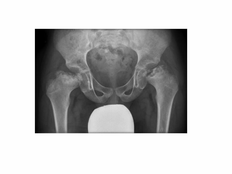
-Acetabular angle : angle that the acetabular line makes with Hilgenreiner's line
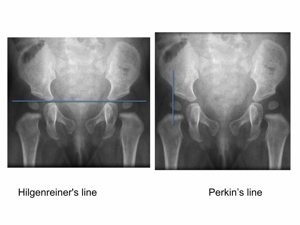
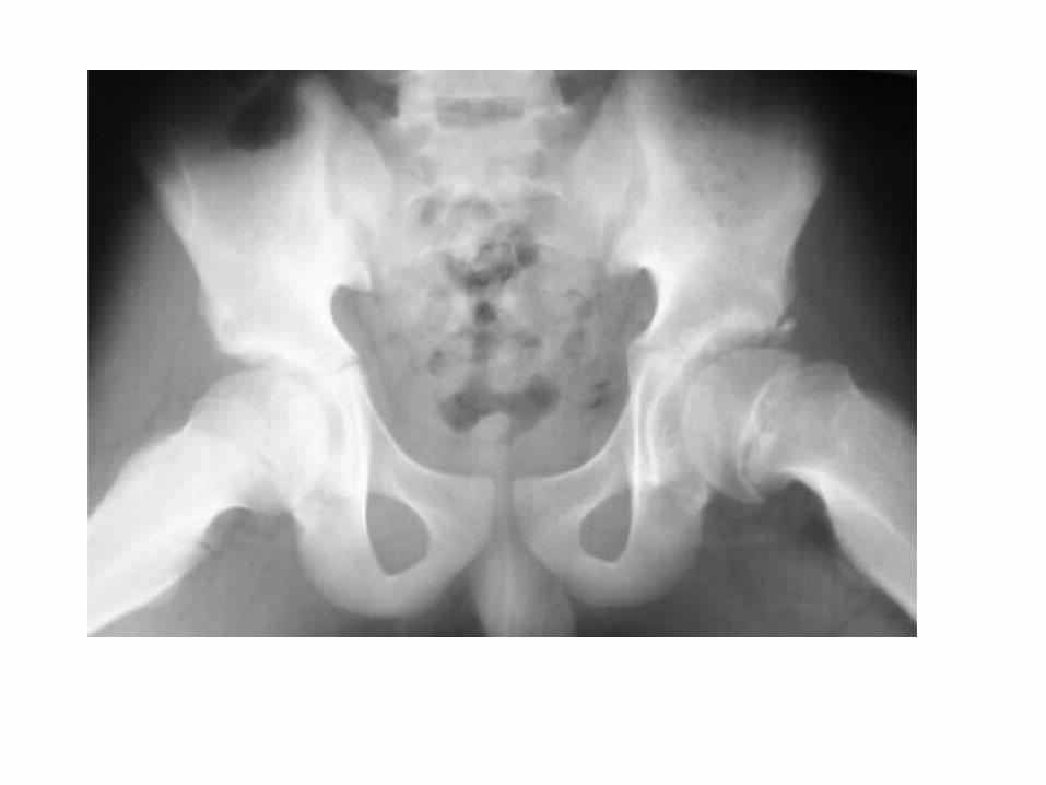
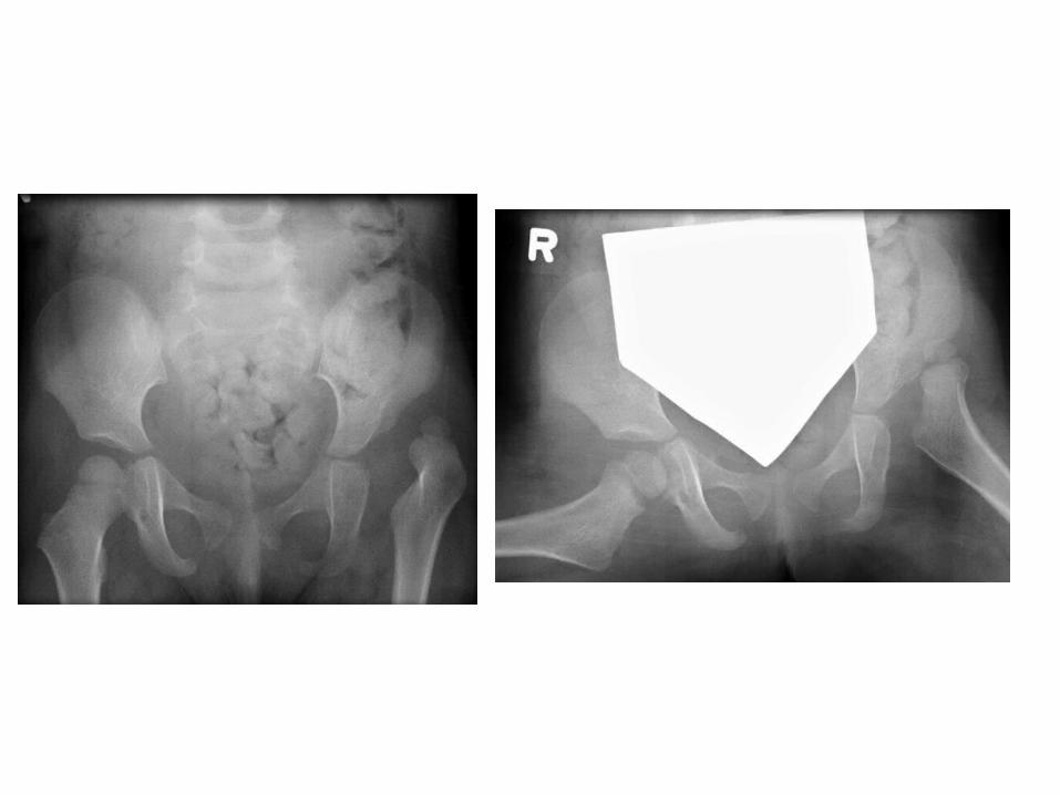
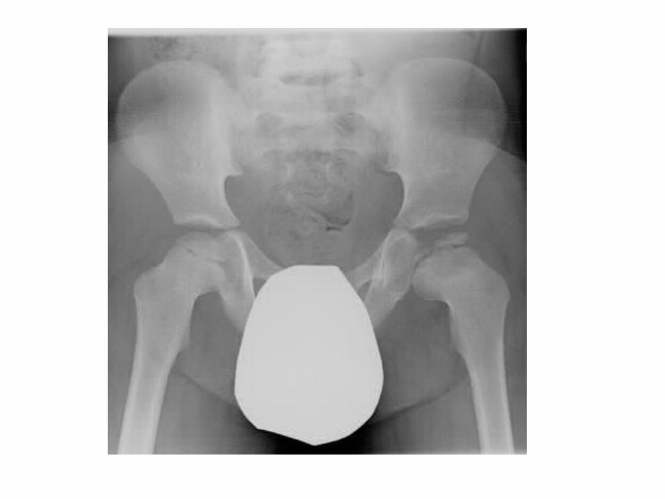
Hilgenreiner's line Perkin’s line
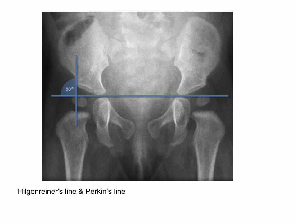
Hilgenreiner's line & Perkin’s line
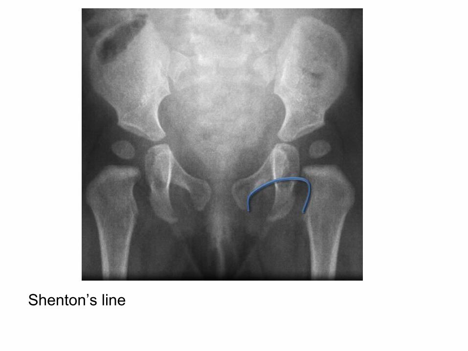
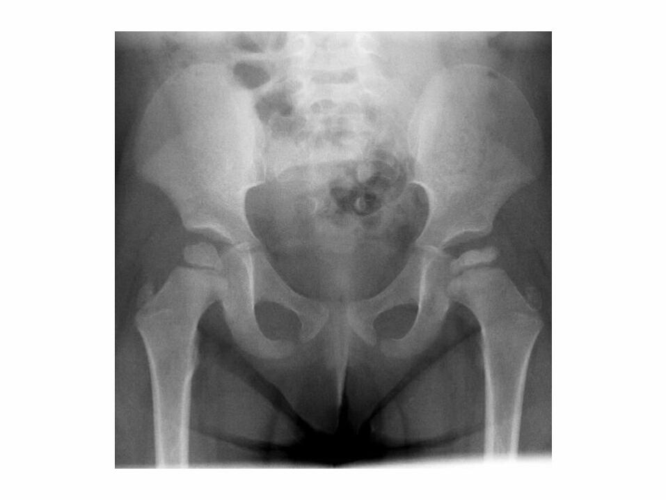
Shenton’s line
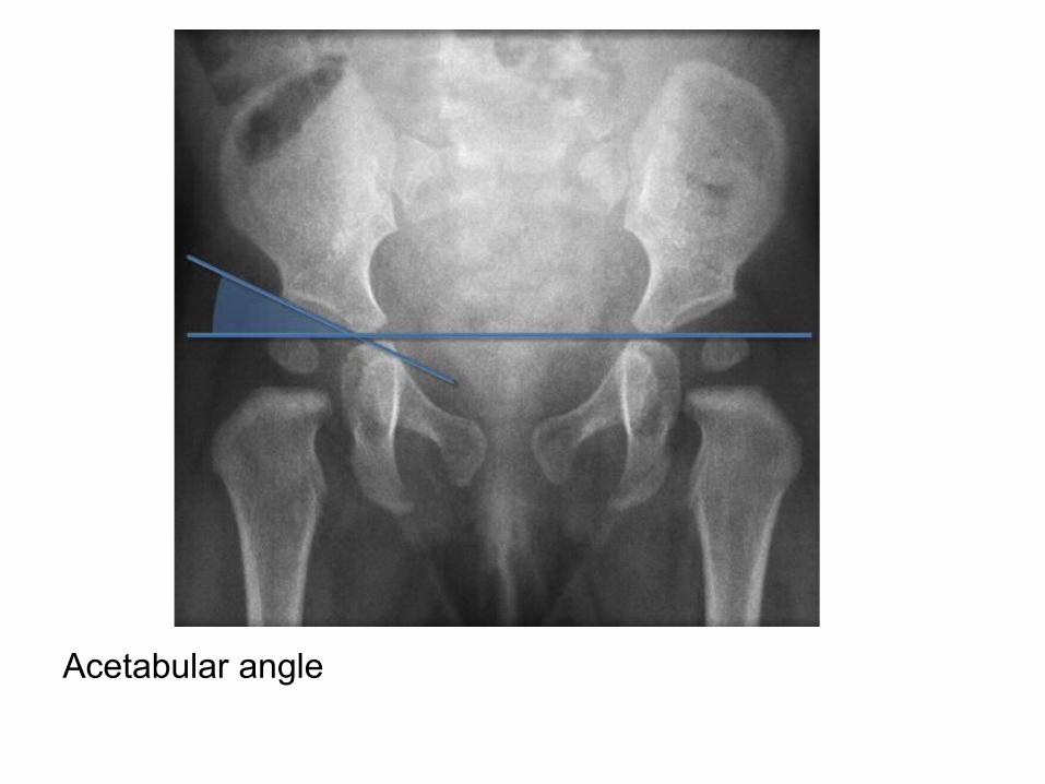
Acetabular angle
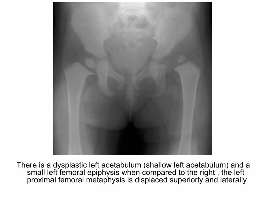
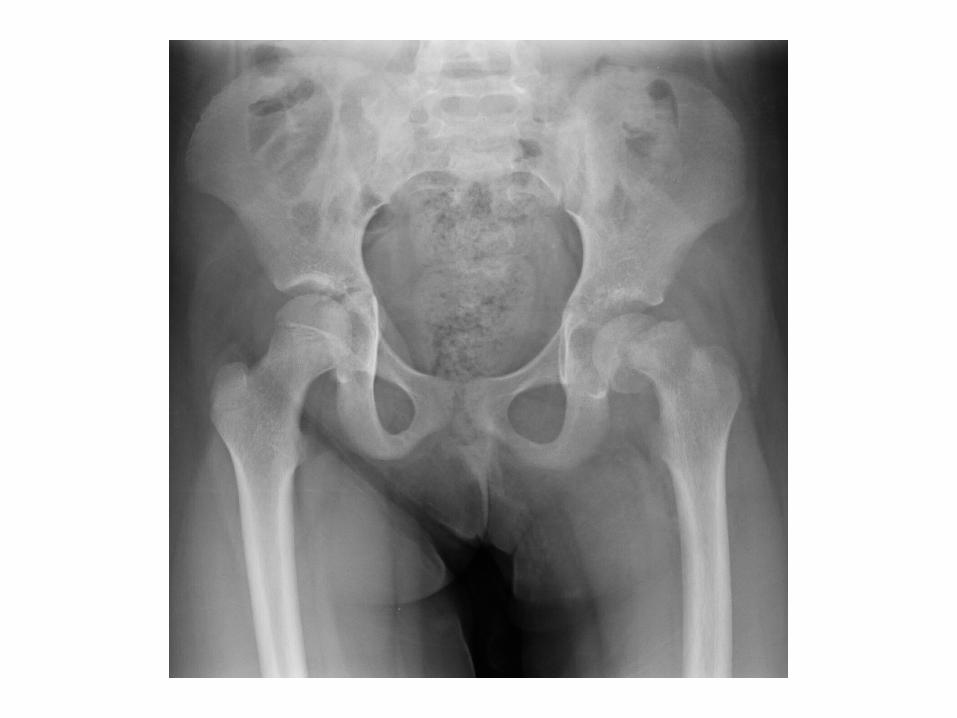
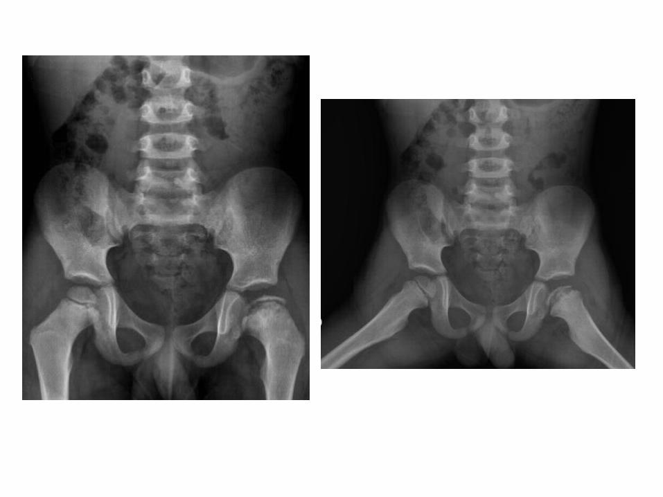
There is a dysplastic left acetabulum (shallow left acetabulum) and a small left femoral epiphysis when compared to the right , the left proximal femoral metaphysis is displaced superiorly and laterally

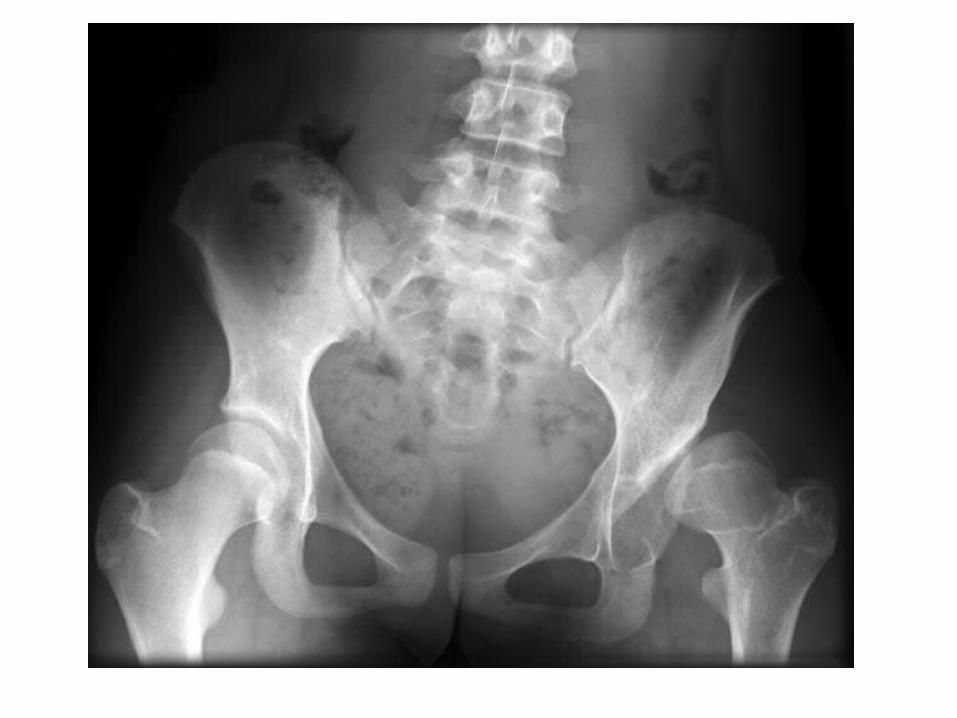
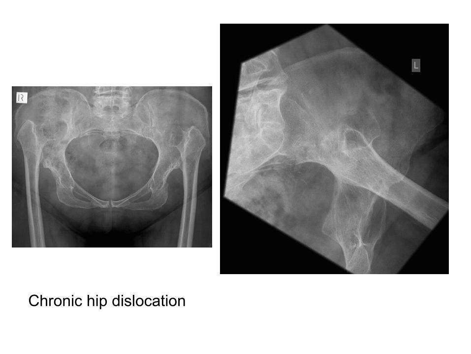
Chronic hip dislocation
2-Legg-Calve-Perthes (LCP) Disease :
a) Incidence
b) Radiographic Features
a) Incidence :
-Osteonecrosis of femoral head
-School age (5-8 years)

a) Incidence :
-Osteonecrosis of femoral head
-School age (5-8 years)
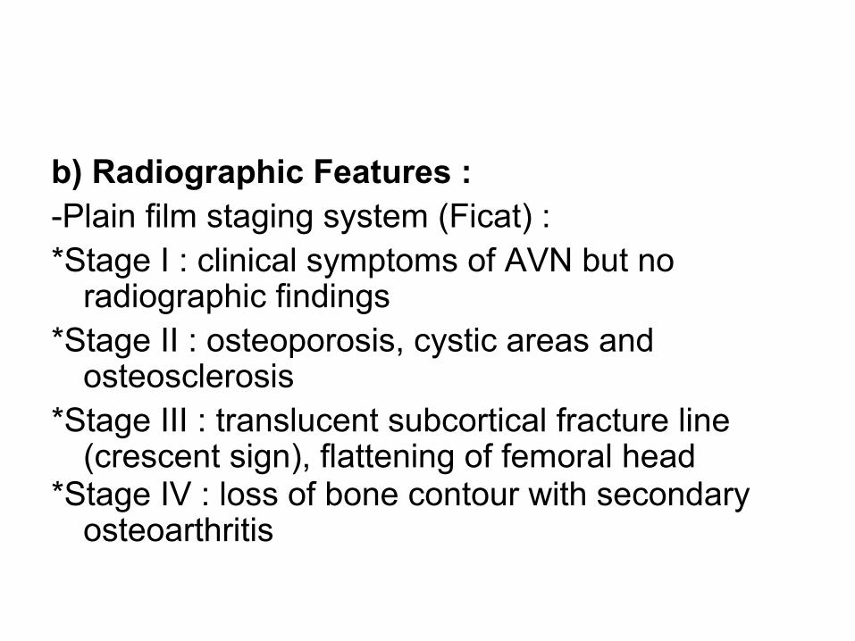
b) Radiographic Features :-Plain film staging system (Ficat) :*Stage I : clinical symptoms of AVN but no
radiographic findings*Stage II : osteoporosis, cystic areas and
osteosclerosis*Stage III : translucent subcortical fracture line
(crescent sign), flattening of femoral head*Stage IV : loss of bone contour with secondary
osteoarthritis
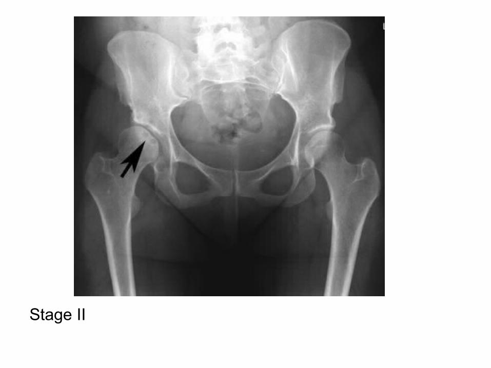
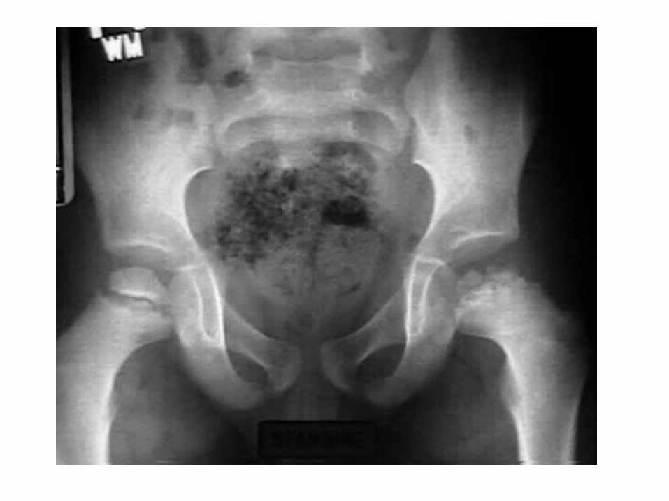
Stage II
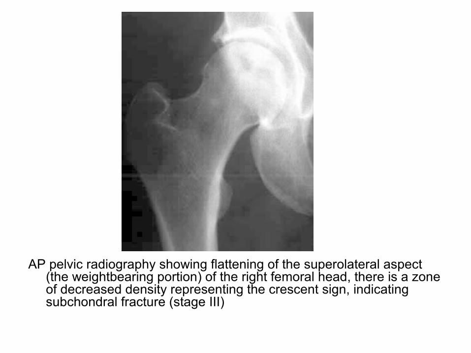
AP pelvic radiography showing flattening of the superolateral aspect (the weightbearing portion) of the right femoral head, there is a zone of decreased density representing the crescent sign, indicating subchondral fracture (stage III)
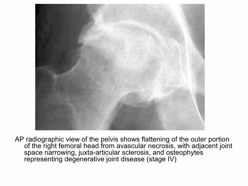
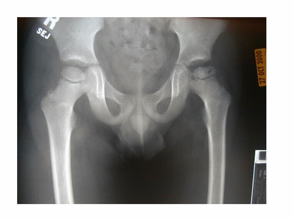
AP radiographic view of the pelvis shows flattening of the outer portion of the right femoral head from avascular necrosis, with adjacent joint space narrowing, juxta-articular sclerosis, and osteophytes representing degenerative joint disease (stage IV)
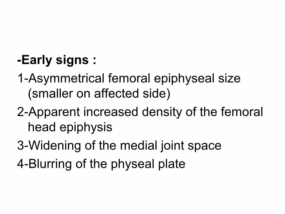
-Early signs :
1-Asymmetrical femoral epiphyseal size (smaller on affected side)
2-Apparent increased density of the femoral head epiphysis
3-Widening of the medial joint space
4-Blurring of the physeal plate
-Late signs :1-The femoral head begins to fragment with
subchondral lucency (crescent sign) 2-Femoral head deformity with widening and
flattening3-Osteoarthritis
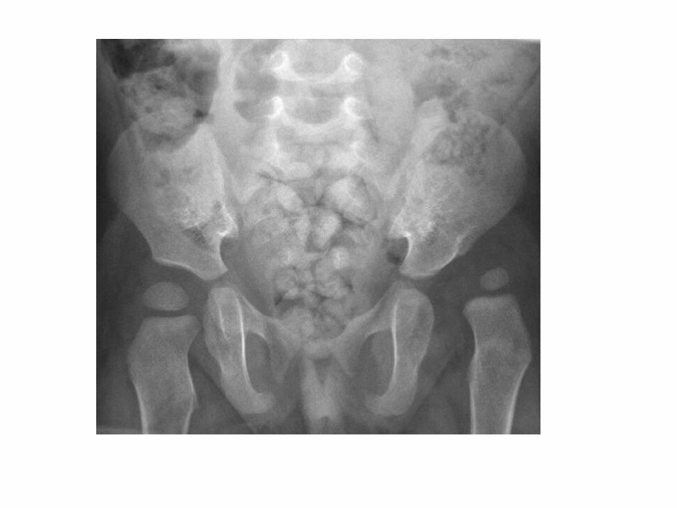
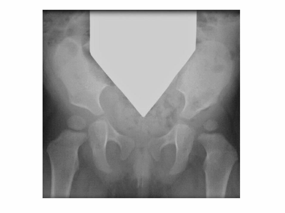
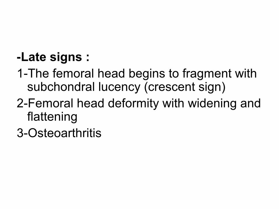
Bilateral Perthes
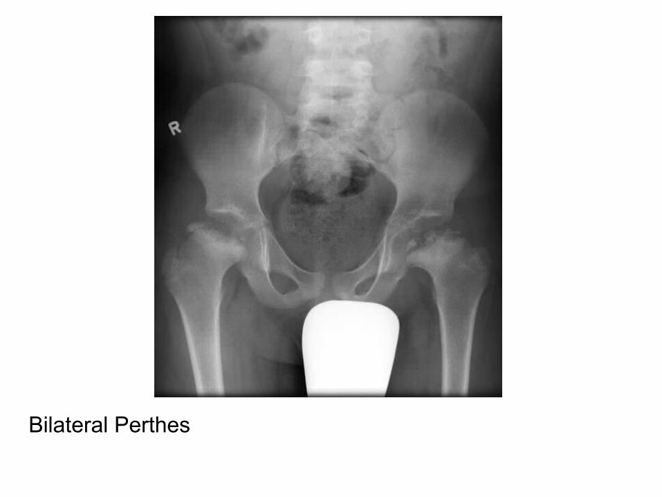
Bilateral

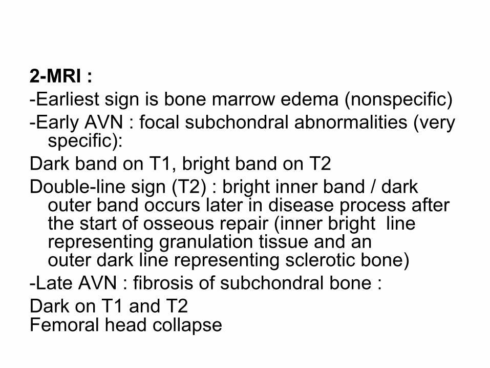
2-MRI :-Earliest sign is bone marrow edema (nonspecific)-Early AVN : focal subchondral abnormalities (very
specific):Dark band on T1, bright band on T2Double-line sign (T2) : bright inner band / dark
outer band occurs later in disease process after the start of osseous repair (inner bright line representing granulation tissue and an outer dark line representing sclerotic bone)
-Late AVN : fibrosis of subchondral bone :Dark on T1 and T2Femoral head collapse
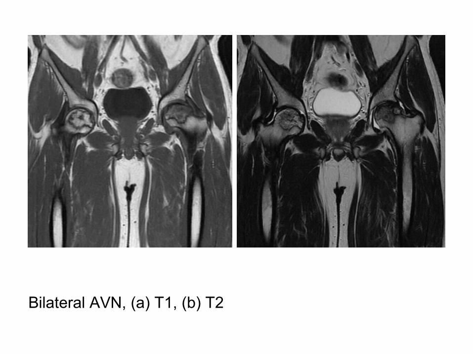
Bilateral AVN, (a) T1, (b) T2
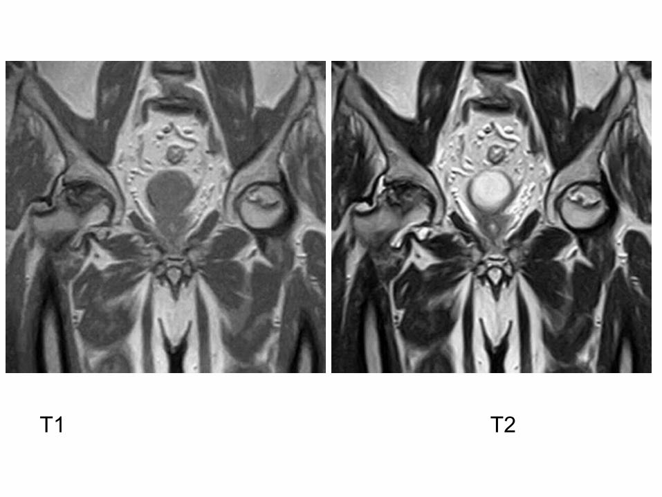
T1 T2
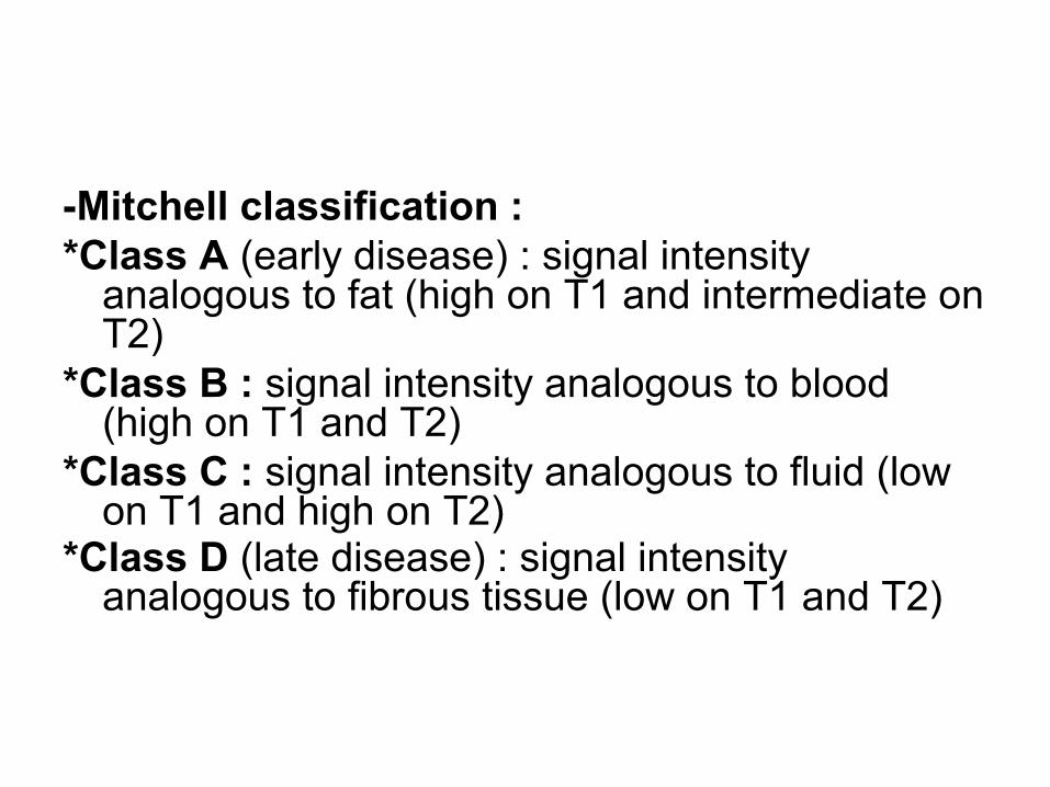
-Mitchell classification :*Class A (early disease) : signal intensity
analogous to fat (high on T1 and intermediate on T2)
*Class B : signal intensity analogous to blood (high on T1 and T2)
*Class C : signal intensity analogous to fluid (low on T1 and high on T2)
*Class D (late disease) : signal intensity analogous to fibrous tissue (low on T1 and T2)
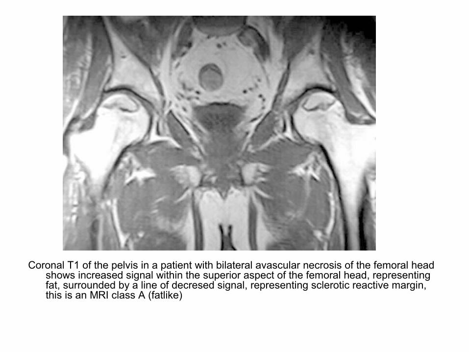
Coronal T1 of the pelvis in a patient with bilateral avascular necrosis of the femoral head shows increased signal within the superior aspect of the femoral head, representing fat, surrounded by a line of decresed signal, representing sclerotic reactive margin, this is an MRI class A (fatlike)
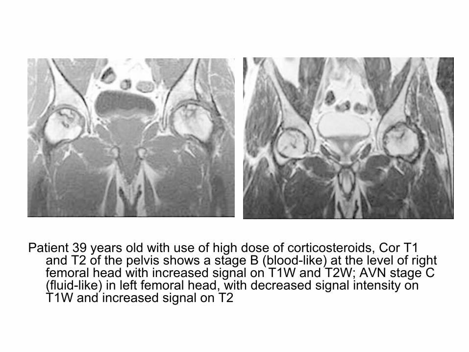
Patient 39 years old with use of high dose of corticosteroids, Cor T1 and T2 of the pelvis shows a stage B (blood-like) at the level of right femoral head with increased signal on T1W and T2W; AVN stage C (fluid-like) in left femoral head, with decreased signal intensity on T1W and increased signal on T2
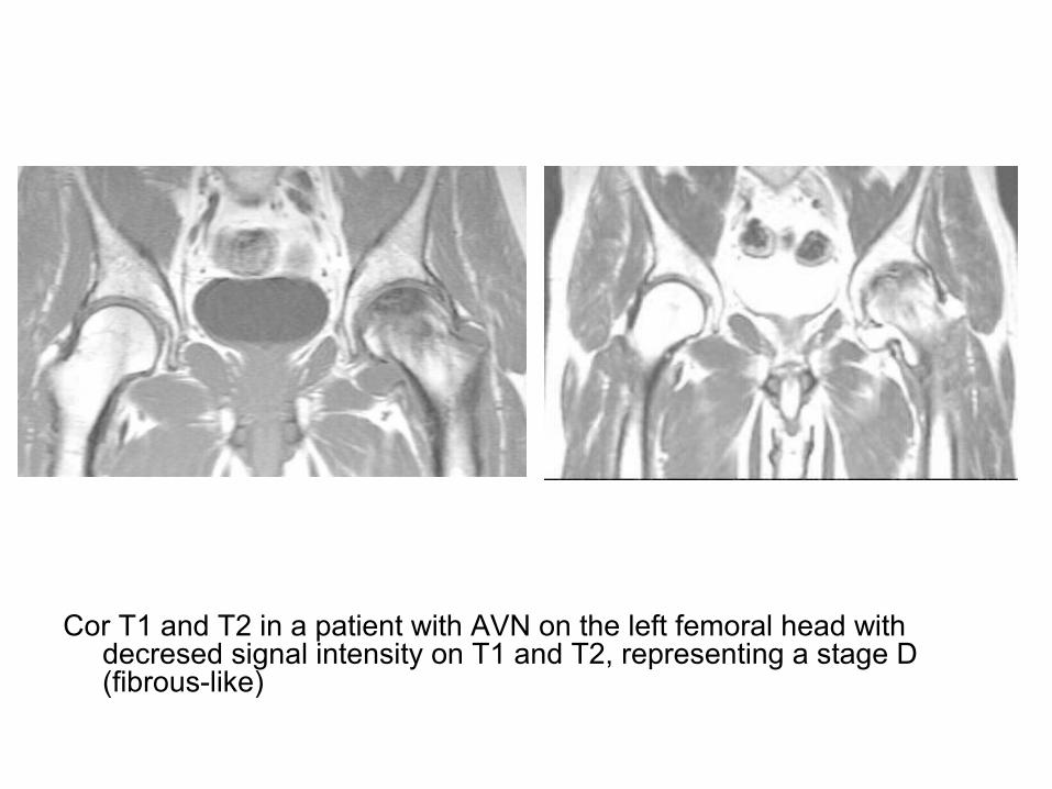
Cor T1 and T2 in a patient with AVN on the left femoral head with decresed signal intensity on T1 and T2, representing a stage D (fibrous-like)
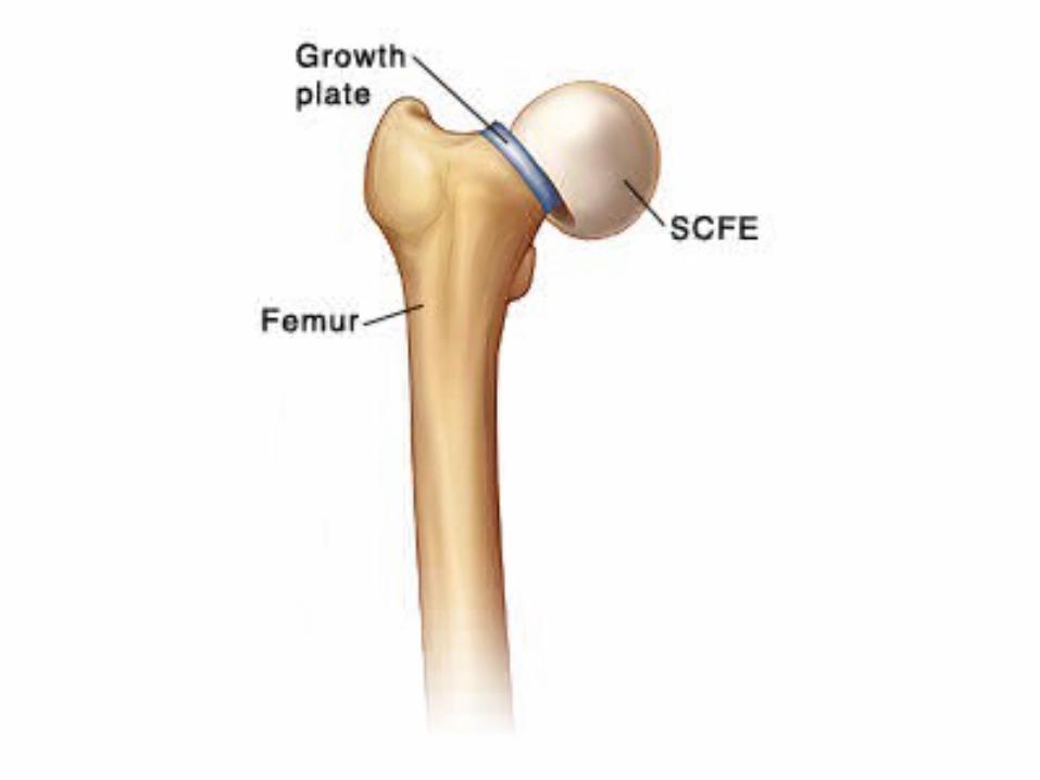
3-Slipped Capital Femoral Epiphysis (SCFE):
a) Incidence
b) Radiographic Features
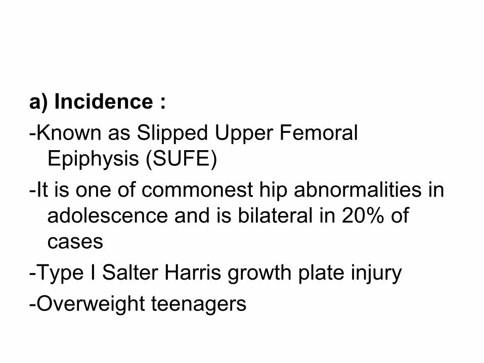
a) Incidence :
-Known as Slipped Upper Femoral Epiphysis (SUFE)
-It is one of commonest hip abnormalities in adolescence and is bilateral in 20% of cases
-Type I Salter Harris growth plate injury
-Overweight teenagers
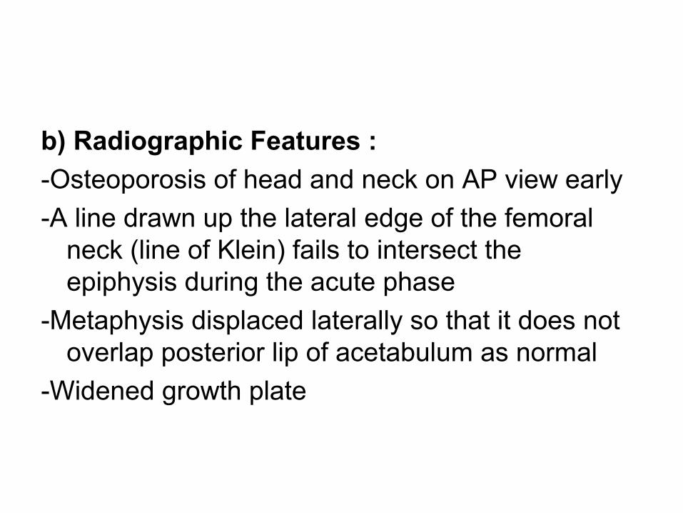
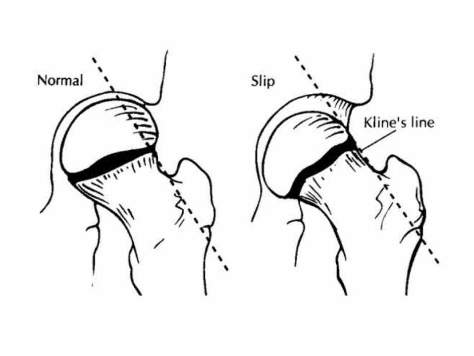
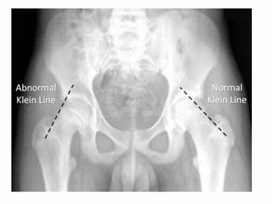
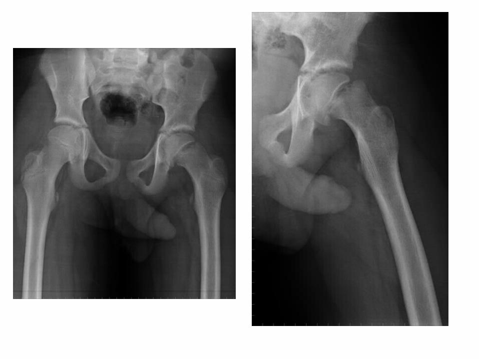
b) Radiographic Features :
-Osteoporosis of head and neck on AP view early
-A line drawn up the lateral edge of the femoral neck (line of Klein) fails to intersect the epiphysis during the acute phase
-Metaphysis displaced laterally so that it does not overlap posterior lip of acetabulum as normal
-Widened growth plate