Embed Size (px)
Citation preview

Development of Ear
20-04-2015
Dr. Laxman Khanal
Assistant Professor
Department of Human Anatomy

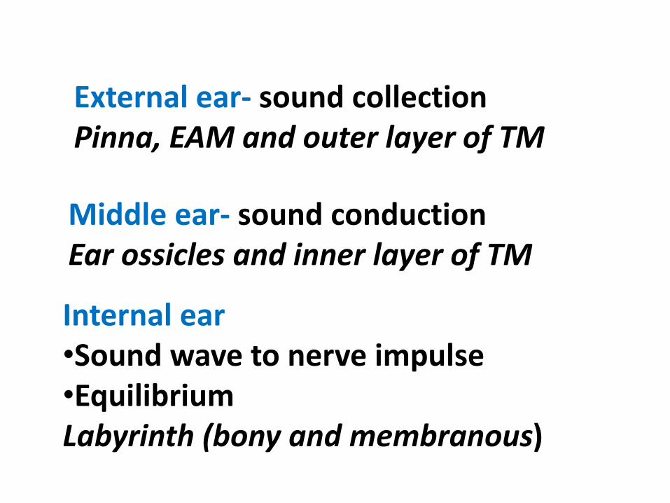
External ear- sound collectionPinna, EAM and outer layer of TM
Middle ear- sound conductionEar ossicles and inner layer of TM
Internal ear•Sound wave to nerve impulse•Equilibrium Labyrinth (bony and membranous)

Q. Vestibulocochlear ganglia is derived froma.Otic vesicleb.Neural crest cellsc. Both a and bd.Neuroectoderm of hindbrain

Development of Internal Ear
Otic Placode @22nd dayOn each side of hindbrain

Invaginating placode Otic pit Otic vesicle
Forms membranous labyrinth
Q. Ductus reunion connecta.Utricluar to saccular portionb.Utricle to Semicircular ductc. Cochlear duct to sacculed.Membranous labyrinth to bony labyrinth

Ventral partSaccules Cochlear ductDorsal partSemicircular ductUtricleEndolymphatic duct
Utriculo-saccular ductCochlear ductDuctus reunions

Q. Which of the following parts of SCD fused during opening to utricle ?a. Dilated part of ant and post SCDb. Non-dilated part of ant and post SCDc. Dilated part of post and lat SCDd. Non-dilated part of post and lat SCD

Development of Utricle and SCD(Dorsal Part of Otic vesicle)
SCDs appear as flat out pocketing of Utricular portionof Otic vesicle.•Three pairs of SCD are formed –Ant/Post/LatOne end of SCD form dilatation (Crus Ampullare) andthe other does not widen (Crus Nonampulare) Because two crus nonampullare fuse, there will be3 crus ampullare and 2 crus nonampullare


Formation of Cochlea
• Mesenchyme surrounding CD Differentiate into cartilage.
• Cartilage undergo vacuolization and form Scala vestibuli and scala tympani.
• Lateral part of CD attached to the cartilage by Spiral ligament and medial edge is attached and supported by modiolus.

Q. Epithelium of which of the following part of CD forms the Organ of Corti?a. Outer ridgeb. Inner ridgec. Endolymphd. All of the above

Epithelium of CD form two ridge.• inner ridge•Outer ridge
Outer ridge forms innerand outer rows of haircells covered bytectorial membrane -Organ of Corti

Q. Middle ear cavity develops from.a.First pharyngeal pouchb.Second pharyngeal pouchc. First pharyngeal cleftd.Second pharyngeal cleft
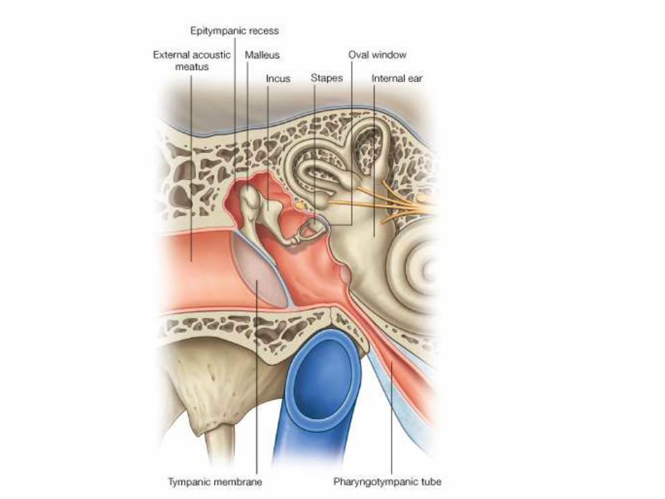

Development of Middle Ear
Q. What is the motor nerve to stapedius muscle?a.Mandibular nerveb.Facial nervec. Glossopharyngeal nerved.Vagus nerve

First pharyngeal pouchDistal part- tympanic cavityProximal part- auditory tube
Malleus /Incus and Tensor Tympani- 1st ArchStapes and Stapedius muscle- 2nd arch

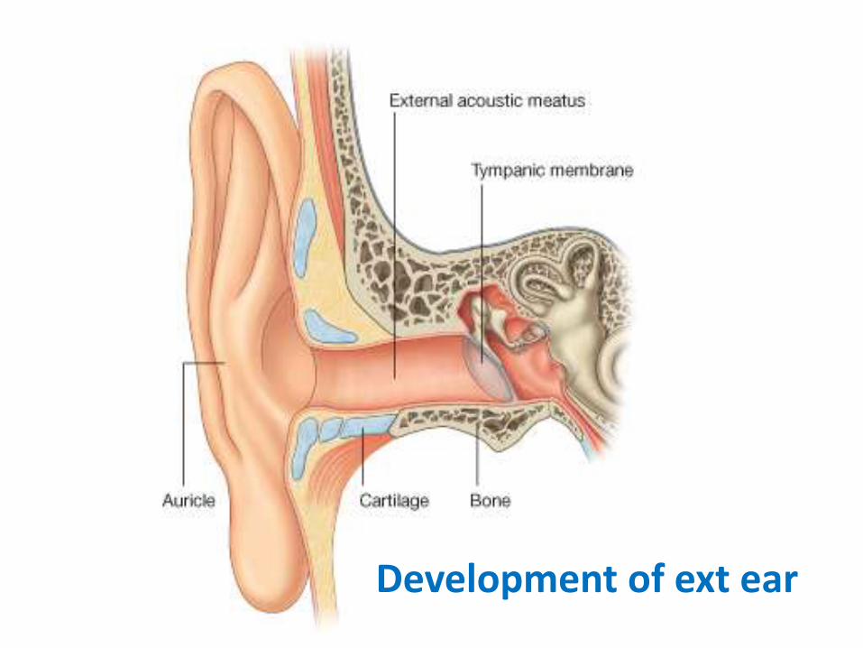
Development of ext ear

•Six auricular Hillocks forms the Pinna.•First pharyngeal cleft forms the EAM

•Six auricular Hillocks forms the Pinna.•First pharyngeal cleft forms the EAM

External ear malformation
Preauricual pit
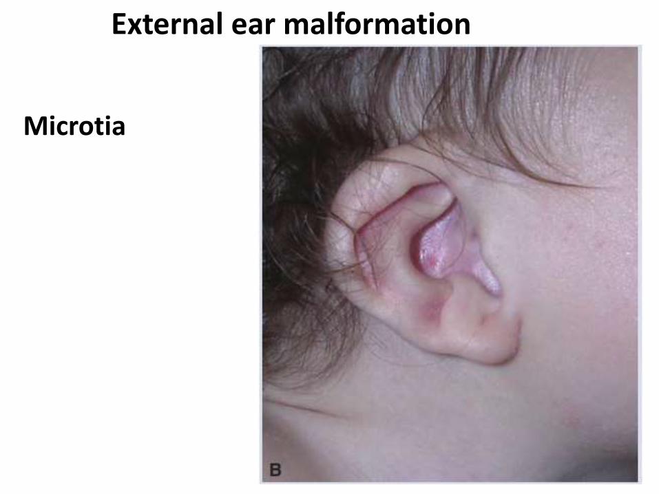
External ear malformation
Microtia

External ear malformation
Preauricualrappendages

External ear malformation
Anotia

Congenital deafness
• Genetic cause
Down’s syndrome
Crouzon syndrome
Treacher collin syndrome
• Retention of meatal plug
• Rubella infection
• Cytomegalo virus infection
• Malformation of middle and internal ear

Q. Vestibulocochlear ganglia is derived froma.Otic vesicleb.Neural crest cellsc. Both a and bd.Neuroectoderm of hindbrain

Q. Ductus reunion connecta.Utriclar to saccular portionb.Utricle to Semicircular ductc. Cochlear duct to sacculed.Membranous labyrinth to bony labyrinth

Q. Which of the following parts of SCD fused during opening to utricle ?a. Dilated part of ant and post SCDb. Non-dilated part of ant and post SCDc. Dilated part of post and lat SCDd. Non-dilated part of post and lat SCD

Q. Epithelium of which of the following part of CD forms the Organ of Corti?a. Outer ridgeb. Inner ridgec. Endolymphd. All of the above

Q. Middle ear cavity develops from.a.First pharyngeal pouchb.Second pharyngeal pouchc. First pharyngeal cleftd.Second pharyngeal cleft

Q. What is the motor nerve to stapedius muscle?a.Mandibular nerveb.Facial nervec. Glossopharyngeal nerved.Vagus nerve

Embryonic structure
Adult derivative
Otic vesicleSaccular portion
Utricular portion
Saccule, CD, Spiral ganglion
Utricle, SCD, vestibular ganglion and endolymphatic duct
Pharyngeal membrane 1
Tympanic membrane
Arch 1 Malleus, Incus, Tensor tympani
Arch 2 Stapes, Stapedius
Pouch 1 Middle ear cavity and auditory tube
Pharyngeal cleft 1 External acoustic meatus
6 auricular hillocks Pinna
Mid
dle
arEx
t e
arIn
tern
al e
ar