Embed Size (px)
DESCRIPTION
Dr. Mrs. Minnu Panditrao, goes in depth with the very important topic of Deep Vein Thrombosis, Pulmonary embolism, aetio patheogenesis, clinical features, management etc.
Citation preview
DEEP VEIN THROMBOSIS (DVT) &
PULMONARY EMBOLISM (PE)
Dr. (Mrs.)Minnu M. Panditrao
CONSULTANT
DEPARTMENT OF ANESTHESILOLOGY AND INTENSIVE CAREPUBLIC HOSPITAL AUTHORITY’S
RAND MEMORIAL HOSPITALFREEPORT
GRAND BAHAMACOMMONWEALTH OF THE BAHAMAS
Formerly:
Professor Department of Anaesthesiology
& Critical CareDr. D Y Patil Medical College
Pimpri, Pune
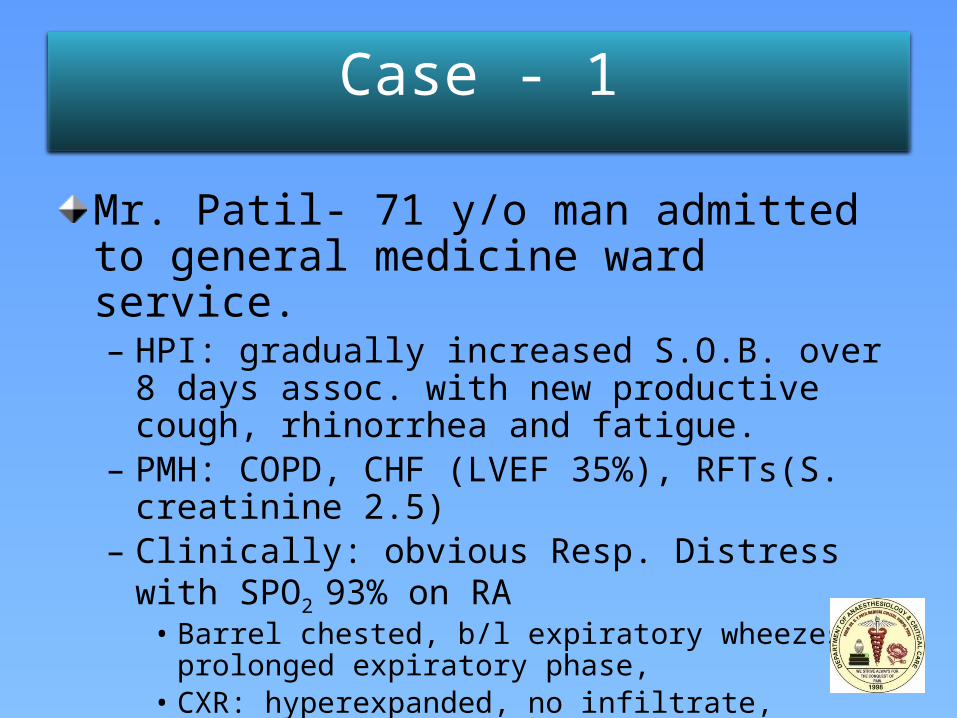
Case - 1
Mr. Patil- 71 y/o man admitted to general medicine ward service.– HPI: gradually increased S.O.B. over 8 days assoc. with
new productive cough, rhinorrhea and fatigue.– PMH: COPD, CHF (LVEF 35%), RFTs(S. creatinine 2.5)– Clinically: obvious Resp. Distress with SPO2 93% on RA
• Barrel chested, b/l expiratory wheezes, prolonged expiratory phase,
• CXR: hyperexpanded, no infiltrate, consolidation or edema. – DX: COPD Exacerbation
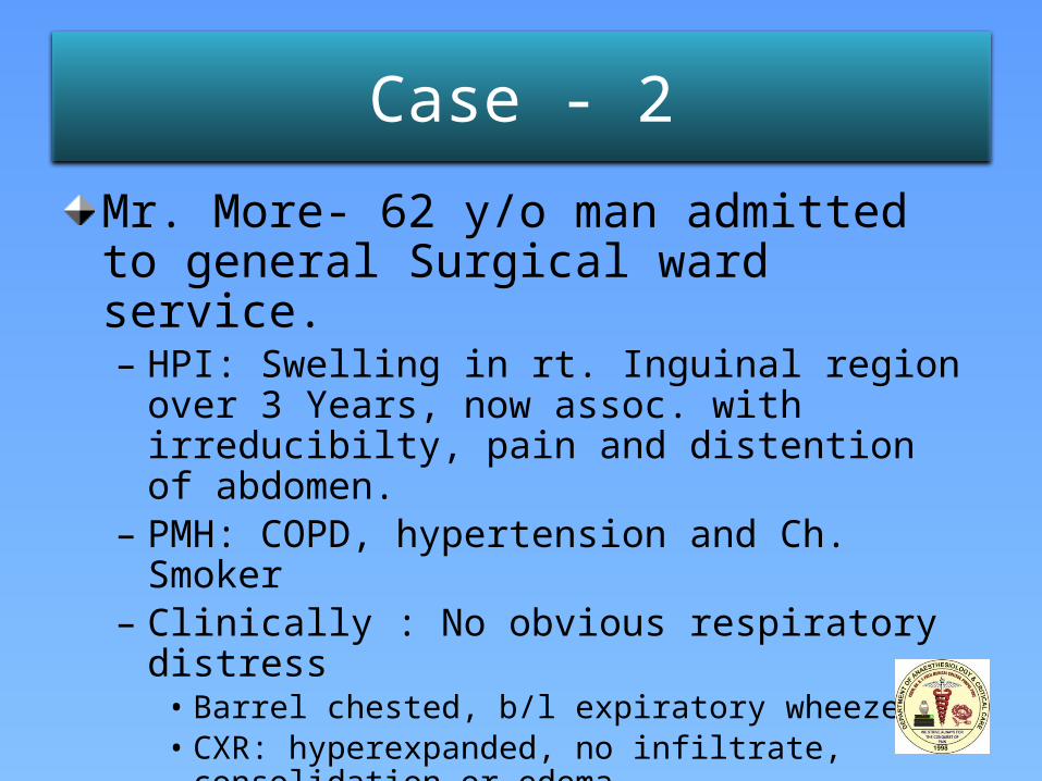
Case - 2
Mr. More- 62 y/o man admitted to general Surgical ward service.– HPI: Swelling in rt. Inguinal region over 3 Years, now
assoc. with irreducibilty, pain and distention of abdomen.
– PMH: COPD, hypertension and Ch. Smoker– Clinically : No obvious respiratory distress
• Barrel chested, b/l expiratory wheezes, • CXR: hyperexpanded, no infiltrate, consolidation or edema.
– DX: rt. indirect Inguinal Hernia, COPD and Hypertension
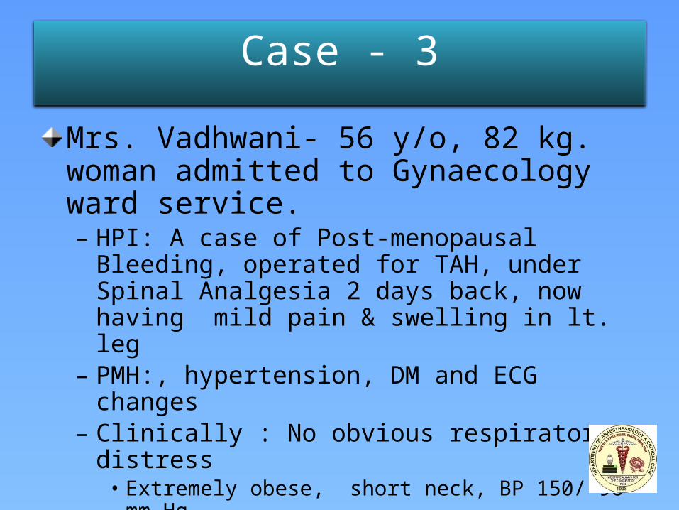
Case - 3
Mrs. Vadhwani- 56 y/o, 82 kg. woman admitted to Gynaecology ward service.– HPI: A case of Post-menopausal Bleeding, operated for
TAH, under Spinal Analgesia 2 days back, now having mild pain & swelling in lt. leg
– PMH:, hypertension, DM and ECG changes– Clinically : No obvious respiratory distress
• Extremely obese, short neck, BP 150/ 96 mm Hg. • CXR: no infiltrate, consolidation or edema.
– DX: Post –op. Pt. Obese, with DM, Hypertension
Are these Potential DVT/ PE Patients?
Are these patients at risk for Venous Thrombo
Embolism (VTE)?
Why worry about VTE in inpatients?
What is the prevalence of DVT/PE in hospitalized
medical and surgical patients?
What are effective methods of prophylaxis?
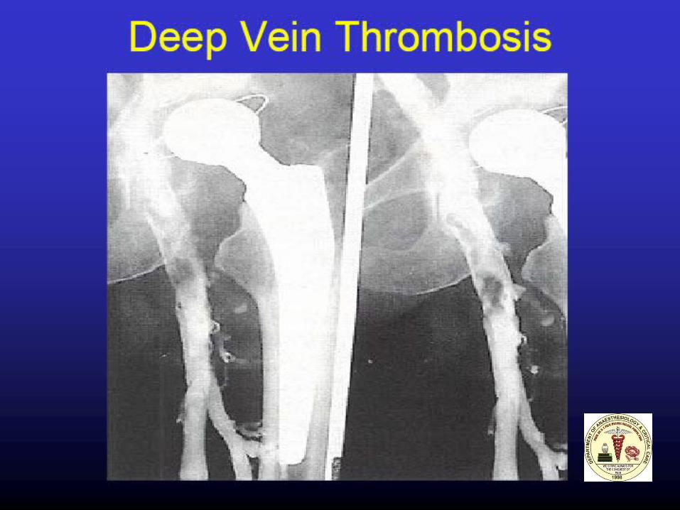
DEEP VEIN THROMBOSIS (DVT)
Enigmatic
Dilemmatic
Mysterious
Dreadful
Potentially Dangerous
Usually fatal
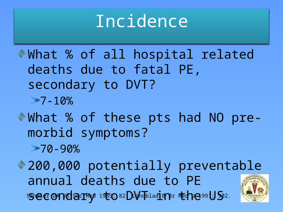
Incidence
What % of all hospital related deaths due to fatal PE, secondary to DVT?
7-10%
What % of these pts had NO pre-morbid symptoms?
70-90%
200,000 potentially preventable annual deaths due to PE secondary to DVT in the US
Sandler DA JR Soc Med 1989; 82, Lindblad B Br Med J 1991; 302.
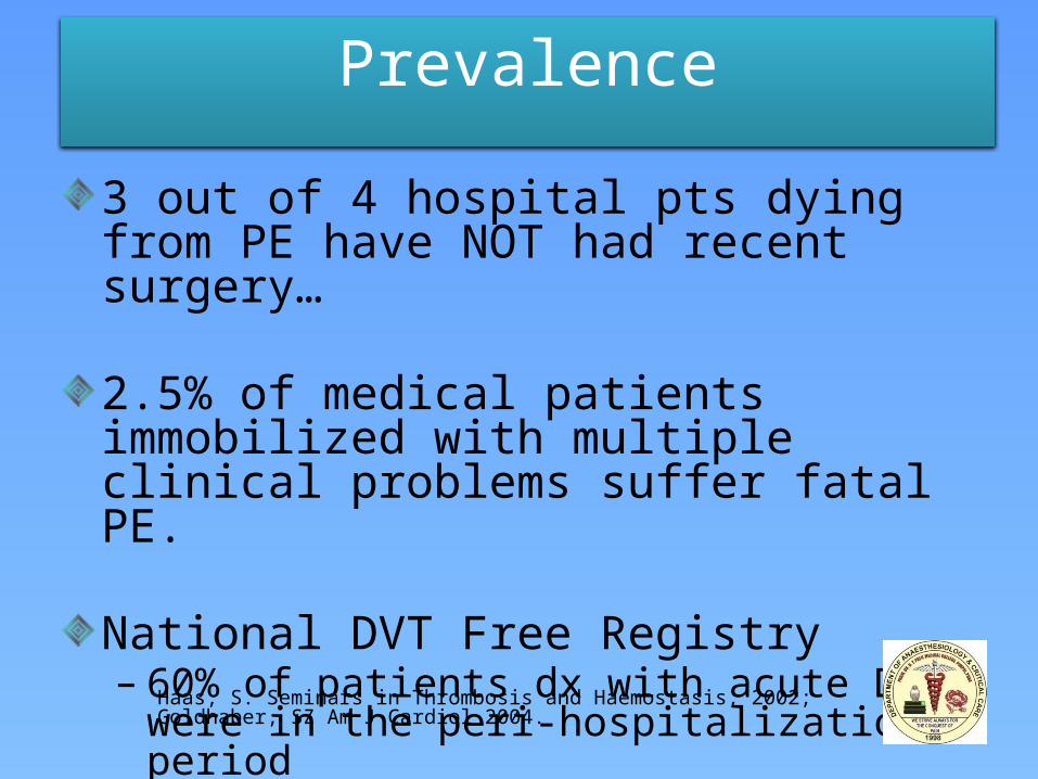
Prevalence
3 out of 4 hospital pts dying from PE have NOT had recent surgery…
2.5% of medical patients immobilized with multiple clinical problems suffer fatal PE.
National DVT Free Registry– 60% of patients dx with acute DVT were in the
peri-hospitalization period– 60% of cases were in non-surgical patients!
Haas, S. Seminars in Thrombosis and Haemostasis, 2002; Goldhaber, SZ Am J Cardiol 2004.
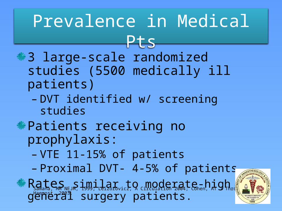
Prevalence in Medical Pts
3 large-scale randomized studies (5500 medically ill patients) – DVT identified w/ screening studies
Patients receiving no prophylaxis:– VTE 11-15% of patients– Proximal DVT- 4-5% of patients
Rates similar to moderate-high risk general surgery patients.
Samana, MM NEJM, 1999; Leizorovicz, A Circulation 2004; Cohen, AT J Thromb Haemost, 2003.
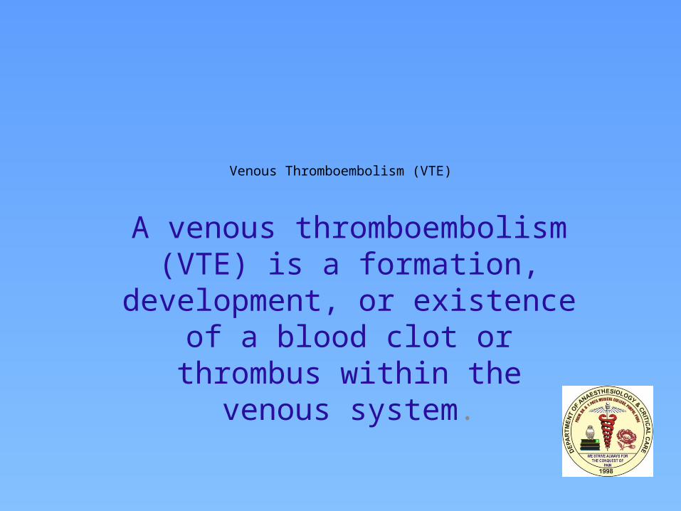
Venous Thromboembolism (VTE)
A venous thromboembolism (VTE) is a formation, development, or
existence of a blood clot or thrombus within the venous system.
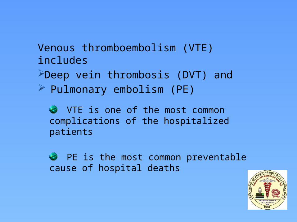
Venous thromboembolism (VTE) includesDeep vein thrombosis (DVT) and Pulmonary embolism (PE)
VTE is one of the most common complications of the hospitalized patients
PE is the most common preventable cause of hospital deaths
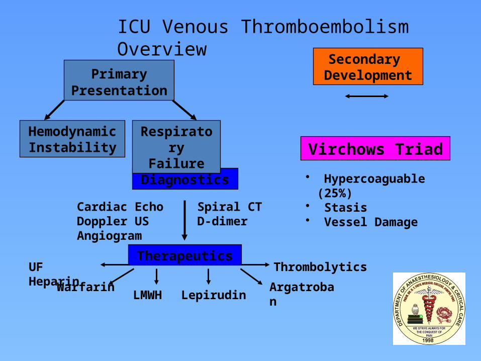
Diagnostics
Therapeutics
Secondary Development
Virchows Triad
Cardiac Echo Spiral CTDoppler US D-dimerAngiogram
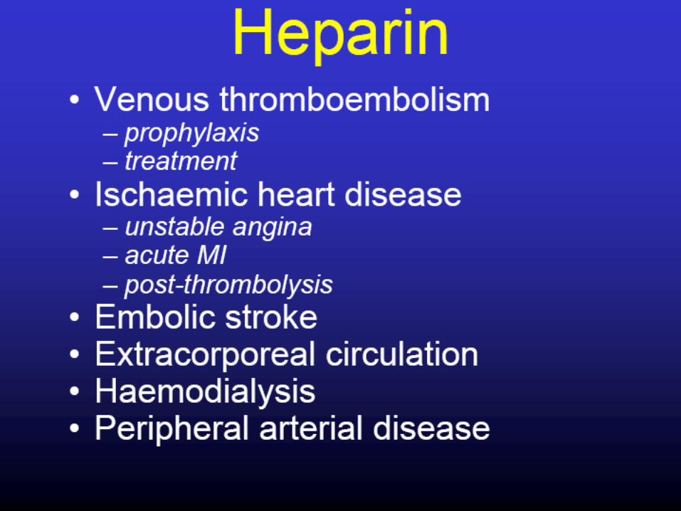
Warfarin
UF Heparin
LMWH LepirudinArgatroban
Thrombolytics
• Hypercoaguable (25%)• Stasis• Vessel Damage
Primary Presentation
Respiratory Failure
Hemodynamic Instability
ICU Venous Thromboembolism Overview
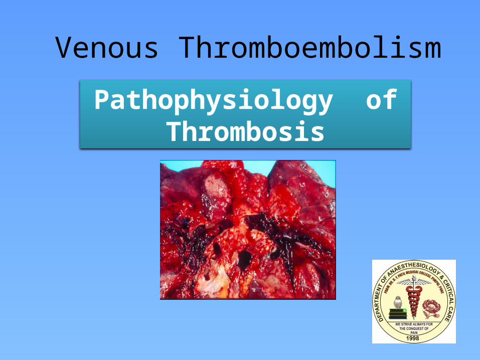
Venous Thromboembolism
Pathophysiology of Thrombosis
Venous Thromboembolism
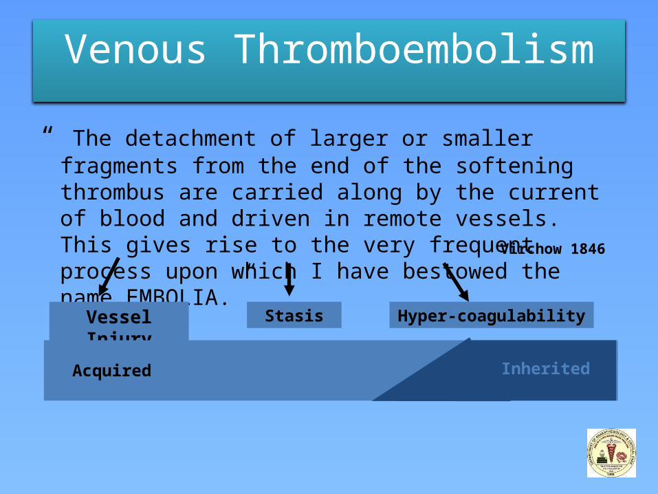
“ The detachment of larger or smaller fragments from the end of the softening thrombus are carried along by the current of blood and driven in remote vessels. This gives rise to the very frequent process upon which I have bestowed the name EMBOLIA.”
Vessel Injury Stasis Hyper-coagulability
Virchow 1846
Acquired Inherited
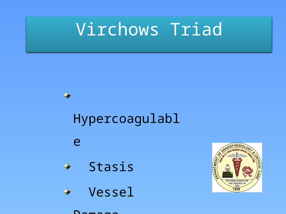
Virchows Triad
Hypercoagulable
Stasis
Vessel Damage
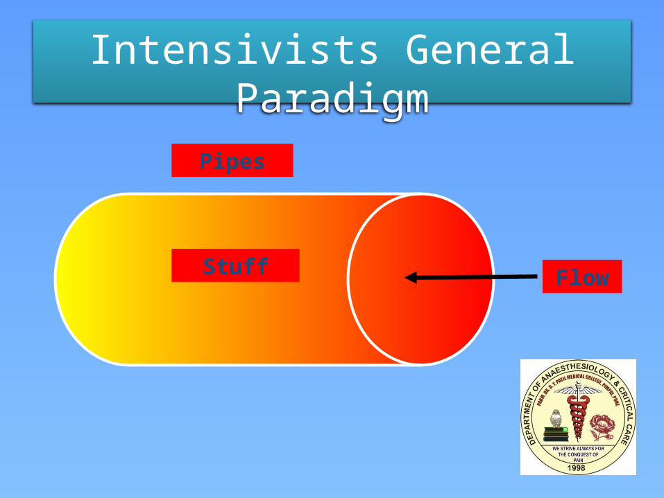
Intensivists General Paradigm
Pipes
Stuff Flow
= Biologically Active Conduit
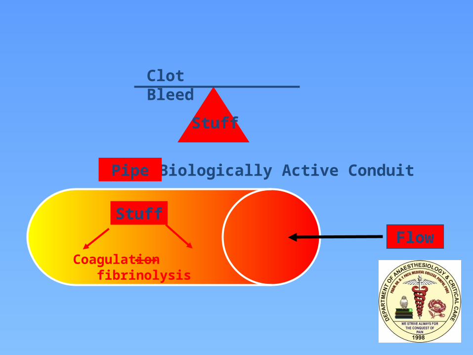
Clot Bleed
Stuff
Pipe
FlowStuff
Coagulation fibrinolysis
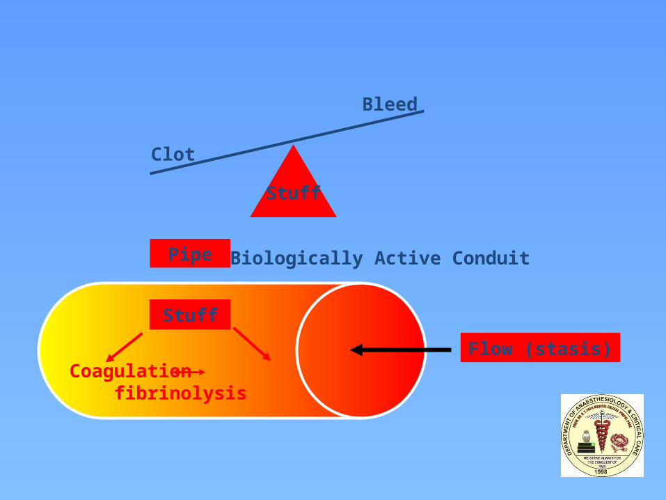
= Biologically Active Conduit
Bleed
Stuff
Pipe
Flow (stasis)
Stuff
Coagulation fibrinolysis
Clot
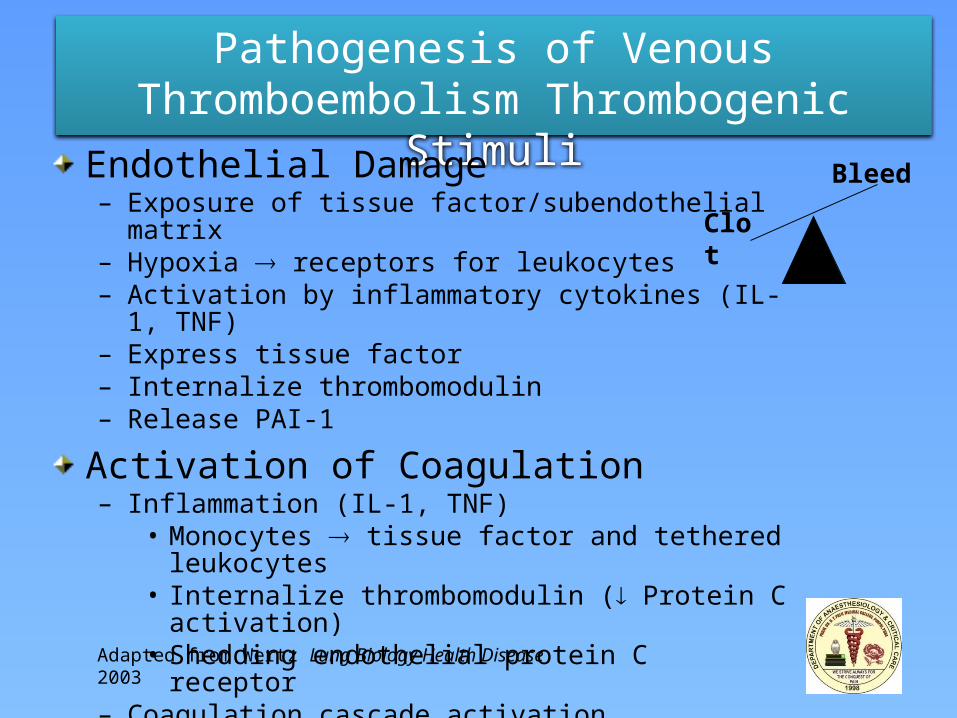
Pathogenesis of Venous Thromboembolism Thrombogenic Stimuli
Endothelial Damage– Exposure of tissue factor/subendothelial matrix– Hypoxia receptors for leukocytes– Activation by inflammatory cytokines (IL-1, TNF)– Express tissue factor– Internalize thrombomodulin– Release PAI-1
Activation of Coagulation– Inflammation (IL-1, TNF)
• Monocytes tissue factor and tethered leukocytes• Internalize thrombomodulin ( Protein C activation)• Shedding endothelial protein C receptor
– Coagulation cascade activation
Clot
Bleed
Adapted from Wertz Lung Biology Health Disease 2003
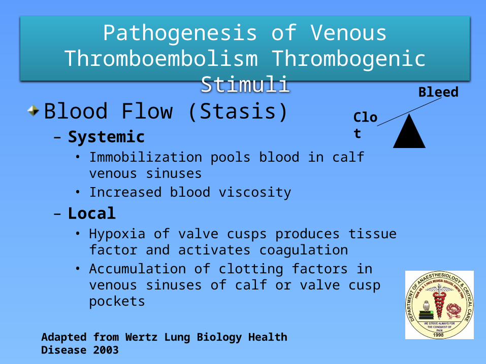
Pathogenesis of Venous Thromboembolism Thrombogenic Stimuli
Blood Flow (Stasis)– Systemic
• Immobilization pools blood in calf venous sinuses• Increased blood viscosity
– Local• Hypoxia of valve cusps produces tissue factor and
activates coagulation• Accumulation of clotting factors in venous sinuses of calf
or valve cusp pockets
Adapted from Wertz Lung Biology Health Disease 2003
Clot
Bleed
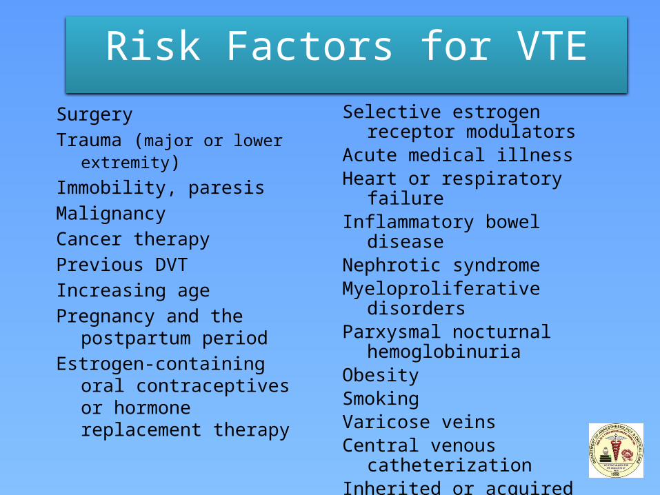
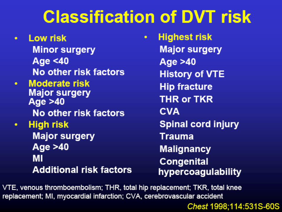
Risk Factors for VTESurgeryTrauma (major or lower extremity)Immobility, paresisMalignancyCancer therapyPrevious DVTIncreasing agePregnancy and the postpartum
periodEstrogen-containing oral
contraceptives or hormone replacement therapy
Selective estrogen receptor modulators
Acute medical illnessHeart or respiratory failureInflammatory bowel diseaseNephrotic syndromeMyeloproliferative disordersParxysmal nocturnal
hemoglobinuriaObesitySmokingVaricose veinsCentral venous catheterizationInherited or acquired
thrombophilia
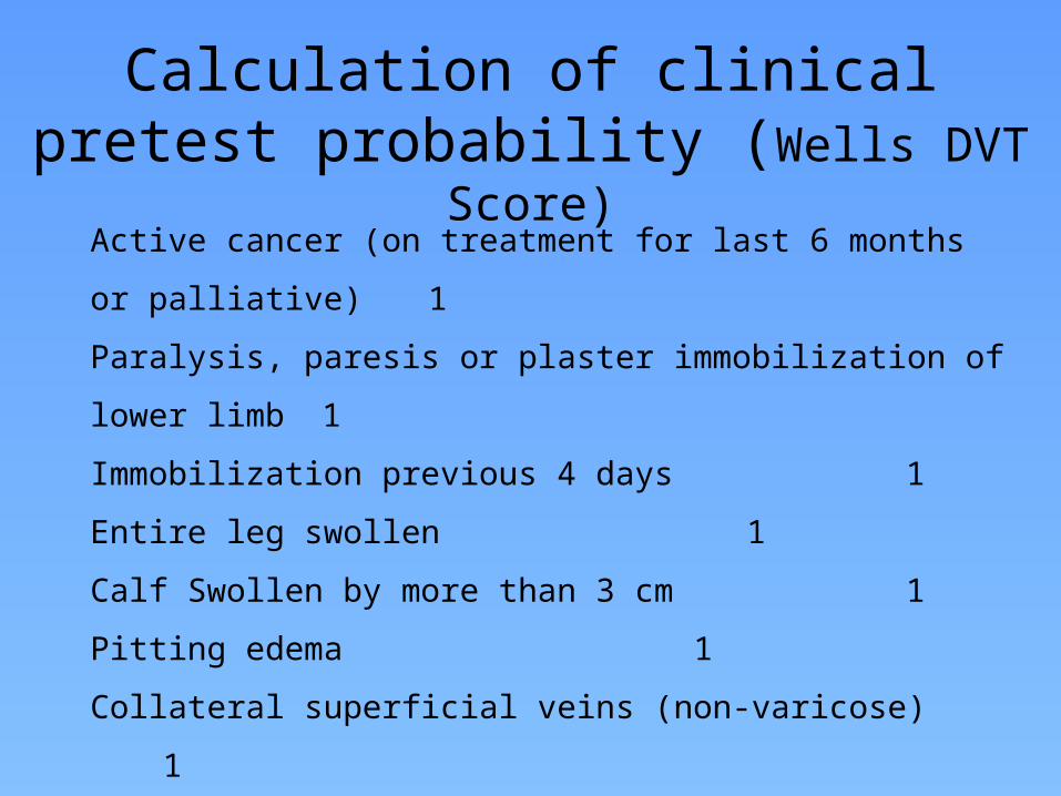
Calculation of clinical pretest probability (Wells DVT Score)
Active cancer (on treatment for last 6 months or palliative) 1
Paralysis, paresis or plaster immobilization of lower limb 1
Immobilization previous 4 days 1
Entire leg swollen 1
Calf Swollen by more than 3 cm 1
Pitting edema 1
Collateral superficial veins (non-varicose) 1
Probable alternative diagnosis -2
(If both legs are symptomatic, score the more severe leg)
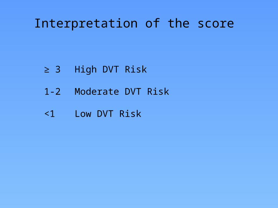
Interpretation of the score
≥ 3 High DVT Risk
1-2 Moderate DVT Risk
<1 Low DVT Risk
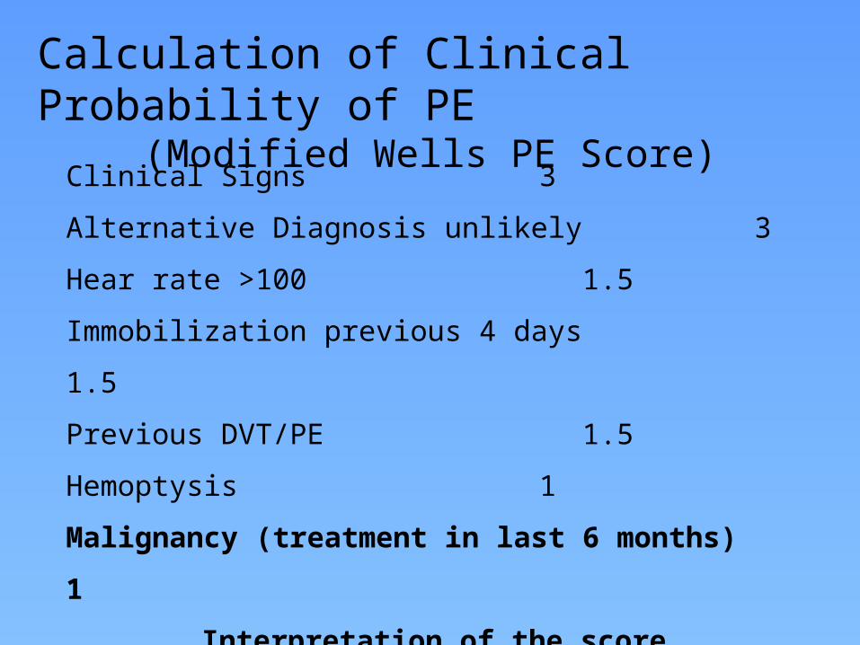
Calculation of Clinical Probability of PE(Modified Wells PE Score)
Clinical Signs 3
Alternative Diagnosis unlikely 3
Hear rate >100 1.5
Immobilization previous 4 days 1.5
Previous DVT/PE 1.5
Hemoptysis 1
Malignancy (treatment in last 6 months) 1
Interpretation of the score
≤ 4 PE less likely > 4 PE likely
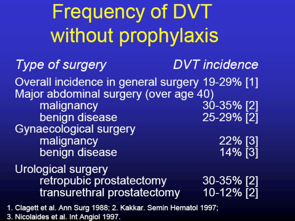
Risk of DVT in
Surgical Patients
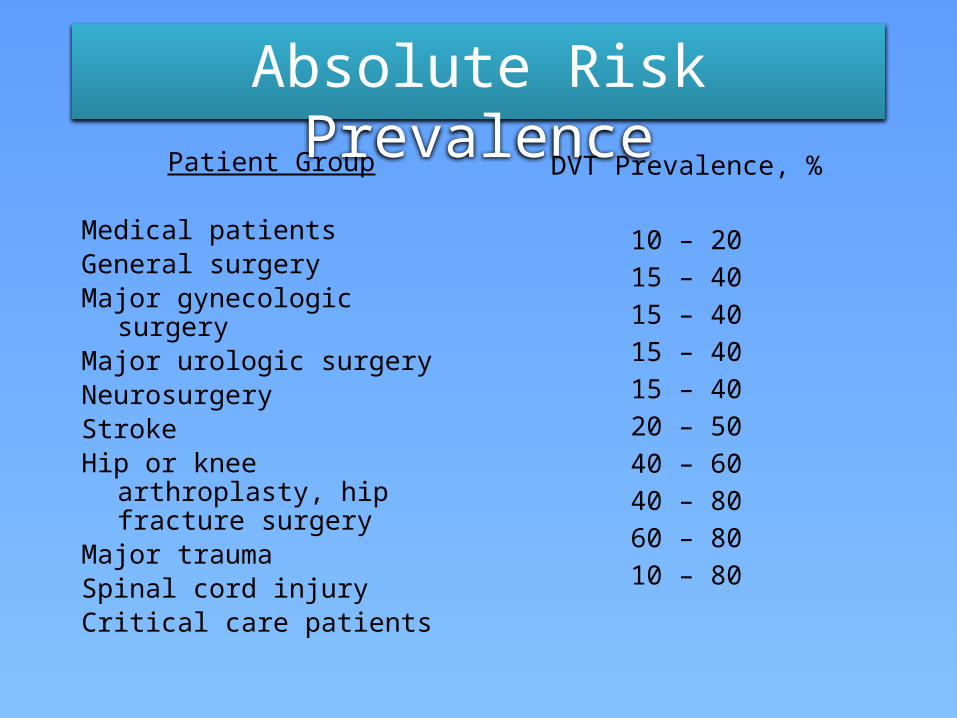
Absolute Risk PrevalencePatient Group
Medical patientsGeneral surgeryMajor gynecologic surgeryMajor urologic surgeryNeurosurgeryStrokeHip or knee arthroplasty, hip
fracture surgeryMajor traumaSpinal cord injuryCritical care patients
DVT Prevalence, %
10 – 2015 – 4015 – 4015 – 4015 – 4020 – 5040 – 6040 – 8060 – 8010 – 80
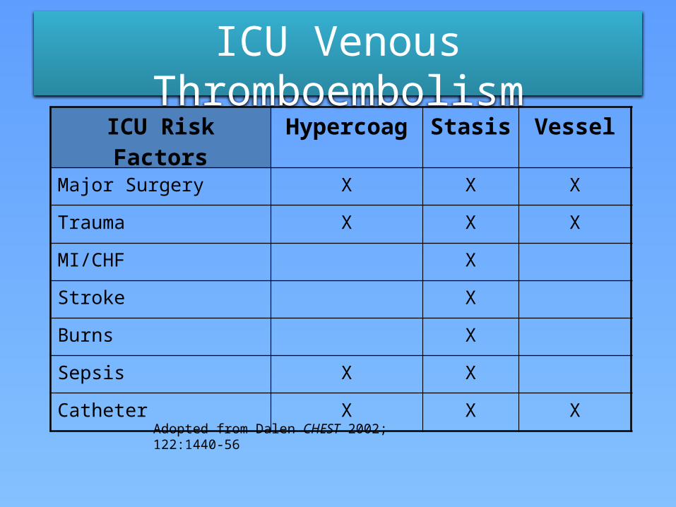
ICU Venous Thromboembolism
Adopted from Dalen CHEST 2002; 122:1440-56
ICU Risk Factors Hypercoag Stasis Vessel
Major Surgery X X X
Trauma X X X
MI/CHF X
Stroke X
Burns X
Sepsis X X
Catheter X X X
DIAGNOSIS OF
DVT/ PE
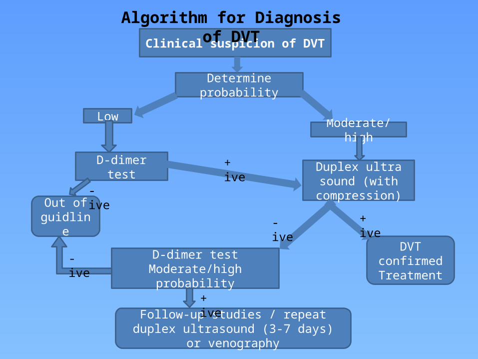
Clinical suspicion of DVT
Determine probability
LowModerate/high
D-dimer test
Out of guidline
Duplex ultra sound (with compression)
D-dimer test Moderate/highprobability
DVT confirmed Treatment
Follow-up studies / repeat duplex ultrasound (3-7 days) or venography
+ ive
+ ive
+ ive
- ive
- ive
- ive
Algorithm for Diagnosis of DVT
Clinical sign & symptoms of PE
Algorithm for Diagnosis of Pulmonary Embolism
Wells ScoreStart heparin if >6, Perform D-dimer test
PE less likely PE likely
D-dimer Perform CTPA
R/w clinical probability & D dimer results Diagnosis of PE
VTE Treatment algorithm
PE less likely & ‘+’ D dimer results
PE likely & ‘-’ D dimer results
PE likely with ‘+’ D dimer results
Perform duplex ultrasound of
the leg
Ultrasound results?
Risk of PE is low. Consider other
diagnosis
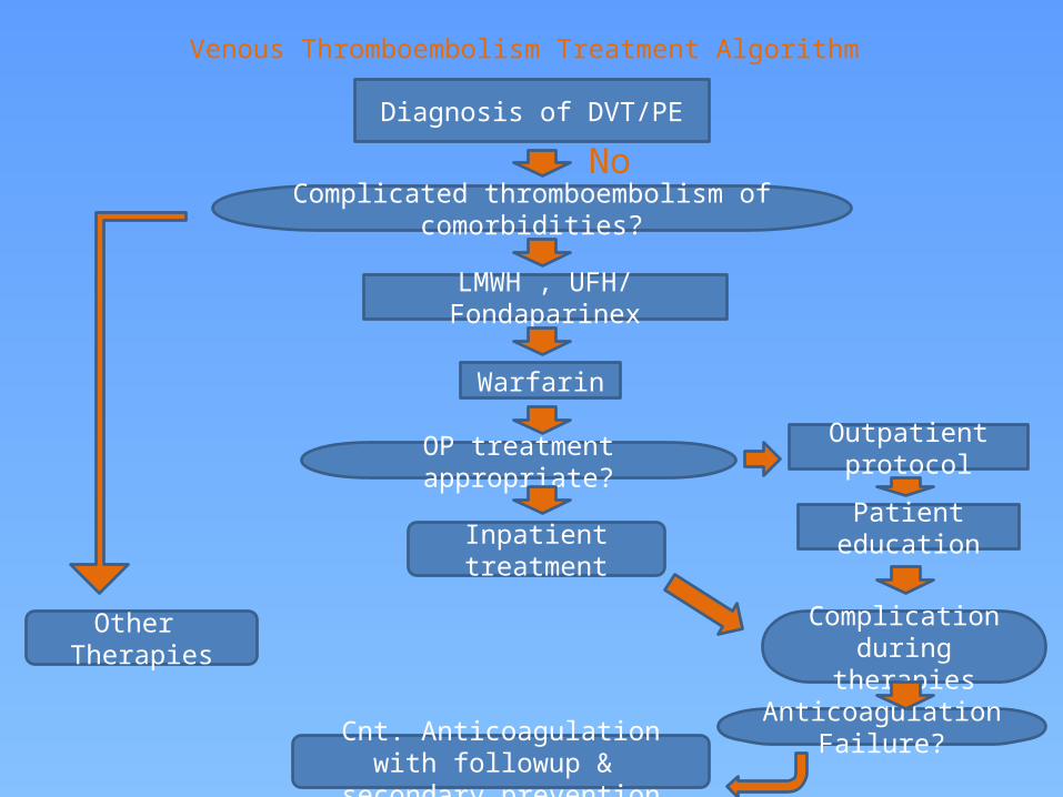
Venous Thromboembolism Treatment Algorithm
Diagnosis of DVT/PE
LMWH , UFH/ Fondaparinex
Warfarin
Inpatient treatment
Outpatient protocol
Patient education
Other Therapies
Cnt. Anticoagulation with followup & secondary prevention
Complicated thromboembolism of comorbidities?
OP treatment appropriate?
Complication during therapies
Anticoagulation Failure?
No

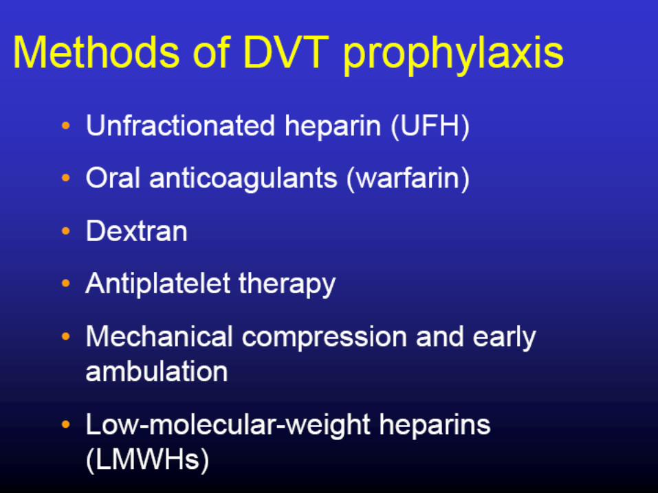
Prophylaxis of DVT
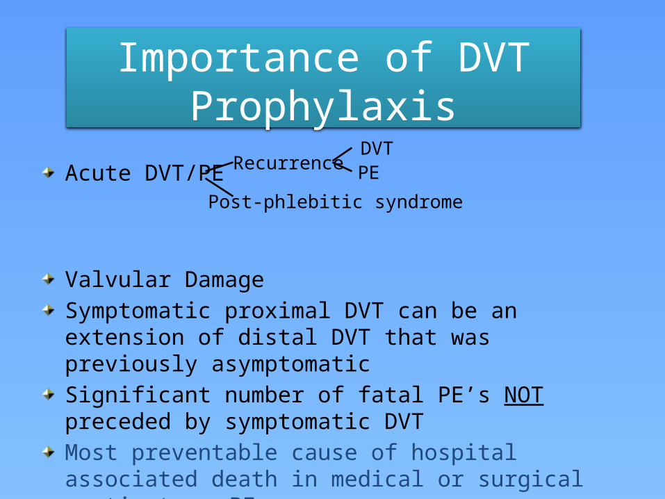
Importance of DVT Prophylaxis
Acute DVT/PE
Valvular DamageSymptomatic proximal DVT can be an extension of distal DVT that was previously asymptomaticSignificant number of fatal PE’s NOT preceded by symptomatic DVTMost preventable cause of hospital associated death in medical or surgical patients PE
Recurrence
Post-phlebitic syndrome
DVTPE
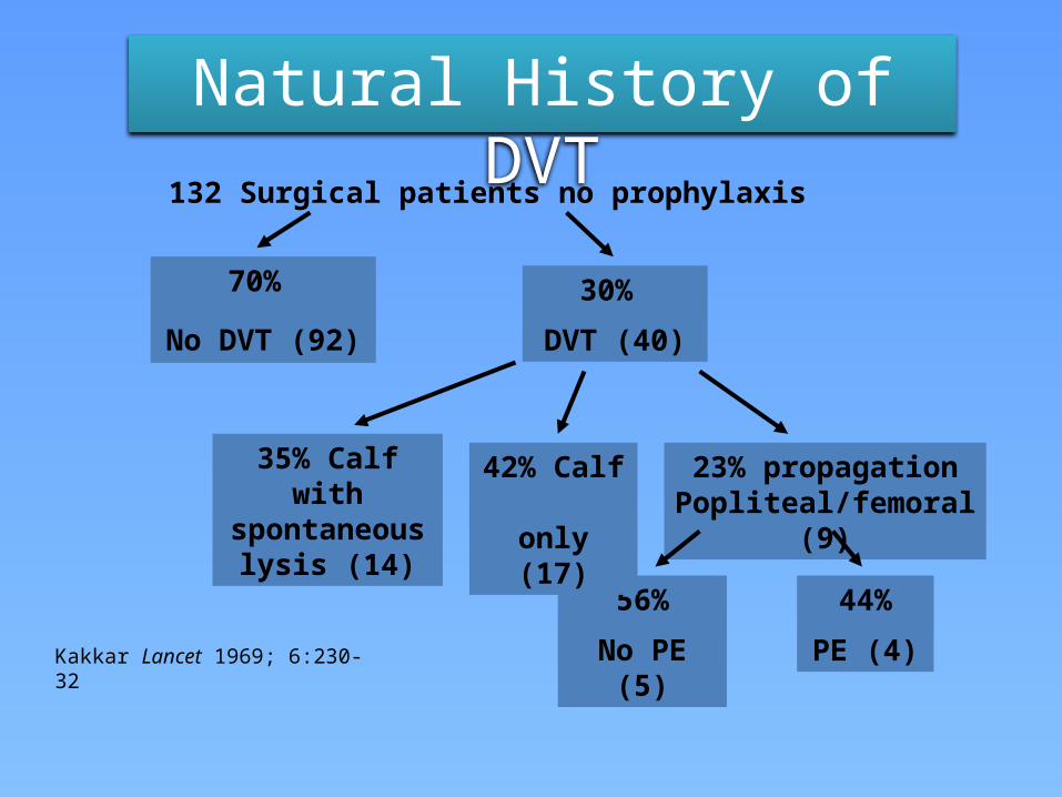
Natural History of DVT132 Surgical patients no prophylaxis
56%
No PE (5)
44%
PE (4)
42% Calf only (17)
23% propagation Popliteal/femoral (9)
35% Calf with spontaneous
lysis (14)
30%
DVT (40)
70%
No DVT (92)
Kakkar Lancet 1969; 6:230-32
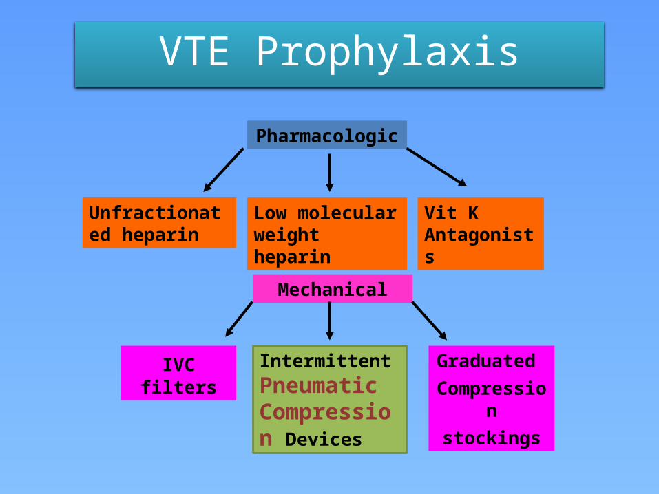
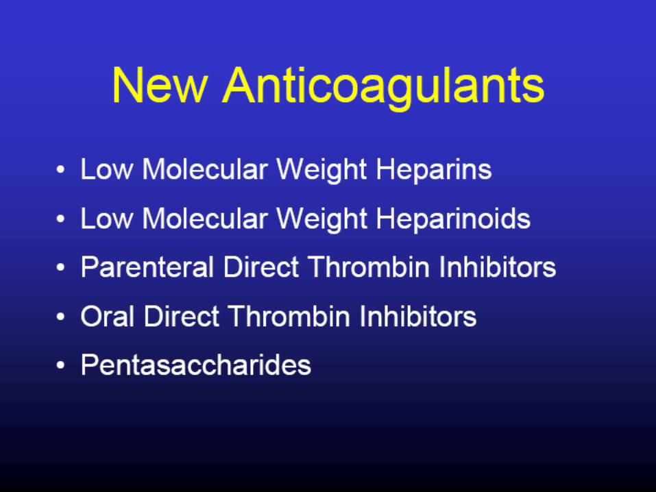
VTE Prophylaxis
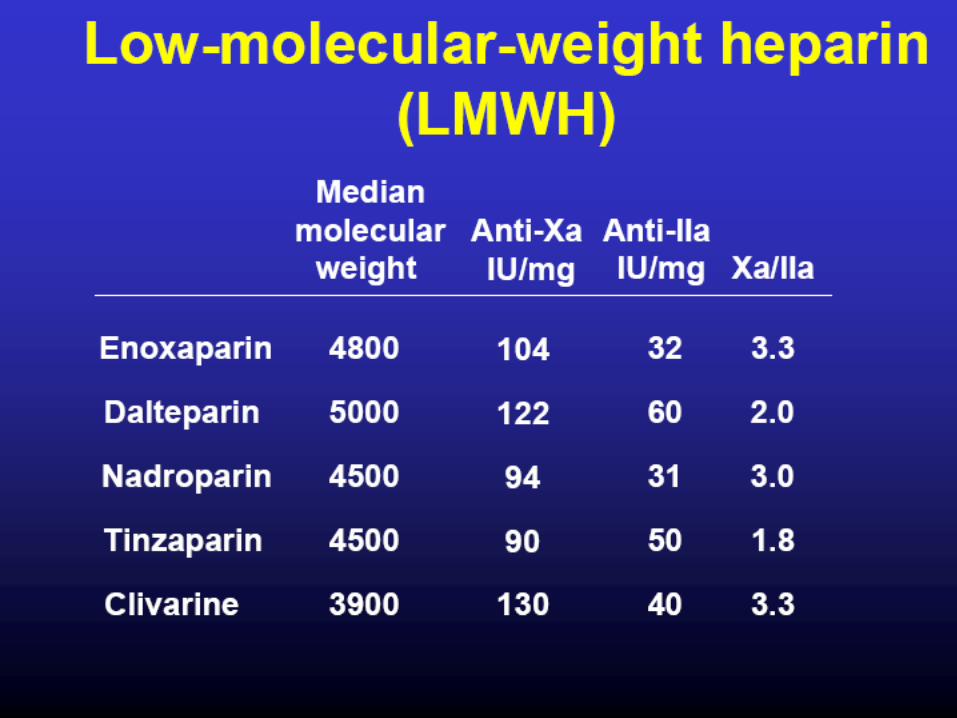
Pharmacologic
Unfractionated heparin
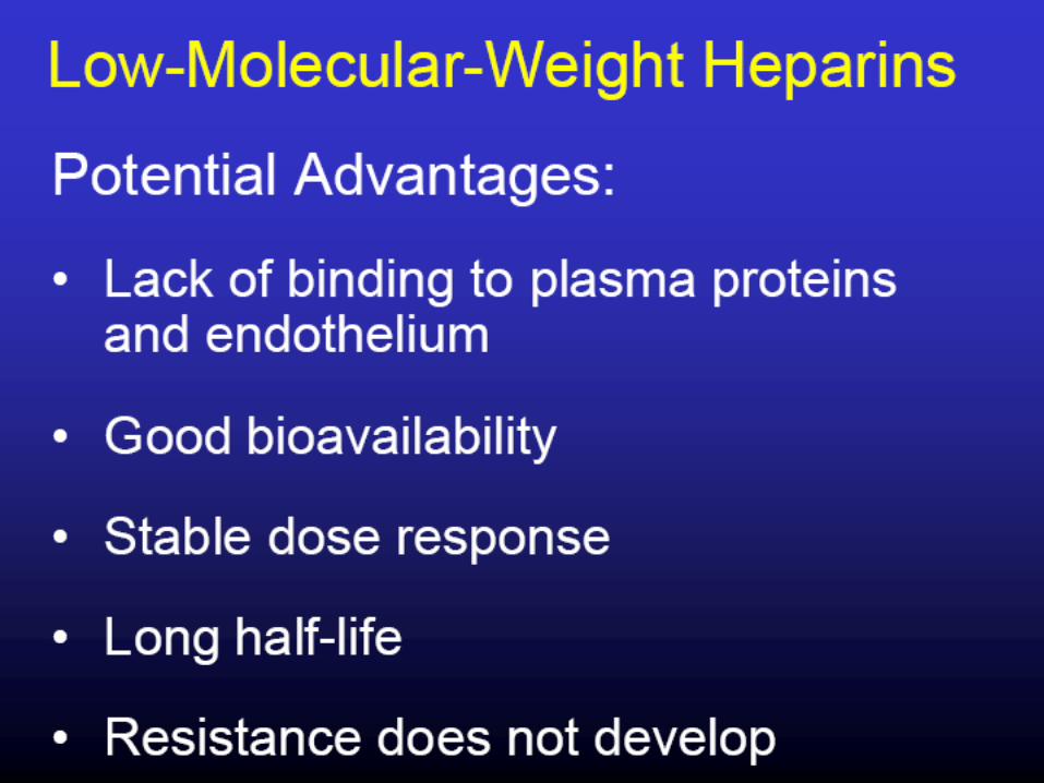
Low molecular weight heparin
Vit K Antagonists
Mechanical
IVC filters Intermittent Pneumatic Compression Devices
Graduated Compression
stockings
49
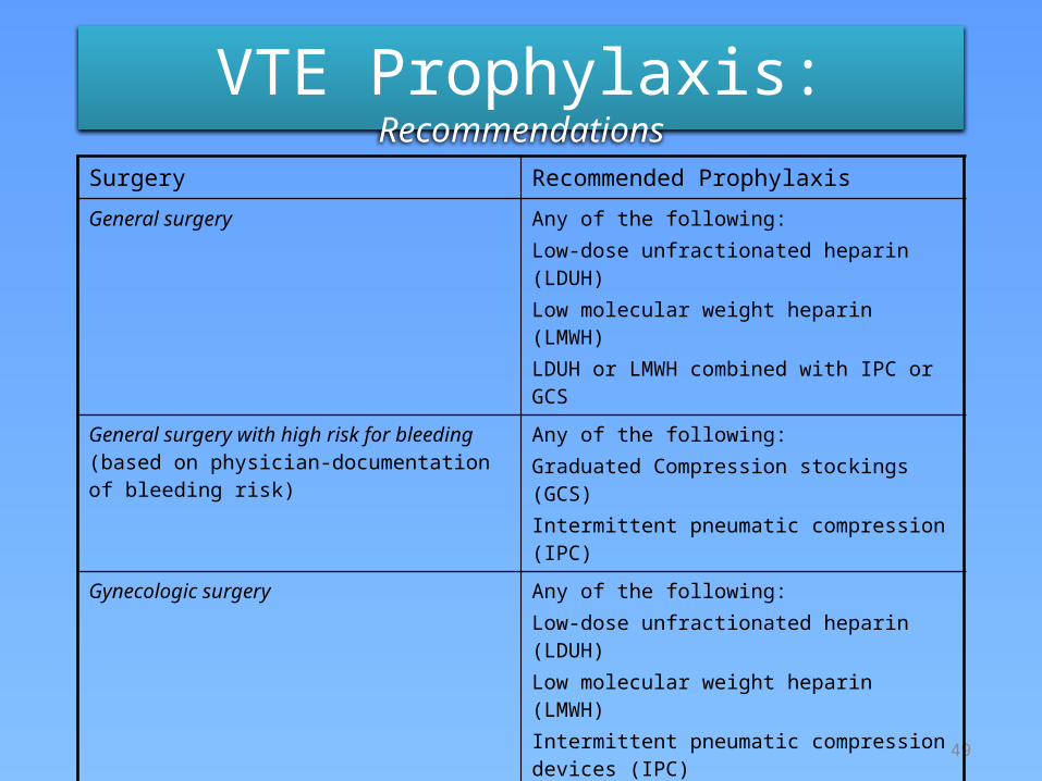
VTE Prophylaxis: Recommendations
Surgery Recommended Prophylaxis
General surgery Any of the following:Low-dose unfractionated heparin (LDUH)Low molecular weight heparin (LMWH) LDUH or LMWH combined with IPC or GCS
General surgery with high risk for bleeding (based on physician-documentation of bleeding risk)
Any of the following:Graduated Compression stockings (GCS)Intermittent pneumatic compression (IPC)
Gynecologic surgery Any of the following:Low-dose unfractionated heparin (LDUH)Low molecular weight heparin (LMWH)Intermittent pneumatic compression devices (IPC) LDUH or LMWH combined with IPC or GCS
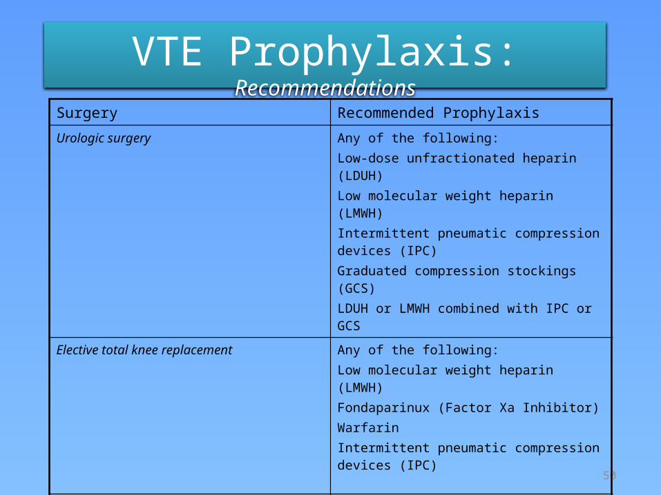
50
VTE Prophylaxis: Recommendations
Surgery Recommended Prophylaxis
Urologic surgery Any of the following:Low-dose unfractionated heparin (LDUH)Low molecular weight heparin (LMWH) Intermittent pneumatic compression devices (IPC) Graduated compression stockings (GCS)LDUH or LMWH combined with IPC or GCS
Elective total knee replacement Any of the following:Low molecular weight heparin (LMWH)Fondaparinux (Factor Xa Inhibitor)WarfarinIntermittent pneumatic compression devices (IPC)
Elective total hip replacement Any of the following:Low molecular weight heparin (LMWH)Fondaparinux (Factor Xa Inhibitor)Warfarin
51
VTE Prophylaxis: Recommendations
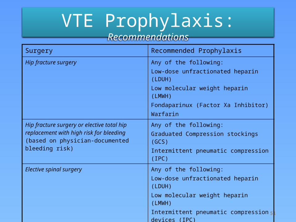
Surgery Recommended Prophylaxis
Hip fracture surgery Any of the following:Low-dose unfractionated heparin (LDUH) Low molecular weight heparin (LMWH)Fondaparinux (Factor Xa Inhibitor)Warfarin
Hip fracture surgery or elective total hip replacement with high risk for bleeding (based on physician-documented bleeding risk)
Any of the following:Graduated Compression stockings (GCS)Intermittent pneumatic compression (IPC)
Elective spinal surgery Any of the following:Low-dose unfractionated heparin (LDUH) Low molecular weight heparin (LMWH) Intermittent pneumatic compression devices (IPC) Graduated compression stockings (GCS)IPC combined with GCSLDUH or LMWH combined with IPC or GCS
52
VTE Prophylaxis: Recommendations
Surgery Recommended Prophylaxis
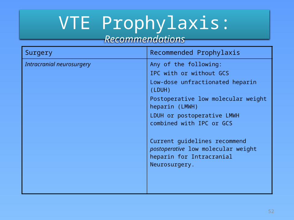
Intracranial neurosurgery Any of the following:IPC with or without GCSLow-dose unfractionated heparin (LDUH) Postoperative low molecular weight heparin (LMWH)LDUH or postoperative LMWH combined with IPC or GCS
Current guidelines recommend postoperative low molecular weight heparin for Intracranial Neurosurgery.
Knudson Ann Surg 2004; 240:490-498
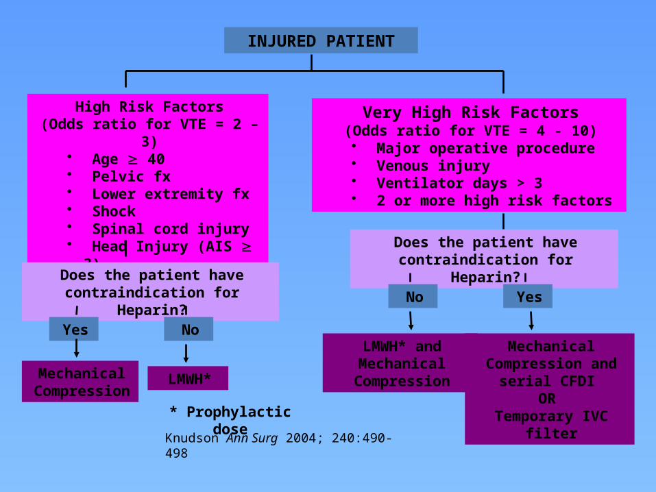
INJURED PATIENT
High Risk Factors(Odds ratio for VTE = 2 – 3)• Age ³ 40• Pelvic fx• Lower extremity fx• Shock• Spinal cord injury• Head Injury (AIS ³ 3)
Very High Risk Factors(Odds ratio for VTE = 4 - 10)
• Major operative procedure• Venous injury• Ventilator days > 3 • 2 or more high risk factors
Does the patient have contraindication for Heparin?
Does the patient have contraindication for Heparin?
Yes No
YesNo
Mechanical Compression
LMWH*
* Prophylactic dose
LMWH* and Mechanical
Compression
Mechanical Compression and serial CFDI
OR Temporary IVC filter
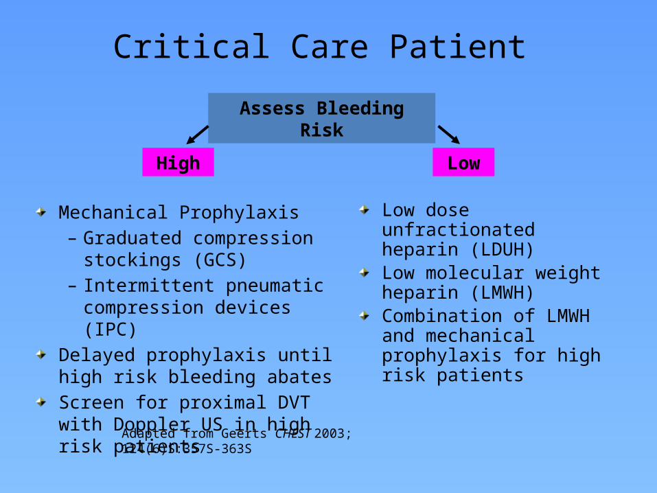
Critical Care Patient
Mechanical Prophylaxis– Graduated compression stockings
(GCS)– Intermittent pneumatic
compression devices (IPC)Delayed prophylaxis until high risk bleeding abatesScreen for proximal DVT with Doppler US in high risk patients
Low dose unfractionated heparin (LDUH)Low molecular weight heparin (LMWH)Combination of LMWH and mechanical prophylaxis for high risk patients
Assess Bleeding Risk
High Low
Adapted from Geerts CHEST 2003; 124(6)S:357S-363S
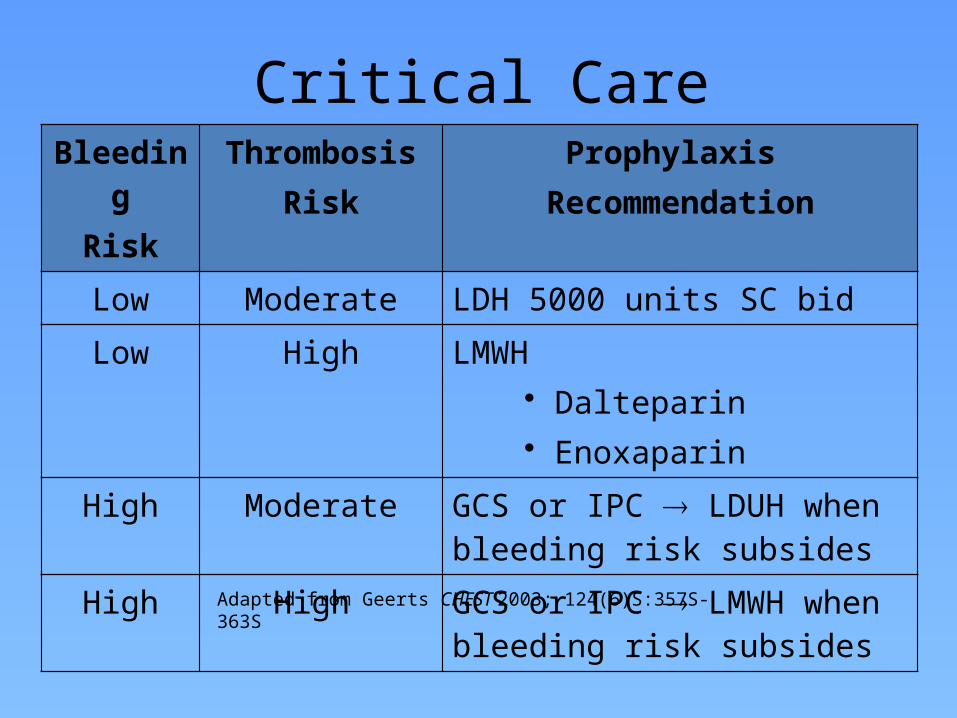
Critical Care PatientBleeding
RiskThrombosis
RiskProphylaxis
Recommendation
Low Moderate LDH 5000 units SC bid
Low High LMWH• Dalteparin• Enoxaparin
High Moderate GCS or IPC LDUH when bleeding risk subsides
High High GCS or IPC LMWH when bleeding risk subsides
Adapted from Geerts CHEST 2003; 124(6)S:357S-363S
Stop DVT Without Risk Of Post-Op Bleeding
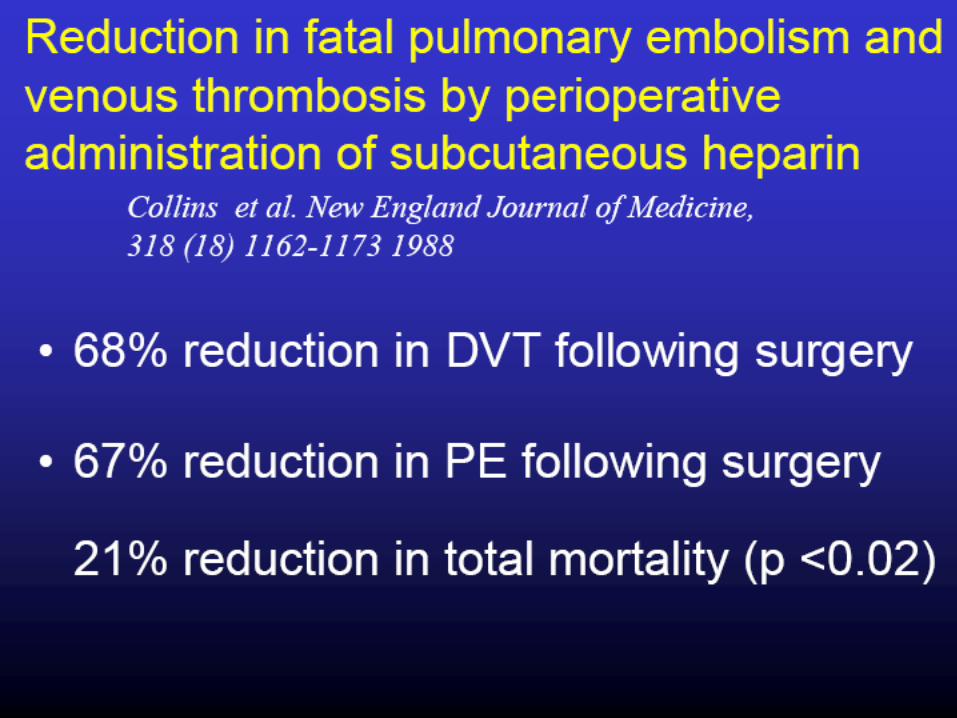
DVT prophylaxis reduces the incidence of DVT during the postoperative period by two-thirds, and prevents death from pulmonary embolism in 1 patient out of every 200 major operations.
The National Institute of Health recommends more extensive use of prophylaxis.
Preventable in most cases with simple cost-effective prophylaxis
Stop DVT Without Risk Of Post-Op Bleeding
Intermittent pneumatic leg compression reduces the risk of DVT by as much as 59% in general surgery patients. It is also virtually free of side effects and is as effective as low-dose heparin in patients undergoing abdominal surgery. Using prophylaxis for DVT is neither complicated nor expensive.
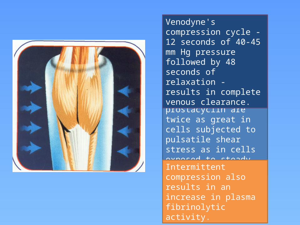
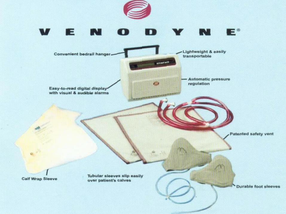
Production rates of prostacyclin are twice as great in cells subjected to pulsatile shear stress as in cells exposed to steady shear stress.
Venodyne's compression cycle -12 seconds of 40-45 mm Hg pressure followed by 48 seconds of relaxation - results in complete venous clearance.
Intermittent compression also results in an increase in plasma fibrinolytic activity.
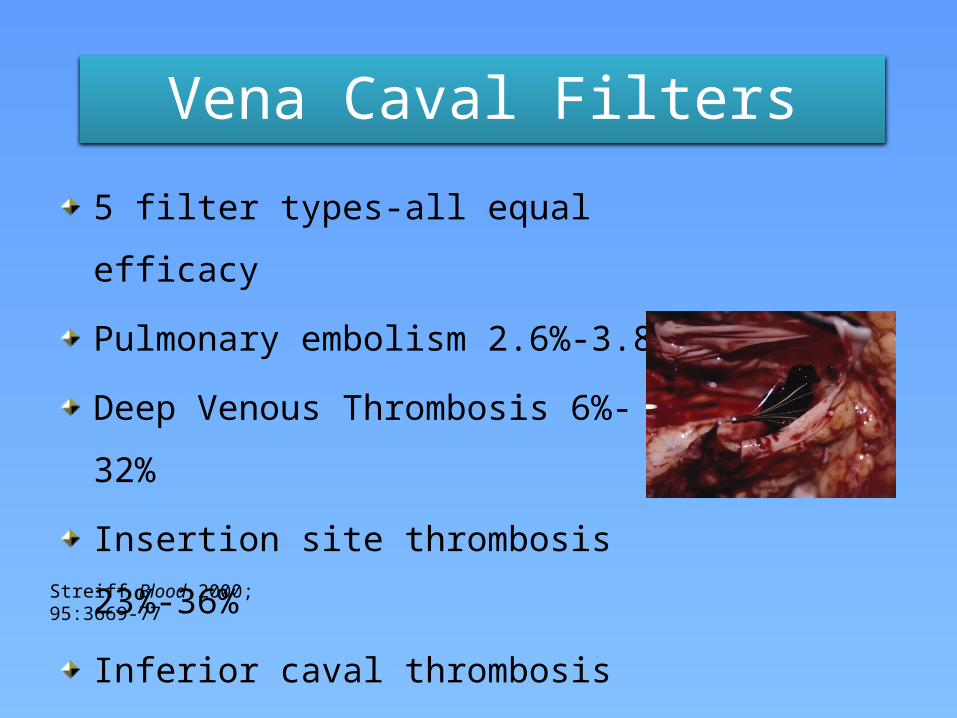
Vena Caval Filters
5 filter types-all equal efficacy
Pulmonary embolism 2.6%-3.8%
Deep Venous Thrombosis 6%-32%
Insertion site thrombosis 23%-36%
Inferior caval thrombosis 3.6%-11.2%
Postphlebitic syndrome 14%-41%Streiff Blood 2000; 95:3669-77

VTE Treatment Algorithm
Venous thromboembolism is common in medical,
surgical as well as critically ill patients
Modifiable risk factors are limited in ICU
DVT prophylaxis is essential
PE risk stratification is crucial to define optimal Tx
Summary
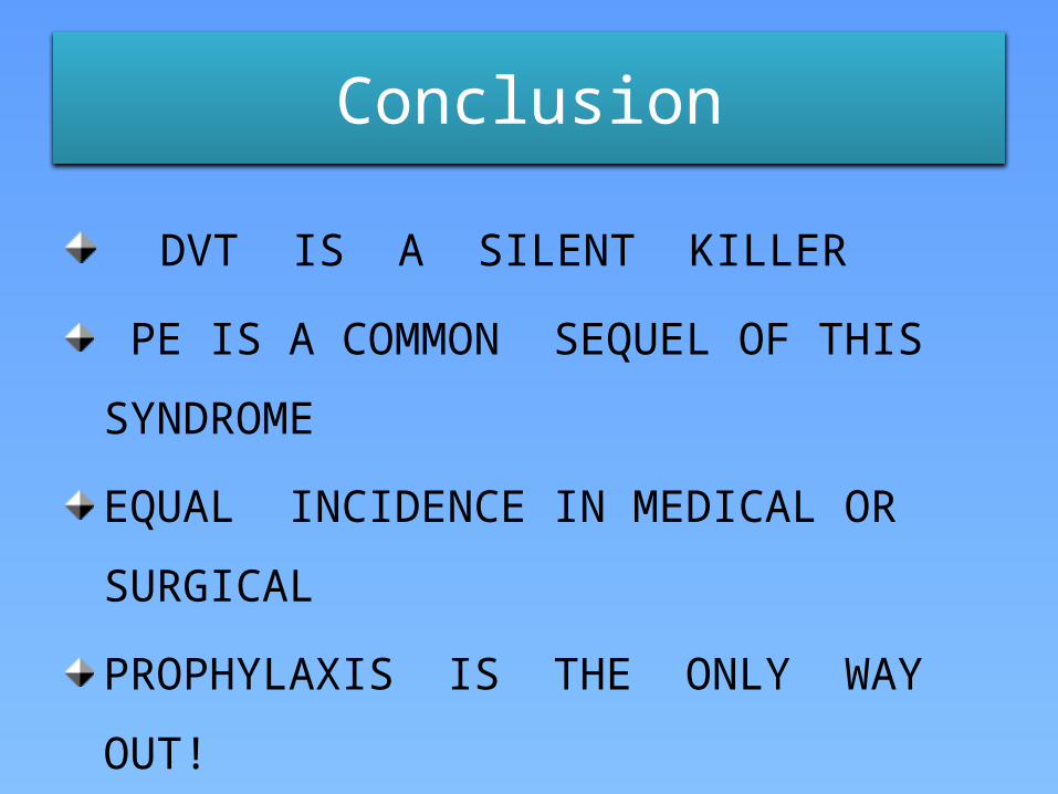
Conclusion
DVT IS A SILENT KILLER
PE IS A COMMON SEQUEL OF THIS SYNDROME
EQUAL INCIDENCE IN MEDICAL OR SURGICAL
PROPHYLAXIS IS THE ONLY WAY OUT!
HEPARIN, LMWHs, & OTHERS EFFECTIVE
PNEUMATIC DEVICES -PROMISING

THANK YOU !