Embed Size (px)
Citation preview

D R . N I S H T H A J A I N
S E N I O R R E S I D E N T ,
D E P A R T M E N T O F N E U R O L O G Y ,
G M C , K O T A .
CRANIAL NERVES XI AND XII

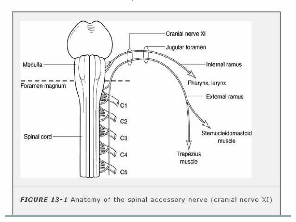
The Spinal Accessory Nerve
The spinal accessory (SA) nerve - two nerves that run
together in a common bundle for a short distance.
The smaller cranial portion (ramus internus) is a
special visceral efferent (SVE) accessory to the vagus.
The cranial root runs to the jugular foramen and unites
with the spinal portion, traveling with it for only a few
millimeters to form the main trunk of CN XI.

The cranial root communicates with the jugular ganglion of
the vagus, and then exits through the jugular foramen
separately from the spinal portion.
It passes through the ganglion nodosum and then blends
with the vagus.
Distributed principally with the recurrent laryngeal nerve to
sixth branchial arch muscles in the larynx except there is
no XI contribution to the cricothyroid muscle.

The major part of CN XI is the spinal portion (ramus
externus).
The fibers of the spinal root arise from SVE motor cells in
the SA nuclei in the ventral horn from C2 to C5, or even C6.
The supranuclear innervation of CN XI arises from the
lower portion of the precentral gyrus.

The bulk of current evidence indicates that both the SCM
and trapezius receive bilateral supranuclear innervation.
The input to the SCM motor neuron pool - ipsilateral and
that to the trapezius motor neuron pool - contralateral.

Somatotopic arrangement present : cord levels C1 and C2 innervate the ipsilateral sternocleidomastoid muscle, and levels C3 and C4 innervate primarily the ipsilateraltrapezius.
The corticobulbar fibers to the sternocleidomastoid are located in the brainstem tegmentum, whereas fibers to the trapezius are located in the ventral brainstem.
Thus, a ventral pontine lesion can cause supranuclearparesis of the trapezius with sparing of the sternocleidomastoid muscle.

To assess SCM power, have the patient turn the head fully
to one side and hold it there, then try to turn the head back
to midline, avoiding any tilting or leaning motion.
The muscle usually stands out well, and its contraction can
be seen and felt.
Significant weakness of rotation can be detected if the
patient tries to counteract firm resistance.

The two sternocleidomastoid muscles can be examined
simultaneously by having the patient flex his neck while the
examiner exerts pressure on the forehead, or by having the
patient turn the head from side to side.
Flexion of the head against resistance may cause deviation
of the head toward the paralyzed side.
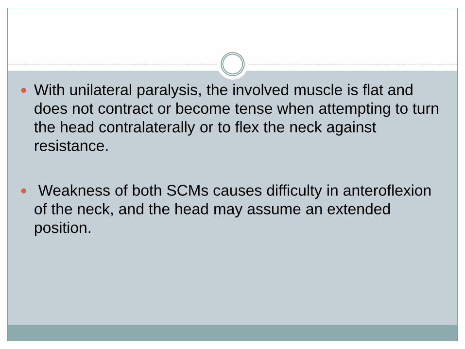
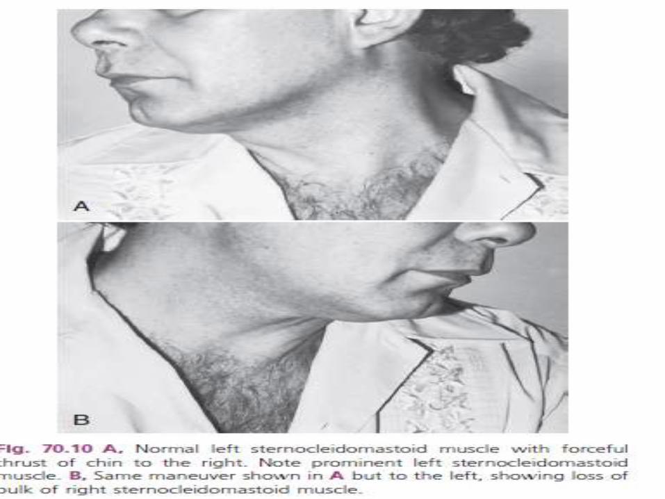
With unilateral paralysis, the involved muscle is flat and
does not contract or become tense when attempting to turn
the head contralaterally or to flex the neck against
resistance.
Weakness of both SCMs causes difficulty in anteroflexion
of the neck, and the head may assume an extended
position.

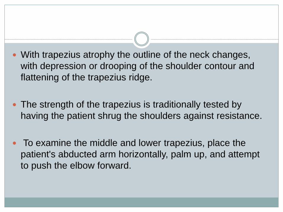
With trapezius atrophy the outline of the neck changes,
with depression or drooping of the shoulder contour and
flattening of the trapezius ridge.
The strength of the trapezius is traditionally tested by
having the patient shrug the shoulders against resistance.
To examine the middle and lower trapezius, place the
patient's abducted arm horizontally, palm up, and attempt
to push the elbow forward.

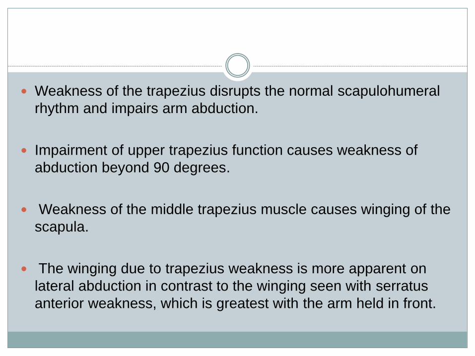
Weakness of the trapezius disrupts the normal scapulohumeral
rhythm and impairs arm abduction.
Impairment of upper trapezius function causes weakness of
abduction beyond 90 degrees.
Weakness of the middle trapezius muscle causes winging of the
scapula.
The winging due to trapezius weakness is more apparent on
lateral abduction in contrast to the winging seen with serratus
anterior weakness, which is greatest with the arm held in front.
When the trapezius is weak, the arm hangs lower on the
affected side, and the fingertips touch the thigh at a lower
level than on the normal side.
Placing the palms together with the arms extended
anteriorly and slightly below horizontal shows the fingers on
the affected side extending beyond those of the normal
side.
The two trapezius muscles can be examined
simultaneously by having the patient extend his neck
against resistance.
Bilateral paralysis causes weakness of neck extension.
The patient cannot raise his chin, and the head may tend to
fall forward (dropped head syndrome).
The shoulders look square or have a drooping, sagging
appearance due to atrophy of both muscles.

Weakness of the muscles supplied by CN XI may be
caused by supranuclear, nuclear, or infranuclear lesions.
Supranuclear involvement usually causes at worst
moderate loss of function since innervation is partially
bilateral.
In hemiplegia there is usually no head deviation, but
testing may reveal slight, weakness of the SCM, with
difficulty turning the face toward the involved limbs.
There may be depression of the shoulder resulting from
trapezius weakness on the affected side.

Irritative supranuclear lesions may cause head turning
away from the discharging hemisphere.
This turning of the head (or head and eyes) may occur as
part of a contraversive, ipsiversive, or jacksonian seizure,
and is often the first manifestation of the seizure.
Extrapyramidal lesions may also involve the
sternocleidomastoid and trapezius muscles, causing
rigidity, akinesis, or hyperkinesis.
Lesions of the lower brainstem or upper cervical spinal cord
may cause dissociated weakness of the SCM and
trapezius muscles depending on the exact location.
Nuclear involvement of the SA nerve may occur in motor
neuron disease, syringobulbia, and syringomyelia.
In nuclear lesions, the weakness is frequently accompanied
by atrophy and fasciculations.
Localisation
Weakness of the trapezius on one side associated with
weakness of the sternocleidomastoid on the other side
(dissociated weakness) indicates an upper motor neuron
lesion ipsilateral to the weak sternocleidomastoid.
Weakness of the trapezius on one side with sparing of the
sternocleidomastoid muscles indicates a ventral brainstem
lesion, a lower cervical cord lesion, or a lower spinal
accessory root lesion.
Weakness of the sternocleidomastoid with trapezius
sparing indicates a lesion of the lower brainstem
tegmentum or upper cervical accessory roots.
Weakness of the sternocleidomastoid and the trapezius
muscles on the same side indicates a contralateral
brainstem lesion, an ipsilateral high cervical cord lesion, or
an accessory nerve lesion before the nerve divides into its
sternocleidomastoid and trapezius branches.

The Hypoglossal Nerve
The hypoglossal nerve (CN XII) - a pure motor nerve,
supply the tongue.
The branches of the hypoglossal nerve are the meningeal,
descending, thyrohyoid, and muscular.
The meningeal branches send filaments derived from
communicating branches with C1 and C2 to the dura of the
posterior fossa.
The descending ramus sends a branch to the omohyoid,
and then joins a descending communicating branch from
C2 and C3 to form the ansa hypoglossi which supplies the
omohyoid, sternohyoid, and sternothyroid muscles.
The thyrohyoid branch supplies the thyrohyoid muscle.
The descending and thyrohyoid branches carry
hypoglossal fibers but are derived mainly from the cervical
plexus.

CN XII supplies the intrinsic muscles, all of the extrinsic
muscles of the tongue except the palatoglossus, and
possibly the geniohyoid muscle.
The cerebral center regulating tongue movements lies in
the lower portion of the precentral gyrus near and within the
sylvian fissure.
Supranuclear control to the genioglossus muscle is
primarily crossed; supply to the other muscles is bilateral
but predominantly crossed.

The clinical examination of hypoglossal nerve function
consists of evaluating the strength, bulk, and dexterity of
the tongue—looking especially for weakness, atrophy,
abnormal movements (particularly fasciculations), and
impairment of rapid movements.
After noting the position and appearance of the tongue at
rest in the mouth, the patient is asked to protrude it, move it
in and out, from side to side, and upward and downward,
both slowly and rapidly.

Motor power can be tested by having the patient press the
tip against each cheek as the examiner tries to dislodge it
with finger pressure.
The normal tongue is powerful and cannot be moved.
When unilateral weakness is present, the tongue deviates
toward the weak side on protrusion because of the action
of the normal genioglossus.
The patient cannot push the tongue against the cheek on
the normal side, but is able to push it against the cheek on
the side toward which it deviates.


Unilateral weakness may cause few symptoms; speech
and swallowing are little affected.
With severe bilateral weakness the tongue cannot be
protruded or moved laterally; the first stage of swallowing is
impaired, and there is difficulty with articulation, especially
in pronouncing linguals.
Rarely, the tongue tending to slip back into the throat may
cause respiratory difficulty.
Supranuclear Lesions
Lesions of the corticobulbar tract anywhere in its course
from the lower precentral gyrus to the hypoglossal nuclei
may result in tongue paralysis.
A lesion of the corticobulbar fibers above their decussation
result in weakness of the contralateral half of the tongue.
A supranuclear lesion is not accompanied by atrophy or
fibrillations of the tongue.

Sudden isolated dysarthria may occur with lacunar infarcts
affecting the contralateral corona radiata or internal
capsule, which interrupt in isolation the cortico-lingual
pathways to the tongue (central monoparesis of the
tongue).
The main decussation of supranuclear projections to the
hypoglossal nucleus in the brainstem is located close to the
pontomedullary junction.

Pontine lesions at the ventral paramedian base close to the
midline affect the contralateral cortico-hypoglossal
projections, whereas lateral lesions at the pontine base
affect ipsilateral projections.

Nuclear Lesions and Intramedullary
Cranial Nerve XII Lesions
Unilateral lesions of the hypoglossal nucleus or nerve result
in paresis, atrophy, furrowing, fibrillations, and
fasciculations that affect the corresponding half of the
tongue.
Because of the close proximity of the two hypoglossal
nuclei, dorsal medullary lesions (e.g., multiple sclerosis,
syringobulbia) often result in bilateral lower motor neuron
lesions of the tongue.
A rare but characteristic syndrome that affects the
hypoglossal nerve in its intramedullary course is the medial
medullary syndrome (Dejerine's anterior bulbar syndrome).
This syndrome results from occlusion of the anterior spinal
artery or its parent vertebral artery.
The anterior spinal artery supplies the ipsilateral pyramid,
medial lemniscus, and hypoglossal nerve; its occlusion
therefore results in three main signs:
Ipsilateral paresis, atrophy, and fibrillations of the tongue
(due to affection of cranial nerve XII).
Contralateral hemiplegia (due to involvement of the
pyramid) with sparing of the face.
Contralateral loss of position and vibratory sensation (due
to involvement of the medial lemniscus).
Peripheral Lesions of Cranial Nerve XII
With neck lesions, the cervical sympathetic chain may be
involved, resulting in an ipsilateral Horner syndrome
(miosis, anhidrosis, and ptosis).
Isolated hypoglossal nerve palsy has been described due
to compression by a kinked vertebral artery (hypoglossal-
vertebral entrapment syndrome).
Skull metastases to the clivus may cause bilateral
hypoglossal nerve palsies.
Combined abducens nerve and hypoglossal nerve palsies
are rare. This ominous combination may be seen with
nasopharyngeal carcinoma (Godtfredsen's syndrome) and
with other clival lesions, especially tumors (three-fourths of
which are malignant).
Lesions, usually tumors or chronic inflammatory lesions, of
the occipital condyle may cause occipital pain associated
with an ipsilateral hypoglossal nerve injury (occipital
condyle syndrome).

The hypoglossal nerve may be injured in isolation in the
neck or in its more distal course near the tongue.
The causes of this peripheral involvement include
carotid aneurysms,
aneurysms of a persistent hypoglossal artery,
vascular entrapment,
spontaneous dissection of the extracranial internal carotid
artery,
local infections,
tuberculosis of the atlantoaxial joint,
rheumatoid arthritis,
surgical (e.g., carotid endarterectomy) or
accidental trauma,
birth injuries,
neck radiation, and
tumors of the retroparotid or retropharyngeal spaces, neck,
salivary glands, and base of the tongue.
Unilateral or bilateral hypoglossal neuropathy may occur in
patients with hereditary neuropathy with liability to pressure
palsy.

Referrences
Dejong’s the neurologic examination, 6th edition.
Localisation in clinical neurology, 5th edition.
Bradley’s neurology, 6th edition.
![The lower cranial nerves: IX, X, XI, XII · The lower cranial nerves: IX, X, ... as is true of the lower cranial nerves [3]. The foramens [5] ... skull between the temporal bone and](https://img.pdfslide.us/doc/110x75/5afcaf0a7f8b9a323490a667/the-lower-cranial-nerves-ix-x-xi-xii-lower-cranial-nerves-ix-x-as-is.jpg)