Embed Size (px)
Citation preview

Department of Pulmonary Medicine
Chronic Obstructive Pulmonary Disease
(COPD)
Dr. Rahul Magazine
M.D. (Medicine); D.T.C.D.
Dept. of Pulmonary Medicine
Department of Pulmonary Medicine

Department of Pulmonary Medicine
Department of Pulmonary Medicine

Department of Pulmonary Medicine
DEFINITION
COPD, a common preventable and treatable disease, is characterized by persistent airflow limitation that is usually progressive and associated with an enhanced chronic inflammatory response in the airways and the lung to noxious particles or gases.

Department of Pulmonary Medicine
Chronic bronchitis has been defined as the presence of chronic productive cough for 3 months during each of two successive years in a patient in whom other causes of chronic cough have been excluded.
Emphysema is defined as a condition of the lung characterized by abnormal permanent enlargement of the air spaces distal to the terminal bronchioles accompanied by destruction of their walls and without obvious fibrosis.
Department of Pulmonary Medicine
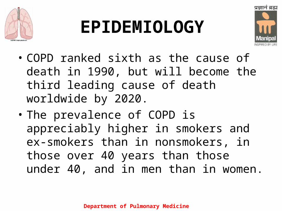
EPIDEMIOLOGY
• COPD ranked sixth as the cause of death in 1990, but will become the third leading cause of death worldwide by 2020.
• The prevalence of COPD is appreciably higher in smokers and ex-smokers than in nonsmokers, in those over 40 years than those under 40, and in men than in women.

Department of Pulmonary Medicine
RISK FACTORS Exposure to particles Tobacco smoke Indoor air pollution from heating and cooking with biomass in poorly vented dwellings (among women in developing countries) Occupational dusts (organic and inorganic) Outdoor air pollution Genes (α1 anti-trypsin deficiency) Airway hyperresponsiveness Lung Growth and Development Oxidative stress Gender Age Respiratory infections Socioeconomic status
Department of Pulmonary Medicine
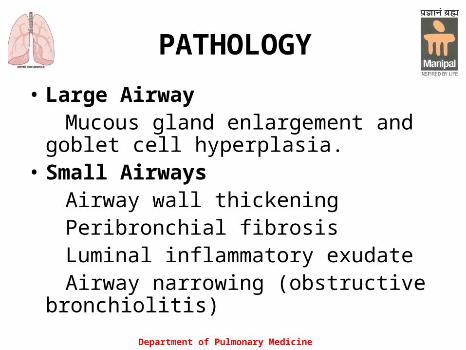
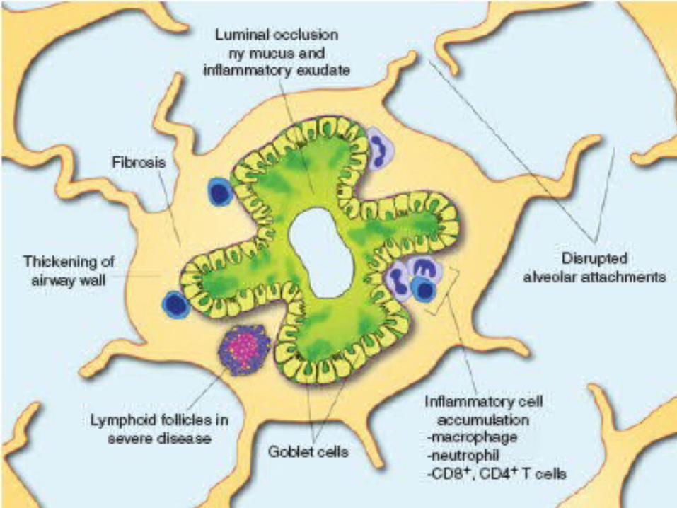
PATHOLOGY
• Large Airway Mucous gland enlargement and goblet cell
hyperplasia. • Small Airways Airway wall thickening Peribronchial fibrosis Luminal inflammatory exudate Airway narrowing (obstructive
bronchiolitis)
Department of Pulmonary Medicine
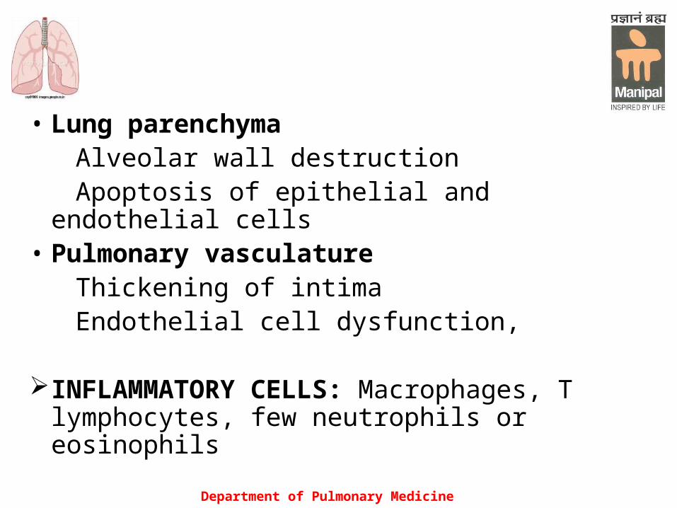
• Lung parenchyma Alveolar wall destruction Apoptosis of epithelial and endothelial cells• Pulmonary vasculature Thickening of intima Endothelial cell dysfunction,
INFLAMMATORY CELLS: Macrophages, T lymphocytes, few neutrophils or eosinophils
Department of Pulmonary Medicine
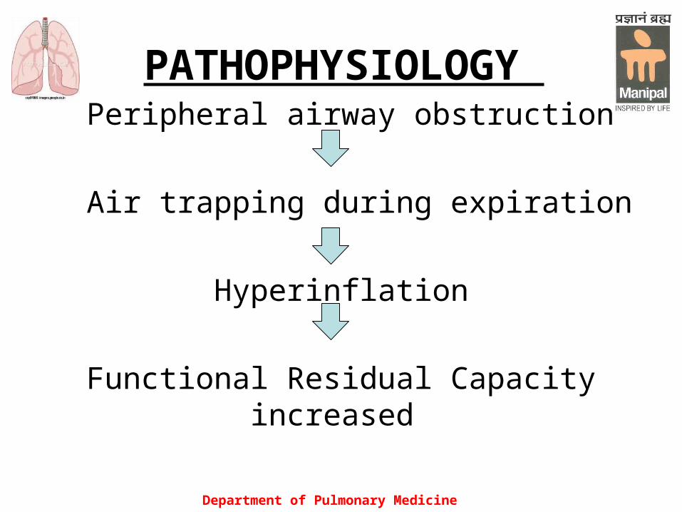
PATHOPHYSIOLOGY Peripheral airway obstruction
Air trapping during expiration
Hyperinflation
Functional Residual Capacity increased
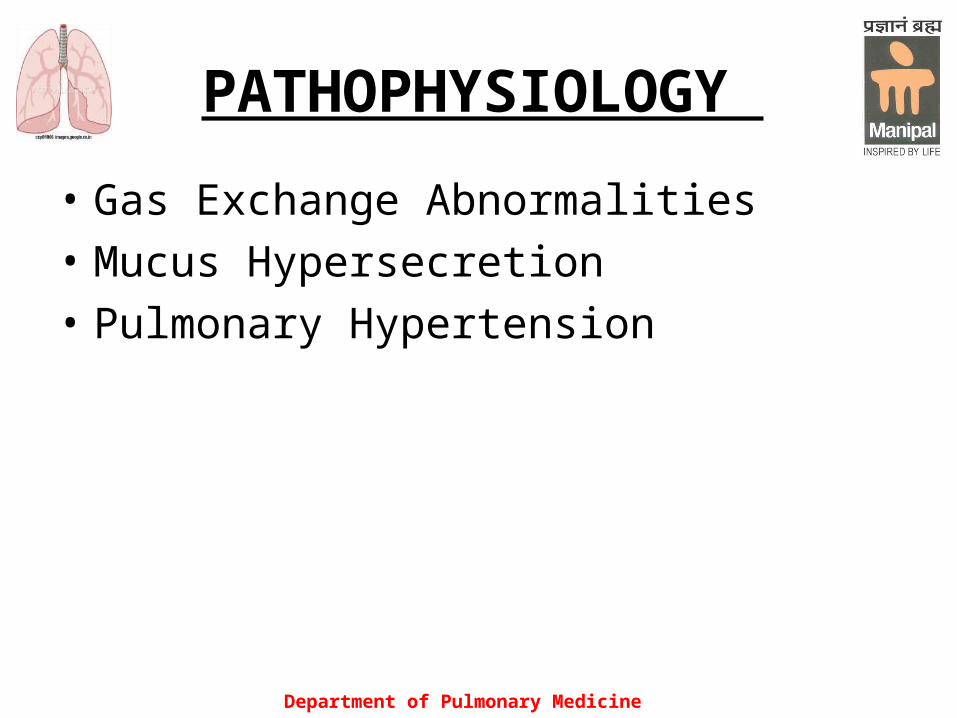
PATHOPHYSIOLOGY
• Gas Exchange Abnormalities
• Mucus Hypersecretion
• Pulmonary Hypertension
Department of Pulmonary Medicine

Department of Pulmonary Medicine
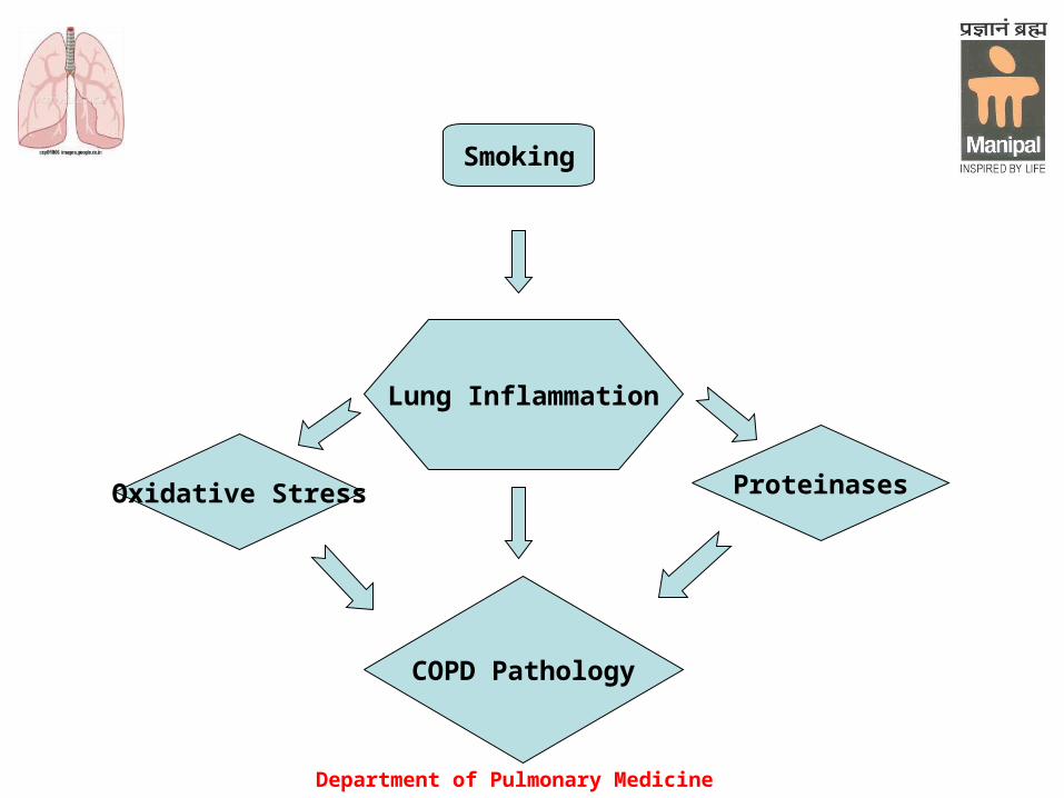
PATHOGENESIS
The inflammation in the respiratory tract of COPD patients appears to be an amplification of the normal inflammatory response of the respiratory tract to chronic irritants such as cigarette smoke
Department of Pulmonary Medicine
Smoking
Lung Inflammation
Oxidative Stress
COPD Pathology
Proteinases
Department of Pulmonary Medicine
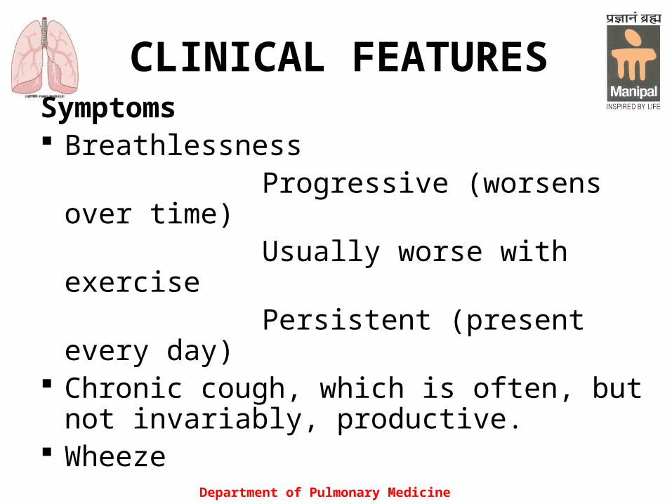
CLINICAL FEATURESSymptoms Breathlessness Progressive (worsens over time) Usually worse with exercise Persistent (present every day) Chronic cough, which is often, but not
invariably, productive. Wheeze

Department of Pulmonary Medicine
History of exposures to risk factors:
A smoking history of at least 20 pack
years is usual before symptoms develop
Smoke from home cooking and heating
fuels
Occupational dusts and chemicals Family history of COPD Weight loss and anorexia are features of severe
COPD Sleep quality is impaired in advanced COPD Hemoptysis
Department of Pulmonary Medicine
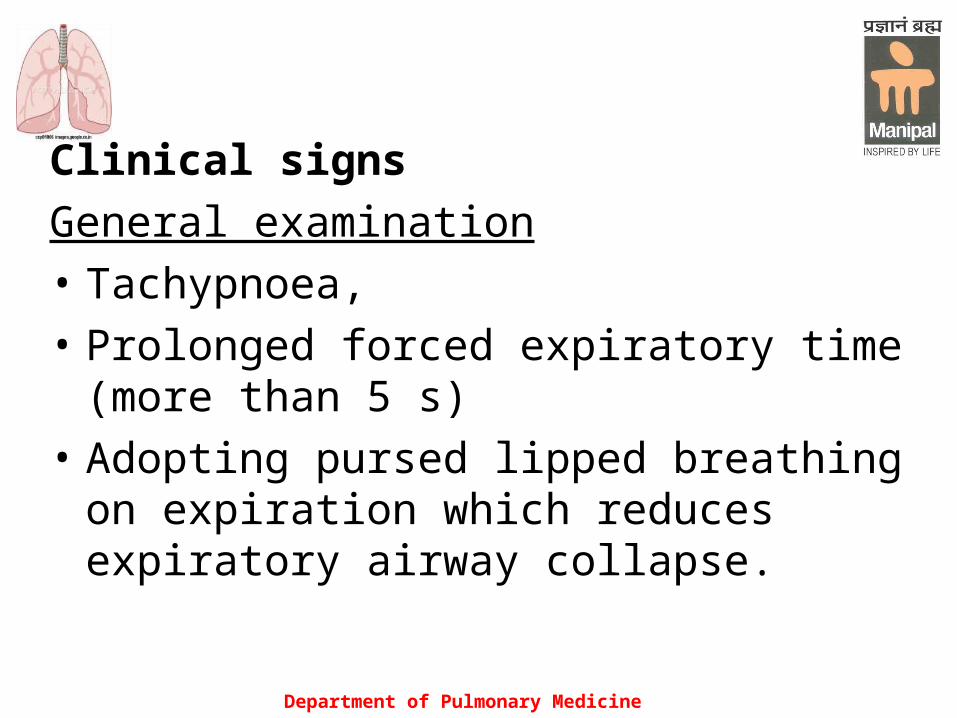
Clinical signs
General examination
• Tachypnoea,
• Prolonged forced expiratory time (more than 5 s)
• Adopting pursed lipped breathing on expiration which reduces expiratory airway collapse.

Department of Pulmonary Medicine
• Use of the accessory muscles of respiration• Adopt the position of leaning forward, supporting
themselves with their arms to fix the shoulder girdle
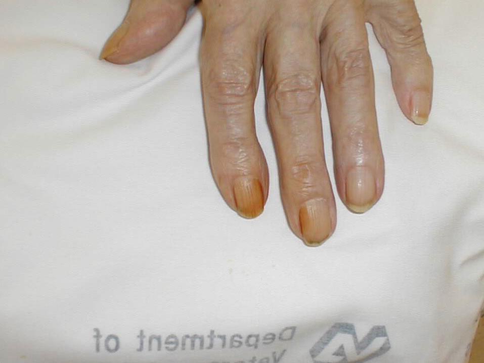
• Tar-stained fingers• Cyanosis in advanced disease • Flapping tremor• Weight loss• Finger clubbing is not a feature of COPD
Department of Pulmonary Medicine
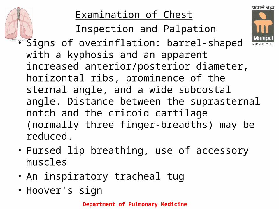
Examination of Chest
Inspection and Palpation• Signs of overinflation: barrel-shaped with a
kyphosis and an apparent increased anterior/posterior diameter, horizontal ribs, prominence of the sternal angle, and a wide subcostal angle. Distance between the suprasternal notch and the cricoid cartilage (normally three finger-breadths) may be reduced.
• Pursed lip breathing, use of accessory muscles • An inspiratory tracheal tug • Hoover's sign
Department of Pulmonary Medicine
• Indrawing of the suprasternal and supraclavicular fossas and of the intercostal muscles
Percussion• Hyper resonant note• Decreased hepatic and cardiac dullness
Ausculatation• Breath sounds may have a prolonged expiratory
phase, or may be uniformly diminished• Wheeze• Crackles may be heard particularly at the lung bases
Department of Pulmonary Medicine
Cardiovascular Examination• Difficulty in localizing the apex beat• Signs of pulmonary artery hypertension• Signs of right heart failure
SYSTEMIC FEATURES: Skeletal muscle wasting Osteoporosis Anxiety and Depression Increased risk of cardiovascular disease, respiratory infections diabetes, lung cancer
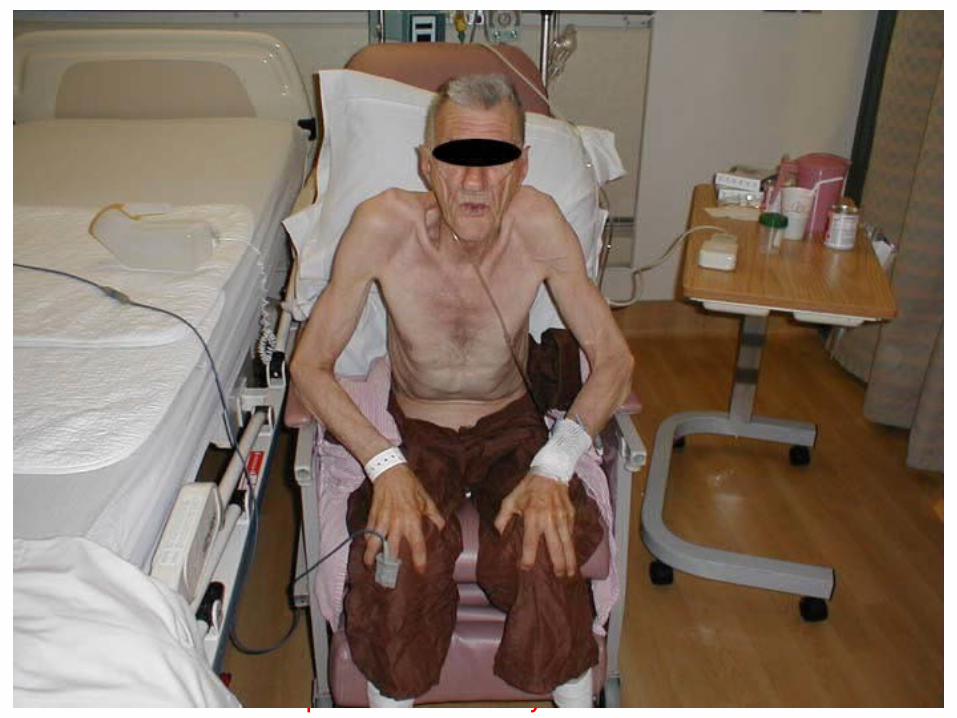
Type A: Pink Puffer (Emphysema Predominant)
• Major complaint is dyspnea,
• Cough is rare, with scant clear, mucoid sputum.
• Patients are thin, with recent weight loss common.
• They appear uncomfortable, with evident use of accessory muscles of respiration.
• Chest is very quiet without adventitious sounds.
• No peripheral edema.
Department of Pulmonary Medicine
Type B: Blue Bloater (Bronchitis Predominant)
• Major complaint is chronic cough, productive of mucopurulent sputum
• Dyspnea usually mild, though patients may note limitations to exercise.
• Patients frequently overweight and cyanotic but seem comfortable at rest.
• Peripheral edema is common.
• Chest is noisy, with rhonchi invariably present
Department of Pulmonary Medicine
Department of Pulmonary Medicine
MANAGEMENT
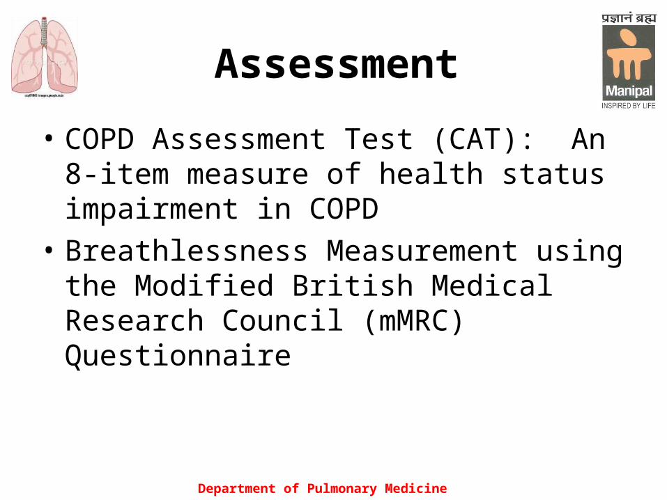
Assessment
• COPD Assessment Test (CAT): An 8-item measure of health status impairment in COPD
• Breathlessness Measurement using the Modified British Medical Research Council (mMRC) Questionnairewell to other measures of health status and predicts future mortality risk.
Department of Pulmonary Medicine
Department of Pulmonary Medicine
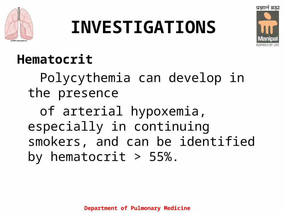
INVESTIGATIONS
Hematocrit
Polycythemia can develop in the presence
of arterial hypoxemia, especially in continuing smokers, and can be identified by hematocrit > 55%.

Department of Pulmonary Medicine
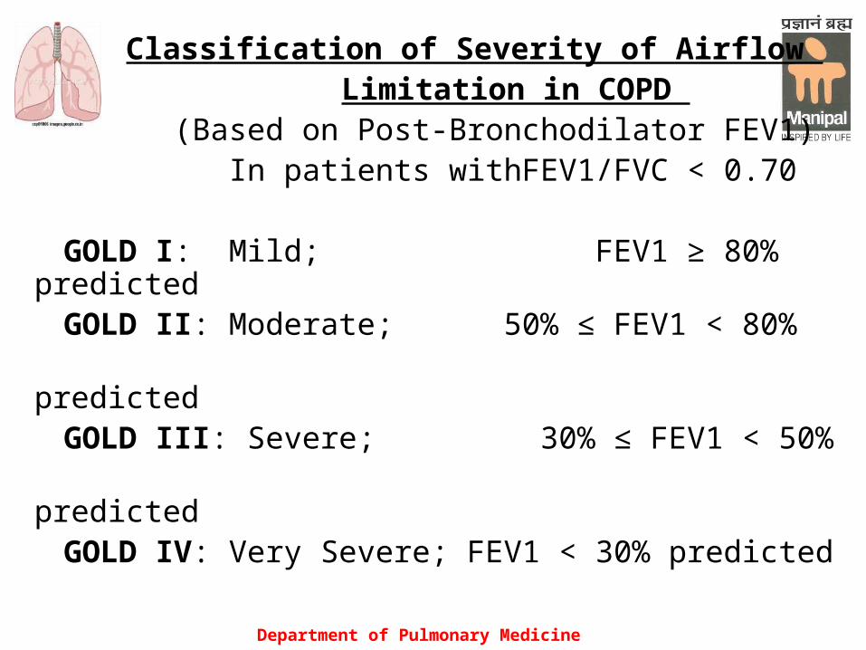
• Spirometry: The presence of a postbronchodilator
FEV1/FVC < 0.70 confirms the presence of persistent airflow limitation and thus COPD
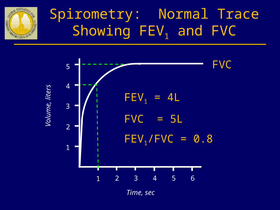
Spirometry: Normal Trace Showing FEV1 and FVC
1 2 3 4 5 6
1
2
3
4
Volu
me,
liters
Time, sec
FVC5
1
FEV1 = 4L
FVC = 5L
FEV1/FVC = 0.8
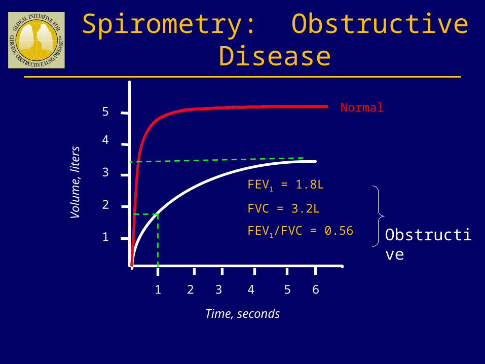
Spirometry: Obstructive Disease
Volu
me,
liters
Time, seconds
5
4
3
2
1
1 2 3 4 5 6
FEV1 = 1.8L
FVC = 3.2L
FEV1/FVC = 0.56
Normal
Obstructive
Department of Pulmonary Medicine
Classification of Severity of Airflow Limitation in COPD
(Based on Post-Bronchodilator FEV1) In patients withFEV1/FVC < 0.70 GOLD I: Mild; FEV1 ≥ 80% predicted GOLD II: Moderate; 50% ≤ FEV1 < 80% predicted GOLD III: Severe; 30% ≤ FEV1 < 50% predicted GOLD IV: Very Severe; FEV1 < 30% predicted
Department of Pulmonary Medicine
• Imaging
Chest X-ray Signs of hyperinflation (flattened diaphragm and an increase in the volume of the retrosternal air space), hyperlucency of the lungs, and rapid tapering of the vascular markings.
Computed tomography (CT)
Department of Pulmonary Medicine
• Arterial blood gas measurement
• Exercise testing• Alpha-1 antitrypsin deficiency
screening (when COPD develops under 45 years or with a strong family history of COPD.)
• Other investigations, including electrocardiography, echocardiography, radionucleotide scintigraphy, and magnetic resonance imaging.
Department of Pulmonary Medicine
TREATMENT 1. Smoking Cessation• Counseling• Pharmacotherapy Nicotine replacement products (nicotine gum,
inhaler, nasal spray, transdermal patch, sublingual tablet, or lozenge)
Other pharmacotherapy:The antidepressants bupropion and nortriptyline. Varenicline, a nicotinic acetylcholine receptor partial agonist that aids smoking cessation by relieving nicotine withdrawal symptoms and reducing the rewarding properties of nicotine

Department of Pulmonary Medicine
Drugs Used in COPD
β2-agonists Short-acting
(Salbutamol, Terbutaline)
Long-acting
(Formoterol, Salmeterol)
Department of Pulmonary Medicine
Drugs Used in COPD
Anticholinergics
Short-acting
Ipratropium bromide
Oxitropium bromide Long-acting Tiotropium
Drugs Used in COPD
Combination short-acting β 2-agonists plus anticholinergic in one inhaler
Salbutamol/Ipratropium
Methylxanthines Aminophylline Theophylline (SR)
Department of Pulmonary Medicine
Department of Pulmonary Medicine
Drugs Used in COPD
Inhaled glucocorticosteroids Beclomethasone, Budesonide, Fluticasone, Triamcinolone
Combination long-acting β 2-agonists plus glucocorticosteroids in one inhaler
Formoterol/Budesonide Salmeterol/Fluticasone
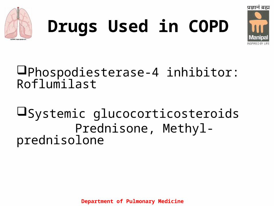
Drugs Used in COPD
Phospodiesterase-4 inhibitor: Roflumilast
Systemic glucocorticosteroids Prednisone, Methyl-prednisolone
Department of Pulmonary Medicine

Department of Pulmonary Medicine
OTHER PHARMACOLOGIC TREATMENTS
• Alpha-1 antitrypsin augmentation therapy.
• Vaccines:
Influenza vaccine (reduces serious illness and
death)
Pneumococcal vaccine (reduces incidence of
CAP)

OTHER PHARMACOLOGIC TREATMENTS
• Mucolytic agents (ambroxol, carbocysteine, iodinated glycerol)
Patients with viscous sputum may benefit
from mucolytics; overall benefits are very
small. • Antibioticsefits are very small• Antioxidant agents• Immunoregulators• Antitussives
Department of Pulmonary Medicine

Department of Pulmonary Medicine
OTHER PHARMACOLOGIC TREATMENTS
Oxygen Therapy Can be administered in three ways: longterm
continuous therapy, during exercise, and to relieve acute dyspnea.
The primary goal of oxygen therapy is to increase the baseline PaO2 to at least 8.0 kPa (60 mm Hg) at sea level and rest, and/or produce an SaO2 at least 90%, which will preserve vital organ function by ensuring adequate delivery of oxygen.

Department of Pulmonary Medicine
The long-term administration of oxygen(> 15 h/d) to patients with chronic respiratory failure has been shown to increase survival.
Long-term oxygen therapy is generally introduced in patients with COPD, who have PaO2 at or below 55 mm Hg or SaO2 at or below 88%, with or without hypercapnia

OTHER TREATMENTS
• Non invasive ventilation with LTOT in
a some selected patients may improve
survival
• Rehabilitation
Department of Pulmonary Medicine

Department of Pulmonary Medicine
Surgical Treatments
Bullectomy
Lung volume reduction surgery
Lung transplantation

Only three interventions influence the natural history of patients with COPD.
1.Smoking cessation
2.Oxygen therapy in chronically hypoxemic patients
3.Lung volume reduction surgery in selected patients with emphysema.There is currently suggestive, but not definitive, evidence that the use of inhaled glucocorticoids may alter mortality rate.
Department of Pulmonary Medicine

Department of Pulmonary Medicine
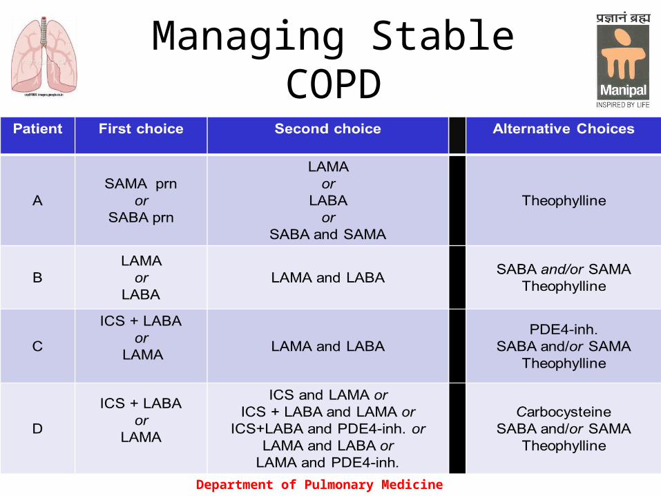
Patient
Characteristic Spirometric Classification
Exacerbations per year
mMRC CAT
ALow Risk
Less SymptomsGOLD 1-2 ≤ 1 0-1 < 10
BLow Risk
More SymptomsGOLD 1-2 ≤ 1 > 2 ≥ 10
CHigh Risk
Less SymptomsGOLD 3-4 > 2 0-1 < 10
DHigh Risk
More SymptomsGOLD 3-4 > 2 > 2
≥ 10
Combined Assessment
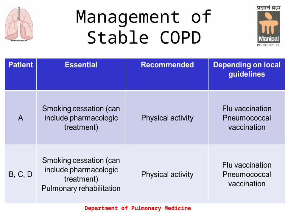
Management of Stable COPD
Department of Pulmonary Medicine
Managing Stable COPD
Department of Pulmonary Medicine

Department of Pulmonary Medicine
MANAGE EXACERBATIONS
An exacerbation of COPD is defined as an event in the natural course of the disease characterized by a change in the patient’s baseline dyspnea, cough, and/or sputum that is beyond normal day-to-day variations, is acute in onset, and may warrant a change in regular medication in a patient with underlying COPD.
Department of Pulmonary Medicine
The most common causes of COPD exacerbations are viral upper respiratory tract infections and infection of the tracheobronchial tree. .
Streptococcus pneumoniae, Hemophilus influenzae, and Moraxella catarrhalis are the most common bacterial pathogens involved in COPD exacerbations.
Department of Pulmonary Medicine
Inhaled bronchodilators (particularly inhaled Β 2-agonists with or without anticholinergics) and oral glucocorticosteroids are effective treatments for exacerbations of COPD.
Antibiotics if clinical signs of airway infection (e.g., increased sputum, purulence)
Department of Pulmonary Medicine
Noninvasive mechanical ventilation in exacerbations improves respiratory acidosis, increases pH, decreases the need for endotracheal intubation, and reduces PaCO2, respiratory rate, severity of breathlessness, the length of hospital stay, and mortality.
Medications and education to help prevent future exacerbations should be considered as part of follow-up
Department of Pulmonary Medicine
OXYGEN THERAPY
Department of Pulmonary Medicine
INTRODUCTION
Oxygen is the substrate that cells use in the greatest quantity and upon which aerobic metabolism and cell integrity depend. Since the tissues have no storage system for oxygen, a continuous supply at a rate that matches changing metabolic requirements is necessary to maintain aerobic metabolism and normal cellular function.
Department of Pulmonary Medicine
PRINCIPLES
In general, maintain SaO2 >90%, though preferably >95%.

Department of Pulmonary Medicine
Devices For Providing Oxygen
• Oxygen supply (cylinder or wall unit)
• Nasal cannula
• Face mask
• Venturi mask

Department of Pulmonary Medicine
NASAL CANNULA• The nasal cannula is a low-flow oxygen administration
system designed to add oxygen to room air when the patient inspires.
• A nasal cannula provides up to 44% oxygen.• The ultimate inspired oxygen concentration is
determined by the oxygen flow rate through the cannula and how deeply the patient breathes (tidal volume).

Department of Pulmonary Medicine
• Increasing the oxygen flow by 1 L/min (starting with 1 L/min) will increase the inspired oxygen concentration by approximately 4%:
— 1 L/min: 21% to 24% — 2 L/min: 25% to 28% — 3 L/min: 29% to 32% — 4 L/min: 33% to 36% — 5 L/min: 37% to 40% — 6 L/min: 41% to 44%

Department of Pulmonary Medicine
Department of Pulmonary Medicine
FACE MASK
• A simple face mask delivers low oxygen flow to the patient’s nose and mouth. A partial rebreathing mask consists of a face mask with an attached reservoir bag
Department of Pulmonary Medicine
• A face mask can supply up to 60% oxygen with flow rates of 6 to 10 L/min. A face mask with oxygen reservoir nonrebreathing mask) provides up to 90% to 100% oxygen with flow rates of 9 to 15 L/min. In this system a constant flow of oxygen enters an attached reservoir.

Department of Pulmonary Medicine
Use a face mask with a reservoir for patients who Are seriously ill, responsive, and have adequate
ventilation but require high oxygen concentrations

Department of Pulmonary Medicine

Department of Pulmonary Medicine
VENTURI MASK• A Venturi mask enables a more reliable and
controlled delivery of oxygen concentrations from 24% to 50%. Use the Venturi mask for patients who retain carbon dioxide (CO2). Patients who have chronic high levels of CO2 in their blood and moderate-to-severe hypoxemia may develop respiratory depression if the drive stimulating them to breathe (oxygen) is reduced.
Department of Pulmonary Medicine
• Delivered oxygen concentrations can be adjusted to 24%, 28%, 35%, and 40% using a flow rate of 4-8 L/min and 40% to 50% using a flow rate of 10-12 L/min.
Department of Pulmonary Medicine
MONITORING
Adequacy and changes in arterial oxygen saturation can be continuously monitored by pulse oximetry and intermittent or continuous invasive blood gas analysis.

Department of Pulmonary Medicine
REFERENCES
• Harrison's Principles of Internal Medicine, 18th Edition
• GOLD. 2011 (revised) • Murray & Nadel's Textbook of Respiratory
Medicine, 4th ed. • CMDT 2010

Department of Pulmonary Medicine
THANK YOU
Department of Pulmonary Medicine
Department of Pulmonary Medicine
Department of Pulmonary Medicine