Embed Size (px)
Citation preview

Narmeen Hassan MS5Final year medical studentDow Medical College, Pakistan

Case Introduction Müllerian Development Congenital Müllerian Anommalities Causes Clinical Presentation Diagnosis Treatment Prognosis

17 yr single female G0P0 presented for evaluation of uterine and vaginal anomaly. Her menarche started at age 12 ½ with regular cycles lasting 5 days. Her pelvic examination showed a longitudinal vaginal septum with cervix on either side indicating a didelphic vagina. An ultrasound showed ill defined uterine anomaly.
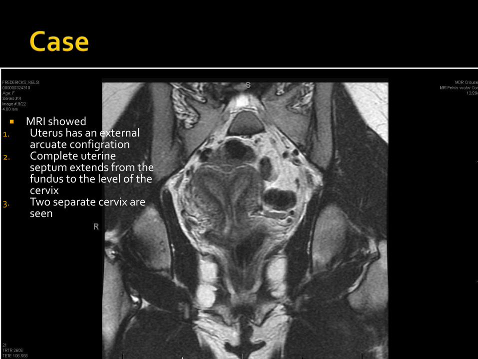
MRI showed 1. Uterus has an external
arcuate configration2. Complete uterine
septum extends from the fundus to the level of the cervix
3. Two separate cervix are seen

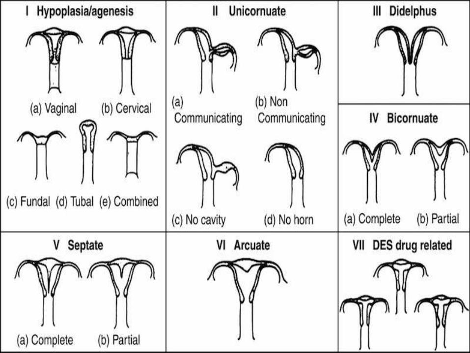
Congenital uterine anomalies are malformations of the uterus that develop during embryonic life. Congenital uterine anomalies Occur in less than 5% of all women, but have been noted in up to 25% of women who have had miscarriages and/or deliveriesof premature babies. When a woman is in her mother’s womb, her uterus develops as two separate halves that fuse together before the woman is born. The images illustrate a normal and abnormaluterus visualized by hysterosalpingogram.
Uterine anomalies are quite often asymptomatic and so are hard to recognize
Uterine anomalies are often an incidental diagnosis while seeing the patient for a different complaint


Unicornuate: only one half of the uterus has developed Didelphys: the two halves of the uterus remain separate Bicornuate: an abnormal, indented external uterine surface and
two endometrial cavities Septate: a normal external uterine surface but two endometrial
cavities Arcuate: a normal external uterine surface with a 1 cm or less
indentation into the endometrial cavity

In the majority of cases, the cause of a congenital uterine anomaly is unknown. Most women with these malformations (more than 90%) have a normal number of chromosomes, 46 XX.
Between 1938 and 1971, some pregnant women were treated with diethylstilbestrol (DES) which is a synthetic estrogen to help prevent miscarriages and premature deliveries. Women who were exposed to DES while in their mother’s womb are at increased risk for having a congenital uterine anomaly and vaginal adenosis
At this time, there are no well-established risk factors for the development of a congenital uterine anomaly, and there is no way to prevent development of a congenital uterine anomaly.

Although congenital uterine anomalies are present at birth, these malformations are usually without symptoms.. Congenital uterine anomalies typically do not cause a woman to have difficulty getting pregnant. However, these malformations are often discovered during evaluations for infertility or pregnancy loss.
The Patient can present with:
Pelvic pain (cyclic or non-cyclic) Dysmenorrhea Abnormal vaginal bleeding Uterine rupture during pregnancy Recurrent pregnancy loss Patient may have a concurrent renal abnormalities

Gynecologic Ultrasonography Pelvic MRI :MRI is considered the preferred
modality due to its multiplanar capabilities as well as its ability to evaluate the uterine outline, junctional zone, and other pelvic anatomy
Hysterosalpingography : Unable to outline the exterior surface of the uterus.
Laparoscopy Hysteroscopy

There are no non-surgical treatments for congenital uterine anomalies. Recommendations for surgical treatment of congenital uterine anomalies depend on the particular anomaly and the woman’s reproductive history.
Surgical intervention should be considered when a septate uterus is found in association with adverse reproductive outcome.
Bicornuate, unicornuate and didelphic uteri rarely require surgical treatment.

Many women with a congenital uterine anomaly have no medical or reproductive problems.
Septate uteri have some of the poorest reproductive outcomes of müllerian duct anomalies.

The septate uterus is a result of absent or incomplete resorption of the intervening uterovaginal septum following fusion of the müllerian ducts. It is the most common congenital anomaly of the uterus, comprising approximately 55% of all anomalies
A septum is primarily composed of fibromusculartissue that may project minimally from the uterine fundus or may extend to the cervical os, almost completely dividing the uterine cavity in two. Septa also may be segmental, resulting in partial communications between the two side

Complications: 90% miscarriage rate, a patient with a septateuterus does not usually not have difficulty conceiving, but a septate uterus is associated with the highest rate of pregnancy loss of the Müllerian duct anomalies Raga et al reported a 25.5% incidence of early miscarriage (< 13 weeks) and a 6.2% incidence of late miscarriage (14 to 22 weeks) in women with septateuterus. Premature birth rates are increased at approximately 21% and fetal survival rates are estimated at 32%
Differential Diagnosis: Bicornuate uterus-- the shape of the external uterine contour is crucial to differentiate a septate uterus from a bicornuate uterus, because widely different clinical and interventional approaches are assigned to each anomaly.

Treatment: Surgical intervention, Hysteroscopicseptal incision/hysteroscopic metroplasty. In septateuterus, but not in bicornuate uterus, the septum can be shaved off during hysteroscopy (metroplasty) to form a single uterine cavity without perforating the uterus
Prognosis: Reproductive outcome has been shown to improve after resection of the septum, with reported decreases in the spontaneous abortion rate from 88% to 5.9% after hysteroscopic metroplasty.

Understanding the embryologic origin of the defect of mullerian anomalies is the key to its correct diagnosis
Women with a history of miscarriage, pre-term deliveries, have a higher incidence of anomalies

American Society of Reproductive Medicine The American Congress of Obstetricians and
Gynecologists Medscape UptoDate