Embed Size (px)
Citation preview


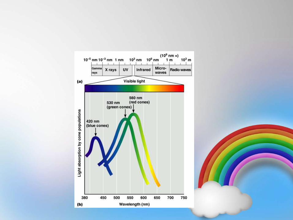
The Electromagnetic Spectrum.
• how we see colour?..
• how colour is described?..
• what colour blindness is?..
COLOUR VISION:Definition: Colour vision is the ability of the
human being to identify & distinguish different
colours.
• Rods & cones: light sensitive receptors
– Rods – Night vision & vision in shades of gray.
– Cones – daylight or bright light vision, colour
vision & acuity of vision
• Any colour has: hue, intensity & saturation.
– Hue – colour that is red, yellow, blue etc.
– Intensity – bright or pale
– Saturation – degree of freedom from dilution with
white
• In man cone vision sensitivity 1/100th – 1/1000th
that of rod vision.
• The peak sensitivity of scotopic vision is ≈ 500 nm
and photopic vision is ≈ 560 nm.
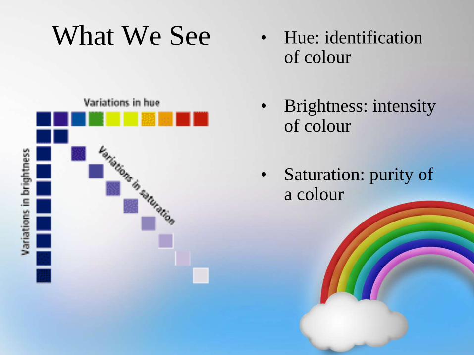
What We See • Hue: identification of colour
• Brightness: intensity of colour
• Saturation: purity of a colour
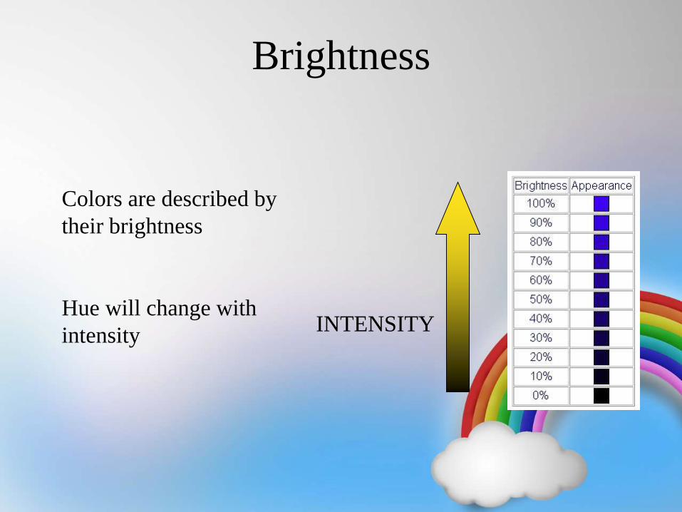
Brightness
Colors are described by
their brightness
Hue will change with
intensityINTENSITY
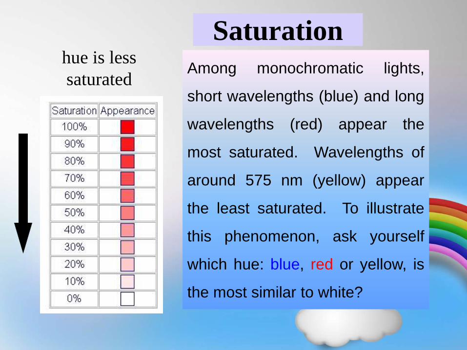
Saturation
Among monochromatic lights,
short wavelengths (blue) and long
wavelengths (red) appear the
most saturated. Wavelengths of
around 575 nm (yellow) appear
the least saturated. To illustrate
this phenomenon, ask yourself
which hue: blue, red or yellow, is
the most similar to white?
hue is less
saturated
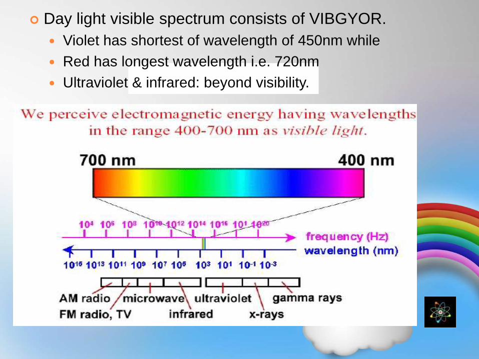
Day light visible spectrum consists of VIBGYOR.
Violet has shortest of wavelength of 450nm while
Red has longest wavelength i.e. 720nm
Ultraviolet & infrared: beyond visibility.
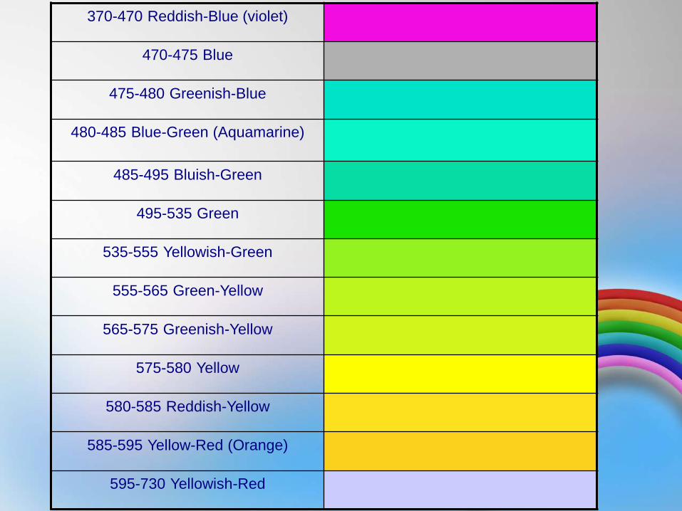
370-470 Reddish-Blue (violet)
470-475 Blue
475-480 Greenish-Blue
480-485 Blue-Green (Aquamarine)
485-495 Bluish-Green
495-535 Green
535-555 Yellowish-Green
555-565 Green-Yellow
565-575 Greenish-Yellow
575-580 Yellow
580-585 Reddish-Yellow
585-595 Yellow-Red (Orange)
595-730 Yellowish-Red
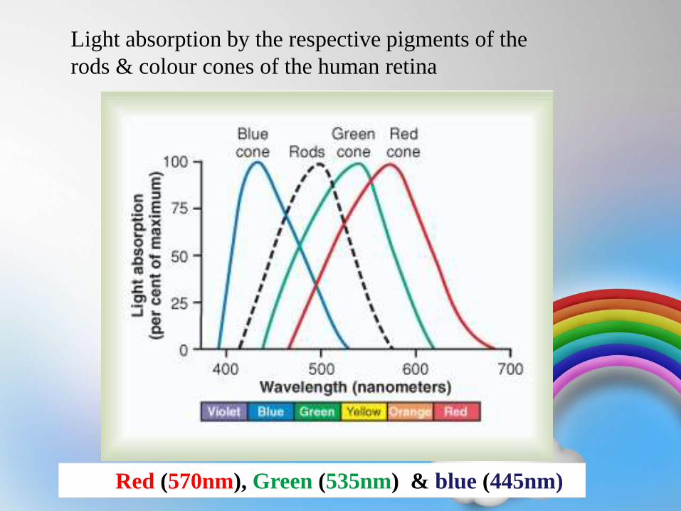
Light absorption by the respective pigments of the
rods & colour cones of the human retina
Red (570nm), Green (535nm) & blue (445nm)
• Primary colours are 3:
– Red(720- 650nm), Green(575-490nm) & blue(490-450nm)
– White – is mixture of all colours (equal stimulation of all
red, green, blue)
– Black is absence of colour but it is a positive sense so blind
eye does not see black, it sees nothing.
• Cones contain a pigment called as cone pigment:-
Photopsin + Retinal. Retinal portion is same in both rods
and cones only opsin part is diff. in cone pigment.
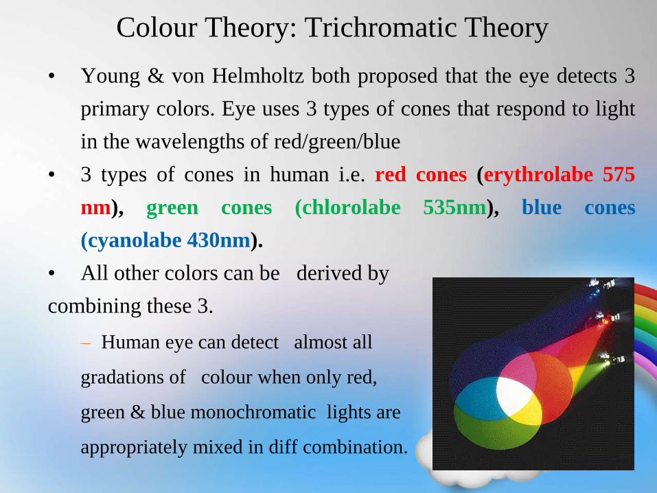
Colour Theory: Trichromatic Theory
• Young & von Helmholtz both proposed that the eye detects 3
primary colors. Eye uses 3 types of cones that respond to light
in the wavelengths of red/green/blue
• 3 types of cones in human i.e. red cones (erythrolabe 575
nm), green cones (chlorolabe 535nm), blue cones
(cyanolabe 430nm).
• All other colors can be derived by
combining these 3.
– Human eye can detect almost all
gradations of colour when only red,
green & blue monochromatic lights are
appropriately mixed in diff combination.
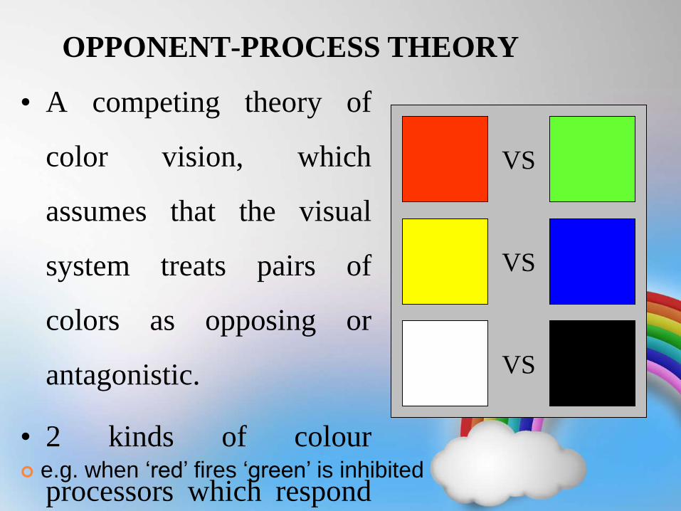
OPPONENT-PROCESS THEORY
• A competing theory of
color vision, which
assumes that the visual
system treats pairs of
colors as opposing or
antagonistic.
• 2 kinds of colour
processors which respond
VS
VS
VS
e.g. when ‘red’ fires ‘green’ is inhibited
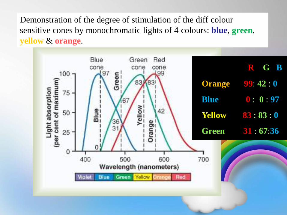
Demonstration of the degree of stimulation of the diff colour
sensitive cones by monochromatic lights of 4 colours: blue, green,
yellow & orange.
R G B
Orange - 99: 42 : 0
Blue - 0 : 0 : 97
Yellow - 83 : 83 : 0
Green - 31 : 67:36
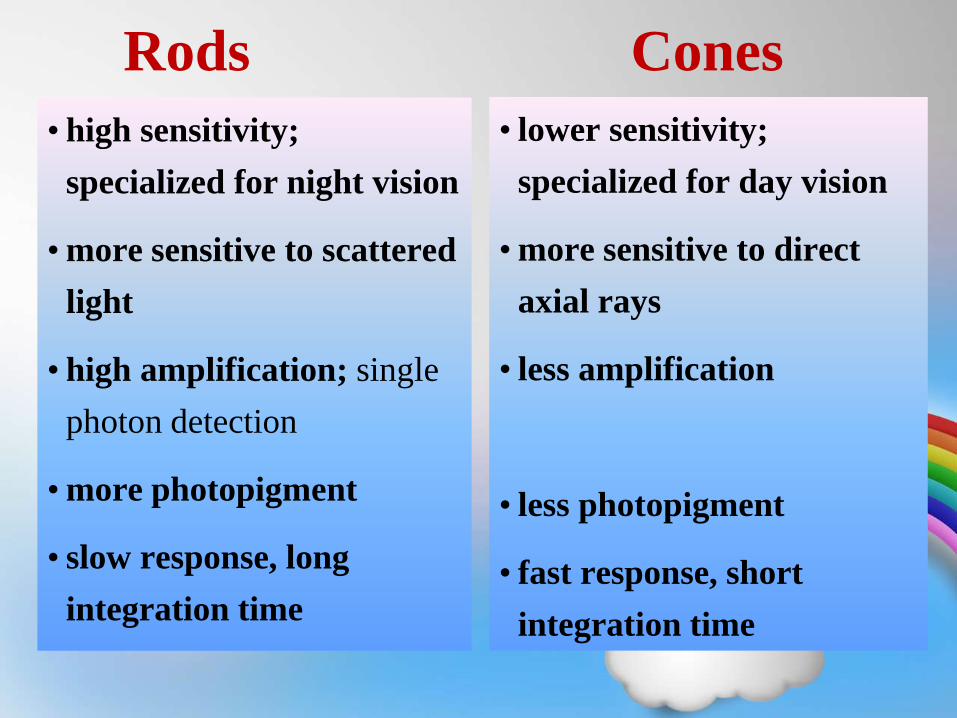
Rods Cones
• high sensitivity;
specialized for night vision
• more sensitive to scattered
light
• high amplification; single
photon detection
• more photopigment
• slow response, long
integration time
• lower sensitivity;
specialized for day vision
• more sensitive to direct
axial rays
• less amplification
• less photopigment
• fast response, short
integration time
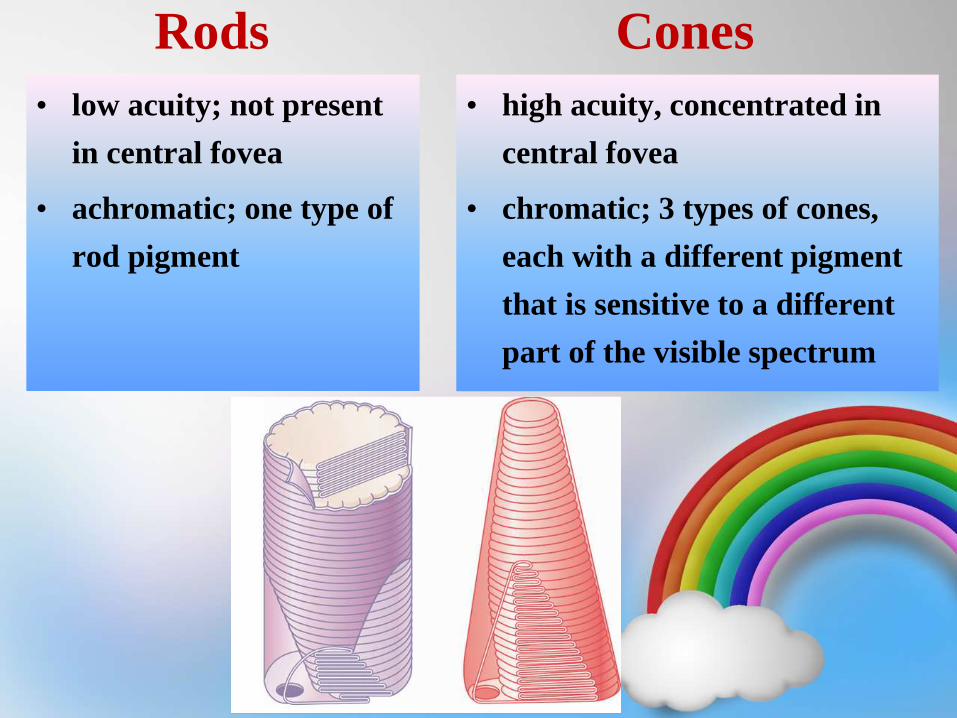
Rods Cones
• low acuity; not present
in central fovea
• achromatic; one type of
rod pigment
• high acuity, concentrated in
central fovea
• chromatic; 3 types of cones,
each with a different pigment
that is sensitive to a different
part of the visible spectrum
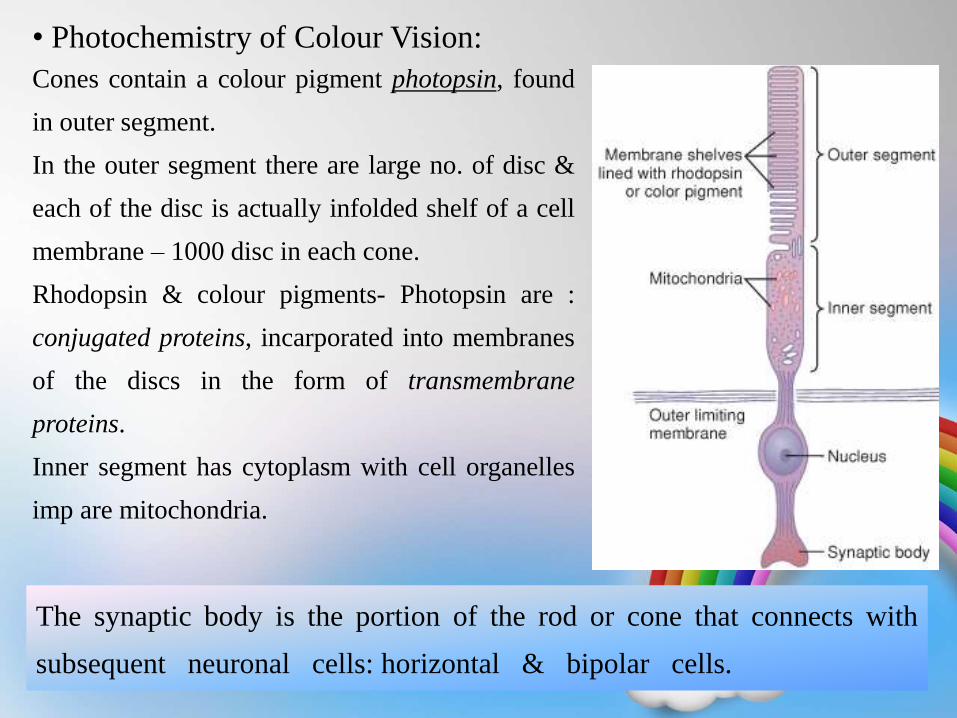
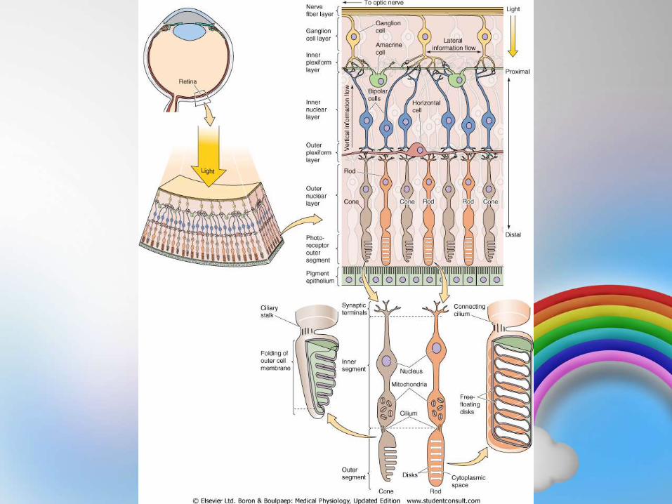
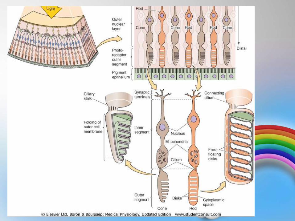
• Photochemistry of Colour Vision:
Cones contain a colour pigment photopsin, found
in outer segment.
In the outer segment there are large no. of disc &
each of the disc is actually infolded shelf of a cell
membrane – 1000 disc in each cone.
Rhodopsin & colour pigments- Photopsin are :
conjugated proteins, incarporated into membranes
of the discs in the form of transmembrane
proteins.
Inner segment has cytoplasm with cell organelles
imp are mitochondria.
The synaptic body is the portion of the rod or cone that connects with
subsequent neuronal cells: horizontal & bipolar cells.
• Cones contain a pigment called as cone
pigment- Photopsin + Retinal.
• For rods it is: Rhodopsin + Retinal
• Thus Retinal portion is same in both
rods and cones, only opsin part is diff
in cone pigment.
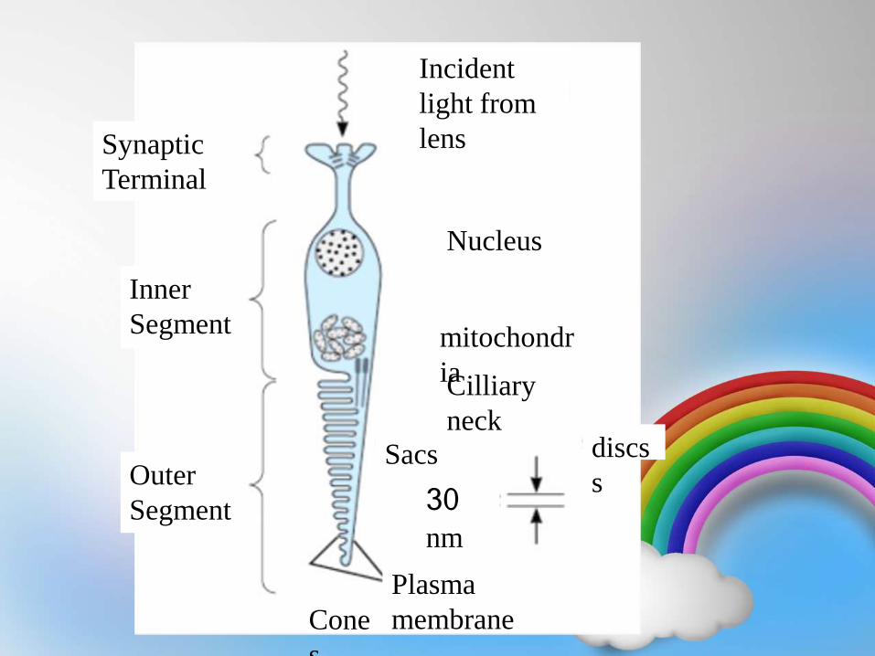
Cone
s
Plasma
membrane
Sacs discs
s
Cilliary
neck
mitochondr
ia
Nucleus
Incident
light from
lensSynaptic
Terminal
Inner
Segment
Outer
Segment 30 nm
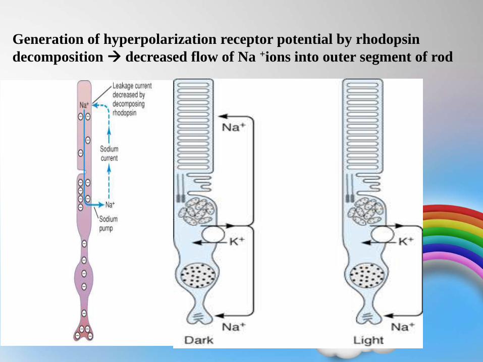
Generation of hyperpolarization receptor potential by rhodopsin
decomposition decreased flow of Na +ions into outer segment of rod
Photopsin /
Colour Pigment /
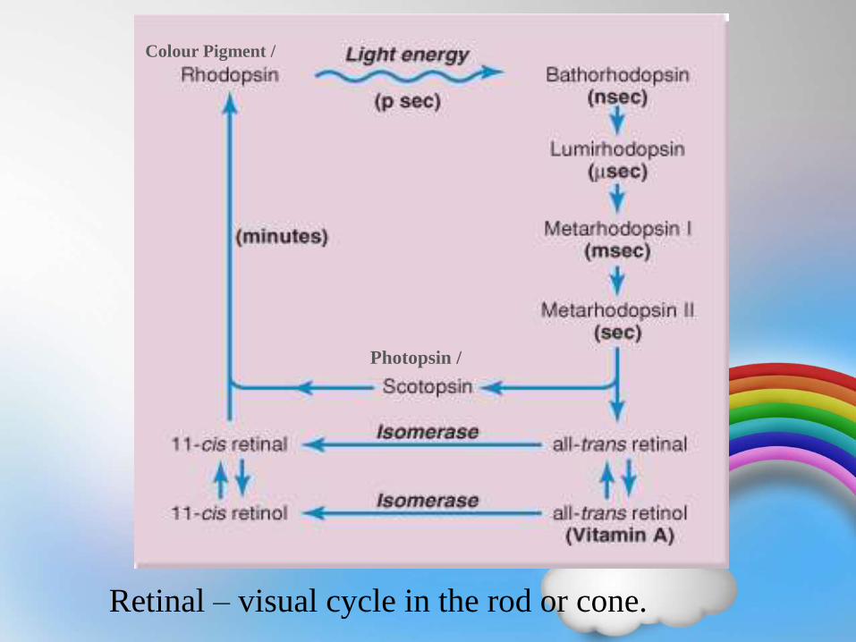
Retinal – visual cycle in the rod or cone.
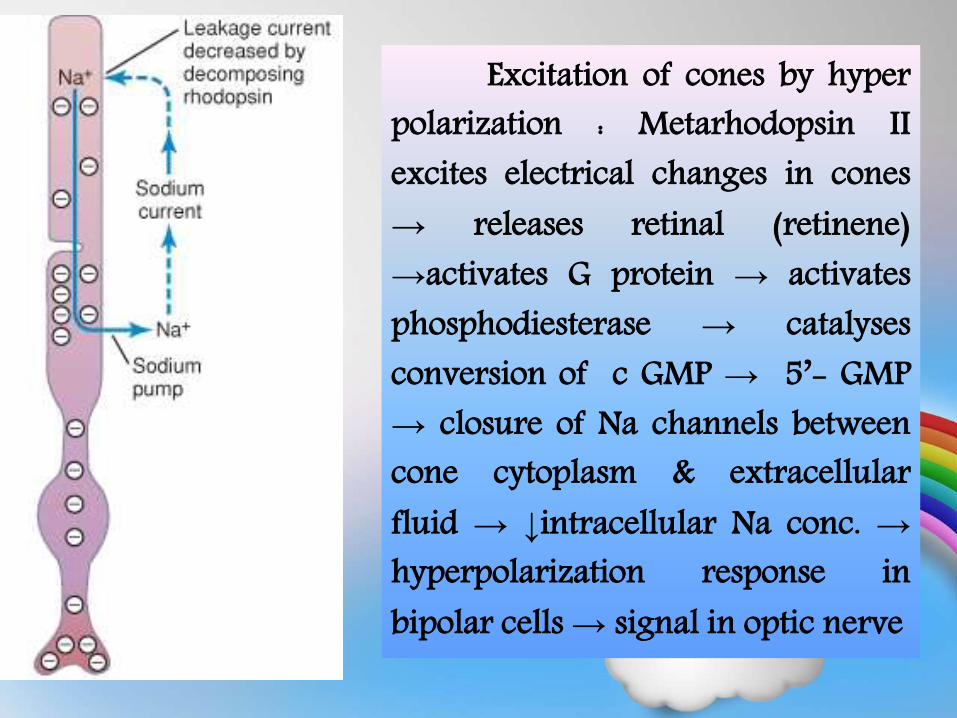
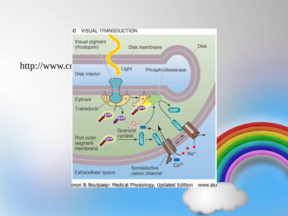
Excitation of cones by hyperpolarization : Metarhodopsin IIexcites electrical changes in cones→ releases retinal (retinene)→activates G protein → activatesphosphodiesterase → catalysesconversion of c GMP → 5’- GMP→ closure of Na channels betweencone cytoplasm & extracellularfluid → ↓intracellular Na conc. →hyperpolarization response inbipolar cells→ signal in optic nerve
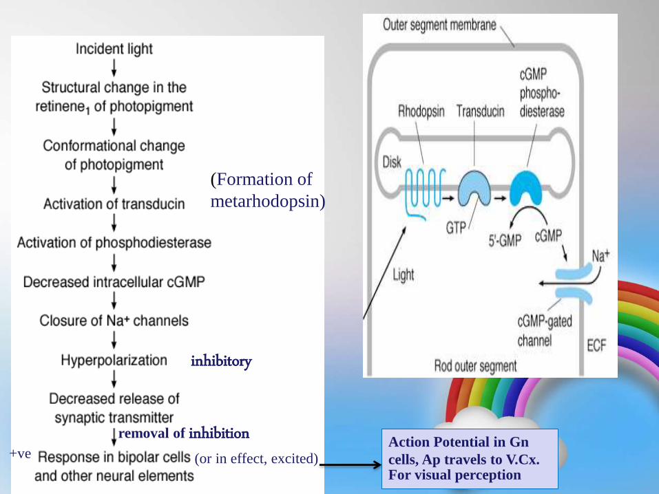
Action Potential in Gn
cells, Ap travels to V.Cx. For visual perception
+ve
removal of inhibition
(or in effect, excited)
(Formation of
metarhodopsin)
inhibitory
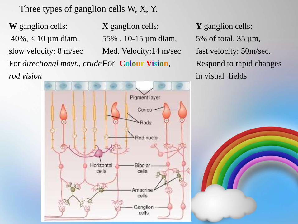
Three types of ganglion cells W, X, Y.
W ganglion cells:
40%, < 10 µm diam.
slow velocity: 8 m/sec
For directional movt., crude
rod vision
X ganglion cells:
55% , 10-15 µm diam,
Med. Velocity:14 m/sec
For Colour Vision,
Y ganglion cells:
5% of total, 35 µm,
fast velocity: 50m/sec.
Respond to rapid changes
in visual fields
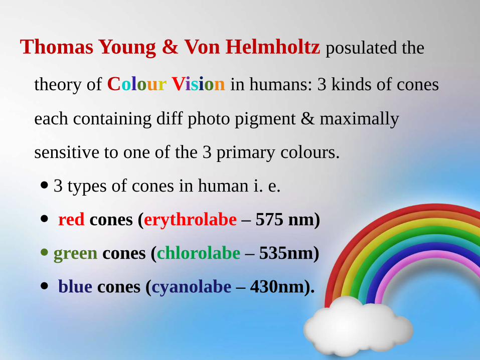
Thomas Young & Von Helmholtz posulated the
theory of Colour Vision in humans: 3 kinds of cones
each containing diff photo pigment & maximally
sensitive to one of the 3 primary colours.
3 types of cones in human i. e.
red cones (erythrolabe – 575 nm)
green cones (chlorolabe – 535nm)
blue cones (cyanolabe – 430nm).
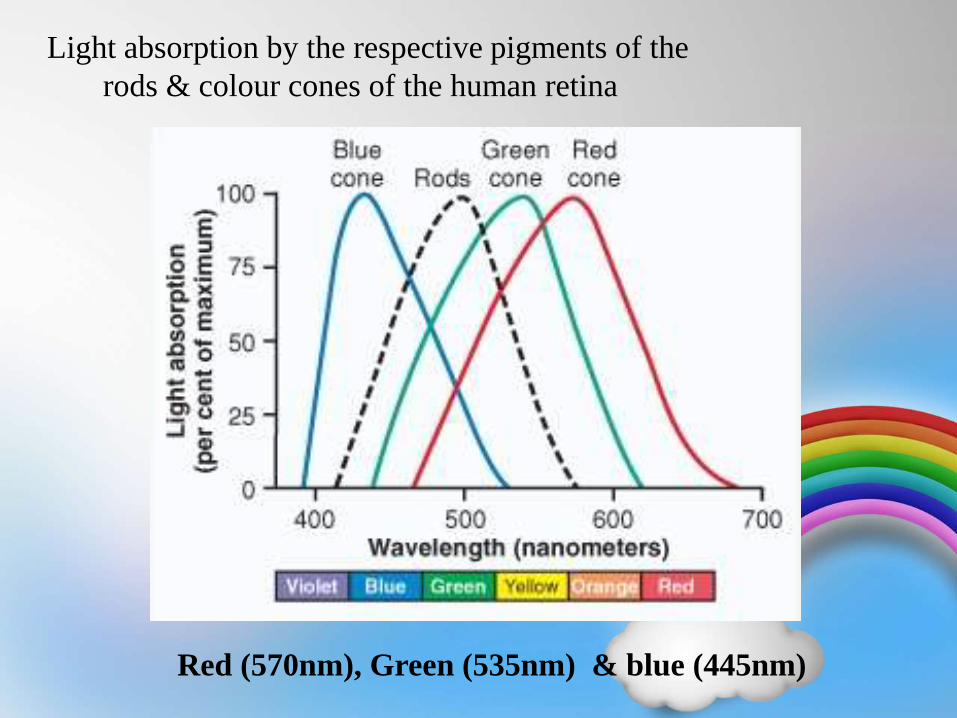
Light absorption by the respective pigments of the
rods & colour cones of the human retina
Red (570nm), Green (535nm) & blue (445nm)
Perception of colour
• At retinal level by ganglion cells
• At lateral geniculate body
• At visual cortex
Colour Contrast Mechanism by Ganglion
Cells:
• Some ganglion cells are excited by only one colour type of cone but
inhibited by second type. e.g. red causing excitation ( by direct
excitatory route thru depolarizing bipolar cells) , green inhibition
(by indirect inhibitory route thru hyperpolarizing bipolar cells) &
vice a versa.
• Similarly blue cones & combination of red & green cones for
yellow colour act as contrast for each other.
• Therefore by this colour contrast mechanism, colour differentiation
begins at retina itself & is not entirely a function of the brain.
– Thus, each colour contrast type of ganglion cell is
excited by one colour but inhibited by the
• Visual pathway-
• Ganglion cells→→optic nerves→→optic chiasma→→ crossing
of nasal half to opposite side →→ optic tract →→ synapse at
LGB →→ Geniculo calcarine fibers →→ Primary visual cortex.
• Analysis of visual detail, colour & conscious vision: From
primary visual cortex (Brodmann’s area no.17) into visual area II
(Brodmann’s area no.18) →→ medial, inferior & ventral region
of occipital and temporal cortex.
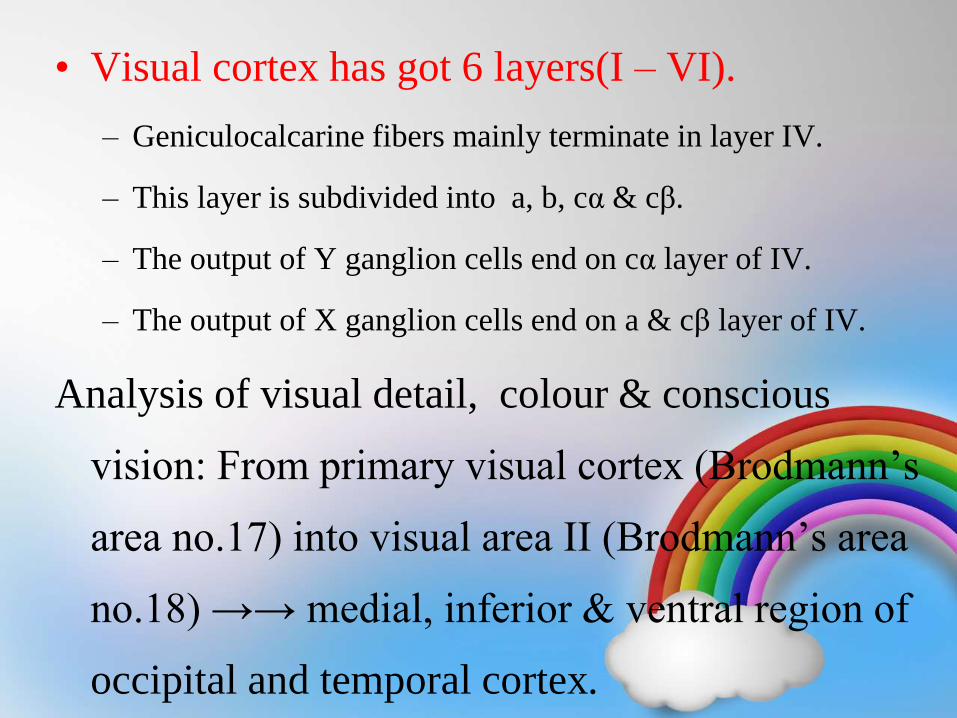
• Visual cortex has got 6 layers(I – VI).
– Geniculocalcarine fibers mainly terminate in layer IV.
– This layer is subdivided into a, b, cα & cβ.
– The output of Y ganglion cells end on cα layer of IV.
– The output of X ganglion cells end on a & cβ layer of IV.
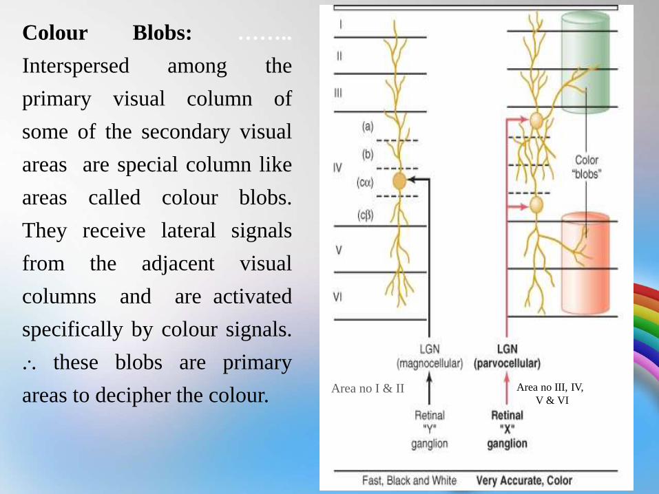
Area no I & II Area no III, IV,
V & VI
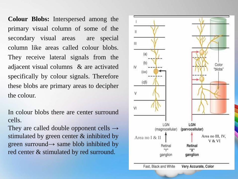
Colour Blobs: Interspersed among the
primary visual column of some of the
secondary visual areas are special
column like areas called colour blobs.
They receive lateral signals from the
adjacent visual columns & are activated
specifically by colour signals. Therefore
these blobs are primary areas to decipher
the colour.
In colour blobs there are center surround
cells.
They are called double opponent cells →
stimulated by green center & inhibited by
green surround→ same blob inhibited by
red center & stimulated by red surround.
•At retinal level : by Ganglion cells
By this colour contrast mechanism, colour
differentiation begins at retina itself & is not entirely a
function of the brain.
Thus, each colour contrast type of ganglion cell is
excited by one colour but inhibited by the “opponent
colour”.
•At lateral geniculate body level : by parvocellualr
pathway
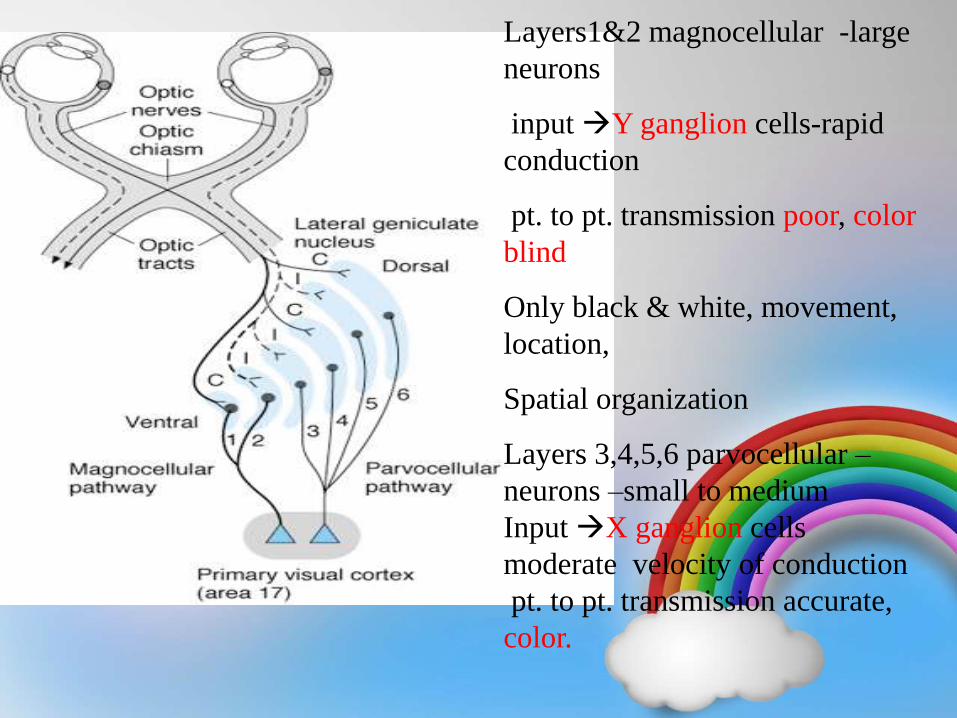
Layers1&2 magnocellular -large
neurons
input Y ganglion cells-rapid
conduction
pt. to pt. transmission poor, color
blind
Only black & white, movement,
location,
Spatial organization
Layers 3,4,5,6 parvocellular –
neurons –small to medium
Input X ganglion cells
moderate velocity of conduction
pt. to pt. transmission accurate,
color.
• Visual cortex has got 6 layers(I – VI).
– Geniculocalcarine fibers mainly terminate in layer IV.
– This layer is subdivided into a, b, cα & cβ.
– The output of Y ganglion cells end on cα layer of IV.
– The output of X ganglion cells end on a & cβ layer of IV.
Analysis of visual detail, colour & conscious
vision: From primary visual cortex (Brodmann’s
area no.17) into visual area II (Brodmann’s area
no.18) →→ medial, inferior & ventral region of
occipital and temporal cortex.
Area no I & II Area no III, IV,
V & VI
Colour Blobs: ……..
Interspersed among the
primary visual column of
some of the secondary visual
areas are special column like
areas called colour blobs.
They receive lateral signals
from the adjacent visual
columns and are activated
specifically by colour signals.
... these blobs are primary
areas to decipher the colour.
APPLIED:
• Colour Blindness : inability to perceive one or more
different colour is called as colour blindness
• if there is weakness for particular colour : anamoly
• complete absence is anopia
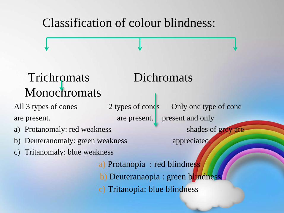
Classification of colour blindness:
Trichromats Dichromats
MonochromatsAll 3 types of cones 2 types of cones Only one type of cone
are present. are present. present and only
a) Protanomaly: red weakness shades of grey are
b) Deuteranomaly: green weakness appreciated.
c) Tritanomaly: blue weakness
a) Protanopia : red blindness
b) Deuteranaopia : green blindness
c) Tritanopia: blue blindness
Monochromatism and
Achromatopsia
•Monochromats have only one
cone pigment instead of the 3
primary color based pigments.
•Achromats have only rods and no
cones and are truly color-blind.
This form is extremely rare. Such
people are able to distinguish
objects only by brightness and
usually have very poor vision due
to the lack of cones.
Anomalous
Trichromacy (most
common) - the
affected person
has all 3 (thus tri-
chrom) cone
pigments but one
is abnormal.
•Protanomaly. A Protanomal has abnormal red-
sensitive cones and requires excess red to match
the yellow standard.
•Deuteranomaly. A Deuteranomal has abnormal
green-sensitive pigment and needs excess green
to match the yellow standard.
•Tritanomaly. Very rare. Abnormal blue-sensitive
cones. Has difficulty in distinguishing blues from
yellows and my use excessive blue to match the
yellow standard.
Anomalous Dichromacy
- the affected person
has only 2 cone
pigments and thus
cannot distinguish
certain colors.
•Protanopia - lack of red-sensitive cone
pigment.
•Deuteranopia - lack of green-sensitive cone
pigment.
•Tritanopia. Very-very rare. Lack of blue-
sensitive pigment. Can't distinguish blue from
yellow. Usually an inherited syndrome with
optic atrophy.
• Red green colour blindness : inheritance is
X linked disorder because the gene for
this pigment is located on short arm of X
chromosome
- Only males are sufferer , females are
carrier.
• Blue colour blindness : very rare , gene is
located on 7th chromosome
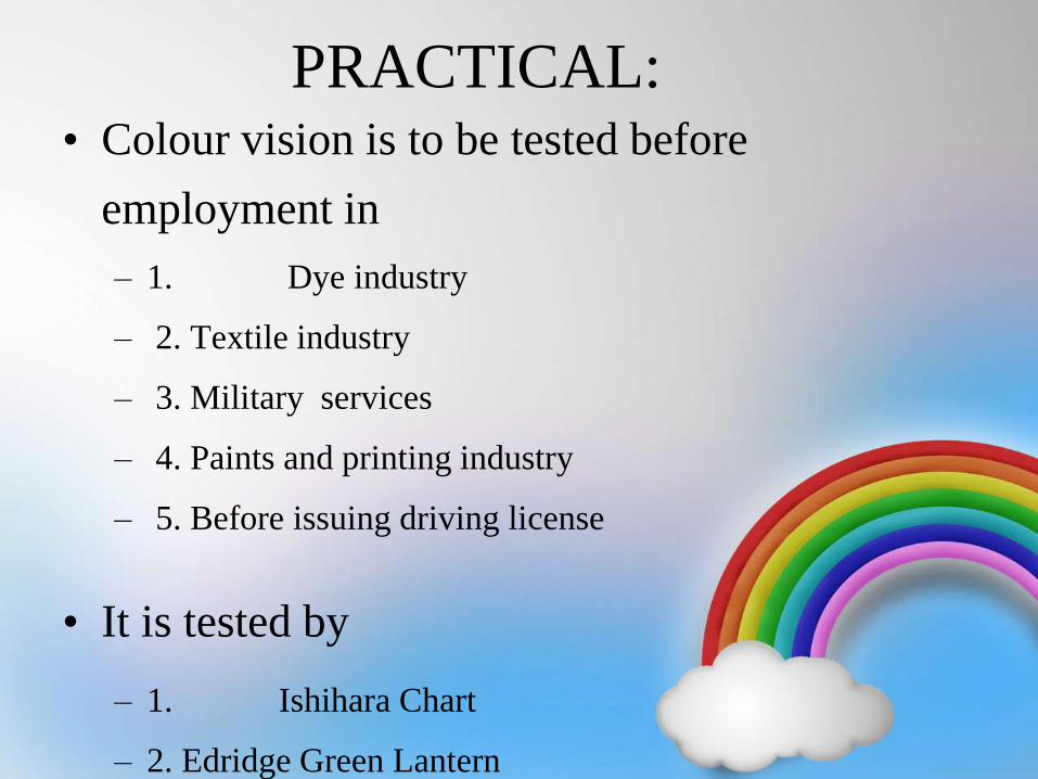
PRACTICAL:• Colour vision is to be tested before
employment in
– 1. Dye industry
– 2. Textile industry
– 3. Military services
– 4. Paints and printing industry
– 5. Before issuing driving license
• It is tested by
– 1. Ishihara Chart
– 2. Edridge Green Lantern
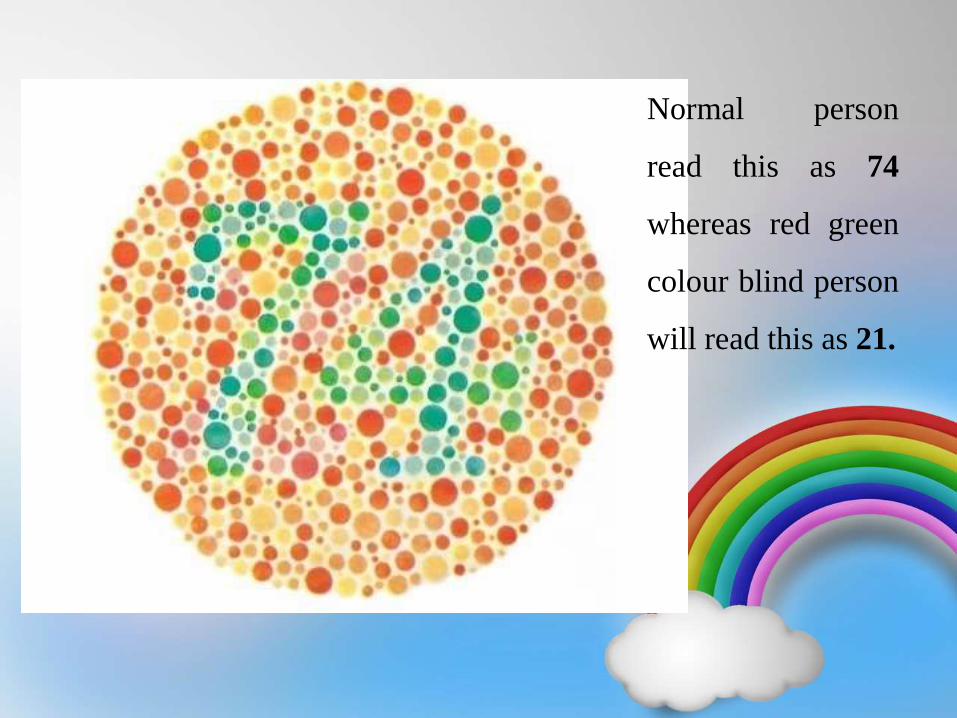
Normal person
read this as 74
whereas red green
colour blind person
will read this as 21.
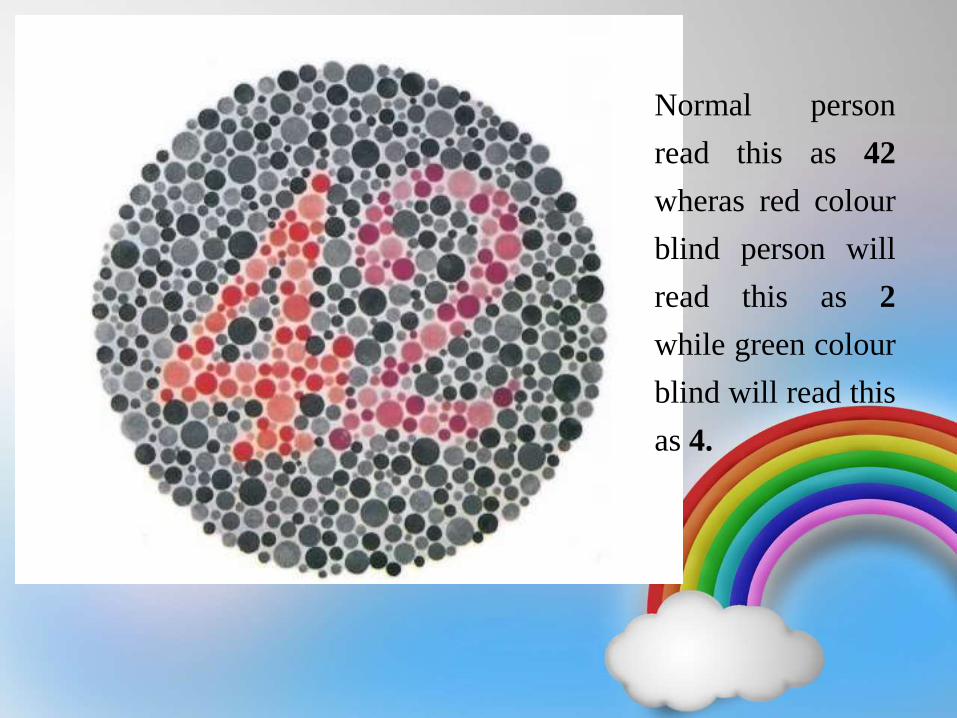
Normal person
read this as 42
wheras red colour
blind person will
read this as 2
while green colour
blind will read this
as 4.
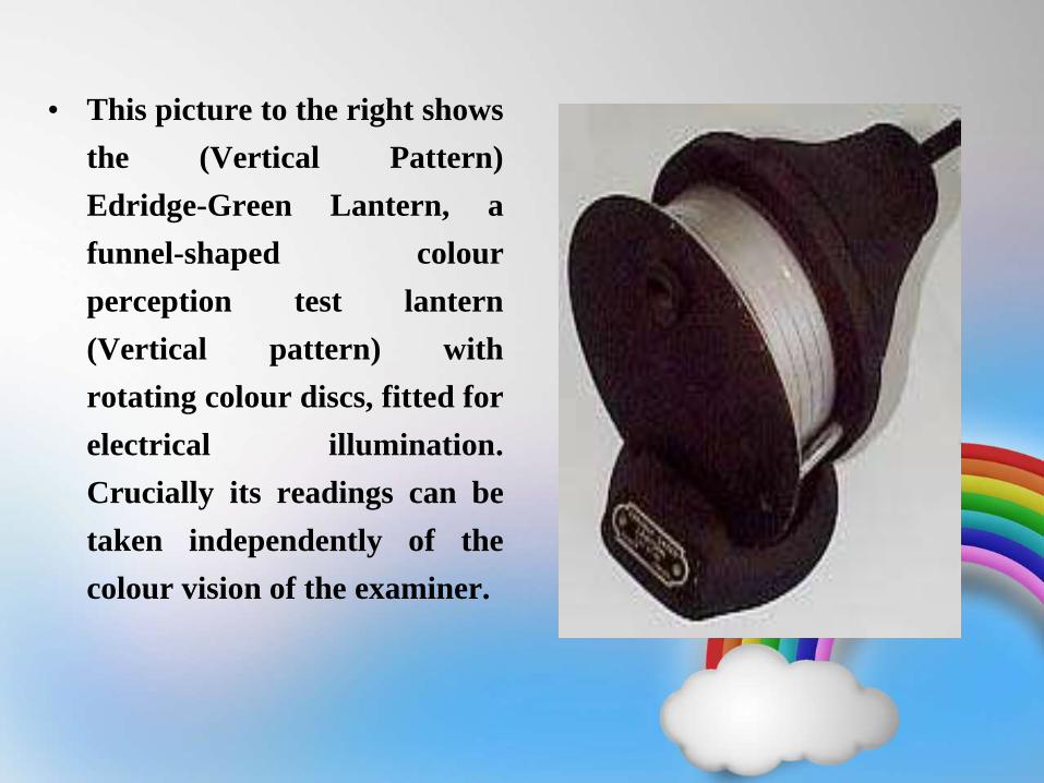
• This picture to the right shows
the (Vertical Pattern)
Edridge-Green Lantern, a
funnel-shaped colour
perception test lantern
(Vertical pattern) with
rotating colour discs, fitted for
electrical illumination.
Crucially its readings can be
taken independently of the
colour vision of the examiner.
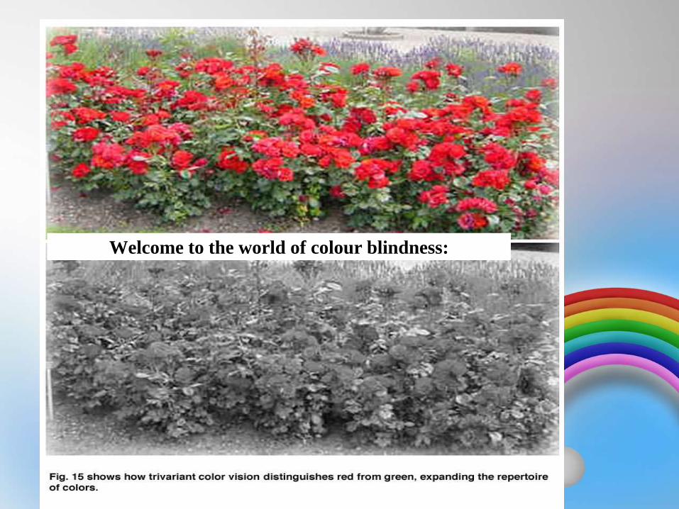
Welcome to the world of colour blindness:
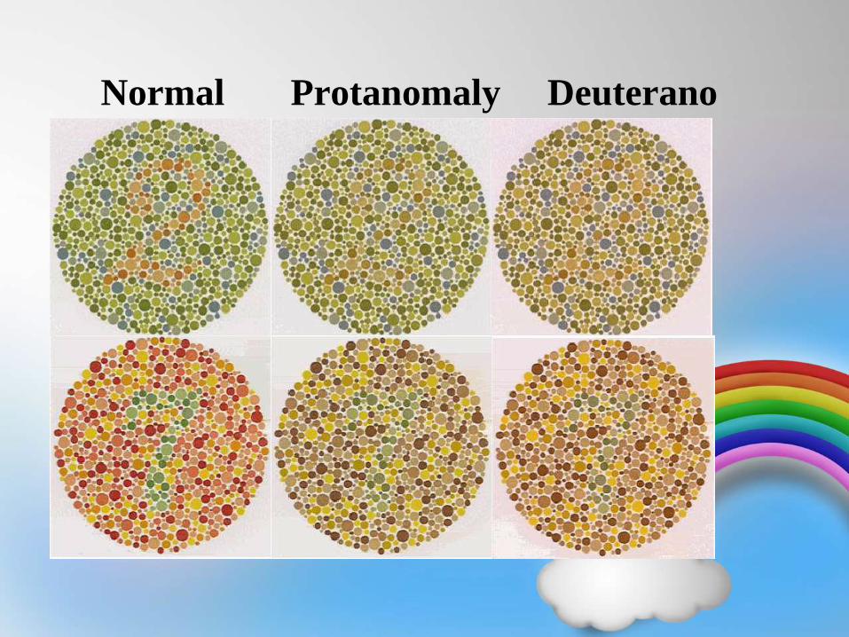
Normal Protanomaly Deuterano
maly
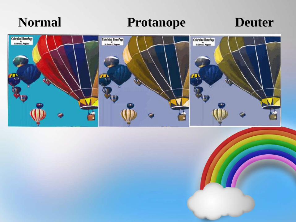
Normal Protanope Deuter
anope
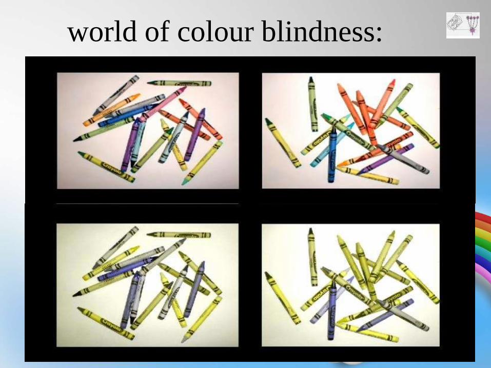
world of colour blindness:

Thank You!
Veena
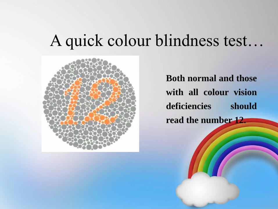
A quick colour blindness test…
Both normal and those
with all colour vision
deficiencies should
read the number 12.
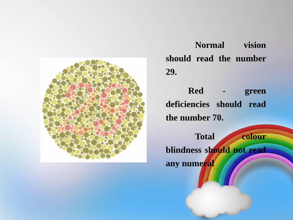
Normal vision
should read the number
29.
Red - green
deficiencies should read
the number 70.
Total colour
blindness should not read
any numeral
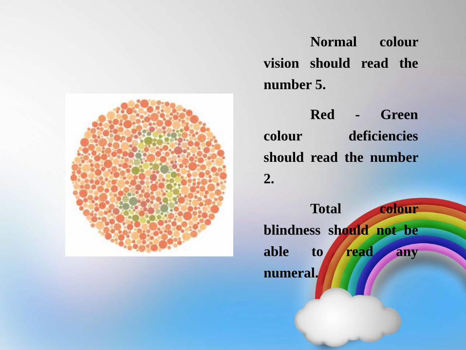
Normal colour
vision should read the
number 5.
Red - Green
colour deficiencies
should read the number
2.
Total colour
blindness should not be
able to read any
numeral.
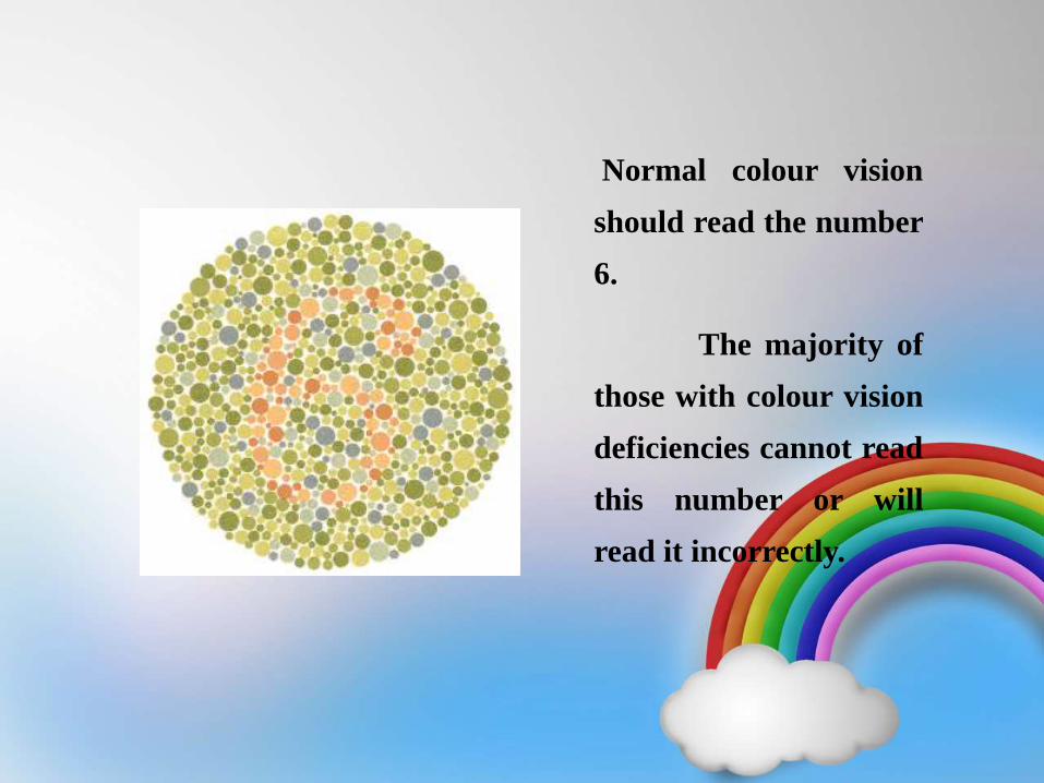
Normal colour vision
should read the number
6.
The majority of
those with colour vision
deficiencies cannot read
this number or will
read it incorrectly.
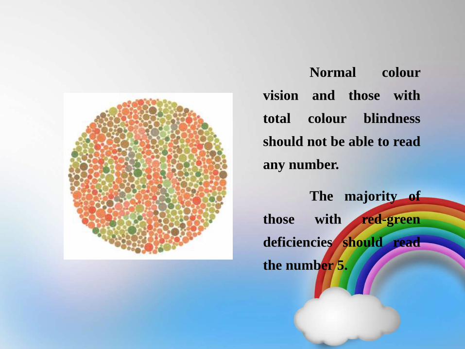
Normal colour
vision and those with
total colour blindness
should not be able to read
any number.
The majority of
those with red-green
deficiencies should read
the number 5.
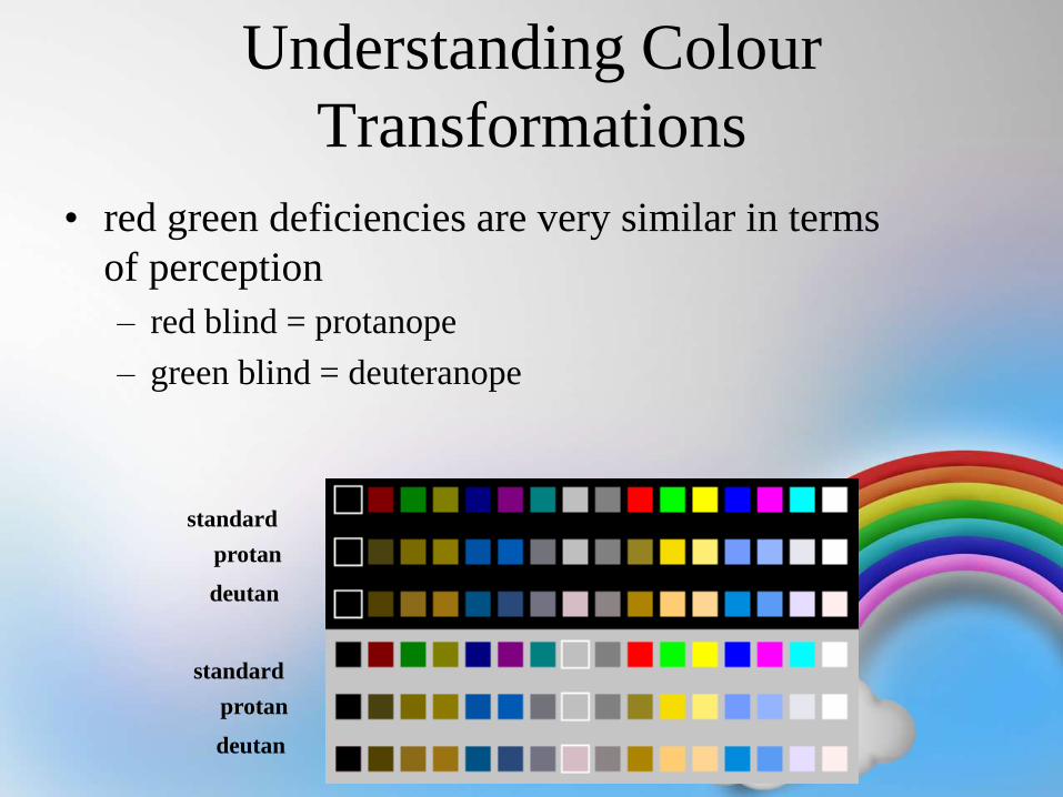
Understanding Colour
Transformations
• red green deficiencies are very similar in terms
of perception
– red blind = protanope
– green blind = deuteranope
standard
protan
deutan
standard
protan
deutan
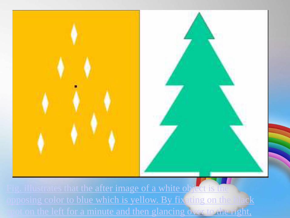
Fig. illustrates that the after image of a white object is the
opposing color to blue which is yellow. By fixating on the black
spot on the left for a minute and then glancing over to the right,
one sees yellow, not black, candles on the tree
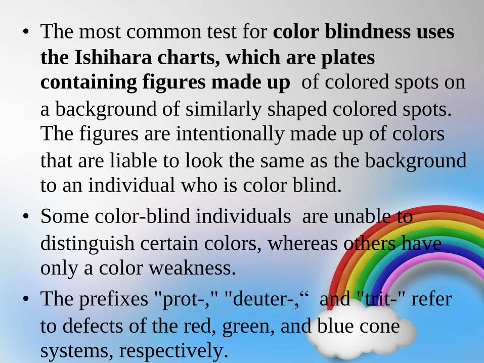
• The most common test for color blindness uses
the Ishihara charts, which are plates
containing figures made up of colored spots on
a background of similarly shaped colored spots.
The figures are intentionally made up of colors
that are liable to look the same as the background
to an individual who is color blind.
• Some color-blind individuals are unable to
distinguish certain colors, whereas others have
only a color weakness.
• The prefixes "prot-," "deuter-,“ and "trit-" refer
to defects of the red, green, and blue cone
systems, respectively.
• Individuals with normal color vision are called
http://www.college-optometrists.org/
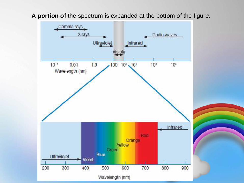
A portion of the spectrum is expanded at the bottom of the figure.
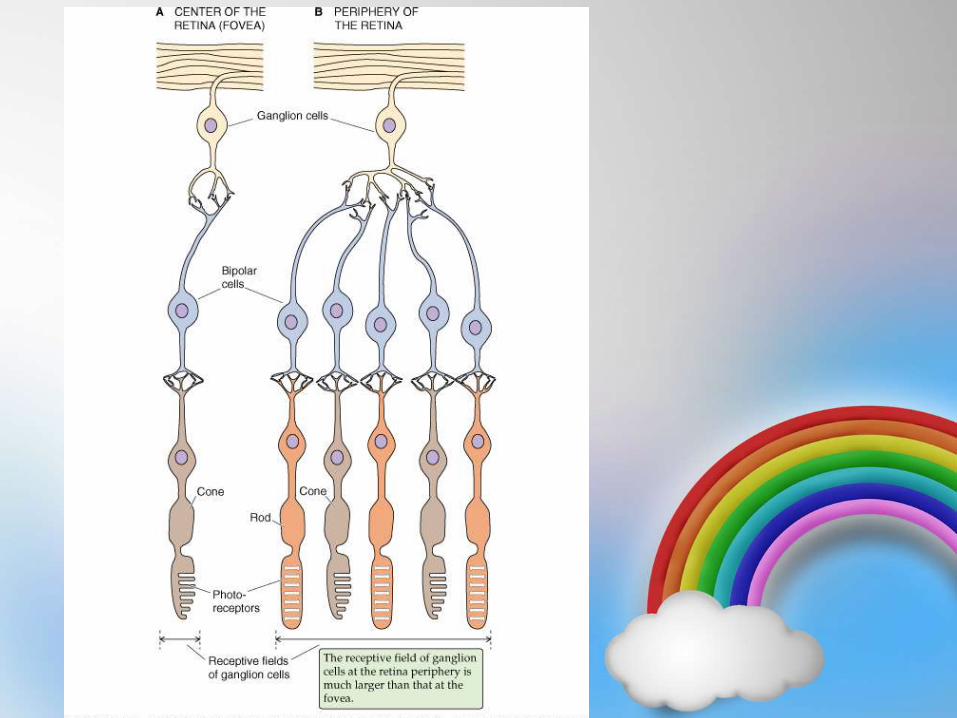
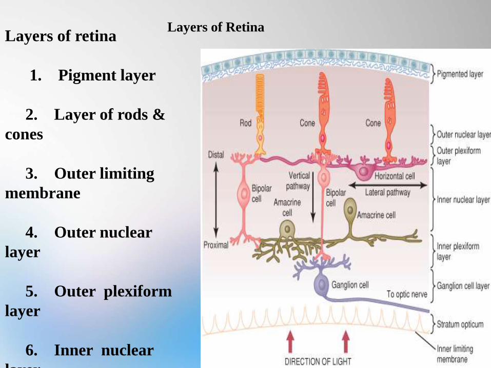
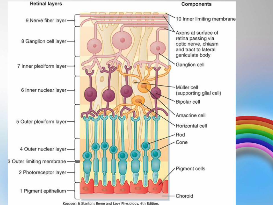
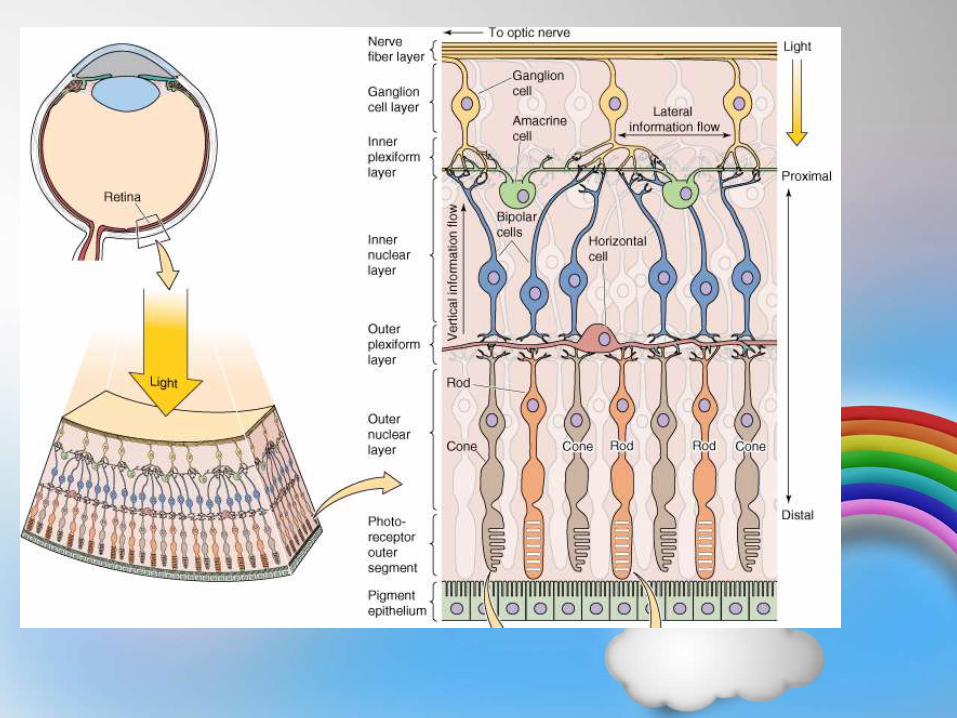
Layers of RetinaLayers of retina
1. Pigment layer
2. Layer of rods &
cones
3. Outer limiting
membrane
4. Outer nuclear
layer
5. Outer plexiform
layer
6. Inner nuclear
layer