Embed Size (px)
Citation preview
CAVITATORY LESIONS OF THE
LUNG
DR Vishwanath Reddy
Yenepoya medical college
Mangalore
Cavity
A cavity is a gas-filled space seen as a lucency
or low attenuation area, within a pulmonary
consolidation, a mass, or a nodule;
hence, a lucent area with in the lung that may
or may not contain a fluid level and that is
surrounded by a wall, usually of varied
thickness”
Bulla
air filled spaces surrounded by a thin wall < 1mm
Cysts
are air containing spaces surrounded by a thin (2mm or
less ) wall
Cavities
air containing spaces surrounded by a thick wall >3mm
and also the air containing lesion that is surrounded by an
infilterate and /or a mass.
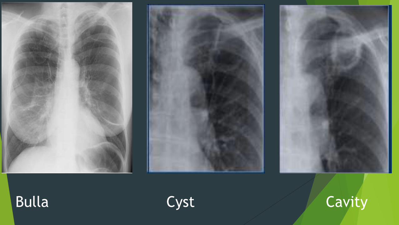
Bulla Cyst Cavity
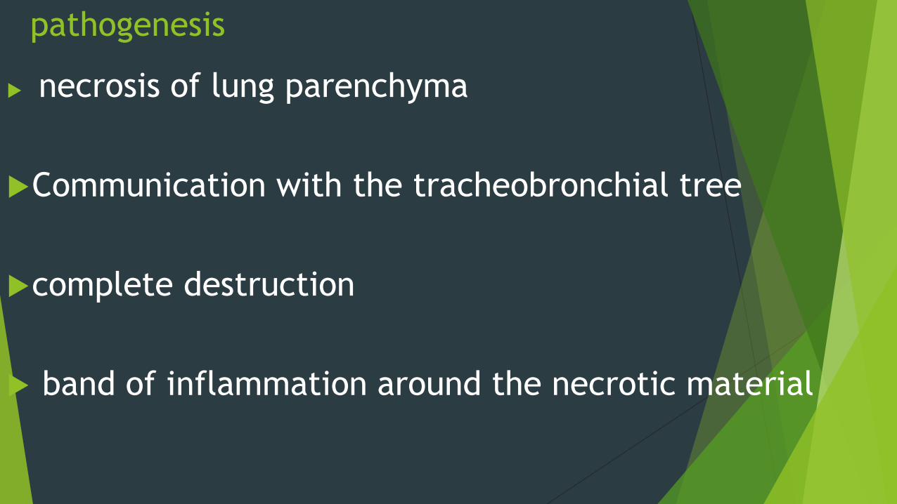
pathogenesis
necrosis of lung parenchyma
Communication with the tracheobronchial tree
complete destruction
band of inflammation around the necrotic material

Imaging modalities
Chest radiograph
Computed tomography
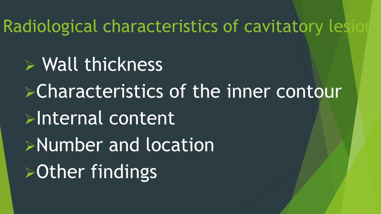
Radiological characteristics of cavitatory lesion
Wall thickness
Characteristics of the inner contour
Internal content
Number and location
Other findings
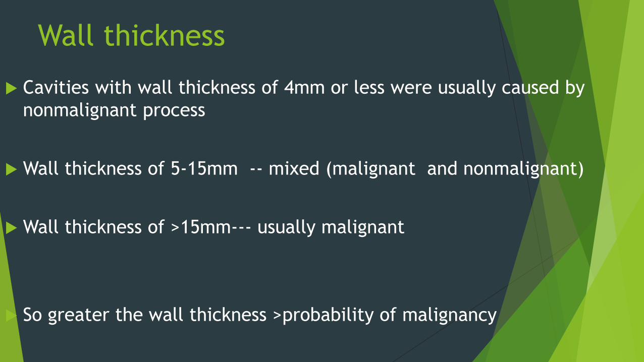
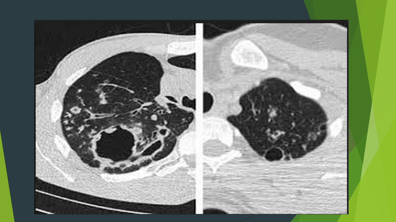
Wall thickness
Cavities with wall thickness of 4mm or less were usually caused by
nonmalignant process
Wall thickness of 5-15mm -- mixed (malignant and nonmalignant)
Wall thickness of >15mm--- usually malignant
So greater the wall thickness >probability of malignancy
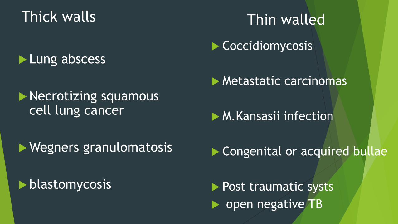
Thick walls
Lung abscess
Necrotizing squamous cell lung cancer
Wegners granulomatosis
blastomycosis
Thin walled
Coccidiomycosis
Metastatic carcinomas
M.Kansasii infection
Congenital or acquired bullae
Post traumatic systs
open negative TB
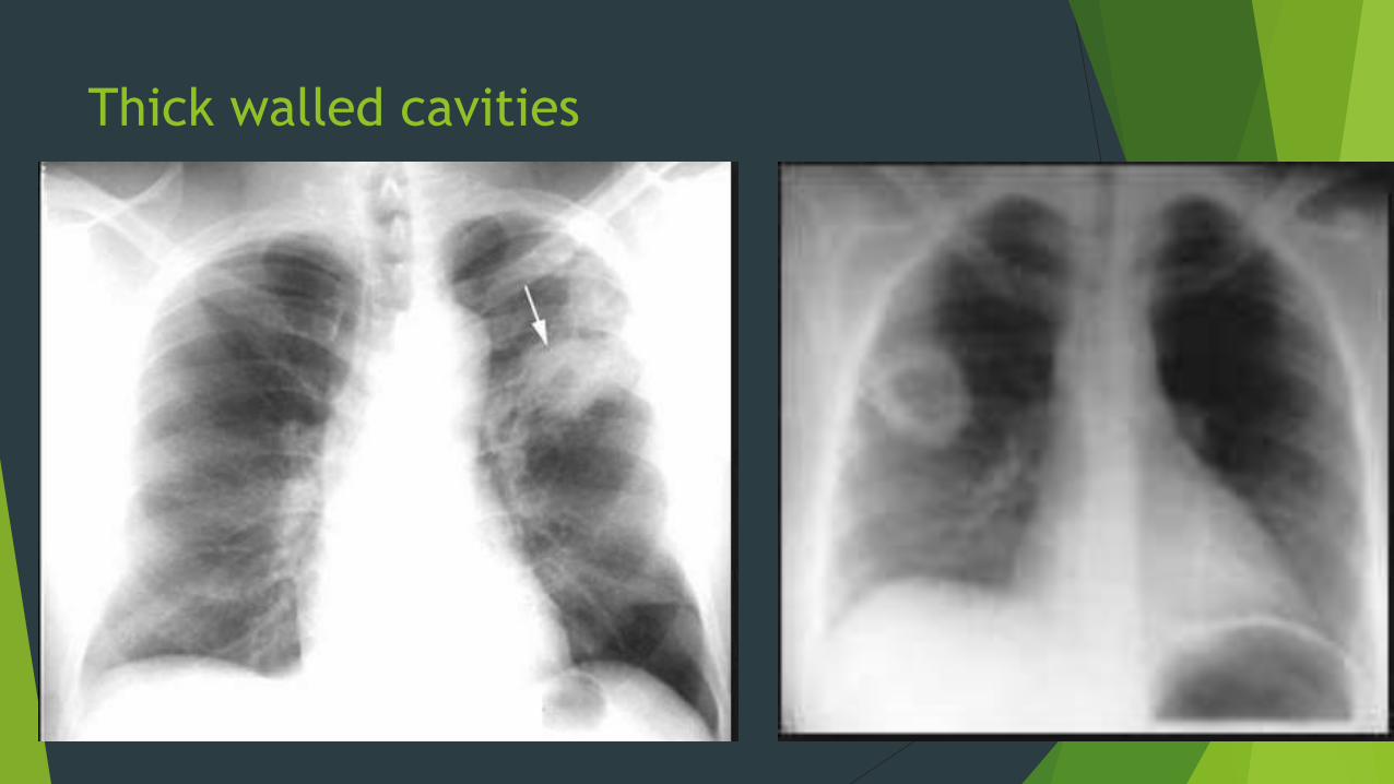
Thick walled cavities

Thin walled
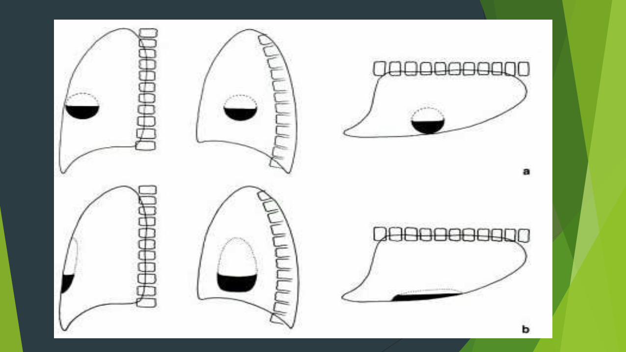
Characteristics of the inner contour
Nodular or irregular –usually in case of neoplasms
Poorly defined /shaggy-corresponds with abscess
Smooth- cavitory lesions of other etiology
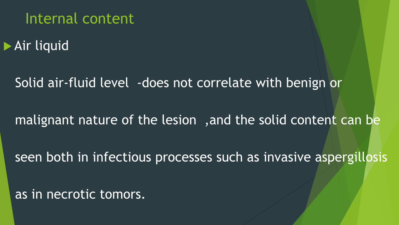
Internal content
Air liquid
Solid air-fluid level -does not correlate with benign or
malignant nature of the lesion ,and the solid content can be
seen both in infectious processes such as invasive aspergillosis
as in necrotic tomors.
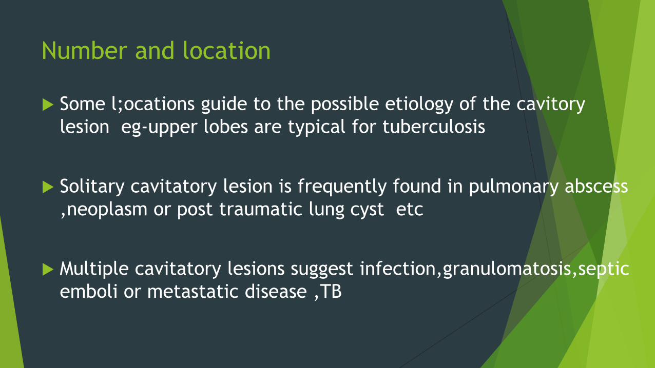
Number and location
Some l;ocations guide to the possible etiology of the cavitory
lesion eg-upper lobes are typical for tuberculosis
Solitary cavitatory lesion is frequently found in pulmonary abscess
,neoplasm or post traumatic lung cyst etc
Multiple cavitatory lesions suggest infection,granulomatosis,septic
emboli or metastatic disease ,TB
Other findings
Like areas of ground glass attenuation, pulmonary opacities
, interstitial disease ,honeycomb pattern etc
Considering the cavitatory lesion and / or associated
findings we will determine if it is a focal / multifocal
affection or diffuse disease ,which will guide us to the most
appropriate diagnosis

Valuation of the clinical context and the time of
the disease process
clinical scenario
duration and evolution
Acute or subacute
Chronic lesions
The differential diagnosis are therefore very broad &
includes
Neoplastic pathology –primary bronchogenic
carcinoma,lymphoma,mets
Infectious-bacterial,mycobacterial,fungal, parasitic
Pulmonary infarcts
Septic emboli
Autoimmune-wegners granulomatosis
- rheumatoid arthritis
Traumatic
Congenital
neoplasms
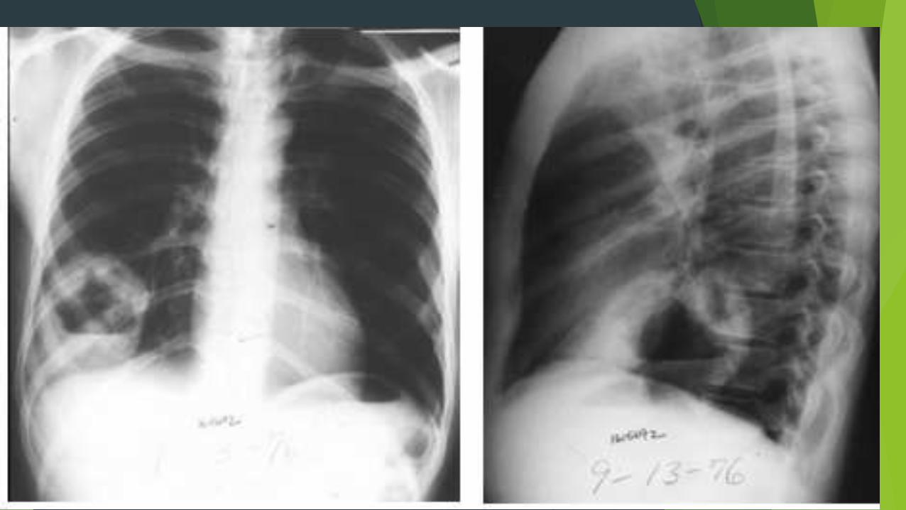
Isolated cavitatory lung lesion
Lesion of variable size
Irregular or speculated margins
Thick walls
Associated with mass and other findings
Air crescent sign may be present in rare cases
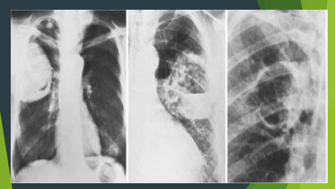
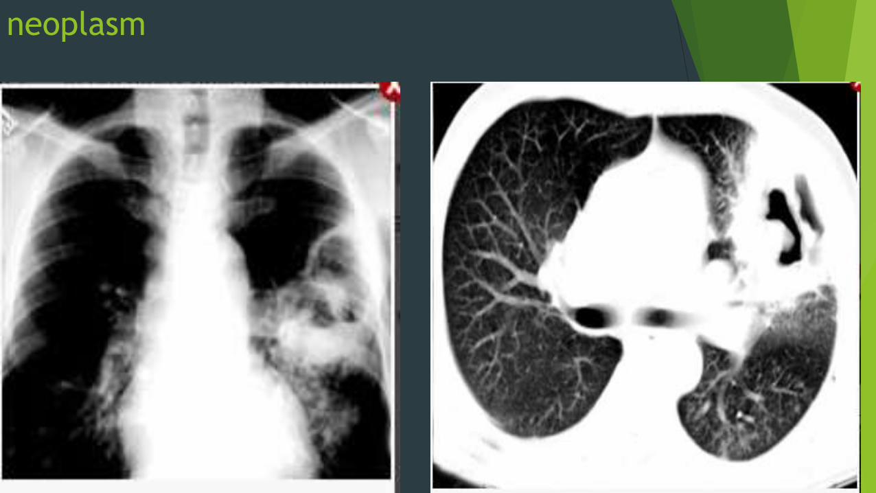
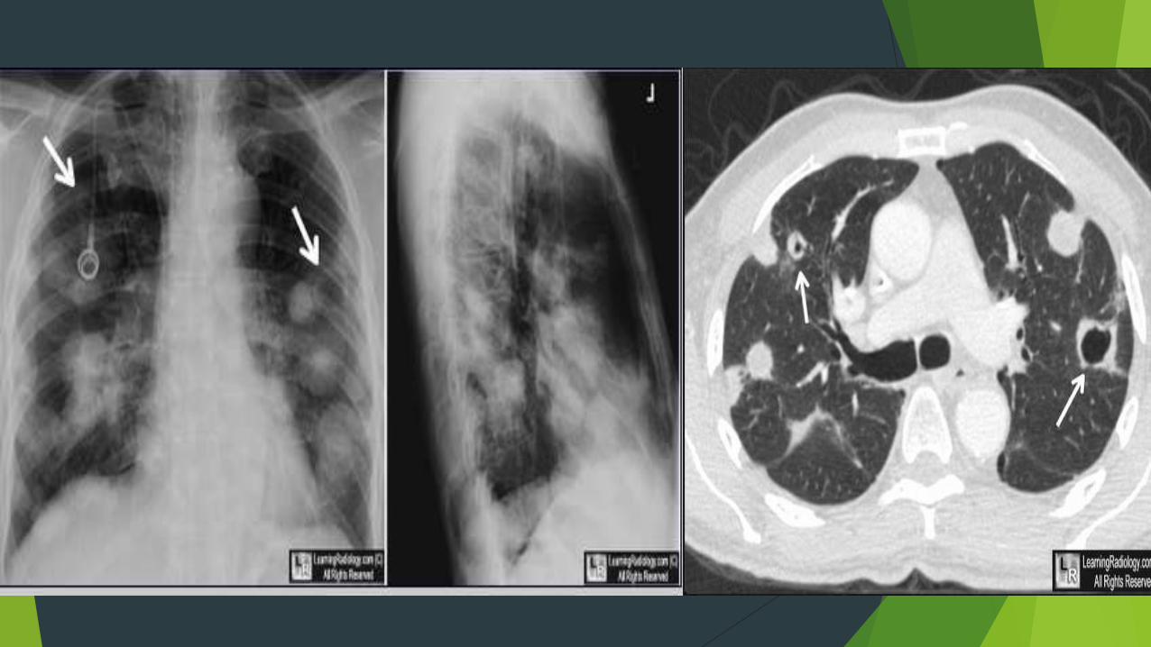
neoplasm
METASTATIC
Metastases are multiple
differing sizes
thick and irregular walls (adenocarcinomas )
Thin-walled cavities ( squamous cell carcinomas)
Cavitation is more common in upper lobe lesions than lower lobe
-
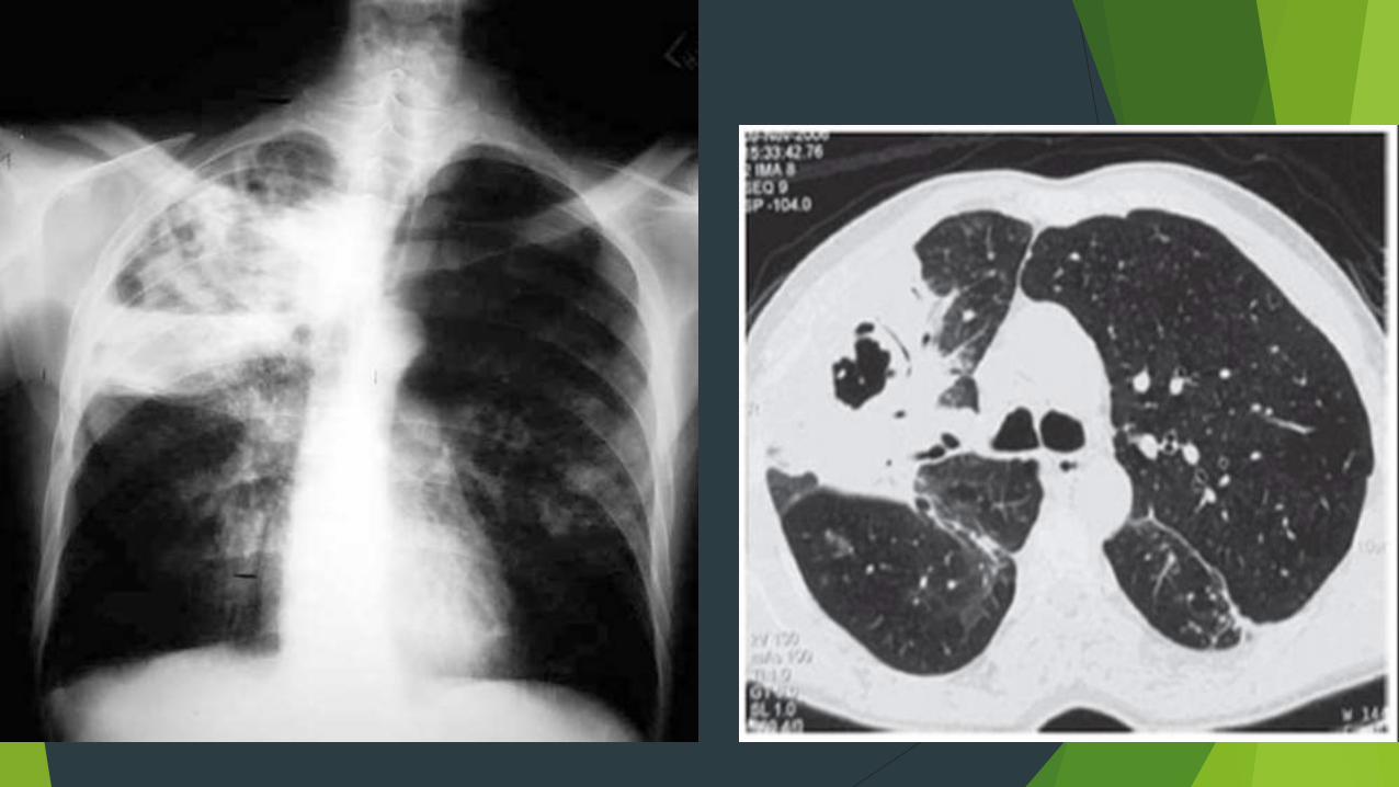
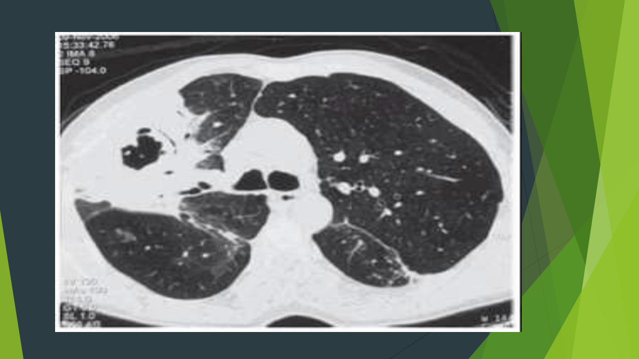
Infectious pathology
Necrotizing bacterial pneumonia
• Caused by staph aureus,gram negative bacteria and anerobic
bacteria
• Primary consolidation that can associate cavitation inside
• In anerobic bacteria it is common to develop abscesses with
thick and irregular walls containing air fluid levels
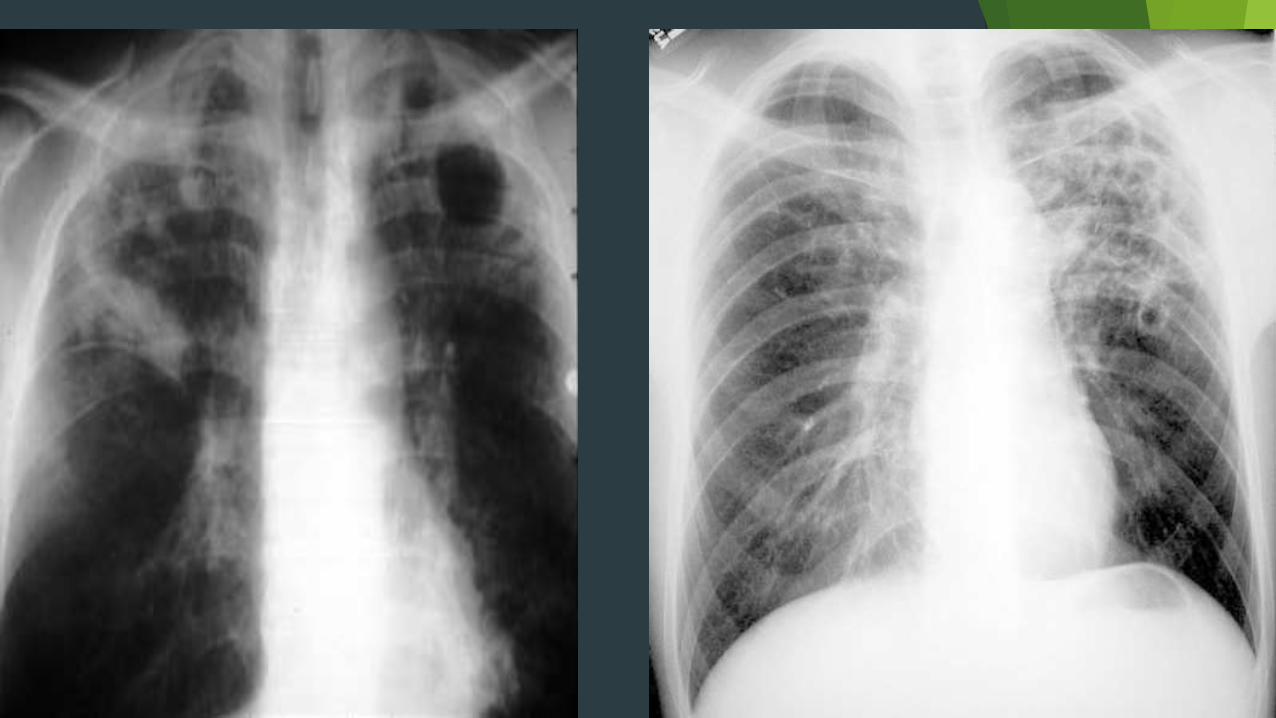
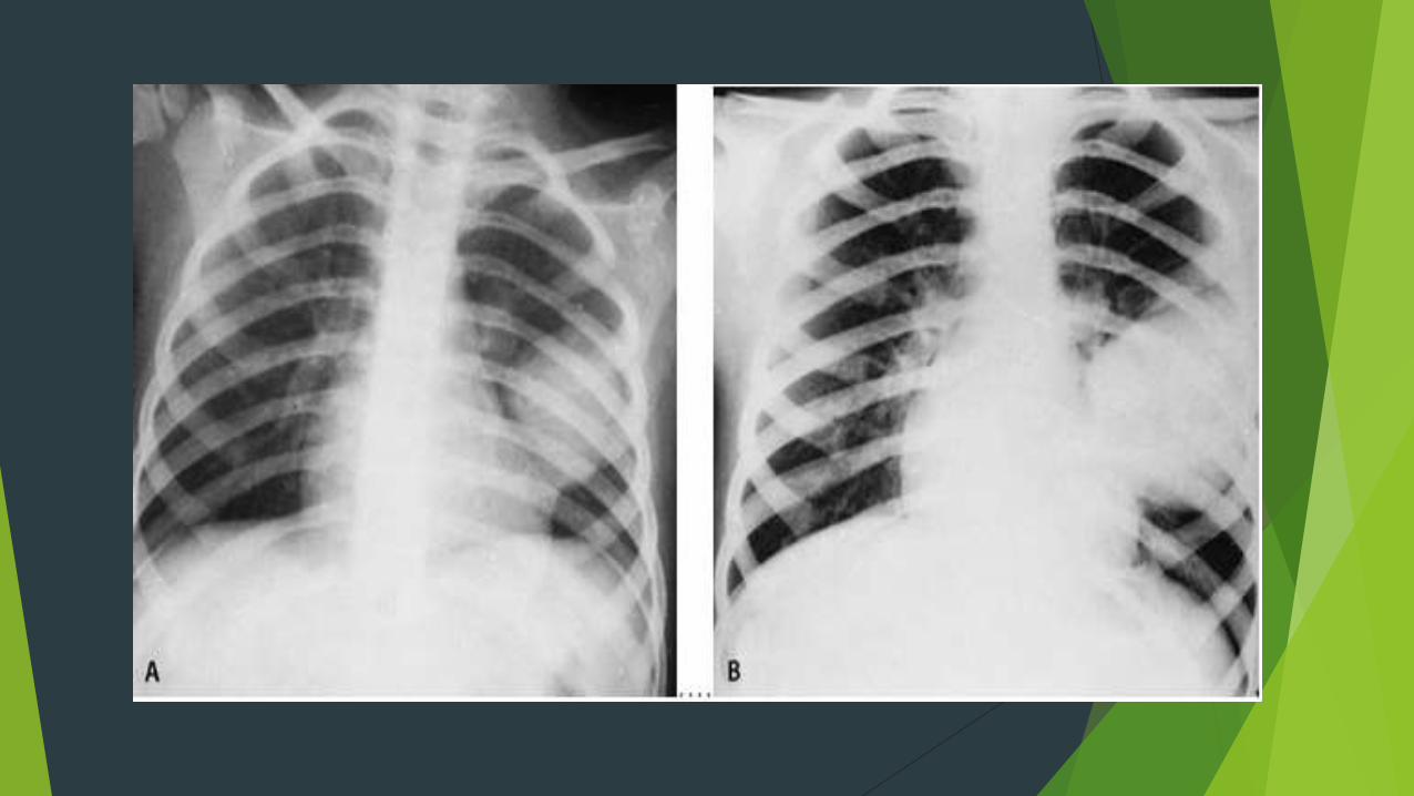
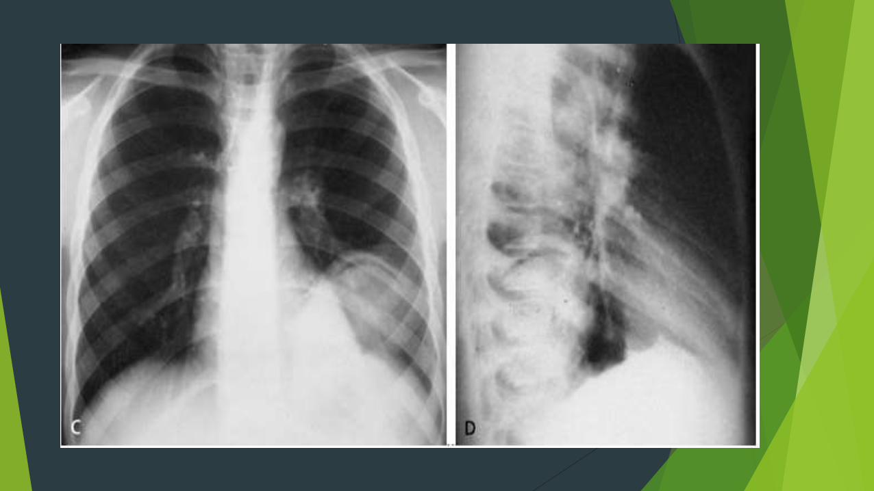
Postprimary tuberculosis
prescence of infiltrates with multiple satellite nodules and cavitatory lesions
located preferentially in the upper lobes and in the apical segments of the lower lobes
Vary widely in size
Both thick & thin walls
Inner walls can be smooth and more irregular and thick
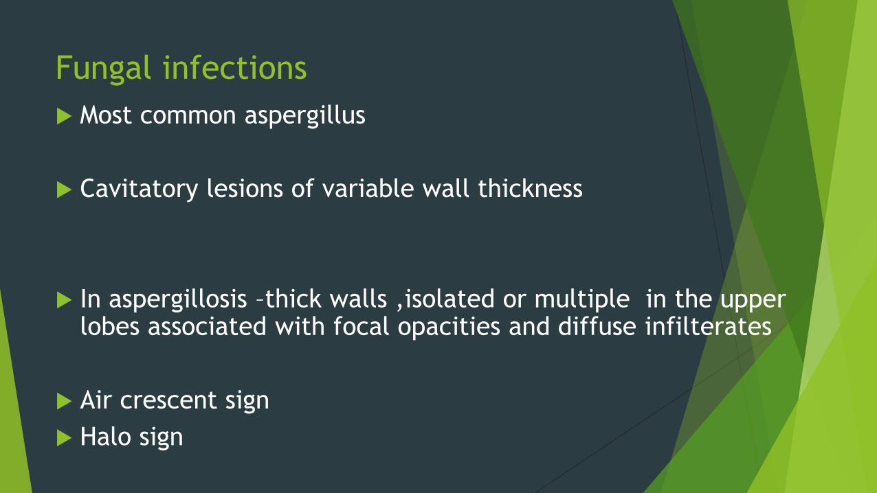
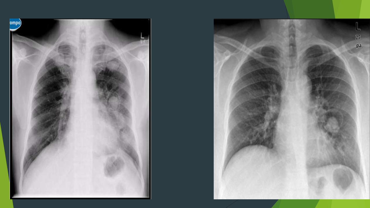
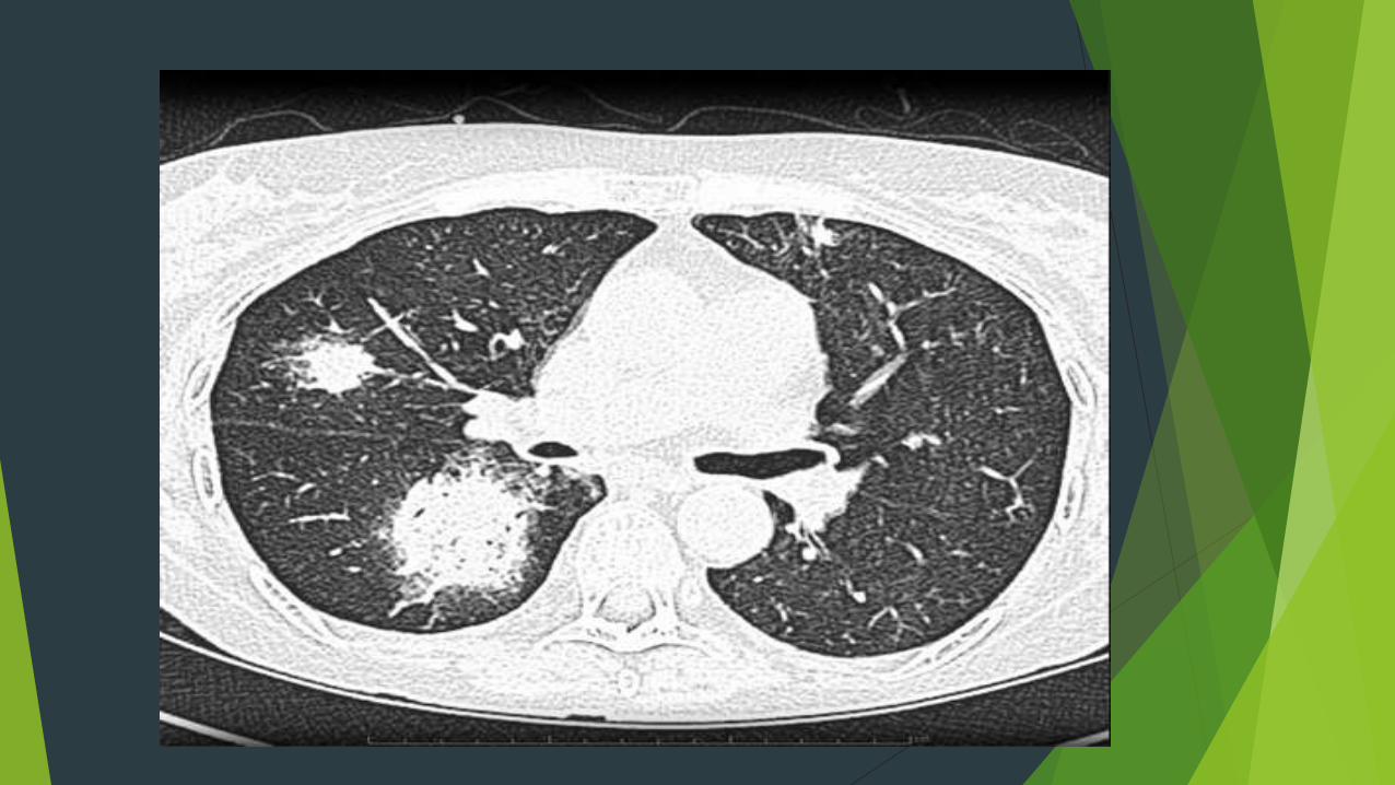
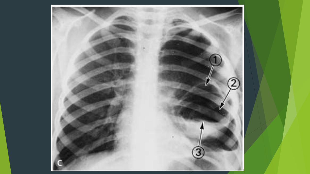
Fungal infections
Most common aspergillus
Cavitatory lesions of variable wall thickness
In aspergillosis –thick walls ,isolated or multiple in the upper lobes associated with focal opacities and diffuse infilterates
Air crescent sign
Halo sign
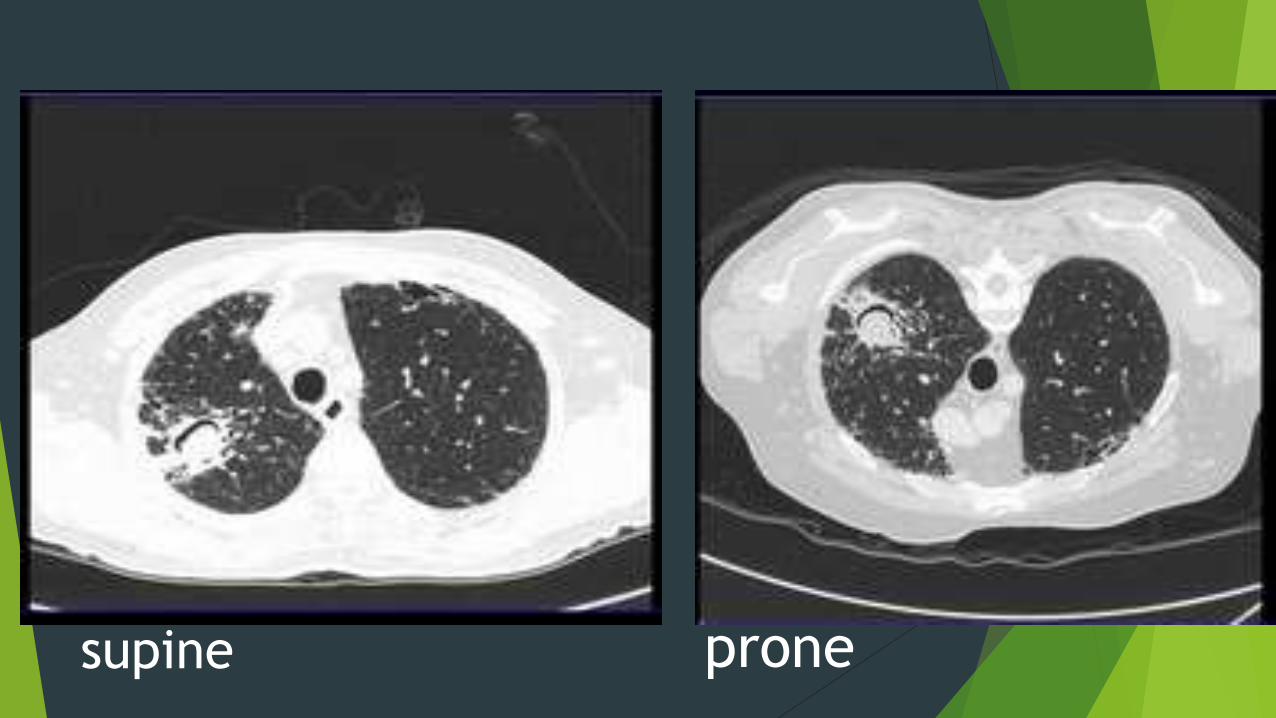
supine prone
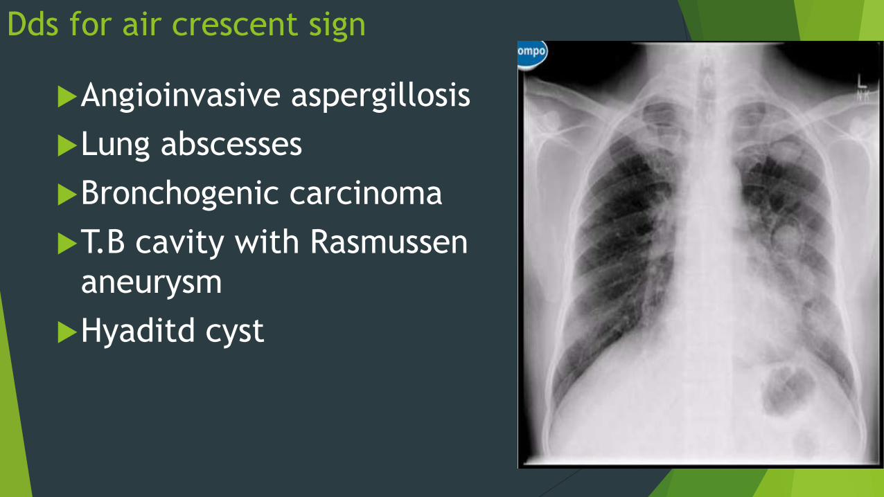
Dds for air crescent sign
Angioinvasive aspergillosis
Lung abscesses
Bronchogenic carcinoma
T.B cavity with Rasmussen
aneurysm
Hyaditd cyst
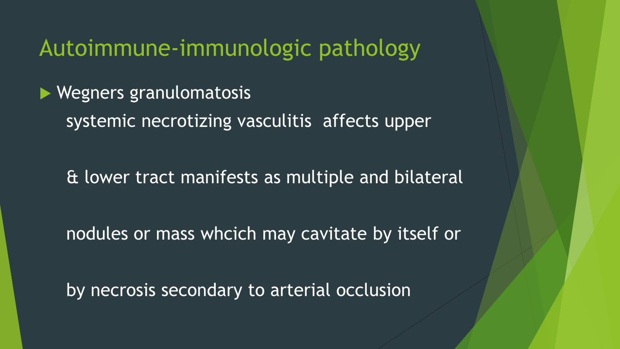
Autoimmune-immunologic pathology
Wegners granulomatosis
systemic necrotizing vasculitis affects upper
& lower tract manifests as multiple and bilateral
nodules or mass whcich may cavitate by itself or
by necrosis secondary to arterial occlusion
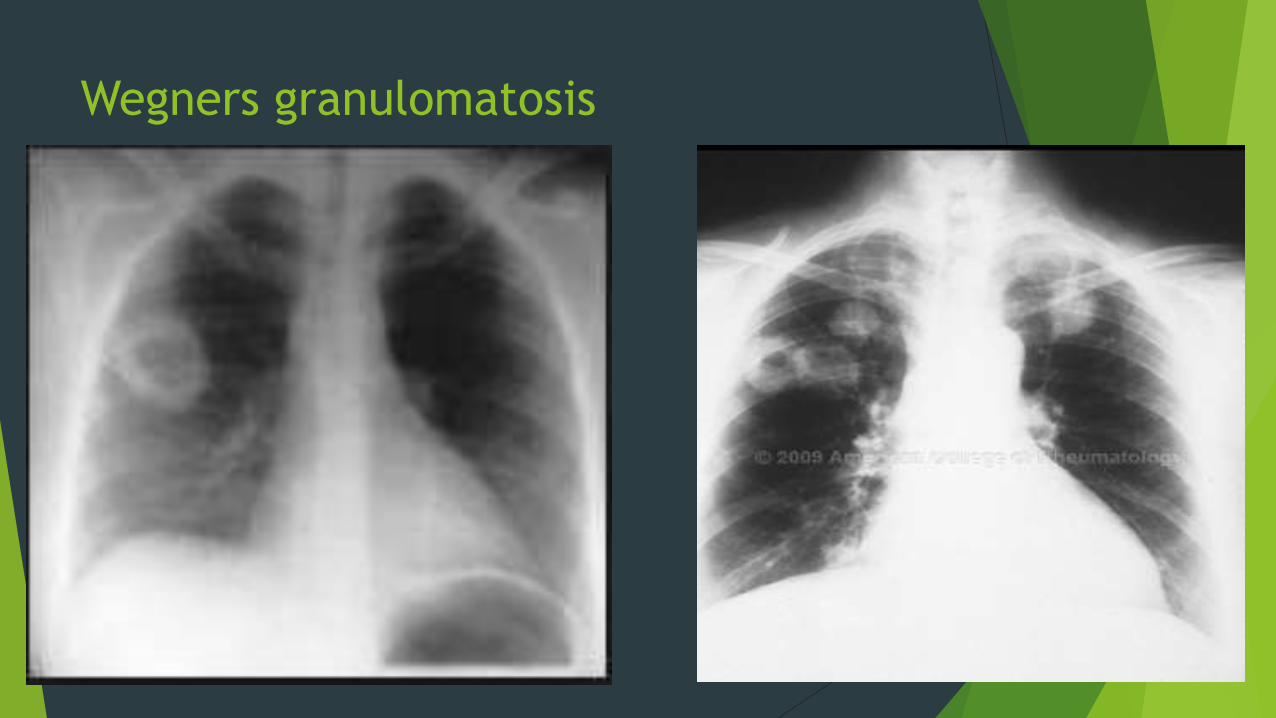
Wegners granulomatosis
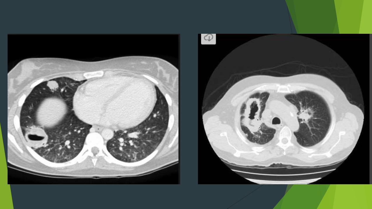
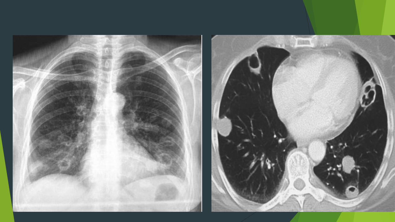
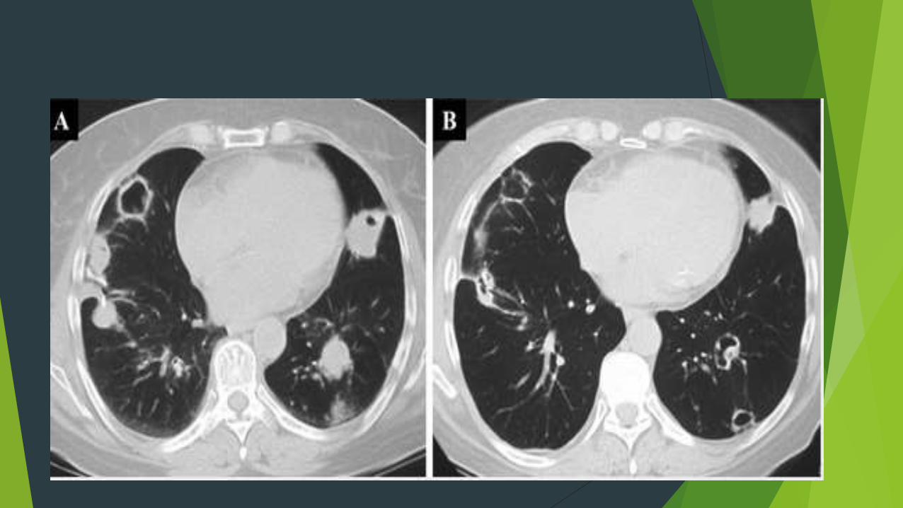
rheumatoid arthritis
Affects mainly women 20 -50 yrs
Bilateral and multiple lung nodules ,usually small in
size and with sub pleural location that may cavitate
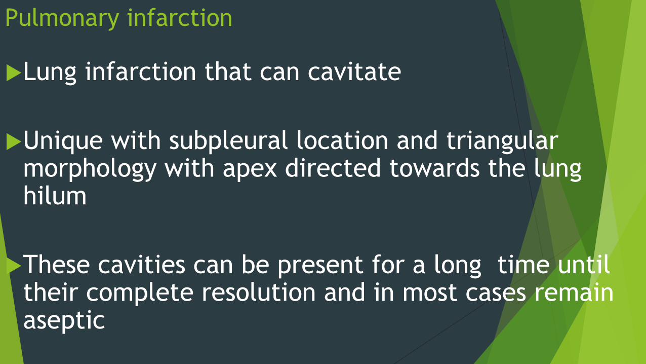
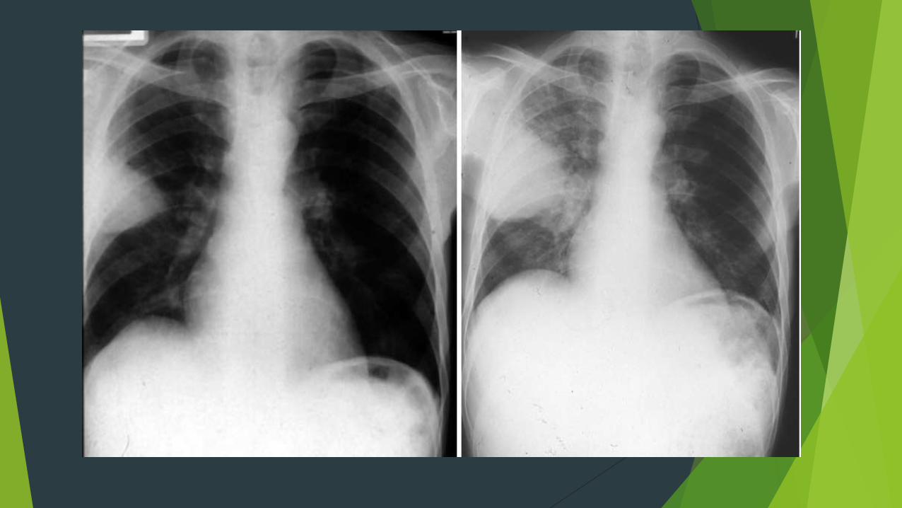
Pulmonary infarction
Lung infarction that can cavitate
Unique with subpleural location and triangular morphology with apex directed towards the lung hilum
These cavities can be present for a long time until their complete resolution and in most cases remain aseptic
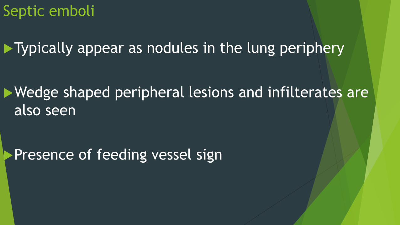
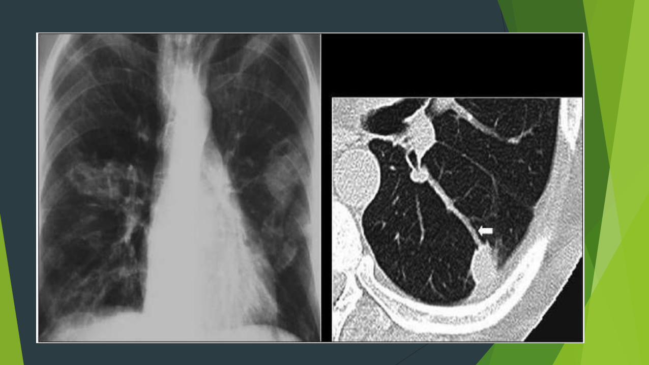
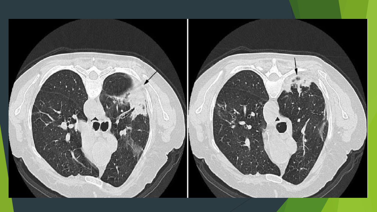
Septic emboli
Typically appear as nodules in the lung periphery
Wedge shaped peripheral lesions and infilterates are
also seen
Presence of feeding vessel sign
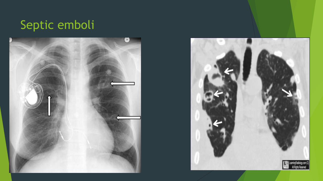
Septic emboli
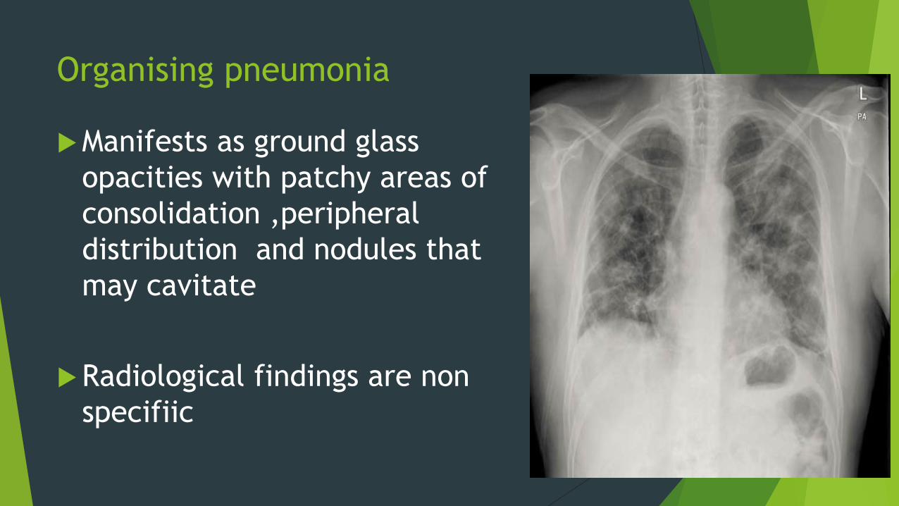
Organising pneumonia
Manifests as ground glass
opacities with patchy areas of
consolidation ,peripheral
distribution and nodules that
may cavitate
Radiological findings are non
specifiic
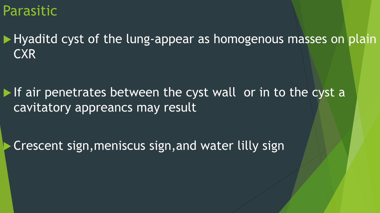
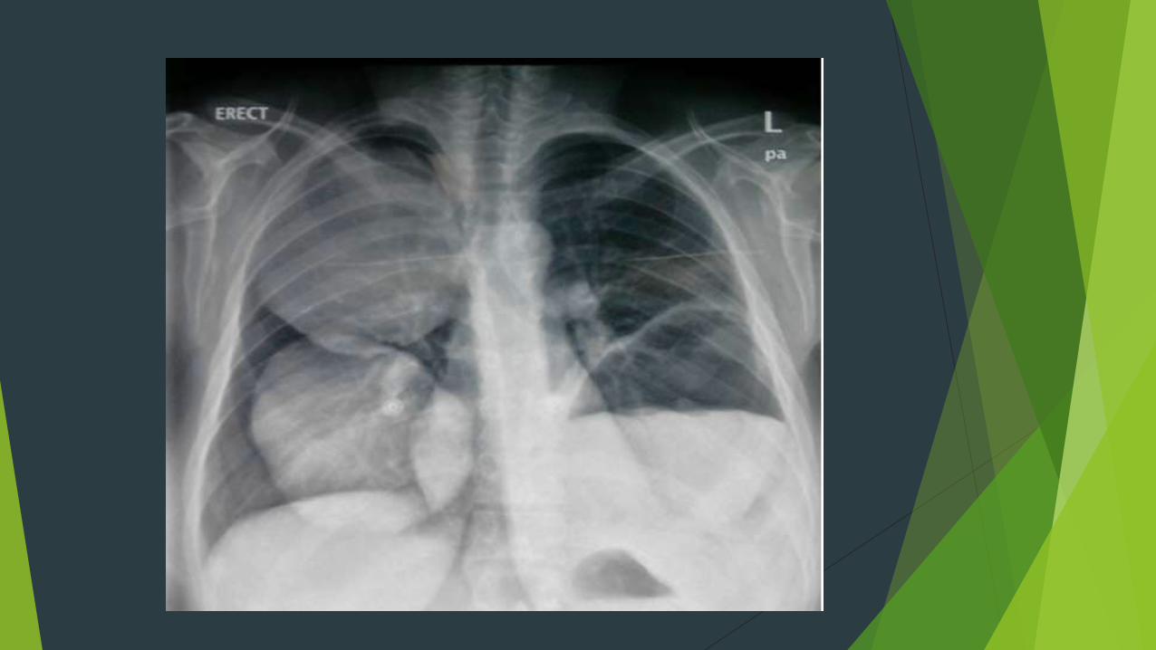
Parasitic
Hyaditd cyst of the lung-appear as homogenous masses on plain
CXR
If air penetrates between the cyst wall or in to the cyst a
cavitatory appreancs may result
Crescent sign,meniscus sign,and water lilly sign
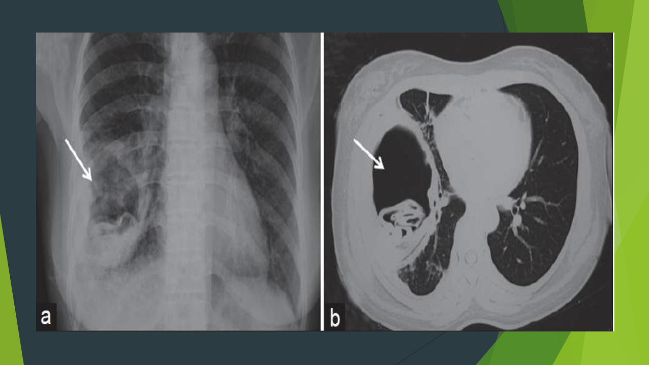
Traumatic
PNEUMATOCELES
variety of sizes and appearances.
thin walled cystic spaces within the lung parenchyma,
containing air
smooth regular inner margins
contain little if any fluid
persist despite absence of symtpoms
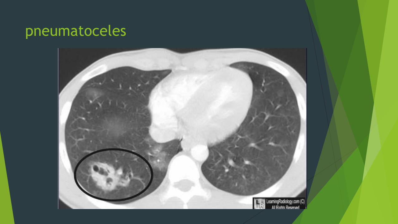
pneumatoceles
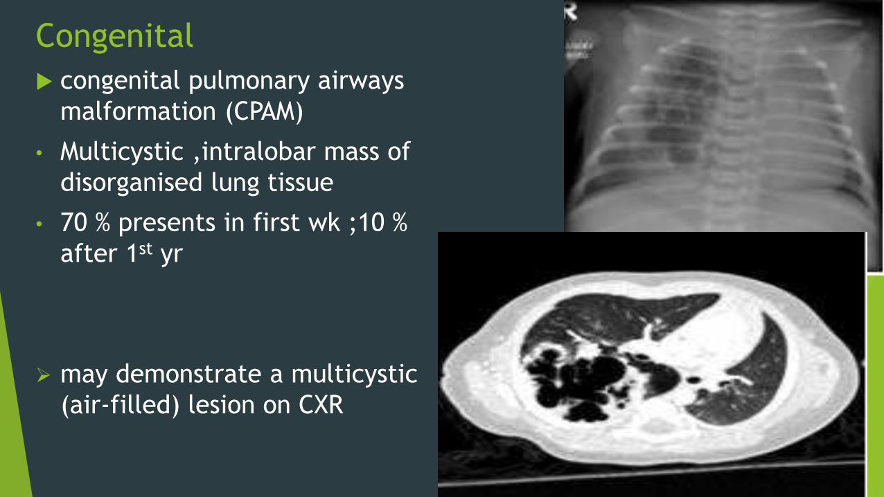
Congenital
congenital pulmonary airways
malformation (CPAM)
• Multicystic ,intralobar mass of
disorganised lung tissue
• 70 % presents in first wk ;10 %
after 1st yr
may demonstrate a multicystic
(air-filled) lesion on CXR
Congenital
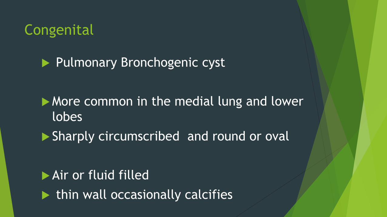
Pulmonary Bronchogenic cyst
More common in the medial lung and lower
lobes
Sharply circumscribed and round or oval
Air or fluid filled
thin wall occasionally calcifies
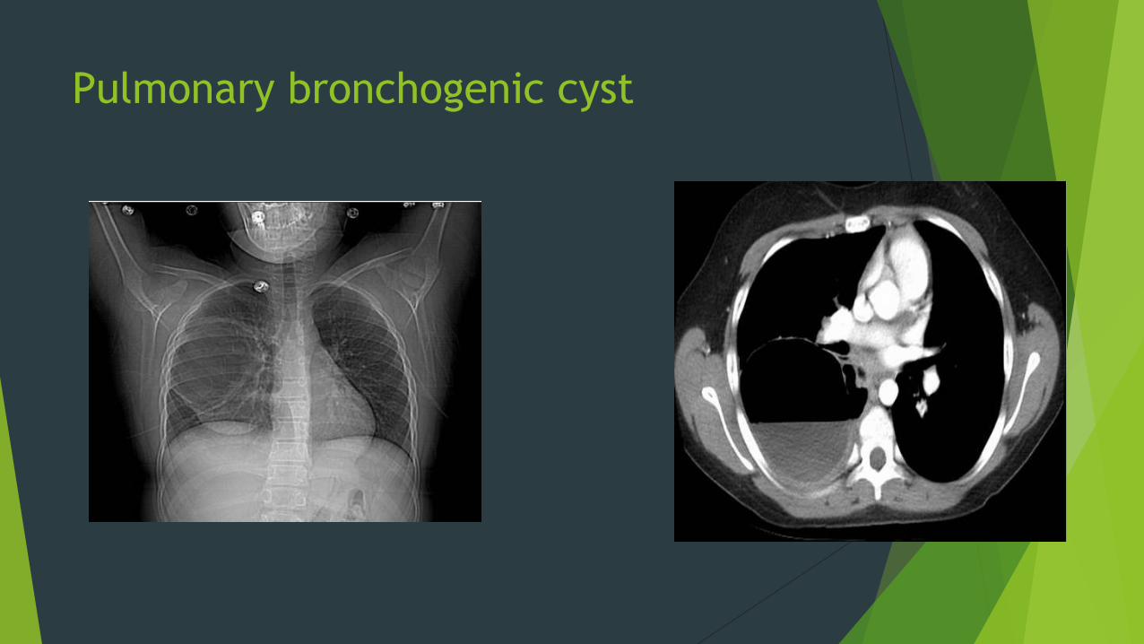
Pulmonary bronchogenic cyst
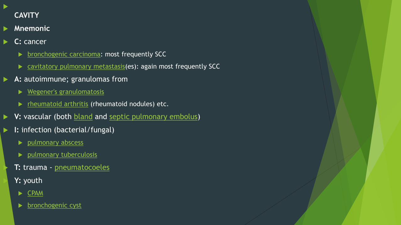
CAVITY
Mnemonic
C: cancer
bronchogenic carcinoma: most frequently SCC
cavitatory pulmonary metastasis(es): again most frequently SCC
A: autoimmune; granulomas from
Wegener's granulomatosis
rheumatoid arthritis (rheumatoid nodules) etc.
V: vascular (both bland and septic pulmonary embolus)
I: infection (bacterial/fungal)
pulmonary abscess
pulmonary tuberculosis
T: trauma - pneumatocoeles
Y: youth
CPAM
bronchogenic cyst

Thank you


![Cavitary Lung Lesion in a Patient with Systemic Lupus ... · CT [3]. Cavitary lesions are more common in lung cancer, tuberculosis, pulmonary abscess, fungus and Wegener’s granulomatosis](https://img.pdfslide.us/doc/110x75/5f7a0835d15e6d3e8b1bef8a/cavitary-lung-lesion-in-a-patient-with-systemic-lupus-ct-3-cavitary-lesions.jpg)