Embed Size (px)
Citation preview


Burn basic facts and management principles
BYTamer Rushdy
جعلها و اآلخره فى عباده بها الله خوف التى النارالدنيا فى للعاصين انذارا و للصالحين ابتالء

Survival was once the only aim of success in managing serious burn cases.
Today, however, the overriding objective of burn care has become reintegration of the patient into the home and community.
This goal has extended the traditional role of the burn care team beyond acute wound closure.
INTRODUCTION
We shall study burn from these aspects in order:
Epidemiology concerning Egypt
Aetiology and types
Pathophysiology
Prehospital care(management at scene)
Management in four stages(modern burn care)
Introduction

Modern burn care may be divided into the following 4 general phases:
The first phase, initial evaluation and resuscitation, occurs on days 1-3 and requires an accurate fluid resuscitation and thorough evaluation for other injuries and comorbid conditions.
The second phase, initial wound excision and biologic closure, includes the maneuver that changes the natural history of the disease. This is accomplished typically by a series of staged operations that are completed during the first few days after injury.
Introduction

The third phase, definitive wound closure, involves replacement of temporary wound covers with a definitive cover; there is also closure and acute reconstruction of areas with small surface area but high complexity, such as the face and hands.
The final stage of care is rehabilitation, reconstruction, and reintegration. Although this begins during the resuscitation period, it becomes time-consuming and involved toward the end of the acute hospital stay.
Introduction
A. EpidemiologyB. Aetiology and typesC. PathophysiologyD. Care at sceneE. Emergency room treatment
Airway Circulation Initial wound assesment Initial wound management
:Today we shall discuss

Epidemiology

Burn injuries in Egypt represent a major problem as compared with heart diseases, malignancy and road accidents.
Studies reveal that about 0.1% of the total population are affected by major burns.
Death rate is still high, about 30%. Of the saved ,36% need later reconstruction. Domestic causes are responsible for 75% of the injuries, the rest are caused by industrial and road.
EPIDEMIOLOGY

This is a study prospectively carried out on 880 acutely burned patients admitted to the Burn Unit of Ain Shams University 1 May 1995 to the 31 October 2001, with the objective to analyze the epidemiological features of burned patients in our country.
Females (53.1%) were more prevalent than males (46.8%).
Adults (61%) superceded children (39%).Females were most commonly burned at home and
mainly suffered from flame and scald burns.
EPIDEMIOLOGY
Males were most commonly burned in outdoor and work locations than at home and mainly sustained electric and flame burns.
Children were mostly burned at home and were mainly victims of scald and flame burns.
They had lower rates of full thickness burns compared to adults.
Epidemiology

Cutaneous burns are caused by the application of heat, cold, or caustic chemicals to the skin.
When heat is applied to the skin, the depth of injury is proportionate to the temperature applied, the duration of contact, and the thickness of the skin
Etiology

In civilian practice, scalds, usually from hot water, are the most common cause of burns. Water at 140°F (60°C) creates a deep dermal or full- thickness burn in 3 s.
At 156°F (69°C) the same burn occurs in 1 s.
Boiling water always causes deep burns.
Exposed areas tend to be burned less deeply than areas covered with thin clothing.
Clothing retains the heat and keeps the liquid in contact with the skin for a longer period.
Scald burns

Scald burn
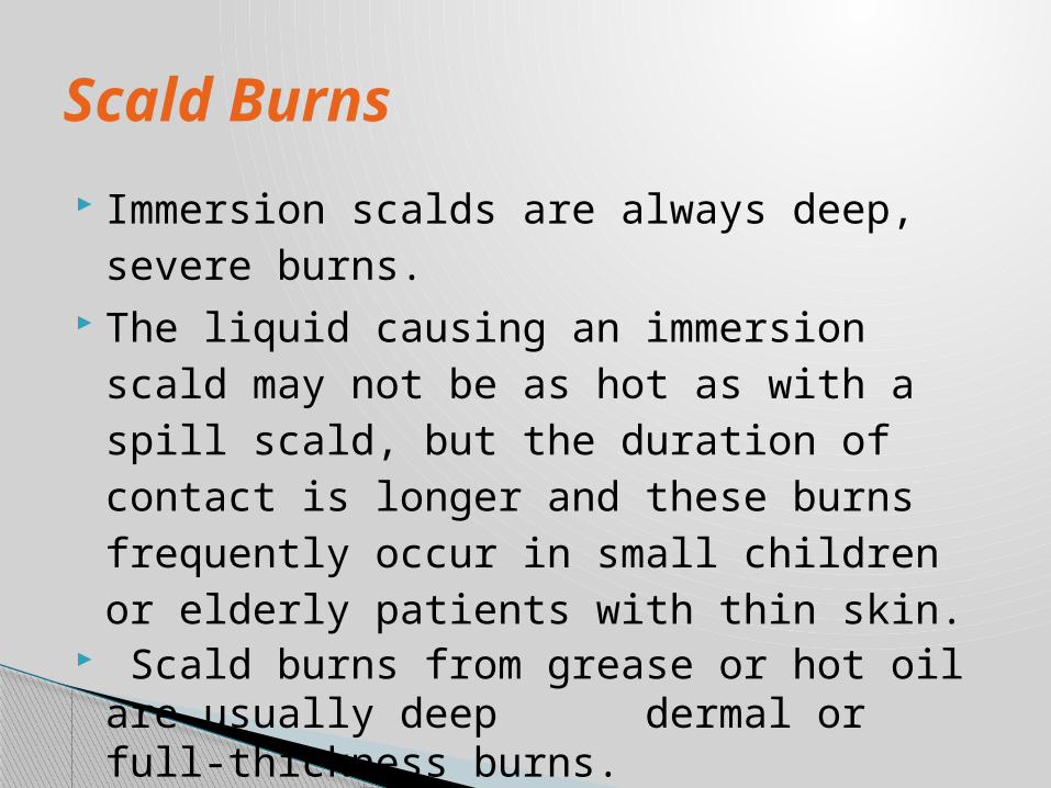
Immersion scalds are always deep, severe burns. The liquid causing an immersion scald may not be as
hot as with a spill scald, but the duration of contact is longer and these burns frequently occur in small children or elderly patients with thin skin.
Scald burns from grease or hot oil are usually deep dermal or full-thickness burns.
Scald Burns
Tar and asphalt burns are a special kind of scald. The tar should be removed by application of a
petroleum-based ointment under a dressing. The dressing may be removed and the ointment
reapplied every 2 to 4 h until the tar has dissolved. Only then can the extent of the injury and the depth
of the burn be estimated accurately.
Scald Burns


Flame burns are the next most common. Patients whose bedding or clothes have been on fire
rarely escape without some full-thickness burns.
Flame Burns

Flame of burn


Flash burns are next in frequency. Explosions of natural gas, propane, gasoline and other
flammable liquids cause intense heat for a very brief time. Clothing, unless it ignites, is protective against flash burns. These burns generally heal without requiring extensive
skin grafting, but they may cover a large skin area and be associated with significant thermal damage to the upper airway.
Flash Burns


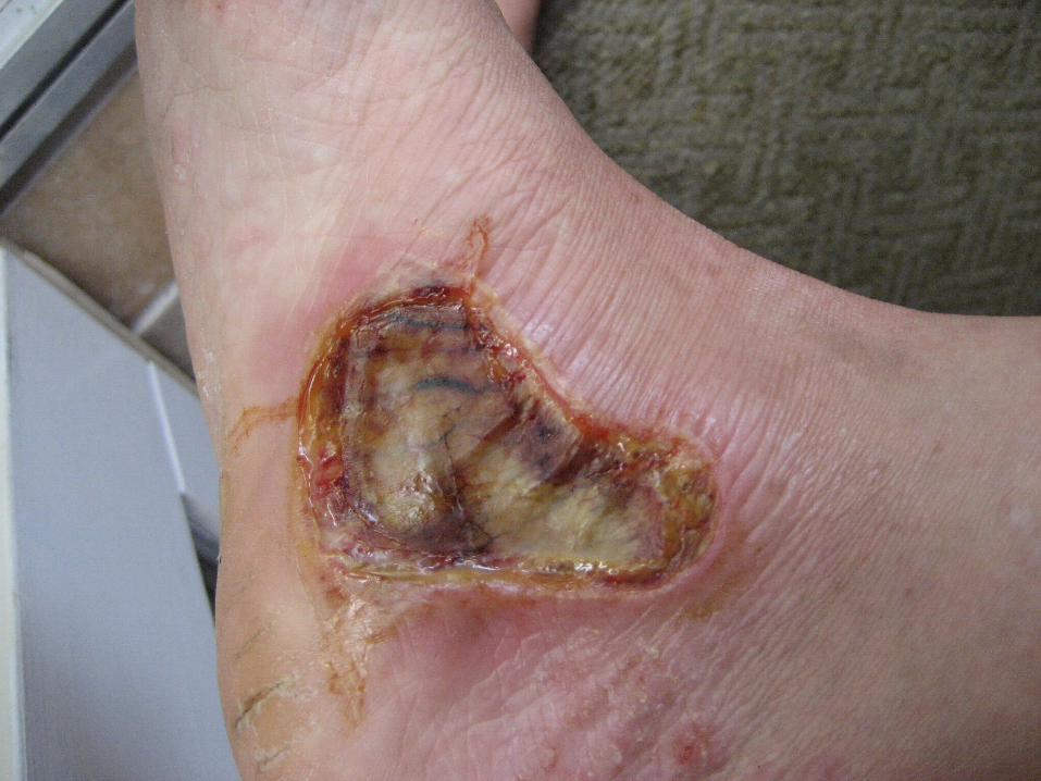
These burns result from contact with hot metals, plastic and glass; they are usually limited in extent, but invariably very deep .
The exhaust pipes of motorcycles cause a characteristic burn of the medial leg that, although small, usually requires excision and grafting.
Contact Burns


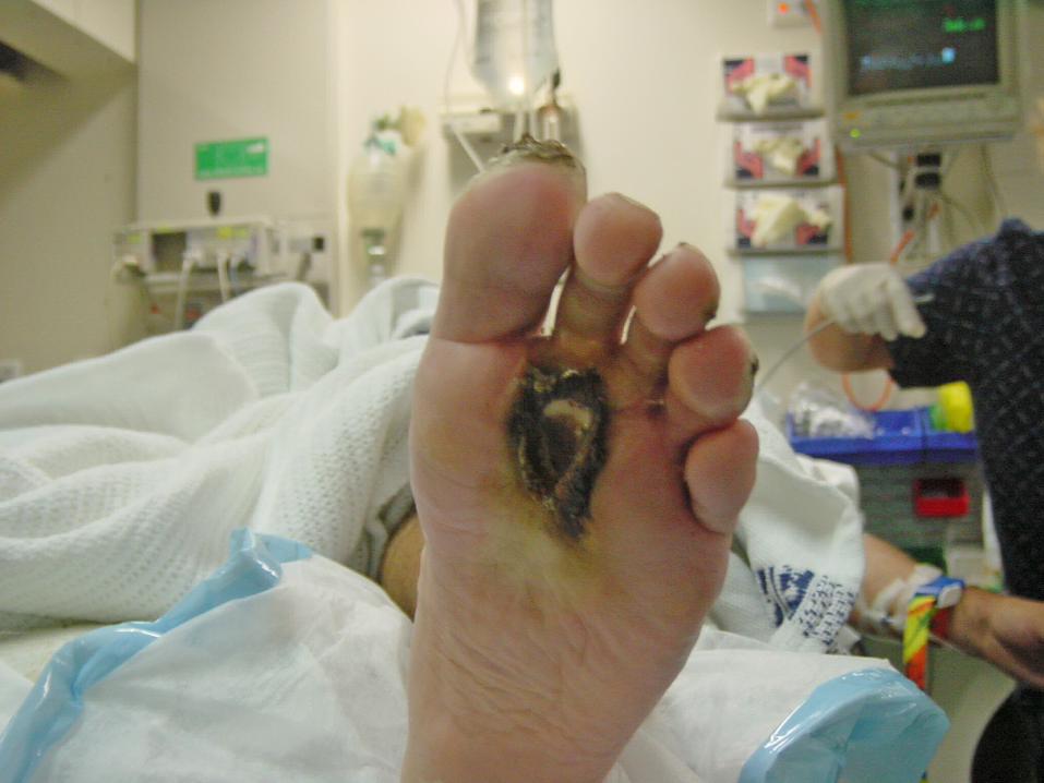
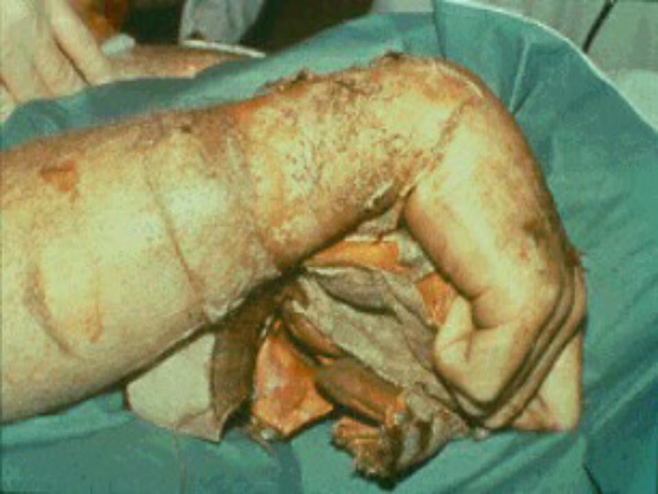
The passage of electric current through the tissues causes heating that results in cellular damage.
Low voltage (<1000 V) such as from a domestic supply causes significant contact wounds and may induce cardiac arrest, but no deep tissue damage .
High-voltage burns (>1000 V) cause damage by two mechanisms: flash and current transmission.
The flash from an arc may cause a cutaneous burn and ignite clothing, but will not result in deep damage but current transmission will result in cutaneous entrance and exit wounds and deep damage..
Electrical burns
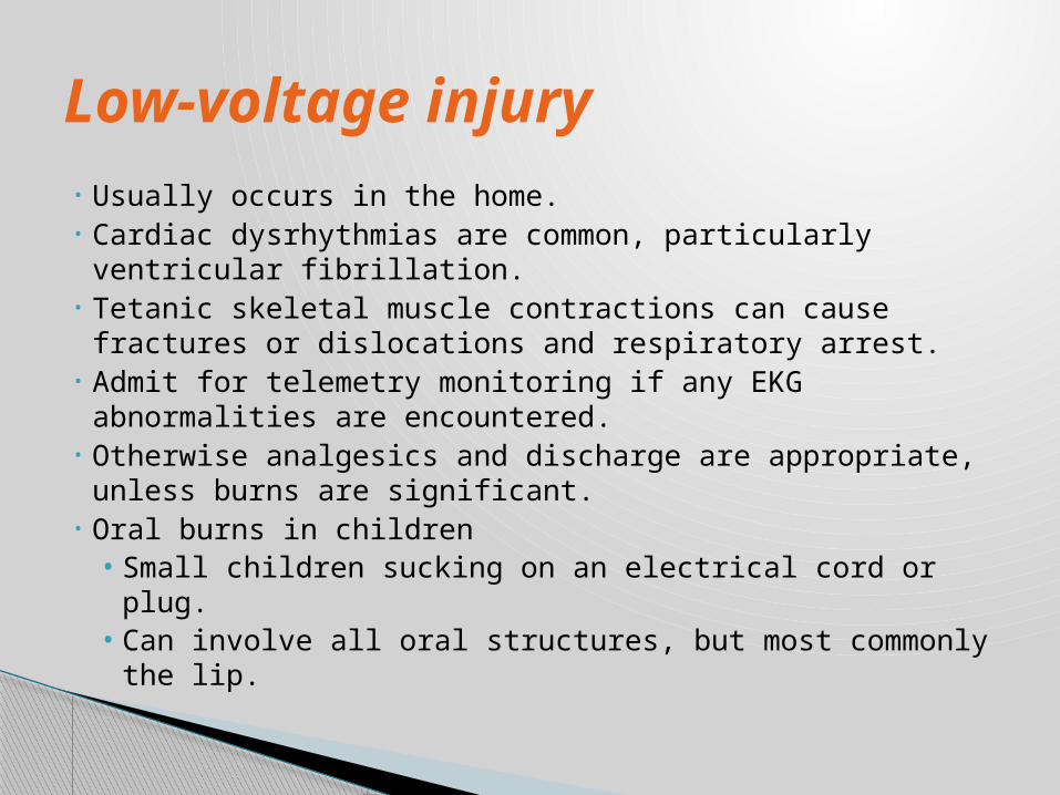
• Usually occurs in the home.• Cardiac dysrhythmias are common, particularly ventricular
fibrillation.• Tetanic skeletal muscle contractions can cause fractures or
dislocations and respiratory arrest.• Admit for telemetry monitoring if any EKG abnormalities are
encountered. • Otherwise analgesics and discharge are appropriate, unless
burns are significant.• Oral burns in children
• Small children sucking on an electrical cord or plug.• Can involve all oral structures, but most commonly the lip.
Low-voltage injury

The extent of tissue damage is typically underestimated because of the unpredictable path of injury.
An associated flash skin burn is not uncommon and can distract from the more important electrical injury to the deeper and remote tissues.
The deep injury is characterized by myonecrosis, especially along deeper tissues adjacent to bone (high resistance area).
Vessel thrombosis and compartment syndrome (both early and delayed) are common sequelae.
High-voltage injury

◦Early, aggressive, and repetitive wound debridements.◦Extremity fasciotomy frequently required.◦Because of the variable tissue necrosis, amputation of
a devitalized extremity may be necessary (even in the presence of adequate blood supply).
High-voltage injury


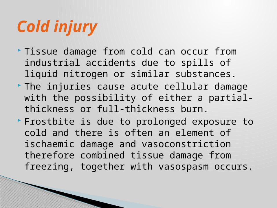
Tissue damage from cold can occur from industrial accidents due to spills of liquid nitrogen or similar substances.
The injuries cause acute cellular damage with the possibility of either a partial-thickness or full-thickness burn.
Frostbite is due to prolonged exposure to cold and there is often an element of ischaemic damage and vasoconstriction therefore combined tissue damage from freezing, together with vasospasm occurs.
Cold injury


X-irradiation may lead to tissue necrosis. Such injuries are exceedingly rare if industrial and medical safety precautions are working.
The tissue necrosis may not develop immediately. These injuries are generally limited in area and surgical
excision, and flap reconstruction may be appropriate management.
Of greater significance is the long-term cumulative effect of ionising radiation in the induction of skin cancers and other tumours.
Ionising radiation

Radiation Burn


Acidic or alkaline chemicals produce burns by the denaturing and breakdown of skin and body proteins.
A burn due to an acid substance is due to coagulation necrosis whereas a burn due to an alkali substance results in liquefaction necrosis.
The harmful effect will continue until the chemical is diluted or neutralised.
The most important initial treatment is dilution with running water.
Chemical burns


PATHOPHYSIOLOGY
BURNS
SKIN LOSS
AIRWAY & LUNG INJURIES
INFLAMMATORY & CIRCULATORY CHANGES
KIDNEYS
STOMACH
INTESTINE
OTHER CHANGES

SKIN LOSS
LOSS OF PROTECTIVE BARRIER
ENTRY OF MICROORGANISMS
LOSS OF FLUID LOSS OF HEAT
IMMUNOSUPPRESSION
SEPTICEMIASUSCEPTIBILITY TO
INFECTIONS
HYPOTHERMIAHYPOVOLEMIA

Cell-mediated immunity is impaired after burn injury, including documented delays in allograft rejection, impairment in responsiveness of lymphocytes, burn-size-related suppression of graft- versus-host activity, suppression of delayed cutaneous sensitivity tests, and diminution of peripheral lymphocytes and thoracic duct
lymphocyte concentration.. Whether this failure is the result of “overuse” or indirectly
the result of down-regulation by cytokine cascades and other products of the inflammatory reaction is unclear.
Cell-Mediated Immunity

INJURIES TO THE AIRWAY & LUNGS
FIRE, BLAST
etc
INHALATIONAL INJURYINHALATIONAL INJURY
CARBON MONOXIDE POISONING
MECHANICAL BLOCK 0N RIB MOVEMENT
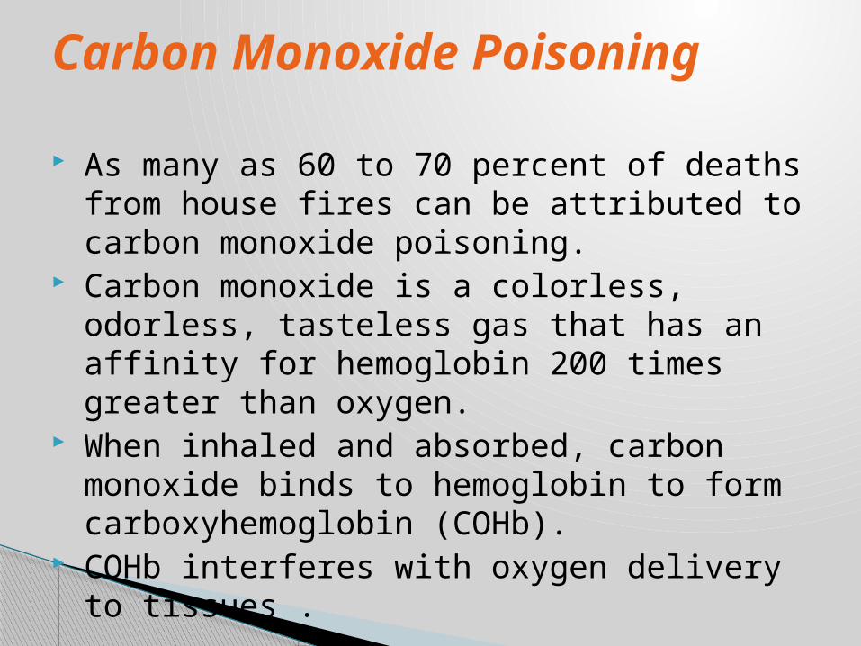
As many as 60 to 70 percent of deaths from house fires can be attributed to carbon monoxide poisoning.
Carbon monoxide is a colorless, odorless, tasteless gas that has an affinity for hemoglobin 200 times greater than oxygen.
When inhaled and absorbed, carbon monoxide binds to hemoglobin to form carboxyhemoglobin (COHb).
COHb interferes with oxygen delivery to tissues .
Carbon Monoxide Poisoning

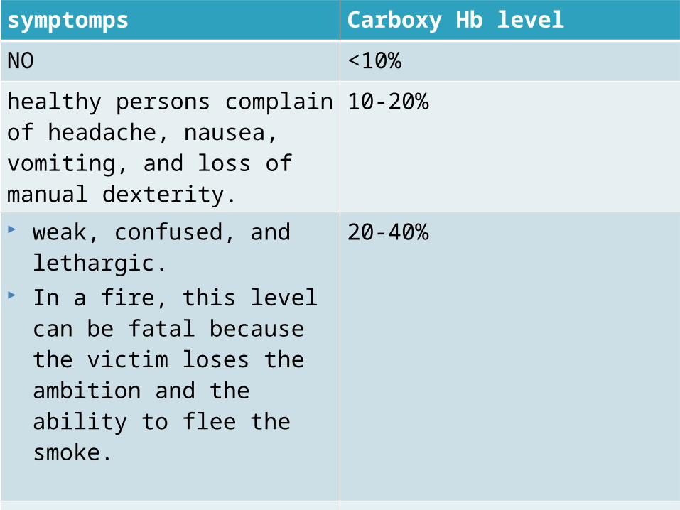
symptomps Carboxy Hb level
NO <10%
healthy persons complain of headache, nausea, vomiting, and loss of manual dexterity.
10-20%
weak, confused, and lethargic. In a fire, this level can be fatal
because the victim loses the ambition and the ability to flee the smoke.
20-40%
coma 40-60%
fatal >60%

Patients burned in an enclosed space or having any suggestion of neurologic symptoms should be placed on 100% oxygen while awaiting measured carboxyhemoglobin levels.
Patients who have not lost consciousness and who have a normal neurologic examination on admission will almost always recover completely without treatment beyond administration of 100% oxygen.
Patients who remain comatosed in the emergency room have a poor prognosis and rarely awaken.
Carbon Monoxide Poisoning
Inhalation injury increases the fluid requirements for resuscitation from burn shock after thermal injury.
Inhalation injury accompanying thermal trauma increases the magnitude of total body injury and requires increased volumes of fluid and sodium to achieve resuscitation.
Inhalation Injury
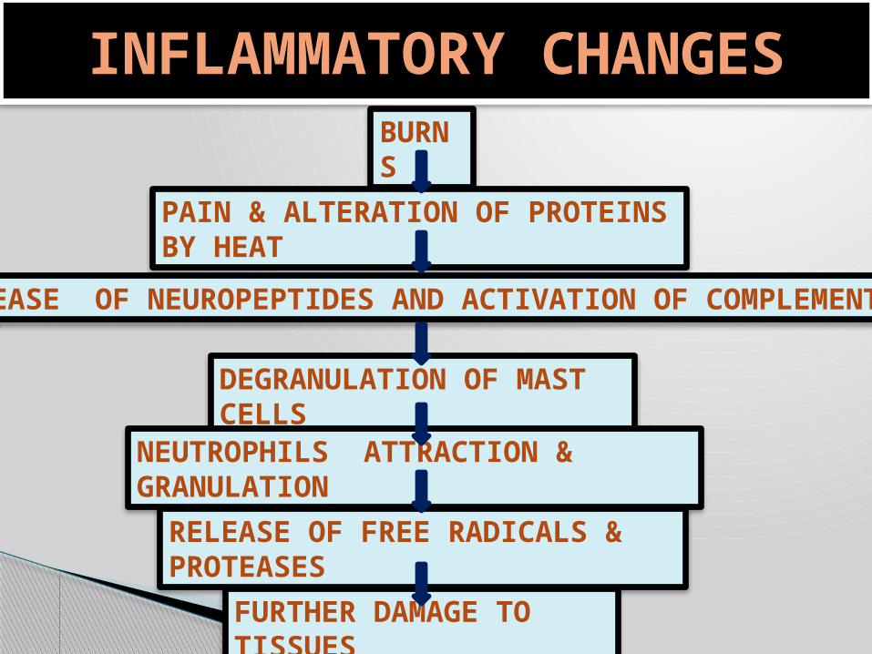
INFLAMMATORY CHANGES
BURNS
PAIN & ALTERATION OF PROTEINS BY HEAT
RELEASE OF NEUROPEPTIDES AND ACTIVATION OF COMPLEMENT
DEGRANULATION OF MAST CELLS
NEUTROPHILS ATTRACTION & GRANULATION
RELEASE OF FREE RADICALS & PROTEASES
FURTHER DAMAGE TO TISSUES
Catecholamines appear to be the major endocrine mediators of the hypermetabolic response in thermally injured patients.
Thyroid hormonal serum concentrations are not elevated in patients with large burns.. Concentrations of free T 3 and T4 fall markedly in the presence of sepsis in burned patients.
Neuroendocrine-Mediator Response

CIRCULATORY CHANGESLOSS OF SKIN
VESSELS PERMEABILITY
DIRECT FLUID LOSS
THIRD SPACE FLUID
COLLECTION
NON-BURN TISSUE
OEDEMA
CIRCULATORY SHOCK

OTHER CHANGES
LUNGS
STOMACH
INTESTINE
KIDNEYS

EMERGENCY CARECare at the Scene

Initial attention must be directed to the airway. Any patient rescued from a burning building or exposed to a
smoky fire should be placed on 100% oxygen by tight-fitting mask if there is any suspicion of smoke inhalation.
If the patient is unconscious, and appropriately trained personnel are present,an endotracheal tube should be placed and attached to a source of 100% oxygen.
Airway
If the airway has to be supported by a tight mask, there is a significant danger of aspiration of gastric contents, because air forced into the stomach will distend it and cause vomiting.
The mask prevents expulsion of the fluid, and gastric contents can flood the tracheobronchial tree.
Airway

Immediate cardiopulmonary resuscitation is rarely necessary, except in electrical injuries that have induced cardiac arrest or in patients with severe carbon monoxide poisoning with hypoxic cardiac arrest.
CPR
Smaller burns, particularly scalds, are treated with immediate application of cool water.
It has been mathematically demonstrated that cooling cannot reduce skin temperature enough to prevent further tissue damage, and that histologic damage is similar with or without cooling, but there is evidence in animals that cooling delays edema formation,probably by reducing initial thromboxane production.
After several minutes have elapsed, further cooling does not alter the pathologic process.
Iced water should never be used even on the smallest of burns. If ice is used on larger burns, systemic hypothermia may follow, and the
associated cutaneous vasoconstriction can extend the thermal damage.
Cold Application

The primary rule for the emergency physician is, “Forget about the burn.” As with any form of trauma the ABC protocol—airway, breathing, circulation—must be followed.
Although a burn is a dramatic injury, a careful search for other life-threatening injuries is the first priority.
Only after making an overall assessment of the patient's condition should attention be directed to the burns.
Emergency Room Care

The history is important. Inhalation injury should be suspected in anyone with a flame burn until proved otherwise.
The acrid smell of smoke on a victim's clothes should raise suspicion.
The rescuers are the most important historians and should be questioned carefully before they leave the emergency facility.
Careful inspection of the mouth and pharynx should be done early.
Hoarseness and expiratory wheezes are signs of potentially serious airway edema or smoke poisoning.
Copious mucus production and carbonaceous sputum are sure signs, but their absence does not rule airway injury out.
Emergency Assessment of Inhalation Injury

A standard Lund-Browder chart is readily available in most emergency departments for a quick assessment of TBSA burns.
Estimation of Burn Size Initial assesment


If the Lund-Browder chart is not available, the "rule of nines" is fairly accurate in adult patients.
A patient's palm is approximately 1% TBSA and can be used for estimating patchy areas.


Palm method
In patient with scattered burns, a method to estimate the percantage of burn is the palm method
The size of the patient´s palm is approximately 1% of TBSA
Fluid resuscitation Goal: Maintain perfusion to vital organs
Based on the TBSA, body weight and whether patient is adult or child.

Fluid resuscitation
Lactated Ringers - preferred solution Contains Na+ - restoration of Na+ loss is essential Free of glucose – high levels of circulating stress
hormones may cause glucose intolerance

Fluid resuscitation Burned patients have large insensible fluid losses
Fluid volumes may increase in patients with co-existing trauma
Vascular access: Two large bore peripheral lines (if possible) or central line.

Fluid resuscitation
Fluid requirement calculations for infusion rates are
based on the time from injury, not from the time fluid
resuscitation is initiated.

Baxter or Parkland Formula 4 cc R/L x % burn x body wt. In kg.
½ of calculated fluid is administered in the first 8 hours
½ is given over the remaining 16 hours.
Maintain urine output at 0.5 cc/kg/hr.
ARF may result from myoglobinuria
Increased fluid volume, mannitol bolus and NaHCO3 into each liter of LR to alkalinize the urine may be indicated

RL at 1.5 mL/kg per percentage burn, colloid at 0.5 mL/kg per percentage burn, and 2000 mL D5W.
Modified Brooke
RL at 2 mL/kg per percentage burn
Brooke
Galveston Formula
Used for pediatric patients
Based on body surface area rather than weight BSA = [87 (H + W) - 2600] / 10,000
More time consuming
5000 mL/m2 TBSA burn plus 2000 mL/m2 BSA, using RL solution plus 12.5 g 25% albumin per liter plus D5W solution as needed for hypoglycemia.
½ of total RL fluid is given in the first 8 hrs and balance over 16 hrs.
Urine output in pediatric patients should be maintained at 1 cc/kg/hr.

Regardless of the resuscitation formula used, the first 24-48 hours require frequent adjustments.
Calculated volumes from all of the formulas should be viewed as educated guesses of the appropriate fluid load.
Blind adherence to a derived number can lead to significant over or under resuscitation if not interpreted within the clinical context.
Over resuscitation can be a major source of morbidity for burn patients and can result in increased pulmonary complications and escharotomies of the chest or extremities.

RL solution is a relatively isotonic crystalloid solution that is the key component of almost all resuscitative strategies, at least for the first 24-48 hours.
It is preferable to normal saline for large-volume resuscitations because its lower sodium concentration (130 mEq/L vs 154 mEq/L) and higher pH concentration (6.5 vs 5.0) are closer to physiologic levels.
Another potential benefit of RL solution is the buffering effect of metabolized lactate on the associated metabolic acidosis.
RL

Assessing adequacy of resuscitation Peripheral blood pressure: may be difficult to obtain –
often misleading
Urine Output: Best indicator unless ARF occurs
A-line: May be inaccurate due to vasospasm
CVP: Better indicator of fluid status
Heart rate: Valuable in early post burn period – should be around 120/min.
> HR indicates need for > fluids or pain control
Invasive cardiac monitoring: Indicated in a minority of patients (elderly or pre-existing cardiac disease)

Effects of hypothermia
Hypothermia may lead to acidosis/coagulopathy
Hypothermia causes peripheral vasoconstriction and impairs oxygen delivery to the tissues
Metabolism changes from aerobic to anaerobic
serum lactate serum pH

Prevention of hypothermia
Cover patients with a dry sheet – keep head covered Pre-warm trauma room Administer warmed IV solutions Avoid application of saline-soaked dressings Avoid prolonged irrigation
Remove wet / bloody clothing and sheets Paralytics – unable to shiver and generate heat Avoid application of antimicrobial creams Continual monitoring of core temperature via foley or
SCG temperature probe

Prophylactic antibiotics are not indicated
in the early postburn period.
Antibiotics

Burns are tetanus-prone wounds. The need for tetanus prophylaxis is determined by the
patient's current immunization status. Previous immunization within 5 years requires no
treatment, immunization within 10 years a tetanus toxoid booster, and unknown immunization status hyperimmune serum (Hyper-Tet).
Tetanus

Many burn centers begin tube feeding on admission, to protect the stomach from stress ulceration and the patient from a paralytic ileus and decreasing catabolism.
If the patient is to be transported, the safest course is usually to decompress the stomach with a nasogastric tube.
Gastric Decompression
During the shock phase of burn care, medications should be given intravenously. Subcutaneous and intramuscular injections are absorbed variably depending on perfusion and should be avoided.
Pain control is best managed with small intravenous doses of morphine, usually 2 to 5 mg, given until pain control is adequate, without affecting blood pressure.
Pain Control

A great deal of interest exists in using antioxidants as adjuncts to resuscitation to try to minimize oxidant-mediated contributions to the inflammatory cascade.
In particular, megadose vitamin C infusion during resuscitation has been studied at some length.
Recently published data using an infusion of 66 mg/kg/h during the first 24 hours demonstrate a 45% decrease in the required fluid resuscitation in a small group of patients.
Vitamin C
Psychosocial care should begin immediately. The patient and family must be comforted and given
a realistic assessment regarding the prognosis of the burns.
Psychosocial Care


INITIAL WOUND ASSESMENT AND FIRST HOUR WOUND CARE
AT EMERGENCY ROOM
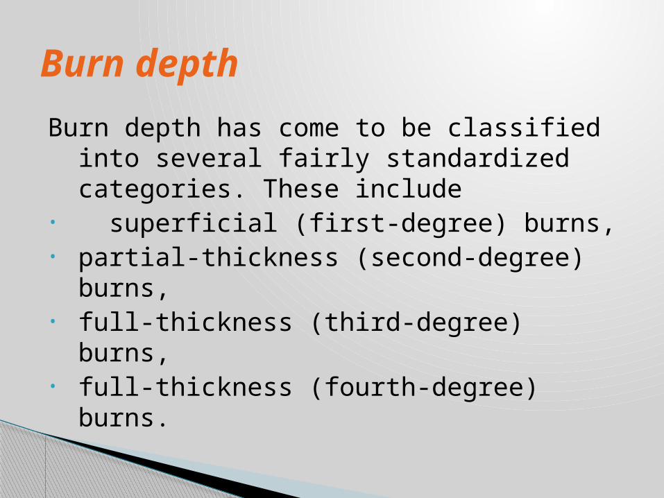
Burn depth has come to be classified into several fairly standardized categories. These include
• superficial (first-degree) burns, • partial-thickness (second-degree) burns, • full-thickness (third-degree) burns, • full-thickness (fourth-degree) burns.
Burn depth
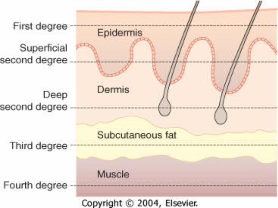

Superficial (first-degree) burns are limited to epidermal layers and are equivalent to a superficial sunburn without blister formation.
Partial-thickness (second-degree) burns are also called dermal burns and can be superficial partial-thickness burns or deep partial-thickness burns.
Superficial partial-thickness burns involve the superficial papillary dermal elements and are pink and moist with pain upon examination. Blister formation appears with the level of the burn. This type of burn is expected to heal well within several weeks, without skin grafting.


Deep partial-thickness burns involve the deeper reticular dermis. They can have a variable appearance ranging from pink to white with a dry surface. Sensation may be present but is usually somewhat diminished, and capillary refill is sluggish or absent. Burns of this depth routinely require excision and grafting for satisfactory healing


Full-thickness (third-degree) burns extend into the subcutaneous tissues and have a firm, leathery texture and complete anesthesia upon examination.
Clotted vessels can be observed through the eschar.

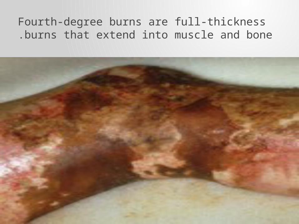
Fourth-degree burns are full-thickness burns that extend into muscle and bone.

• Wash gently with gauze soaked in saline.• Remove any obviously loose skin.• Apply topical agents .• Irrigate debris from the eyes, as needed.• Cover wounds with dry sterile dressings.
Initial burn wound management

To be continued

نلتقى بعد
الفاصل

![Negative Burn Index - Enjolrasworld Arndt/Negative Burn … · Web viewNegative Burn. Negative Burn, Vol. 1. ... [Charles Moore/Randy Green & Craig Gilmore] 6p . 2) The Apparition:](https://img.pdfslide.us/doc/110x75/5b5082fd7f8b9a5a6f8e9ce5/negative-burn-index-arndtnegative-burn-web-viewnegative-burn-negative-burn.jpg)


![5 2 BURN[1]](https://img.pdfslide.us/doc/110x75/546289f3b1af9f03628b4aae/5-2-burn1.jpg)