Embed Size (px)
Citation preview


ها النار التى خوف هللا بها عباده فى اآلخره و جعل
اابتالء للصالحين و انذارا للعاصين فى الدني

❏ total 2º and 3º burns > 10% TBSA in patients < 10 or > 50
years of age
❏ total 2º and 3º burns > 20% TBSA in patients any age
❏ 3º burns > 5% TBSA in patients any age
❏ 2º or 3º burns with threat of serious functional or cosmetic
impairment (i.e. face, hands, feet, genitalia, perineum, major
joints).

❏ inhalation injury (may lead to respiratory distress)
❏ electrical burns (internal injury underestimated by
TBSA)
❏ chemical burns posing threat of functional or
cosmetic impairment
❏ burns associated with major trauma

Focus of burn treatment is then shifted to the definitive
burn wound treatment and to the general support of the
patient, which include:
Wound care and coverage
Nutritional support
Infection diagnosis and management
Rehabilitation and management of burn wound
sequale
Full-thickness circumferential burns result in the formation
of a tough, inelastic mass of burnt tissue (eschar).
The eschar, may due to this inelasticity, results in the burn-
induced compartment syndrome.
This is caused by the accumulation of extracellular and
extravascular fluid within confined anatomic spaces
The excessive fluid causes the intracompartmental pressure
to increase, resulting in collapse of the contained vascular
and lymphatic structures and, hence, loss of tissue viability.

The presence of a circumferential eschar with one of
the following:
Impending or established vascular compromise of
the extremities or digits.
Impending or established respiratory compromise
due to circumferential torso burns

Neurovascular integrity should be monitored frequently
and in a scheduled manner.
Capillary refilling time, Doppler signals, pulse oximetry,
and sensation distal to the burned area should be checked
every hour.
Limb deep compartment pressures should be checked
initially to establish a baseline.

Subsequently, any increase in capillary refill time,
decrease in Doppler signal, or change in sensation
should lead to rechecking the compartment pressures.
Compartment pressures greater than 30 mm Hg
should be treated by immediate decompression via
escharotomy and fasciotomy, if needed.

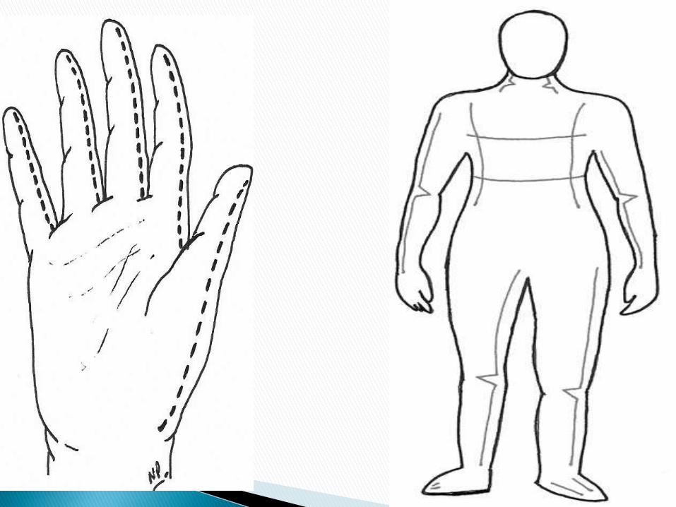
When escharotomy is required in a patient with a
circumferential chest wall burn, it is performed in the
anterior axillary line bilaterally. If there is significant
extension of the burn onto the adjacent abdominal
wall, the escharotomy incisions should be extended to
this area and should be connected by a transverse
incision along the costal margin


Local anesthesia is unnecessary because third- degree
eschar is insensate; small doses of intravenous
narcotics may be utilized to control anxiety.
The incision, which must avoid major nerves, vessels,
and all tendons should extend through the eschar
down to the subcutaneous fat.
Escharotomy is rarely required within the first 6 h
postburn .


Treatment planning depends on the assessment of the
following factors:
• Patient’s general condition and co-morbid factors
• Patient age
• Burn depth
• Burn size
• Anatomical distribution of injury

Treatment optionBurn depth
1-Topical antimicrobials
2-Biological dressings e.g human
placenta
3-Skin substitutes e.g Biobrane®
5-exposure
Small /medium sized superficial
partial thickness wound (< 40%
TBSA)
1-Allograft
2-Xenograft
3-Topical antimicrobials
Large superficial partial thickness
injury(> 40% TBSA)
excision and grafting
Versus
Topical antimicrobials
Deep partial thickness injury
(small and large )
invariably
require excision and skin grafting.
Full thickness injury

DisadvantagesAdvantagesTopical Agents
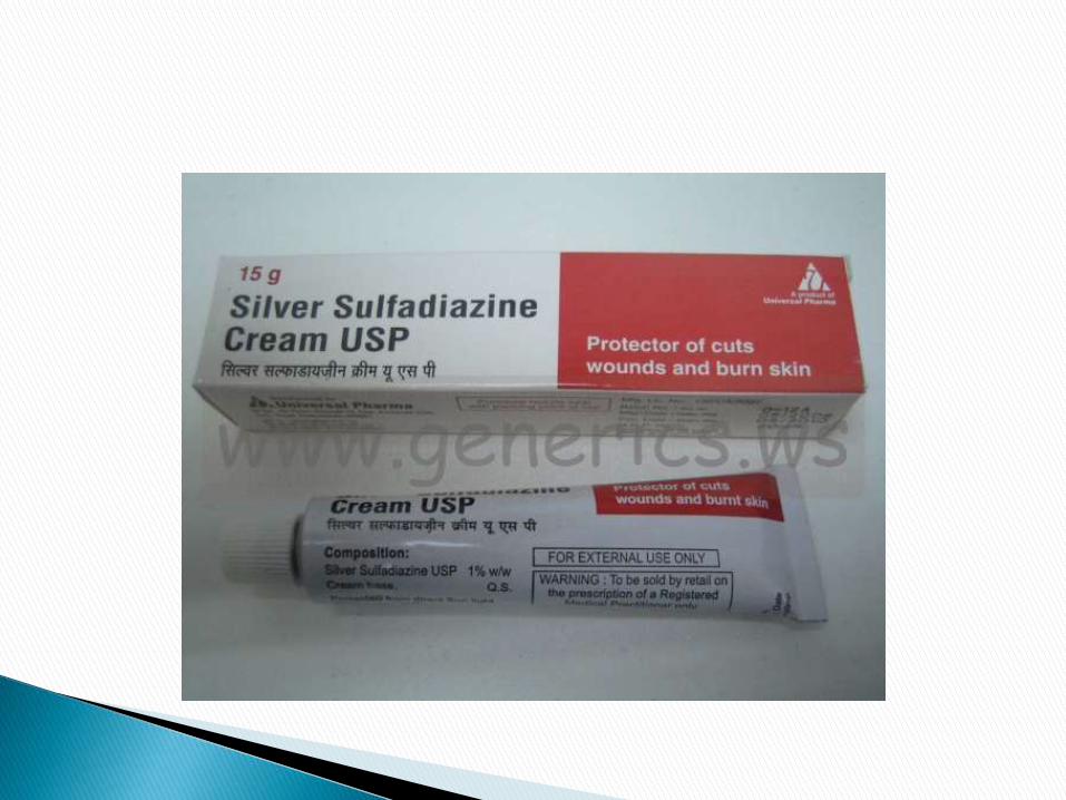
Lack of penetrationPainlessSilver Sulfadiazine
Painful, Carbonic
anhydrase inhibitor
PenetratesMafenide Acetate
Limited penetrationBroad spectrumSilver Nitrate
Impairs wound
healing in high
doses
Broad spectrumSodium
Hypochlorite

disadvantagesadvantagesagent
Minimal
coverage Often
combined with
polymyxin
and neomycin into
triple
ointment
Gram-positive
coverage
Bacitracin
Petroleum-based
Keeps grafts moist
Polymyxin B

Flamazine Dressing

MEBO Dressing


There are numerous products available and can be
differentiated to those that provide temporary wound
cover while the underlying wound re-epithializes or
is ready for autografting (i.e., Biobrane®,
Dermagraft TC®) and those that close the wound
and help reconstitute part of the resultant skin
(Integra®).


usually harvested from cadaveric donors after
appropriate donor selection and screening for
communicable disease, and consent from relatives
has been obtained.
In order of preference of allograft take on the excised
burn wound, fresh allograft is by far the best followed
by cryopreserved, glycerolized, then freeze-dried.
Allograft skin can also be obtained from living donors,
usually parents or relatives of burned children


Skin from different species can be used for temporary
physiological wound closure.
Pig skin is commonly used and is commercially
available.

There are two methods of management of the burn
wound with topical agents.
In exposure therapy, no dressings are applied
over the wound after application of the agent to the
wound twice or three times daily. This approach is
typically used on the face and head. Disadvantages are
increased pain and heat loss as a result of the
exposed wound and an increased risk of cross-
contamination.

In the closed method, an occlusive dressing is
applied over the agent and is usually changed twice
daily. The disadvantage of this method is the potential
increase in bacterial growth if the dressing is not
changed twice daily, particularly when thick eschar is
present. The advantages are less pain, less heat loss, and
less cross-contamination. The closed method is generally
preferred.

In vitro culturing of epidermal cells (keratinocytes)
produced a permanent skin and grafted onto a burn
wound bed, closing massive wounds when donor
sites were limited.
The first successful grafting was reported in children
in 1986.

When the patient is admitted, a 1-cm skin biopsy
specimen is usually sent to a commercial laboratory
for culturing.
Three weeks later 5- by 5-cm 2 sheets of cultured
cells are delivered.
CEAs are expensive.
Engrafted CEAs are poorly adherent and extremely
fragile for months after application.


Excisional procedures should be performed as early
as possible after the patient is stabilized.
This allows the wound to be closed before infection
occurs and, in extensive burns , allows donor sites to
be recropped as soon as possible.
Cosmetic results are better if the wound can be
excised and grafted before the intense inflammatory
response associated with burns becomes well
established.

Any burn projected to take longer than 3 weeks to
heal is a candidate for excision within the first
postburn week.
Wound excision is adaptable to all age groups, but
infants, small children, and elderly patients require
close perioperative monitoring.

Excision can be performed to include the burn and
subcutaneous fat to the level of the investing fascia
(fascial excision), or by sequentially removing thin
slices of burned tissue until a viable bed remains
(sequential excision).

The principle is to shave very thin layers of burn eschar
sequentially until viable tissue is reached.
The burn can be removed with a variety of instruments,
usually power- or hand-driven dermatomes.
Slices are taken until a viable bed of dermis or
subcutanbed does not bleed briskly, another slice of the
same depth eous fat is reached.
If inspection of the dermal or fatty bed reveals a surface
that appears gray or dull rather than white and shiny, or
if there is evidence of clotted vessels, the excision should
be carried deeper.

Any fat that has a brownish discoloration, has blood
staining, or contains clotted blood vessels will not support
a skin graft and must be excised until the bed contains
uniformly yellow fat with briskly bleeding vessels.
Bleeding is controlled with sponges soaked in 1:10,000
epinephrine solution applied to the excision bed for 10
min.
Continued bleeding is then controlled with an
electrocautery.

Fascial excision is reserved for patients with very
deep or for patients with very large, life-threatening,
full-thickness burns.

(1) It results in a reliable bed of known viability.
(2) Tourniquets can be routinely used for extremities.
(3) Operative blood loss is less than with sequential
excision.
(4) Less experience is required to ensure an optimal
bed.

(1) The operative time is longer.
(2) There may be severe cosmetic deformity,
especially in obese patients.
(3) There is a higher incidence of distal edema when
excision is circumferential.

Skin graft junctures should be avoided over joints,
and grafts should be placed transversely when
possible.
Thick skin grafts yield a better appearance than thin
skin grafts so should be used on the face, neck, and
other cosmetically important areas.

The resultant donor sites can be overgrafted with thin
skin grafts to minimize hypertrophic scarring of the
donor site.
Whenever possible, cosmetically important areas
should be grafted with sheet skin grafts.

Although meshed skin grafts provide cover with
excellent function, the meshed pattern persists as a
permanent reminder of the burn.
Adjacent pieces of skin graft should be approximated
carefully.
While staples are adequate for areas in which
cosmetics is not an issue, for critical areas, such as
the face, suturing the edges together is preferred.


Superficial burns of the face should be left exposed.
The face is washed twice daily with a mild soap and
water, and a thin layer of a bland ointment
(bacitracin) is applied to the open wounds to prevent
drying.
Superficial burns of the ear should be treated with a
bland ointment.
Deeper injuries must be treated with topical
antibiotics; excessive pressure may cause chondritis,
and should be avoided.

Suspected corneal burns should be stained with
fluorescein for confirmation of diagnoses.
Superficial corneal burns should be treated similarly
to corneal abrasions, with vigorous irrigation, the
application of ophthalmologic antibiotic ointment,
and eye patching.

Superficial burns of the hand should be elevated for
24 to 48 h to minimize swelling.
Circumferential hand burns may require
hospitalization for observation of adequate
circulation.
Range-of-motion exercises should begin as soon as
possible after injury.
.

Although burns of the feet are painful, walking and
range-of-motion exercises should be performed.
Crutches should not be allowed.
To prevent edema, burned feet should be elevated
when the patient is not walking or exercising.
An elastic bandage should be applied over the
wound dressing when the patient is walking or
sitting, but it should be removed at night when the
feet are elevated.

Perineal burns frequently require hospitalization for
24 to 48 h for observation of urinary obstruction
secondary to edema.
Minor perineal burns can be treated with a bland
ointment.
Extensive superficial perineal burns, e.g., pediatric
bathtub scald injuries, are best treated with topical
(silver sulfadiazine), utilizing a diaper as the wound
dressing.

To prevent contracture Aim
Extended (no pillow)Head and neck
apply eye ointment 3
times daily
Eyelids
apply moisturizing agent
(Vaseline)
Lips
apply maintainer Lip commissure
elevation and apply splint
in functional position
Hand
(abducted )Axilla
dorsiflexed with foot
support.
Foot


1- Early release of tension over flexion creases of joints. Tension in a scar encourages hypertrophy, so that releasing it by grafting or local flaps may
prevent its occurrence.
2- Continuous scar massage, after application of skin
emollient, can be quite effective.

3- Pressure on maturing scar tissue, appears to
reduce the incidence of hypertrophic changes. Such
pressure is most likely maintained by compressive
garments for 24hrs./day, for at least six to twelve
months.

1- The release of the contracture by re-arrangingthe tissues by local flaps (e.g.: Z- plasty) or by the application of skin graft. 2- Intralesional steroid injection(e.g.: triamcinolone acetonide; 1-2 cc of 40 mg./ccat one or two weeks interval.). It inhibits collagenaseinhibitors causing degradation of collagen, thus decreasing dermal thickening.

3- Application of silicone gel sheet as an
occlusive dressing.
Ideally it should be placed 24hrs./day for about ayear.

5- Laser therapy :
The modalities are :
- Pulse-dyed laser ----- microvascular thrombosis
- CO2 laser & Argon laser----- collagen shrinkage
through heating.
- Nd-YAG laser----- inhibits collagen metabolism
and production.
However the recurrence rate with laser therapy is
high.

6- Interferon therapy : The newest therapeutic
modality on the horizon is intralesional injection Of Interferon.
They reduce fibroblast synthesis and collagen
type I, III and possibly IV and increase the
collagenase activity.

Management of burn sequelaein specific regions
1- Head and Neck
2- Upper extremity
3- Lower extremity
4- Trunk

3- Reconstruction :a- Minor defect: advancement and rotation of adjacent scalp flaps will be enough to fill the defect.
b- Moderate defect: Tissue expansion is the final treatment of choice. This allows the area to be reconstructed with like tissue and with no donor defect.


c- Extensive defect: This is a difficult situation.
Defects in this range may be too large to be
corrected by tissue expansion. If periosteum is
intact, a skin graft is applied. Otherwise free tissue
transfer is required. The most common flaps are the
omentum and the latissimus myocutaneous flaps.
1- The forehead : is best resurfaced with asingle sheet of split thickness skin graft.With bony exposure or destruction, flap reconstruction is indicated.2- The cheeks : the best is tissue expansionfrom adjacent non-injured tissue (e.g.: neck).Thin free flaps may be considered (e.g.: radialforearm flap). Others describe the use of alarge full-thickness graft as one aesthetic unit.

Eye lid reconstruction :Indications : exposed cornea, contractor
ectropion of upper and/or lower eye lid and contractures at the canthi regions.1- Total loss of eye lids : the exposed cornea can be covered by mobilizing the conjunctiva which is covered with skin graft. Later on the lids can be reconstructed with local flaps (e.g.: cheek flap or median forehead flap with septalmucoperichondrial graft as lining).
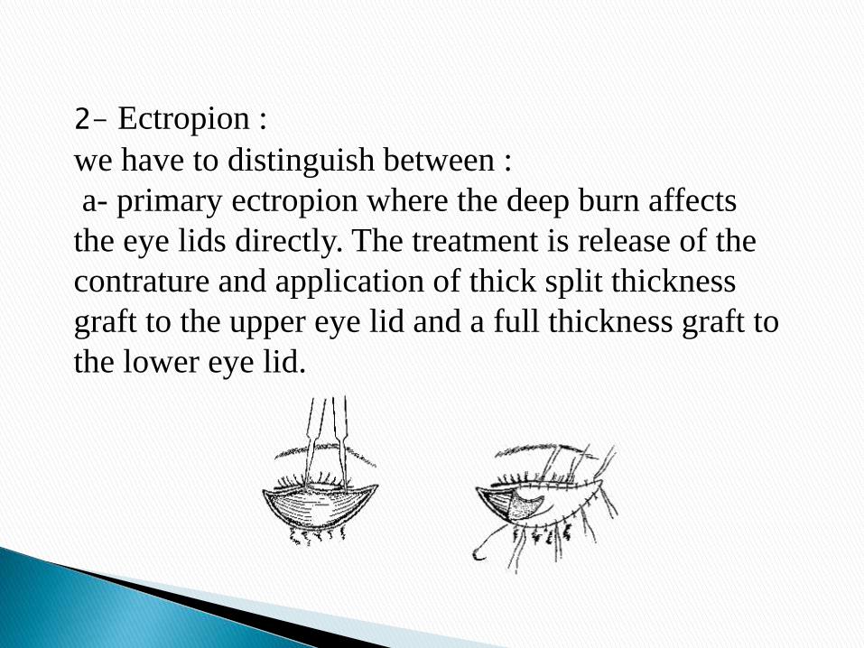
2- Ectropion :
we have to distinguish between :
a- primary ectropion where the deep burn affects
the eye lids directly. The treatment is release of the
contrature and application of thick split thickness
graft to the upper eye lid and a full thickness graft to
the lower eye lid.

b- secondary ectropion, due to contracture of
forehead, cheek or neck pulling on the eye lids.
Treating the cause will alleviate the condition.

Eye brow reconstruction :* Loss of the hair may be compensated by the simple simulation done by an eye brow pencil ( specially in women ).However surgical reconstruction of the eye brow may be done through :
1- Hair transplantation: single hair transplantation is
better than a punch graft.
2-Hair-bearing flap from the temporal scalp. It is
based on the superficial temporal artery and it is an island flap.

3- Strip graft taken anywhere from the hairy
scalp with the dimension and shape of the eye
brow. Care is taken :
- not to exceed 4 mm. in width.
- not to injure the hair follicles during elevation of
the flap by the scalpel.
- the direction of the hair should be oriented from
medial to lateral.

Lip and mouth reconstruction :1- Extensive scarring of the upper or lower lip:excision and full thickness graft within theaesthetic unit of the involved lip.2- Microstomia (oral commissurecontracture):corrected by full thickness incisions at each angleof the mouth as far as a line dropped verticallyfrom the pupil of the eye. Then the oral mucosa

is mobilized and everted onto the lip skin, forminga new commissure. Some overcorrection isgenerally advisable.

Nasal reconstruction :1- Total destruction of the nose requires :a- Flap reconstruction either regional, like the forehead flap, or distant by microvascular transfer.b- Prosthetic reconstruction. 2- Unacceptable hypertrophic or hypopigmentedscars over a large surface of the nose may be treated by dermabrasion, either mechanical or bylaser, and application of a single sheet of skingraft within the nasal aesthetic units.

3- Alar rim reconstruction is done using a composite
graft from the ear.
4- Nostril stenosis is treated by release and skin
grafting. Splints must be worn for at least six months
after surgery to prevent recurrence.
5- Web contracture between columella and upper lip,
may be released by V-Y advancement flap.

Ear reconstruction:- Indications: Partial or total loss of the externalear.- Classification: Help to determine the treatment.Mild defect: loss of helix and upper part of the auricle, without extensive scarring.Moderate defect: concha nearly normal; upper halfof the ear missing; antihelix and its posterior cruramissing.Severe defect: remnant of concha; local soft tissuescarred; external ear orifice normal or stenosed.

Head & neck reconstruction(Ear reconstr.)
Treatment :1- Total absence of the auricle :
- Surgical reconstruction using a costochondral
graft, as described for microtia.
- Osteointegrated prosthesis.
2- Subtotal absence of helical rim :
- Local flap reconstruction is preferred.
- When the entire helix is missing, a tubed
cervical skin flap is used.

3- Ear lobe deformity:
- Adherence of the ear lobe to the neck is the main
deformity. Z-plasty or local flaps are generally
sufficient for correction.
4- Meatal stenosis :
- Splinting may be used as a preventive measure
and may eliminate the need for surgical
correction
- After release, use local flaps if available. If not
use skin graft.
- A conformer is worn by the patient for 4 - 6
months to prevent recurrence.

* Treating established contractures :1- Mild cases: mild scar bands can generally be corrected surgically by using local flaps or Z-plasties.2- Moderate cases: contractures involving 1/3 - 2/3 of anterior neck, can be treated using tissue expansion. The unscarred lateral aspects of the neck are expanded.
3- Severe cases: contractures involving more than
2/3 of the anterior neck, are better treated by
release and split thickness skin graft or distant flap
by microvascular technique. Local flaps are not
adequate.

* Treating established contractures :1- Scar bands and minor contractures are bettertreated by local flaps e.g.: Z-plasty or V-Y plasty.They may be combined with the application of skin graft, kept in place by tie-over dressing.

* Treating established contractures :2- Moderate contracture may be released and the
defect filled with a latissimus dorsi fasciocutaneous
flap.

* Treating established contractures :3- Severe contracture, producing large defect on
release, are best treated with skin graft.

Plaster of paris is applied at the
end of the operation where the
joint is kept as fully abducted
as possible.
Splintage should be maintained
for several weeks until the
patient can put the joint
through a full range of
movement.
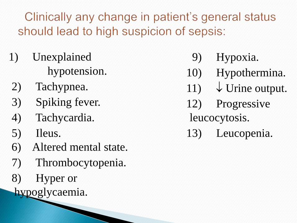
1) Unexplained
hypotension.
2) Tachypnea.
3) Spiking fever.
4) Tachycardia.
5) Ileus.
6) Altered mental state.
7) Thrombocytopenia.
8) Hyper or
hypoglycaemia.
9) Hypoxia.
10) Hypothermina.
11) Urine output.
12) Progressive
leucocytosis.
13) Leucopenia.

Management of clinically septic patien
1) Support of cardiopulmonary and G.I. systems.
2) Eschar debridement.3) Empiric antibiotic.4) Send for culture/sensitivity.5) Adequate fluid to maintain intravascular volume.
6) Invasive monitoring.7) Change in frequency of dressing.8) Change in topical antibiotic.
t

Before the availability of penicillin, streptococci and
staphylococci were the predominant infecting
organisms.
By the late 1950s, gram-negative bacteria (
Pseudomonas species) had emerged as the dominant
organism causing fatal wound infections in burn
patients.

All burn wounds become contaminated soon after
injury with the patient's endogenous flora or with
resident organisms in the treatment facilities
The likelihood of septicemia increases in proportion
to the size of the burn wound.

One result of the prolonged survival of severely burned
patients in critical care units, made possible by modern
patient support techniques, is that the respiratory tract has
become the most common locus of infection
A diagnosis of pneumonia is confirmed by the presence of
characteristic chest radiograph patterns, and the presence of
offending organisms and inflammatory cells in the sputum
For the diagnosis of bronchopneumonia, analysis of
sputum samples may be adequate

Suppurative thrombophlebitis is a major cause of
sepsis in burn patients, occurring in up to 5 percent of
patients with major burns.

Endocarditis is occasionally the cause of occult sepsis in
burn patients, and its incidence continues to rise with the
increasing use of intravenous catheters for hemodynamic
monitoring. Endocarditis should be suspected in patients
with positive blood cultures and no other identifiable
source of bacteremia. These patients should be examined
repeatedly
by echocardiography until the source of the septicemia is
identified.

Most patients with burns greater than 20 percent
TBSA require indwelling urinary catheters to guide
fluid resuscitation.
Aseptic techniques of insertion and catheter care, the
use of a closed drainage system, and the removal of
the catheter at the earliest clinically indicated time are
effective measures for preventing urinary tract
infections.

The pinna of the ear is composed almost entirely of
cartilage with minimal blood supply and is vulnerable
to infection.
It is a rare complication.
When chondritis does occur, conservative approach
with drainage of the helix centrally, in an attempt to
preserve the outer cartilages, is usually successful.

The nutritional effects of the hypermetabolic response
to thermal injury are manifested as exaggerated energy
expenditure and massive nitrogen loss.
Nutritional support is directed primarily toward
supply of calories to match energy expenditure and
provision of nitrogen to replace or support body protein
stores.

Caloric requirements in adult burn patients are
calculated using the Curreri
formula, which calls for 25 kcal/kg/day plus 40
kcal/% TBSA burned/day.

Patients with burns under 25 percent TBSA that are
not complicated by facial injury, inhalation injury, or
malnutrition, and are not associated with psychological
difficulties can usually be maintained on high-calorie,
high-protein diets ingested orally.
The nutritional requirements of patients with large
burns cannot be met by the oralroute alone, and these
patients should be fed gastrointestinally or
nasoenterally.

A functionally intact alimentary tract always should
be used.
Enteral nutrients seem to maintain the integrity of the
gastrointestinal tract, and increased hepatic protein
synthesis may reduce the incidence of bacterial
translocation from the gut.

An oral diet preserves gut mucosal mass and
maintains digestive enzyme content; parenteral feeding
results in decreased mucosal cell turnover.
Total parenteral nutrition should be instituted when
enteral feedings alone cannot provide adequate
nutritional support


![5 2 BURN[1]](https://img.pdfslide.us/doc/110x75/546289f3b1af9f03628b4aae/5-2-burn1.jpg)


![Burn 20PowerPoint[1]](https://img.pdfslide.us/doc/110x75/55cf8c4e5503462b138b479d/burn-20powerpoint1.jpg)


![Burn Management students[1]](https://img.pdfslide.us/doc/110x75/577d2daf1a28ab4e1eae1b91/burn-management-students1.jpg)