Embed Size (px)
Citation preview
Body Fitness and Exercise:
Basic Theory and Practice for Therapists,
Second Edition
Mo Rosser
Hodder Murray

Body Fitness and ExerciseSECOND EDITION
Basic theory and practice for therapists

This page intentionally left blank
Body Fitness and ExerciseSECOND EDITION
Basic theory and practice for therapists
Mo Rosser
Hodder MurrayA MEMBER OF THE HODDER HEADLINE GROUP

British Library Cataloguing in Publication Data
ISBN-10 0 340 789565ISBN-13 978 0 340 789568
Second edition 2001First published 1995Impression number 10 9 8 7 6Year 2010 2009 2008 2007 2006
Copyright, © 2001 Mo Rosser
All rights reserved. No part of this publication may be reproduced ortransmitted in any form or by any means, electronic or mechanical,including photocopy, recording, or any information storage andretrieval system, without permission in writing from the publisher orunder licence from the Copyright Licensing Agency Limited. Furtherdetails of such licences (for reprographic reproduction) may beobtained from the Copyright Licensing Agency Limited, of 90Tottenham Court Road, London W1T 4LP.
Typeset by Servis Filmsetting LtdPrinted in India for Hodder Arnold, an imprint of Hodder Education,a member of the Hodder Headline Group, 338 Euston Road, London NW1 3BH
Contents
PART A UNDERPINNING KNOWLEDGE: ANATOMICAL, PHYSIOLOGICAL ANDPHYSICAL CONCEPTS
Introduction 3
Chapter 1 Organisational Levels 5Chapter 2 The Skeletal System 22Chapter 3 The Joints of the Skeletal System 48Chapter 4 Skeletal Muscle 63Chapter 5 The Support Systems: Nervous, Cardio-vascular and Respiratory 83Chapter 6 Muscle Work 107Chapter 7 Physical Principles Relating to Exercise 120Chapter 8 Starting Positions 135
PART B TRAINING FOR FITNESS
Introduction 141
Chapter 9 The Components of Fitness 142Chapter 10 Relaxation and Posture 228Chapter 11 Specific Exercise for Correction of Postural Problems 242
PART C SAFETY CONSIDERATIONS, ASSESSMENT AND EXERCISE GUIDELINES
Chapter 12 General Exercises 259Chapter 13 Safety and Hygiene Factors Related to Exercise 273Chapter 14 Exercise Classes 305Chapter 15 First Aid for Sports Injuries 317
Practice Tasks 332
References and Further Reading 334Index 335

Er Côf am rhieni annwylWilliam Aldwyn a Catherine Read

AcknowledgementsI am indebted to many people for their advice and support during the preparation of this book.In particular, my thanks to Sue Wandless for reading and advising on the text. For theirencouragement and patience, I thank Elsie Rosser, Sue Rosser and Helen Price, and myhusband, Gwyn, for his consistent support. Special thanks also to Suzie Robertson and JeffRosser, who meticulously typed, prepared and organised the manuscript.
The author and publisher would like to thank the following, for permission and assistancein the reproduction of copyright photographs and material: Accoson Ltd, fig. 10.5; BritishMedical Association, fig. 10.4; Pharma Plast Ltd, fig. 8.2; Powersport International Ltd, fig. 8.5 &p. 91; Ragdale Hall Health Hydro, fig. 10.1 & p. 135; Vitalograph Ltd, fig. 10.3. Permission hasbeen granted by the publisher, Edward Arnold, for the reproduction of several figures from: Sears,W. Gordon (1985) Anatomy and Physiology for Nurses, eds. R. S. Winwood and E. Sears,London: Edward Arnold (figs. 2.8, 3.4–3.17, 3.19, 3.21, 4.6, 5.1, 5.2, 5.4).
Every effort has been made to obtain permission for the reproduction of copyrightmaterial. Any queries regarding such should be addressed to the publisher.
Photographs on pages 1, 139, 145, 148, 257 appear courtesy of Action Plus.
Cover photograph appears courtesy of The Stock Market, London.

This page intentionally left blank
PART AUnderpinningKnowledgeAnatomical,
Physiological andPhysical Concepts

This page intentionally left blank

Introduction
Regular exercise will produce beneficial effects for any age group providing the exercise is specificand appropriate to the level of fitness of the individual. Progressive exercise, correctly performedwill increase the level of fitness and improve health. It will also create a sense of well-being,produce greater energy and reduce the risk of developing many diseases. Exercise makes demandson the body systems over and above normal everyday activities and as a result the systems adaptanatomically and physiologically. All activities involve the co-ordinated interaction of many bodysystems. The muscular system and the skeletal system interact to produce movement, the con-tracting muscles exert a force or pull on the bones, resulting in movement at the joints. Muscle con-traction requires energy, which is supplied by nutrients from the digestive system and oxygen fromthe respiratory system. These products are delivered to the muscles by the cardio-vascular systemwhich also transports the waste products of metabolism such as carbon dioxide and lactic acidaway from the contracting muscles. The nervous system and endocrine system are also involvedwith the control and regulation of movement.
These systems will cope efficiently with everyday activities as they are physiologicallyadapted to that level. However, if the activities suddenly increase, the systems are stressed and areinitially unable to cope with the extra demand. But if the higher level of activity is maintained overa period of time, the systems gradually adapt and improve until they are able to cope efficientlyagain. This is the fundamental principle of training, i.e. gradually increasing the stress or demandon the systems in order to produce physiological adaptation and improvement.
You will be familiar with the symptoms felt when the body systems are over stressed, e.g. sud-denly running for a bus will produce breathlessness and panting: the first sporting event of the seasonwill produce muscle soreness. However if these activities are repeated on a regular basis these symp-toms diminish with time because certain physiological adaptations and improvements have takenplace enabling the systems to function more efficiently.
Training programmes may be adapted and tailored to meet the needs of a specific sport orathletic performance, e.g weight training to improve muscle strength for weight lifters or aerobictraining to improve endurance for distance runners.
It is important to remember that beneficial effects are only derived from exercise that isappropriate, progressive and correctly performed. Inappropriate exercise, casually performed, mayresult in injury, pain and stiffness.
Teachers of exercise have to be aware of their responsibility for the safety of those in theircare. They are educators, advisers and role models and therefore require sufficient knowledge todeliver safe and effective exercise and to give accurate advice.

This book covers the basic theory of fitness and exercise and will enable the student to con-struct suitable programmes to meet a variety of needs. It is impossible to cover all types of exer-cise in one book and further reading about specific training regimes is required. Students must keepabreast of new developments and use knowledge carefully for the benefit of their clients.
To fully understand how the body benefits from exercise it is important to have a basicunderstanding of body structure and function. The following chapters will provide you with a basicknowledge of anatomy and physiology and how the systems interact and adapt as a result of exer-cise.
4 Body Fitness and Exercise 2E
Chapter 1Organisational levels
To appreciate fully the effects of exercise and to educate their clients, all exercise therapists musthave a sound basic knowledge of anatomy and physiology.
■ Anatomy is the study of the structure of the body.■ Physiology is the study of the functions of the body
The structure of each system is adapted and designed to carry out certain specific functions.The systems interact with each other in a co-ordinated manner to maintain the stable internal envi-ronment required by cells for proper functioning. The maintenance of a stable internal environ-ment is known as homeostasis. An in-depth study of these subjects is not within the scope of thisbook and the therapist should refer to a specialist anatomy and physiology textbook.
This first section will review the body systems and elaborate specifically on those involvedin movement, namely the skeletal and muscular systems.
THE ORGANISATIONAL LEVELS OF THE BODYChemical → Cellular → Tissue → Organ → Body System
CHEMICALAt the very basic level, we have the chemical elements which form the body mass and are essen-tial for maintaining life. Reactions in which these chemicals are combined or broken down under-lie all the processes necessary for sustaining life.

CELLULARThe cells are the basic structural and functional units of the body. All the activities that maintainlife are carried out by the cells. The body is made up of billions of cells: they all have a similar basicstructure, but change slightly to suit their function, for example blood cells differ from fat cells.
Cells are bathed in extracellular fluid; this provides a medium for the exchange of nutrientsand oxygen from the capillary blood into the cells and the removal of the waste products of metab-olism from the cells into the capillary blood.
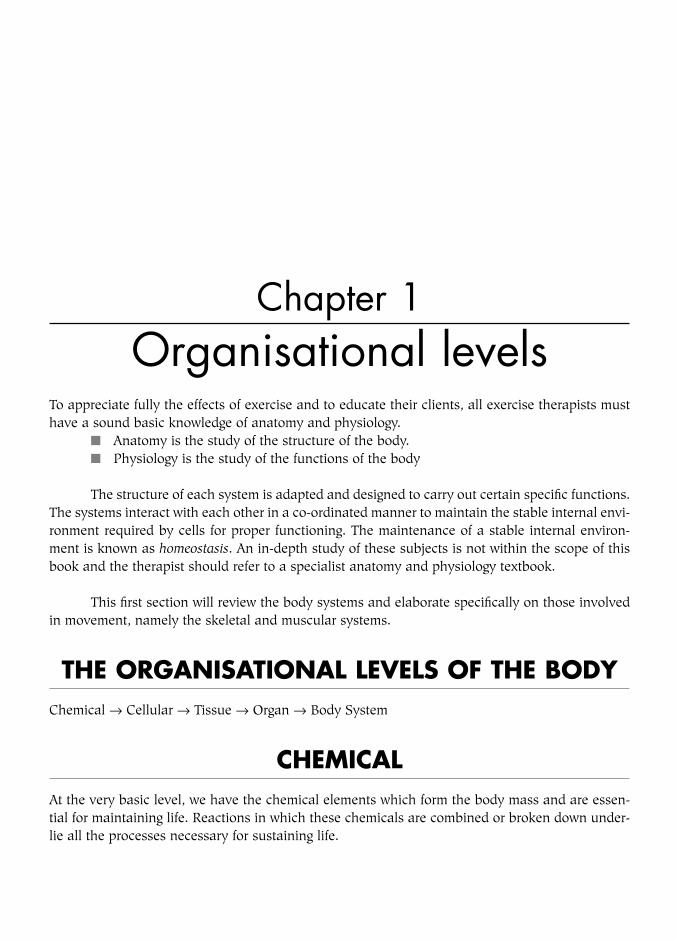
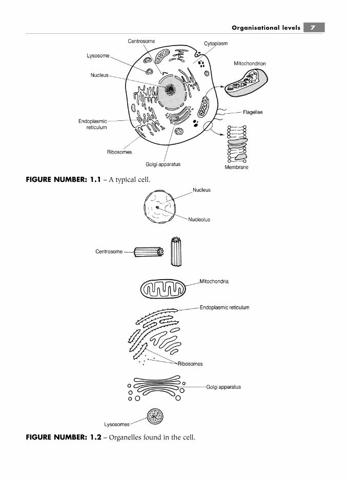
The structure of a typical cell
The cell membrane or plasma membraneThis is the outer layer or boundary of the cell. It gives shape to the cell and protects it, separatingthings inside the cell (intracellular) from those outside the cell (extracellular). It regulates thepassage of substances in and out of the cell.
The cytoplasmThis is a soft, jelly-like substance where the functions of the cell are carried out. It contains variousstructures called organelles (mini-organs), each of which has a specific function. Also in the cyto-plasm are various chemical substances called inclusions.
The organellesThese mini-organs each have a characteristic shape and a specific role to perform. The type andnumber of organelles in different kinds of cells vary depending on the activities of the cell; forexample, muscle cells have large numbers of mitochondria, because they have a high level ofenergy output.
■ The largest of the organelles is the nucleus. It controls the activities of the cell and itcontains the body’s genetic material (DNA).
Other organelles include:■ Mitochondria, which generate ATP/energy; there are large numbers in muscle cells;■ Ribosomes, which synthesise protein;■ Lysosomes, which digest and deal with waste;■ The Golgi apparatus which is concerned with the production of membrane and protein
lipids and glycoproteins;■ Endoplasmic reticulum – a series of channels for transporting substances within the
cell;■ The centrosome, involved in cell division.
The inclusionsThese are chemical substances produced by cells. They may not be present in all cells. Forexample, melanin is a pigment found in certain cells of the skin and hair; it protects the body by
6 Body Fitness and Exercise 2E
Organisational levels 7
FIGURE NUMBER: 1.1 – A typical cell.
FIGURE NUMBER: 1.2 – Organelles found in the cell.

screening out ultra-violet light, and gives the skin its brown colour on exposure to sunlight. Lipids(fat) is found in fat cells; this is broken down to provide energy when required.
The characteristics of cellsAll living things, whether they be single-celled or multi-celled organisms, have certain characteris-tics or functions in common that are essential to life:
MetabolismThis is the sum total of all the cells’ chemical activities. There are two phases of metabolism:
■ Catabolism is the breaking down of chemical substances derived from food to providethe energy and heat needed to sustain life;
■ Anabolism uses the energy of catabolism to build new chemical compounds and repairtissues.
RespirationThis involves two processes – external respiration: the movement of gases in and out of the lungsand circulating blood, and internal respiration: the metabolic activities within the cells.
Cells are capable of producing energy from food substances taken in by or stored in thebody. When oxygen is utilised in this process, it is termed aerobic respiration; when oxygen is notutilised in the process, it is termed anaerobic respiration.
GrowthCells grow in size up to a certain limit. When this limit is reached the cells divide.
ReproductionWhen the growth of cells is complete, they divide to produce two daughter cells that are identicalto each other. This process of cell division is known as mitosis.
ExcretionCells are capable of getting rid of the waste products resulting from metabolism; these pass out ofthe cell through the cell membrane into the tissue fluid, they then pass through capillary walls intothe circulating blood.
IrritabilityCells are capable of responding to stimuli, which may be physical, chemical or thermal.
MovementSome cells are capable of movement. They move by pushing out fingers of cytoplasm calledpseudopodia or by the movement of flagellae.
The body is made up of billions of cells; they are similar in structure but not exactly thesame, as they are modified (changed) to carry out specific functions. All the metabolic functionsnecessary for sustaining life are carried out by the cells.
8 Body Fitness and Exercise 2E

The energy for the metabolic activities of each cell is derived from the breakdown of a par-ticular chemical compound – adenosine triphosphate, or ATP. Only a small amount of ATP isstored in the cells; once this is used up it must be continually resynthesised (produced). The pro-duction of ATP is a continuous process and comes from the energy released from certain highenergy chemicals and the breakdown of the foods we eat, mainly the carbohydrates and fats.Muscle cells expend far more energy (for muscle contraction) than other cells and must be able tocontinually resynthesise large amounts of ATP/Energy.
Three metabolic systems contribute to this energy production: the phosphagen system,anaerobic glycolysis, and the aerobic system. These are discussed in chapter 4.
Summary and aid to learningThe body is made up of billions of cells similar in structure but not identical as they are modifiedto carry out specific functions. All the metabolic functions necessary for sustaining life are carriedout by the cells.
Cells are composed of cytoplasm, containing organelles which is surrounded and containedby the cell membrane.
The nucleus is the largest organelle; it controls all the functions carried out by the cell andcontains the body’s genetic material or DNA.
Each organelle has a specific, life-sustaining role to play.
The energy for the metabolic activities of every cell is derived from the breakdown of a par-ticular chemical compound – adenosine triphosphate, or ATP.
Mitochondria produce ATP; they produce the energy for all the activities of the cell and con-sequently are known as powerhouses or power plants.
Only a small amount of ATP is stored in the cells; once this is used up it must be continu-ally resynthesised (produced).
The production of ATP is a continuous process and comes from the energy released fromcertain high energy chemical compounds and the breakdown of the foods we eat, mainly carbo-hydrates and fats.
Organisational levels 9
LEARNCharacteristics or functions of cells:■ Metabolism ■ Excretion■ Respiration ■ Irritability■ Growth ■ Movement■ Reproduction

TISSUESThe tissues of the body are made up of groups of similar cells that work together to perform aspecific function. All the cells of one tissue will be identical, but the cells of different tissues will bemodified to suit tissue function. There are four main types of tissue in the body:
■ Epithelial tissue covers the body’s surfaces, lines the organs and tubes and forms glands;■ Connective tissue supports and protects organs, binds and connects tissues and organs
together and provides storage of fat for energy reserves;■ Muscle tissue is able to contract and relax to produce movement;■ Nervous tissue initiates and transmits impulses to co-ordinate the activities of the body.
It is the communication system of the body.
Epithelial tissue or epitheliumThis tissue forms the outer covering of body surfaces and body organs. It also forms the inner liningof organs, tracts, vessels and ducts. Glandular epithelium lines glands and secretes substances.
Epithelium is composed of closely packed cells. There are two main classifications:■ simple epithelium, which is a single layer of cells;■ stratified or compound epithelium, which consists of many layers of cells.
It may be further sub-classified according to the shape of the cells. The many types ofepithelium may be summarised as shown in Tables 1.1 and 1.2.
These chemicals regulate certain physiological processes.
Glandular epithelium contains cells that secrete substances and is found in glands.■ Exocrine glands secrete substances into ducts or directly onto surfaces: for example,
sweat glands secrete sweat, salivary glands secrete saliva and various digestive tractglands secrete digestive juices.
10 Body Fitness and Exercise 2E
Table 1.1Types of simple epithelium
Name Type Location
Squamous (a) flat cells lines heart and blood vessels,alveoli
Cuboidal (b) cube-shaped cells lines kidney tubules, ducts ofglands
Columnar (c) cells like columns lines stomach and digestive tract
Columnar ciliated (d) columns with hair-like cilia lines respiratory tract andfallopian tubes
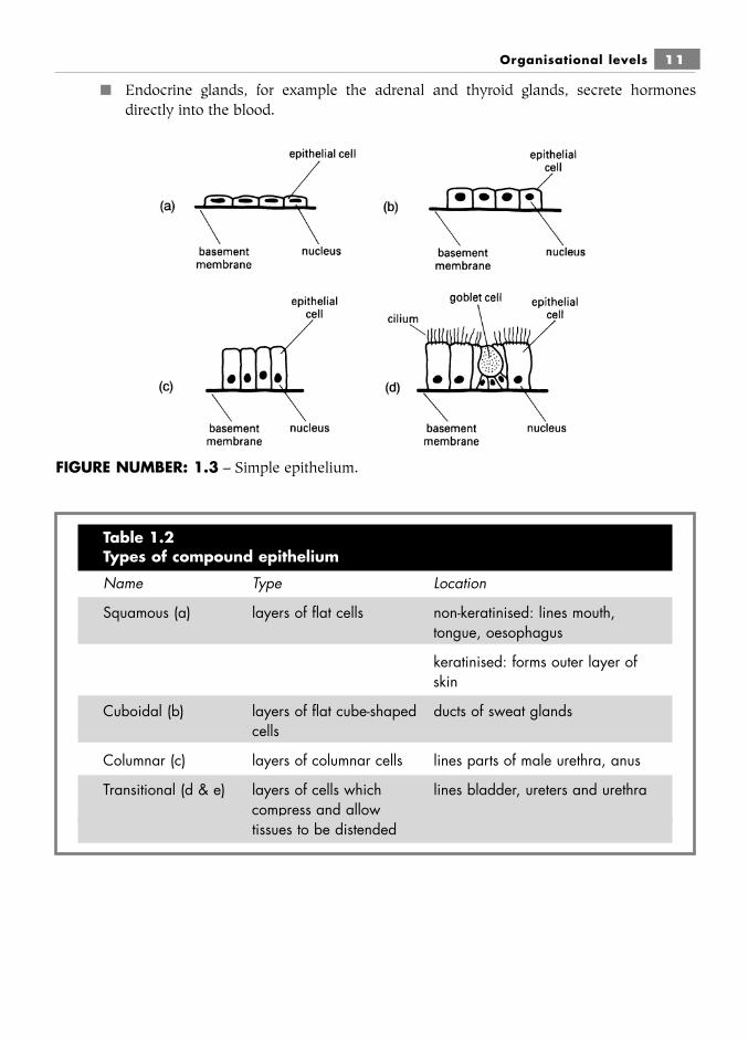
■ Endocrine glands, for example the adrenal and thyroid glands, secrete hormonesdirectly into the blood.
Organisational levels 11
FIGURE NUMBER: 1.3 – Simple epithelium.
Table 1.2Types of compound epithelium
Name Type Location
Squamous (a) layers of flat cells non-keratinised: lines mouth,tongue, oesophagus
keratinised: forms outer layer ofskin
Cuboidal (b) layers of flat cube-shaped ducts of sweat glandscells
Columnar (c) layers of columnar cells lines parts of male urethra, anus
Transitional (d & e) layers of cells which lines bladder, ureters and urethracompress and allowtissues to be distended
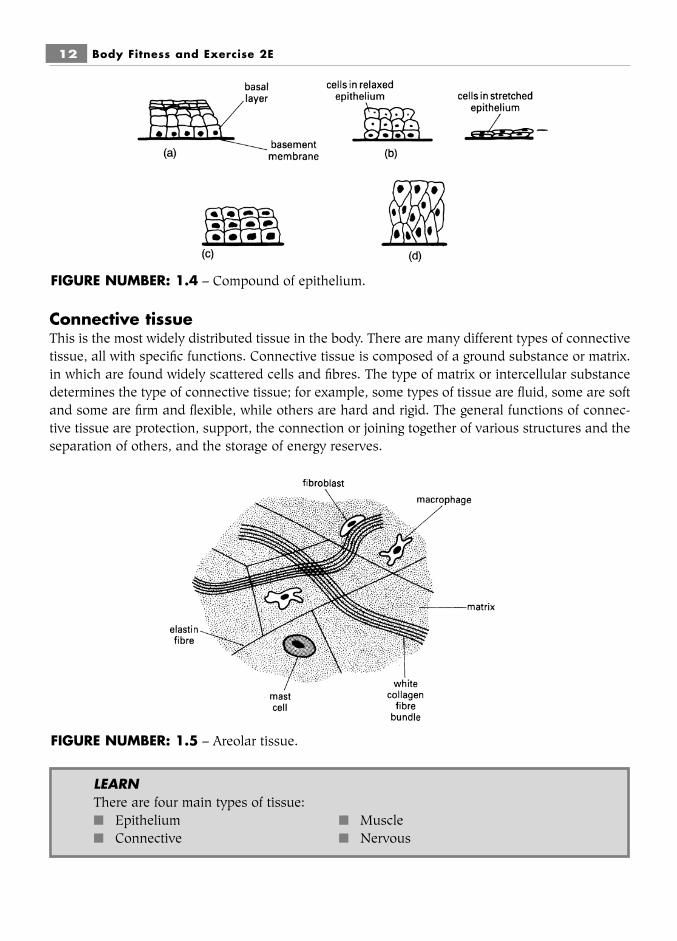
Connective tissueThis is the most widely distributed tissue in the body. There are many different types of connectivetissue, all with specific functions. Connective tissue is composed of a ground substance or matrix.in which are found widely scattered cells and fibres. The type of matrix or intercellular substancedetermines the type of connective tissue; for example, some types of tissue are fluid, some are softand some are firm and flexible, while others are hard and rigid. The general functions of connec-tive tissue are protection, support, the connection or joining together of various structures and theseparation of others, and the storage of energy reserves.
12 Body Fitness and Exercise 2E
FIGURE NUMBER: 1.4 – Compound of epithelium.
FIGURE NUMBER: 1.5 – Areolar tissue.
LEARNThere are four main types of tissue:■ Epithelium ■ Muscle■ Connective ■ Nervous

Organisational levels 13
FIGURE NUMBER: 1.6 – Adipose tissue.
FIGURE NUMBER: 1.7 – Hyaline cartilage.
Table 1.3Types of connective tissue
Name Structure Location/function
Areolar tissue loose moist tissue with a widely distributed as dermisviscous matrix and a loose, of skin and under the skinirregular arrangement of as superficial fascia; foundfibres: white fibres for between muscles and otherstrength and yellow fibres for tissues and around organs.elasticity. A variety of cells It gives strength, elasticityare found scattered and supportthroughout
[continued]

14 Body Fitness and Exercise 2E
Name Structure Location/function
Adipose tissue loose connective tissue, with subcutaneous layer of skin,large numbers of specialised the amount varying between cells, called adipocytes, for thin and obese people; fat storage. The cytoplasm around heart and kidney; in and nucleus of the cell are the marrow of long bones; pushed to one side and fat as padding around joints. fills the cell Regular aerobic exercise will
utilise the fats from these stores as a source of energy
Dense or white fibrous composed of closely packed forms tendons and connective tissue bundles of fibres, mainly aponeuroses that attach muscle
white collagen fibres, to bone, and ligaments thatinterspersed with cells hold bones together; provides a
protective covering for organs,e.g.kidney, heart, liver, testes
Yellow elastic tissue composed mainly of yellow forms the walls of arteries,elastic fibres with few trachea, bronchial tubes fibroblasts. This tissue gives and the lungs. It allows elasticity and strength, organs to stretch and recoilrecoiling to its original shapeafter stretching
Reticular tissue reticular fibres form a forms delicate supportdelicate network with cells in organs such as spleen,wrapped around them lymph nodes, liver
Cartilage (three types)hyaline cartilage consists of a gelatinous covers the ends or
intercellular matrix with fine articulating surfaces of collagen fibres and cells bones; forms the costal called chondrocytes. Hyaline cartilages, the rings of the cartilage is smooth, tough, trachea and bronchi and resilient and flexible. It is the nasal septum; provides milky white with a bluish a smooth surface to tinge. It is commonly called minimise friction at joints. gristle With age, injury or disease
this cartilage may bedamaged or eroded, andfriction at the joint increasesas bone rubs on bone,producing pain and stiffness
[continued]

Summary and aid to learningEach tissue of the body is composed of a mass of identical cells grouped together.
The cells of different tissues are basically the same but have changed slightly to suit theirparticular function.
There are four main tissue groups: Epithelium, connective, muscle, and nervous.
Each of these groups may be further subdivided. Read the text and list all the different typesof cells found in each group.
Study the function of each type (see Table 1.3) and explain why the cells of adipose tissuediffer from the cells of hyaline cartilage.
Organisational levels 15
Name Structure Location/function
fibro-cartilage similar to hyaline, but the found in the symphysis matrix contains bundles of pubis, inter-vertebral discs collagen fibres with widely and the menisci of the knee. dispersed chondrocytes. The It supports and cushions. fibres give strength, Severe compression and toughness and flexibility. It abnormal movements can gives a slight cushioning damage discs and menisci. effect when compressed These are common injuries
in sport and exercise (seechapter 3)
elastic cartilage similar to hyaline, but the found in the epiglottis and matrix consists of freely external ear, giving shape branching elastic fibres with and supportdispersed chondrocytes. It isflexible and resilient. It givessupport and shape
Bone or osseous tissue (two types)compact bone hard, dense, ivory-like tissue forms the outer layer of bones
cancellous bone sponge-like structure with found inside most bonestrabeculae and large spaces
Blood fluid connective tissue transports substances consisting of plasma and around the body. Regulates circulating cells body heat. Prevents blood
loss by coagulation
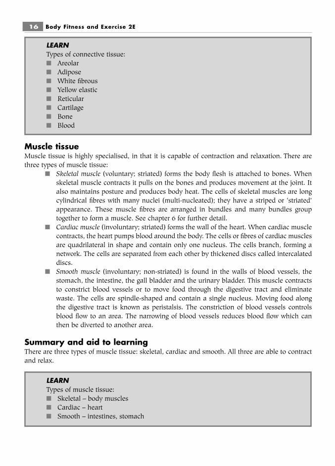
Muscle tissueMuscle tissue is highly specialised, in that it is capable of contraction and relaxation. There arethree types of muscle tissue:
■ Skeletal muscle (voluntary; striated) forms the body flesh is attached to bones. Whenskeletal muscle contracts it pulls on the bones and produces movement at the joint. Italso maintains posture and produces body heat. The cells of skeletal muscles are longcylindrical fibres with many nuclei (multi-nucleated); they have a striped or ‘striated’appearance. These muscle fibres are arranged in bundles and many bundles grouptogether to form a muscle. See chapter 6 for further detail.
■ Cardiac muscle (involuntary; striated) forms the wall of the heart. When cardiac musclecontracts, the heart pumps blood around the body. The cells or fibres of cardiac musclesare quadrilateral in shape and contain only one nucleus. The cells branch, forming anetwork. The cells are separated from each other by thickened discs called intercalateddiscs.
■ Smooth muscle (involuntary; non-striated) is found in the walls of blood vessels, thestomach, the intestine, the gall bladder and the urinary bladder. This muscle contractsto constrict blood vessels or to move food through the digestive tract and eliminatewaste. The cells are spindle-shaped and contain a single nucleus. Moving food alongthe digestive tract is known as peristalsis. The constriction of blood vessels controlsblood flow to an area. The narrowing of blood vessels reduces blood flow which canthen be diverted to another area.
Summary and aid to learningThere are three types of muscle tissue: skeletal, cardiac and smooth. All three are able to contractand relax.
16 Body Fitness and Exercise 2E
LEARNTypes of connective tissue:■ Areolar■ Adipose■ White fibrous■ Yellow elastic■ Reticular■ Cartilage■ Bone■ Blood
LEARNTypes of muscle tissue:■ Skeletal – body muscles■ Cardiac – heart■ Smooth – intestines, stomach

Skeletal muscle forms the body flesh and gives the body shape.
Skeletal muscle is known as voluntary because it is under the control of the will: we candecide if we want to move an arm or leg.
It is also known as striated because when looked at under a powerful microscope, stripes orstriations can be seen across the length of the fibres.
The cells of skeletal muscle are long cylinder-like fibres with many nuclei.
When skeletal muscle contracts it pulls on bones which results in movement at the under-lying joints.
Cardiac muscle is striated and found only in the walls of the heart.
Organisational levels 17
FIGURE NUMBER: 1.8 – Types of muscle tissue.
skeletal
cardiac
smooth
Cardiac muscle is involuntary because it is not under the control of the will, we cannotcontrol the contraction of the heart. This tissue is under the control of the autonomic nervoussystem.
The cells are quadrilateral in shape and contain one nucleus.
Smooth muscle is also involuntary and is under the control of the autonomic system.
It is non–striated, there are no stripes along its length.
The cells are spindle shaped with a single nucleus.
Smooth muscle is found in the walls of blood vessels, intestines, stomach etc.
Smooth muscle of the intestine contracts to move food along; this contraction and relax-ation is known as peristalsis.
Smooth muscle also contracts to constrict blood vessels; this controls the blood flow to anarea. When the vessels contract blood flow is reduced, when the vessels dilate more blood flows.Increased blood flow is an important factor during exercise when the contracting muscles dependon an increased delivery of nutrients and oxygen by the blood.
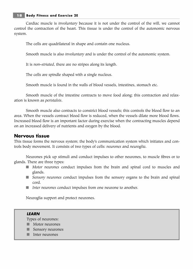
Nervous tissueThis tissue forms the nervous system; the body’s communication system which initiates and con-trols body movement. It consists of two types of cells: neurones and neuroglia.
Neurones pick up stimuli and conduct impulses to other neurones, to muscle fibres or toglands. There are three types:
■ Motor neurones conduct impulses from the brain and spinal cord to muscles and glands.
■ Sensory neurones conduct impulses from the sensory organs to the brain and spinalcord.
■ Inter neurones conduct impulses from one neurone to another.
Neuroglia support and protect neurones.
18 Body Fitness and Exercise 2E
LEARNTypes of neurones:■ Motor neurones■ Sensory neurones■ Inter neurones

ORGANSMany tissues combine to form the organs of the body. Each organ has a specific function or func-tions to perform.
For example, the stomach digests food, the lungs exchange gases, the heart pumps blood,the kidneys filter fluids and form urine, the ovaries produce and release ova. Organs combine tomake up the systems of the body.
BODY SYSTEMSEach body system consists of many organs that co-operate to perform various functions. All thesystems are interrelated and function together to maintain life. There are eleven body systems, asshown in Table 1.4. During exercise and training the systems interact in a complex way to ensureoptimum performance.
Organisational levels 19
LEARNThe eleven body systems are:■ Integumentary ■ Respiratory■ Skeletal ■ Digestive■ Muscular ■ Urinary■ Nervous ■ Reproductive■ Cardio-vascular ■ Endocrine■ Lymphatic
Table 1.4The eleven body systems
System Location Function
Integumentary system the skin and all its structures; protects; regulatesnails; hair; sweat and temperature; eliminatessebaceous (oil) glands waste; makes vitamin D;
receives stimuli
Skeletal system the bones, joints and supports; protects; allowscartilages movement; stores fat and
minerals; protects cells thatproduce blood cells
[continued]
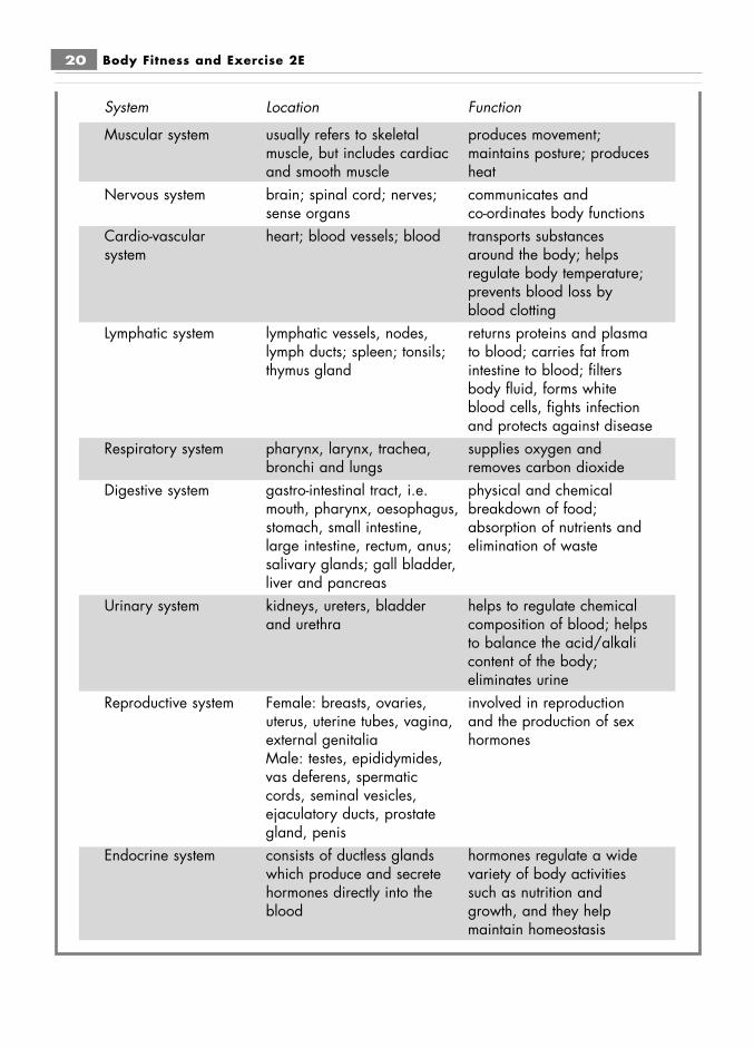
20 Body Fitness and Exercise 2E
System Location Function
Muscular system usually refers to skeletal produces movement;muscle, but includes cardiac maintains posture; producesand smooth muscle heat
Nervous system brain; spinal cord; nerves; communicates andsense organs co-ordinates body functions
Cardio-vascular heart; blood vessels; blood transports substancessystem around the body; helps
regulate body temperature;prevents blood loss byblood clotting
Lymphatic system lymphatic vessels, nodes, returns proteins and plasmalymph ducts; spleen; tonsils; to blood; carries fat fromthymus gland intestine to blood; filters
body fluid, forms whiteblood cells, fights infectionand protects against disease
Respiratory system pharynx, larynx, trachea, supplies oxygen andbronchi and lungs removes carbon dioxide
Digestive system gastro-intestinal tract, i.e. physical and chemicalmouth, pharynx, oesophagus, breakdown of food; stomach, small intestine, absorption of nutrients and large intestine, rectum, anus; elimination of wastesalivary glands; gall bladder,liver and pancreas
Urinary system kidneys, ureters, bladder helps to regulate chemicaland urethra composition of blood; helps
to balance the acid/alkalicontent of the body;eliminates urine
Reproductive system Female: breasts, ovaries, involved in reproductionuterus, uterine tubes, vagina, and the production of sex external genitalia hormonesMale: testes, epididymides,vas deferens, spermaticcords, seminal vesicles,ejaculatory ducts, prostategland, penis
Endocrine system consists of ductless glands hormones regulate a widewhich produce and secrete variety of body activitieshormones directly into the such as nutrition andblood growth, and they help
maintain homeostasis

Organisational levels 21
QUESTIONS1. List the organisational levels of the body.2. Give three functions of the cell membrane.3. Name the organelles that carry out the following functions:
a synthesise proteinb deal with wastec generate energy.
4. Define the term metabolism, and name the two phases involved.5. Complete the following sentences:
… group together to form body tissues.Body systems are made up of many … .
6. List all the types of epithelial tissue and give the location of each.7. a Name the tissue that stores body fat.
b List three locations where fat is stored.8. a List the three types of cartilage.
b Name the cartilage that covers the articulating surfaces of bones.9. Give the location of the following muscle tissues:
a skeletalb smoothc cardiac.Draw a simple diagram of each tissue.
10. Name and give the function of the three types of neurone.

Chapter 2The skeletal system
This chapter will help you to understand the structure and functions of bones, cartilages and joints.■ Bones form the framework of the body and act as levers.■ Cartilage protects the ends of bones allowing smooth movement and acts as a shock
absorber.■ Joints are formed where two or more bones meet; this is where body movement takes
place.
THE ANATOMICAL POSITIONBefore we can describe body movement, we must have a basic position or static posture that isused as a common reference point for describing surfaces, relationships and directions of move-ment. This is known as the anatomical position.
DefinitionIn the anatomical position, the body is upright, with feet slightly apart and toes pointing forward.The arms hang at the sides with the palms of the hands facing forwards. (Note the difference fromthe normal relaxed standing position, where the palms of the hands face the sides of the body.)
With the body in this position the terminology related to structures and joint movement canbe described.
Body planesThese are imaginary surfaces along which movements take place. There are three planes and theylie at right angles to each other:
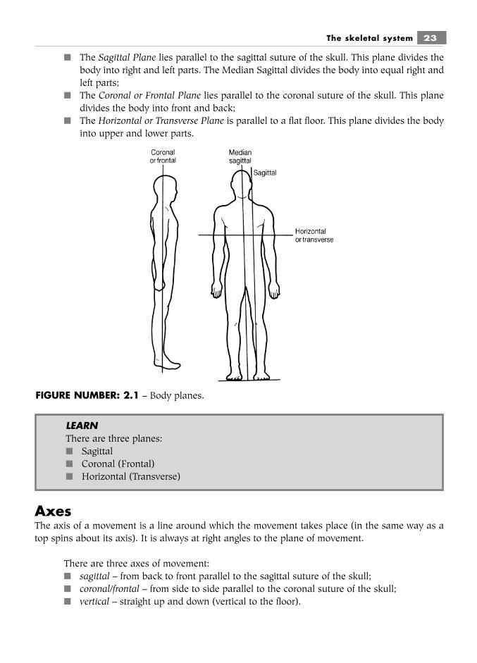
■ The Sagittal Plane lies parallel to the sagittal suture of the skull. This plane divides thebody into right and left parts. The Median Sagittal divides the body into equal right andleft parts;
■ The Coronal or Frontal Plane lies parallel to the coronal suture of the skull. This planedivides the body into front and back;
■ The Horizontal or Transverse Plane is parallel to a flat floor. This plane divides the bodyinto upper and lower parts.
AxesThe axis of a movement is a line around which the movement takes place (in the same way as atop spins about its axis). It is always at right angles to the plane of movement.
There are three axes of movement:■ sagittal – from back to front parallel to the sagittal suture of the skull;■ coronal/frontal – from side to side parallel to the coronal suture of the skull;■ vertical – straight up and down (vertical to the floor).
The skeletal system 23
FIGURE NUMBER: 2.1 – Body planes.
LEARNThere are three planes:■ Sagittal■ Coronal (Frontal)■ Horizontal (Transverse)

Examples of the planes and axes of certain movements when the body is in the anatomicalposition:
■ flexion (bending) of the elbow is movement in a sagittal plane with a frontal axis;■ abduction of the hip (taking it out to the side) is movement in a frontal plane with a
sagittal axis;■ turning the head from right to left is movement in a horizontal plane with a vertical axis.
The functions of the skeletal system■ Support – the bony framework gives shape to the body, supports the soft tissues and
provides attachment for muscles.■ Protection – the bony framework protects delicate internal organs from injury. For
example, the brain is protected by the skull, the heart and lungs are protected by the ribcage.
■ Movement – is produced by a system of bones, joints and muscles. The bones act aslevers and muscles pull on the bones, resulting in movement at the joints.
■ Storage of minerals – bones store many minerals, particularly calcium and phosphorus.■ Storage of energy – fats or lipids stored in the yellow bone marrow provide energy when
required.■ Storage of tissue that forms blood cells – both red and white blood cells are produced
by red bone marrow which is found in the spongy bone of the pelvis, vertebrae, ribs,sternum and in the ends of the femur and humerus.
24 Body Fitness and Exercise 2E
LEARNThere are three axes:■ Sagittal■ Coronal (Frontal)■ Vertical
LEARNIdentify the plane of movement, then the axis will be at right angles to it.
TASKTry the movements just described and work out others; remember that the move-ment must be in one of three planes and that the axis of the movement will be atright angles to that plane.
LEARNFunctions of the skeletal system are:■ Support ■ Movement■ Protection ■ Storage of minerals, fats and tissue-forming blood cells
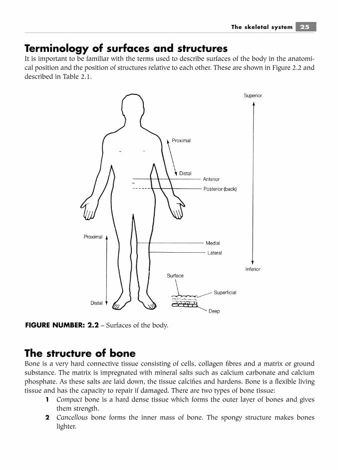
Terminology of surfaces and structuresIt is important to be familiar with the terms used to describe surfaces of the body in the anatomi-cal position and the position of structures relative to each other. These are shown in Figure 2.2 anddescribed in Table 2.1.
The structure of boneBone is a very hard connective tissue consisting of cells, collagen fibres and a matrix or groundsubstance. The matrix is impregnated with mineral salts such as calcium carbonate and calciumphosphate. As these salts are laid down, the tissue calcifies and hardens. Bone is a flexible livingtissue and has the capacity to repair if damaged. There are two types of bone tissue:
1 Compact bone is a hard dense tissue which forms the outer layer of bones and givesthem strength.
2 Cancellous bone forms the inner mass of bone. The spongy structure makes boneslighter.
The skeletal system 25
FIGURE NUMBER: 2.2 – Surfaces of the body.

Bones are enclosed in a dense layer of fibrous connective tissue known as the periosteum. This layercontains blood vessels which deliver nutrients to the bone, nerve and bone cells. Tendons (whichattach muscles to bone) and ligaments (which join bones together) blend with the periosteum.
There are different types of bone cells, which are found in the periosteum or scatteredthroughout compact and spongy bone. They include:
■ Osteoblasts: the bone builders, which produce minerals and collagen needed for strongbones
■ Osteocytes: the main cells of bone tissue, which carry out the activities necessary formaintaining healthy bones
■ Osteoclasts: the bone clearers, which absorb and remove bone.
Exercise strengthens bones because they adapt to stress by laying down more calcium andother minerals, and also by increasing collagen fibres.
26 Body Fitness and Exercise 2E
Table 2.1Terminology used to describe the structure of the body
Description of surfaceor structure Position
Anterior or ventral a surface that faces forwards; a structure that is furtherforwards than another
Posterior or dorsal a surface that faces backwards; a structure that is furtherback than another
Medial a surface or structure that is nearer to the mid-line thananother
Lateral a surface or structure that is further away from the mid-linethan another
Proximal a structure that is towards the root or origin, i.e. nearer thetrunk
Distal a structure that is further away from the root or origin, i.e.further away from the trunk
Superficial a structure that is nearer the surface than others
Deep a structure that lies beneath others, i.e. is further from thesurface
Superior a structure higher than others, i.e. nearer the head
Inferior a structure lower than others, i.e. nearer the foot
Fractures and other injuries to bones may occur in sports, and other physical activities.These must be quickly diagnosed and fixed to limit damage. An adequate length of time must beallowed for the fracture to heal.
Types of bones forming the skeletonThere are four different types of bones named according to their shape:
■ Long bones are longer than their width, e.g. femur, tibia, fibula, humerus, radius, ulna,metacarpals, phalanges.
■ Short bones of almost equal width and length, e.g. carpal and tarsal bones.■ Flat bones are flat thin bones, found where protection is needed and also where a
broad surface is required for the attachment of muscles, e.g. skull bones, scapulae,sternum, ribs.
■ Irregular bones are all the bones with complex shapes that do not fit into the abovecategories, e.g. vertebrae, sacrum, innominate bone, sphenoid, ethmoid.
Other small bones found in the body but not named according to shape are calledsesamoid bones: small rounded bones that develop within tendons, such as the patella. Theyenable the tendon to move smoothly over the underlying bone.
Summary and aid to learningBefore we can describe anything relating to body structure and be understood by others we mustuse common reference points and terminology. We must refer to the body in the anatomical posi-tion.
Read the text and write a definition of the anatomical position; now stand or instruct apartner to stand in this position.
We also have words for describing surfaces and structures related to one another. These arebest learnt as opposites:
The skeletal system 27
LEARNThere are two types of bone tissue:■ Compact bone – hard, dense. Forms the outer layer of bone for strength■ Cancellous bone – spongy or honeycomb-like structure. Forms inner mass of
bone for lightness
LEARNThe four types of bones forming the skeleton:■ Long■ Short■ Flat■ Irregular
Work with a partner and test each other by indicating to an aspect of the body which the othermust identify.
The skeletal system includes the bones, joints and cartilages that make up the frameworkof the body.
The skeleton has specific functions. Read the text and explain the following:■ The skeleton gives the body a framework (how?)■ The skeleton protects (what?)■ The skeleton allows movement (how?)■ The skeleton stores (what and where?)
Bone is a connective tissue; a bone is composed of two different tissues:■ Compact bone which is dense, hard tissue and forms the outer covering of bones. It gives
the bone its strength.■ Cancellous bone which is spongy or honeycomb-like in structure. It is light, making
bones less heavy.
There are different types of bones which make up the skeleton. They are named accordingto their shape: long bones, short bones, flat bones and irregular bones. Can you give some exam-ples of each type?
THE BONES OF THE SKELETONIt is difficult to study and visualise bones simply by using diagrams. It is easier to learn and muchmore interesting when a model skeleton and model bones are used. These can be examined andthe important features identified and related to one’s own body. Only the important and relevantfeatures have been included in the following text. The bones are clearly labelled for easy learning.However, remember to identify the features on model bones and palpate (feel) on your own bodywhere possible.
The human skeleton is made up of 206 bones. These are grouped into two main divisions:the axial skeleton, which forms the core or axis of the body, and the appendicular skeleton, whichforms the girdles and limbs.
28 Body Fitness and Exercise 2E
anterior: towards the front posterior: towards the back
proximal: nearer the body distal: further away from the body
medial: towards mid-line lateral: away from mid-line
superior: higher than another inferior: lower than another
superficial: nearer the surface deep: lies beneath another
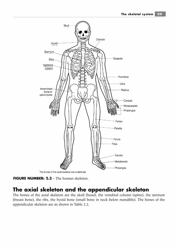
The axial skeleton and the appendicular skeletonThe bones of the axial skeleton are the skull (head), the vertebral column (spine), the sternum(breast bone), the ribs, the hyoid bone (small bone in neck below mandible). The bones of theappendicular skeleton are as shown in Table 2.2.
The skeletal system 29
FIGURE NUMBER: 2.3 – The human skeleton.
The bones of the skullThese include the cranial and facial bones.
The sutures of the skullThese are the joints between the bones of the skull. They are immovable fibrous joints. There arefour main sutures: coronal, sagittal, lambdoidal and squamous.
30 Body Fitness and Exercise 2E
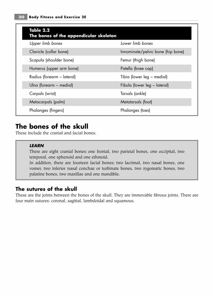
Table 2.2The bones of the appendicular skeleton
Upper limb bones Lower limb bones
Clavicle (collar bone) Innominate/pelvic bone (hip bone)
Scapula (shoulder bone) Femur (thigh bone)
Humerus (upper arm bone) Patella (knee cap)
Radius (forearm – lateral) Tibia (lower leg – medial)
Ulna (forearm – medial) Fibula (lower leg – lateral)
Carpals (wrist) Tarsals (ankle)
Metacarpals (palm) Metatarsals (foot)
Phalanges (fingers) Phalanges (toes)
LEARNThere are eight cranial bones: one frontal, two parietal bones, one occipital, twotemporal, one sphenoid and one ethmoid.In addition, there are fourteen facial bones; two lacrimal, two nasal bones, onevomer, two inferior nasal conchae or turbinate bones, two zygomatic bones, twopalatine bones, two maxillae and one mandible.

The skeletal system 31
FIGURE NUMBER: 2.4 – The bones of the skull.
FIGURE NUMBER: 2.5 – Lateral view of the skull.
THE FEATURES OF THE SKELETAL BONES
32 Body Fitness and Exercise 2E
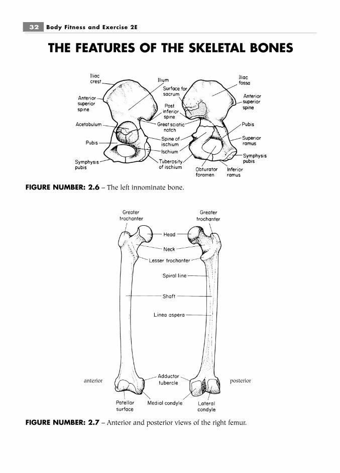
FIGURE NUMBER: 2.6 – The left innominate bone.
FIGURE NUMBER: 2.7 – Anterior and posterior views of the right femur.
anterior posterior
The skeletal system 33
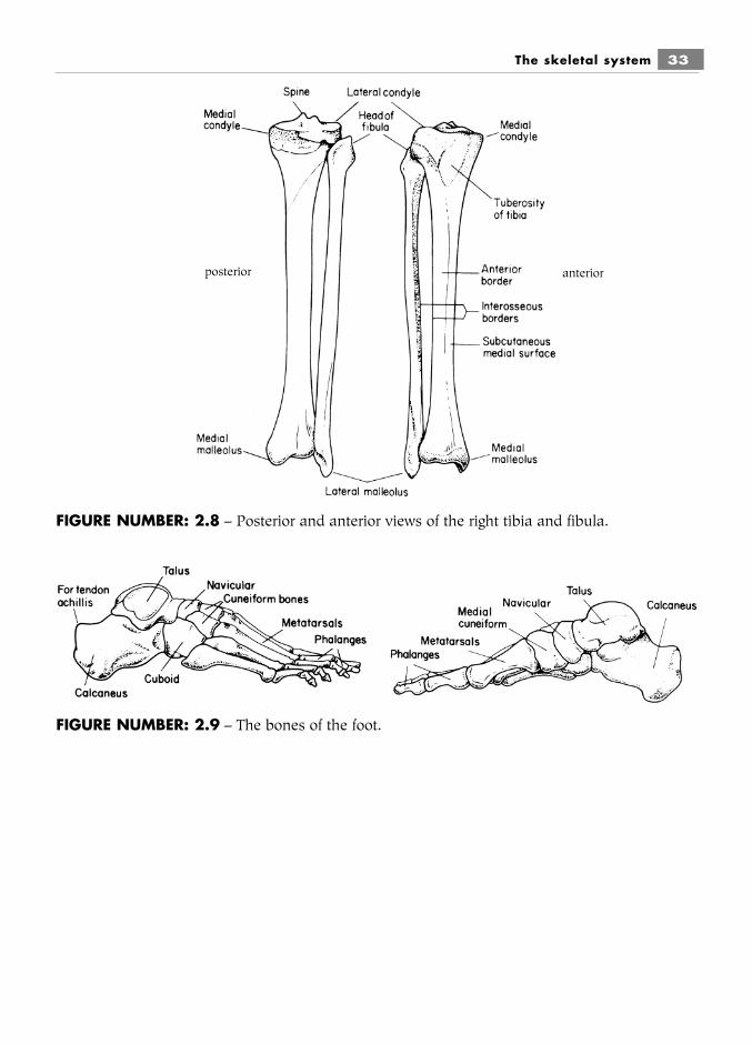
FIGURE NUMBER: 2.8 – Posterior and anterior views of the right tibia and fibula.
FIGURE NUMBER: 2.9 – The bones of the foot.
anteriorposterior

34 Body Fitness and Exercise 2E
FIGURE NUMBER: 2.10 – The posterior surface of the scapula.
FIGURE NUMBER: 2.11 – The left clavicle.
The skeletal system 35
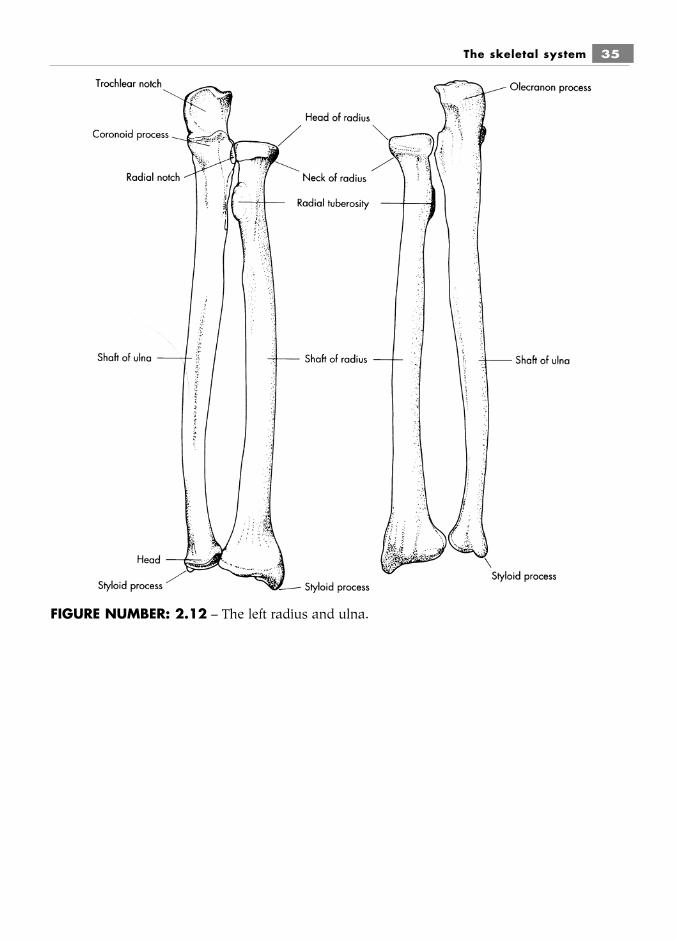
FIGURE NUMBER: 2.12 – The left radius and ulna.

36 Body Fitness and Exercise 2E
FIGURE NUMBER: 2.13 – The left humerus.
FIGURE NUMBER: 2.14 – The left hand.
The skeletal system 37
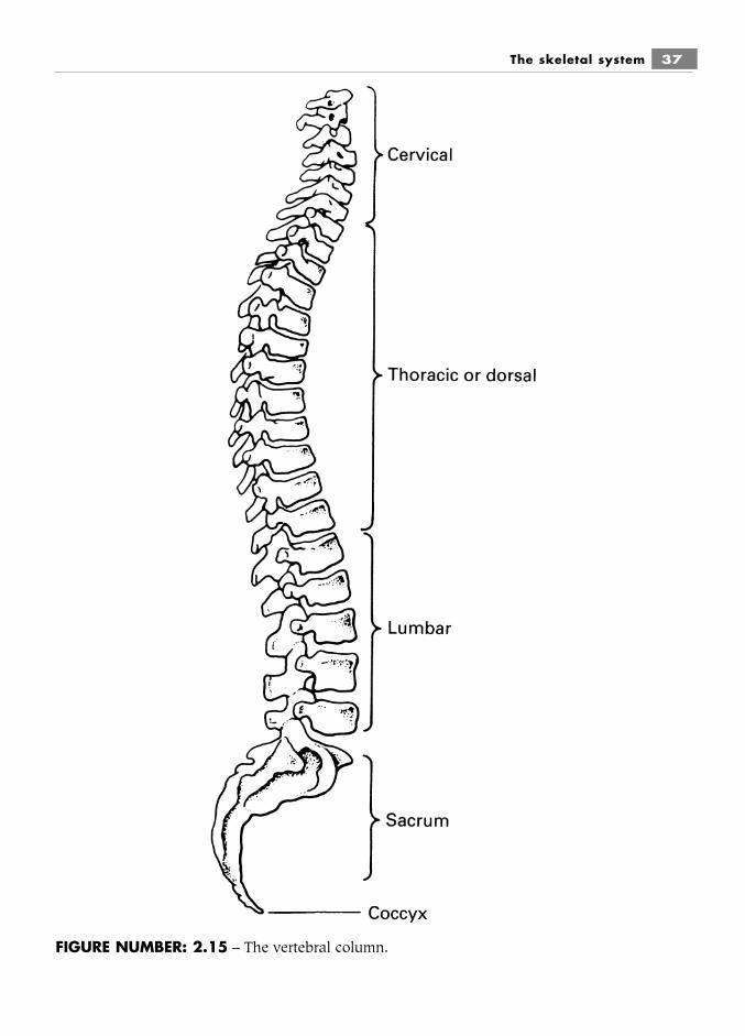
FIGURE NUMBER: 2.15 – The vertebral column.
The vertebral column (spinal column)The vertebral column is composed of 33 vertebrae. Some are fused together, so that in fact thereare only 26 bones. Between the bodies of adjacent vertebrae are discs of fibro cartilage which actas shock absorbers. These are called inter-vertebral discs. The column is divided into five regions:
■ Cervical – seven vertebrae (neck)■ Thoracic – twelve vertebrae (upper back)■ Lumbar – five vertebrae (small of back)■ Sacral – five fused vertebrae (sacrum)■ Coccygeal – four fused vertebrae (coccyx).
The functions of the vertebral column■ It allows movement forward, backward and laterally.■ It protects the spinal cord.■ It supports the head.■ It provides rigidity to maintain the upright posture.■ It provides posterior attachment for the ribs.■ It provides attachment for muscles.■ It acts as a shock absorber due to the cushioning effect of the intervertebral discs.■ The cancellous bone of the vertebrae stores red bone marrow, which forms blood cells.■ It stores minerals.■ It provides the fulcrum for numerous movements.
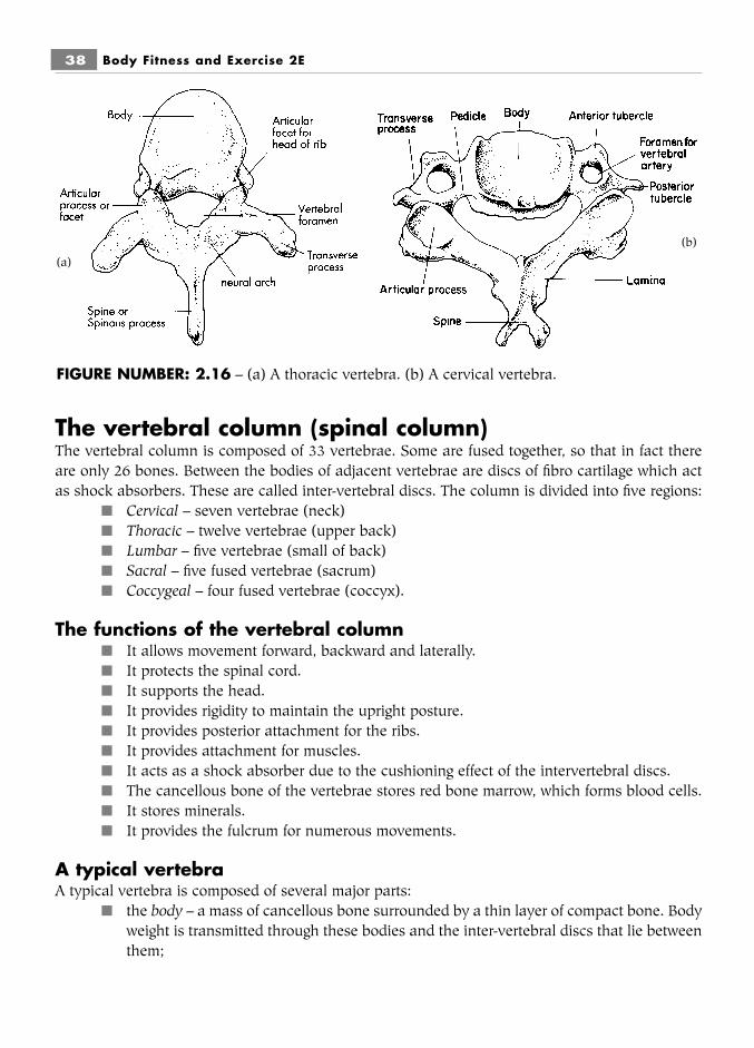
A typical vertebraA typical vertebra is composed of several major parts:
■ the body – a mass of cancellous bone surrounded by a thin layer of compact bone. Bodyweight is transmitted through these bodies and the inter-vertebral discs that lie betweenthem;
38 Body Fitness and Exercise 2E
FIGURE NUMBER: 2.16 – (a) A thoracic vertebra. (b) A cervical vertebra.
(a)
(b)

■ the neural or vertebral arch – a strong arch of bone enclosing the vertebral foramen. It ismade up of several fused parts. It protects the spinal cord, which passes down from thebrain through the vertebral foramen;
■ the spinous process – a spikelike backward projection. It provides attachment for manymuscles and ligaments;
■ the transverse process – two projections, one on either side. They also provide attach-ment for muscles and ligaments;
■ four facets – surfaces (two above and two below) for articulating with the adjacent ver-tebrae.
Spaces between the vertebrae known as the inter-vertebral foramina allow the passage ofnerves entering and leaving the spinal cord along its length.
All vertebrae except the first and second cervical (atlas and axis) have these features incommon, but they vary in size, becoming larger lower down for weight bearing. The fused verte-brae of the sacrum and coccyx also differ.
The inter-vertebral discsThese lie between the bodies of the vertebrae; they act as shock absorbers and allow for compres-sion and distortion along the column. The core of the disc is the nucleus pulposus, which is a jelly-like material consisting of 85 per cent water. Surrounding this is the annulus fibrosus, which is com-posed of many rings of elastic fibres woven at angles to each other. It is thus able to expand andmove to absorb compression forces.
As we grow older, the nucleus loses its water-binding capacity, fibro-cartilage replaces thegelatinous substance and the nucleus gradually hardens. The annulus fibrosus also loses its elas-ticity. As elasticity and flexibility are lost, the hardened rigid disc becomes more susceptible toinjury. If the compression forces are abnormally strong or sudden, the annulus fibrosus may tearor rupture, allowing the nucleus to protrude into the space. This is known as a ‘slipped disc’ or discprolapse. If this protrusion presses against a nerve as it passes out of the spinal canal through the
The skeletal system 39
FIGURE NUMBER: 2.17 – A section through the vertebral column

inter-vertebral foramen, then neurological symptoms will be felt along the path of the nerve, forexample if the prolapse is in the lumbar spine, pain, tingling, pins and needles, numbness may befelt down the leg.
Disc problems can occur at any time, but the likelihood increases as we get older. It is there-fore extremely important to consider the age and medical condition of clients when giving any neckand trunk exercises. Failure to do so can result in very serious injury.
Movement of the spinal columnThe vertebrae and discs are bound together by strong, powerful ligaments. There is very little move-ment between adjacent vertebrae, but the total combined movement along the whole length allowsconsiderable movement of the trunk. The movements of the vertebral column are:
■ flexion■ extension■ side flexion■ rotation
There is a greater range of movement in the cervical and lumbar regions than in the tho-racic. These variations are due to the length and direction of the spinous processes, the ratiobetween the height of the discs and the height of the vertebral body, and the tension of the sup-porting ligaments.
Flexion and extension of the neck occur in the cervical region. Flexion and extension ofthe trunk occur mainly in the lumbar region. Rotation of the trunk occurs mainly in the thoracicregion.
Dangerous movementsThe most hazardous movement is trunk forward flexion, as this movement takes place mainly inthe lumbar spine; the leverage is long being the length of the head and trunk. About 20 per cent ofthe movement occurs between the fourth and fifth lumbar vertebrae, and 60 to 70 per cent occursbetween the fifth lumbar vertebra and the first sacral vertebra. There is therefore a high risk ofdamage to this vulnerable area of the lower back. Hyperextension is also hazardous due to theextreme compression forces on the discs which may rupture.
The curves of the vertebral columnThe vertebral column shows curves along its length. These are seen in the cervical, thoracic,lumbar and sacral regions. The thoracic and sacral curves are primary curves, being present beforebirth. The cervical and lumbar curves are secondary curves and develop after birth. The cervicalcurve develops when the baby lifts its head, the lumbar curve develops as the baby learns to sitand stand. When viewed posteriorly:
■ the cervical curve is concave;■ the thoracic curve is convex;■ the lumbar curve is concave;■ the sacral curve is convex.
40 Body Fitness and Exercise 2E

Spinal problemsCertain spinal problems result in exaggerated or abnormal spinal curves. When the spine is viewedposteriorly the following curves may be seen:
■ Kyphosis is an exaggerated thoracic curve with increased convexity and forward flexion.■ Lordosis is an exaggerated lumbar curve with increased concavity and extension.■ Kypho-lordosis is a combination of the above.■ Scoliosis is a lateral deviation of the spine. It may deviate to the right or to the left and
may show a long C curve or an S curve.
These curves are accompanied by muscle imbalance: some muscles will be too tight and theopposite groups will be over-stretched. Exercises can help to correct these problems.
These problems are fully discussed in Chapter 11.
The skeletal system 41
FIGURE NUMBER: 2.18 – Vertebral curves in the embryo and adult.
TASKSWork with a partner.■ Examine your partner’s back and identify the five regions of the vertebral
column.■ Run your index finger firmly down the spinous processes, leaving a red line. If
the line deviates to the right or left it indicates a spinal problem. Name thisspinal problem.
■ Perform all the movements of the vertebral column.

The thorax or thoracic cavityThis is the bony cage of the chest, composed of the sternum, the 24 ribs and the twelve thoracicvertebrae.
The sternumThe sternum or breast bone is a flat narrow bone made up of three parts:
■ the manubrium – the top part, squarish in shape;■ the body – the long middle part;■ the xiphoid process – the small pointed lower end.
The ribsThe ribs are narrow flat bones articulating with the thoracic vertebrae behind and with the sternumin front. The ribs are arranged in pairs, one on the right and the other on the left:
■ Seven pairs are true ribs, which join the sternum.■ Five pairs are false ribs, which join the rib above. Two of these are called floating ribs
as they have no attachment in front.
Each rib is joined to the sternum or to the adjacent ribs by a strip of hyaline cartilage. Theseare called the costal cartilages.
Small muscles known as the intercostal muscles fill the spaces between the ribs. They liein two layers; eleven internal intercostals and eleven external intercostals on each side of the chest.a large muscle called the diaphragm forms the floor of the thoracic cavity. The lungs lie within andare protected by the thoracic cavity.
The mechanism of breathingThe capacity of the thorax must increase so that air can be taken in and then must decrease sothat air can be forced out. During inspiration (breathing in), the intercostal muscles contract and
42 Body Fitness and Exercise 2E
FIGURE NUMBER: 2.19 – Skeleton of the thorax.

swing the ribs upwards and outwards; the sternum is pushed forwards, and the diaphragm movesdownwards. Thus the capacity of the thorax increases sideways, forwards and downwards and thepressure inside the thorax is lowered. When the pressure is reduced below atmospheric pressure(i.e. the pressure of the air outside the body), air rushes in and fills the lungs. Oxygen passes intothe bloodstream through the walls of the capillaries surrounding the lungs and carbon dioxidepasses the other way. During expiration (breathing out) the intercostal muscles relax, thediaphragm moves upwards, the ribs and sternum collapse back and the lungs recoil. This increasesthe pressure in the lungs and air is forced out.
During exercise, more oxygen is required to maintain energy for muscle contraction.Therefore the intercostals and diaphragm work harder and as a result they improve in strength andcondition. The elasticity and condition of the lungs improves in the same way.
The skeletal system 43
LEARNDuring inspiration (breathing in) the following actions take place:■ Intercostals contract – ribs swing out and up■ Sternum pulled forward■ Diaphragm moves downThese actions increase the size of the chest cavity and air rushes in and fills thelungs.■ During expiration (breathing out):■ Intercostals relax■ Sternum moves back■ Diaphragm moves up■ Air is squeezed out as lungs recoil.
Air moves in and out of the lungs due to a difference in pressure. It moves fromareas of high pressure to low pressure.
TASKS■ Place your hands on the sides of the lower ribs.
Breathe in deeply and feel the ribs moving outwards and upwards. Breathe outand feel the ribs moving back.Repeat six times.
■ Repeat this procedure with the hands over:a) the front of the midriff – breathe in and the abdomen moves out, breathe out:b) the body of the sternum – breathe in and the sternum swings forward,
breathe out.
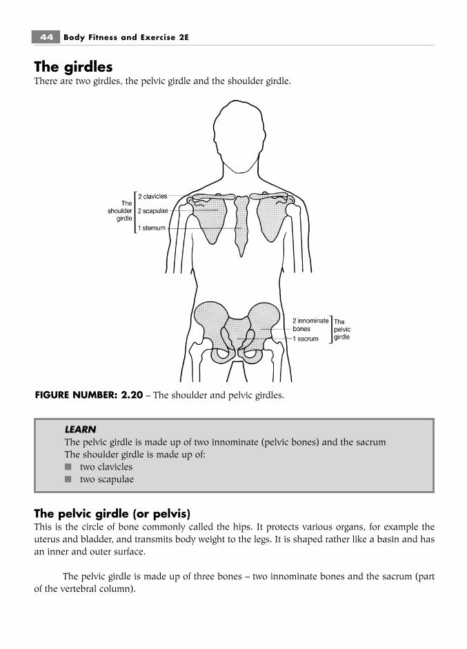
The girdlesThere are two girdles, the pelvic girdle and the shoulder girdle.
The pelvic girdle (or pelvis)This is the circle of bone commonly called the hips. It protects various organs, for example theuterus and bladder, and transmits body weight to the legs. It is shaped rather like a basin and hasan inner and outer surface.
The pelvic girdle is made up of three bones – two innominate bones and the sacrum (partof the vertebral column).
44 Body Fitness and Exercise 2E
FIGURE NUMBER: 2.20 – The shoulder and pelvic girdles.
LEARNThe pelvic girdle is made up of two innominate (pelvic bones) and the sacrumThe shoulder girdle is made up of:■ two clavicles■ two scapulae

The two large innominate or pelvic bones articulate in front at a cartilaginous joint calledthe pubic symphysis. At the back they articulate on each side of the sacrum at gliding synovialjoints called the sacro-iliac joints. There is hardly any movement at these joints as they fit tightlytogether and are held in place by very strong ligaments. The pelvis is supported on the femoralheads and may tilt forward, backward or sideways. Pelvic tilt accompanies movements of the trunkand hip joints, as is shown in chapter 4.
The shoulder girdle (the pectoral girdle)The shoulder girdle is composed of two clavicles in front and two scapulae at the back. Anteriorly,each clavicle articulates with the sternum at the sterno-clavicular joint. Laterally, the clavicles artic-ulate with the acromion process of the scapula at the acromio-clavicular joint.
The shoulder girdle forms an incomplete ring of bone around the upper thorax, joining theupper limbs to the axial skeleton.
The movement of the shoulder girdle accompany movements of the shoulder joint and con-tribute to a wide range of arm movements.
Response to exerciseBone tissue will adapt in response to exercise, and the degree of improvement relates to the inten-sity of the applied forces. Adaptation will occur only in those bones subjected to stress. The adap-tations include:
■ Increased enzyme activity which improves the condition of the bones■ Increased strength of bones reducing the risk of fractures■ Increase in bone girth following intense weight training programmes■ Increased bone mineral density: the deposits of minerals such as calcium increase in
response to the stresses applied to the bones.
Exercise and increased calcium intake is particularly beneficial for post menopausal womenas it can delay and protect against osteoporosis. This is condition where calcium is lost, bonesbecome brittle and fracture easily.
Summary and aid to learningThe skeleton can be thought of in two parts; the axial and appendicular part:
■ The axial skeleton is the central part. It includes: the skull, vertebral column, ribs,sternum and hyoid.
■ The appendicular skeleton includes all the bones of the limbs, i.e the shoulder, arm,hand, hip, leg, foot.
The skeletal system 45
LEARNThe pelvic girdle is a rigid structure and contributes little to the movement of the hipjoint and leg but the shoulder girdle is freely moveable and contributes greatly tothe wide range of movement of the shoulder joint and arm.

■ List all the bones of the appendicular skeleton.The vertebral column or spinal column is made up of separate bones called vertebrae (the singularis vertebra). In between the bodies of the vertebrae, are discs of fibro-cartilage, which act as shockabsorbers; these are called intervertebral discs.
The medical term ‘slipped disc’ refers to movement or damage of one or more of these discs.The condition causes pain because the displaced disc may press on a nerve leaving the spinal cord.Spinal nerves leave the spinal cord through a small space found between the vertebrae, called theintervertebral foramen.
The vertebral column is divided into five regions: the cervical region, the thoracic region, thelumbar region, the sacrum and the coccyx; the bones of the sacrum and coccyx are fused together.
Read the text, list the regions of the vertebral column and give the number of vertebrae ineach region.
Which region is most vulnerable to damage?
The major parts of a typical vertebra are: the body, neural arch, spinous process, two trans-verse processes and four facets.Examine a vertebra and identify each part.
Work with a partner to perform the movements of the spinal column then identify eachmovement: discuss the movements which may cause damage and give reasons why they are haz-ardous. Think of exercises that you would avoid including in any exercise plan because theyinvolve these movements.
The thorax is the chest area. It is made up of: the sternum in front, 12 pairs of ribs and the12 thoracic vertebrae. The diaphragm, a large muscle, forms the base and separates the thoraciccavity from the abdominal cavity. In the spaces between the ribs lie the intercostal muscles. Thesemuscles expand the chest during breathing.
Explain the difference between true ribs, false ribs and floating ribs. Examine a skeleton andpoint these out to a friend.
The shoulder girdle is an incomplete ring of bone around the top of the trunk. It is made upof the two clavicles and the sternum in front, and the two scapulae behind. It is a freely moveablestructure.
The pelvic girdle is a complete ring of bone around the base of the trunk. It is made up oftwo innominate or pelvic bones and the sacrum. It is a very rigid structure.
Explain why the shoulder girdle contributes to the movement of the shoulder joint but thepelvic girdle does not contribute to the movement of the hip joint.
46 Body Fitness and Exercise 2E

The skeletal system 47
QUESTIONS1. Compare the two main divisions of the human skeleton.2. List the bones in each division.3. Explain the functions of the skeletal system.4. Explain why cancellous bone is sometimes known as spongy bone.5. List the four main types of bones and give one example of each.6. Describe the anatomical position.7. Define the following terms:
a anterior surfaceb proximal endc mediald superior structuree deep muscle
8. List the bones of the skull.9. Name the regions of the vertebral column and give the number of vertebrae in
each.10. Give two functions of the inter-vertebral discs.11. Compare the following spinal problems: kyphosis, lordosis, scoliosis.12. List the bones that form the thoracic cavity or thorax.13. Where is the xiphold process located?14. Explain the terms true and false ribs.15. Label the diagram below:
FIGURE NUMBER: 2.21 – A typical vertebra.
Chapter 3The joints of theskeletal system
JOINTSWhen two or more bones meet they form a joint, sometimes called an articulation. All body move-ment occurs at joints, from the small movements of the fingers to the large movements of the shoul-der. The bones are held together by connective tissue and are moved by the contraction of skele-tal muscle.
The shape of the articulating bones and the flexibility and tensile strength of the surround-ing connective tissue determines the strength, stability and movement of joints.
Bones with curved surfaces that fit into each other and are close together form strong stablejoints with less movement. Bones with little curvature that fit together loosely form joints that areless stable but allow greater movement.
LEARNA joint is formed when two or more bones meet. It is sometimes referred to as anarticulation.
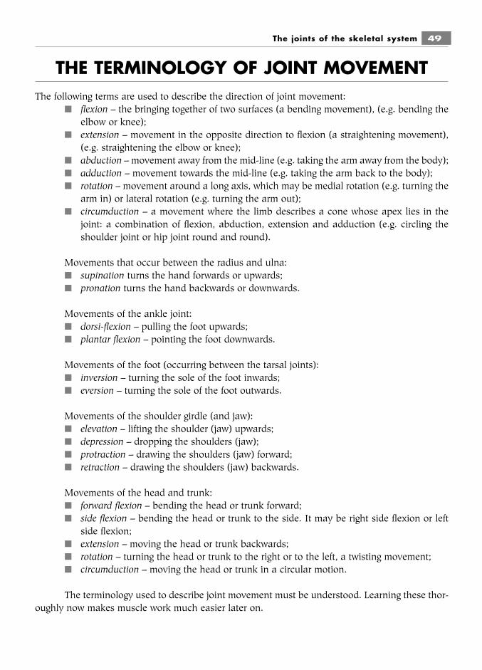
THE TERMINOLOGY OF JOINT MOVEMENTThe following terms are used to describe the direction of joint movement:
■ flexion – the bringing together of two surfaces (a bending movement), (e.g. bending theelbow or knee);
■ extension – movement in the opposite direction to flexion (a straightening movement),(e.g. straightening the elbow or knee);
■ abduction – movement away from the mid-line (e.g. taking the arm away from the body);■ adduction – movement towards the mid-line (e.g. taking the arm back to the body);■ rotation – movement around a long axis, which may be medial rotation (e.g. turning the
arm in) or lateral rotation (e.g. turning the arm out);■ circumduction – a movement where the limb describes a cone whose apex lies in the
joint: a combination of flexion, abduction, extension and adduction (e.g. circling theshoulder joint or hip joint round and round).
Movements that occur between the radius and ulna:■ supination turns the hand forwards or upwards;■ pronation turns the hand backwards or downwards.
Movements of the ankle joint:■ dorsi-flexion – pulling the foot upwards;■ plantar flexion – pointing the foot downwards.
Movements of the foot (occurring between the tarsal joints):■ inversion – turning the sole of the foot inwards;■ eversion – turning the sole of the foot outwards.
Movements of the shoulder girdle (and jaw):■ elevation – lifting the shoulder (jaw) upwards;■ depression – dropping the shoulders (jaw);■ protraction – drawing the shoulders (jaw) forward;■ retraction – drawing the shoulders (jaw) backwards.
Movements of the head and trunk:■ forward flexion – bending the head or trunk forward;■ side flexion – bending the head or trunk to the side. It may be right side flexion or left
side flexion;■ extension – moving the head or trunk backwards;■ rotation – turning the head or trunk to the right or to the left, a twisting movement;■ circumduction – moving the head or trunk in a circular motion.
The terminology used to describe joint movement must be understood. Learning these thor-oughly now makes muscle work much easier later on.
The joints of the skeletal system 49

Some joints only move in two directions, for example the elbow and knee, whilst otherswill move in six directions, for example the shoulder and hip joints. As has previously been men-tioned, muscles pull on the bones to produce these movements. Therefore some muscles will beflexors, producing flexion at the joint, whilst other muscles will be extensors, producing extensionat the joint, and so on. When one group of muscles contracts to produce movement (the agonists)the opposite groups must relax to allow the movement to take place (the antagonists). See page00.
THE CLASSIFICATION OF JOINTSThere are three main groups:
■ Fibrous joints are immovable. The bones fit tightly together and are held firmly byfibrous tissue. There is no joint cavity. Examples are the sutures of the skull.
■ Cartilaginous joints are slightly movable. The bones are connected by a disc of fibro-car-tilage. There is no joint cavity. Examples are the symphysis pubis (between the pubicbones) and the inter-vertebral joints (between the vertebral bodies).
■ Synovial joints are freely movable. These are the most numerous in the body. There aresix different types of synovial joints. They are classified according to their planes ofmovement, which depend on the shape of the articulating bones. All the freely movablejoints of the body are synovial joints and although their shape and movements vary,they all have certain characteristics in common.
50 Body Fitness and Exercise 2E
FIGURE NUMBER: 3.1 – A fibrous joint.
FIGURE NUMBER: 3.2 – A cartilaginous joint.

The joints of the skeletal system 51
Features of a typical synovial joint■ A joint cavity (space within the joint)■ Hyaline cartilage, which covers the surfaces of the articulating bones. Sometimes called
articular cartilage, it reduces friction and allows smooth movement. As previously men-tioned, with age, injury or disease there may be erosion or damage of this cartilage.Friction will increase as bone moves over bone, the joint will be stiff and movementspainful. Regular exercise will delay the onset of these problems, but if there is jointdamage exercises must only be performed under medical supervision
■ The capsule or articulating capsule, which surrounds the joint like a sleeve. It holds thebones together and encloses the cavity. The capsule is strengthened on the outside byligaments, which help to stabilise and strengthen the joints. Ligaments may also befound inside a joint, holding the bones together in order to increase stability. The move-ment at any joint will be limited by the tightness or rigidity of the capsule and ligaments.Flexibility exercises and full-range mobility exercises will maintain and increase theextensibility of these structures and maintain full-range joint movement
■ The synovial membrane lining the capsule, which produces synovial fluid■ Synovial fluid or synovium, a viscous fluid which lubricates and nourishes the joint.
Regular exercise stimulates an increase in the production of synovial fluid, so that lubri-cation and nourishment of the cartilage is increased.
Discs (menisci)Some joints, such as the knee, have pads of fibro-cartilage called discs. They are attached to thebones and give the joint a better ‘fit’. They also cushion movement. These structures are prone todamage and tearing, usually as a result of excessive stress and rotational forces.
FIGURE NUMBER: 3.3 – A synovial joint.
LEARNThere are three main types of joints:■ Fibrous – no movement■ Cartilaginous – slight movement■ Synovial – freely moveable

52 Body Fitness and Exercise 2E
LEARNParts of a synovial joint are:■ Joint cavity■ Hyaline cartilage – covers bone ends■ Capsule surrounds the joint like a sleeve, ligaments around the joint strengthen
the capsule■ Synovial membrane – lines capsule and secretes synovial fluid■ Synovial fluid lubricates the joint and nourishes the cartilage
Table 3.1The synovial joints
Type of joint Examples Movements
Gliding joints intercarpal and intertarsal multiaxial; movements limited tojoints gliding or shifting
Hinge joints elbow, knee, ankle, uniaxial and one plane onlyinterphalangeal joints (sagittal plane, frontal axis);(joints of fingers and toes) movements – flexion and extension
Pivot joints superior radio-ulnar joint uniaxial and one plane onlyand atlas on axis (moves (horizontal plane, vertical axis);the head left and right) movement – rotation
Ellipsoid (condyloid) wrist (radio-carpal), biaxial and in two planes (frontaljoints knuckle (metacarpo and sagittal axes, sagittal and
phalangeal joint) frontal planes); movements – flexion,extension, adduction, abduction,circumduction
Saddle joints carpo-metacarpal joint of multiaxial – sagittal, frontal andthumb (base of thumb) vertical axes with corresponding
planes; movements – flexion,extension, adduction, abduction,rotation (limited), circumduction
Ball and socket joints hip and shoulder joints multiaxial – sagittal, frontal andvertical axes with correspondingplanes; movements – flexion,extension, adduction, abduction,rotation (medial and lateral),circumduction
Classification of the six synovial joints
BursaeAny movement produces friction between the moving parts. In order to reduce friction, sac-likestructures containing synovial fluid are found between tissues. These are called bursae and areusually found between tendons and bone. They may become inflamed following injury or repeti-tive stress. This results in swelling, stiffness and pain of the joint.
THE RANGE OF MOVEMENT AT JOINTSThe range and degree of movement at joints will vary from individual to individual and will dependon many factors. An understanding of these factors will enable the therapist to plan realistic objec-tives and avoid being over-ambitious.
■ The shape and contour of the articulating surfaces. The range of movement will be limitedwhen the bones fit tightly into each other. Examine the hip and shoulder joints: both aresynovial ball and socket joints capable of the same number of movements, but theshoulder joint allows a far greater range than the hip joint. This is because the shoulderhas a shallow socket (the glenoid cavity) for articulating with the large ball (the head ofthe humerus) so the movement is not restricted by the depth of the socket. The hip, onthe other hand, has a deep socket (acetabulum) into which the head of the femur fitstightly and securely restricting movement.
■ The tension of the connective tissue components – the capsule and the ligaments sup-porting the joint. Ligaments are made of tough, non-elastic, white fibrous tissue. Theyare found strengthening the capsule around the outside of joints and sometimes insidethe joints. They hold the bones together to stabilise and support the joint. They are par-ticularly important for loosely-fitting joints such as the shoulder and weight-bearingjoints such as the knee. These ligaments prevent abnormal movements, but if a joint ispushed beyond its range with great enough force these ligaments may tear. Ligaments
The joints of the skeletal system 53
LEARNLigaments are found outside and sometimes inside the joint. They are tough bandsof connective tissue which help to hold the bones together.
LEARNThe range or degree of movement at a joint will depend on:■ The shape of the articulating surfaces of the bones■ The tension of the capsule and ligaments■ The tension of muscles and tendons around the joint■ The contact between the soft tissue around the joint■ Age

may be partially torn, as in sprains, or they may rupture completely and the joint maydislocate.
■ The tension of muscles and tendons around the joint. Tight muscles will limit the move-ment in underlying joints. Cold muscles are not as extensible as warm muscles and theirtension may prevent full joint movement. Forcing a joint when the muscles around it arecold may result in tears or strains of the muscle fibres. It is therefore important toperform warm-up routines before exercising joints through their full range. Somemuscles, such as the hamstrings, pass over two joints and the position of one joint limitsmovement in the other. Note the difference in the range of movement when flexing thehip with the knee straight and flexing the hip with the knee bent. The range of move-ment is far greater in the latter.
■ The approximation of soft tissue near the joint. Joint movement is limited when surfacescome into contact with each other, preventing further movement, for example, flexion ofthe elbow joint is limited when the muscles of the forearm touch the biceps.
■ Ageing will affect joint range. Children are more supple than young adults, and theyoung adults more supple than the elderly, because tissues and ligaments tighten withage. Good regular exercise routines will help to maintain range. So called double-jointedpeople have a greater range of joint movement because they are born with lax liga-ments.
Because of the importance of joint movements in exercises, the basic structure and move-ments of each joint must be clearly understood. This knowledge enables the therapist to selectappropriate exercises to maintain range and mobility and, most importantly, to give advice on theprevention of injury, i.e. strains, sprains, dislocation and fractures.
THE MAJOR FEATURES OF SKELETAL JOINTS
JOINTS OF THE LEG (LOWER LIMB)
The hip jointType: Synovial – ball and socketBones: The head of the femur articulates with the acetabulum of the innominate bone
54 Body Fitness and Exercise 2E
TASKS■ Examine diagrams 3.4–3.11 and learn the major features.■ Examine models of joints and identify the major features.■ Relate each joint to your own body and perform the possible movement.■ Working in pairs, ask your partner to perform named movements, such as
flexion of the hip joint or extension of the knee joint.

The joints of the skeletal system 55
Movements: Flexion, extension (sagittal plane)Abduction, adduction (frontal plane)Rotation (medial and lateral) (horizontal plane)Circumduction (a combination of flexionabduction, extension and adduction).
True flexion and extension of the hip joint are limited to 90° flexion and only 10° extension, butthese movements are greatly increased by tilting and rotation of the pelvis forwards and backwardsand by associated movements of the vertebral column.
The knee jointType: Synovial – hingeBones: The condyles of the femur articulate with the condyles of the tibia. (The posterior
aspect of the patella also articulates)Movements: Flexion and extension (sagittal plane). In flexion there is slight rotation
The knee joint is susceptible to many injuries as its stability depends on its powerful ligaments andmuscles. Severe stresses can cause sprains, tears or ruptures of any of the ligaments, i.e. the medialand lateral collateral ligaments or the cruciate ligaments. The menisci or cartilages may also bedamaged and may require surgical removal.
FIGURE NUMBER: 3.4 – The hip joint.

56 Body Fitness and Exercise 2E
The ankle jointType: Synovial – hingeBones: The malleoli of the tibia and fibula articulate with the talusMovements: Plantar flexion – pointing toe down (flexion)
Dorsi-flexion – pulling foot up (extension)
FIGURE NUMBER: 3.5 – The knee joint. (a) Viewed from the front. (b) Viewed from the side.
FIGURE NUMBER: 3.6 – The ankle joint.

The subtalar and talo-calcaneo navicular jointsType: Synovial – glidingBones: Tarsal bonesMovements: Inversion – turning sole inwards
Eversion – turning sole outwards
The ligaments around the ankle joint are susceptible to injury, a condition commonly calledsprained ankle. The lateral ligament is the most vulnerable as there is a greater range of inversionif the ankle is forced inward. However, tears of the medial ligament occur in forced eversioninjuries. Forced plantar flexion will tear the capsular ligament anteriorly.
The joints of the footThe 26 bones of the foot articulate with each other, forming a variety of joints. The bones of thefoot form three arches, which help to absorb shock and prevent jarring during walking, running,etc.
The joints of the skeletal system 57
FIGURE NUMBER: 3.7 – The Medial arch of the foot.
FIGURE NUMBER: 3.8 – The lateral arch of the foot.

■ The medial arch runs along the inside of the foot from the heel (calcaneus) to the threemedial toes. This arch is supported by the tendons of the tibialis anterior and tibialisposterior muscles, which act as slings lifting the arch. Normally this arch is not incontact with the ground during weight bearing. If the muscles and ligaments are weak,the arch drops to create the condition known as flat feet. If the muscles and ligamentsare tight the arch is held high, and this is known as high instep.
■ The lateral arch runs along the outside of the foot from the heel to the two lateral toes.This arch is supported by the tendons of the peroneus longus and peroneus brevismuscles. This is low to the ground and transmits body weight from the heel along theouter border of the foot to the toes during weight bearing.
■ The anterior transverse arch lies under the ball of the foot along the metatarsal heads. Itis supported by ligaments and the lumbrical muscles. Collapse of this arch can lead tosevere pain under the metatarsal heads. Numerous ligaments and small musclessupport these bones in the sole of the foot. They are arranged in four layers, and areprotected and separated from the skin by the plantar fascia.
Two important ligaments are:■ the spring ligament, which passes from calcaneus to navicular;■ the long plantar ligament, which passes from calcaneus to cuboid and the middle three
metatarsals.
The movements of the foot during walking and running are very complex and good footfunction is essential to prevent stresses in higher joints. The walking action should begin by strik-ing with the heel, then transfer the weight to the outer border and push off from the toes. The skinover the sole, meanwhile, relays important sensory stimuli to the brain. Impaired foot function cangive rise to many problems, such as poor co-ordination, strains on ligaments, stresses at joints andimpairment of the function of muscles, with accompanying pain and stiffness. Care of the feet is apriority for everyone partaking in sport and exercise, and choice of footwear is exceedingly impor-tant.
JOINTS OF THE ARM (UPPER LIMB)
The shoulderType: Synovial – ball and socketBones: The head of the humerus articulates with the glenoid cavity of the scapulaMovement: Flexion and extension, abduction and adduction, rotation (medial and lateral), cir-
cumduction
The range of movement at the shoulder joint is greatly increased by accompanying movements ofthe shoulder girdle.
58 Body Fitness and Exercise 2E

The joints of the skeletal system 59
The elbow jointType: Synovial – hinge jointBones: The trochlea of the humerus articulates with the trochlear notch of the ulna and the
head of the radius with the capitulum of the humerusMovements: Flexion and extension
FIGURE NUMBER: 3.9 – The shoulder joint.
FIGURE NUMBER: 3.10 – The elbow joint.

The superior radio-ulnar jointType: Synovial – pivotBones: The head of the radius articulates with the radial notch of the ulnaMovements: Pronation and supination
The wrist jointType: Synovial – ellipsoid (condyloid)Bones: The lower end of the radius and disc of the ulna articulate with the scaphoid, lunate
and triquetralMovements: Flexion and extension, abduction and adduction, circumduction
Response to exerciseLigaments and tendons increase in strength in response to training and their flexibility alsoincreases. These connective tissue components are therefore more able to accommodate anyincreased or excessive forces applied to them. This improvement in tensile strength reduces the riskof injury.
Joint function improves as there is an increase in synovial fluid to the joint which lubricatesthe joint, facilitating movement.
The cartilage is nourished and lubricated which improves its condition and the function ofthe joint.
60 Body Fitness and Exercise 2E
FIGURE NUMBER: 3.11 – The wrist joint.
Following a programme of training there is an increase in the thickness of the hyaline car-tilage which increases its cushioning properties.
Summary and aid to learningA joint is formed when two or more bones meet.
All body movement takes place at joints. Muscles produce the movement by contractingand pulling on bones, then the movement takes place at the joint.
Some joints allow more movement than others; this is largely dependent on the shape of thearticulating surfaces.
Read the text and learn all the terminology regarding movement. Then, with a partner, con-sider every joint and work out every movement possible at each joint.
There are three main groups of joints:■ Fibrous joints – held together with fibrous tissue and are immovable.■ Cartilaginous joints – have a disc of cartilage between the bones. There is a slight
amount of shifting movement at these joints.■ Synovial joints – contain synovial fluid secreted by the synovial membrane, which lubri-
cates the joint. These joints are freely moveable.
Can you give an example of each of the joints listed above?
There are six different types of synovial joint. Name these joints and give one example ofeach.
Draw an example of a typical synovial joint and label the following parts: joint cavity, syn-ovial membrane, hyaline cartilage, capsule, ligaments and synovial fluid.
The joints of the skeletal system 61

62 Body Fitness and Exercise 2E
QUESTIONS1. Define the term articulation.2. List the three main groups of joints and give an example from each group.3. Give the functions of the following parts of a synovial joint:
a the synovial membraneb the synovial fluidc the hyaline cartilage.
4. Name one joint where discs or menisci are to be found.5. List the six types of synovial joint.6. Briefly explain any four factors that limit the range of movement at joints.7. List and define the movements of the hip joint.8. Give two reasons why there is a greater range of movement in the shoulder joint
compared with the hip joint, although both are ball and socket.9. Give the movements of the ankle joint (remember that this is a hinge joint).
10. Name and describe the arches of the foot.11. Describe the action of walking.12. Name and describe the movements that occur between the radius and ulna.13. Explain briefly how bones, ligaments and tendons improve in response to exercise.

Chapter 4Skeletal muscle
Skeletal muscle forms the body flesh and gives the body shape. It is able to generate great force toproduce and control movement. It possesses three important properties:
■ Contractability – this is the ability to contract and shorten in response to a stimulus fromthe central nervous system.
■ Extensibility – this is the ability to lengthen while generating force or contracting, if it isacted on by an external force.
■ Elasticity – this is the ability to return to its relaxed normal length after it has beenstretched, rather like a piece of elastic.
Muscle tissue is totally under the control of the nervous system. Muscles are richly suppliedwith blood vessels and nerves, both sensory and motor. Stimuli initiated in the brain are transmit-ted via motor nerves to the muscle fibres resulting in their contraction. As large numbers of mus-cle fibres are stimulated to contract, the whole muscle contracts and shortens. This shortening ofthe muscle exerts a force on the bones of attachment resulting in movement.
In addition to producing movement, muscle tissue has other important functions.
Functions of skeletal muscle tissueProducing movement – as explained above, this is the main function of skeletal muscle.
When muscles are stimulated to contract, they exert a force or pull on bones at their point ofattachment which results in movement at the joint.
Maintaining posture – muscles maintain the upright posture. A proportion of fibres within amuscle will contract to keep the body upright.
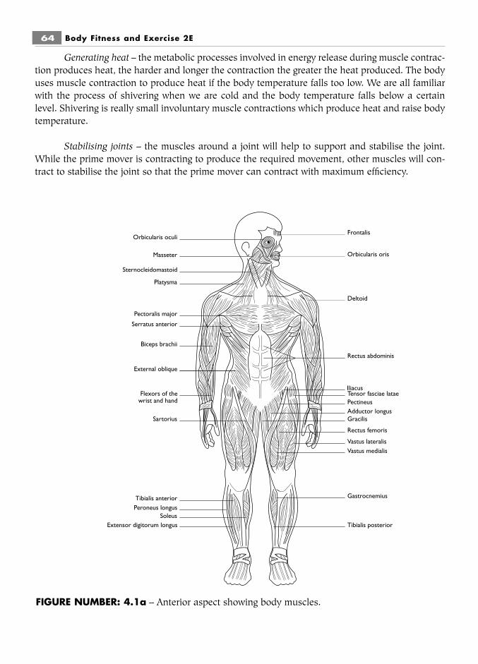
Generating heat – the metabolic processes involved in energy release during muscle contrac-tion produces heat, the harder and longer the contraction the greater the heat produced. The bodyuses muscle contraction to produce heat if the body temperature falls too low. We are all familiarwith the process of shivering when we are cold and the body temperature falls below a certainlevel. Shivering is really small involuntary muscle contractions which produce heat and raise bodytemperature.
Stabilising joints – the muscles around a joint will help to support and stabilise the joint.While the prime mover is contracting to produce the required movement, other muscles will con-tract to stabilise the joint so that the prime mover can contract with maximum efficiency.
64 Body Fitness and Exercise 2E
Vastus lateralis
Frontalis
Orbicularis oris
Deltoid
Rectus abdominis
Vastus medialis
Rectus femoris
GracilisAdductor longus
Tensor fasciae lataePectineus
Tibialis posterior
Gastrocnemius
Extensor digitorum longusSoleus
Peroneus longusTibialis anterior
Sartorius
External oblique
Biceps brachii
Serratus anterior
Flexors of the wrist and hand
Pectoralis major
Platysma
Sternocleidomastoid
Masseter
Orbicularis oculi
Iliacus
FIGURE NUMBER: 4.1a – Anterior aspect showing body muscles.

Skeletal muscle 65
THE STRUCTURE OF SKELETAL MUSCLESkeletal muscle is composed of muscle fibres arranged in bundles called fasciculi; many bundlesof fibres make up the complete muscle. The fibres, bundles and muscles are surrounded and pro-tected by connective tissue sheaths:
■ The connective tissue around each fibre is called the endomysium.■ The connective tissue around each bundle is called the perimysium.■ The connective tissue around the muscle is called the epimysium.
This connective tissue blends at each end of the muscle to form tendons which attach themuscles to the underlying bones.
Occipitalis
Trapezius
DeltoidInfraspinatus
Teres majorTeres minor
Brachioradialis
Extensors of thewrist and hand
Adductor magnusGracilis
Semitendinosus
Semimembranosus
Achilles tendon
Gastrocnemius
Soleus
Biceps Femoris
Iliotibial tract
Gluteus maximus
Gluteus medius
External oblique
Latissimus dorsi
Triceps brachii
Splenius capitis
FIGURE NUMBER: 4.1b – Posterior aspect showing body muscles.

66 Body Fitness and Exercise 2E
Muscle fibresMuscle fibres are long, thin multi-nucleate cells. The fibres vary from 10 to 100 microns in diame-ter and from a few millimetres to many centimetres in length. The long fibres extend the full lengthof the muscle, while the short fibres end in connective tissue intersections within the muscle.
Each muscle fibre is bound by a cell membrane known as the sarcolemma, just beneathwhich lie the nuclei. The cytoplasm of the muscle cell is known as the sarcoplasm. It contains large
FIGURE NUMBER: 4.2 – (a) The structure of a muscle.(b) A single muscle fibre showing striations.(c) A myofibril, illustrating a sarcomere.
LEARNMuscles are composed of:■ Muscle fibres covered by the endomysium (lots of fibres together form bundles)■ Muscle bundles covered by the perimysium (lots of bundles form the muscle)The muscle is covered by the epimysium

numbers of mitochondria and other organelles. Muscle fibres are made up of smaller proteinthreads called myofibrils. These run the whole length of the fibre and are the elements which con-tract and relax. Myofibrils are made up of even smaller threads called myofilaments.
Under an electron microscope, myofibrils are seen to have alternate light and dark bandscalled I and A bands. In the middle of the dark A band is a lighter zone, the H zone. In the mid-dle of the light I band is a dark line, the Z line.
The segment between two Z lines is known as the sarcomere. These sarcomeres are repeatedalong the whole length of the myofibril. Each sarcomere contains overlapping thick and thin myo-filaments. The thin myofilaments are made of the protein actin. They begin at the Z line and extendinto the A band, where they overlap with the thick myofilaments, which are made of the proteinmyosin. These thick bands have small cross-bridges projecting sideways towards active sites on thethin bands. These are very important: when a stimulus from the nervous system is received by themuscle fibre, a series of chemical reactions takes place which results in the cross-bridges linkingand pulling the thin bands towards the thick bands. The sliding thin bands pull on the Z lines andeach sarcomere shortens. Consequently, the myofibrils and fibres shorten and the whole musclecontracts. The energy for this contraction is obtained from the breakdown of ATP (adenosine tri-phosphate) stored in the myosine cross-bridges.
Muscle relaxation occurs when no stimulus is received from the nervous system. The thinbands slide back to their precontracted state and the muscle relaxes.
Muscle elongation occurs only as a result of some pulling force on the muscle. This forcemay be the pull of antagonistic muscles (i.e. on the opposite side of the joint), the pull of gravity,the pull of weights, springs, etc., or manual pulling by oneself or another person. The fibres elon-gate because the thin filaments move away from the thick filaments and each sarcomere getslonger. The pull must allow at least one cross-bridge to remain intact; otherwise, the sarcomere willrupture. Strong forces can cause small tears within a muscle because the cross-bridges are nolonger intact. During exercise and sports, excessive stress may result in muscle tears.
Skeletal muscle 67
FIGURE NUMBER: 4.3 – The sarcomere. (a) During relaxation. (b) During contraction.

Neuromuscular functionThe contraction of skeletal muscle is controlled by the nervous system. When the brain receivesinformation that movement is required, stimuli/impulses are initiated in the motor cortex whichwill activate the muscles to contract. These impulses, transmitted via motor nerve neurones, passdown from the brain via the spinal cord to the anterior horn cells. The axons of many anterior horncells emerge together as a motor nerve and pass to a muscle.
The point at which the motor nerve enters the muscle is known as the motor point. Eachaxon will then divide into many branches and each branch will supply one muscle fibre.
The neuromuscular junction, i.e. the point at which the axon branch meets the muscle fibre,is known as the motor end plate.
The anterior horn cell, its axon, axon branches and all the muscle fibres it supplies, isknown as a motor unit.
A stimulus of sufficient strength will result in all the muscle fibres within a motor unitcontracting together to maximum strength but a stimulus below this intensity will not produce anyresponse. The stimulus of sufficient intensity to produce a contraction is known as the thresholdstimulus.
68 Body Fitness and Exercise 2E
Synaptic cleftSynaptic vesicles
Muscle fibreMotorend plate Acetylcholine
Synaptic knob
Sarcolemma
Sarcoplasm
Motor point
FIGURE NUMBER: 4.4 – Synapse.

Remember that muscle fibres contract according to the ‘all or none law’ which states thatmuscle fibres contract with maximum force or not at all.
The number of motor units recruited will influence the strength of the contraction; the moremotor units activated the greater the strength of the contraction. Strength training results in therecruitment of more motor units and hence greater muscle strength.
Another important consideration is the frequency of the impulses. A low frequency will pro-duce a ‘twitch’ of the muscle, which is a quick contraction followed by complete relaxation.Increasing the frequency will result in a second contraction occurring before complete relaxation.The length of contraction will now be the sum of the two twitches; this will give a longer contrac-tion and is known as wave summation. However, the contraction will not be smooth as the musclewill partially relax between the twitches. Increasing the frequency until the muscle cannot relaxbetween stimuli will result in a smooth contraction of the muscle known as a tetanic contraction.
Muscle fibre typesA muscle is composed of different types of muscle fibres which differ in their metabolism and func-tion. The proportion of each fibre within a muscle will depend on the function of the muscle. Thereare two main types:
Type II Red fibres:Type II White fibres: (which can be subdivided into type IIa, IIb and IIc).
Type I fibres are slow twitch fibres which are equipped to contract slowly but repeatedlyover a long period of time and utilise the aerobic energy system. They are used during enduranceactivities such as marathon running or swimming.
Type II fibres are fast twitch fibres that are equipped to contract rapidly for short periodsonly and utilise the anaerobic energy systems. They are used in fast rapid activities such as squashor short sprints.
The different properties of the fibres are shown opposite.
Muscle shapeMuscle shape varies depending on the function of the muscle. The fleshy bulk of the muscle isknown as the belly. The bundles of muscle fibres lie either parallel or obliquely to the line of pullof the muscle. Parallel fibres are found in strap-like and fusiform muscles. These long fibres allow
Skeletal muscle 69
LEARNThe all or none law.When muscle fibres respond to a stimulus, they contract with maximum force or notat all.

70 Body Fitness and Exercise 2E
for a wide range of movement. The shorter oblique fibres are found in triangular and pennate mus-cles, where muscle strength is required.
Muscle attachmentsAs previously explained, a muscle is composed of muscle fibres and connective tissue components,namely the endomysium, perimysium and epimysium. Certain muscles have connective tissueintersections, dividing the muscle into several bellies, as seen in the rectus abdominus.
Type 1 Slow twitch Type II Fast twitch
Utilises aerobic pathway Utilises anaerobic pathway
High endurance capacity Low endurance capacity
Slow contraction Fast contraction
High resistance to fatigue Low resistance to fatigue
Small fibre diameter Large fibre diameter
Low phosphocreatine stores High phosphocreatine stores
High capillary density Low capillary density
Low glycogen stores High glycogen stores
Low glycolytic enzyme stores High glycolytic enzyme stores
High triglyceride stores Low triglyceride stores
High myoglobin content Medium myoglobin content
FIGURE NUMBER: 4.5 – Muscle shape.
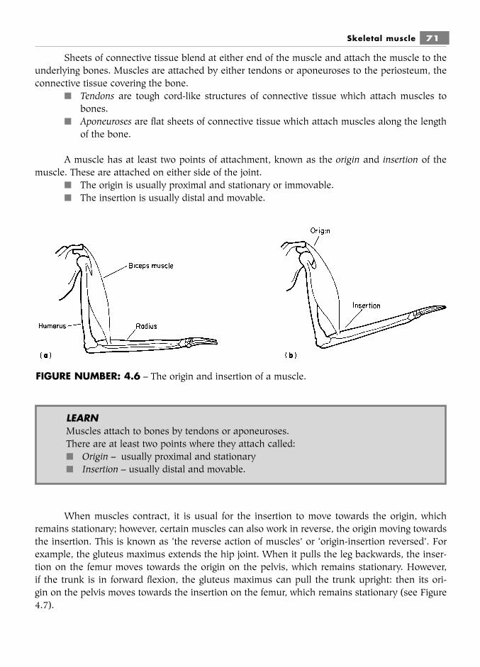
Sheets of connective tissue blend at either end of the muscle and attach the muscle to theunderlying bones. Muscles are attached by either tendons or aponeuroses to the periosteum, theconnective tissue covering the bone.
■ Tendons are tough cord-like structures of connective tissue which attach muscles tobones.
■ Aponeuroses are flat sheets of connective tissue which attach muscles along the lengthof the bone.
A muscle has at least two points of attachment, known as the origin and insertion of themuscle. These are attached on either side of the joint.
■ The origin is usually proximal and stationary or immovable.■ The insertion is usually distal and movable.
When muscles contract, it is usual for the insertion to move towards the origin, whichremains stationary; however, certain muscles can also work in reverse, the origin moving towardsthe insertion. This is known as ‘the reverse action of muscles’ or ‘origin-insertion reversed’. Forexample, the gluteus maximus extends the hip joint. When it pulls the leg backwards, the inser-tion on the femur moves towards the origin on the pelvis, which remains stationary. However,if the trunk is in forward flexion, the gluteus maximus can pull the trunk upright: then its ori-gin on the pelvis moves towards the insertion on the femur, which remains stationary (see Figure4.7).
Skeletal muscle 71
FIGURE NUMBER: 4.6 – The origin and insertion of a muscle.
LEARNMuscles attach to bones by tendons or aponeuroses.There are at least two points where they attach called:■ Origin – usually proximal and stationary■ Insertion – usually distal and movable.

Muscle toneMuscle tone is the state of partial contraction or tension found in muscles even when at rest. Asmall number of muscle fibres will always be in a state of contraction. The degree of tension willdepend on the number of motor units recruited. The recruitment of a few motor units will be justsufficient to produce tautness in the muscle, but not to result in full contraction and movement. Asdifferent motor units are stimulated different groups of fibres will contract alternately, working a‘shift’ system to prevent fatigue of the few. Changes in muscle tone are adjusted according to theinformation received from sensory receptors within the muscles and their tendons. Muscle spindlestransmit information on the degree of stretch within the muscle. Tendon receptors called Golgiorgans transmit information on the amount of tension applied to the tendon by muscle contrac-tion. Too much stretch and tension will be counteracted by the recruitment of fewer motor unitsand a reduction in muscle tone. Too little will be counteracted by an increase in number of motorunits and in muscle tone. Muscle tone is essential for maintaining upright posture.
■ Hypotonic muscles, i.e. those with less than the normal degree of tone, are said to beflaccid.
■ Hypertonic muscles, i.e. those with a greater degree of muscle tone and where fibres areover-contracted and rigid, are said to be spastic.
■ A contraction that increases muscle tone but does not change the length of the muscleis called isometric contraction (equal length).
■ A contraction where muscle tone remains the same but the muscle changes in length iscalled isotonic contraction (equal tone).
Blood supply to skeletal muscleThe oxygen and nutrients required by the contracting muscles are delivered by the circulatingblood while the waste products of the energy-producing reactions are removed. During exercisethere is a marked increase in blood flow to the exercising muscles. Blood is redirected from other
72 Body Fitness and Exercise 2E
FIGURE NUMBER: 4.7 – Actions of gluteus maximus.(a) Extension of the hip joint, the insertion moves towards the origin.(b) Raising the trunk, the origin moves towards the insertion.

organs such as the stomach and liver. Blood flow may increase from around 20 percent of cardiacoutput to 80 percent. The redirecting of blood from other areas is known as blood shunting oraccommodation. The supplies of oxygen and nutrients are brought by the blood via the arteries,and the waste products are removed via the veins. The arteries branch to form smaller arteries andarterioles within the perimysium. They then divide further to form capillary networks within theendomysium, where they join venules, which lead to veins. When muscles are relaxed, the capil-lary network delivers blood to the muscle fibres.
When muscles contract, the pressure impedes the flow of blood through the capillary beds.This reduces the supply of oxygen and nutrients and limits the removal of waste. During exercise,muscle fibres alternately contract and relax and the capillaries deliver blood during the relaxationphase. However, repeated or sustained contraction, such as isometric work or exercising withoutsufficient rest periods, prevents the flow of blood to the muscle fibres, due to compression on theblood vessels and the capillaries. This results in muscle fatigue due to lack of oxygen and nutrientsand the accumulation of waste products such as lactic acid. The strength and speed of contractionbecome progressively weaker, and as fatigue continues the muscle fails to relax completely, result-ing in muscle spasm and pain. Regardless of the activity, muscles must be given sufficient time torelax completely. This will ensure an adequate blood supply and prevent fatigue.
Regular endurance aerobic exercise results in an increase in blood vessels and capillary net-works to the muscles. This will improve the blood supply, increase levels of oxygen and nutrientsand reduce levels of lactic acid. Thus the capacity to exercise without fatigue will improve.
Energy for muscle contractionEnergy may be defined as the ability or capacity to perform work. All human activity is dependentupon the ability to provide energy on a continuous basis, all tissue cells require energy to carry outtheir activities. Energy is provided by the food we eat but the energy liberated from the breakdownof food is not directly employed to do work but rather, is used to manufacture a chemical com-pound, adenosine triphosphate (ATP). The energy supplied by the breakdown of this high energychemical compound provides the energy for cells to perform their work.
Muscle cells expend far more energy than other cells, and convert chemical energy tomechanical energy. Only a limited quantity of ATP is stored in muscle cells and this is depletedafter a few seconds of activity and must be continually replenished if muscle contraction is to con-tinue. The body has adapted different systems for the resynthesis of ATP and hence energy pro-duction. ATP may be resynthesised through the breakdown of another high energy chemical com-pound, namely phosphocreatine (PC), also stored in muscle cells, but the main source of energyis provided by carbohydrates and fats stored as glycogen in muscle cells and in the liver. If theenergy is generated by reactions which do not utilise oxygen, they are anaerobic energy systems. Ifthe reactions utilise oxygen, they are aerobic energy systems.
The system used will depend on the intensity and duration of the activity and on the avail-ability and use of oxygen.
Skeletal muscle 73

Remember that throughout the following text ATP represents adenosine triphosphate. PCrepresents phosphocreatine.
There are three energy systems for resynthesising ATP; two are anaerobic energy systemsand one is the aerobic energy system.
The systems are:■ the alactic ATP – PC system or phosphagen system (anaerobic).■ the lactic acid system (anaerobic glycolysis).■ the aerobic (oxygen) system (aerobic glycolysis).
The alactic ATP – PC systemThis system provides the most rapidly available source of energy as it utilises the ATP and PCstored in muscle cells. It does not depend on transporting oxygen from the lungs nor on a longchain of chemical reactions. Therefore when a muscle is stimulated to contract, immediate energyis obtained from stored ATP and the activity will continue for 5–6 seconds until the ATP is depleted.ATP is then replenished by the breakdown of PC. This supply will provide energy for 10–15 sec-onds of maximum effort, until the stored PC is depleted. This fast system would be used in activ-ities of short duration such as sprinting, kicking, jumping, throwing actions and in the initial effortof push off from the starting block. The amount of energy obtainable from this system is limiteddue to the limited stores of ATP and PC in the muscle. However, they are replenished very quick-ly after three to four minutes of recovery. This is important as the rapid activity can be performedagain after a short rest.
Oxygen is not utilised, therefore it is anaerobic.
The lactic acid systemIf the activity continues after the stored phosphocreatine is depleted, ATP is resynthesised from gly-cogen (carbohydrate).
Carbohydrate is eaten as sugars and starch and converted to glycogen which is found in theblood and stored in muscle tissue and in the liver.
The breakdown of the glucose molecule to resynthesise ATP and hence provide energy,involves a series of reactions known as glycolysis. Initially, if oxygen is not available quicklyenough, energy is liberated through the partial breakdown of glycogen known as anaerobic glycol-ysis which occurs in the intracellular fluid of the muscle cell.
The glycogen molecule is broken down to pyruvic acid which in the absence of oxygen isconverted to lactic acid. This system utilises carbohydrate only.
This system is not particularly efficient as it yields only 2–3 moles of ATP for each moleculeof glucose, which releases only a small amount of energy. However, it is an important source of fast
74 Body Fitness and Exercise 2E
energy for 2–3 minutes of high intensity activity such as 400–800 metre running or 100–200 metreswimming. It is also used at the end of a run during the final ‘burst’.
Anaerobic glycolysis results in the formation of lactic acid which contributes to musclefatigue. With the build of lactic acid within the muscle, contraction will diminish and activity willstop.
The aerobic systemThis system of energy release requires a continuing supply of oxygen and is therefore termed theaerobic system. When oxygen is available, glycogen is completely broken down through a seriesof complicated reactions in the mitochondria of the cell. The many reactions of the aerobic systeminclude aerobic glycolysis, the Krebs cycle and the electron transport system. The initial stage isthe same as with the previous system as glycogen is broken down to pyruvic acid but when oxygenis available lactic acid does not accumulate as the breakdown of pyruvic acid continues. It ischemically altered enabling it to enter the Krebs cycle and the electron transport system where thebreakdown continues to yield carbon dioxide, water and energy (ATP).
This system provides far more energy than the other two systems as 1 molecule of glycogenwill provide enough energy to resynthesise 38 moles of ATP. This system will therefore provide acontinual supply of energy for prolonged endurance activities without producing fatiguing by-products. The activity can continue indefinitely providing the supply of oxygen and the fuel storescan be maintained. This system utilises both carbohydrate and fat as fuel but protein will only beused in extreme conditions of starvation or ultra marathon running.
Which system?The systems used will depend on the intensity and duration of the activity. Most activities will util-ise both anaerobic and aerobic systems depending on the availability of oxygen. However, as ageneral rule, short bursts of fast intense activity will utilise the anaerobic systems while prolongedlow intensity activity will utilise the aerobic system.
Which fuel?The fuel used to resynthesise ATP is provided by digested nutrients which include carbohydrates,fats and proteins.
Carbohydrates are starches and sugars which are converted to glucose or glycogen andstored in the blood, liver and muscle tissue. Carbohydrates provide primary fast energy duringshort bursts of intense anaerobic activity and moderate aerobic activity. However, glycogen storesare limited and become depleted during prolonged activities. Acute feelings of exhaustion are expe-rienced as glycogen is depleted, as in ‘hitting the wall’ in marathon running.
Fats in the form of fatty acids and triglycerides are stored in the liver and adipose tissue.Fats are the body’s most concentrated source of energy. The body is able to store far more fat thanglycogen and twice as much energy is stored in one gram of fat, than in one gram of carbohydrate.Therefore fats provide a far greater source of potential energy for prolonged endurance activities.
Skeletal muscle 75

However, fat requires a small amount of glycogen for its combustion and if glycogen stores aredepleted, fat cannot be broken down. Fat cannot provide energy for fast activity, because itdepends on the availability of oxygen, which depends on a person’s aerobic capacity. Trainingimproves aerobic capacity and a fit person with a high aerobic capacity will burn fat more easily.During endurance activities, the sooner fat can be utilised, the more glycogen will be spared whichwill prolong activity.
Proteins are broken down to amino acids and are used for growth, body building and tissuerepair. Proteins are only used as an energy source if stores of carbohydrates and fats are very lowor depleted as in conditions of starvation.
Aerobic and anaerobic exercisesThese names are derived from the energy systems used. Most activities involve both aerobic andanaerobic metabolism.
Aerobic exercisesThese are endurance activities that utilise oxygen for energy production. They are slow, steady-state exercises, which allow for the systems to supply sufficient oxygen for the complete breakdownof glycogen. Oxygen supply is maintained throughout and no oxygen debt is incurred. There istherefore no gasping or deep breathing at the end of these activities. Aerobic activities include jog-ging, walking, swimming, cycling, aerobic classes. (Remember that if the exercises become too fastand vigorous in aerobic classes, the exercise will be anaerobic. Clients should not be short ofbreath during aerobic activities and should be able to talk or sing whilst exercising.)
76 Body Fitness and Exercise 2E
Table 4.1The three metabolic systems
Anaerobic (alactic): ATP-PC Anaerobic: lactic acid Aerobic: oxygen systemsystem system
Uses stored ATP and PC Uses glycogen Uses glycogen, fattyacids, triglycerol
Beginning of all activities Fast activity up to 2–3 Slow steady moderateand very fast short bursts minutes to low intensity, longup to 10–15 seconds duration activity
Example: quick dash, Example: 400 metre run Example: jogging,sprint marathon running
Contraction stops when Contraction stops due to Contraction maintainedATP and PC are used up lack of oxygen and lactic indefinitely until
acid build-up. glycogen is depletedand exhaustion isreached

Anaerobic exercisesThese are activities that do not use oxygen for energy production. All activities begin anaerobicallyand continue until all readily available energy within the muscles is used up; this will last for 10–15seconds, until supplies of ATP and PC are exhausted and is known as the alactic phase, as lacticacid is not produced. Further vigorous, fast-moving activities – too fast for the systems to supplyoxygen – will result in the incomplete breakdown of glycogen and the breakdown of pyruvic acidinto lactic acid. This is known as the lactic phase. Lactic acid builds up within the muscle and willeventually inhibit its contraction. Anaerobic activities include squash, sprinting, hurdling and fastvigorous actions. There will be deep breathing or panting at the end of this activity as extra oxygenis required to re-establish homeostasis (body balance).
Most sports utilise both energy systems, where the fast vigorous phases are anaerobic andthe slower steadier phases are aerobic.
Oxygen uptakeOxygen uptake is the amount of oxygen consumed within a certain time: usually one minute. It isknown as VO2. Maximum oxygen uptake is the maximum amount of oxygen taken in, transportedand utilised by the muscles per minute to produce energy. The amount of oxygen consumed at restis around 0.2–0.3 litres per minute, but this increases considerably during exercise, to a pointwhere the system is unable to meet further demand. This point is an individual’s aerobic capacityor VO2 maximum.
VO2 maximum can be measured and is used to assess a person’s aerobic power or fitness.With training, fitness develops, the heart pumps out more blood, the lungs improve and ventila-tion increases, with more oxygen taken in. This in turn is delivered to the muscles and used moreefficiently. As fitness increases the amount of oxygen taken in increases. Trained athletes have farhigher VO2 maxima than untrained individuals.
Regular training increases the capacity for oxygen uptake and the ability to exercise aerobi-cally for longer periods.
Oxygen debtThis is the excess amount of oxygen taken in during recovery, over and above that normally con-sumed. As previously explained, during vigorous muscular activity, oxygen cannot be supplied fastenough to the muscle fibres and oxygen supplies are depleted. Energy is therefore generatedfrom the anaerobic breakdown of pyruvic acid, which produces lactic acid. A large percentage ofthe lactic acid is transported from the muscle to the liver, where it is converted back to glucose orglycogen, but some lactic acid remains in the muscle. After exercise has stopped, extra oxygen isrequired to metabolise the lactic acid, and to replenish ATP, phosphocreatine and glycogen, aswell as increasing the supply of oxygen in the blood and lungs in order to restore the bodysystems to their normal state. The increase of lactic acid and carbon dioxide in the blood
Skeletal muscle 77

stimulates the respiratory system, so that breathing increases in depth and rate and oxygen debtis repaid.
Muscle fatigueMuscle fatigue is the inability of a muscle to sustain a contraction. The contraction becomes pro-gressively weaker and then fails completely as the muscle is unable to produce sufficient energy tomeet its needs. During fast, vigorous exercise it is thought to be due to a build up of lactic acid inthe muscle and blood. During low intensity exercise it is thought to be due to depletion of glyco-gen and dehydration.
Muscular sorenessTwo types of muscular soreness have been identified:Acute muscular soreness and delayed muscular soreness.
Acute muscular soreness is experienced during or immediately following activity. It is thoughtthat this is due to an insufficient blood supply to the contracting muscles. This may be due to thetension which builds up in the contracting muscles pressing on the blood vessels, reducing oroccluding blood flow. Metabolic waste products such as lactic acid are not removed and built upwithin the muscle to such an extent that they stimulate the pain receptors within the muscle. Thisacute soreness is quickly relieved when exercise stops and blood flows freely again flushing out thelactic acid.
Delayed muscle soreness or delayed onset muscle soreness (DOMS)The onset of this soreness is delayed for 24–48 hours after the activity. The exact cause is notknown but it is thought that it may be due to damage of the tissues. This damage may includeminor tears of the muscle fibres; damage of the connective tissue components within the muscle;or damage to tendons and ligaments through over stretching.
Another theory suggests that muscle spasm occurs; this exerts pressure on the blood ves-sels reducing blood flow, resulting in pain.
Stretching warm muscles before activity helps prevent soreness and stretching after theactivity helps alleviate it. The stretches must be smooth and slow as any jerking or bounding mightincrease the damage.
Delayed muscular soreness is greatest following eccentric contraction, less following con-centric contraction and least following isokinetic contraction (Chapter 6).
SKELETAL MUSCLE RESPONSE TO EXERCISEThe adaptations of skeletal muscle will depend on the type of activity undertaken.
78 Body Fitness and Exercise 2E

Endurance training■ Muscle endurance improves when a muscle is made to contract repeatedly against low
or moderate resistance. As a result of endurance training other changes occur:■ Increase in the number and size of blood vessels supplying blood to muscle fibres;■ an increase in myoglobin content;■ an increase in the density of the capillary networks supplying blood to muscle fibres;■ an increase in the blood flow aided by blood shunting thus improving the delivery of
oxygen and the removal of waste;■ an increase in the size and number of mitochondria in the muscle cells, and therefore
greater efficiency in utilising oxygen and generating energy stores;■ an increase in glygocen stores. The increased availability of oxygen and glycogen raises
the anaerobic threshold, so that the muscles use aerobic energy for longer periods, thusreducing levels of lactic acid. The muscles can continue contracting for longer periodswithout fatigue.
■ An increase in glycolitic and oxidative enzymes.
Strength training■ Muscle strength will increase, providing the muscle is made to work against sub-maximal
or maximal loads. As the muscle strengthens the load must be progressively increased.■ More motor units are recruited, which increases the strength of contraction.■ Muscle size (bulk) increases. Research to date suggests that this is due to the following
adaptations:■ an increase in the size and number of myofibrils■ an increase in the contractile proteins myosin and actin■ an increase in stored energy supplies (ATP, PC) and enzymes, giving a greater source of
quick energy■ an increase in connective tissue components.
Flexibility training■ An increase in elasticity and extensibility of the muscle.■ Neurological adaptation delaying the stretch reflex.■ Flexibility exercises gently stretch connective tissue in the muscle and at a joint.
Flexibility exercises performed before but particularly after an activity session willreduce muscle soreness.
Summary and aid to learningSkeletal muscle forms the body flesh. Its functions are: to produce movement; to maintain posture;to generate heat; to stabilise joints.
It has three properties:
■ Contractability: it is able to contract■ Extensibility: it is able to lengthen■ Elasticity: it is able to return to its original length after being stretched.
Skeletal muscle 79

Skeletal muscle cells are elongated fibres with many nuclei along their length. These fibresare grouped together into bundles called fasciculi. Many bundles arranged together form themuscle. The fibres, bundles and muscles are surrounded by ‘sleeves’ of connective tissue. Thisconnective tissue blends together at the ends of the muscle to form the tendons.
Give the function of tendons.
When viewed under a powerful microscope light and dark bands can be seen along thelength of the fibres. These are thick and thin filaments of the proteins, actin and myosin. It is thesliding of the thin actin filaments towards the thick myosin filaments that shortens the muscle, pro-ducing muscle contraction. This is known as the sliding filament theory.
There are two main types of muscle fibres within a muscle:■ Type II Slow twitch, red fibres■ Type II Fast twitch, white fibres
Study the text and write down six differences between these fibres.
Muscles are under the control of the nervous system, they contract in response to stimuliinitiated in the brain.
The all or none law states that, stimulated muscle fibres will contract with maximum forceor not at all.
Muscle tone – this is the state of partial contraction found in a muscle even at rest.
Sensory receptors within the muscles, known as muscle spindles, register the degree ofstretch within a muscle.
The energy for muscle contraction is obtained from the breakdown of a chemical compoundcalled adenosine triphosphate (ATP). All cells require ATP to carry out their functions but musclecells require large amounts to maintain muscle contraction during exercise.
Only a small amount of ATP is stored in muscle cells, therefore when this is used up it mustbe continually remade (resynthesised). It is resynthesised from another chemical called phospho-creatine (PC) and from the food we eat, mainly carbohydrates and fats in the form of glycogen andtriglycerides.
Therefore the energy for muscle contraction comes from stored ATP and from the other chem-ical reactions or systems which resynthesise it from foods. Some of the systems utilise oxygen andare known as aerobic systems while others do not need oxygen and are called anaerobic systems.
Remember: aerobic means with oxygenanaerobic means without oxygen
80 Body Fitness and Exercise 2E

There are three systems which provide the energy for muscle contraction:■ ATP – PC system uses stored ATP, PC, anaerobic.■ Lactic acid system involves the incomplete breakdown of glycogen, anaerobic, and as its
name suggests, lactic acid is produced which builds up in the muscle.■ Aerobic system involves the complete breakdown of glycogen and triglycerides in the
presence of oxygen, aerobic, carbon dioxide and water are produced.
The system used will depend on the intensity and duration of the activity.
Study the text and answer the following:■ Which system would be used during marathon running and why?■ Explain the main factor which limits contraction when very fast activity is performed.
Aerobic power (VO2 maximum) is the maximal rate that an individual can take in andutilise oxygen during maximal performance. It is an indicator of cardiorespiratory fitness.
Oxygen debt or recovery oxygen is the amount of oxygen consumed after exercise, overand above that normally consumed. It is the amount of oxygen required to resynthesise ATP andPC stores in the muscle and to restore homeostasis.
Muscle fatigue is the inability of a muscle to sustain a contraction. The contractionbecomes weaker and weaker and then fails completely. During fast, high intensity exercise, it isthought to be due to the build up of lactic acid in the muscles and the blood. During low intensityexercises it is thought to be caused mainly by depletion of glycogen, and dehydration.
Muscle soreness there are two types:■ acute muscular soreness – this occurs immediately after performance;■ delayed muscular soreness or delayed onset muscular soreness DOMS. This
occurs 24–48 hours after performance.
Read the text and discuss the different causes of the two types of muscular soreness.
Skeletal muscle 81
82 Body Fitness and Exercise 2E
QUESTIONS1. Give three functions of muscle tissue.2. Name the connective tissue sheath which surrounds a muscle.3. Name two contractile proteins found in muscle fibres.4. Complete the following sentence: Muscle contraction occurs as a result of a stimulus
from the … .5. Explain what happens to a muscle fibre if no cross-bridges remain intact.6. Name two types of muscle fibre.7. State which type of muscle fibre depends on aerobic metabolism and which type
depends on anaerobic metabolism.8. Explain what is meant by the term threshold stimulus.9. List any three factors which affect the strength of muscle contraction.
10. Define the following:a tendonb aponeurosisc origind insertion.
11. Define the term muscle tone.12. Name the structures which transmit information on the degree of tension found in a
muscle.13. Name the chemical which supplies energy for muscle contraction.14. Briefly explain why energy is supplied by anaerobic metabolism for the first fifteen to
twenty seconds of muscular activity.15. Explain why lactic acid is produced during short, vigorous bursts of activity.16. Explain the term oxygen debt.17. State what types of exercise and activities utilise aerobic metabolism.18. Differentiate between aerobic and anaerobic exercises.19. Explain what is meant by the term VO2 maximum.20. Define the term muscle fatigue and explain how it occurs.
Chapter 5The Support Systems:
Nervous, Cardio-vascularand RespiratoryTHE NERVOUS SYSTEM
The nervous system is the communication and control system of the body. It works with the endo-crine system to maintain homeostasis (body balance). The nervous system will sense changes,interpret them and initiate appropriate action.
The nervous system is made up of:1 The central nervous system comprising the brain and spinal cord which controls both
reflex and voluntary movements. Here, incoming stimuli are processed, information isstored, thoughts are initiated and motor skills (movements) are controlled.
2 The peripheral nervous system comprising 12 pairs of cranial nerves arising from thebrain and 31 pairs of spinal nerves arising from the spinal cord. These nerves conductimpulses from sensory organs to the spinal cord and brain and conduct impulses fromthe brain and spinal cord to muscles and glands.
3 The autonomic system comprising both sympathetic and parasympathetic parts whichexercise involuntary control of body functions.

84 Body Fitness and Exercise 2E
The autonomic system conducts information from the viscera to the central nervous systemand information from the brain to smooth muscle, cardiac muscle and glands. This part is invol-untary as it is not under conscious control.
Structure of the neuroneNervous tissue is composed of the functional units which conduct impulses called neurones andthe supporting tissue called neuroglia. There are three types of neurones:
■ Sensory neurones which transmit stimuli from sensory organs to the spinal cord andbrain.
LEGS
EYES
TOUCH
TASTE
HEARING
SMELLHANDS
MOUTH SIGHT
CerebrumThe main part of the brain. It is the largest
in volume, and occupies most of the cranium.Divided into two halves, this part of the brainis responsible for thought, speech, memory,
learning, imagination and decision making. Theright side of the cerebrum controls activitieson the left side of the body, and vice versa.
CerebellumControls balance and
coordination of muscularactivities and movement.
Medulla oblongataControls the unconscious
activities such as breathing,heart beat and digestion. This
part of the brain is anextension of the spinal cord.
ThalamusA small area ofthe brain which registers pain.
HypothalamusControls sleeping
and waking, eating,drinking and speech.
FIGURE NUMBER: 5.1 – The brain.

The Support Systems: Nervous, Cardio-vascular and Respiratory 85
■ Motor neurones which transmit stimuli from the brain and spinal cord to muscles andglands.
■ Inter-neurones which form a connection between neurones.
All neurones have a similar structure: they have a cell body, one long nerve fibre called anaxon and several short nerve fibres called dendrons ending in dendrites.
■ Axons carry impulses away from the cell body.■ Dendrons and dendrites carry impulses towards the cell body.
The axons of some nerves are protected by a fatty myeline sheath; this sheath is interruptedat intervals by small spaces called nodes of Ranvier. These determine the speed at which the nerveimpulse is transmitted.
The nerve impulseA nerve impulse is electrical in nature. It is generated in response to a stimulus and is transmittedalong a nerve fibre due to changes in its electrically charged state. Impulses are transmitted inaxons and dendrons in one direction only. The fatty myeline sheath insulates parts of the nerveand the impulse must jump from one node of Ranvier to another. This jumping from node to nodeserves to increase the speed of transmission.
FIGURE NUMBER: 5.2 – Types of neurone. (a) Motorneurone. (b) Sensoryneurone.(c) Interneurone.
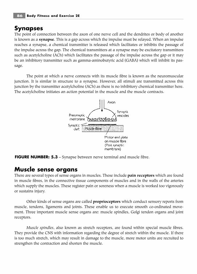
SynapsesThe point of connection between the axon of one nerve cell and the dendrites or body of anotheris known as a synapse. This is a gap across which the impulse must be relayed. When an impulsereaches a synapse, a chemical transmitter is released which facilitates or inhibits the passage ofthe impulse across the gap. The chemical transmitters at a synapse may be excitatory transmitterssuch as acetylcholine (ACh) which facilitates the passage of the impulse across the gap or it maybe an inhibitory transmitter such as gamma-aminobutyric acid (GABA) which will inhibit its pas-sage.
The point at which a nerve connects with its muscle fibre is known as the neuromuscularjunction. It is similar in structure to a synapse. However, all stimuli are transmitted across thisjunction by the transmitter acetylcholine (ACh) as there is no inhibitory chemical transmitter here.The acetylcholine initiates an action potential in the muscle and the muscle contracts.
Muscle sense organsThere are several types of sense organs in muscles. These include pain receptors which are foundin muscle fibres, in the connective tissue components of muscles and in the walls of the arterieswhich supply the muscles. These register pain or soreness when a muscle is worked too vigorouslyor sustains injury.
Other kinds of sense organs are called proprioceptors which conduct sensory reports frommuscle, tendons, ligaments and joints. These enable us to execute smooth co-ordinated move-ment. Three important muscle sense organs are: muscle spindles, Golgi tendon organs and jointreceptors.
Muscle spindles, also known as stretch receptors, are found within special muscle fibres.They provide the CNS with information regarding the degree of stretch within the muscle. If thereis too much stretch, which may result in damage to the muscle, more motor units are recruited tostrengthen the contraction and shorten the muscle.
86 Body Fitness and Exercise 2E
FIGURE NUMBER: 5.3 – Synapse between nerve terminal and muscle fibre.

Golgi tendon organs are located in tendon fibres near the junction between the tendon andthe muscle. They are also sensitive to stretch and are stretched when the muscle in whose tendonthey lie contracts. When the contraction is too strong the stretch in the GTO is relayed to the CNScausing the contracted muscle to relax. This protects the muscle from injury.
Joint receptors are found in tendons, ligaments, joint capsules and in the periostium cover-ing bones. They provide information concerning joint position and angle and together with otherreflexes are concerned with the maintenance of posture.
Neuromuscular controlThe selection and performance of motor movement is initiated in the motor cortex of the brain. Theprimary motor area is subdivided according to body areas and is responsible for specific patternsof movement, while the pre-motor area is responsible for more complex patterns; this is also calledthe sports skill area. The three areas of the brain most involved in the execution of movement arethe cerebral cortex, basal ganglia, and cerebellum. Impulses are transmitted from the brain via theupper motor neurone to the anterior horn of the spinal cord. Here the impulse will cross a synapseto stimulate the lower motor neurone and will be transmitted via its axon to the muscle fibres,resulting in contraction of the muscle. Information from the sense organs will be transmitted backto the brain regarding the spatial location of body parts. etc., and the degree of stretch in musclesand tendons. The other areas of the brain such as the cerebellum and basal ganglia are respon-sible for synchronising actions and with the execution of smooth, co-ordinated movement. Trainingand practice reinforces patterns of movement in the brain and has a positive effect on reflex actionand on the transmission of impulses. As a result, skill, co-ordination, balance, rhythm, timing andreaction will improve.
Summary and aid to learningThe nervous system is a communication and control system.
The nervous system is made up of:■ central nervous system composed of the brain and spinal cord.■ peripheral nervous system comprising 12 pairs of cranial nerves and 31 pairs of spinal
nerves.■ autonomic nervous system comprising the sympathetic and parasympathetic parts.
Nerve cells are called neurones; there are three types:■ Sensory neurones■ Motor neurones■ Inter neurones
Explain the function of each type.
A synapse is the point of connection between two neurones – it is a gap across which theimpulses must be transmitted.
The Support Systems: Nervous, Cardio-vascular and Respiratory 87
The chemical acetylcholine facilitates the passage of the impulse across the gap. The chemi-cal gamma-aminobutyric acid will inhibit its passage.
The point of connection between a nerve fibre and a muscle fibre is known as the neuro-muscular junction. It is similar in structure to a synapse but only acetylcholine is released here.
The point where the nerve enters the muscle is known as the motor point. The point on themuscle fibre where the nerve connects is known as the motor end plate.
There are sense organs within a muscle which register pain or soreness. There are also stretchreceptors which register the degree of stretch within the muscle; these are the muscle spindles.
Name the sensory receptors found in tendons and explain their function.
The nervous system initiates and coordinates movement during exercise.
THE CARDIO-VASCULAR SYSTEM(BLOOD CIRCULATORY SYSTEM)
The cardio-vascular system is a closed circuit. It is composed of a pump, called the heart, a net-work of inter-connecting tubes, called blood vessels, and the fluid which flows through them, calledblood.
The system is designed to carry oxygenated blood to the cells and to take away deoxygen-ated blood from the cells. (Oxygenated blood contains oxygen, nutrients, hormones, enzymes, etc.,while deoxygenated blood contains carbon dioxide and the waste products of metabolism.)
Parts of the cardio-vascular system■ The heart■ Arteries and arterioles■ Capillaries■ Veins and venules■ Blood
All the body cells are bathed in interstitial fluid (tissue fluid). This fluid provides the medium forsubstances to move in and out of the cells and capillaries. Oxygenated blood flows from the heart,through the arteries and arterioles to the capillaries. The walls of the capillaries are very thin, con-sequently the oxygen and nutrients easily pass out through the walls into the interstitial fluid andthen into the cells.
The waste products of metabolism (metabolites) pass out through the cell walls intothe tissue fluid and then into the capillaries in the same way. They are then transported in thedeoxygenated blood via the venules and veins back to the heart. The heart then pumps this
88 Body Fitness and Exercise 2E

The Support Systems: Nervous, Cardio-vascular and Respiratory 89
deoxygenated blood to the lungs to be reoxygenated. The passage of substances across the cellmembranes and blood vessel walls is controlled by differences in pressure on each side of the wallor membrane.
The structure of the heartThe heart is a muscular organ and contains four chambers. The heart lies in the thoracic cavitybetween the lungs. It is about 12cm in length, and weighs 250–350 grams. It is somewhat coneshaped, having a base above and an apex below. It lies to the left of mid-line, approximately two-thirds to the left and one-third to the right. It is protected on each side by the lungs; in front by thesternum; below by the diaphragm. It also further protected by the rib cage and chest muscles.
The wall of the heartThis is composed of three layers of tissue: the pericardium, the myocardium and the endocardium.
■ The pericardium is the tough outer coat, which is composed of an outer sac of toughfibrous tissue lined with a double layer of serous membrane. This serous membranesecretes serous fluid which lubricates, reduces friction and allows smooth movement asthe heart beats.
Branch ofpulmonaryartery
Left atrium
Branches ofpulmonaryvein
Left ventricle
Septum
Inferior vena cava
Fat
Muscle(myocardium)
Right ventricle
Aortic valve
Right atrium
Pulmonary valve
Superior vena cava
Aorta
FIGURE NUMBER: 5.4 – The heart.
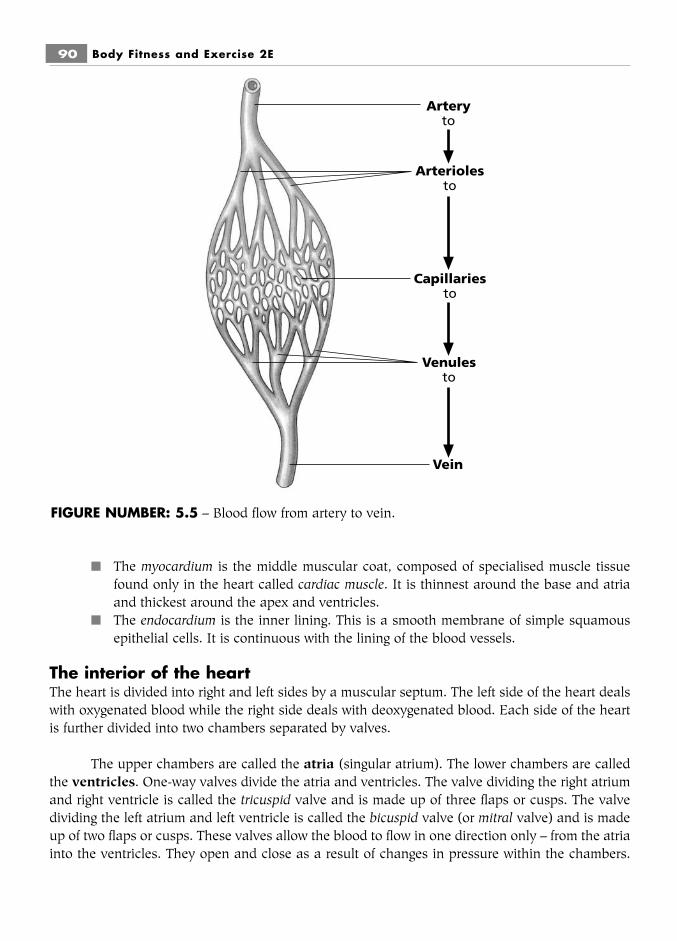
90 Body Fitness and Exercise 2E
■ The myocardium is the middle muscular coat, composed of specialised muscle tissuefound only in the heart called cardiac muscle. It is thinnest around the base and atriaand thickest around the apex and ventricles.
■ The endocardium is the inner lining. This is a smooth membrane of simple squamousepithelial cells. It is continuous with the lining of the blood vessels.
The interior of the heartThe heart is divided into right and left sides by a muscular septum. The left side of the heart dealswith oxygenated blood while the right side deals with deoxygenated blood. Each side of the heartis further divided into two chambers separated by valves.
The upper chambers are called the atria (singular atrium). The lower chambers are calledthe ventricles. One-way valves divide the atria and ventricles. The valve dividing the right atriumand right ventricle is called the tricuspid valve and is made up of three flaps or cusps. The valvedividing the left atrium and left ventricle is called the bicuspid valve (or mitral valve) and is madeup of two flaps or cusps. These valves allow the blood to flow in one direction only – from the atriainto the ventricles. They open and close as a result of changes in pressure within the chambers.
Arteryto
Arteriolesto
Capillariesto
Venulesto
Vein
FIGURE NUMBER: 5.5 – Blood flow from artery to vein.
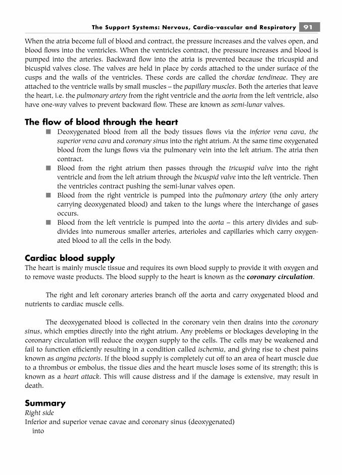
When the atria become full of blood and contract, the pressure increases and the valves open, andblood flows into the ventricles. When the ventricles contract, the pressure increases and blood ispumped into the arteries. Backward flow into the atria is prevented because the tricuspid andbicuspid valves close. The valves are held in place by cords attached to the under surface of thecusps and the walls of the ventricles. These cords are called the chordae tendineae. They areattached to the ventricle walls by small muscles – the papillary muscles. Both the arteries that leavethe heart, i.e. the pulmonary artery from the right ventricle and the aorta from the left ventricle, alsohave one-way valves to prevent backward flow. These are known as semi-lunar valves.
The flow of blood through the heart■ Deoxygenated blood from all the body tissues flows via the inferior vena cava, the
superior vena cava and coronary sinus into the right atrium. At the same time oxygenatedblood from the lungs flows via the pulmonary vein into the left atrium. The atria thencontract.
■ Blood from the right atrium then passes through the tricuspid valve into the rightventricle and from the left atrium through the bicuspid valve into the left ventricle. Thenthe ventricles contract pushing the semi-lunar valves open.
■ Blood from the right ventricle is pumped into the pulmonary artery (the only arterycarrying deoxygenated blood) and taken to the lungs where the interchange of gasesoccurs.
■ Blood from the left ventricle is pumped into the aorta – this artery divides and sub-divides into numerous smaller arteries, arterioles and capillaries which carry oxygen-ated blood to all the cells in the body.
Cardiac blood supplyThe heart is mainly muscle tissue and requires its own blood supply to provide it with oxygen andto remove waste products. The blood supply to the heart is known as the coronary circulation.
The right and left coronary arteries branch off the aorta and carry oxygenated blood andnutrients to cardiac muscle cells.
The deoxygenated blood is collected in the coronary vein then drains into the coronarysinus, which empties directly into the right atrium. Any problems or blockages developing in thecoronary circulation will reduce the oxygen supply to the cells. The cells may be weakened andfail to function efficiently resulting in a condition called ischemia, and giving rise to chest painsknown as angina pectoris. If the blood supply is completely cut off to an area of heart muscle dueto a thrombus or embolus, the tissue dies and the heart muscle loses some of its strength; this isknown as a heart attack. This will cause distress and if the damage is extensive, may result indeath.
SummaryRight sideInferior and superior venae cavae and coronary sinus (deoxygenated)
into
The Support Systems: Nervous, Cardio-vascular and Respiratory 91
right atrium then through tricuspid valveinto
Right ventricleinto
Pulmonary artery – to lungs
Left sidePulmonary vein from lungs (oxygenated)
intoLeft atrium then through bicuspid valve
intoLeft ventricle
intothe aorta and via branching arteries on to tissue cells.
Blood vesselsThese are tubes through which blood flows. There are three types of blood vessels, namely arter-ies, veins and capillaries.
ArteriesTransport blood away from the heart. Arteries carry oxygenated blood, nutrients, etc., to the cells,with the exception of the pulmonary artery, which carries deoxygenated blood from the heart to thelungs. Artery walls are composed of three layers of tissue. These layers surround a hollow corecalled a lumen.
The outer layer is fibrous tissue with elastin and collagen.
The middle layer is smooth muscular tissue.
The inner layer is simple squamous epithelium called endothelium.
As the heart pumps blood into the arteries, they expand to accommodate the blood, then,as the heart relaxes, the artery walls recoil propelling the blood forwards.
Arteries leaving and near the heart are large, but these branch and become smaller untilthey become small arterioles with thinner walls.
VeinsVeins transport blood back to the heart. The walls of the veins are similar in structure to those ofthe arteries, except that the outer layer is thicker and the middle muscular layer is thinner. Theinner layer endothelium folds to form valves. These valves prevent the backward flow of blood. Thelumen of the veins is larger. Veins carry deoxygenated blood and waste products from cells backto the heart, except for the pulmonary vein, which carries oxygenated blood from the lungs to the
92 Body Fitness and Exercise 2E

heart. Blood flows back from the cells to the right side of the heart and is known as venous return.Venous return is helped along the veins by the contraction and relaxation of muscles and by theexpansion and contraction of the thorax and diaphragm during breathing; these actions are knownas ‘the muscle pump’ and ‘the respiratory pump’. If the muscles are not contracting, e.g. duringlong periods of standing or inactivity, gravity exerts a downward force on the blood. If the valvesare weak this pressure overloads the vein and the wall bulges outwards causing the conditionknown as ‘varicose veins’. If exercise stops suddenly blood pools in the legs depleting the supplyto the brain which can result in dizziness and fainting. The very small veins at the capillary endare known as venules.
CapillariesThese are tiny vessels connecting arterioles and venules. The walls of the capillaries are very thin,composed of a single layer of squamous epithelium (endothelium). Capillaries form networksamong tissue cells with arterioles leading into them and venules leading away. The primary func-tion of capillaries is to allow exchange of gases, nutrients and waste products between the cellsand the blood. When metabolic needs are low, part of the network can shut off and blood flowsthrough a small portion only of the capillary network. When metabolic needs are high, the net-works dilate increasing blood flow. As activity increases, there will be an increase in demand fornutrients and oxygen by the contracting muscles, hence the blood vessels and capillaries in themuscles dilate but at the same time the vessels and capillaries in other organs such as the stom-ach, kidneys and liver constrict, so that extra blood is delivered to the muscles. This is known asblood shunting.
BloodBlood is a liquid connective tissue which performs many functions. It is a viscous (slightly sticky)fluid which flows through the heart and blood vessels. Its temperature is around 38°C and its pHis around 7.4 (slightly alkaline). The total volume of blood in the human body is around 51⁄2 litres(5–6 litres in men and 4–5 litres in women).
It is composed of a faintly yellow transparent fluid known as plasma which contains dis-solved substances and different types of cells.
■ 55 percent of the total volume is plasma.■ 45 percent of the total volume is cells.
PlasmaA straw-coloured transparent fluid composed of approximately 91 percent water, 7 percent proteinsand 2 percent other solutes.
Substances found in plasma:1 Plasma proteins: albumin, globulin, fibrinogen and prothrombin (important for blood
clotting).2 Mineral salts (electrolytes): sodium, potassium, magnesium, phosphorus, calcium, etc.3 Dissolved foods (nutrients): amino acids, fatty acids, glycerol glucose and vitamins.
The Support Systems: Nervous, Cardio-vascular and Respiratory 93

4 Hormones produced by endocrine glands.5 Enzymes secreted by specialised cells.6 Gases: oxygen, carbon dioxide, etc.7 Antibodies and antitoxins.8 Waste products of metabolism: urea, uric acid, etc.
Blood cellsThere are 3 main types of cells in blood:
1 Erythrocytes – red blood cells; these contain haemoglobin, whose function is the trans-portation of oxygen from the lungs to body cells and carbon dioxide from cells to thelungs.
2 Leucocytes – white blood cells; there are many types. They are phagocytic cells i.e. theydestroy and ingest micro-organisms thus protecting the body against infection anddisease.
3 Platelets or throbocytes – these play an important role in preventing blood loss. Theyinitiate a chain of reactions resulting in blood clotting.
Functions of blood1 It transports substances around the body.
i Oxygen from the lungs to body cells, mainly by combining with haemoglobin in theblood, to form oxyhaemoglobin. The concentration of haemoglobin in the bloodincreases through endurance training.
ii Carbon dioxide from cells to the lungs: some is transported by combining withhaemoglobin but most is transported in the form of bicarbonate ions.
iii Nutrients from digestive tract to body cells.iv Metabolic waste products from cells to excretory organs.v Hormones from endocrine glands to the cells.vi Any drugs taken for medicinal purposes.
2 It regulates:i The water content of cells.ii Body heat, maintaining normal body temperature.iii pH by means of buffers.
3 It protectsi Against disease and infection through lencocytes, destroying micro-organisms,
phagocytic action and production of antibodies.ii Against blood loss by the process of blood clotting.
Conduction system of the heartThe beating of the heart is under the influence of the autonomic nervous system which controlsthe strength and rate of contraction. The sympathetic nervous system increases heart rate and theparasympathetic slows it down. These are regulated by the cardio-acceleratory centre and thecardio-inhibitory centres of the medulla which respond to variations in blood pressure, to the con-centration of chemicals in the blood and to temperature variations.
94 Body Fitness and Exercise 2E
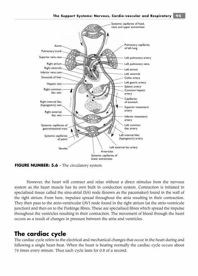
The Support Systems: Nervous, Cardio-vascular and Respiratory 95
However, the heart will contract and relax without a direct stimulus from the nervoussystem as the heart muscle has its own built in conduction system. Contraction is initiated inspecialised tissue called the sino-atrial (SA) node (known as the pacemaker) found in the wall ofthe right atrium. From here, impulses spread throughout the atria resulting in their contraction.They then pass to the atrio-ventricular (AV) node found in the right atrium (at the atrio-ventriculejunction) and then on to the Purkinge fibres. These are specialised fibres which spread the impulsethroughout the ventricles resulting in their contraction. The movement of blood through the heartoccurs as a result of changes in pressure between the atria and ventricles.
The cardiac cycleThe cardiac cycle refers to the electrical and mechanical changes that occur in the heart during andfollowing a single heart beat. When the heart is beating normally the cardiac cycle occurs about74 times every minute. Thus each cycle lasts for 0.8 of a second.
Pulmonary capillariesof left lung
Left pulmonary artery
Left pulmonary veins
Left atrium
Left ventricleCeliac artery
Left gastric arterySplenic arteryCommon hepaticartery
Capillariesof stomach
Superior mesentericartery
Inferior mesentericartery
Left commoniliac artery
Left internal liliac(hypogastric) artery
Left external liac arteryArterioles
Systemic capillaries oflower extremities
Venules
Systemic capillariesof pelvis
Systemic capillaries ofgastrointestinal tract
Right externaliliac vein
Right internal iliac(hypogastric) vein
Hepatic vein
Sinusoids of liver
Inferior vena cava
Right ventricleRight atrium
Superior vena cava
Pulmonary trunk
Aorta
Systemic capillaries of head,neck, and upper extremities
Right commoniliac vein
FIGURE NUMBER: 5.6 – The circulatory system.
96 Body Fitness and Exercise 2E
■ The term systole is used for the contraction time.■ The term diastole is used for the relaxation time.1 Atrial systole – contraction of the atria for 0.1 of a second.2 Atrial diastole – relaxation of the atria for 0.7 of a second.3 Ventricular systole – contraction of the ventricles for 0.3 of a second.4 Ventricular diastole – relaxation of the ventricles of 0.5 of a second.
(The sum of atrial and ventricular systole is 0.4 of a second therefore the heart is totallyrelaxed for 0.4 of a second.)
The superior and inferior vena cavae pour blood into the right atrium; at the same time thepulmonary veins pour blood into the left atrium. The SA node sends an impulse to contract and theatria contract for 0.1 of a second and blood is pushed through the valves into the ventricles. The AVnode sends an impulse to contract and the ventricles contract for 0.3 of a second. Blood is pushedby the right ventricle into the pulmonary artery and by the left ventricle into the aorta. After contrac-tion of the ventricles the heart rests for 0.4 of a second then the cycle begins again.
Cardiac outputCardiac output is the amount of blood ejected per minute from the left ventricle of the heart. It isa product of stroke volume and heart rate.
Stroke volume is the amount of blood ejected by the left ventricle during one contraction orsystole (average stroke volume in the adult is around 70 ml).
Heart rate is the number of beats/minute (average is around 74 beats/min).
Cardiac output is calculated as follows:cardiac output � stroke volume � heart rate (number of beats/min)
Therefore using average valuescardiac output � 70 ml � 74/min
� 5180 ml/min or 5.2 litres/min.
Blood pressureThis is the force or pressure which the blood exerts on the walls of the blood vessels in which it iscontained. The blood pressure in the arteries is higher than that in the veins. The arterial blood pres-sure is the result of the left ventricle pushing blood into the aorta. This pressure is known as systol-ic blood pressure and is found to be around 120 mmHg (millimetres of mercury). During completecardiac diastole, i.e. the heart resting, the blood pressure falls to around 80 mmHg. These figures varydepending on the individual, the degree of activity and the time of day – it tends to be lower at rest.
It is measured using a SPHYGMOMANOMETER and expressed BP � mmHg.120
80

Factors affecting normal blood pressure (BP)1 Cardiac output: the amount of blood ejected by the left ventricle into the aorta affects
blood pressure. An increase in cardiac output raises blood pressure while a decrease incardiac output lowers blood pressure.
2 Volume of blood: enough blood must be circulating through the system (normallyaround 51⁄2 litres) to maintain normal BP. Blood loss during haemorrhage reduces quan-tity of blood flowing in the system and blood pressure drops. If the blood volumeincreases the blood pressure increases.
3 Peripheral resistance: BP varies with constriction and dilation of the arterioles knownas vaso-dilation and vasoconstriction. Constriction of arterioles raises BP, dilation ofarterioles lowers BP (remember this happens when the body is heated).
4 Elasticity of the arterial walls – the amount of stretch and recoil of the arterial wallsaffects BP.
The pulse rateThe pulse rate is the same as the heart rate, being around 74 beats/min. The pulse can be feltbecause of the expansion and elastic recoil of the arteries during each ventricular systole (contrac-tion). The pulse is strongest in the arteries closest to the heart. The pulse is usually taken at theradial artery at the wrist but can be taken at the carotid artery in the neck or the brachial artery,medial to the biceps muscle.
The clotting or coagulation of bloodWhen a blood vessel is damaged blood will escape. To prevent blood loss the body reacts by amechanism called clotting or coagulation of blood. It is a very complex process. Certain substancesmust be present for clotting to occur. They are:
Prothrombin These three substancesCalcium are always presentFibrinogen } in the blood.andThromboplastin which is only released from damaged thrombocytes when injury occurs.
Stages of blood clottingWhen bleeding starts the release of Thromboplastin triggers a series of reactions which end withthe formation of a blood clot. In a simplified form the stages are as follows:
Prothrombin acted on by Thromboplastin in the presence of calcium will be converted intoThrombin. Thrombin then acts upon Fibrinogen which is converted into threads of fibrin. Thesefibrin threads form a mesh which together with trapped blood cells result in a blockage or clotwhich stops further bleeding. After a time the clot shrinks and dries as serum is released andhealing takes place. (The drug Heparin is an anti-coagulant which prevents prothrombin beingconverted to thrombin.)
The Support Systems: Nervous, Cardio-vascular and Respiratory 97

Cardio-vascular response to exerciseAs previously discussed in chapter 4, muscle contraction is dependent on ATP, which is the energysource for all cellular activity. Once exercise begins, the small quantity of ATP stored in musclecells is quickly used up. If contraction continues, ATP must be resynthesised using glycogen fromliver stores and oxygen from the lungs. These are transported to the muscles via the blood. Hencethe cardio-vascular system must quickly respond to this increased demand for oxygen and glyco-gen by the contracting muscles and the following changes occur.
■ The heart rate increases dramatically, from around 80 beats/min to 180 beats/min.■ The heart pumps out a larger volume of blood per beat (stroke volume).■ The cardiac output therefore increases greatly, from around 5–6 L/min at rest to around
30 L/min during maximal activity.■ Changes in blood pressure depend on the type of exercises. During aerobic exercises,
systolic pressure increases as a result of cardiac output, but diastolic pressure remainsconstant. During anaerobic and isometric exercises, both systolic and diastolic pressurerises, due to pressure on, and resistance of, the blood vessels.
■ Blood flow to the muscles and other organs changes once exercise commences. At rest,only 15–20 percent of cardiac output is transported to skeletal muscle. However, duringintense exercise this may go up to around 80 percent of cardiac output as blood is redi-rected from the other organs such as the stomach, liver and kidneys to the exercisingmuscles. This is controlled by vasoconstriction and vasodilation of the arterioles and isknown as blood shunting.
After a period of training there will be further adaptations:■ The heart increases in size and volume.■ The muscular wall of the heart becomes thicker.■ More blood flows into the left ventricle; this contracts more forcefully pumping a larger
volume of blood around the body.■ The resting heart rate decreases: because the heart pumps out more blood with each
beat, fewer beats per minute are necessary. Endurance athletes may have heart rates aslow as 40 beats per minute.
■ There is an increase in the size and number of blood vessels and in the density of cap-illary networks in the heart and in the muscles. This improves the delivery of oxygenand nutrients and speeds up the removal of waste.
■ There is an increase in the haemoglobin content of the blood which increases its oxy-gen carrying capacity.
The functions of the blood during exercise are:■ To transport oxygen from the lungs to the contracting muscles■ To transport carbon dioxide from the muscles to the lungs■ To transport lactic acid from the muscles to the liver■ To transport glucose from the liver stores to the muscles■ To regulate body temperature by transporting heat to the surface■ To maintain body homeostasis, i.e. body equilibrium.
98 Body Fitness and Exercise 2E
The Support Systems: Nervous, Cardio-vascular and Respiratory 99
Summary and aid to learningThe function of the cardio-vascular system is the transportation of substances around the body.
The cardio-vascular system is composed of the heart, arteries, arterioles, capillaries, veins, venulesand blood.
The heart has four chambers: right and left atrium, right and left ventricles.
The right side of the heart deals with deoxygenated blood.
The left side deals with oxygenated blood.
StructureThe wall of the heart is made up of three coats or layers.
1 Pericardium – tough outer coat of fibrous tissue lined with serous membrane, lubricatedfor smooth movement.
2 Myocardium – middle muscular coat, special muscle tissue called cardiac muscle.3 Endocardium – inner smooth coat of squamous epithelium.
The heart tissue is supplied with blood via the coronary circulation.
Valves of the heart1 Tricuspid valve has three flaps and divides the right atrium and right ventricle.2 Bicuspid (Mital) valve has two flaps and divides the left atrium and left ventricle.3 Semi-lunar valves are found at the openings of the arteries, the aorta on the left and the
pulmonary artery on the right.
Blood Vessels of the HeartInferior and superior venae cavae and coronary sinus enter the right atrium (deoxygenated).
Pulmonary vein enters the left atrium (oxygenated).
Pulmonary artery leaves the right ventricle (deoxygenated).
Aorta leaves the left ventricle (oxygenated).
Venous returnThis is the deoxygenated blood carried from the tissues back to the heart. It is aided by the musclepump – contraction of muscles, and the respiratory pump – movement of thorax and contractionof diaphragm.
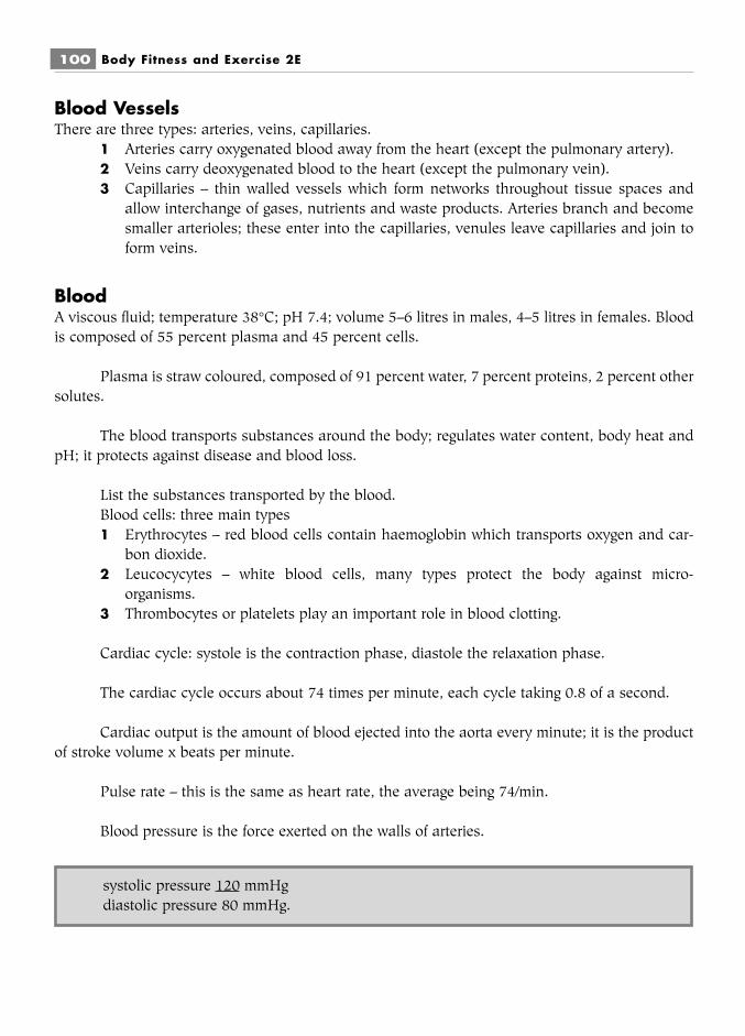
100 Body Fitness and Exercise 2E
Blood VesselsThere are three types: arteries, veins, capillaries.
1 Arteries carry oxygenated blood away from the heart (except the pulmonary artery).2 Veins carry deoxygenated blood to the heart (except the pulmonary vein).3 Capillaries – thin walled vessels which form networks throughout tissue spaces and
allow interchange of gases, nutrients and waste products. Arteries branch and becomesmaller arterioles; these enter into the capillaries, venules leave capillaries and join toform veins.
BloodA viscous fluid; temperature 38°C; pH 7.4; volume 5–6 litres in males, 4–5 litres in females. Bloodis composed of 55 percent plasma and 45 percent cells.
Plasma is straw coloured, composed of 91 percent water, 7 percent proteins, 2 percent othersolutes.
The blood transports substances around the body; regulates water content, body heat andpH; it protects against disease and blood loss.
List the substances transported by the blood.Blood cells: three main types1 Erythrocytes – red blood cells contain haemoglobin which transports oxygen and car-
bon dioxide.2 Leucocycytes – white blood cells, many types protect the body against micro-
organisms.3 Thrombocytes or platelets play an important role in blood clotting.
Cardiac cycle: systole is the contraction phase, diastole the relaxation phase.
The cardiac cycle occurs about 74 times per minute, each cycle taking 0.8 of a second.
Cardiac output is the amount of blood ejected into the aorta every minute; it is the productof stroke volume x beats per minute.
Pulse rate – this is the same as heart rate, the average being 74/min.
Blood pressure is the force exerted on the walls of arteries.
systolic pressure 120 mmHgdiastolic pressure 80 mmHg.

Blood clottingThromboplastin released from damaged throbocytes acts on Prothrombin in the presence ofcalcium, converting it to Thrombin; this acts on fibrinogen converting it into threads of fibrin. Thesethreads form a mesh with released blood cells forming a scab/clot which covers and seals thewound, preventing further blood loss.
Main circulatory routes are:1 The Systemic or General circulation to the body, heart and brain.2 The Pulmonary circulation to the lungs.3 The Portal circulation to the liver.
The heart plays a major role during exercise because the cardio-vascular system must increasethe delivery of oxygen to the contracting muscles and increase the speed of removal of wasteproducts. In response to regular exercise the heart will improve in strength and function.
Study the text and write a list of the changes and improvements found in the cardio-vascular system following regular training.
THE RESPIRATORY SYSTEMThis system is responsible for the exchange of oxygen and carbon dioxide between the externalenvironment and the internal environment of the body. It is closely linked with the cardio-vascularsystem as the exchange of gases takes place between the alveoli of the lungs and the blood in thepulmonary capillaries.
The system is composed of:■ The nose and nasal passages■ The pharynx■ The larynx■ The trachea■ The bronchi and bronchioles■ The lungs which are composed of alveoli.
The noseThe nose serves as the first section of the passageway for air going into the lungs. It is also theorgan of smell as the olfactory receptors are located in the nose. The inner lining of the nose is aciliated mucous membrane with a rich supply of blood vessels. It has two functions:
1 To filter the air as it enters the system, trapping organisms and dust particles preventingtheir entry into the lungs.
2 To moisten and warm the air as it passes through.
The Support Systems: Nervous, Cardio-vascular and Respiratory 101

102 Body Fitness and Exercise 2E
The pharynxBoth the respiratory and digestive tracts share the pharynx as both air and food pass through thispassageway. The tonsils are located here.
The larynxThis is the voice box which lies between the pharynx and the trachea. It is composed of cartilagesand smooth muscle tissue. The larynx plays a part in respiration, speech and swallowing. Air
Air is taken into the bodythrough the mouth andnasal cavity. In the nostrils,the air is filtered by tinyhair (cilia), warmed, andmoistened by mucus.
At the top of the throat is aflap of skin, the epiglottis,which prevents food or otherparticles entering the lungs.
Air passes through the larynx,which is sometimes known asthe voice box. Sound is producedby passing air over the vocalchords in the larynx.
The trachea is a large,flexible but strong tube,also known as the windpipe.Rings of cartilage maintainits shape.
The trachea branchesinto two to enter thelungs as bronchi.
The bronchus in eachlung divides intobronchioles.
Bronchioles sub-divide into smallair sacs, alveoli. Most of the lungtissue is made up of millions ofalveoli, which is where the exchangeof oxygen into the blood and carbondioxide out of the blood occurs.
Lung
The alveoli are covered with very small vessels,capillaries, which allow oxygen and carbondioxide to pass to and from the blood stream.
Vein
Venule
Arteriole
ArteryAlveoli
FIGURE NUMBER: 5.7 – The respiratory system.

passes through the larynx to the trachea; the passage of air over the vocal chords causes them tovibrate, producing sound. During the swallowing of food the larynx protects the airway as it isdrawn up, shutting off the airway to prevent food entering.
The tracheaThis is a tube about 4.5 inches long and 1 inch in diameter which extends from the larynx to thebronchi. It is composed of smooth muscle with C-shaped bands of cartilage at regular intervalsalong its length. These cartilagenous bands prevent the walls of the trachea from collapsing inwards.The function of the trachea is to maintain a permanently open pathway to the lungs. Any obstruc-tion of this vital airway, even for a few minutes, will result in asphyxia (suffocation) and death.
The bronchiThe trachea eventually divides into two primary bronchi.
The right bronchus leads to the right lung.
The left bronchus leads to the left lung.
In structure, each bronchus is similar to the trachea, being composed of smooth musclewith C-shaped rings of cartilage at intervals along its length. As the bronchi enter the lungs theyfurther subdivide into smaller secondary bronchi and then into even smaller bronchioles. Thebronchioles further divide into minute tubes called alveolar ducts which terminate in sponge-likesacs called alveoli.
The lungsThe left and right lungs are cone-shaped organs which extend from their base on the diaphragmbelow, to their apex above the clavicles. They lie within the thoracic cavity protected by the ribsand the sternum. The left lung is composed of two lobes and the right lung is composed of threelobes. They are covered by the visceral pleura containing serous fluid which reduces friction andfacilitates movement of the lungs during breathing. The lungs are composed of the tubes of thebronchial tree and the numerous sponge-like alveoli. The alveoli are surrounded by dense capillarynetworks. The function of the lungs is to provide a large surface area where the inspired air cancome into close contact with the blood thus facilitating the rapid exchange of gases. This exchangetakes place across the alveoli-pulmonary capillary interface. Oxygen, from inspired air, diffusesthrough the walls of the alveoli, through the walls of the surrounding capillary networks, into theblood to be transported around the body, to the tissue cells. Carbon dioxide from tissue cells iscarried via the blood to the lungs. Here it diffuses through the capillary walls then in through thewalls of the alveoli to be expired out of the lungs. This gaseous exchange is regulated by the partialpressure of the gases in the alveolar air and in the pulmonary blood. The gases diffuse across thegradient from high pressure to low pressure until equilibrium is reached. (Diffusion is the move-ment of molecules across a permeable membrane.)
The Support Systems: Nervous, Cardio-vascular and Respiratory 103

VentilationVentilation is the movement of air in and out of the lungs. As explained previously, ventilation isbrought about by the contraction of the skeletal muscles which expand the thorax. The musclesinvolved are the diaphragm and the intercostal muscles which lie between the ribs. Ventilation iscomposed of two phases, inspiration, taking air into the lungs and expiration, expelling air out ofthe lungs.
During inspiration the external intercostal muscles contract, swinging the ribs outwardsand upwards, rather like a bucket handle, and the sternum is pushed forward. This increases thethoracic cavity from side to side and from front to back. At the same time the diaphragm contractsto increase the cavity longitudinally. This increases the volume of the thorax and reduces the pres-sure within to below atmospheric pressure, consequently air rushes into the lungs. During normalexpiration these muscles relax, the thoracic cavity returns to its normal size, the pressureincreases to above atmospheric pressure and air rushes out. During forced respiration the inter-nal intercostals contract pulling the ribs in and down, the diaphragm moves upwards, forcing airout.
Minute ventilation is the amount of air inspired (Vi) or expired (Ve) in one minute.
This will depend on how much air we breathe in per breath (at rest this is around400–600 mL) and the number of breaths per minute (at rest 10–25 breaths/min).
Definitions of lung volume and capacities■ Pulmonary ventilation is the movement of air in and out of the lungs.■ Tidal Volume (Vt) is the volume of air inhaled or exhaled in one breath.■ Minute ventilation (VE) is the amount of air we exhale in one minute. At rest this is
between 6–15 L/min but during maximal exercise this can increase to between 145–200L/min. These increases are the result of an increase in the depth and rate of breathing.
■ Vital capacity (VC) is the maximum volume forcefully expired after maximal inspiration.■ Residual volume (RV) is the volume remaining in the lung at the end of maximal expira-
tion.■ Total lung capacity (TLC) is the volume of air in the lung at the end of maximal inspi-
ration.
Respiratory response to exercise■ The respiratory and the cardiovascular systems closely interact to take in and transport
oxygen to the contracting muscles and to remove carbon dioxide, a waste product ofaerobic activity, from the muscles to the lungs. During exercise ventilation may be15–30 times greater than at rest.
■ When exercise commences there is a rapid rise in ventilation which may increase froma resting level of 6–15 L/min up to 145–200 L/min. This increase is possible because of:increased rate of breathing, and increased depth of breathing.
104 Body Fitness and Exercise 2E

The initial rapid rise will tend to level off during steady state sub maximal exercise but willcontinue to rise during maximal effort.
■ Improved strength and condition of the muscles of respiration.■ Improved elasticity and recoil property of lung tissue.■ Improved diffusion capacity across the alveolar–pulmonary capillary interface.■ An increase in the blood supply to and from the lungs.
Summary and aid to learningThe function of the respiratory system is the exchange of gases between the human body and theenvironment.
The system is composed of: the nose, pharynx, larynx, trachea, bronchi, bronchioles andthe lungs made up of alveoli.
As air passes in through the nose it is warmed and filtered. It is further warmed as it passesthrough the pharynx and larynx.
The trachea divides into two primary bronchi: the right bronchus to the right lung and theleft bronchus to the left lung.
The bronchi subdivide as they enter the lungs into smaller bronchi which further divide intobrochioles. These end as small ducts which enter the alveoli.
There are two lungs containing the branches and alveoli.
How many lobes are there to each lung?
The inspired air must be brought into close contact with the blood for the exchange of gasesto take place.
Study the text and state where the exchange takes place. Name the main gas that movesout of the lung into the blood and name the main gas that passes out of the blood into the lungsto be exhaled.
Ventilation is the movement of air in and out of the lungs. Inspiration is the taking of air intothe lungs, in other words breathing in. Expiration is expelling air out of the lungs, i.e. breathing out.
The muscles of respiration contract to expand the chest during inspiration; they relax duringexpiration. Name the muscles of respiration.
Minute ventilation is the amount of air inspired or expired in one minute.
This will depend on the number of breaths per minute and the amount of air taken in perbreath. Study the list of lung volume and capacities.
The Support Systems: Nervous, Cardio-vascular and Respiratory 105

The respiratory system must work harder and faster during exercise to take in increasedoxygen and to eliminate carbon dioxide.
Discuss the improvements that can be expected following a period of training.
106 Body Fitness and Exercise 2E
QUESTIONS1. Name the three main parts that make up the nervous system.2. Give the function of each type of neurone.3. Complete the following:
a Axons carry impulses … .b Dendrites carry impulses … .
4. Name the chemical transmitter that transports an impulse across a synaptic gap.5. Explain the function of proprioceptors.6. Name the sensory receptor which relays information regarding the degree of stretch
within a muscle.7. Discuss the function of the cardio-vascular system during exercise.8. Describe the pathway of oxygenated blood after it leaves the lungs.9. Outline how the gases oxygen and carbon dioxide are transported in the blood.
10. Explain the term ‘blood shunting’ and outline why it occurs.11. Explain the term venous return. How is it aided back to the heart.12. Define the following:
a Stroke volumeb Heart ratec Cardiac output.
13. Explain how each of these is affected by exercise.14. Discuss the effects on the heart of a period of long term training.15. Explain the interaction between the cardio-vascular and respiratory systems during
exercise.16. Describe how oxygen passes from the external environment to the muscles.17. Define the terms:
a Tidal volumeb Vital capacityc Total lung capacity.
18. Discuss how ‘minute ventilation’ increases from rest to maximal exercise and givevalues.
19. List the improvements found in the respiratory system following a period of training.
Chapter 6Muscle work
Muscles work to produce or control movement at joints. When a muscle is working, tension forcebuilds up within the muscle and the muscle may shorten, lengthen or remain the same lengthdepending on the action required.
The muscle may be required to move a part, to control the effect of an external force or tohold a specific static position.
ISOTONIC, ISOMETRIC AND ISOKINETIC WORKMuscle work or contraction is classified into isotonic and isometric and recently defined isokinetic.
Isotonic contraction – equal tone; as force is generated, the muscle changes in lengththroughout the movement but the tone remains the same. The muscle may shorten, when the workis known as concentric work, or the muscle may lengthen, when the work is known as eccentricwork.
In practice it is not possible for the tone to remain the same throughout the full range ofmovement because of the difference in the angle of pull across the range. The degree of tensionwill vary with the position of the joint. For example, when performing a biceps curl with a weightin the hand, the tension developed will vary depending on the joint angle. The greatest tension isdeveloped just before the angle of the elbow approaches a right angle. In fact, it has been shownthat tension is maximal at 120 degrees; it decreases to the weakest point at 30 degrees. It followsthat a muscle is only as strong as its weakest point which will limit the maximum weight that can

be lifted. The muscle would only generate maximal tension at its weakest point, it will not be con-tracting maximally through the rest of the range. This is an obvious disadvantage to the athlete whorequires strength throughout the range. Machines have now been developed to overcome thisproblem as explained below.
Isometric contraction – equal length; the length of the muscle does not change but thereis an increase in tone. This is also known as static work.
Isokinetic contraction – equal speed; this is maximal contraction at constant speedthroughout the range. Machines have been developed which adjust automatically to providemaximal resistance and constant speed throughout the range. This is of great benefit to athletes,as strength and speed gives power which is a major factor in peak performance. Although the liftwill be a concentric contraction as in isotonic work, the tension will be maximal throughout therange.
Definitions and ExamplesIsotonic work may be concentric or eccentric:
■ Concentric work (isotonic shortening) – a muscle working concentrically shortens andthickens, the origin and insertion move towards each other and movement is producedin the joint.For example, in bending the elbow to lift a weight, the elbow flexors shorten to flex theelbow.
■ Eccentric work (isotonic lengthening) – a muscle working eccentrically becomes longerand thinner as the origin and insertion move away from each other. The muscle paysout gradually to control the movement produced by some external force such as gravity,springs, etc.For example, when lowering a bucket of water to the ground, the elbow flexors lengthenand pay out gradually to lower the bucket smoothly downwards. If these musclesstopped working the bucket would drop rapidly due to the force of gravity.
Isometric work is also known as static work.■ A muscle working statically does not change in length, but there is an increase in muscle
tone. The origin and insertion do not move and there is no joint movement.For example, when holding a bucket of water above the ground, the elbow flexors haveto increase in tone to maintain the position, but there is no movement at the elbow joint.
108 Body Fitness and Exercise 2E
LEARNIsotonic – the muscle changes in length; in concentric contraction the muscleshortens; in eccentric contraction the muscle lengthensIsometric or static workNo change in muscle length but there is an increase in toneIsokinetic – maximal contraction at constant speed throughout the range.
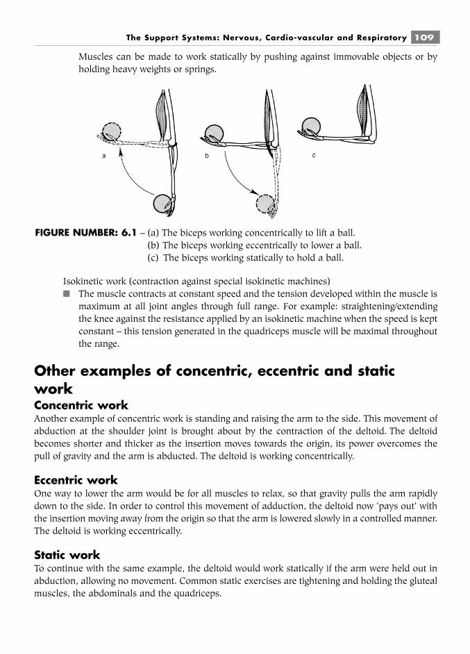
Muscles can be made to work statically by pushing against immovable objects or byholding heavy weights or springs.
Isokinetic work (contraction against special isokinetic machines)■ The muscle contracts at constant speed and the tension developed within the muscle is
maximum at all joint angles through full range. For example: straightening/extendingthe knee against the resistance applied by an isokinetic machine when the speed is keptconstant – this tension generated in the quadriceps muscle will be maximal throughoutthe range.
Other examples of concentric, eccentric and staticworkConcentric workAnother example of concentric work is standing and raising the arm to the side. This movement ofabduction at the shoulder joint is brought about by the contraction of the deltoid. The deltoidbecomes shorter and thicker as the insertion moves towards the origin, its power overcomes thepull of gravity and the arm is abducted. The deltoid is working concentrically.
Eccentric workOne way to lower the arm would be for all muscles to relax, so that gravity pulls the arm rapidlydown to the side. In order to control this movement of adduction, the deltoid now ‘pays out’ withthe insertion moving away from the origin so that the arm is lowered slowly in a controlled manner.The deltoid is working eccentrically.
Static workTo continue with the same example, the deltoid would work statically if the arm were held out inabduction, allowing no movement. Common static exercises are tightening and holding the glutealmuscles, the abdominals and the quadriceps.
The Support Systems: Nervous, Cardio-vascular and Respiratory 109
FIGURE NUMBER: 6.1 – (a) The biceps working concentrically to lift a ball.(b) The biceps working eccentrically to lower a ball.(c) The biceps working statically to hold a ball.

Isokinetic workThe movement of the arms through the water during freestyle swimming.
The uses of muscle workConcentric muscle work is the usual method used for muscle strengthening, although eccentric andstatic work should also be included. Maximal strength gains will not be achieved throughout therange.
Eccentric muscle work can sometimes be easier to perform and is useful when re-educatingmuscles if they cannot perform concentric work. Eccentric work in full and outer range (see below)maintains flexibility. Muscle soreness is greatest following eccentric contraction.
Static work is easy to perform, but muscle fatigue develops quickly. This is because the con-stant compression on the blood vessels and capillary networks impedes the blood flow, thus reduc-ing the delivery of oxygen and nutrients and the removal of waste products.
Static work increases the blood pressure and should not therefore be performed by thosewith heart and blood pressure problems.
Static work should be practised for short periods, with frequent rest intervals. It is alsoimportant to perform static holding at different points throughout the muscle range.
Isokinetic work is the most effective work for strengthening muscles as maximal resistanceis applied throughout the range which will result in equal improvement throughout the range.Muscle soreness is least following isokenetic contact.
RANGE OF MOVEMENTWhen muscles contract they move the joint through a certain range.
There are four ranges that a muscle or joint can work through:1 Full range – from full stretch to full contraction, or vice versa.2 Outer range – from full stretch to the mid-point of contraction, or vice versa.3 Inner range – from the mid-point to full contraction, or vice versa.
110 Body Fitness and Exercise 2E
TASKWork with a partner.While one of you performs a movement, the other should try to identify the type ofmuscle work, for example standing, swing the right leg out sideways. The abductorsare working concentrically.
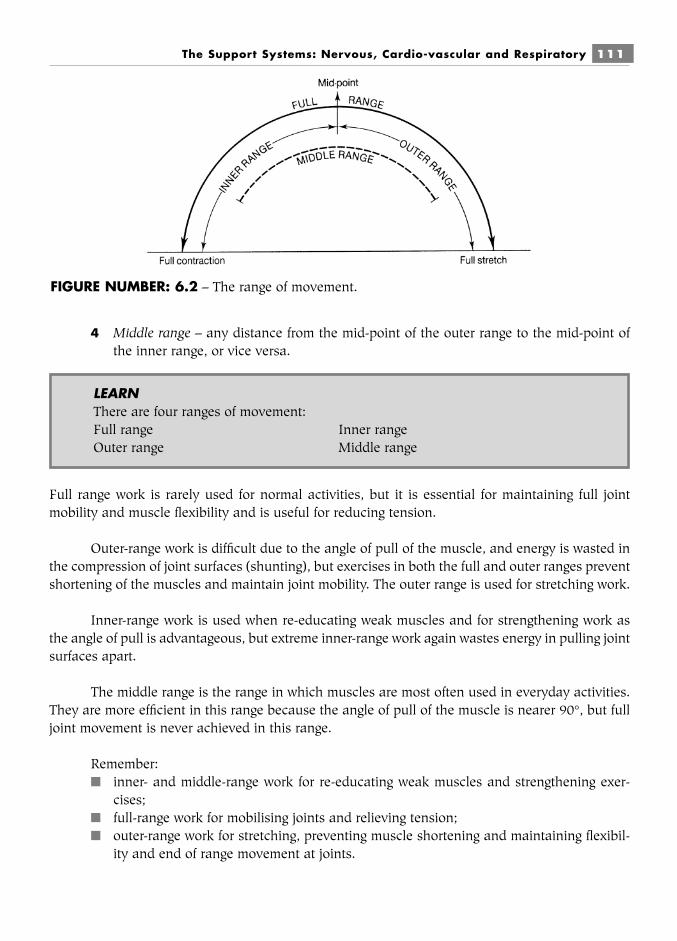
The Support Systems: Nervous, Cardio-vascular and Respiratory 111
4 Middle range – any distance from the mid-point of the outer range to the mid-point ofthe inner range, or vice versa.
Full range work is rarely used for normal activities, but it is essential for maintaining full jointmobility and muscle flexibility and is useful for reducing tension.
Outer-range work is difficult due to the angle of pull of the muscle, and energy is wasted inthe compression of joint surfaces (shunting), but exercises in both the full and outer ranges preventshortening of the muscles and maintain joint mobility. The outer range is used for stretching work.
Inner-range work is used when re-educating weak muscles and for strengthening work asthe angle of pull is advantageous, but extreme inner-range work again wastes energy in pulling jointsurfaces apart.
The middle range is the range in which muscles are most often used in everyday activities.They are more efficient in this range because the angle of pull of the muscle is nearer 90°, but fulljoint movement is never achieved in this range.
Remember:■ inner- and middle-range work for re-educating weak muscles and strengthening exer-
cises;■ full-range work for mobilising joints and relieving tension;■ outer-range work for stretching, preventing muscle shortening and maintaining flexibil-
ity and end of range movement at joints.
FIGURE NUMBER: 6.2 – The range of movement.
LEARNThere are four ranges of movement:Full range Inner rangeOuter range Middle range

THE GROUP ACTION OF MUSCLES
When muscles contract to produce movement, they work in groups. Each member of the group hasa particular role to play, rather like the members of an orchestra. They work together in a syn-chronised manner to produce smooth, co-ordinated, efficient movement.
There are four different members, which are named according to their function. They are theagonists or prime movers, the antagonists, the synergists and the fixators:
■ The agonists or prime movers are the muscles that contract to produce the requiredmovement (prime action).
For example, abduction of the hip joint is produced by the abductors; therefore thegluteus medius, the gluteus minimus and the tensor fasciae latae are the agonists orprime movers.
■ The antagonists lie on the opposite side of the joint from the agonists. They are theopposite group, which must relax and lengthen in a controlled manner so that the move-ment produced by the agonists is performed smoothly.
For example, when the abductors are contracting to abduct the hip the oppositegroup, the adductors, must relax. Therefore the adductors magnus, longus, brevis,pectineus and gracilis are the antagonists.
■ The synergists assist the prime movers to produce the most efficient movement. Theymay alter the angle of pull of the prime mover or prevent unwanted movement.
For example, during abduction of the hip the deep hip muscles will prevent the hiprotating, so that maximum effort is put into abduction. Therefore the piriformis and theobturator muscles are the synergists.
■ The fixators ensure that the prime movers act from a fixed base. They stabilise andprevent unnecessary movements in surrounding joints.
For example, during abduction of the hip joint the pelvis is held steady. Therefore thetrunk side flexors and abductors of the opposite side are the fixators.
112 Body Fitness and Exercise 2E
LEARNThere are four different muscles or groups working when movement occurs. They are■ prime movers (agonists) – these produce the movement■ antagonists are the opposite group which relax■ synergists – these help the prime movers■ fixators – these fix or stabilise other joints
LEARNMuscles acting on a joint are arranged around a joint. Those with opposite actionslie on opposite sides of the joint.Flexors opposite extensorsAbductors opposite adductorsMedial rotators opposite lateral rotators

The agonists and antagonists are the most vital members of the group and requireidentification when analysing muscle work. When the agonists are contracting to produce move-ment, the antagonists must relax to allow the movement to take place. This is known as reciprocalrelaxation and can be used as a technique for stretching muscles (see chapter 9). It is sufficient toremember that synergists and fixators are also contributing to the movement, as their identificationis frequently difficult.
The muscles acting on a joint are arranged around the joint. Some are superficial whileothers are deep. The agonists and antagonists are arranged as opposite pairs – flexors oppositeextensors, abductors opposite adductors, medial rotators opposite lateral rotators. When theflexors are the agonists, the extensors will be the antagonists and vice versa. Other smaller muscleswill be the synergists and fixators. The patterns of movement are synchronised in the motor cortexand the appropriate impulses are conveyed to the muscles via their motor nerves.
The balance between agonists and antagonists is very important as tightness and shorten-ing or over-stretching and weakness of one group will affect the function of the other. A muscleimbalance will be produced, resulting in stresses on the underlying joints and ligaments which mayadversely affect performance. These stresses may also result in deformity and pain. Exercises mustalways be planned to maintain a balance between agonists and antagonists.
ANALYSIS OF MUSCLE WORKAll exercise schemes require careful planning to ensure that the set objectives are realised. For cor-rective schemes, the exercises must be carefully planned to target specific muscles or groups: somewill require strengthening, while others will require stretching. For general schemes, all the mainmuscle groups must be included and balance maintained between opposing muscles.
Planning exercise schemes therefore requires the ability to analyse muscle work. First of all,the starting position must be considered, as this determines the effect of gravity on the movement.This is followed by identification of the moving joint, the direction of movement and the musclesproducing that movement. Then we consider the type of muscle work and the range of movement.
To analyse muscle work follow this procedure:1 Give the starting position.2 Name the moving joint, e.g. hip, shoulder, etc.3 Name the direction of movement, e.g. flexion, abduction, etc.4 Name the prime movers, i.e. the muscles producing the movement.5 Name the type of muscle work, i.e. concentric, eccentric or static.6 Name the range of movement, i.e. inner, outer, middle or full.
The type of muscle work poses the most difficult problem to most students. Remember:■ If the muscle is shortening and the origin and insertion are moving nearer to each other,
the work is concentric.
The Support Systems: Nervous, Cardio-vascular and Respiratory 113

■ If the movement is produced by an external force such as gravity, weights or springs andthe muscle is lengthening and paying out to control the movement, so that the originand insertion are moving away from each other, the work is eccentric.
■ If the muscle is contracting but producing no movement at the joint, the work is static.
Exercises performed in different starting positions will have different muscle work, for exampleabduction and adduction of the hip joint performed in different positions, as shown below.
Position: lying supineMovement: part legsMoving joint: hipDirection of movement: abductionPrime movers: the abductors (gluteus medius,
minimus and tensor fasciae latae)Muscle work: concentric (muscle shortens producing the movement)Range: inner
Movement: close legsMoving joint: hipDirection of movement: adductionPrime movers: adductors (adductors longus, magnus, brevis, pectineus, gra-
cilis)
114 Body Fitness and Exercise 2E
LEARNTo work out which muscle or group is working, ask yourself the following questions:What is the starting position?Which joint is moving?What is the direction of movement?Which muscles are producing that movement?Which type of muscle work is it?(is the muscle shortening, lengthening or staying the same length)?Which range is it moving through?
FIGURE NUMBER: 6.3a

Muscle work: concentricRange: outer
When the legs are opened and closed in the lying position gravity does not affect the movement,since gravitational pull is downwards and this movement is in the horizontal plane.
Both the abductors and adductors work concentrically.
Now if we change the starting position but do the same movement the muscle workchanges.
Position: lying on sideMovement: upper leg raiseMoving joint: hip jointDirection of movement: abductionPrime movers: abductors (as before)Muscle work: concentric (muscle shortens)Range: inner
(This movement is against the pull of gravity.)Movement: lowering legMoving joint: hip jointDirection of movement: adductionPrime movers: abductors (because gravity will pull the leg down)Muscle work: eccentricRange: inner
Gravity pulls the leg down, therefore the adductors do not need to work, but the abductors workeccentrically to prevent the leg from falling.
Thus in this starting position the muscle work changes: only the abductors work, first con-centrically and then eccentrically to produce controlled movement and counteract the pull ofgravity.
If we change the starting position yet again, the muscle work will change again.Position: lying with legs at right angles to trunkMovement: part legs
The Support Systems: Nervous, Cardio-vascular and Respiratory 115
FIGURE NUMBER: 6.3b
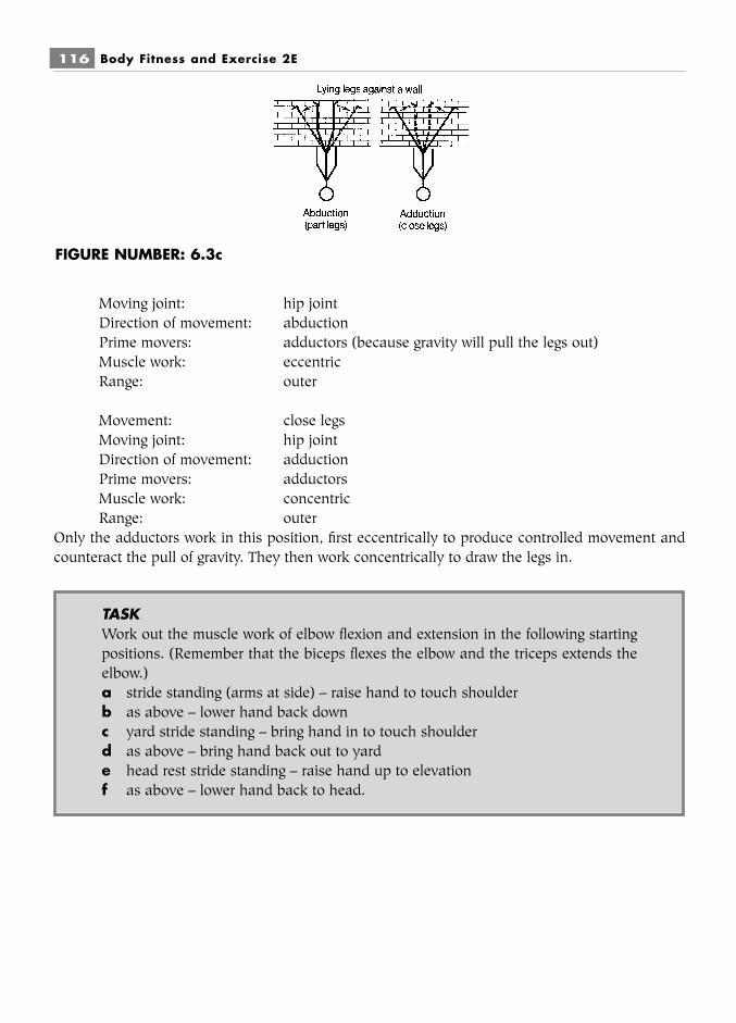
116 Body Fitness and Exercise 2E
Moving joint: hip jointDirection of movement: abductionPrime movers: adductors (because gravity will pull the legs out)Muscle work: eccentricRange: outer
Movement: close legsMoving joint: hip jointDirection of movement: adductionPrime movers: adductorsMuscle work: concentricRange: outer
Only the adductors work in this position, first eccentrically to produce controlled movement andcounteract the pull of gravity. They then work concentrically to draw the legs in.
FIGURE NUMBER: 6.3c
TASKWork out the muscle work of elbow flexion and extension in the following startingpositions. (Remember that the biceps flexes the elbow and the triceps extends theelbow.)a stride standing (arms at side) – raise hand to touch shoulderb as above – lower hand back downc yard stride standing – bring hand in to touch shoulderd as above – bring hand back out to yarde head rest stride standing – raise hand up to elevationf as above – lower hand back to head.
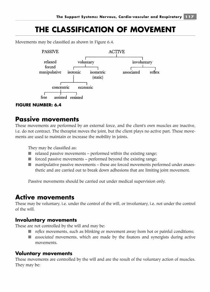
THE CLASSIFICATION OF MOVEMENTMovements may be classified as shown in Figure 6.4.
Passive movementsThese movements are performed by an external force, and the client’s own muscles are inactive,i.e. do not contract. The therapist moves the joint, but the client plays no active part. These move-ments are used to maintain or increase the mobility in joints.
They may be classified as:■ relaxed passive movements – performed within the existing range;■ forced passive movements – performed beyond the existing range;■ manipulative passive movements – these are forced movements performed under anaes-
thetic and are carried out to break down adhesions that are limiting joint movement.
Passive movements should be carried out under medical supervision only.
Active movementsThese may be voluntary, i.e. under the control of the will, or involuntary, i.e. not under the controlof the will.
Involuntary movementsThese are not controlled by the will and may be:
■ reflex movements, such as blinking or movement away from hot or painful conditions;■ associated movements, which are made by the fixators and synergists during active
movements.
Voluntary movementsThese movements are controlled by the will and are the result of the voluntary action of muscles.They may be:
The Support Systems: Nervous, Cardio-vascular and Respiratory 117
FIGURE NUMBER: 6.4
■ isometric (static), where the muscles do not change in length but increase in tone andno movement is produced at the joint;
■ isotonic, where the muscles change in length and produce movement at the joints.Isotonic movements may be concentric (muscle shortening) or eccentric (muscle length-ening).
Concentric movements may be further subdivided into assisted, free and resisted:■ Assisted active exercise – when muscle power is inadequate to produce a desired move-
ment, its power can be helped by the use of an external force acting with the musclepull. The movement is thus assisted.
■ Free active exercise is movement where the working muscles are subjected only to theforces of gravity acting upon the part being moved.
■ Resisted active exercise is movement where the action of the muscles is resisted by anexternal force, e.g. weights, springs, etc. This resistance acts against the muscle pull. Itcan be increased progressively to develop muscle power and endurance.
Summary and aid to learningThere are three basic types of muscle contraction:
Isotonic, isometric and isokinetic.
Isotonic contraction can be subdivided into:concentric contraction – when the muscle shortens, the origin and insertion come closer
together.eccentric contraction – when the muscle lengthens, the origin and insertion move away from
each other, as the muscle pays out slowly to control any external forces acting on it.
Isometric contraction is also known as static contraction. The muscle develops tensionbut there is no change in length.
Isokinetic contraction – this is recently defined as maximum contraction at constantspeed. Machines have been developed to keep the speed constant throughout the range. It has anadvantage over isotonic contraction as strength gains will be equal throughout the entire range.
Work with a partner and perform concentric, eccentric and static contractions for differentmuscles in different starting positions.
When a muscle contracts it moves and moves the joint through a certain distance. This isknown as ‘the range of movement’. There are four ranges of movement: full, middle, inner and
118 Body Fitness and Exercise 2E
LEARNMovement may be■ passive: these are performed by someone else or some extended force■ active: these are performed by the person’s own muscles contracting

outer. If you think about these words they explain the range. Explain and demonstrate each rangeto a partner, using the biceps muscle.
Muscles must work as a group in order to perform smooth coordinated movement. Themuscle which contracts to produce the movement is called the prime mover or agonist. The muscleon the other side of the joint, i.e. opposite the prime mover, must relax slowly to allow smoothmovement to take place; this is called the antagonist. There are muscles that help the prime movercalled synergists. Other muscles hold adjacent joints steady; these are called fixators.
When analysing muscle work you must give the starting position and identify:■ the moving joint■ the direction of movement, e.g. flexion, etc.■ the prime mover■ the type of muscle work, e.g. concentric, etc.■ the range of movement, e.g. middle, etc.
Practise this analysis with a partner; choose any movement and consider each of the above.
Passive movement is produced by some external force it requires no muscle contraction.Active movement is produced by muscle contraction.
The Support Systems: Nervous, Cardio-vascular and Respiratory 119
QUESTIONS1. Name the three main types of muscle work.2. Define the terms concentric work and eccentric work and give one example of each.3. Explain the four ranges of movement.4. Name each member which contributes to the group action of muscles.5. Define the terms:
a prime mover (agonist)b antagonist.
6. List the points to consider when analysing muscle work.7. Discuss the advantage of isokinetic contraction compared with isotonic contraction.8. Analyse the muscle work of the following actions carried out in stride-standing
position:a raise the arm out to the side to shoulder levelb lower the arm back to the side.Now analyse these movements when the body is lying supine.
9. Define the terms active movement and passive movement.
Chapter 7Physical principles relating
to exerciseThe science or study of body movement is known as kinesiology. As previously explained, move-ment is normally produced by muscles acting or pulling on bones, resulting in movement at joints.These movements are affected and governed by certain scientific principles. These need to beunderstood in order to identify muscle work and to devise effective and appropriate exerciseschemes.
FORCEA force is that which changes the state of rest or motion of an object. Any force acting on the bodywill make it move or affect its movement. When muscles contract they exert a force, which, ifstrong enough, will produce movement at the joint, for example the biceps muscle must contractwith sufficient force to lift the forearm and bend the elbow. The power of the muscle must be greatenough to overcome the resistance of any force pulling the other way, in other words the muscleforce must be greater than the resisting force for movement to occur.
There are various external forces that can be used to resist movement, such as the pull ofgravity or the use of weights, springs, pulley systems, multigyms, etc. Muscles are strengthenedwhen they are made to work against progressively increasing forces.

Certain postural muscles are continually working against the force of gravity to maintainposture. If these muscles relaxed, the body would fall to the ground.
GravityThis is the force that attracts or pulls everything towards the ground. It is a continual pull in adownward direction.
Gravitational pull affects most body movement and must be considered when planningexercises. Movements performed downwards (with gravity) will use different muscle work fromthose performed upwards (against gravity). Movements in the sagittal and frontal planes areaffected by gravity, but movements in the horizontal plane are not.
Movements upwards, downwards or sideways will have different relationships with gravityand will require different muscle work. It is therefore very important to consider gravitational pulland to select appropriate starting positions when compiling exercise schemes (see chapter 8).
Anti-gravity musclesThe upright posture is maintained by particular muscle groups known as postural muscles or anti-gravity muscles. They must work continuously to oppose the pull of gravity and keep the bodyupright. Any weakness or imbalance of these muscles will affect body alignment and may result inpostural deformities.
Physical principles relating to exercise 121
FIGURE NUMBER: 7.1 – Postural muscles.

Regular exercise will maintain muscle strength and balance, thus preventing abnormal pos-tures. If deformities have developed, specific exercises must be practised to stretch the tightmuscles and strengthen weak muscles.
The anti-gravity muscles are the anterior tibials, the posterior tibials, the quadriceps, the hipextensors (gluteus maximus and hamstrings), the erector spinae, the abdominals, the trapeziusand the rhomboids, the neck extensors and the neck flexors.
The centre of gravityThis is an imaginary point at the centre of a body around which it is perfectly balanced. In thestanding position the centre of gravity of the human body lies approximately at the second sacralvertebra, but this will vary with the shape and weight distribution of the individual. More weighton the top half raises the centre of gravity, whereas bending the knees or kneeling will lower it.
The lower the centre of gravity the more stable the object will be, so that a person in thelying position is more stable than a person in the standing position.
Stability is an important consideration when planning exercise, as the more stable the bodyis the easier it is to perform an exercise.
The line of gravityThis is an imaginary line which falls perpendicularly (vertically) through the centre of gravity.When a person stands upright, the line of gravity passes through the vertex (top of the head),through the mid-cervical vertebrae, in front of the thoracic vertebrae, behind the bodies of thelumbar vertebrae, through the second sacral vertebra, slightly in front of the knee joint and in frontof the ankle joint, ending between the ball of the foot and the heel.
The line of gravity is a useful measure when examining posture.
When the body adopts the correct posture, a line in the same plane but lateral to the lineof gravity will fall through the lobe of the ear, the point of the shoulder (the acromion process), thehip joint, to the front of the knee joint (but behind the patella) and in front of the ankle joint, endingbetween the ball of the foot and the heel (see chapter 10). The line of gravity will not fall throughall these points if posture is incorrect and will move as the position of the body changes.
StabilityThe base of an object is that part that touches the ground. Any object with two or more feet on theground will have a base that includes the area of the feet and the area of the space in between. Thelarger the area of the base the greater the stability.
122 Body Fitness and Exercise 2E
LEARNThe lower the centre of gravity, the greater the stability
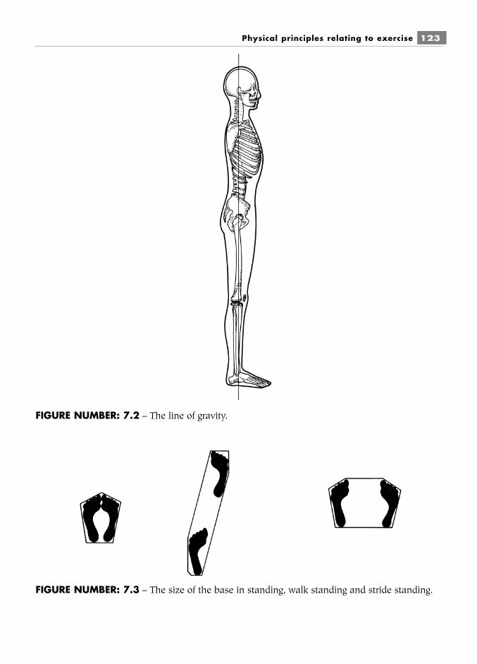
Physical principles relating to exercise 123
FIGURE NUMBER: 7.2 – The line of gravity.
FIGURE NUMBER: 7.3 – The size of the base in standing, walk standing and stride standing.

When a person sits on a chair, the base includes the feet of the person and the area betweenthe legs of the chair. When a person is lying down the area of the body surface on the ground isthe base. This gives great stability, as it is a large base with a low centre of gravity.
The stability of a body depends on the relationships between the centre of gravity and line ofgravity and the base. As has already been mentioned, the lower the centre of gravity, the morestable the body will be. When the line of gravity falls near the centre of the base, the body is stable.As the line of gravity moves towards the edge of the base the body becomes increasingly unstable.If the line of gravity falls outside the base, the body falls over.
When the base is small it is difficult for the line of gravity to remain within it and the bodyeasily falls over, but if the base becomes larger it is easier for the line of gravity to remain within itand the body is more stable. For example, if a person stands with the feet close together, the baseis relatively small, being the area of the feet alone. Therefore, if the body moves forward, sidewaysor backward, the line of gravity will easily fall outside the base and the body will fall over.
However, if a person stands with the feet apart, as in stride standing, the base is muchlarger, being the area in between the feet as well as the area of the feet. The line of gravity will nowstay within the base when the person leans over, and the body will not fall over as it is more stable.Arm and trunk movements are easier to perform when the body is more stable. Whenever the bodymoves into an unstable position, muscles are immediately brought into play to prevent the bodyfalling over. The smaller the base and the higher the centre of gravity, the greater the muscle powerand co-ordination needed to maintain the upright posture and the more difficult it will be toperform exercise. Other factors which increase stability are:
■ increased body mass;■ friction between the feet and the ground;■ focusing the vision on a stationary object.
Newton’s Laws of MotionAn understanding of these laws is useful when considering exercise, but in-depth study is notrequired.
Newton’s laws state:1 A body will continue in a state of rest or uniform motion in a straight line unless it is
acted on by a force.2 A change in acceleration of a body is directly proportional to the force and inversely pro-
portional to the mass.3 To every action there is an equal and opposite reaction.
124 Body Fitness and Exercise 2E
LEARNThe larger the base the greater the stability
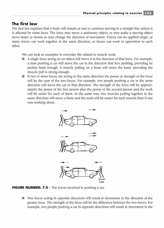
The first lawThe first law explains that a body will remain at rest or continue moving in a straight line unless itis affected by some force. The force may move a stationary object, or may make a moving objectmove faster or slower or may change the direction of movement. Forces can be applied singly, ormany forces can work together in the same direction, or forces can work in opposition to eachother.
We can look at examples in everyday life related to muscle work.■ A single force acting on an object will move it in the direction of that force. For example,
a man pushing a car will move the car in the direction that he’s pushing, providing hepushes hard enough. A muscle pulling on a bone will move the bone, providing themuscle pull is strong enough.
■ If two or more forces are acting in the same direction the power or strength of the forcewill be the sum of the two forces. For example, two people pushing a car in the samedirection will move the car in that direction. The strength of the force will be approxi-mately the power of the first person plus the power of the second person and the workwill be easier for each of them. In the same way, two muscles pulling together in thesame direction will move a bone and the work will be easier for each muscle than if onewas working alone.
■ Two forces acting in opposite directions will result in movement in the direction of thegreater force. The strength of the force will be the difference between the two forces. Forexample, two people pushing a car in opposite directions will result in movement in the
Physical principles relating to exercise 125
FIGURE NUMBER: 7.4 – The forces involved in pushing a car.

direction of the one pushing harder. Opposing muscles cannot contract together,because as the prime mover contracts the opposing antagonist always relaxes (this iscontrolled by nerve impulses and is known as reciprocal relaxation). However, musclescan be made to contract against external forces such as gravity, weights, springs, pulleysor machines. If the muscle force is greater than the external force, movement will occurin the direction of muscle pull. This principle is applied to improve muscle strength. Aweight is selected that the muscle is just strong enough to lift, and this weight is lifted aset number of times. As the muscle responds and strengthens a greater weight is usedand the procedure is repeated until the required strength is reached. If the muscle powerand the weight are equal there will be equilibrium, and therefore no movement. If theweight is greater than the muscle power, movement will occur towards the weight. If themuscle is forced to lift too great a weight the myofibrils may tear, damaging the muscle.
The second lawThe second law explains that an increase in speed will be directly proportional to the force, so thatthe greater the force, the greater the acceleration. It also depends on the mass: the greater the mass,the lower the acceleration.
For example, if two athletes of equal weight are pushing off from a starting block with equalforce, they will accelerate at the same speed. However, if one is much heavier than the other he orshe will accelerate more slowly, and will have to use greater force, i.e. muscle power, to producethe same acceleration.
The third lawThe third law explains that every action has an equal and opposite reaction. This is important inball games such as tennis and squash. The harder the ball is hit, the harder it hits the surface andthe harder it rebounds. The surface applies a resistance force against the force of the striking ball.The resistance force from hard surfaces is greater than that from softer surfaces, which absorbsome of the force. Hard court tennis is faster than grass court tennis, although the standard of playis equal. It is easier to run on hard surfaces than on soft surfaces, as there is a greater oppositereaction propelling one forward.
These opposing forces can cause problems for runners, as the constant jarring as the feethit the ground may cause repetitive stress injuries such as shin splints and spinal problems.
When exercising it is an advantage to work on a sprung floor, which dissipates impact forcesand reduces the risk of injury. Well-manufactured training footwear with cushioned soles shouldbe worn to dissipate these forces.
(Some students may require other physical laws and principles related to speed, velocityand so on. These are not within the scope of this book and specialist texts should be referred to.)
Skeletal muscles produce the forces required for mobility. They start and stop movement,they maintain movement, they change the speed of movement, they may accelerate or decelerate
126 Body Fitness and Exercise 2E

actions and they change the direction of movement. Considerably more force is required to startand stop movement and to change direction than to maintain movement in the same direction.
LEVERSLevers apply a force to produce movement about a point. Levers can be used to make work easieror harder. We are all familiar with the use of a lever to prise the lid off a tin of paint. When a coinis placed under the lid, and a force applied on the other side, the lid lifts up. If the coin does notwork, we use a spoon handle or some longer rigid bar, and this will lift the lid because it has greatermechanical advantage. The principles of leverage also apply to body movement.
A lever is a rigid bar which moves around a fixed point called a fulcrum.
A force or effort (E) applied at one point on the lever moves a second force or weight (W)applied at another point.
The distance between the effort and the fulcrum is known as the effort arm (EA) and thedistance from the weight to the fulcrum is known as the weight arm (WA).
A lever is balanced whenW � WA � E � EA
If the weight or length of the weight arm increases, the effort or length of the effort arm must alsoincrease. Later we will see how this relates to increasing the work done by muscles.
In the body:■ the rigid bar is the bone;■ the fulcrum is the joint;■ the effort is the pull of the muscle at its point of insertion;■ the weight is the part being moved.
There are three different classes or orders of levers. They are different because of the posi-tion of the fulcrum in relation to the effort and the weight.
Physical principles relating to exercise 127
FIGURE NUMBER: 7.5 – A lever.

First order or class (EFW)In the first order the fulcrum (F) lies between the effort (E) and the weight (W).
Here, the fulcrum may be nearer the weight, giving a longer effort arm, or may be nearer theeffort, giving a longer weight arm. When the effort arm is longer than the weight arm there ismechanical advantage. If the weight arm is longer than the effort arm there is mechanical disad-vantage.
We find examples of this first order in everyday life (e.g. a see-saw), but few in the body.
If weight � weight arm � effort � effort arm, then the see-saw is balanced, but when oneside is greater than the other the see-saw will move down at the end with the greater force. In thehuman body, during extension of the head the fulcrum lies at the cervical joints. The effort is sup-plied by the muscle pull at the point of insertion (upper fibres of trapezius) and the weight is thehead being moved. In order to move the head backwards, the muscle power and the distance fromthe fulcrum must be greater than the weight of the head and its distance from the fulcrum.
128 Body Fitness and Exercise 2E
FIGURE NUMBER: 7.6a – The first order of levers.
FIGURE NUMBER: 7.6b – Equilibrium on a see-saw.
FIGURE NUMBER: 7.6c – Extension of the head.

Second order or class (FWE)In this order the weight lies between the fulcrum and the effort.
Here, the effort arm will always be longer than the weight arm and consequently there willalways be mechanical advantage. This is a lever of power.
A wheelbarrow has a fulcrum at the wheel, the weight in the middle and the effort at thehandle. Because the effort arm is always longer than the weight arm, it is quite easy to lift a heavyload in a wheelbarrow.
When raising the heel off the ground, the fulcrum is at the metatarso-phalangeal joints, thebody weight falls down the leg to the ankle and the effort to lift the heel is from the plantar flexors(gastrocnemius and soleus) at their point of insertion.
Physical principles relating to exercise 129
FIGURE NUMBER: 7.7a – The second order of lever.
FIGURE NUMBER: 7.7b – Lifting a load in a wheelbarrow.
FIGURE NUMBER: 7.7c – Lifting the heel off the ground.
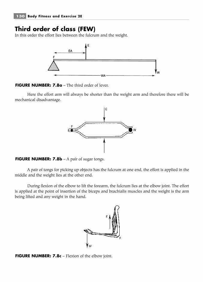
Third order of class (FEW)In this order the effort lies between the fulcrum and the weight.
Here the effort arm will always be shorter than the weight arm and therefore there will bemechanical disadvantage.
A pair of tongs for picking up objects has the fulcrum at one end, the effort is applied in themiddle and the weight lies at the other end.
During flexion of the elbow to lift the forearm, the fulcrum lies at the elbow joint. The effortis applied at the point of insertion of the biceps and brachialis muscles and the weight is the armbeing lifted and any weight in the hand.
130 Body Fitness and Exercise 2E
FIGURE NUMBER: 7.8a – The third order of lever.
FIGURE NUMBER: 7.8b – A pair of sugar tongs.
FIGURE NUMBER: 7.8c – Flexion of the elbow joint.

There is a larger number of third order levers in the body than any other. Although they arelevers of mechanical disadvantage, they allow for speed and a wide range of movement.
Leverage related to muscle workAs explained previously, there are examples of all three types of lever to be found in the body, butthere are far more of the third class, giving speed and a larger range of movement. When power isrequired we find the second class.
■ The fulcrum is the joint where movement is taking place.■ The effort is provided by the muscle power exerted when the muscle contracts.■ The effort arm is the distance from the joint to the point where the muscle inserts (this
cannot be changed).■ The weight is the part being moved, which can be increased by adding weight to the
part.■ The weight arm is the distance from the fulcrum to the end of the moving part, which
can be increased by adding length, such as a pole or dumb-bell.
Therefore we can increase the effort for the muscle by increasing the weight or lengtheningthe weight arm.
If muscle power (effort) � effort arm � weight � weight arm, everything is balanced and nomovement will occur.
If we increase the muscle power so that muscle power (effort � effort arm) is greater thanweight � weight arm, movement will occur.
If we then increase the weight or the length of the weight arm, greater muscle power will berequired to produce movement.
This principle is used to strengthen muscles. The weight is progressively increased and themuscle is made to lift it a set number of times, with the result that the muscle become stronger (seechapter 9).
Examples1 Increasing the work to strengthen abdominal muscles:
Crook lying:■ arms across chest, curl up (short weight arm)■ hands on ears, curl up (longer weight arm)■ arms stretched above head, curl up (longer weight arm)■ arms across chest holding weight, curl up (increased weight)■ arms stretched above head holding weight, curl up (increased weight arm and
weight).This progression continues by increasing the weight to be lifted. Once the muscle canlift the weight 10 to 15 times, the weight can be increased.
Physical principles relating to exercise 131
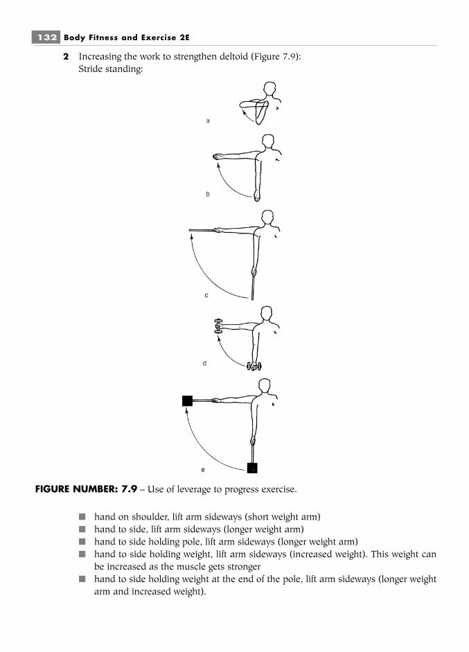
2 Increasing the work to strengthen deltoid (Figure 7.9):Stride standing:
■ hand on shoulder, lift arm sideways (short weight arm)■ hand to side, lift arm sideways (longer weight arm)■ hand to side holding pole, lift arm sideways (longer weight arm)■ hand to side holding weight, lift arm sideways (increased weight). This weight can
be increased as the muscle gets stronger■ hand to side holding weight at the end of the pole, lift arm sideways (longer weight
arm and increased weight).
132 Body Fitness and Exercise 2E
FIGURE NUMBER: 7.9 – Use of leverage to progress exercise.

Physical principles relating to exercise 133
SummaryA force is that which changes the state of rest or motion of an object.
When a muscle contracts it exerts a pulling force on the bone of attachment, making itmove.
Gravity is the force which pulls everything towards the ground.
Anti-gravity muscles are the muscles which contract to keep us upright. They work againstthe force of gravity; if they relaxed the body would fall to the ground.
Centre of gravity refers to the point at the centre of an object around which it is perfectly bal-anced.
The centre of gravity of the human body, in the standing position, is around the second sacralvertebra but this will vary with weight distribution. The lower the centre of gravity the more stablethe object.
The line of gravity falls perpendicularly through the centre of gravity.
The base of an object is that part that touches the ground; the larger the base the more stablethe object.
Explain how the base and the centre of gravity of an object affects its stability.
Levers apply a force to produce movement about a point.
A lever is a rigid bar which moves around a fixed point called a fulcrum. A force or effort isapplied at one point to move a second force or weight applied at another point.
List the parts of a lever related to body parts and movement.
There are three types/orders of levers which are different because of the position of thefulcrum in relation to the effort and weight.
Draw a diagram of each type of lever and give an example of where each is to be found inthe body.
TASKShow progression for the abductors of the hip joint in the side-lying position usingthe above five steps.

We can increase the effort for a muscle, that is make it work harder, by increasing the weightor lengthening the weight arm.
There are more type three levers in the body than any other. These are levers of speed.
134 Body Fitness and Exercise 2E
QUESTIONS1. Define the terms gravity and centre of gravity.2. Where is the approximate position of the centre of gravity in the human body?3. When assessing posture, list the points through which the line of gravity will fall.4. Complete the following:
The base of an object is that part which … .5. Give any three factors which influence the stability of a body.6. Explain why it is preferable to exercise on a sprung floor.7. Draw diagrams to illustrate the three classes of levers.8. Relate the parts of a lever to the human body.9. Give two ways in which leverage can be used to increase the resistance to muscle
work.10. Show two ways of using leverage to make the following exercise harder for the
gluteus maximus:prone lying, raise the leg off the floor, knee bent to right angle.
Chapter 8Starting positions
When writing out exercise schemes it is vital to state the starting position. This is easily done ifschemes are written in two columns: one for the starting position, the other giving instructions forthe exercise. The age and agility of the client will influence the choice of the starting position. Theolder or less agile client may well require a more stable starting position.
There are five basic starting positions:■ Lying (also known as supine lying)■ Kneeling■ Sitting■ Standing■ Hanging.
These basic positions can be modified to increase or reduce the difficulty of the exercise.
Modifications are made to:■ raise or lower the centre of gravity;■ increase or decrease the size of the base to change stability;■ increase or decrease leverage;■ provide adequate fixation of the body so that specific movements can be performed with
maximum concentration;■ increase or decrease the muscle work required to maintain the position;■ ensure maximum support for relaxation.

136 Body Fitness and Exercise 2E
Modifications of starting positions
■ Lying: prone lying, side lying, half lying, crook lying, crook lying with pelvis lifted.
■ Kneeling: prone kneeling, inclined prone kneeling, heel sitting, half kneeling.
FIGURE NUMBER: 8.1 – Modifications of lying.
FIGURE NUMBER: 8.2 – Modifications of kneeling.

Starting positions 137
■ Sitting: crook sitting, long sitting, astride sitting, side sitting, stoop sitting, fall out sitting.
■ Standing: toe standing, stride standing, walk standing, step standing, lax stoop stand-ing, stoop standing.
FIGURE NUMBER: 8.3 – Modifications of sitting.
FIGURE NUMBER: 8.4 – Modifications of standing.

■ Hanging: stride hanging, knee bend hanging.
The position of the arms is also very important and this is usually written first, e.g. bendstride standing.
■ Arm positions: wing, low wing, across bend, under bend, bend, reach, yard, stretch,head rest.
138 Body Fitness and Exercise 2E
FIGURE NUMBER: 8.5 – Modifications of hanging.
FIGURE NUMBER: 8.6 – Arm positions.
PART BTraining for Fitness

This page intentionally left blank

Introduction
Fitness may be defined as having sufficient energy and skill to cope in one’s environment. Fitnessis specific to the individual and ranges from the optimum fitness required by top athletes throughto the lowest level required barely to cope with daily tasks.
Fitness and health are interrelated, but it is important to distinguish between the two.Health may be defined as ‘freedom from disease’ or ‘a state of physical, mental and social well-being’; it is therefore possible to be healthy (free from disease) but unfit. It is also possible to besuperbly fit and compete at the highest level while suffering ill health. Many top athletes continuecompeting or training when suffering from colds, infections, and so on.
Improving fitness means improving the physiological functioning of the various bodysystems, which in turn will improve one’s capacity to function, resulting in an improved quality oflife. Improvement is gained by overloading the systems, in other words by making demands onthem over and above those required by normal activities, and continuing progressively to workthem harder.

Chapter 9The components of fitness
Before we can begin to improve fitness levels we must understand all the components that contrib-ute to overall fitness. They can be defined in the following way:
Cardio-respiratory endurance means improving the condition of the heart and lungs, thusimproving the delivery of nutrients and oxygen to the tissue cells.
Muscle strength means improving muscle maximum strength which will make activitieseasier.
Muscle endurance means improving the ability of muscles to contract for longer periods.
Flexibility means improving the suppleness of connective tissue components within themuscles which will improve extensibility and elasticity. It also improves the tensile strength andextensibility of ligaments and tendons. These factors will enable the tissues to withstand excessiveforces, reducing the likelihood of injury.
Skill refers to accurate performance with little or no waste of energy. It is largely a functionof the nervous system which initiates, controls and co-ordinates all activities.
Speed refers to the distance moved in a specific time. Some activities will require fast speedwhile others require slow control. Speed is an important factor in most sport because improvedspeed and strength will result in greater power.

Body composition refers to the ratio of body fat and fat-free tissue. It is dependent on energybalance which is the balance between energy consumed (the food eaten – calories in) and energyexpended, (the amount of activity undertaken – calories out).
Nutrition refers to the foods we eat and the role they play in sustaining life. Good nutritionmeans eating the required amount of a variety of foods which will enable the body to function atmaximum efficiency.
Rest and relaxation means allowing the body sufficient time to recover following activity. Themore intense the activity the longer the rest period should be. Many physiological improvementscontinue into the rest period. Adequate rest will prevent chronic fatigue.
Relaxation means freedom from stress and is related to rest and anxiety states. Stress adver-sly affects the function of many body systems. Consequently reducing stress factors will enable thebody to function more efficiently.
When we consider the concept of improving fitness it becomes obvious that this means dif-ferent things to different people. Fitness is specific to the individual and the required improvementdepends on the current level of fitness and desired fitness outcome. For an elderly person animprovement may be achieved simply through improved nutrition and a low level of increasedstrength and flexibility, while the peak performing athlete will require maximum levels of fitness inthose systems related to his/her activity. Therefore a training programme must target the compo-nents where improvement is required. The weight lifter will require the maximum possible strength,the sprinter will require strength in the ‘push off the block’ muscles and anaerobic capacity; themarathon runner will require aerobic capacity; the ballet dancer will require flexibility, strength and
The components of fitness 143
FIGURE NUMBER: 9.1 – Input necessary for fitness.

aerobic capacity. Most sports will require a combination of many components and it is importantto guard against overdevelopment of one at the expense of another. The most important questionsto consider before embarking on a training programme are:
What is the current level of fitness?
What is the desired outcome or goal?
PlanningThere are several important principles to consider when planning or embarking on any trainingprogramme.
SpecificityThis means that the exercise/training routine must be specific to those muscles or energy systemstargeted for conditioning, i.e. those used in the sport or performance. This is because trainingeffects are generally not transferable; strength training will not improve cardio-vascular enduranceor vice versa.
OverloadThis means that the appropriate systems are stressed beyond normal requirements. Overload mustalways be appropriate to the fitness level of the individual; too little will not produce any improve-ment, too much can cause damage. Overload may be adjusted by increasing or decreasing one orall of the following:
■ intensity: the degree of stress applied; it should be sufficient to produce some discom-fort
■ frequency: the frequency of the training sessions; the number of sessions per week;■ duration: the length of time of the exercise session or programme.
ProgressionThis means that the exercises/overload must become progressively harder for improvement andadaptation to be achieved. Progression must be steady and gradual.
Training thresholdThis refers to the minimum intensity of exercise required to bring about improvement, e.g. toimprove cardio-vascular endurance an individual must exercise at 60–80 percent of maximum heartrate (MHR) for 20–30 minutes, three times per week. 60 percent indicates the minimum intensity,this must be increased progressively as fitness develops. The very fit can work 90 percent MHR.
ReversibilityThis refers to the loss of training effect if an individual stops exercising or if the intensity of train-ing decreases. Training effect will regress within just two weeks and there will be considerable lossof fitness after a period of four to six weeks after training stops. The effects of short duration train-ing regress more quickly than the effects of long duration training.
144 Body Fitness and Exercise 2E

CARDIO-RESPIRATORY ENDURANCE(Cardio-vascular, cardio-pulmonary and aerobic endurance are all terms used for this aspect offitness). They all refer to the efficiency of the heart, circulation and lungs to take in, transport andtransfer oxygen to muscle tissue.
Before exercise begins, the body is in a balanced state known as homeostasis, where thesystems meet the body’s metabolic needs. When exercise begins, the systems must respond rapidlyto the increased demand for nutrients and particularly oxygen to produce ATP. If the demand (over-load) is regularly and progressively increased the systems will respond and their physiologicalfunctioning will improve. The capacity of the body to work aerobically will increase, i.e. to utilisethe aerobic energy system.
Cardio-respiratory endurance improves as a result of low intensity, long duration training,which will improve the capacity to utilise the aerobic energy system. If the activity becomes too fastand vigorous, the systems will be unable to supply oxygen fast enough to meet the demand and thework will become anaerobic and endurance will not improve. Short bursts of fast activity will improvethe capacity to utilise the anaerobic systems, i.e. ATP – PC system and the lactic acid system.
Training methods to improve cardio-respiratoryendurance (aerobic capacity)Cardio-respiratory endurance will improve in response to regular long duration, low intensityaerobic activities such as:
■ jogging/running■ swimming;■ walking;■ treadmill running■ cross-country skiing;■ any aerobic dance or exercise
programmes.
The components of fitness 145
LEARNEndurance training involves low intensity work over a period of time. It trains thebody to provide and utilise oxygen, thus increasing aerobic capacity
FIGURE NUMBER: 9.2

In order to maintain a training effect and improve fitness the work load must be graduallyand slowly increased. As previously explained the three variables to consider are:
■ the intensity of the exercise – how hard it is;■ the frequency of exercise – how often it is performed;■ the duration of exercise – how long the programme lasts.
IntensityHeart rate increases with exercise, therefore it is an excellent indicator of how hard the body isworking. The heart rate varies considerably between different people and in the same person underdifferent conditions. The heart rate is the same as the pulse rate and can be taken at pulse pointsthroughout the body. The usual point for reading pulse rate is at the wrist (the radial pulse point).Participants in any training programme must be taught to take his/her pulse rate (see Chapter 14).
■ The average male resting pulse is between 72–76 beats per minute.■ The average female resting pulse is between 76–80 beats per minute.
These are average values and considerable variations will be found. Fit people have a far lowerresting pulse rate than the unfit. Endurance athletes may have pulse rates as low as 30–40 beatsper minute.
Maximum heart rateTo obtain an estimate of maximum heart rate (MHR) deduct the client’s age from 220. For example,for a 40-year-old, maximum heart rate will be 220 � 40 � 180 beats per minute.
The pulse rate must not exceed this MHR during aerobic exercise as it will produce toomuch stress. Healthy adults should exercise at a target heart rate of 60–90 per cent of theirmaximal heart rate depending on their current level of fitness. Those who are unfit should exer-cise at the lower end while those who are already quite fit should exercise at the higher end oftheir maximum range. Initially effort should be 60 per cent of MHR. For a 40-year-old this will be:
146 Body Fitness and Exercise 2E
LEARNThe heart rate indicates the intensity at which a person is exercising
LEARNA person’s maximum heart rate is the maximum rate at which it is safe to exercise.
An unfit person should not exceed 60 percent of maximum heart rate but shouldincrease gradually up to 85–90 percent as fitness improves.
LEARNTo calculate a person’s maximum heart rate deduct his/her age from 220

180 � � 108 beats/min. (this is the target heart rate or training rate)
The client should exercise with sufficient intensity to maintain this pulse rate for 15–30 minutes’duration two to three times per week. After four to six weeks it will be safe to exercise at 70 percent of maximum heart rate providing there have been no problems.
180 � � 126 beats/min.
Eventually, this will build up to 80–85 per cent even 90 per cent of MHR
Therefore, for this clientAge MHR Target Zone40 180 108–161
DurationThis is the actual time that a person is exercising at the target heart rate. It does not include thewarm-up and cool-down periods. This part of an exercise programme will last for 15–30 minutes.Initially, after a warm-up and stretch of 15–20 minutes, the client will perform a 15-minute pro-gramme, maintaining the target heart rate, and then cool down. This time will gradually increaseto 30 minutes as fitness improves.
Duration is also used to describe the length of the training programme, e.g. 12–16 weeks.
FrequencyThis is the number of sessions per week, which may be two to three times a week for an unfit begin-ner, moving up to three to four times per week as fitness improves. Athletes who compete in endu-rance, activities train for 4–5 days/week or more.
70
100
60
100
The components of fitness 147
LEARNThe target heart rate is the rate at which the person should be exercising. It will bea percentage of maximum heart rate (60–90 percent).
Beginners Intermediate Advanced60–70% 70–80% 80–90%
These will increase as fitness develops
LEARNTo maintain or increase levels of fitness any one or any combination of the follow-ing must increase: intensity, duration, frequency
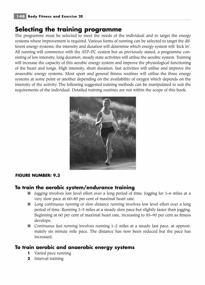
Selecting the training programmeThe programme must be selected to meet the needs of the individual and to target the energysystems where improvement is required. Various forms of running can be selected to target the dif-ferent energy systems; the intensity and duration will determine which energy system will ‘kick in’.All running will commence with the ATP–PC system but as previously stated, a programme con-sisting of low intensity, long duration, steady state activities will utilise the aerobic system. Trainingwill increase the capacity of this aerobic energy system and improve the physiological functioningof the heart and lungs. High intensity, short duration, fast activities will utilise and improve theanaerobic energy systems. Most sport and general fitness routines will utilise the three energysystems at some point or another depending on the availability of oxygen which depends on theintensity of the activity. The following suggested training methods can be manipulated to suit therequirements of the individual. Detailed training routines are not within the scope of this book.
To train the aerobic system/endurance training■ Jogging involves low level effort over a long period of time. Jogging for 3–6 miles at a
very slow pace at 60–80 per cent of maximal heart rate.■ Long continuous running or slow distance running involves low level effort over a long
period of time. Running 3–5 miles at a steady slow pace but slightly faster than jogging.Beginning at 60 per cent of maximal heart rate, increasing to 85–90 per cent as fitnessdevelops.
■ Continuous fast running involves running 1–2 miles at a steady fast pace, at approxi-mately six minute mile pace. The distance has now been reduced but the pace hasincreased.
To train aerobic and anaerobic energy systems1 Varied pace running2 Interval training
148 Body Fitness and Exercise 2E
FIGURE NUMBER: 9.3
3 Pick up sprint training■ Varied-pace running (Swedish Fartlek). This varies the pace at specific intervals through-
out the run. Continuous steady-pace running is interrupted at intervals by quick sprints.This will combine both anaerobic and aerobic energy systems and is a more realistictraining for most sports. It can be varied to use only aerobic systems if low effort is usedin all stages.
It may involve running over different terrain, through forests and fields, uphill anddownhill, on sand, gravel and grass.
Tracks are very carefully graded for their degree of difficulty. A programme mayinclude:1 10–15 minutes’ jogging;2 five minutes’ rapid walk;3 one mile slow distance running;4 ten minutes’ rapid walk;5 five sprints interspersed with jogging over 100 metres.
■ Interval training. This involves fast training interspersed with slow work, performed in acertain amount of time or over a measured distance. The slow or light work periodallows the oxygen debt incurred during the fast work to be repaid. This method willimprove both aerobic and anaerobic systems.
■ Pick-up sprint training. This increases the speed of work. It begins with walking, movingon to jogging, then striding and sprinting, and ending up with walking again. Theprocess is repeated as often as possible. This improves both aerobic and anaerobicsystems and trains athletes to pick up speed quickly.
To train the anaerobic systems■ Sprint training involves repeated sprints of 60–70 yards with complete recovery in
between.■ Hollow sprints involves sprinting interrupted by a recovery period of jogging or walking,
e.g. sprint 60 yards, jog 60 yards, walk 60 yards, repeat until fatigued.
■ Cross-training. Many athletes believe in and derive benefit from cross-training. Thiscombines different forms of training. For example, cycling may be combined with swim-ming: the swimming will not improve cycling prowess, but it will maintain endurancequalities while allowing the recovery of the cycling muscles. It is also more interestingand motivating to add variety rather than keep to one training mode.
The effects of endurance training
Immediate Response■ The heart rate increases from around 80 to 180 beats per minute.■ The heart pumps out a larger volume of blood per beat (stroke volume).■ An increase in cardiac output which is stroke volume × heart rate from around
6 L/minute at rest to 30 L/minute during exercise.
The components of fitness 149

■ The flow of blood to the contracting muscles is greatly increased as blood is shuntedfrom other organs such as the stomach.
■ The rate and depth of ventilation increases. During exercise the volume of air breathedin and out of the lungs may be 30 times greater than that breathed in at rest.
After a period of training there will be further effects■ The heart increases in size and volume.■ The efficiency of the heart improves: it pumps out a larger volume of blood per beat
(stroke volume) and therefore a larger quantity per minute (known as cardiac output).■ The heart rate decreases: the heart rests for a longer period, which reduces the workload
on the heart. Endurance athletes may have heart rates as low as 40 beats per minute.■ There is an increase in the size and number of blood vessels in the heart and skeletal
muscle.■ There is an increase in the size and density of capillary networks, supplying blood to
the heart and skeletal muscle, with improvement in the delivery of oxygen and theremoval of waste.
■ There is an increase in the haemoglobin content of the blood which increases its oxygencarrying capacity.
■ There is an increase in the size and number of mitochondria, which enables oxygen tobe used more efficiently.
■ There is an increase in glycogen stores and glycolytic enzymes.■ The aerobic capacity (VO2max) is increased.■ The anaerobic threshold is raised, so that aerobic metabolism is used for longer periods,
increasing the capacity to exercise without fatigue.■ The rate and depth of respiration increases, improving ventilation.■ Fats are utilised for energy production, which reduces body fat.■ Bones are strengthened in response to the stresses placed on them.
Dangers of Endurance Training■ Repetitive stress injuries, mainly of the ankles, knees and back, and shin splint injuries.■ Dehydration.
Precautions■ Perform adequate warm-up.■ Build up the training gradually. Do not exceed maximum heart rate (MHR). Begin at 60
per cent of MHR, working up to 80–90 per cent as fitness develops.■ Do not exercise if there is any pain present in musculoskeletal structures.■ Do not exercise if suffering from colds, fevers, flu, etc.■ Stop exercising if pain develops.■ Drink fluid after the training session or during long-distance running to prevent de-
hydration.■ Wear well-fitting, well-cushioned, appropriate footwear.■ Wear loose, absorbent clothing.■ Perform adequate cool-down.
150 Body Fitness and Exercise 2E
Improving muscle fitnessMuscle response to overload training will differ, depending on the type of training:
■ Muscle strength will increase in response to progressive overload over a period of time.Strength develops through high resistance with low repetitions.
■ Muscle bulk will increase with strength, but one or two lifts at maximal resistance mustbe performed to improve bulking.
■ Muscle endurance will increase in response to low resistance with high repetitions.■ Muscle power and explosive power will increase in response to plyometric training,
involving muscle contraction following a rapid stretch.
Muscle strengthThis refers to the maximal force a muscle can develop against resistance. It is measured by themaximum weight that can be lifted in a single effort. This is known as one repetition maximum(1 RM). It is established by trial and error, using increasingly heavy weights, until the maximum isreached, for example by performing one lift of 10 kg, one of 12 kg and one at 14 kg, etc., until themaximum weight that can be lifted is reached.
Overloading a muscle with a weight of over two-thirds of the muscle’s maximum load overa period of time will increase strength and bulk.
Initially, an increase in strength is the result of the recruitment of more motor units. Eachmotor unit stimulates a large number of muscle fibres to contract. Therefore, the more motor unitsrecruited, the more fibres will be contracting, which will increase strength. Further strength devel-ops as a result of an increase in the contractile proteins (myosin and actin) and in the size andnumber of myofibrils. These factors also increase muscle bulk.Muscles will strengthen if they are made to contract against progressively increasing resistance.
Resistance may be applied in many ways, but some of these methods are not measurable, and soaccurate progression is not possible. Muscles can be made to work against:
■ one’s own body weight, e.g. press-ups – not measurable;■ the resistance provided by a partner, e.g. pushing or pulling against a partner – not mea-
surable;■ water – not measurable;
The components of fitness 151
LEARNIn response to progressive overload, muscles respond and improve. Musclesbecome stronger and bigger. Muscle power and explosive power will also improve
LEARNMuscle strength is the maximal force a muscle can develop against resistance

■ specialised equipment, such as rowing machines, exercise bikes, multigyms;■ free weights, e.g. sand bags, weight boots, ankle and wrist weights, dumb-bells and bar
bells;■ springs and pulleys;■ weight machines are now very sophisticated and technologically advanced and can be
found in gyms and fitness centres. They are the most popular method of weight trainingfor athletes and fitness enthusiasts.
Strength training programmesProgrammes may be required to improve overall body strength or to target specific muscles orgroups related to sport or athletic performance. The following principles apply to any strength train-ing programme.
Weight Training Principles
OverloadMuscles must be overloaded to gain strength; resistance must exceed that normally encountered.The resistance should be 66 percent of 1 (RM) repetition maximum or over. This is the minimumoverload that will achieve a strength gain. If a muscle is underloaded, the present level of strengthwill be maintained but will not increase.
ProgressionThe overload must be progressive, it must increase as strength develops. As soon as the musclecan perform 2–4 extra lifts without fatigue, the load must be increased.
Arrangement of exerciseThe routines should be organised so that succesive exercises do not involve the same muscle orgroup of muscles. The routine must be planned to include different areas consecutively, i.e. thigh,chest, lower back, buttocks, calf, shoulders and upper back, abdomen, upper arms. This ensuresthat muscles have an adequate time to recover, preventing fatigue.
SpecificityIf strength is required for a specific sport or activity, the programme should be planned so that themuscles used in that sport or activity are exercised. If possible the exercise should simulate thenormal pattern of movement used in the sport to reinforce the pattern of movement in the brain.
152 Body Fitness and Exercise 2E
LEARNResistance may be applied to
Isotonic contraction � eccentricconcentric
Isometric contractionIsokinetic contraction

RecoveryTime should be allowed for recovery both between sets of lifts and between training days. Trainingthree times per week will allow adequate rest time but still achieve strength gains.
Planning strength programmesMuscle strengthening, otherwise known as weight training, usually involves concentric contrac-tions against resistance. However, as discussed in chapter 6 there are three other types of contrac-tion, namely eccentric, isometric and isokinetic contractions which can also be used.
Weight training programmes can be designed for each type of contraction as outlined below.
ISOTONIC strength training programmesThese exercises are dynamic and involve concentric muscle shortening and/or eccentric musclelengthening, which will result in joint movement.
Contraction is usually performed against weights, barbels or weight machines and uses therepetition maximum (RM) principle.
As the muscle contracts to lift the weight, the resistance is applied to concentric work. Thisis referred to as the positive phase and is the more effective phase for increasing strength and bulk.
Resistance is applied to eccentric work as the part is lowered back to the starting position.This is referred to as the negative phase.
The weight selected will depend on strength but must be over 66 percent of 1 RM.
The components of fitness 153
FIGURE NUMBER: 9.5 – (a) Weight training for biceps.(b) Weight training for latissimus dorsi.

As previously explained, repetition maximum is the maximum load that a muscle or groupof muscles can lift a set number of times.
One repetition maximum (1 RM) is the maximum load that a muscle or group can lift onlyonce before fatigue. Six repetition maximum (6 RM) is the maximum load that a muscle or groupcan lift six times before fatigue.
The contractions are organised into sets. One to five sets are repeated with a short pausebetween each set. Each set will consist of a certain number of contractions known as repetitions.A typical programme might be:
1 to 5 sets, of 6 to 10 contractions, 3–5 times per week for 6–10 weeks.
These factors can be manipulated to suit the individual and will depend on the rate at whichstrength gains have been achieved. Adequate rest is essential both between sets and between ses-sions to avoid chronic fatigue.
It is possible to use heavier loads for eccentric work thus gaining extra strength and bulk.This is favoured by very experienced lifters as part of their routine. The technique involves usingtwo assistants to lift a heavy load into position (slightly too heavy for the concentric phase), whichis then lowered slowly by the lifter.
Recording dataIt is very important to record accurate details of the programme and progress. Measurements ofbulk and strength taken at regular intervals under the same conditions will indicate progress. Thesemeasurements must be recorded before commencing the programme, and then every four weeks.Firstly:
■ measure bulk with a tape measure placed around the widest point; always measure atthe same point; record
■ measure strength by the maximum weight which can be lifted in a single lift (1 RM);record 1 RM
During each session, the following data must be recorded:■ the load (the weight that is lifted)■ the repetitions (the number of times the weight is lifted without a rest – it is usual to
select 6–10 lifts)■ the number of sets performed. Select 1–5 sets and allow 1 minute rest period between
each set. These are recorded as follows:10 kg � 8 reps � 3 sets
As strength develops, the weight or number of sets is increased.For improving strength, ‘heavy load low repetitions’ is the format: the weight lifted should be atleast two-thirds of the maximum possible weight – over 66 percent of 1 RM. Beginners should prac-tise initially with low weights until they have perfected the technique.
It is possible to vary the routines; the following are examples of effective but simple systems.
154 Body Fitness and Exercise 2E
EXAMPLE 1Establish 1 RM and calculate 80 percent of this, which will be the starting weight. If 1 RM � 25 kg,the weight will be 20 kg. This would be lifted 6–8 times and recorded as:
20 kg � 8 � 120 kg � 8 � 220 kg � 8 � 3
EXAMPLE 2Establish 10 RM (the weight that can be lifted 10 times). The first set of 10 lifts is performed with50 percent of this weight, the second set with 75 percent and the third set with 100 percent. If 10RM was 20 kg, this would be recorded as:
10 kg � 10 � 115 kg � 10 � 220 kg � 10 � 3
The weight is increased as strength develops. The repetitions and sets may also be increased, butincreasing the weight is the most essential.
Allow 48 hours between training sessions to ensure adequate recovery.
ISOMETRIC strength training programmesIsometric exercise involves muscle contraction against an immovable resistance. This is static exer-cise as tension develops in the muscle but there is no change in length. The disadvantage of isomet-ric strength training is that strength develops only at the joint angle at which the exercise is performedand not throughout the range. This may be the requirement of some sporting activities, but if strengthis required throughout the range, contractions at different points in the range must be performed.
Much research has been conducted into the effectiveness of different numbers of contrac-tions at different percentages of maximum force. As it is difficult to judge percentages of maximumforce without special apparatus, it is now accepted that it is practical to perform contractions atmaximum force. It is generally agreed that substantial gains in strength will be achieved followinga programme of:5–10 maximum contractions held for 5 seconds, 3–5 days per week for 4–6 weeks.
Contractions should be performed at different points of the range, e.g.■ 5 contractions at the end of inner range■ 5 contractions at the beginning of middle range■ 5 contractions at the end of middle range■ 5 contractions at the end of outer range
Each contraction should be held for at least 5 seconds and repeated 5–10 times.
These exercises have the advantage that expensive equipment is not necessarily required.There are, however, disadvantages to isometric training.
The components of fitness 155

Disadvantages of isometric resistance■ It is difficult to ensure that the appropriate overload is applied, and to measure accu-
rate progression.■ Strength is developed at the point where overload is applied and therefore is not devel-
oped through the entire range.■ There is no alternate contraction–relaxation of the muscle, therefore no pumping action
to deliver blood. The maintained pressure on the capillary networks in the muscles pre-vents the delivery of nutrients and oxygen – fatigue will quickly develop.
■ This type of exercise raises blood pressure; isometric work should NOT be performed byanyone with heart or blood pressure problems.
ISOKINETIC strength training programmesAn isokinetic contraction develops maximum tension throughout the full range of movement. Thisis only possible using machines which keep the speed of movement constant.
The speed of contraction can be graded from slow to fast. Research indicates two interest-ing facts:
Training at slow speed will increase the strength of slow speed movements only. Training atfast speed will increase the strength of all speeds of movement. There are many routines that canbe followed. One example consists of:5 sets of 6–12 maximum contractions at fast speed 3 days/week for 6–8 weeks.
Which method is best?Research indicates that isokinetic training programmes are the best for improving strength andmuscle endurance. There is no muscle soreness with isokinetic work and no loss of strength.However, machines required for this work may not be available and some athletes prefer usingweights which are more readily available.
Both muscle strength and endurance are also gained from isotonic programmes using eitherlow repetitions and high resistance or high repetitions and low resistance. There is some degree ofmuscle soreness following concentric contractions but a higher level of soreness following eccen-tric contraction. There is also some strength loss during the recovery period.
Isometric programmes produce strength gains only at the joint angle that is exercised.Contractions should therefore be performed at three or more angles throughout the range. There islittle endurance gain and there is a greater degree of muscle soreness.
Use of resistance strength training1 To strengthen specific muscles required for sport, athletic performance, etc.2 To improve speed and power
156 Body Fitness and Exercise 2E

3 To improve muscle endurance.4 To strengthen specific muscles for improvement of posture.5 To generally strengthen body muscles to improve body shape.6 To increase lean body tissue and decrease body fat.7 To rehabilitate muscles following injury.
Planning strengthening programmes■ Assess goals, establish to what end is improvement required. The goals should be real-
istic and achievable, and must be discussed and agreed with the client.■ Discuss and agree a time scale.■ Assess present level of strength – this will indicate the initial starting resistance and the
repetitions. Record assessment data.■ Select the training method and the equipment.■ Plan the warm-up.■ Plan the stretch routine.■ Plan the core conditioning phase.■ Plan cool-down and include some stretch.■ Consider the rest or recovery time.■ Record the load, the number of repetitions and the number of sets.
Precautions to be observed during strength training■ Set realistic objectives.■ Select weights appropriate to strength level.■ Check weights for safety.■ Start with an easy programme with light weights and few lifts.■ Perform warm-up.■ Choose a correct stable starting position.■ Secure the weights so that they cannot move or slide.■ Keep good body alignment.■ Perform the lift carefully and slowly for maximum effect with a heavy weight (momen-
tum plays a part in fast movement – it is less effective and can result in trauma). Fastmovement with low weight for endurance is safer.
■ The rest between each lift should be minimal – 1–2 seconds. The rest between each setshould be 1–2 minutes to allow for recovery.
■ Increase number of lifts up to 30 and then increase the weight when 2–3 extra lifts canbe performed.
■ Do not hold the breath when lifting, as this can cause an increase in blood pressure andincreases the load on the heart. Holding the breath can also increase intra-abdominalpressure which can cause hernia. Keep mouth open and breathe regularly.
■ Exhale as you lift, inhale as you lower.■ Work different muscle groups. Change the exercises so that different muscles are
stressed. Allow recovery time.■ Maintain a balance between agonist and antagonist.■ Replace weights and all apparatus neatly and safely.
The components of fitness 157
Effects of strength trainingThe physiological effects of muscle strengthening are:
■ The recruitment of more motor units, which increases the strength of the contraction.■ A faster neuro-muscular response, which increases the speed of contraction.■ An increase in the size and number of myofibrils, which increases strength and bulk.
Recent research indicates that in some instances there may be an increase in thenumber of muscle fibres.
■ An increase in contractile proteins myosine and actine.■ An increase in ATP, PC, enzymes and glycogen stores.■ An increase in blood flow to the muscles due to dilation (although there is no increase
in the number of blood vessels or capillary networks as with endurance training).■ An increase in the mineral content of bones.■ An increase in strength of tendons and ligaments.
On average, the absolute strength in males is greater than in females. This is due to bodycomposition and the fact that males bulk more readily than females. Research indicates that thisis due to the increased presence of the male hormone testosterone, which is necessary for the syn-thesis of actin and myosin. However, females will develop strength in response to progressiveweight training.
Muscles do not respond equally to programmes of equal intensity. The average rate ofstrength gain is around 5 percent per week, but some muscles gain only 1 percent, while gains of100 percent have been recorded. Gains are greatest in the more active, fitter muscles and fast twitchfibres are more responsive to resistance training than slow twitch fibres. Improvement is greatest atthe beginning of a strength training programme, levelling out as the programme continues.
Once the desired level of strength has been achieved it is easily maintained by a onceweekly programme using maximum weight.
Dangers of strength training■ Muscle strain and even rupture of fibres if too much overload is applied.■ Muscle fatigue and soreness if repetitions are too high and rest periods are too short.■ Trauma – if weights are not properly secure they may fall and cause damage.■ Damage to the moving joints and their connective tissue components if the positioning
of the joint is incorrect or if the lifts are casually performed.■ Overstress of other joints through poor posture and poor technique.
General strength trainingThese schemes include strengthening exercises for all the large muscle groups of the legs, trunk andarms. General schemes may include exercises against gravity, against one’s own body weight,against resistance from a partner, or using weights such as dumb-bells, poles, ankle and wristweights, medicine balls and so on. They may be performed individually or as a class. Exercise classesgenerally include some strengthening work. Circuits are also useful for general strengthening. This
158 Body Fitness and Exercise 2E

involves performing a number of exercises in sequence, with a set number of repetitions within aspecified time.
Circuit training
A circuit is composed of eight or more ‘stations’ arranged around the room. A different exercise isplanned for each station. It is important to construct the circuit so that a different muscle or group ofmuscles is exercised at consecutive stations. This allows the muscles time to recover, preventing fatigue.
This is a very flexible form of training as the exercises can be selected to stress thosesystems where improvement is required.
For example, if the goal is to improve general strength then a resisted exercise for each ofthe large muscle groups would be selected; working one group at each station. The degree of resis-tance can be varied to suit the fitness level of the individual.
The programme normally consists of the initial warm up, followed by performing as manycontractions as possible in 30 seconds at each station, with a break of 15–30 seconds betweeneach station, until one circuit is complete. This is repeated until three circuits have been completedthen cool down is performed.
Frequency will be three times per week for 8–10 weeks.
Decreasing the resistance but increasing the number of contractions will improve muscleendurance.
If the speed of contraction is maintained at a high level and the rest periods kept short, thisprogramme will result in strength gains and an improvement in cardio-respiratory endurance. To
The components of fitness 159
FIGURE NUMBER: 9.5 – Circuit training.

improve cardio-respiratory endurance still further, some aerobic activity can be included in the pro-gramme; these are performed in between stations, e.g. skipping, walking, jogging or cycling.
If flexibility only is required then a stretch exercise would be planned at each station. If bothstrength and flexibility are required then both types of exercise would be included.
Circuits are very adaptable training programmes as the exercises can be selected to improvemuscle strength, muscle endurance, flexibility, or cardio-respiratory endurance.
Points to consider■ What is the goal?■ What are the time constraints?■ Which system requires improvement?■ Select the muscles that require strengthening to improve body contours.■ Organise a circuit of 8–10 exercises, one per station.■ Select exercises for different muscle groups at each station.■ Examples of strengthening exercises can be found later in this chapter.■ Use any resistance apparatus you have access to. If this is limited remember that exer-
cises against gravity or own body weight can be included.■ Ensure that there is adequate space between station.■ Assess the client’s present level of strength at each stations.■ Use a resistance of 60–66 percent 1 RM.■ Teach each exercise, making sure that the client understands what is required and is
performing it correctly before starting the programme.■ Explain that she is to perform as many contractions as possible in 30 seconds but they
must be accurately performed. Accuracy must not be sacrificed for speed. She shouldrest for 30 seconds between each station or longer if there are signs of stress such asbreathlessness, profuse sweating or extreme reddening. She may complete the circuit 2or 3 times depending on her stress level. Keep accurate records of weights used, clientweight and muscle girth measurement. Check progress each week that there were noadverse effects. Stress the importance of attending regularly, for example, 3 times perweek.
160 Body Fitness and Exercise 2E
TASKWorking with a friend, role play client and therapist. Plan a circuit training pro-gramme to improve general strength for a 30-year-old female who wishes to improvebody contours, before her holiday in two months time.This could also be done as an eight week project and form part of your portfolio
It is essential to keep accurate records of the programme and progress.
The components of fitness 161
QUESTIONS1. Explain briefly what is meant by cardio-respiratory fitness.2. At what level of intensity should an individual exercise in order to improve
cardio-respiratory endurance?3. Explain briefly how you would calculate an individual’s maximum heart rate.4. At what percentage of maximum heart rate should a beginner exercise?5. List ten important effects of endurance training.6. Define the following terms
a Muscle strengthb Muscle endurance
7. Explain what is meant by the term ‘One repetition maximum’.8. List four disadvantages of isometric strength training.9. Explain briefly how you would construct a circuit for strengthening the large
muscle groups of the legs and arms. Name the muscles and explain theselected exercise.
10. List six effects of muscle strength training.11. Explain the principal difference between training for muscle strength and
muscle endurance.12. Explain the following principles of training: specificity, overload, progression,
training threshold.
LEARNThe maximum weight that can be lifted by a muscle just once is known as one rep-etition maximum (1 RM).
LEARNFor strength training, a muscle must be made to lift over 66 percent of 1 RM
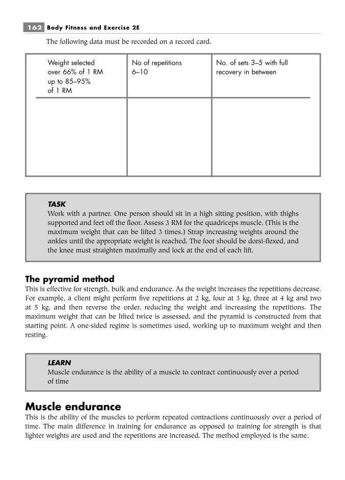
The following data must be recorded on a record card.
The pyramid methodThis is effective for strength, bulk and endurance. As the weight increases the repetitions decrease.For example, a client might perform five repetitions at 2 kg, four at 3 kg, three at 4 kg and twoat 5 kg, and then reverse the order, reducing the weight and increasing the repetitions. Themaximum weight that can be lifted twice is assessed, and the pyramid is constructed from thatstarting point. A one-sided regime is sometimes used, working up to maximum weight and thenresting.
Muscle enduranceThis is the ability of the muscles to perform repeated contractions continuously over a period oftime. The main difference in training for endurance as opposed to training for strength is thatlighter weights are used and the repetitions are increased. The method employed is the same.
162 Body Fitness and Exercise 2E
LEARNMuscle endurance is the ability of a muscle to contract continuously over a periodof time
Weight selected No of repetitions No. of sets 3–5 with fullover 66% of 1 RM 6–10 recovery in betweenup to 85–95%of 1 RM
TASKWork with a partner. One person should sit in a high sitting position, with thighssupported and feet off the floor. Assess 3 RM for the quadriceps muscle. (This is themaximum weight that can be lifted 3 times.) Strap increasing weights around theankles until the appropriate weight is reached. The foot should be dorsi-flexed, andthe knee must straighten maximally and lock at the end of each lift.

Endurance trainingWeight selected No. of Repetitions Setsunder 60% of 1 RM 20–30–50 3from 40–60% 1 RM
For endurance, the weight is kept below 66 per cent of the maximum and repeated 25–50 times ormore. Training must be repeated 3–5 times per week.
■ Speed can be improved using high-speed contractions with low resistance.■ To increase power (speed � strength), 3 sets of 15 high-speed contractions with 30–60
per cent of maximal load is generally recommended.
The effects of muscle endurance trainingThe physiological effects of training for endurance are:
■ an increase in the number and size of blood vessels in the muscles;■ an increase in the number of capillary networks;■ an increase in the number of mitochondria in the muscle cells;■ an increase in the number of oxidative enzymes, which extract oxygen from the blood
– the aerobic capacity of the muscles is therefore improved;■ an increase in glycogen stores and glycolitic enzymes used for energy.
PlyometricsExplosive power will improve using plyometrics. These are jumping, leaping and hopping move-ments, where the prime mover is stretched before contraction. The speed of the stretch is an impor-tant factor. When a muscle is stretched, the stretch receptors within the muscle are stimulated. Thisincreases the strength of the following contraction. The longer and faster the stretch, the greater thefollowing concentric contraction. There is a danger of damaging joints and producing micro-tearsin muscle fibres when performing these exercises. They must only be performed by the very fit andonly after adequate warm-up.
The components of fitness 163
FIGURE NUMBER: 9.6 – Plyometric training.
Summary and aid to learningFitness means having the energy and skill to cope in one’s environment.
This obviously means different things to different people. For some people it simply meanscoping with the tasks of daily living but at the other end of the scale, for the athlete, it will meanperforming skills at the highest level.
When we consider training for improving fitness we must ask two questions:■ Fitness for what? (what is the desired outcome?)■ Where are we starting from? (what is the present level of fitness?)
We can improve fitness by considering all the factors that contribute to fitness.
Cardio-respiratory endurance means improving the condition of the heart and lungs, thusimproving the delivery of nutrients and oxygen to the tissue cells.
Muscle strength means the maximal force a muscle can develop against resistance whichwill make activities easier.
Muscle endurance means improving the ability of muscles to contract for longer periods.
Flexibility means improving the suppleness of connective tissue components within themuscles which will improve extensibility and elasticity. It also improves the tensile strength andextensibility of ligaments and tendons. These factors will enable the tissues to withstand excessiveforces, reducing the likelihood of injury.
Skill refers to accurate performance with little or no waste of energy. It is largely a functionof the nervous system which initiates, controls and coordinates all activities.
Speed refers to the distance moved in a specific time. Some activities will require fast speedwhile others require slow control. Speed is an important factor in most sport because improvedspeed and strength will result in greater power.
Body composition refers to the ratio of body fat and fat-free tissue. It is dependent on energybalance which is the balance between energy consumed (the food eaten), and energy expended(the amount of activity undertaken).
Nutrition refers to the foods we eat and the role they play in sustaining life. Good nutritionmeans eating the required amount of a variety of foods which will enable the body to function atmaximum efficiency.
164 Body Fitness and Exercise 2E
LEARNFor endurance training the weight is kept below 66 percent of 1RM but repetitionsare high, 20–50 lifts
Rest and relaxation means allowing the body sufficient time to recover following activity. Themore intense the activity the longer the rest period should be. Many physiological improvementscontinue into the rest period. Adequate rest will prevent chronic fatigue.
Relaxation means freedom from stress and is related to rest. Stress adversly affects the func-tion of many body systems. Consequently reducing stress factors will enable the body to functionmore efficiently.
Planning programmesThe following principles of training must be considered when planning a training programme:
Specificity means that the programme must be specific to the component or systems thatrequire improving, e.g. muscle strength.
Overload means that the training load must be greater than that normally encountered.
Progression means that the overload must be progressively increased in line with improve-ment. The work must become harder as improvement is gained.
Training threshold is the minimum intensity of exercise required to gain improvement.
Reversibility refers to the fact that training gains will regress when training stops.
Cardio-respiratory endurance
Also known as aerobic capacity or endurance, this refers to the capability of the lungs totake in adequate oxygen and the ability of the heart to deliver it to the muscles.
Any form of low intensity, long duration activity, performed over a period of time willimprove endurance.
Select four activities which will improve aerobic endurance.
IntensityThe intensity must be greater than that normally encountered. The heart rate/pulse rate is used toestablish the appropriate intensity.
An individual must not exercise above their maximum heart rate. To determine maximumheart rate deduct the person’s age from 220.
The maximum heart rate for a 30 year old would be 190 beats per minute. The heart ratemust not rise above this during training. However, to produce a training effect the person mustexercise at above 60 per cent of this maximum heart rate but this can increase to 90 per cent as
The components of fitness 165
fitness develops; this is the target zone. The rate which should be maintained throughout thesession is known as the target rate.
Calculate the target rate for a 30 year old exercising at 60 per cent of maximum heart rate.
FrequencyThis refers to the number of training sessions per week; for endurance this will be 4–5 times perweek.
DurationThis can refer to two things:
■ the duration of each session for endurance must be over 30 minutes; it is usually longer.■ the duration of the training programme – for endurance this will be 12–16 weeks.
Following an endurance training programme one would expect an improvement in thephysiological functioning of the lungs, the heart and blood, and of the muscles. Study the text andwrite a list of these improvements.
Endurance training improves the capacity to use fat as fuel, therefore a programme of endu-rance training and a controlled diet is the best way of reducing body fat.
Muscle strengthThis refers to the maximal force a muscle can develop against resistance. Strength is measured bythe maximum weight a muscle can lift. This is known as one repetition maximum (1 RM).
Strength improves in response to lifting increasing loads; this is refered to as weight training.
The principles of weight training programmes are:
Overload means that the overload must be more than is generally encountered. It must beover 66 per cent of 1 RM.
Progression means the load must be increased as strength develops.
Arrangement of the exercises means successive exercises should not work the same muscle.
Specificity means the exercises must target the specific muscles where strength is required.
Recovery means the muscles must be given time to recover, both between sets and betweentraining days. Overtraining without adequate rest can result in chronic fatigue.
Although weight training usually refers to isotonic contraction, both isometric and isokineticcontractions can be used.
166 Body Fitness and Exercise 2E

Explain the difference between these types of contraction.
Strength gains are achieved using a low number of contractions but high resistance. Bulkwill increase with strength but one or two lifts at maximum load should be included.
A programme might include 6–10 contractions grouped into a set; if 1–5 sets are performedthe duration will be 6–10 weeks. This is just one example. The numbers can be varied slightly tosuit the individual and type of contraction.
Muscle endurance refers to the ability of a muscle to continue contracting over a longperiod of time.
To improve muscle endurance the programme will involve high repetitions and low resis-tance.
A programme might include 30–50 repetitions of low weight between 40–60 per cent of1 RM, frequency 3 times per week for 6–10 weeks.
Muscle power is a product of strength and speed and is a requirement of some sports. Thisis achieved through plyometric training which includes jumping, leaping and hopping movementswhere the prime mover is stretched before contraction.
STRENGTHENING EXERCISES
Calf strengthening exercisesNAME: GASTROCNEMIUS, SOLEUS ACTION: PLANTAR FLEXORSPOSITION: CALF MUSCLES
Starting position Exercise
■ Standing lift up onto toes and down.
■ Standing on one leg lift up onto toes and down. Repeat withother leg.
■ Standing step onto a step, then lift onto toes. Repeatwith other leg.
When these three exercises become too easy and the client can perform 20–30 without dif-ficulty, weights can be used to increase the effort. Hold equal weights in each hand, begin-ning with 1 kg and increasing gradually as muscle strength improves.
The components of fitness 167
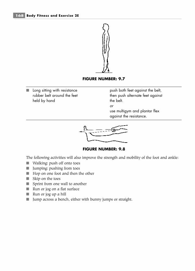
■ Long sitting with resistance push both feet against the belt,rubber belt around the feet then push alternate feet againstheld by hand the belt.
oruse multigym and plantar flexagainst the resistance.
The following activities will also improve the strength and mobility of the foot and ankle:■ Walking: push off onto toes■ Jumping: pushing from toes■ Hop on one foot and then the other■ Skip on the toes■ Sprint from one wall to another■ Run or jog on a flat surface■ Run or jog up a hill■ Jump across a bench, either with bunny jumps or straight.
168 Body Fitness and Exercise 2E
FIGURE NUMBER: 9.7
FIGURE NUMBER: 9.8

Quadriceps strengthening exercisesNAME: RECTUS FEMORIS, VASTUS MEDIALIS, VASTUS LATERALIS, VASTUS INTERMEDIUSACTION: KNEE EXTENSORSPOSITION: FRONT OF THIGH
Starting position Exercise
■ Long sitting press the back of the knee downinto the bed and tighten thequadriceps muscle: dorsi-flex thefoot and try to lift the heel just offthe bed. Hold and release.
■ Long sitting with a rolled towel press the back of the kneeunder the heel downwards and tighten the
quadriceps muscle; dorsi-flex thefoot. Hold and release.
■ Long sitting dorsi-flex the foot and tighten theknee. Raise the leg just off the bedkeeping the knee tight and straight.
If the knee bends slightly it indicates that the muscle is weak and that the above exercisesmust be continued until the leg can be lifted without any give.
■ Long sitting dorsi-flex the foot and tighten theknee, then lift the leg and circle itslowly around.
■ Long sitting dorsi-flex the foot and tighten theknee, lift the leg and lower italmost to the bed, then lift it againseveral times.
The components of fitness 169
FIGURE NUMBER: 9.9 FIGURE NUMBER: 9.10 FIGURE NUMBER: 9.11
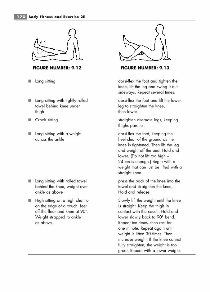
■ Long sitting dorsi-flex the foot and tighten theknee, lift the leg and swing it outsideways. Repeat several times.
■ Long sitting with tightly rolled dorsi-flex the foot and lift the lower towel behind knee under leg to straighten the knee,thigh then lower.
■ Crook sitting straighten alternate legs, keepingthighs parallel.
■ Long sitting with a weight dorsi-flex the foot, keeping theacross the ankle heel clear of the ground as the
knee is tightened. Then lift the legand weight off the bed. Hold andlower. (Do not lift too high –24 cm is enough.) Begin with aweight that can just be lifted with astraight knee.
■ Long sitting with rolled towel press the back of the knee into thebehind the knee, weight over towel and straighten the knee,ankle as above Hold and release.
■ High sitting on a high chair or Slowly lift the weight until the kneeon the edge of a couch, feet is straight. Keep the thigh inoff the floor and knee at 90°. contact with the couch. Hold andWeight strapped to ankle lower slowly back to 90° bend.as above. Repeat ten times, then rest for
one minute. Repeat again untilweight is lifted 30 times. Thenincrease weight. If the knee cannotfully straighten, the weight is toogreat. Repeat with a lower weight.
170 Body Fitness and Exercise 2E
FIGURE NUMBER: 9.12 FIGURE NUMBER: 9.13

This last exercise can be performed with an elasticated band or spring attached to the chair,level with the ankle, against which the leg then pulls. Use a leg-press machine if available.
Note: before using weights to improve strength, read the notes on weight training earlier inthis chapter.
■ Wing standing bend the knees and lower thebody slowly, bending to just abovea right angle. Keep the backstraight. Push up straight and lockthe knees by pulling patellaupwards. (Caution: do not takethe buttocks below knee level.)
This can be progressed by holding a weight in the arms or by placing them over the shoul-ders. A fit person could perform this with one leg at a time.
■ Using a multigym or sliding bend the knees fully. Then pushsprung board or rowing out to straighten and tighten themachine knees, hold it and bend the knees
again slowly. Keep the backstraight.
■ Standing in front of a bench or step up, straighten the knee fully,stairs then step down. Do ten or more
per leg. A fit person could step upand down two stairs at a time.
Cycling is also beneficial to the quadriceps muscle. Make sure that the leg can straightenduring each downward movement of the pedal. To progress, increase the resistance as nec-essary on an exercise bike or cycle uphill on an ordinary bicycle.
The components of fitness 171
FIGURE NUMBER: 9.15FIGURE NUMBER: 9.14
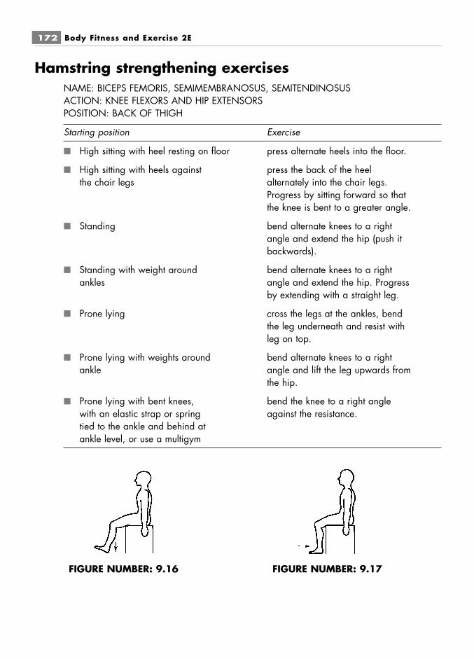
Hamstring strengthening exercisesNAME: BICEPS FEMORIS, SEMIMEMBRANOSUS, SEMITENDINOSUSACTION: KNEE FLEXORS AND HIP EXTENSORSPOSITION: BACK OF THIGH
Starting position Exercise
■ High sitting with heel resting on floor press alternate heels into the floor.
■ High sitting with heels against press the back of the heelthe chair legs alternately into the chair legs.
Progress by sitting forward so thatthe knee is bent to a greater angle.
■ Standing bend alternate knees to a rightangle and extend the hip (push itbackwards).
■ Standing with weight around bend alternate knees to a rightankles angle and extend the hip. Progress
by extending with a straight leg.
■ Prone lying cross the legs at the ankles, bendthe leg underneath and resist withleg on top.
■ Prone lying with weights around bend alternate knees to a rightankle angle and lift the leg upwards from
the hip.
■ Prone lying with bent knees, bend the knee to a right anglewith an elastic strap or spring against the resistance.tied to the ankle and behind atankle level, or use a multigym
172 Body Fitness and Exercise 2E
FIGURE NUMBER: 9.16 FIGURE NUMBER: 9.17

Hip extensor strengthening exercisesNAME: GLUTEUS MAXIMUS ACTION: HIP EXTENSORPOSITION: BUTTOCK
Starting position Exercise
■ Supine lying or high sitting tighten buttocks, then release.
■ Supine lying tighten buttocks and lift slightly offthe floor.
■ Crook lying lift buttocks off the floor.do not hyper extend the back.
■ Standing swing alternate legs forwards andbackwards, lowering slowly.
■ Stride standing slide arms down the legs, bendtrunk forwards and return toupright.
■ Standing with weights on ankles swing alternate legs backwards andlower slowly.
■ Standing with weights on ankles bend the knee to a right angle andpress the leg backwards with shortmovements.
■ High sitting stand up and sit down slowly.
■ High sitting press the thighs downwards intothe seat and rotate themoutwards.
■ Prone lying bend alternate knees and lift theleg off the floor. (Caution: keepthe hips against the floor.) Ankleweights can be used forprogression.
The components of fitness 173
FIGURE NUMBER: 9.18 FIGURE NUMBER: 9.19 FIGURE NUMBER: 9.20
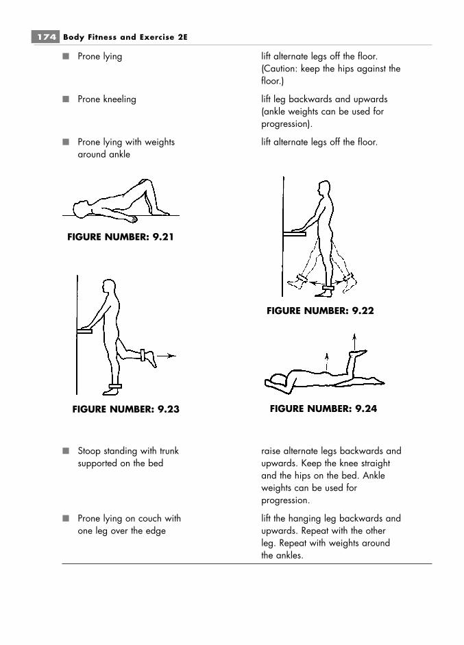
■ Prone lying lift alternate legs off the floor.(Caution: keep the hips against thefloor.)
■ Prone kneeling lift leg backwards and upwards(ankle weights can be used forprogression).
■ Prone lying with weights lift alternate legs off the floor.around ankle
■ Stoop standing with trunk raise alternate legs backwards andsupported on the bed upwards. Keep the knee straight
and the hips on the bed. Ankleweights can be used forprogression.
■ Prone lying on couch with lift the hanging leg backwards andone leg over the edge upwards. Repeat with the other
leg. Repeat with weights aroundthe ankles.
174 Body Fitness and Exercise 2E
FIGURE NUMBER: 9.21
FIGURE NUMBER: 9.22
FIGURE NUMBER: 9.23 FIGURE NUMBER: 9.24
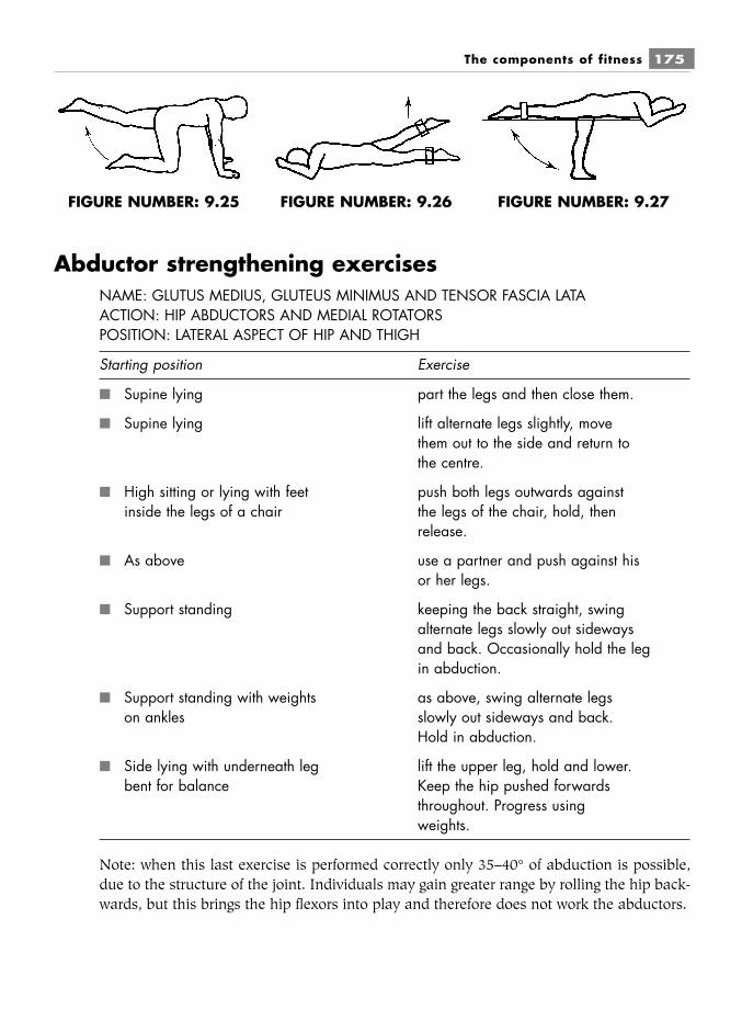
Abductor strengthening exercisesNAME: GLUTUS MEDIUS, GLUTEUS MINIMUS AND TENSOR FASCIA LATAACTION: HIP ABDUCTORS AND MEDIAL ROTATORSPOSITION: LATERAL ASPECT OF HIP AND THIGH
Starting position Exercise
■ Supine lying part the legs and then close them.
■ Supine lying lift alternate legs slightly, movethem out to the side and return tothe centre.
■ High sitting or lying with feet push both legs outwards againstinside the legs of a chair the legs of the chair, hold, then
release.
■ As above use a partner and push against hisor her legs.
■ Support standing keeping the back straight, swingalternate legs slowly out sidewaysand back. Occasionally hold the legin abduction.
■ Support standing with weights as above, swing alternate legson ankles slowly out sideways and back.
Hold in abduction.
■ Side lying with underneath leg lift the upper leg, hold and lower.bent for balance Keep the hip pushed forwards
throughout. Progress usingweights.
Note: when this last exercise is performed correctly only 35–40° of abduction is possible,due to the structure of the joint. Individuals may gain greater range by rolling the hip back-wards, but this brings the hip flexors into play and therefore does not work the abductors.
The components of fitness 175
FIGURE NUMBER: 9.25 FIGURE NUMBER: 9.26 FIGURE NUMBER: 9.27
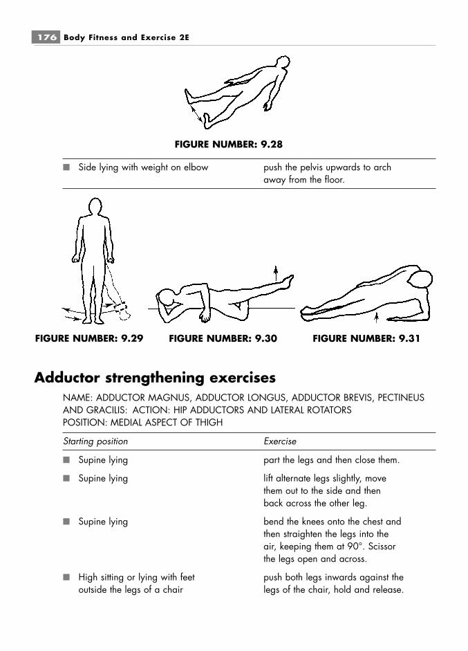
■ Side lying with weight on elbow push the pelvis upwards to archaway from the floor.
Adductor strengthening exercisesNAME: ADDUCTOR MAGNUS, ADDUCTOR LONGUS, ADDUCTOR BREVIS, PECTINEUSAND GRACILIS: ACTION: HIP ADDUCTORS AND LATERAL ROTATORSPOSITION: MEDIAL ASPECT OF THIGH
Starting position Exercise
■ Supine lying part the legs and then close them.
■ Supine lying lift alternate legs slightly, movethem out to the side and thenback across the other leg.
■ Supine lying bend the knees onto the chest andthen straighten the legs into theair, keeping them at 90°. Scissorthe legs open and across.
■ High sitting or lying with feet push both legs inwards against theoutside the legs of a chair legs of the chair, hold and release.
176 Body Fitness and Exercise 2E
FIGURE NUMBER: 9.28
FIGURE NUMBER: 9.29 FIGURE NUMBER: 9.30 FIGURE NUMBER: 9.31
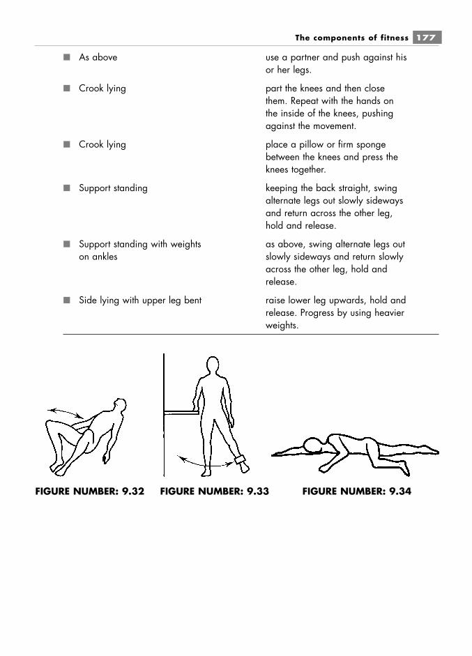
■ As above use a partner and push against hisor her legs.
■ Crook lying part the knees and then closethem. Repeat with the hands onthe inside of the knees, pushingagainst the movement.
■ Crook lying place a pillow or firm spongebetween the knees and press theknees together.
■ Support standing keeping the back straight, swingalternate legs out slowly sidewaysand return across the other leg,hold and release.
■ Support standing with weights as above, swing alternate legs outon ankles slowly sideways and return slowly
across the other leg, hold andrelease.
■ Side lying with upper leg bent raise lower leg upwards, hold andrelease. Progress by using heavierweights.
The components of fitness 177
FIGURE NUMBER: 9.32 FIGURE NUMBER: 9.34FIGURE NUMBER: 9.33

Abdominal strengthening exercisesNAME: RECTUS ABDOMINUS, EXTERNAL OBLIQUE, INTERNAL OBLIQUE, TRANSVERSUSABDOMINUSACTION: TRUNK FLEXORS, SIDE FLEXORS AND ROTATORSPOSITION: COVER THE ABDOMEN
Starting position Exercise
■ Crook lying press the small of the back intothe floor, tilt the pelvis backwardsand pull the stomach in. Hold andrelease.
■ Crook lying press the small of the back intothe floor. Tuck the head downonto the chest, then raise the headand shoulders to look at the knees.Hold and release.
■ Crook lying bring both knees up to form rightangles at hip and knee. Reach uptowards the ceiling with alternateknees.
■ Crook lying, arms at side curl up (Caution: returnslowly from the base of the spine upwards.)
■ Crook lying, arms across chest curl up.
■ Crook lying, hands on shoulders curl up.
■ Crook lying, hands on ears curl up. (Do not put thehands behind the neck as this candamage the neck.)
■ Stretch crook lying curl up. (Caution: onlyfor those with strong abdominals.)Keep the arms back: do notswing them forwards.
■ Crook lying, holding a weight or curl up. (Caution: onlymedicine ball on the chest for those with strong abdominals.)
178 Body Fitness and Exercise 2E
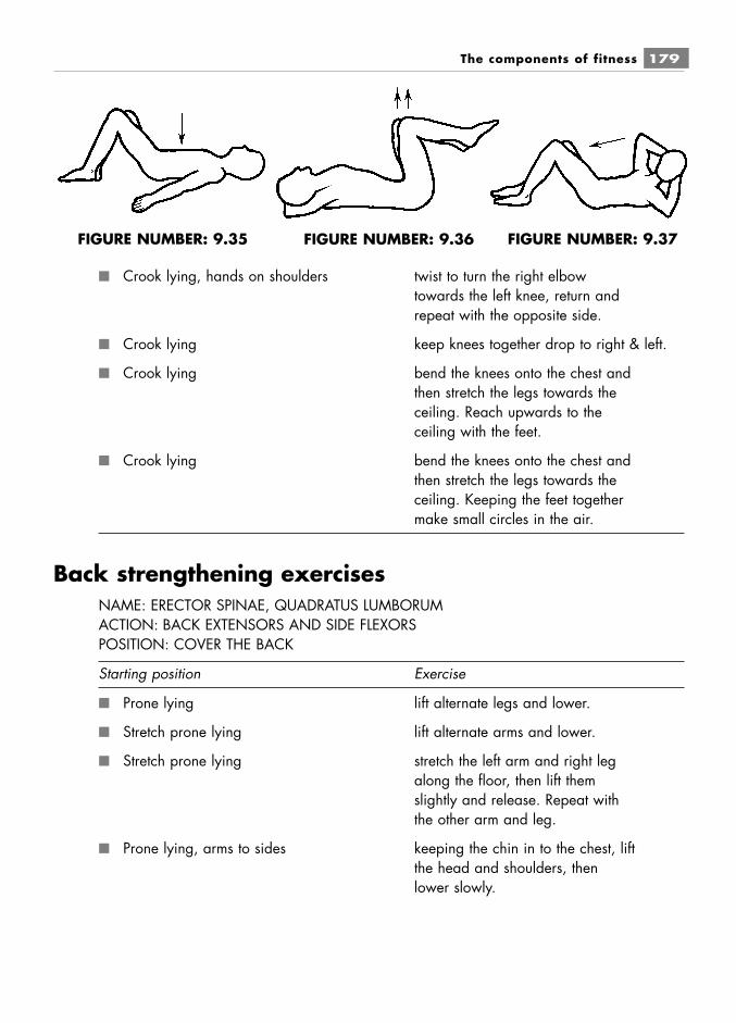
■ Crook lying, hands on shoulders twist to turn the right elbowtowards the left knee, return andrepeat with the opposite side.
■ Crook lying keep knees together drop to right & left.
■ Crook lying bend the knees onto the chest andthen stretch the legs towards theceiling. Reach upwards to theceiling with the feet.
■ Crook lying bend the knees onto the chest andthen stretch the legs towards theceiling. Keeping the feet togethermake small circles in the air.
Back strengthening exercisesNAME: ERECTOR SPINAE, QUADRATUS LUMBORUMACTION: BACK EXTENSORS AND SIDE FLEXORSPOSITION: COVER THE BACK
Starting position Exercise
■ Prone lying lift alternate legs and lower.
■ Stretch prone lying lift alternate arms and lower.
■ Stretch prone lying stretch the left arm and right legalong the floor, then lift themslightly and release. Repeat withthe other arm and leg.
■ Prone lying, arms to sides keeping the chin in to the chest, liftthe head and shoulders, thenlower slowly.
The components of fitness 179
FIGURE NUMBER: 9.35 FIGURE NUMBER: 9.37FIGURE NUMBER: 9.36
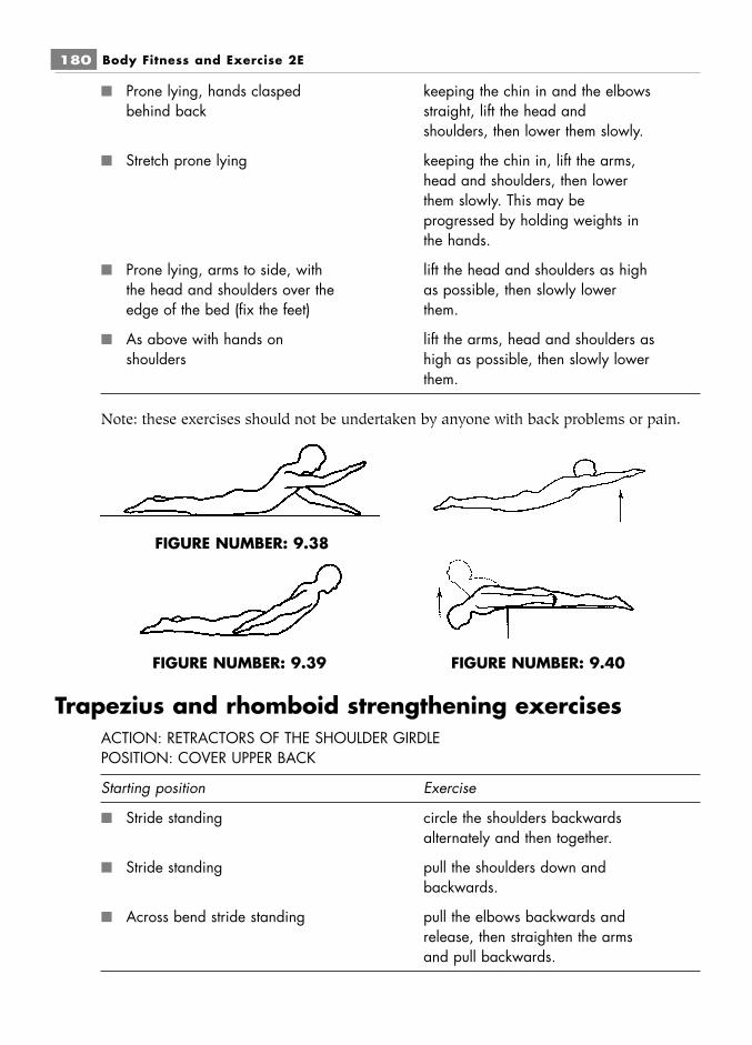
■ Prone lying, hands clasped keeping the chin in and the elbowsbehind back straight, lift the head and
shoulders, then lower them slowly.
■ Stretch prone lying keeping the chin in, lift the arms,head and shoulders, then lowerthem slowly. This may beprogressed by holding weights inthe hands.
■ Prone lying, arms to side, with lift the head and shoulders as highthe head and shoulders over the as possible, then slowly loweredge of the bed (fix the feet) them.
■ As above with hands on lift the arms, head and shoulders asshoulders high as possible, then slowly lower
them.
Note: these exercises should not be undertaken by anyone with back problems or pain.
Trapezius and rhomboid strengthening exercisesACTION: RETRACTORS OF THE SHOULDER GIRDLEPOSITION: COVER UPPER BACK
Starting position Exercise
■ Stride standing circle the shoulders backwardsalternately and then together.
■ Stride standing pull the shoulders down andbackwards.
■ Across bend stride standing pull the elbows backwards andrelease, then straighten the armsand pull backwards.
180 Body Fitness and Exercise 2E
FIGURE NUMBER: 9.38
FIGURE NUMBER: 9.39 FIGURE NUMBER: 9.40
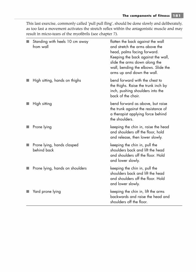
This last exercise, commonly called ‘pull pull fling’, should be done slowly and deliberately,as too fast a movement activates the stretch reflex within the antagonistic muscle and mayresult in micro-tears of the myofibrils (see chapter 7).
■ Standing with heels 10 cm away flatten the back against the wallfrom wall and stretch the arms above the
head, palms facing forward.Keeping the back against the wall,slide the arms down along thewall, bending the elbows. Slide thearms up and down the wall.
■ High sitting, hands on thighs bend forward with the chest tothe thighs. Raise the trunk inch byinch, pushing shoulders into theback of the chair.
■ High sitting bend forward as above, but raisethe trunk against the resistance ofa therapist applying force behindthe shoulders.
■ Prone lying keeping the chin in, raise the headand shoulders off the floor, holdand release, then lower slowly.
■ Prone lying, hands clasped keeping the chin in, pull thebehind back shoulders back and lift the head
and shoulders off the floor. Holdand lower slowly.
■ Prone lying, hands on shoulders keeping the chin in, pull theshoulders back and lift the headand shoulders off the floor. Holdand lower slowly.
■ Yard prone lying keeping the chin in, lift the armsbackwards and raise the head andshoulders off the floor.
The components of fitness 181
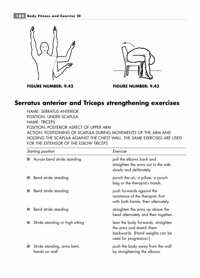
Serratus anterior and Triceps strengthening exercisesNAME: SERRATUS ANTERIORPOSITION: UNDER SCAPULANAME: TRICEPSPOSITION: POSTERIOR ASPECT OF UPPER ARMACTION: POSITIONING OF SCAPULA DURING MOVEMENTS OF THE ARM ANDHOLDING THE SCAPULA AGAINST THE CHEST WALL. THE SAME EXERCISES ARE USEDFOR THE EXTENSOR OF THE ELBOW TRICEPS
Starting position Exercise
■ Across bend stride standing pull the elbows back andstraighten the arms out to the sideslowly and delibrately.
■ Bend stride standing punch the air, a pillow, a punchbag or the therapist’s hands.
■ Bend stride standing push forwards against theresistance of the therapist, firstwith both hands, then alternately.
■ Bend stride standing straighten the arms up above thehead alternately and then together.
■ Stride standing or high sitting lean the body forwards, straightenthe arms and stretch thembackwards. (Hand weights can beused for progression.)
■ Stride standing, arms bent, push the body away from the wallhands on wall by straightening the elbows.
182 Body Fitness and Exercise 2E
FIGURE NUMBER: 9.42 FIGURE NUMBER: 9.43
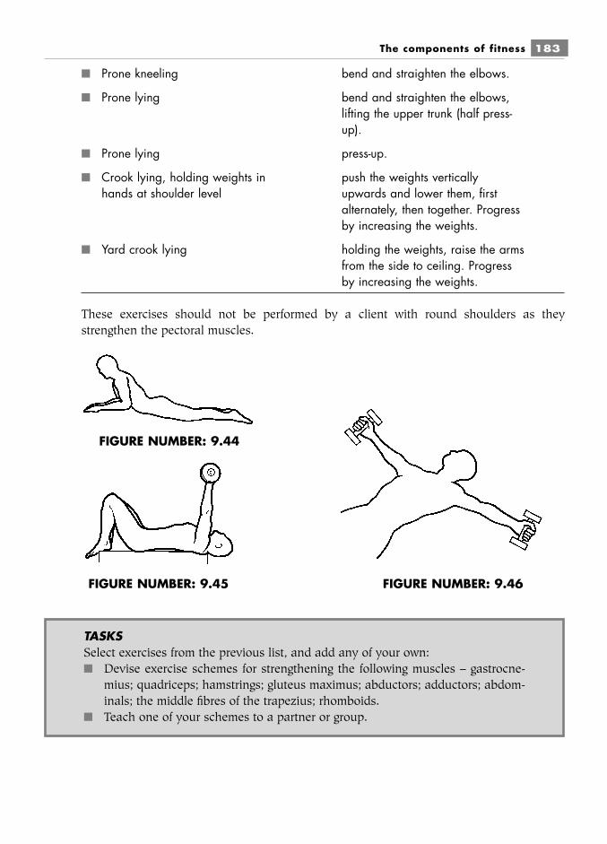
■ Prone kneeling bend and straighten the elbows.
■ Prone lying bend and straighten the elbows,lifting the upper trunk (half press-up).
■ Prone lying press-up.
■ Crook lying, holding weights in push the weights verticallyhands at shoulder level upwards and lower them, first
alternately, then together. Progressby increasing the weights.
■ Yard crook lying holding the weights, raise the armsfrom the side to ceiling. Progressby increasing the weights.
These exercises should not be performed by a client with round shoulders as theystrengthen the pectoral muscles.
The components of fitness 183
FIGURE NUMBER: 9.44
FIGURE NUMBER: 9.45 FIGURE NUMBER: 9.46
TASKSSelect exercises from the previous list, and add any of your own:■ Devise exercise schemes for strengthening the following muscles – gastrocne-
mius; quadriceps; hamstrings; gluteus maximus; abductors; adductors; abdom-inals; the middle fibres of the trapezius; rhomboids.
■ Teach one of your schemes to a partner or group.
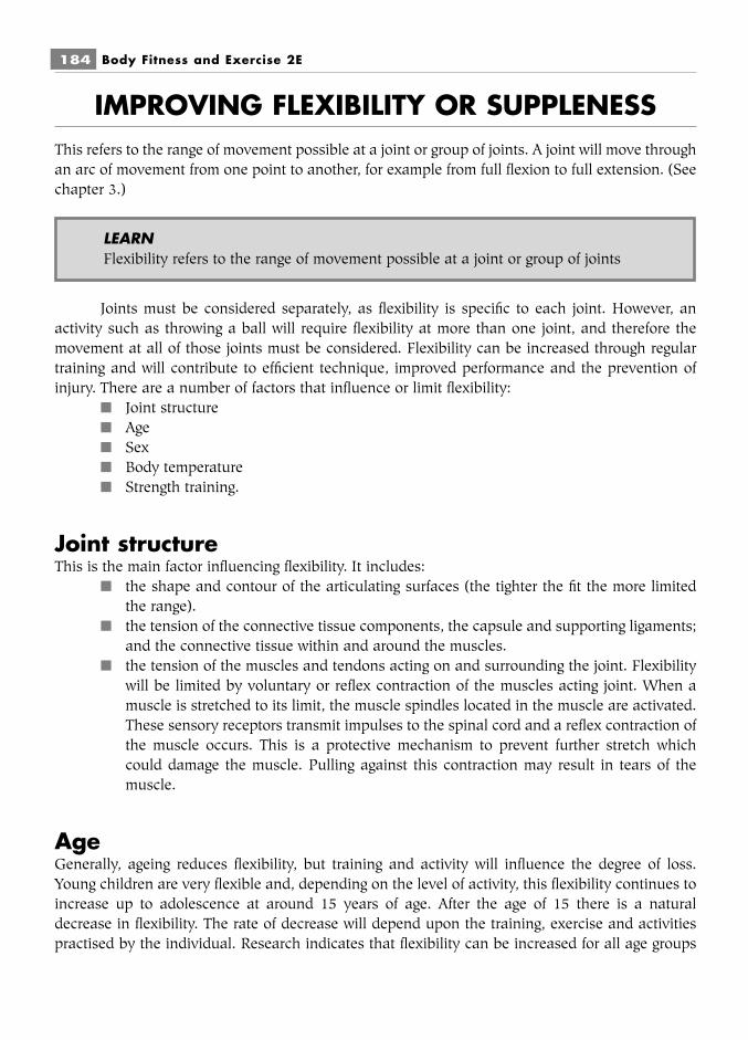
IMPROVING FLEXIBILITY OR SUPPLENESSThis refers to the range of movement possible at a joint or group of joints. A joint will move throughan arc of movement from one point to another, for example from full flexion to full extension. (Seechapter 3.)
Joints must be considered separately, as flexibility is specific to each joint. However, anactivity such as throwing a ball will require flexibility at more than one joint, and therefore themovement at all of those joints must be considered. Flexibility can be increased through regulartraining and will contribute to efficient technique, improved performance and the prevention ofinjury. There are a number of factors that influence or limit flexibility:
■ Joint structure■ Age■ Sex■ Body temperature■ Strength training.
Joint structureThis is the main factor influencing flexibility. It includes:
■ the shape and contour of the articulating surfaces (the tighter the fit the more limitedthe range).
■ the tension of the connective tissue components, the capsule and supporting ligaments;and the connective tissue within and around the muscles.
■ the tension of the muscles and tendons acting on and surrounding the joint. Flexibilitywill be limited by voluntary or reflex contraction of the muscles acting joint. When amuscle is stretched to its limit, the muscle spindles located in the muscle are activated.These sensory receptors transmit impulses to the spinal cord and a reflex contraction ofthe muscle occurs. This is a protective mechanism to prevent further stretch whichcould damage the muscle. Pulling against this contraction may result in tears of themuscle.
AgeGenerally, ageing reduces flexibility, but training and activity will influence the degree of loss.Young children are very flexible and, depending on the level of activity, this flexibility continues toincrease up to adolescence at around 15 years of age. After the age of 15 there is a naturaldecrease in flexibility. The rate of decrease will depend upon the training, exercise and activitiespractised by the individual. Research indicates that flexibility can be increased for all age groups
184 Body Fitness and Exercise 2E
LEARNFlexibility refers to the range of movement possible at a joint or group of joints

if appropriate exercises are undertaken, but the rate of increase will be greater in younger agegroups and will decline with age.
TrainingAs stated above, training and the selection of appropriate exercise will increase flexibility for all agegroups. Those individuals, such as gymnasts and ballet dancers, who continue with uninterruptedtraining programmes will have greater flexibility than the untrained.
Sex (Gender)It has been suggested that females are more flexible than males, although the evidence is incon-clusive. Females on the whole have lighter and smaller bones, which may influence flexibility. Inthe main they have a shorter leg length and lower centre of gravity, making certain movementseasier. They are also designed for flexibility of the pelvic region to facilitate childbirth.
Body temperatureThe elevation of body temperature increases flexibility. Therefore, warm-up exercises must be per-formed before flexibility training. Pre-heating with hot packs, heat lamps, hot baths or showers,diathermy or massage will increase the effect. These methods may be used before warm-up but notinstead of exercises. The heat reduces viscosity and relaxes tissues, which thus offer less resistanceto movement. Heat also increases the extensibility of connective and muscle tissue surroundingthe joint.
Strength trainingCertain strength training routines can limit joint flexibility. Strength training must be planned toinclude full-range movements and eccentric work.
In the same way as we overload a muscle in order to increase strength, we must overstretchin order to increase flexibility. The body continually adapts to increasing demand placed upon it,so that moving a joint beyond its existing range and stretching the surrounding tissues will resultin increased range as the tissues become more extensible.
Uses of stretching exercisesStretching increases the range of movement and will improve the performance of simple activitiesrequired for daily living and also the complex activities required by the elite performer.
Stretching increases the suppleness and range of the tissues and joints, enabling them toaccommodate any increased stresses; this will reduce the risk of injury.
Gentle, uniform stretching eases tension and spasm which reduces pain and stiffness.
The components of fitness 185

Stretching of tight muscles will improve muscle balance which will contribute to goodposture.
Stretching is an important part of the rehabilitation following injury. The healing processmay result in the formation of scar tissue within the muscle. This must be kept supple and its elas-ticity maintained, otherwise it will contract and impair the contractile properties of the muscle.
Stretching exercises should aways be included in any exercise scheme. They should bepractised after the warm-up in order to enhance performance and reduce the risk of injury. Theyshould be performed during the cool-down to reduce muscle soreness and stiffness. Stretchingexercises can also be used as a complete programme, designed progressively to increase the rangein all body joints. This type of slow stretch programme allows time for thought and meditation and,as in yoga, pursues a harmony of body, mind and spirit.
Methods of stretchingThere are various methods of stretching, including ballistic, dynamic, static, proprioceptive neuro-muscular facilitation (PNF) and others.
Ballistic stretchingThis type of stretching involves fast, jerky movements where the increased momentum created bya ‘bounce’ is used to increase movement at the end of the range, for example bouncing to touchthe toes. Ballistics are not generally recommended and should be avoided in class work.
The arguments against their use are physiologically sound:■ A quick or rapid stretch does not allow sufficient time for the tissues to adapt, resulting
in strain.■ A sudden jerk applied to a muscle will initiate the stretch reflex and muscle tension will
increase. Further pulling against this tension may result in microscopic tears of the myo-fibrils. Healing will result in the formation of fibrous scar tissue, which impairs the func-tion of the muscle.
■ A quick stretch does not allow for neurological adaptation. Research has shown that thetension generated in a muscle by a fast stretch is far greater than that generated by aslow stretch. Therefore the tensile resistance to fast stretching is much greater.
■ Bouncing movements are not easy to control. Therefore the positioning of joints and thedirection of movement may not be correct, increasing the likelihood of injury.
Despite all these reasons for not attempting ballistics, some people favour their use for specifictraining. There is some evidence that following ballistic or fast stretch training the stretch reflex
186 Body Fitness and Exercise 2E
LEARNBallistic stretching is not recommended and should be avoided. Jerking may resultin tears of myofibrils which will impair muscle function.
may be delayed and will be activated nearer the end of range of movement, thus improving perfor-mance. Gymnasts and dancers may include ballistic exercises prior to specific actions or routines,but generally they should be avoided.
Dynamic stretchingThese are movements where a muscle or muscles are worked gradually through their range.Beginning with short-range movements, the actions move progressively through to maximum fullrange. Dynamic flexibility is required by ball kickers in rugby and soccer, e.g. a gradual stretchingof the hamstrings in stages before kicking ensures that the effort of the muscles kicking the ball isnot hampered by tight hamstrings. Repetitive free knee extension is performed, increasing therange each time.
Static stretchingThese are movements which take a muscle slowly and deliberately to the end of its range. The posi-tion is then held and further stretch applied. During the holding time, the muscle adapts to thestretch. The stretch reflex controlled by the muscle spindles is inhibited, there is a slow decreasein muscle tension and the muscle relaxes. This allows an increase in muscle length and in therange of joint movement. Research has shown that low-force, long-duration stretching at a raisedtemperature will result in permanent lengthening.
Static stretching is safer and more effective than ballistic or dynamic stretching, as thetissues have time to relax. Maximum stretch is achieved when a muscle is fully relaxed and con-nective tissue is fully stretched. The stretch reflex is inhibited, and so there is no risk of tearing,muscle soreness and damage. Movements are slower, more controlled and more functionally accu-rate, and there is therefore less risk of injury. Static stretching requires less energy consumption.
Static stretching may be classified into active and passive stretching.■ Active stretching is stretching alone, without external aid.■ Active assisted stretching is a stretch performed alone until a limit is reached, at which
point a partner helps to gain a further stretch.■ Passive stretching is achieved by an external force such as traction or a partner, while
the individual remains inactive.
All static stretching must be controlled and performed with care. Particular care, effectivecommunication and trust must exist between partners in active assisted and passive stretching.These should only be practised by competent, well-trained individuals.
Proprioceptive neuromuscular facilitationThere are many PNF techniques, which are an excellent way of increasing range of movement, butthey require an in-depth knowledge of neurophysiology and are not within the scope of this book.However, one of the techniques, alternate contract/relax, is straightforward and useful, especiallyfollowing recovery from injury.
This form of increased range is achieved in the following way.
The components of fitness 187
A muscle is moved to its point of slight stretch, i.e. at the end of joint movement, and anisometric contraction of that muscle against resistance is performed and held. This is followed byrelaxation and further joint movement, which will now be possible.
For example, in prone lying (face down), lift one leg to stretch the hip flexors. Ask a partnerto support the leg and to resist an isometric contraction (i.e. not to allow movement as you pushdown against his or her hand). Relax, then lift the leg higher, as the hip flexors will now allow agreater range of movement.
In sitting, tilt the head to the left to stretch the right sterno-cleido-mastoid. Now place thehand against the right side of the face. Contract the muscle statically against the hand resistance,hold and relax. The head will now move further to the left.
Techniques of static stretching■ Select a suitable venue that is warm and well ventilated. Ensure that there is sufficient
space to perform all movements.■ Wear warm clothing to maintain and increase body temperature.■ Check that the floor surface is clean, smooth and non-slip.■ Do not stretch if any of the following contra-indications are present: hyper-mobility,
strains or sprains, inflammation of joints, pain in joints, fevers, heart problems, high orlow blood pressure, or after a heavy meal.
■ Identify the goals (where flexibility is required). Always warm up with a set of exercisesdesigned to work the large muscle groups. This warm-up will increase muscle tempera-ture, reduce muscle viscosity, decrease muscle tension and promote relaxation and willmake tissues more extensible.
■ Set the mind into a tranquil and relaxed state.■ Isolate the muscle or group for stretching and place the joint in the correct position.
Stretch slowly and evenly, feeling the pull in the belly of the muscle and not at thetendon ends. There should be a feeling of mild discomfort, not pain. Hold the stretch forsix to ten seconds to begin with, increasing to 20–30 seconds over time. As the tensiondecreases, stretch a little further – do not jerk or bounce at the end of the movement.Let pain be the guide. If the pain increases, relax; if the muscle begins to quiver, relax;if muscle tension increases, relax. Move slowly out of the stretch.
■ Repeat the stretch five times at the beginning of a programme, eventually working up toten to fifteen repetitions.
■ Exhale as you move into the stretch and relax.■ Stretching programmes should be performed once or twice a day if rapid improvement
is required.
The effects of stretch trainingThe physiological effects of flexibility training are:
■ an increased range of movement at joints;
188 Body Fitness and Exercise 2E
■ increased flexibility of the supporting structures;■ increased elasticity and extensibility of muscles;■ reduced tension and increased relaxation in the muscles;■ increased circulation to the muscles;■ improved balance and co-ordination between muscle groups;■ improved posture;■ improved mechanical efficiency and improved speed and skill;■ improved techique and performance, since relaxation of the antagonistic muscles allows
the agonist to maximise performance;■ neurological adaptation delaying the stretch reflex;■ if performed after performance, particularly eccentric work, a reduction of muscle sore-
ness.
Dangers of stretching exercises■ Damage to muscles by causing micro-tears of muscle fibres, caused by sudden stretch
of cold muscles, or ballistrics;■ Damage to joints caused by poor technique;■ Over-stretching of ligaments caused by poor technique or forcing at the end of the range;■ Straining other body areas due to incorrect positioning;■ Raising blood pressure caused by incorrect breathing.
Summary and aid to learningFlexibility:
This refers to the range of movement possible at a joint.
The factors which affect flexibility are:
Joint structure: this includes the shape of the bones; the suppleness of the capsule, muscles,ligaments and tendons surrounding the joint.
The components of fitness 189
FIGURE NUMBER: 9.47 –Active stretch for the gastrocenius.
FIGURE NUMBER: 9.48 –Active assisted stretching of triceps.
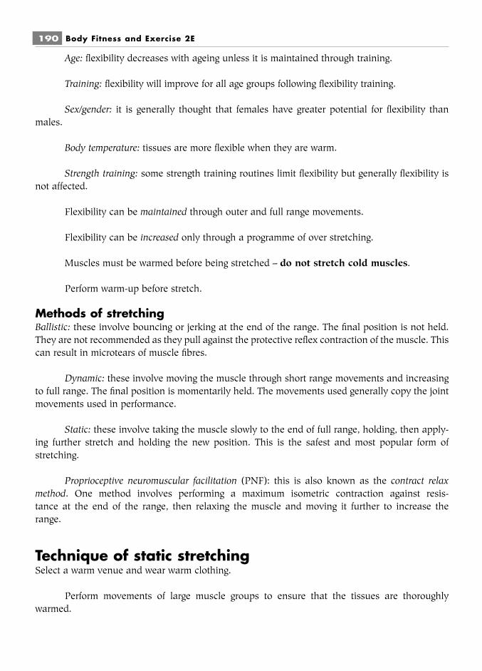
Age: flexibility decreases with ageing unless it is maintained through training.
Training: flexibility will improve for all age groups following flexibility training.
Sex/gender: it is generally thought that females have greater potential for flexibility thanmales.
Body temperature: tissues are more flexible when they are warm.
Strength training: some strength training routines limit flexibility but generally flexibility isnot affected.
Flexibility can be maintained through outer and full range movements.
Flexibility can be increased only through a programme of over stretching.
Muscles must be warmed before being stretched – do not stretch cold muscles.
Perform warm-up before stretch.
Methods of stretchingBallistic: these involve bouncing or jerking at the end of the range. The final position is not held.They are not recommended as they pull against the protective reflex contraction of the muscle. Thiscan result in microtears of muscle fibres.
Dynamic: these involve moving the muscle through short range movements and increasingto full range. The final position is momentarily held. The movements used generally copy the jointmovements used in performance.
Static: these involve taking the muscle slowly to the end of full range, holding, then apply-ing further stretch and holding the new position. This is the safest and most popular form ofstretching.
Proprioceptive neuromuscular facilitation (PNF): this is also known as the contract relaxmethod. One method involves performing a maximum isometric contraction against resis-tance at the end of the range, then relaxing the muscle and moving it further to increase the range.
Technique of static stretchingSelect a warm venue and wear warm clothing.
Perform movements of large muscle groups to ensure that the tissues are thoroughlywarmed.
190 Body Fitness and Exercise 2E
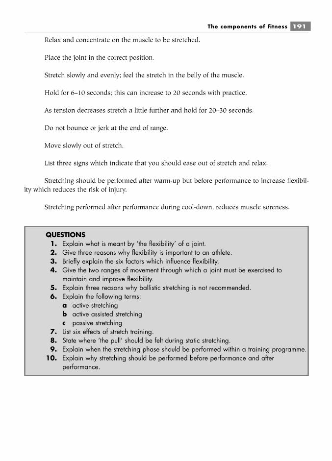
Relax and concentrate on the muscle to be stretched.
Place the joint in the correct position.
Stretch slowly and evenly; feel the stretch in the belly of the muscle.
Hold for 6–10 seconds; this can increase to 20 seconds with practice.
As tension decreases stretch a little further and hold for 20–30 seconds.
Do not bounce or jerk at the end of range.
Move slowly out of stretch.
List three signs which indicate that you should ease out of stretch and relax.
Stretching should be performed after warm-up but before performance to increase flexibil-ity which reduces the risk of injury.
Stretching performed after performance during cool-down, reduces muscle soreness.
The components of fitness 191
QUESTIONS1. Explain what is meant by ‘the flexibility’ of a joint.2. Give three reasons why flexibility is important to an athlete.3. Briefly explain the six factors which influence flexibility.4. Give the two ranges of movement through which a joint must be exercised to
maintain and improve flexibility.5. Explain three reasons why ballistic stretching is not recommended.6. Explain the following terms:
a active stretchingb active assisted stretchingc passive stretching
7. List six effects of stretch training.8. State where ‘the pull’ should be felt during static stretching.9. Explain when the stretching phase should be performed within a training programme.
10. Explain why stretching should be performed before performance and afterperformance.
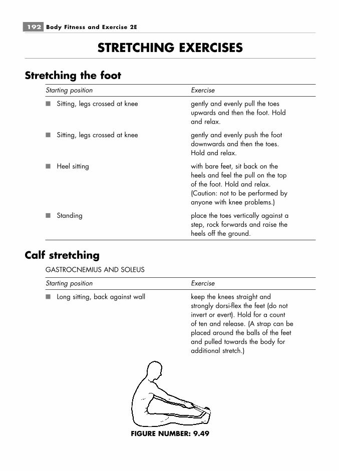
STRETCHING EXERCISES
Stretching the footStarting position Exercise
■ Sitting, legs crossed at knee gently and evenly pull the toesupwards and then the foot. Holdand relax.
■ Sitting, legs crossed at knee gently and evenly push the footdownwards and then the toes.Hold and relax.
■ Heel sitting with bare feet, sit back on theheels and feel the pull on the topof the foot. Hold and relax.(Caution: not to be performed byanyone with knee problems.)
■ Standing place the toes vertically against astep, rock forwards and raise theheels off the ground.
Calf stretchingGASTROCNEMIUS AND SOLEUS
Starting position Exercise
■ Long sitting, back against wall keep the knees straight andstrongly dorsi-flex the feet (do notinvert or evert). Hold for a countof ten and release. (A strap can beplaced around the balls of the feetand pulled towards the body foradditional stretch.)
192 Body Fitness and Exercise 2E
FIGURE NUMBER: 9.49

■ Walk standing, one foot directly keep the back heel firmly on thein front of the other ground and bend the front knee
gently until a pull is felt in the calfof the hind leg. Hold for a count often, then slowly release. Repeatwith the other leg.
■ Reach standing, facing a wall, walk the feet backwards, keepingwith hands against wall the heels on the ground, until a
pull is felt in the calves. Hold for acount of ten.
■ Standing with hands against walk the feet backwards, keepingwall as above the heels on the ground. When
the pull is just being felt, bend theelbows slowly and the pull willincrease. Hold for a count of ten.
■ Standing on an incline lean forward, keeping the heels onthe ground, until a pull is felt in thecalf.
■ Toe standing on the edge of lift up onto the toes and align thea step body over the feet, then lower the
heels until the pull is felt in the calf.Hold for a count of ten, then release.
The components of fitness 193
FIGURE NUMBER: 9.50
FIGURE NUMBER: 9.51
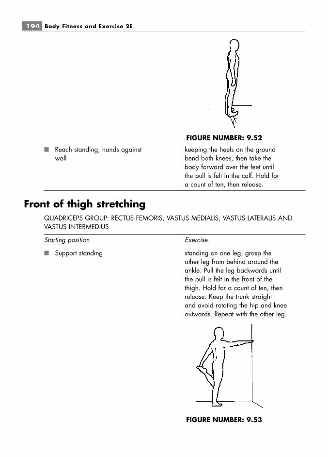
■ Reach standing, hands against keeping the heels on the groundwall bend both knees, then take the
body forward over the feet untilthe pull is felt in the calf. Hold fora count of ten, then release.
Front of thigh stretchingQUADRICEPS GROUP: RECTUS FEMORIS, VASTUS MEDIALIS, VASTUS LATERALIS ANDVASTUS INTERMEDIUS
Starting position Exercise
■ Support standing standing on one leg, grasp theother leg from behind around theankle. Pull the leg backwards untilthe pull is felt in the front of thethigh. Hold for a count of ten, thenrelease. Keep the trunk straightand avoid rotating the hip and kneeoutwards. Repeat with the other leg.
194 Body Fitness and Exercise 2E
FIGURE NUMBER: 9.52
FIGURE NUMBER: 9.53
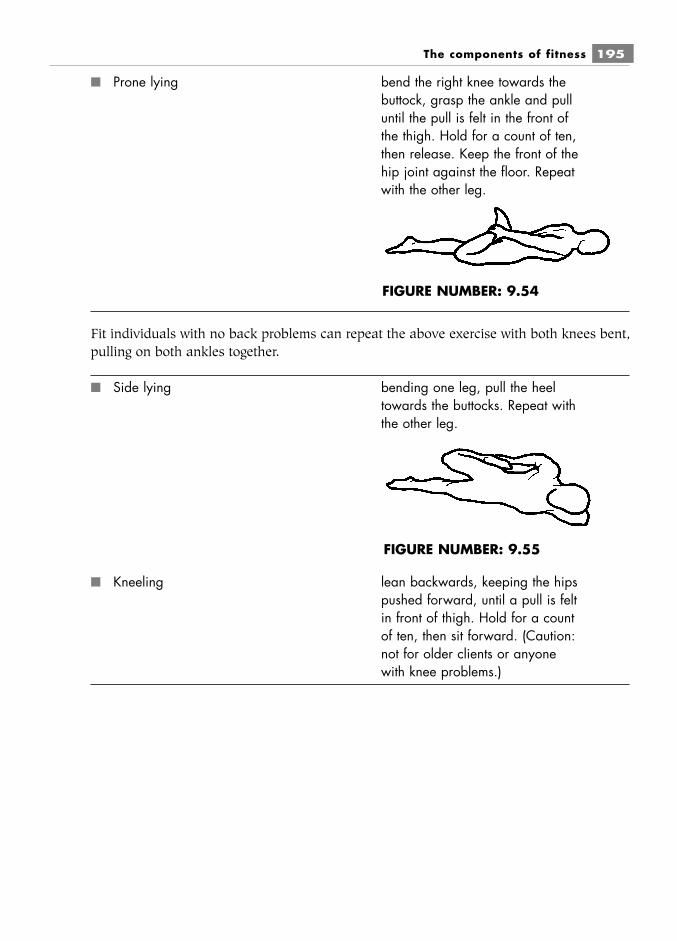
■ Prone lying bend the right knee towards thebuttock, grasp the ankle and pulluntil the pull is felt in the front ofthe thigh. Hold for a count of ten,then release. Keep the front of thehip joint against the floor. Repeatwith the other leg.
Fit individuals with no back problems can repeat the above exercise with both knees bent,pulling on both ankles together.
■ Side lying bending one leg, pull the heeltowards the buttocks. Repeat withthe other leg.
■ Kneeling lean backwards, keeping the hipspushed forward, until a pull is feltin front of thigh. Hold for a countof ten, then sit forward. (Caution:not for older clients or anyonewith knee problems.)
The components of fitness 195
FIGURE NUMBER: 9.54
FIGURE NUMBER: 9.55
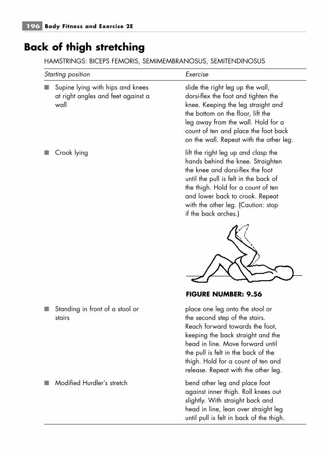
Back of thigh stretchingHAMSTRINGS: BICEPS FEMORIS, SEMIMEMBRANOSUS, SEMITENDINOSUS
Starting position Exercise
■ Supine lying with hips and knees slide the right leg up the wall,at right angles and feet against a dorsi-flex the foot and tighten thewall knee. Keeping the leg straight and
the bottom on the floor, lift theleg away from the wall. Hold for acount of ten and place the foot backon the wall. Repeat with the other leg.
■ Crook lying lift the right leg up and clasp thehands behind the knee. Straightenthe knee and dorsi-flex the footuntil the pull is felt in the back ofthe thigh. Hold for a count of tenand lower back to crook. Repeatwith the other leg. (Caution: stopif the back arches.)
■ Standing in front of a stool or place one leg onto the stool orstairs the second step of the stairs.
Reach forward towards the foot,keeping the back straight and thehead in line. Move forward untilthe pull is felt in the back of thethigh. Hold for a count of ten andrelease. Repeat with the other leg.
■ Modified Hurdler’s stretch bend other leg and place footagainst inner thigh. Roll knees outslightly. With straight back andhead in line, lean over straight leguntil pull is felt in back of the thigh.
196 Body Fitness and Exercise 2E
FIGURE NUMBER: 9.56

Inner thigh stretchingADDUCTOR GROUP: ADDUCTOR LONGUS, ADDUCTOR MAGNUS, ADDUCTOR BREVIS,PECTINEUS, GRACILIS
Starting position Exercise
■ Long sitting open the legs as far as possible.Keep the back straight and leanforwards until a pull is felt in theinner thigh. Hold for a count often and release. (Caution: do notround the trunk or slouch.)
■ Crook sitting keep the feet together and dropthe knees open as far as possible.Then pull the feet towards thebody until the pull is felt in theinner thigh. Hold for a count often and release. (Caution: not tobe done by anyone with knee orhip problems.)
The components of fitness 197
FIGURE NUMBER: 9.57
FIGURE NUMBER: 9.58

■ Crook lying, hands on knees part the knees as far as possible,then press apart with the handsuntil the pull is felt on the inside ofthe thigh. Hold for a count of tenand release. (Caution: not to bedone by anyone with knee or hipproblems.)
■ Supine lying, legs up against part the legs by sliding them alonga wall the wall until a stretch is felt in the
inner thigh. Hold for a count often and release.
■ Stride standing stretch the right leg out as farsideways as possible, withoutrotating the leg outwards, thenbend the left leg until a pull is felton inner thigh of the right leg.Hold for a count of ten andrelease. Repeat with the other leg.
198 Body Fitness and Exercise 2E
FIGURE NUMBER: 9.59
FIGURE NUMBER: 9.60

Outer thigh stretchingHIP ABDUCTORS: GLUTEUS MEDIUS, GLUTEUS MINIMUS, TENSOR FASCIA LATA
Starting position Exercise
■ Supine lying raise the right leg and swing it overthe left leg. Lift the right leg slightlyand dorsi-flex the foot until a pullis felt at the outer thigh. Hold for acount of ten, then release. Repeatwith the other leg.
■ Supine lying bend the left knee to the chest,then push the knee across to theright until a pull is felt in the outerthigh. Hold for a count of ten andrelease. Repeat with the other leg.
■ Supine lying raise the leg to vertical and moveit across the body until a pull is felt in theouter thigh. Repeat with the other leg.
The components of fitness 199
FIGURE NUMBER: 9.61
FIGURE NUMBER: 9.62
FIGURE NUMBER: 9.63
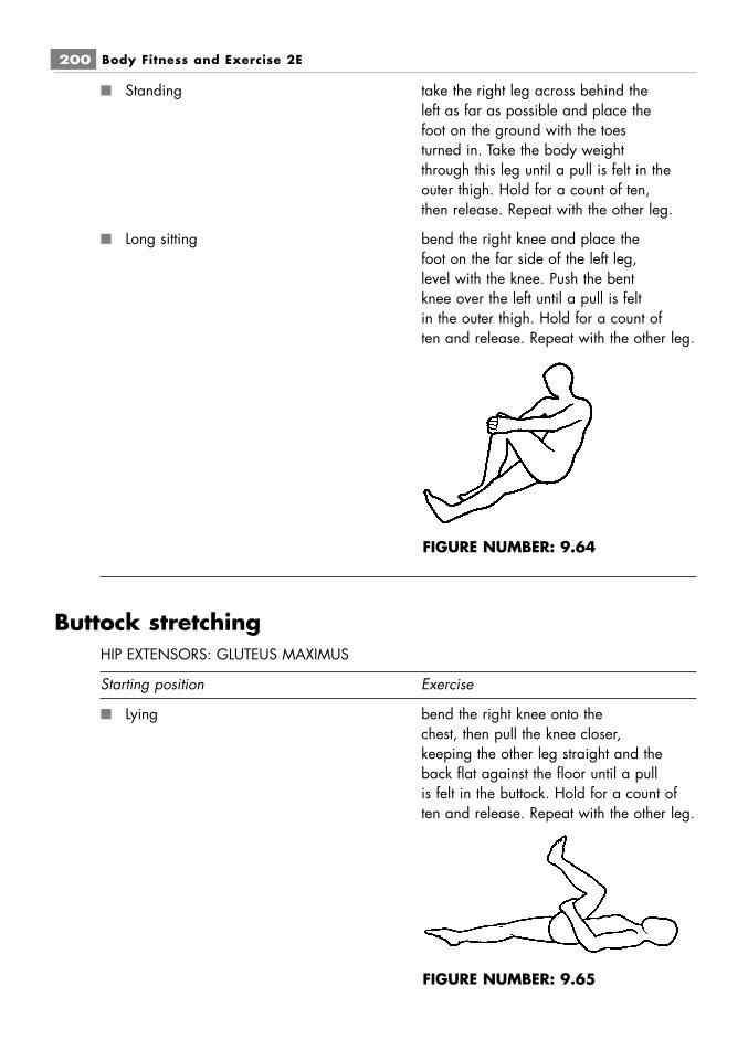
■ Standing take the right leg across behind theleft as far as possible and place thefoot on the ground with the toesturned in. Take the body weightthrough this leg until a pull is felt in theouter thigh. Hold for a count of ten,then release. Repeat with the other leg.
■ Long sitting bend the right knee and place thefoot on the far side of the left leg,level with the knee. Push the bentknee over the left until a pull is feltin the outer thigh. Hold for a count often and release. Repeat with the other leg.
Buttock stretchingHIP EXTENSORS: GLUTEUS MAXIMUS
Starting position Exercise
■ Lying bend the right knee onto thechest, then pull the knee closer,keeping the other leg straight and theback flat against the floor until a pullis felt in the buttock. Hold for a count often and release. Repeat with the other leg.
200 Body Fitness and Exercise 2E
FIGURE NUMBER: 9.64
FIGURE NUMBER: 9.65

■ Inclined prone kneeling stretch the hands forwards ontothe floor, bend the right kneetowards the hands, then drop thetrunk onto the thigh. Hold for acount of ten and release. Repeatwith the other leg. (Caution: not to bedone by anyone with knee problems.)
■ Standing bend the right knee to the chestthen pull the knee closer, but do notarch the back. Hold for a count of ten andrelease. Repeat with the other leg.
■ Standing place the right foot on a step, thendrop the trunk forward and insidethe leg, bending the left knee untila pull is felt in the right buttock.Hold for a count of ten andrelease. Repeat with the other leg.
■ Crook lying bend the right leg up and place theankle across the left thigh. Lift theleft leg up and back to applypressure on the right leg until apull is felt in the right buttock.Hold for a count of ten andrelease. Repeat with the other leg.
Hip flexor stretchingPSOAS AND ILIACUS, SARTORIUS
Starting position Exercise
■ Walk standing bend the forward knee, feeling thepull in the other hip. Repeat withthe other leg.
The components of fitness 201
FIGURE NUMBER: 9.66
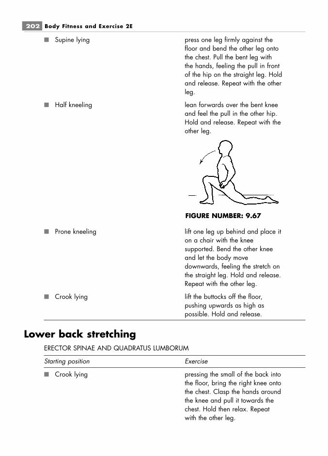
■ Supine lying press one leg firmly against thefloor and bend the other leg ontothe chest. Pull the bent leg withthe hands, feeling the pull in frontof the hip on the straight leg. Holdand release. Repeat with the otherleg.
■ Half kneeling lean forwards over the bent kneeand feel the pull in the other hip.Hold and release. Repeat with theother leg.
■ Prone kneeling lift one leg up behind and place iton a chair with the kneesupported. Bend the other kneeand let the body movedownwards, feeling the stretch onthe straight leg. Hold and release.Repeat with the other leg.
■ Crook lying lift the buttocks off the floor,pushing upwards as high aspossible. Hold and release.
Lower back stretchingERECTOR SPINAE AND QUADRATUS LUMBORUM
Starting position Exercise
■ Crook lying pressing the small of the back intothe floor, bring the right knee ontothe chest. Clasp the hands aroundthe knee and pull it towards thechest. Hold then relax. Repeatwith the other leg.
202 Body Fitness and Exercise 2E
FIGURE NUMBER: 9.67
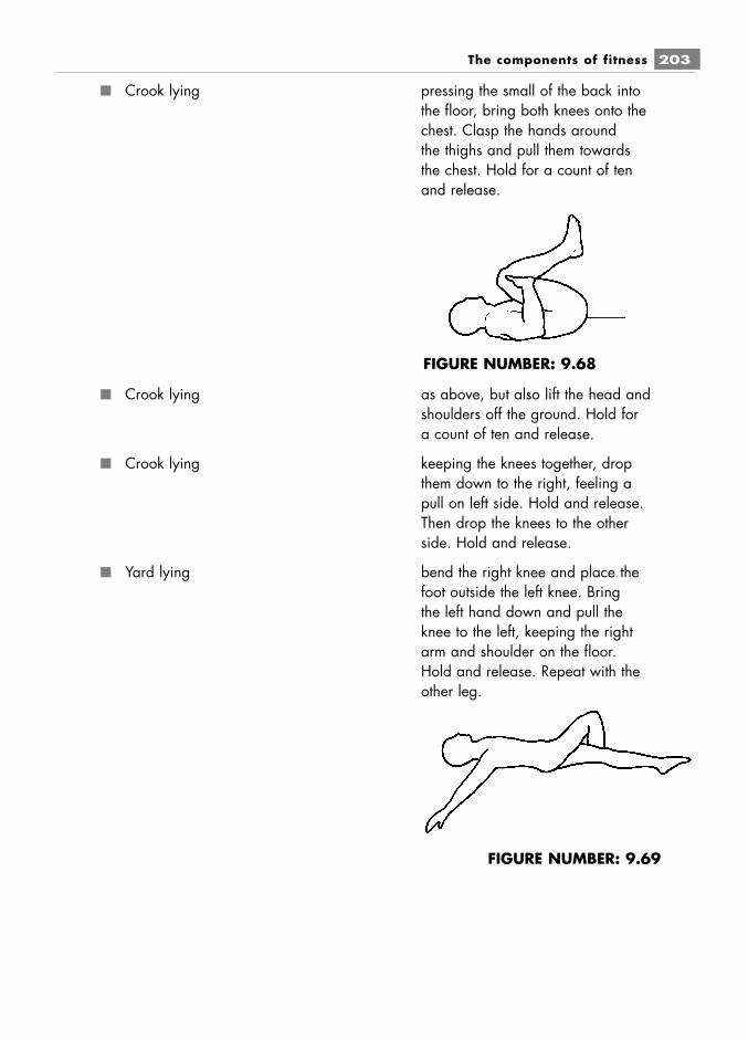
■ Crook lying pressing the small of the back intothe floor, bring both knees onto thechest. Clasp the hands aroundthe thighs and pull them towardsthe chest. Hold for a count of tenand release.
■ Crook lying as above, but also lift the head andshoulders off the ground. Hold fora count of ten and release.
■ Crook lying keeping the knees together, dropthem down to the right, feeling apull on left side. Hold and release.Then drop the knees to the otherside. Hold and release.
■ Yard lying bend the right knee and place thefoot outside the left knee. Bringthe left hand down and pull theknee to the left, keeping the rightarm and shoulder on the floor.Hold and release. Repeat with theother leg.
The components of fitness 203
FIGURE NUMBER: 9.68
FIGURE NUMBER: 9.69
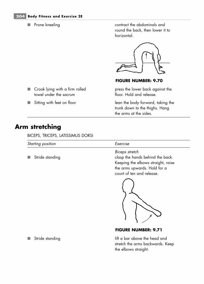
■ Prone kneeling contract the abdominals andround the back, then lower it tohorizontal.
■ Crook lying with a firm rolled press the lower back against thetowel under the sacrum floor. Hold and release.
■ Sitting with feet on floor lean the body forward, taking thetrunk down to the thighs. Hangthe arms at the sides.
Arm stretchingBICEPS, TRICEPS, LATISSIMUS DORSI
Starting position Exercise
Biceps stretch■ Stride standing clasp the hands behind the back.
Keeping the elbows straight, raisethe arms upwards. Hold for acount of ten and release.
■ Stride standing lift a bar above the head andstretch the arms backwards. Keepthe elbows straight.
204 Body Fitness and Exercise 2E
FIGURE NUMBER: 9.70
FIGURE NUMBER: 9.71

Triceps stretch■ Stride standing lift the right arm upwards and
bend the elbow so that the handlies behind the head. Use theother hand behind the head topush the upper arm down further.Repeat with the other arm.
■ Stride standing clasp the hands above the head,and pull the arms backwards as faras possible behind the head. Holdfor a count of ten and release.
Latissimus dorsi stretch■ Sitting Lift one arm upwards and bend
the elbow so that the hand liesbehind the head. Use the otherhand to pull the arm towards thebody and bend the trunk to thesame side. Hold and release.Repeat with the other arm.(Caution do not side Flex Trunk. Bendonly until pull is felt below armpit. Onlysuitable for young or agile.)
The components of fitness 205
FIGURE NUMBER: 9.72
FIGURE NUMBER: 9.73
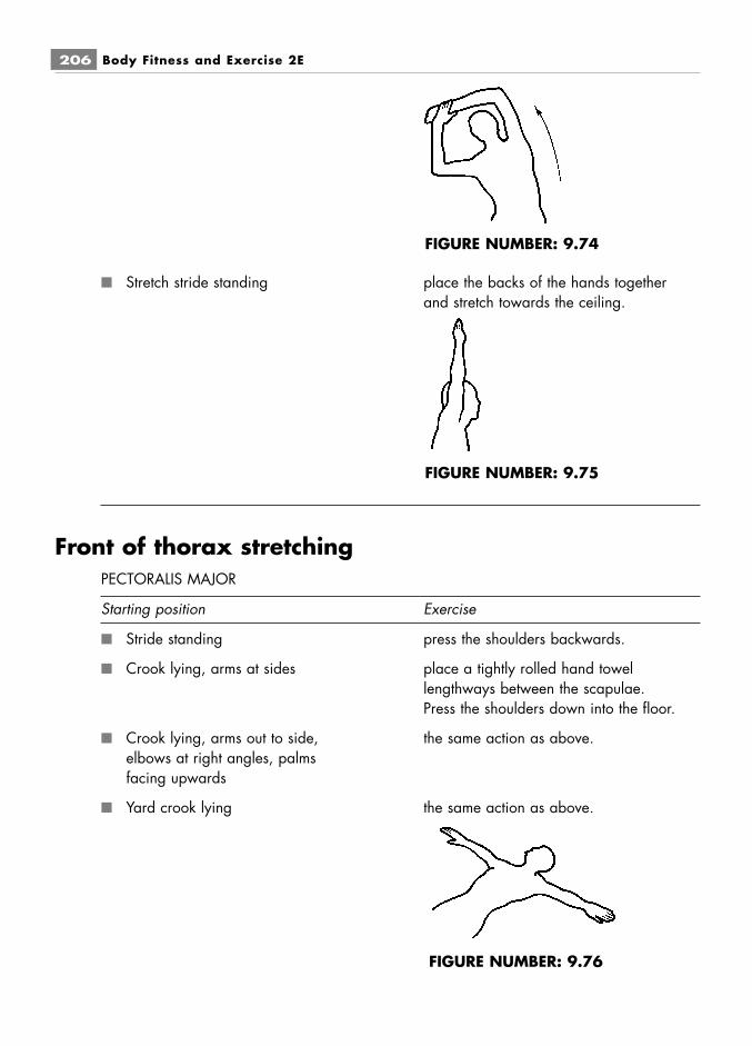
■ Stretch stride standing place the backs of the hands togetherand stretch towards the ceiling.
Front of thorax stretchingPECTORALIS MAJOR
Starting position Exercise
■ Stride standing press the shoulders backwards.
■ Crook lying, arms at sides place a tightly rolled hand towellengthways between the scapulae.Press the shoulders down into the floor.
■ Crook lying, arms out to side, the same action as above.elbows at right angles, palmsfacing upwards
■ Yard crook lying the same action as above.
206 Body Fitness and Exercise 2E
FIGURE NUMBER: 9.74
FIGURE NUMBER: 9.75
FIGURE NUMBER: 9.76

■ Prone kneeling stretch the arms forwards and outwards until the elbows are straight. Extend thewrists and drop the chest forwards,pulling the shoulders backwards.
■ Lying with a pillow between the raise the arms above the head andshoulders press them into the floor.
■ Stride standing or high sitting place one bent arm behind thehead with the elbow pointingupwards, the other behind theback with the elbow pointingdownwards. Clasp the hands ifpossible, or link them with a towelor strap. Pull downwards, bringingthe upper arm back and nearerthe head. Repeat with the otherarm.
■ Stride standing or high sitting use a bar that is shoulder-widthlong or just over. Hold the bar atthe ends, lift it upwards above thehead and then lower itdownwards behind the head.
■ Long sitting with back to a chair place the arms behind and graspor wall bars the sides of the chair. Keep the
elbows straight and thrust thechest upwards and forwards, keepingthe chin in. Hold then release.
■ Standing with back to wall bars place the arms behind and graspthe wall bar just below shoulderheight. Drop the body forwardand pull back between thescapulae. Keep the chin in. Holdand release.
The components of fitness 207
FIGURE NUMBER: 9.77
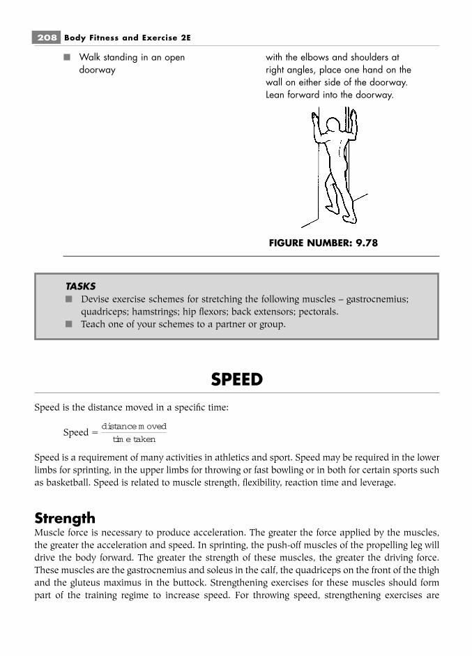
■ Walk standing in an open with the elbows and shoulders atdoorway right angles, place one hand on the
wall on either side of the doorway.Lean forward into the doorway.
SPEEDSpeed is the distance moved in a specific time:
Speed �
Speed is a requirement of many activities in athletics and sport. Speed may be required in the lowerlimbs for sprinting, in the upper limbs for throwing or fast bowling or in both for certain sports suchas basketball. Speed is related to muscle strength, flexibility, reaction time and leverage.
StrengthMuscle force is necessary to produce acceleration. The greater the force applied by the muscles,the greater the acceleration and speed. In sprinting, the push-off muscles of the propelling leg willdrive the body forward. The greater the strength of these muscles, the greater the driving force.These muscles are the gastrocnemius and soleus in the calf, the quadriceps on the front of the thighand the gluteus maximus in the buttock. Strengthening exercises for these muscles should formpart of the training regime to increase speed. For throwing speed, strengthening exercises are
distance m oved
tim e taken
208 Body Fitness and Exercise 2E
FIGURE NUMBER: 9.78
TASKS■ Devise exercise schemes for stretching the following muscles – gastrocnemius;
quadriceps; hamstrings; hip flexors; back extensors; pectorals.■ Teach one of your schemes to a partner or group.

required for the serratus anterior, pectoralis major, anterior deltoid, triceps, wrist and finger exten-sors.
FlexibilityA large range of movement at the joints will allow for longer strides, and long fast strides willincrease speed. Increased flexibility of the ankle, knee and hip will thus increase running speed.Increased flexibility of the shoulder, elbow and wrist will increase the propulsion force when throw-ing. Flexibility exercises should therefore be included in training programmes.
Reaction timeThe ability to react quickly to a stimulus is vital in many sports. Muscles must be able to contractinstantly in response to a stimulus such as a starting gun or the hitting of a service ball. Instantreactions to stimuli will improve speed. Reaction time can be improved by repetitive practice of therequired action, optimising body position and nervous response. Exercises where a quick responseis required, such as ball throwing, catching and running, are also used.
LeverageBody activities involve the movement of many levers. As previously explained (chapter 7), thelevers of the human body are mainly of the third order. These are designed for speed and range ofmovement. The longer the lever, the greater the speed, providing the force or muscle strength isgreat enough.
The effect of lactic acid on speedThe build-up of lactic acid has an inhibitory effect on muscle contraction. Bursts of fast activity ini-tially use stored ATP and PC, but these are soon used up and fast energy is obtained from theanaerobic breakdown of glycogen into pyruvic acid with the production of lactic acid. This build-up of lactic acid inhibits muscle contraction and therefore reduces speed. Athletes must thereforetrain to increase their aerobic capacity and to run close to their VO2max. Since the aerobic metab-olism is used for a longer period, there will be little or no lactic acid and little impairment of themuscle action.
This form of training will consist initially of short bursts of intense activity for 30–60 secondsand then rests of the same duration. The time is then progressively increased. These activities mayinclude shuttle running or a circuit of set exercises interspersed with running. Training forspeed involves different forms of running, beginning with jogging, moving on to running and finish-ing with maximum-speed sprinting. Variations include maximum-pace running interspersed withwalking or jogging. Training must also include specific training at maximum speed for set distances.
Other physical principles affecting speed include reaction forces, friction, resistance forcesand mass.
The components of fitness 209

SKILLSkilled movement is balanced, co-ordinated, graceful and precise, with accurate timing and overallrhythmic flow. There is no wastage of energy.
Skill is learnt behaviour and improves as a result of practice and experience. Acquiring askill is goal-directed: the learner must be aware of the desired end result and how to achieve it.Constant repetitions of patterns of movement must be practised until they are registered in thebrain and can eventually be performed automatically (see chapter 12). In order to master an activ-ity and give a skilled performance, the activity should be broken down into small chunks or parts.Each part should be practised until a satisfactory standard is achieved. The sequence of move-ments is then linked, and the complete activity is practised until all errors are eliminated and theperformance is automatic. Muscle strength and flexibility contribute to a skilled performance, butother factors are involved, such as neuro-muscular co-ordination, balance, etc.
Balance will improve by gradually reducing stability. Practice of a movement should beginin a stable position and progress to a less stable position. The speed of performance can also varyfrom normal to slow and then fast. Rhythm and timing will improve as the activity is mastered andco-ordination, grace and precision are achieved.
Summary and aid to learningSpeed is the distance moved in a specific time.
Speed times strength equals power.
Speed is a requirement of many sports and contributes to maximum performance.
List six sporting or athletic activities where you think speed is important.
The factors which influence speed are:strength – greater strength means greater driving force or propulsion; flexibility – greater flex-
ibility means greater range of movement, longer strides, etc.; reaction time – the faster the responseto a stimulus or instant reaction will improve speed; leverage – long levers improve speed; lactic acidbuild up – because the build up of lactic acid inhibits muscle contraction speed will be reduced.
Motor skill is precision of movement with no wastage of energy.
Skill is learnt behaviour and goal directed.
Factors which contribute to motor skill are:strength, flexibility, neuro-muscular coordination, balance, rhythm, etc.
Skill is an area of study which is not within the scope of this book.
210 Body Fitness and Exercise 2E

NUTRITION AND DIETThis is a vast and specialised field, the detail of which is not within the scope of this book.Therapists requiring detailed information should therefore refer to specialist texts. However, thischapter should provide you with a solid foundation for this important topic.
The food we eat is broken down by the digestive system and used to maintain body func-tions such as:
■ providing energy for cellular activity;■ providing building material for tissue growth and repair;■ producing hormones, enzymes and antibodies.
A good balanced diet must be eaten to supply all the nutrients necessary for the body toperform its functions and promote health. Any food which is surplus to requirements is convertedinto fat and stored in adipose tissue. These fat stores are our fuel storage tanks; in times of highdemand or low calorie intake, this fat is removed and broken down to provide energy for musclecontraction and cellular activity.
EnergyEnergy may be defined as the capacity to perform work. The energy requirements of the body willvary depending on the activities carried out. The basic energy requirement of the body for main-taining body functions in the waking state is known as the basal metabolic rate (BMR). This refersto the condition of the body first thing in the morning before getting up, or after lying down for atleast 60 minutes. BMR is proportional to body weight: the heavier you are, the higher your BMRwill be. It is generally higher for men and decreases with age. The demand for energy will propor-tionally increase as body activities increase, i.e. the greater the intensity of the activity, the greaterthe energy expenditure will be.
CaloriesA calorie is a unit of heat used to express the energy value of food.
■ A calorie is the heat required to raise the temperature of one gram of water by 1 degreeCelsius.
■ A Kilocalorie (Kcal) is the heat required to raise one Kilogram of water by 1 degreeCelsius.
The metric measurement of the energy value of food is the kilojoule. To convert Kcal to KJ:1 Kcal � 4.2 KJ
Calorie intake should never be below one’s basal metabolic rate; low calorie diets of 1,000Kcals or below are dangerous. A well-balanced diet of around 1,500 Kcals per day should provideadequate nutrients for those leading sedentary lives, but most people require more. The averageactive female consumes over 2,000 Kcals per day and the average male consumes over 3,000
The components of fitness 211

Kcals. However, for anyone involved in intense physical activity, e.g. athletes, sports people etc.,this will rise to 4,000–6,000 Kcals and above. These individuals must carefully consider their dietto ensure that they are meeting their energy demands.
Essential nutrientsThere are six basic nutrients necessary for a healthy diet:
■ carbohydrates■ fats (lipids)■ proteins■ vitamins■ minerals■ water.
Fibre is an additional requirement, as it aids the functioning of the digestive tract and pro-tects against many diseases.
Carbohydrates, fats and proteins are the energy providers and are known as energy nutri-ents or macronutrients. The other nutrients play important roles but they do not provide energy;they are known as micronutrients. Other chemical compounds derived from plants known as phytochemicals have recently been identified which offer protection against carcinogens (substanceswhich produce cancer).
CarbohydratesCarbohydrates give the body its source of quick energy, and are the starches, sugars and cellulosefound in pulses, cereals, bread, honey, potatoes, pasta, rice, and root vegetables. They are com-posed of carbon, hydrogen, and oxygen.
Carbohydrates are divided into three main groups:1 Monosaccharides (glucose, fructose and galactose);2 Dissaccharides (sucrose, lactose and maltose);3 Polysaccharides (starches, glycogen, and cellulose).
During digestion, complex sugars (polysaccharides) are broken down to simple sugars (di-saccharides) and then to one unit sugars (monosaccharides). These are absorbed in the smallintestine and transported in the blood to the liver, where fructose and galactose are converted toglucose.
Sources of carbohydrate in our dietSimple carbohydrates (monosaccharides and disaccharides) are the sugars found in honey, syrups,jams or fruits, etc. They give a quick surge of energy which quickly disappears, making us feel tiredand craving for more. Sugar and refined carbohydrates (white flour, white rice, etc.) are low in vita-mins, minerals and fibre and are therefore not as nutritious as unrefined and complex carbohy-drates.
212 Body Fitness and Exercise 2E

Complex carbohydrates (polysaccharides) are starches and fibre. They are found in bread,cakes, pasta, cereals, pulses, fruit and vegetables. They should form the greater percentage of ourdietary intake because they provide vitamins, minerals, fibre and phyto chemicals. Unrefined,wholemeal products (e.g. brown rice, wholemeal bread and pasta) have a high nutritional value.
GlucoseGlucose obtained from the breakdown of carbohydrates is used to produce ATP (adenosine tri-phosphate) required for muscle contraction. After eating a meal, blood sugar (glucose) levels willrise, which stimulates the pancreas to secrete insulin. Insulin reduces blood sugar levels, as it aidsthe transport of glucose from the blood into the cells. Glucose is then either used directly by thecells for immediate energy, or is converted in the liver to glycogen by a process known as glycogen-esis. This glycogen is then stored in the liver or in muscle tissue, and will provide a reserve ofenergy for future use. When these stores are full, surplus glucose is converted in the liver to fat,which is then stored as triglycerides in adipose tissue. Thus, a low fat but high carbohydrate dietmay still increase body fat if all the carbohydrate is not utilised for energy. When energy demandsare high, stored glycogen is needed. The liver then converts glycogen back to glucose, to supply theenergy required. This process is known as glycogenolysis.
The body is only able to store a small amount of glycogen – around 500 g – which amountsto around 2000 Kcals of energy. Of this, approximately 80 percent is stored in skeletal muscletissue and 20% in the liver. This is only enough to provide energy for one day of normal activity,so a regular intake of carbohydrate is necessary to maintain glycogen stores.
The average person with a sedentary life style should derive over 50 percent of total dailycalories from carbohydrates (this means consuming over 300 g daily). The athlete or anyone withhigh energy expenditure will require over 60–70 percent which means over 400–600 g daily.
The use of carbohydrates during exerciseGlycogen will be used to produce ATP during all forms of exercise, but the proportion usedincreases with the intensity and decreases with the duration of the activity. During short bursts ofintense anaerobic activity, such as fast short duration sprints, stored ATP, phosphocreatine and gly-cogen provide the energy. During prolonged moderate or intense aerobic activity, carbohydrateswill be the main source, but a proportion will be supplied by fats. Prolonged low intensity activitysuch as distance walking or jogging will use a greater percentage of fat.
Following any intense activity, muscle and liver glycogen will be depleted. It is important torestore glycogen levels as quickly as possible after exercise, by eating a high carbohydratemeal. The more depleted the store, the longer it will take to restore and the more carbohydratewill be required. It is recommended that 1 g of carbohydrate is eaten for every kilogram of bodyweight.
The components of fitness 213
LEARNOne gram of carbohydrate yields approximately 4.0 Kcals of energy

Exercising with low glycogen levels will lead to early muscle fatigue and poor performance.It is therefore vital for athletes, sports people, etc., to ensure a high carbohydrate intake as this willenable them to exercise for longer and to improve performance.
Research has shown that a high carbohydrate diet significantly increases endurance. Longdistance runners increase carbohydrate intake to around 80 percent of total calorie intake toenhance performance. These athletes may practise the technique of carbohydrate loading, to sub-stantially increase glycogen stores. This involves depleting glycogen stores for three days throughexercise and diet, followed by a high consumption of carbohydrates two to three days before theevent. However, this is no longer recommended, and at most should only be done twice or threetimes a year, as it can lead to fatigue and health risks. Different techniques of carbohydrate loadingare being introduced, including gradually decreasing the duration of training and increasing carbo-hydrate intake for seven days before performance.
During any activity of 60 minutes or over, carbohydrate intake can help to delay fatigue.Suitable consumables include carbohydrate sports drinks, or a banana.
FatsFats are the body’s secondary source of energy: carbohydrates provide primary fast energy, whilefats provide long-term energy. Like carbohydrates, they are composed of carbon, hydrogen, andoxygen but in a different ratio – there is less oxygen in fats. Fats or lipids are obtained from animaland vegetable sources.
Sources of fat in our dietThere are two main types of fat:
■ saturated■ unsaturated.
Saturated fatsThese contain the maximum number of hydrogen atoms in each molecule, and so are saturated withrespect to hydrogen. They tend to be solid at room temperature. Saturated fats are found mainly inanimal products such as pork, beef, lamb, butter, milk, cheese, eggs, etc. They are also found incertain plant products such as cocoa butter, palm oil and coconut oil. These fats contribute to highblood cholesterol levels and heart disease, and their excessive consumption should be discouraged.Unsaturated fatsThese contain fewer hydrogen atoms, and so are called unsaturated. This group include monoun-satured fats and polyunsaturated fats. These come mainly from plant sources, and tend to be liquidat room temperature.
■ Monounsaturates include olive oil and peanut oil.■ Polyunsaturates include corn oil, sunflower oil, sesame oil, cotton seed oil, soybean oil.
These fats help to reduce cholesterol levels; the consumption of these fats, in particular themonosaturates, is preferable to saturated fats.
214 Body Fitness and Exercise 2E
The use of fats during exerciseBoth saturated and unsaturated fats provide the same amount of energy per unit weight:
In addition to providing energy, fats protect vital organs such as the heart and kidneys, theyprovide body insulation, and are used in many physiological processes such as resynthesisingtissues e.g. the myelin sheaths of nerves and thromboplastin for blood clotting. They also transportthe fat soluble vitamins.
During digestion, fats are broken down to fatty acids and glycerol. If these fatty acids arenot required for immediate energy, they are converted and stored in adipose tissue and in the liverin the form of triglycerides.
Fats are the body’s most concentrated source of energy. Twice as much energy is stored in onegram of fat, than in one gram of carbohydrate. The body is able to store far more fat than glycogen,and so it is a greater store of potential energy which allows us to exercise for very long periods; evenmarathon runners do not run out of fat. However, fat requires a small amount of glycogen for its com-bustion and if glycogen stores are depleted, fat cannot be broken down to provide energy.
Fat cannot provide energy for fast activity, because it depends on the availability of oxygen,which depends on a person’s aerobic capacity. A fit person with high aerobic capacity will burn fatmore easily than an unfit person. During medium pace activities, fat provides around 50 percent ofthe required energy. If the effort is prolonged, fat will provide up to 90 percent of the required energy.
High fat intake increases the likelihood of developing high cholesterol levels, hypertensionand heart diseases. It is also linked with the development of many cancers, such as breast andcolon cancers. It is recommended that fat intake should not exceed 20–30 percent of total energyintake; of this only 6–10 percent should be saturated fat.
CholesterolCholesterol is a fatty like substance belonging to the chemical group known as sterols. Cholesterolis consumed in the diet, but is also synthesised in the liver. A high intake of saturated fat increasescholesterol levels, but it is also found in red meats, liver, kidney, egg yolk and dairy products. (Itis not found in vegetables.) A certain amount of cholesterol is needed by the body for building cellsand producing hormones, but too much is harmful, as it contributes to plaque formation in thearteries, which causes blockages and clots and increases the risk of coronary heart disease.Cholesterol is bound to two types of lipoproteins:
1 high density lipoproteins (HDL) – this is ‘good’ cholesterol as it does not adhere tovessel walls and may even protect against heart disease.
2 low density lipoproteins (LDL) – this is the ‘bad’ high risk cholesterol.
The components of fitness 215
LEARNOne gram of fat yields approximately 9.0 Kcals of energy
A low intake of saturated fats and cholestrol rich foods is recommended to protect againstheart disease.
ProteinsProteins are chemically different from carbohydrates and fats, and are more complex in structure.They are composed of carbon, hydrogen and oxygen, but also contain nitrogen, sulphur and iron.
Proteins are the tissue builders of the body, used for growth, body building and tissue repair.Proteins are:
■ used in the growth and repair of keratin, collagen, elastin■ used in the production of actin and myosin, which increase the size of myofibrils and
the strength of muscle contraction.■ needed to repair damage following injuries such as bone fractures, muscle strains,
tendon and ligamentous injuries etc.■ essential in rebuilding muscle cells after intense effort■ catalysts for many chemical reactions■ thought to have an effect on the nervous system, increasing arousal and alertness,
which are important for the elite performer■ used for making antibodies, enzymes and hormones.
Proteins are constructed from amino acids. There are 20 amino acids required by the body,and most can be manufactured by the body. However, eight cannot, and must be taken in from thediet; and these are known as essential amino acids.
Proteins are manufactured in the cells of plants and animals. Those containing all theessential amino acids are called complete proteins (high quality proteins). These are generallyobtained from animal sources such as lean meat, fish, eggs, milk and cheese. Those which do notcontain all the essential amino acids are called incomplete proteins (low quality), which areobtained from plant sources such as grains, pulses, fruit and vegetables. Vegans and vegetariansmust ensure that they eat a wide variety of these products, to ensure an adequate intake of essen-tial amino acids.
Food containing protein is broken down by the digestive system into amino acids. Thosetaken directly into cells are synthesised into new proteins, and the remainder are taken to the liverwhere some are synthesised into plasma proteins, others are de-animated and used for energy ifrequired, or converted to glycogen or fat and stored.
There are no protein stores in the body, unlike carbohydrate and fat stores. All body proteinis functional, therefore the body must ingest enough protein to meet its needs. Protein intakeshould be 10–15 percent of total energy intake i.e. 70–100 g per day. This should increase in timesof illness, growth, tissue repair, during pregnancy or training and performance. Many elite perform-ers increase their protein intake with supplements of amino acids (but there is no evidence to dateto prove that taking supplements enhances performance).
216 Body Fitness and Exercise 2E

Proteins are only used as an energy source in cases of starvation, or very long distancerunning when all stores of carbohydrates and fats are running low.
VitaminsVitamins are chemical compounds. They are essential nutrients which enable the body to functionefficiently, but they do not provide energy. They do, however, play important roles:
■ they regulate metabolic processes;■ they are important for growth, for the functioning of the nervous system and the
immune system;■ they are involved in enzyme production, and in many other biological functions.
Vitamins are obtained from plant and animals food sources, and a balanced diet will ensurean adequate intake. Vitamins K and B6 are formed by bacterial action in the large intestine, butthe others must be obtained from the diet.
Vitamins may be grouped into:■ Fat soluble vitamins (A, D, E, K)■ Water soluble vitamins (B complex and C)
Fat soluble vitamins are stored in the liver and in fatty tissue whereas water soluble vita-mins are excreted in the urine.
Fat soluble vitamins
Vitamin ASource: fish oils, butter, milk, cheese, eggs. Our body can produce this vitamin from carotenoids,e.g. beta-carotene found in yellow vegetables and fruits such as carrots, peaches, apricots, melonand in green leafy vegetables such as spinach and broccoli.Function: aids growth and repair of tissues; maintains mucous membranes, epithelial linings, andskin. Provides a visual pigment required for night vision. Beta-carotene may protect against heartattack, cancer, and reduce muscle soreness.Deficiency: nightblindness.
Vitamin DSource: fish oils, eggs, dairy products, fortified cereals, margarines. Produced in the skin by theaction of sunlight on dehydrocholesterol.Function: increases the absorption of calcium; promotes the growth of bones.Deficiency: rickets, brittle bones and bone deformities.
The components of fitness 217
LEARNOne gram of protein yields 4.0 Kcals of energy
Vitamin ESource: Wheatgerm, wholemeal cereals and bread, nuts, seeds, egg yolk, vegetable oils.Function: As an antioxidant it helps to protect against cancer, heart disease; it protects cell mem-branes; it helps muscles to utilise oxygen and may aid recovery after exercise.Deficiency: possibly anaemia.
Vitamin KSource: green vegetables, fruits, cereals, meat.Function: involved in the formation or prothrombin; essential in blood clotting.Deficiency: increased risk of haemorrhages.
Water soluble vitaminsVitamin B complexThis is a large group of vitamins; each has a particular function.Source: obtained from a wide variety of sources: lean meats, vegetables, pulses, legumes, wholegrains, dairy products, eggs.Function: They are essential for:
■ creating energy;■ converting carbohydrates into glucose;■ the metabolism of fats and proteins.
Some are associated with the manufacture of red blood cells, the growth and developmentof cells, and with the functioning of the nervous system.Deficiency: as this group of vitamins has a wide range of functions, deficiency will result in manydisorders and conditions – beriberi, pellagra, fatigue, muscular twitching, anaemia, nervous disor-ders, gastrointestinal problems.Folic acid: this is included in the B vitamins. It helps to form heme, the iron containing proteinwhich is needed to form red blood cells. It protects coronary vessels and is required for brain devel-opment and function.
Vitamin CSource: citrus fruits, berries, tomatoes, peppers, leafy green vegetables.Functions: growth and tissue repair; collagen formation; important for healthy gums, teeth andblood vessels; it helps to absorb iron and utilise folic acid; it forms adrenaline; it helps fight bacte-ria, is an antioxidant and may help fight cancer.Deficiency: scurvy, anaemia, poor connective tissue growth and repair, tender swollen gums andloose teeth, bleeding, decrease in exercise performance as it affects aerobic capacity.
MineralsMinerals are found in all body cells and fluids, and they form part of the body’s structure.Although these elements are only required in small quantities, they are esential for regulating andmaintaining life processes. They cannot be manufactured by the body and must be obtained fromthe diet.
218 Body Fitness and Exercise 2E

CalciumCalcium is required for the formation of teeth and bones. Inadequate intake of calcium results inporous bones, known as osteoporosis. Menopausal women are particularly susceptible to thisdisease, as there is a loss of bone mass and a high risk of sustaining bone fractures. Regular exer-cise, an active lifestyle and an adequate calcium intake will protect against this disease.
Iron is an important mineral, as it is a component of haemoglobin and muscle myoglobin.An adequate iron intake is therefore important for transportation and the storage of oxygen. Femaleathletes in particular must guard against iron deficiency (anaemia), as this will affect aerobiccapacity.
Other minerals such as sodium, potassium and magnesium are important for nerveimpulse conduction and fluid balance. Iodine is required for proper functioning of the thyroidgland.
Profuse sweating will result in water and mineral loss which must be replaced through a bal-anced diet. Sports drinks can help in cases of severe fluid loss.
Vitamin and mineral requirements of the athleteManufacturers imply that the athlete will derive great benefit from taking vitamin and mineral sup-plements. These micronutrients do not provide energy, and research indicates that there is nobenefit in consuming extra vitamins if recommended levels are maintained through a varied andbalanced diet. It is true that these nutrients are essential for the proper functioning of bodily pro-cesses but they are only required in small amounts.
Nutritional requirements will vary depending on age, size, levels of activity and metabolism.Individuals who are very active will require more than those who are inactive, but because theygenerally eat more, they will obtain an adequate supply of nutrients naturally from food.Supplements may be advisable for those who are restricting calories or for vegans; they may alsobe recommended following illness, during pregnancy, or for those suffering from anaemia or otherdeficiency diseases.
Performance may be adversely affected if the intake of vitamins and minerals is below therecommended levels, but providing intake meets the recommended levels, performance will not befurther improved by consuming large doses above this level. Advice should be sought from a doctoror qualified nutritionist before taking supplements.
When deficiency has been investigated and established, supplements must be limited to therecommended dose, and balanced to include all the nutrients which contribute in some way to per-formance. For example:
■ vitamin A, for repair of tissues■ vitamin B complex, involved in energy metabolism
The components of fitness 219

■ vitamin C, necessary for the absorption of iron and the forming of red blood cells whichtransport oxygen
■ vitamins C, E and beta-carotene to neutralise free radicals, thus limiting post exercisepain and soreness (explained below)
■ calcium, for strong bones■ iron, to improve the oxygen capacity of the blood■ magnesium, for nerve-muscle function, regulation of body temperature and to activate
the enzymes involved in energy production■ potassium and sodium, for nerve-muscle function■ phosphorus as part of ATP, for energy release■ zinc, for tissue growth and repair and as a part of the enzymes required for metabolism
of macro-nutrients.
Chemicals which affect the body
Free radicalsFree radicals are reactive chemicals; they are unstable atoms or molecules with unpaired electrons.They are continually produced in the body as a result of metabolic reactions, and are constantlytrying to pair up with other electrons to regain stability. In their effort to become stable, theybombard other cells, damaging the cells and their DNA.
They may damage skin cells causing ageing, liver spots or skin cancers; they also damagethe cells of other tissues producing various types of cancer. They attack and oxidise LDL choleste-rol in the blood stream, resulting in a ‘furry’ plaque which blocks arteries and increases the risk ofheart disease. Research has shown that there is a marked rise in free radical levels following exer-cise, and that they may be responsible for post exercise pain and stiffness. Fortunately substanceshave been identified that counter the effects of free radicals, which are known as antioxidants.
AntioxidantsThese act as scavengers, neutralising free radicals and thus protecting the body from damage.Some are found in the body as parts of enzymes, whereas others must be consumed in the diet;e.g., vitamins C, E and beta carotene, minerals such as zinc, copper, selenium and the many phytochemicals found in fruit and vegetables. Research indicates that these phyto chemicals affordeffective protection against free radicals and cancer.
Phyto chemicalsThese are chemicals such as flavonoids, sulphoraphane and chlorogenic acid. They are found infruits and plants, which, when eaten in adequate amounts, protect the body from carcinogens andpromote health.
Research indicates that at least five portions of fruits and vegetables should be eaten daily.The following fruits and vegetables will provide a variety of different phyto chemical, each havingdifferent protective effects:
220 Body Fitness and Exercise 2E

■ Broccoli, cauliflower, cabbage, sprouts, kale, tomatoes, peppers, pineapples, strawber-ries, grapes, raspberries, onion, garlic and soya beans.
In fact, most fruit and vegetables offer some form of protection.
Water and other liquidsWater represents 40–70 percent of total body mass. Individuals who are lean and muscular havea higher water content than fatter individuals with the same body mass, because fat contains lesswater. The body does not store water – it is excreted in urine. If water loss is high, the body willquickly become dehydrated and death will occur within days. Under normal conditions, the bodymaintains a balance between fluid intake and output; feelings of thirst indicate that the body isdehydrated and that water is needed.
Water is essential for the biological functioning of the body:■ it plays a vital role in regulating body temperature■ substances dissolve in water and are transported around the body in blood plasma and
lymph;■ it is a component of cells and tissue fluid and provides a medium for the exchange of
oxygen, nutrients and waste products between cells and the blood;■ as part of synovial fluid, it lubricates joints and reduces friction;■ it bathes tissues such as the eyes, brain and spinal cord;■ waste products are excreted in water, as urine and faeces;■ it absorbs heat and cools the body through evaporation. Dehydration occurs quickly
during vigorous activity through profuse sweating and expired air.
To maintain water balance, the average person with a sedentary lifestyle should drinkbetween two and three litres of water or diluted fruit juices every day. This is in addition to any tea,coffee or alcohol consumed because these are diuretics, which increase fluid loss.
Although it is a very unusual occurrence, it is important to remember that drinking too muchwater (over nine to ten litres per day) is dangerous. This will produce symptoms such as headache,blurred vision, sweating and vomiting. In extreme cases the brain is affected and the personbecomes delirious, comatosed and may eventually die.
Dehydration during exerciseContracting muscles generate a great deal of heat during exercise, up to 100 times more thanresting muscles. The body must get rid of this extra heat, or the core body temperature will rise todangerous levels. The blood transports this heat from the muscles to the skin surface where it islost through convection, radiation and evaporation (sweating). In hot weather, there is little or noheat loss through convection and radiation, therefore heat loss must be through evaporation withincreased sweating.
Vigorous activity produces profuse sweating, which can result in a fluid loss of 4–5 percentof body mass. The longer and harder the exercise and the hotter and more humid the conditions,
The components of fitness 221

the greater the fluid loss. Long distance runners may lose as much as two litres every hour throughthe lungs and skin. As fluid loss increases, the body becomes dehydrated. Water will be lost fromall body compartments and there will be a reduction in blood volume. Because there is less bloodfor the heart to pump per beat, cardiac output is reduced, which in turn reduces the delivery ofoxygen and nutrients to the contracting muscles; performance will be limited. The circulatorysystem tries to maintain blood volume to the muscles by constricting vessels and reducing bloodflow to the skin, therefore less heat is lost and temperature rises.
The symptoms of dehydration include: a decrease in level of performance, nausea, irritabil-ity, dizziness, fatigue, confusion and eventually complete exhaustion and collapse.
It is possible to prevent dehydration by ensuring an adequate intake of fluid before, duringand after vigorous or prolonged activity. Performers are recommended to:
1 Drink plenty of water the day before the event.2 Drink two cups of water two hours before the event.3 Drink one cup of water about 15–30 minutes before the event.4 Drink a quarter to half a cup every 15–20 minutes during the event (this is not neces-
sary for events lasting up to 30 minutes).5 Rehydrate fully after the event (this will depend on the degree of dehydration).
Recording the weight before and after performance will give an indication of how muchwater has been lost and how much needs replacing after exercise. For every 1 kilogram of weightlost, 1 litre of water should be drunk to restore balance.
Sports drinksDrinking water alone may not be enough to rehydrate the body, as the electrolytic balance mustalso be considered. Drinking water quickly removes the feeling of thirst (to protect against lowplasma electrolyte levels), and stimulates the kidneys to excrete urine. This occurs before rehydra-tion is complete. It is important to continue drinking even if feelings of thirst have diminished.Sports drinks are continually being developed, containing sodium and/or carbohydrate (glucose)and other electrolytes. Many of these drinks contain a glucose polymer, which is an easily digest-ible form of complex carbohydrate. The main aim of these drinks is to speed up rehydration; thosecontaining carbohydrate also maintain blood sugar levels, delay depletion of muscle glycogen, andthus increase endurance.
Research indicates that for athletes exercising at low intensity for up to an hour, water is aseffective as expensive sports drinks for preventing dehydration. Exercising for this length of time willnot utilise the glucose provided by sports drinks, and the sodium lost is easily replaced through diet.
However, when exercising for long duration at moderate to high intensity, sports drinks arerecommended as they rehydrate faster, and if consumed during exercise, can enhance perfor-mance. The intake of glucose raises blood sugar levels and spares muscle glycogen, so that themuscles contract harder for longer. Sodium helps to retain water in the blood without inhibitingthirst; it also limits urine production, therefore hydration is faster.
222 Body Fitness and Exercise 2E

Many nutritionists believe that the diets of most people are already too high in sodium andso do not require it as an addition to drinks. It must also be remembered that carbohydrates supplycalories, and those athletes wishing to control weight should avoid the extra calories found in thesedrinks.
Sports drinks come in different concentrations and selection is important. There are threekinds of sports drinks:
1 hypotonic2 isotonic3 hypertonic.
Although all these drinks will rehydrate the body, the main difference lies in their rate of absorp-tion which is dependent on their osmolarity (this refers to the concentration of solutes in a solu-tion). A drink with low osmolarity will have fewer particles in solution than a drink with high osmo-larity.
Hypotonic drinksHypo means: less than. These drinks have low osmolarity; they have a lower concentration ofsolutes (less particles), usually less than 4 grams of sugar per 100 ml. They pass quickly out of thestomach and are absorbed for fast rehydration. These are geared to the low/moderate, short dura-tion (up to one hour) athlete. Water would be just as suitable under these conditions, but somewill enjoy the flavour of these drinks and consequently drink more.
Isotonic drinksIso means: the same. These drinks have a higher concentration of solutes than hypotonic drinksand are absorbed at about the same rate as water. They contain 4–8 grams of sugar per 100 ml andwill refuel and rehydrate. These drinks are geared to the endurance athlete, exercising for one tothree hours; if consumed during performance, they provide extra glycogen.
Hypertonic drinksHyper means: more than. These drinks have a much higher osmolarity, and are more concentratedthan isotonic drinks. They slow gastric emptying, and are absorbed more slowly than water.Containing over 8 grams of sugar per 100 ml, they rehydrate slowly, spare muscle glycogen andprolong endurance. These drinks are geared to the ultramarathon runners, cyclists and others whomust maintain effort all day.
FibreFibre provides roughage and bulk which stimulates peristalsis and facilitates the movement ofwaste through the large intestine for excretion. We obtain fibre from wholemeal foods (brownbread, rice and pasta) and from fruit and vegetables. An intake of 20–35 grams per day is recom-mended. High fibre intake:
■ reduces the risk of heart disease■ helps to lower cholesterol level■ decreases the risk of cancer, particularly colon cancer■ reduces the risk of gastrointestinal problems and diseases■ protects against diabetes.
The components of fitness 223
A high fibre diet must be accompanied by high fluid intake to keep the colon functioningefficiently.
Weight controlWeight control is largely a balance between energy input (food eaten) with energy output (energyused).
■ If energy input equals energy output, weight remains stable (neither gained or lost).■ If energy input is greater than energy output, weight increases as the excess fuel is
stored on the body as fat.■ If the energy input is less than energy output, weight is lost as fuel is taken from the fat
stores.
Therefore the most effective way to lose weight is to eat less and increase the level of aerobicactivity. Aerobic exercise is the most effective form of activity for losing weight, as it utilises fat aswell as carbohydrate for energy.
Any diet should aim at reducing weight by around 1 kilo per week. It is important to eat abalanced and varied diet and to include each of the foods necessary for health. It is potentiallydangerous to reduce calorie intake to 1,000 cals or under. Most people will lose weight on a1,500–2,000 cals diet. It is worth noting that:
■ 1 gram of carbohydrate provides: 4 Kcals (17 kilojoules) of energy.■ 1 gram of protein provides: 4 Kcals (17 kilojoules) of energy.
but
■ 1 gram of fat provides: 9 Kcals (39 kilojoules) of energy.
Therefore, eating more carbohydrate and protein and cutting down on fat will result in lessenergy intake and quicker weight loss.
Body compositionBody weight is dependent on the major structural components of the body, which include bone,muscle and fat. Weighing machines tell us how heavy we are, and by regularly weighing ourselves,we know whether we have gained or lost weight. We can also compare our weight against the socalled ‘norm’ for our height, by checking established height–weight tables. If our weight is greaterthan the average values, we are classed as overweight; if our weight is less than the stated values,we are underweight. Being very overweight or very underweight can increase health risks.However, it is important to remember that these are only ‘average’ values; we may not fit into the‘average mould’, and weight alone tells us little about our individual state of health.
The Body Mass Index (BMI) is used to assess health risks in relation to weight and height.The BMI is calculated by dividing weight and height. The BMI is calculated by dividing weight inkilograms, by the square of the height in metres. The result is then checked against the followingrisk table.
224 Body Fitness and Exercise 2E

However, body composition is the important factor (i.e. the ratio of fat to lean tissue). Toomuch fat increases the risk of developing many serious diseases such as hypertension, heartdisease, vascular problems, stroke, diabetes, gall bladder disease, arthritis and certain cancers.
It is possible to be classed as overweight, even though the percentage of body fat is low.Many athletes and sports people develop large muscles through specific training which will addconsiderably to their weight. This is desirable weight gain from their point of view, and does notconstitute a health problem. They may be overweight when compared with average tables, but theywill not be ‘overfat’, which is the critical issue.
Although diet is the major factor in gaining fat, activity levels play a crucial part, as do otherfactors such as hormonal influences, inherited characteristics and somatotypes. These factorsmake it more difficult for some individuals to control their weight:
1 Endomorphs: These are short, stocky, curvaceous and plump. For this group weight/fatgain is easy but weight loss is difficult.
2 Ectomorphs: These are long limbed, slim and slightly muscular. They do not easily gainweight.
3 Mesomorphs: These are muscular and stocky. They gain weight/fat slowly but increasemuscle strength easily.
Individuals are predominantly of one type but may have aspects of another.
Summary and aid to learningAthletes and sportspeople require a low fat to lean tissue ratio, because fat means surplus baggageto carry around. This is costly in terms of energy expenditure which limits endurance. Transportingexcess fat will increase inertia, reduce speed and agility. The greater the weight, the greater theinertia and the greater the effort required to overcome it. Fat laid down in muscle tissue increasesfriction which impairs strength and function. Some athletes and dancers reduce calories to danger-ous levels in pursuit of leanness as it is aesthetically desirable and enhances performance.
The components of fitness 225
Table 9.1Ranges of body Mass Index
BMI Males Diagnosis BMI Females
Light frame Heavy frame Light frame Heavy frame
20� – 25 Acceptable 19� – 24
26� – 30 Overweight 25� – 29
31� – 40 Obese 30� – 40
41� Dangerously 41�
obese

However, athletes must balance training and diet, and consume extra calories to meet high energyand muscle building requirements, but guard against fat gain.
High carbohydrate, moderate protein, low fat intake is recommended, together with an ade-quate intake of vitamins, minerals, water and fibre.
Obesity or high fat ratio (i.e., 20 percent over desirable weight) is a major health problem inWestern society. The majority of the population is too fat and should change their diet and increaseexercise levels. A combination of healthy eating and increased levels of aerobic activity is the keyto success. Regular, moderate/low intensity aerobic exercise of 20–30 minutes duration 3–5 timesper week combined with reduced calorie intake of around 2,000 calories per day will result in fatloss, improved body shape, and an increase in lean tissue. It is an advantage to have a high pro-portion of muscle tissue as it has a high metabolic rate, therefore more calories are burnt.
Quick guide to healthy eating■ Reduce intake fat, particularly saturated fat. When cooking boil, steam or bake food; do
not fry.■ Reduce intake of red meat as it has high fat content, replace with poultry. Cut off all
visible fat from meat and remove the skin from poultry. Avoid eating prepared foodssuch as sausages, pate, pies, etc.
■ Reduce sugar and salt intake.■ Eat plenty of fish, particularly oily fish such as mackerel, salmon, herring, trout.■ Eat plenty of fresh or frozen vegetables – at least five portions per day (but do not over-
cook).■ Eat plenty of fresh, frozen or dried fruit.■ Eat wholemeal foods such as bread, pasta, rice, cereals, pulses, beans.■ Eat a wide variety of foods.■ Cut down on alcohol.■ Eat plenty of fibre.■ Drink two to three litres of water or diluted fruit juices per day.■ Low fat, high carbohydrate and moderate protein is recommended. Remember that fat
has twice the number of calories as carbohydrate and protein (weight for weight).
226 Body Fitness and Exercise 2E
The components of fitness 227
QUESTIONS1. Define the following terms
a basal metabolic rateb kilocaloriec macronutrientsd micronutrients
2. List the six basic nutrients necessary for a healthy diet.3. Explain the importance of including plenty of fibre in the diet.4. Give six examples of foods which provide carbohydrates.5. Name the two main types of carbohydrates and give examples of where each is
found.6. Explain why a high carbohydrate intake is vital for distance runners.7. List the two main types of fat and give examples of where each is found.8. Compare the energy yield of one gram of fat with one gram of carbohydrate and
protein.9. Explain why a fit person with a high aerobic capacity will burn fat more easily than
an unfit person.10. Explain what is meant by the term ‘essential amino acids’.11. Define the terms:
a Complete proteinsb Incomplete proteins.
12. Name the fat soluble vitamins.13. List four functions of vitamins.14. Name two important minerals and give their function.15. Explain what is meant by ‘free radicals’. Why are they undesirable in the body?16. Give three examples of antioxidants and explain their importance in the diet.17. Explain why water is an important component of the diet.18. Explain why vigorous exercise may result in dehydration.19. Give the symptoms of dehydration.20. Explain the difference between hypotonic, isotonic and hypertonic sports drinks.
Chapter 10Relaxation and posture
RELAXATIONRelaxation means freedom from tension and anxiety and involves both a physiological and apsychological state. Tension and anxiety are caused by stress, which upsets the body balance,known as homeostasis. The body ceases to function efficiently, resulting in lethargy, illness and dis-ease.
Stress has been defined as a non-specific response of the body to any demand made on it.Stressors, those factors causing stress, may be social, chemical, bacterial, physical, climatic orpsychological. People differ in their ability to cope with stress; some are more affected than others.We are all aware of the symptoms and may well have experienced some ourselves, e.g. increasedsweating, increased heart rate, higher blood pressure, rapid breathing, dryness of the mouth, inabil-ity to cope, feeling overwhelmed and out of control, inability to concentrate or make decisions,trembling, nail biting, frequent urination, non-stop talking, pacing and other nervous habits.
It is impossible to remove all stressors from daily life. Indeed, a certain degree of stress isdesirable and productive, and it can produce feelings of thrill and excitement. However, the ability
LEARNThe relaxed state means freedom from tension and anxiety

to relax is very important as it combats stress and reduces its harmful effects. It conservesenergy, reduces fatigue, lethargy and overtiredness and helps the body to return to a state ofhomeostasis.
Allowing the body to rest and recover is essential for those participating in gymnastics, ath-letics, sport and fitness activities. It is important for all participants in these activities to practiseand master relaxation techniques, since the ability to relax at the right moment can improve per-formance. Total relaxation conserves energy and concentrates the mind before events and soshould be practised both before events and during breaks or intervals.
Aids to relaxationA variety of aids can be used to promote relaxation:
■ heat therapy, e.g. heat packs, heat blankets, hot baths, showers, sauna and steam bathsand infra-red lamps;
■ cold therapy, e.g. cold packs and wraps;■ massage performed in a deep, slow and rhythmic manner;■ preparations such as analgesic liniments, wintergreen and other muscle relaxants.
Relaxation techniquesTo achieve long-term benefits, the individual must learn to recognise the difference between beingin a tense state and being in a relaxed state. As physical and mental relaxation are interdependent,both must be taught. Although relaxation techniques may appear simple, they are skills that mustbe learned and practised regularly.
Examples of relaxation techniques:■ The relaxation response■ Progressive relaxation (contract/relax technique)■ Visualisation or imagery■ Biofeedback.
Regardless of the technique, the selection of a warm, quiet environment and the position-ing and comfort of the client are important considerations. These factors alone may be sufficient toelicit the relaxation response, as explained below.
Preparation of the room■ The area should be warm and well ventilated.■ The area should be quiet and away from any distracting noises or activities.■ The lighting should be low and diffused.■ The colour scheme should be soft and warming, using pastel colours rather than harsh,
bold colours.■ The area should be spotlessly clean and tidy. All linen and towels should be boil-
washed and well laundered.
Relaxation and posture 229
■ A comfortable mattress on the floor provides the best support, with pillows for the headand knees. Two low plinths pushed together and covered with a thin mattress can beused. (Clients feel more secure nearer the ground and on a wide rather than a narrowsurface.)
■ Light blankets can be used for additional warmth.■ Very soft relaxing music may be played in the background. This depends on client pref-
erence, as some clients do not like absolute quiet and become tense.
Client careA full client consultation should be carried out.
■ Allow the client time to discuss their lifestyle and any problems that may be contribut-ing to stress and anxiety levels.
■ Discuss stress levels at work or during sport or training that may be affecting perfor-mance. Advise and suggest strategies for coping where possible. Explain how relaxationwill help.
■ Suggest suitable clothing, such as a loose-fitting cotton vest, T-shirt or sweater andloose-fitting pyjama or track suit bottoms. Loose socks can be worn on the feet. (Do notallow the client to walk around in socks as there is a danger of slipping.)
■ If suitable clothing is not available, loosen the clothing, remove the tie and belt, loosenthe collar and trousers or skirt and remove the shoes.
■ Suggest that the client uses the toilet, as it is impossible to relax with a full bladder.■ Use some form of heat, if available, prior to the commencement of relaxation training.
(Follow the correct procedure when applying heat.)■ Create an atmosphere conducive to relaxation. Smile, be calm, pleasant and relaxed,
speak slowly and clearly, keep your voice low and do not rush or hurry the client.Explain the procedure clearly and carefully to alleviate any anxiety.
The relaxation responseThe client relaxes in response to four basic conditions:
■ a quiet environment – this cuts out noise, limits distraction and allows the individual toswitch off;
■ a comfortable position – the position selected for all relaxation techniques is very impor-tant. The position should be selected to suit the preference of the client: lying, half lyingor the recovery position may be chosen. The body must be well supported with pillowsto minimise muscle effort and to enable the client to remain in this position for a con-siderable length of time;
■ mental concentration – this can be an image on which to concentrate, such as a sphere,box or vase, or any object in the room, such as a clock or mirror. The client concentrateshard on this one image and empties the mind of other thoughts or images;
230 Body Fitness and Exercise 2E
LEARNCreate an atmosphere conducive to relaxation. Allow the client time to talk andexplain their problems. Listen attentively and quietly.
■ a passive attitude – this is the most difficult, especially for those with extreme mentalanxiety. It involves letting go and emptying the mind of thoughts and distractions.
Progressive relaxationThis method was developed by Dr. Edmund Jacobson, one of the pioneers in the field of relaxa-tion. It aims to develop an awareness of the difference between feelings of tension and relaxationwithin muscles and muscle groups. The client is taught to contract and relax each muscle group insequence, from the foot to the head. With practice, the client will appreciate the difference betweenbeing tense and relaxed and will develop the ability to adopt the relaxed state quickly. The clientcan then be taught to recognise differing degrees of tension within muscles by using the samesequence but varying the contraction, from full contraction to part contraction and minimalcontraction, for each muscle or group.
The therapist should select a suitable venue and prepare the client (see page 00). The clientshould lie on a mattress and be fully supported. Modifications of the lying position can be used,for example the recovery position, with the body well supported with pillows, the supine position(on the back) with a pillow under the head and knees, or half lying, with pillows for the head andknees. Encourage the client to ‘let go’, breathe deeply and close the eyes gently.
The technique is then practised as follows, beginning with the feet and repeating eachmovement three times:
■ Pull the feet up hard (dorsi-flexion), then let go.■ Push the feet down hard (plantar flexion), then let go.■ Push the knees down hard against the floor, then let go.■ Push the leg down hard against the floor, then let go.■ Tighten the buttock muscles hard, then let go.■ Pull the abdominal muscles in hard, then let go.■ Raise the shoulders off the floor, then let go.■ Press the shoulders hard into the floor, then let go.■ Press the arms hard into the floor, then let go.■ Curl the fingers to make a fist, then let go.■ Press the head into the floor, then let go.■ Screw up and tighten the face, then let go.■ Tighten all groups, then let go.
The client should breathe out as he or she ‘lets go’.
The therapist must use her voice to good effect when teaching relaxation. The command‘tighten hard’ should be firm, and ‘let go’ should be spoken in a lower tone and drawn out longerto encourage the feeling of letting go. The terms ‘relax’ and ‘release’ can be used or interchangedwith ‘let go’.
Clients can then practise the sequence on their own until they are free of tension and sleepy.They should be left for 15 to 20 minutes and then woken up slowly.
Relaxation and posture 231
As clients develop the ability they can be taught to appreciate differing degrees of tension.This is done in the same way as above, except that the first contraction should be maximal,the second contraction partial and the third contraction minimal. The commands would be as fol-lows:
■ Pull the feet up hard, and relax.■ Gently pull the feet up, just feel the muscle pulling, and relax.■ Move the foot upwards ever so slightly, and relax.
Clients should then practise the three different contractions in their own time, feeling thedifference in muscle tension each time.
The therapist will work through the body in this way, using the same sequence as above.
Visualisation or imageryThis technique requires the individual to visualise situations or conditions conducive to relaxation.For example:
■ Imagine lying on the beach in warm sunshine. It is quiet and peaceful, you feel warmand heavy.
■ Imagine lying in a field in warm sunshine. You smell the grass. You feel warm andheavy.
■ Imagine sinking into a feather duvet. It feels soft and warm and wraps around you.■ Think of any situation that recalls warmth, comfort and peace.■ Concentrate entirely on the rhythm of breathing, letting the breathing become deeper
and slower.
Any examples that enhance relaxation can be included.
Visualisation with breathing can be used to good effect when performing static-stretch andflexibility exercises that require the relaxed state.
The client should exhale and move into the stretch position, stretching until tension is feltin the muscle belly. This position is held while the client breathes in and out slowly for a few cycles.The client then breathes in then out slowly while moving into a further stretch, imagining themuscle fibres letting go and lengthening. When tension develops the position is held briefly, fol-lowed by relaxation.
232 Body Fitness and Exercise 2E
TASKWork with a partner.■ Position your partner comfortably in the recovery position, using pillows as
required.■ Teach your partner to relax using the progressive relaxation technique.

BiofeedbackWhen it is difficult to appreciate the difference between muscle tension and relaxation, biofeed-back techniques using special equipment may be applied. The equipment gives a reading thatrelates to the degree of tension, and the mind is then used to attempt a change in the reading.
POSTUREPosture is the term used to describe the alignment of the body, in other words how the body is held.Good posture means that the body is balanced and the muscle work required to maintain anupright position is minimal. Poor posture means that the body is out of balance and certain mus-cles must contract strongly to maintain this position. Over time, this means that those muscles willtighten and shorten, while others weaken and stretch. This muscle imbalance imposes stresses onthe underlying structures, the ligaments and joints, resulting in deformities, stiffness and pain. Thebody loses its ability to function at maximum efficiency and the performance of everyday activities,movement and exercise can become severely limited.
Poor alignment of one part of the body can affect other parts. This can be more clearlyunderstood if we think of the body in terms of segments. Each segment must be perfectly balancedon the one below. If one segment moves forwards, backwards or sideways, adjustments have to bemade in all the other segments for balance to be restored.
Relaxation and posture 233
FIGURE NUMBER: 10.1 – Body segments must be balanced one on the other.

Posture is dynamic, constantly adjusting to counteract the forces acting upon the body.Postural adjustments may be made consciously or unconsciously. The cerebral cortex, basalnuclei, cerebellum and brain stem all play a part in the control of posture.
These higher centres respond to different impulses arriving from various sensory receptors.Information on the body’s position in space is received from muscle spindles, from tendon andjoint receptors, from the eyes and ears and from the skin on the soles of the feet. The higher cen-tres respond to incoming information and relay impulses back to the muscles, initiating musclecontraction to produce corrective action.
Posture is influenced by many factors, both physical and psychological. A large proportionof the population leads a sedentary life and takes little exercise, which leads to muscle imbalanceand poor body alignment. Other factors that influence posture include heredity, weight distribu-tion, height, nervous tension, illness, fatigue, occupational stress, poor working conditions andpoor sitting positions. Psychological and emotional states also have an effect – people who arehappy, confident and extrovert, with high self-esteem, exhibit good posture, while those who areunhappy, sad, introverted and lacking in confidence, with low self-esteem, have poor posture.
Poor posture results in muscle imbalance. Some muscles become tight, while others will beoverstretched. This imposes stress on the ligaments, tendons and underlying joints, producing painand stiffness. In addition, certain muscles are unable to work through their full range. This not onlyrestricts the activities of daily living but severely limits the capacity to perform at maximum poten-tial in athletics, sports, dancing, etc.
Poor posture affects general health. The natural movements of the thorax may be restrictedand its expansion limited, which results in shallow breathing. This reduces the intake of oxygenand the elimination of carbon dioxide. The circulation is affected due to the tension in muscles andthe reduction in thoracic movement, which mean that blood is unable to flow freely around thebody. This limits the delivery of nutrients and the elimination of waste products.
Good postural habits should be developed when young and maintained throughout life. Itis possible to improve posture for all age groups through appropriate exercise. The extent of theimprovement will depend on the degree of deformity, the age and the commitment of the individ-ual.
Good posture is important for the following reasons:■ maintaining muscle balance;■ improving body shape and appearance;■ preventing muscle tension, spasm and pain;■ preventing stresses on ligaments, tendons and joints;■ preventing skeletal deformities and associated pain;■ increasing the movement of the thorax, resulting in deeper breathing with an increase
in oxygen intake and the elimination of carbon dioxide;■ improving the efficiency of the circulatory system;
234 Body Fitness and Exercise 2E

■ improving the performance of all activities and exercises and enhancing peak perfor-mance;
■ reducing the risk of musculo-skeletal injuries.
Evaluation of posturePosture must be accurately examined and evaluated before correction can take place. An accurateassessment of posture should form part of the client consultation. All findings should be carefullyrecorded and appropriate exercises devised for correction of the faults.
Aids to postural assessmentThe following aids can be used to make assessment easier and more accurate.
■ a plumb line to check body alignment;■ a mirror to provide visual feedback for the client. First, look at the client’s normal pos-
ture and discuss any problem areas. Correct the posture and discuss the improvements.View the posture from the front, side and back. On the front view, lines can be drawnto check the level of the ear lobes and shoulders, the waist angles, and the level of theright and left anterior superior iliac spines and the knees. On the side view, draw a ver-tical line from the ear lobe to just in front of the lateral malleolus – does it fall throughthe plumb-line points? Check for round shoulders, kyphosis, lordosis, flat back, swayback and hyper-extended knees. On the back view, check for winged scapulae, scolio-sis, pelvic level and buttock folds.
■ a graphed board – the client stands in front of the board and the relevant bony points(ear lobes, shoulders, waist angles, knees) are marked, examined for any deviation anddiscussed.
Relaxation and posture 235
FIGURE NUMBER: 10.2 – Points that the line of gravity will pass through when posture iscorrect.

It is not necessary to use all these aids, but one or two will help the client to appreciate hisor her problems.
Procedure■ Welcome and observe the client as she/he walks into the room.■ Ask the client to sit, observing how he or she sits down.■ Take the client’s details – names, address, doctor’s address, medical history, occupation.
Discuss fully any stresses at work, working positions, seating, etc. Discuss lifestyle,associated activities, and nutritional standards. Try to assess the client’s psychologicalstate while talking – is he or she tense, under stress, fatigued or exhausted? Is theclient an introvert or extrovert, or are there any other factors that might influence pos-ture?
■ Ask the client to undress down to pants only, making sure that there is complete privacy.
■ If possible observe the posture as the client walks around the room (this may not bepossible in a small cubicle). Many problems can be observed when the body is inmotion. Observe the client sitting down and standing up: are the movements evenly bal-anced or does he or she sit and stand unevenly?
■ Ask the client to adopt a normal stance and assess the posture from the front, side andback.
■ Discuss any problem areas with the client. Stand him or her in front of a mirror, indi-cate the postural faults and show how they may be corrected.
■ Correct the client’s posture and ask him or her to hold the adjustments until good pos-ture is obtained (the new positioning will seem very unnatural at first).
■ Tell the client to relax and then to make the adjustments independently and hold thecorrected stance.
■ Explain to the client that frequent practice is needed throughout the day.■ Teach appropriate exercises to restore muscle balance.
The examination of posture
From the frontHead position:
■ Are the ear lobes level? If they are not there is muscle imbalance. The sterno-cleido-mastoid and the upper fibres of the trapezius are tight on the lower side, while those onthe other side will be stretched.
Shoulders:■ Are they level, or is one higher than the other, indicating muscle imbalance? The upper
fibres of the trapezius and levator scapulae are tight on the raised side. A difference inlevel may also indicate scoliosis, so check for that also. (A slight difference is consid-ered normal.)
■ Are both shoulders held high? This indicates tension in the muscles on both sides. Theright and left upper fibres of the trapezius and the levator scapulae are tight.
236 Body Fitness and Exercise 2E

■ Are the shoulders drawn forwards, rounded? This indicates muscle imbalance. The pec-toral muscles are tight but the middle fibres of the trapezius and the rhomboids arestretched.
■ Are there hollows above the clavicles? This indicates muscle tension, which may be dueto respiratory problems such as asthma.
Breasts:■ Are the breasts held high or sagging? If there is breast sag and round shoulders, correc-
tion of the posture may help to lift the breasts.Waist:
■ Are the waist angles on the right and left level? If one is lower than the other, there maybe spinal deformity or a difference in leg length.
Anterior superior iliac spines:■ Are they level? If not, there may be spinal deformity or a difference in leg length.■ Are they dropped forward? This indicates a lordosis with a tight erector spinae and
quadratus lumborum and weak abdominals.■ Are they dropped backwards? This indicates a flat back or sway back, with weak back
extensors, i.e. the erector spinae and quadratus lumborum, and tight abdominals.Patellae:■ Do they point forwards? If not there may be knock knees (genu valgum) or bow legs
(genu varum).Toes:■ Do they point forwards? If they point outwards there may be flattening of the medial
arch and flat feet.■ If they point inwards or outwards, the weight distribution over the foot will be wrong,
causing foot problems.■ Look for bunions, where the big toe deviates towards and sometimes across the other
toes and there is swelling at the metatarso-phalangeal joint.■ Look for hammer toes, where the inter-phalangeal joints are deformed.
From the sideUse a plumb line. This should fall through the lobe of the ear, the point of the shoulder and thehip joint, behind the patella and just in front of the lateral malleolus.Head position:
■ Is the neck or cervical curve exaggerated and the chin forward? This means that theneck extensors, the upper fibres of the trapezius at the back of the neck, are tight andthe neck flexors are weak.
Thoracic curve:■ Is there kyphosis, i.e. an exaggerated thoracic curve, giving a humped look? This means
that the pectoral muscles are tight and the middle fibres of the trapezius and rhomboidsare weak.
Abdomen:■ Is the abdomen protruding or sagging forwards, indicating weakness of the abdominal
muscles? The pelvis may be tilted forward. This is known as visceroptosis as the weakabdominals allow the viscera to sag forward.
Relaxation and posture 237

Lumbar curve:■ Is there lordosis, i.e. an exaggerated lumbar curve with the spine curved inwards? This
means that there will be an anterior pelvic tilt with weak abdominals and a tight erec-tor spinae and quadratus lumborum.
■ If the lumbar region is flat, which is much less common, the erector spinae and quad-ratus lumborum will be weak.
Buttocks:■ Are the buttocks well toned with strong muscles, or are the gluteal muscles weak and
sagging?Knees:
■ Are the knees hyper-extended?
From the backHead:
■ Are the ear lobes level or is the head tilted, indicating muscle imbalance? (See front.)Shoulders:
■ Are they level? (See front.)■ Are there winged scapulae, i.e. the inferior angle and medial border of the scapulae lift
away from the chest wall? This indicates a weakness of the serratus anterior and thelower fibres of the trapezius.
Spine:■ Is there scoliosis, i.e. a lateral deviation of the spine? This may be an S or C curve to the
right or left. If you are unsure, pull a finger firmly down the spinous processes: the red lineshould be straight, and will show up any deviation. A scoliosis may be structural (presentfrom birth). Or it may be postural, and will straighten out when the body is flexed forward.
Buttocks:■ Are the buttock folds level? If they are not, scoliosis, lateral pelvic tilt or different leg
length may be present.Heels:
■ Are these square and firmly planted on the ground? If not, the weight distribution willbe uneven.
Correction of the postureThe correction of the posture should begin at the feet. Each position should be maintained as thesubsequent one is practised.
FeetStand with the feet four to six inches apart, with the toes pointing forward. The weight should beevenly distributed between the balls of the feet and the heels.
Practise the following:■ Raise the toes off the ground, feel the weight evenly distributed between the balls and
heels, then lower the toes.
238 Body Fitness and Exercise 2E

■ Sway the body forwards, feeling more weight on the balls.■ Sway the body backwards, feeling more weight on the heels.■ Position the body so that the weight is evenly distributed between the balls and the
heels. Lift the medial arch slightly, but do not curl the toes.
Knees■ Press the knees backwards hard, ease the knees by bending them slightly, then find the
mid-point and pull the kneecaps upwards by tightening the quadriceps muscle.■ If the knees are hyper-extended, ease them slightly and pull the kneecaps upwards as
above.■ If the knees are bowed or knock-kneed, tighten the kneecaps, rotate the thighs outwards
and tighten the buttocks to bring the kneecaps to point forward.
Check the feet again after performing these movements.
PelvisTilt the pelvis forwards and then backwards; pull it forwards again slightly, tucking the tail under,and hold this balance. Pull the abdomen in and breathe out as the pelvis is pulled forward, thenhold this position while breathing normally.
ThoraxPull the thorax upwards from the waist as you breathe in, drawing the shoulders backwards anddownwards. Hold this position while breathing normally. Do not thrust the chest forwards.
Neck and headElongate the neck and pull the chin backwards. Feel as though someone is pulling the hairupwards at the crown.
Check the feet, knees, pelvis and thorax again, hold this position and then relax.
Practise this correction several times a day and during various activities; correct the postureduring inhalation and hold the balance during exhalation.
If the new posture is maintained while walking around, it will eventually become habitual.
Summary and aid to learningRelaxation means being free from tension and anxiety which are normally caused by stress.
Aids to relaxation include any form of mild heat, cold packs, slow deep massage, analgesicointments and types of muscle relaxants.
List the sources of heat that could be used to promote relaxation.
The preparation of the room is very important.
Relaxation and posture 239
Prepare a room or cubicle for a client needing to practise relaxation.
Relaxation techniques include:
The relaxation response involves the client’s response to a quiet soothing environment, totalconcentration on a particular object while trying to let go of all tension.
Progressive relaxation aims to develop an awareness of the difference between feelings oftension and relaxation. Contraction, relaxation of all the large muscle groups is performed workingsystematically around the body.
Visualisation or imagery involves visualising situations or conditions conducive to relaxa-tion.
Biofeedback involves a meter which registers tension; the client then attempts through willpower to lower the tension.
Practise these relaxation techniques with a partner.
Posture refers to the alignment of the body.
The upright posture is maintained by the contraction of the postural muscles.
List the postural muscles.
Poor alignment of one part will impose stresses on other parts.
Poor posture may affect health, as the movement of the thorax may be restricted; this willreduce oxygen intake. The tension of the shortened muscles and restricted movement of the tho-rax will adversly affect the blood flow around the body.
Give reasons why good posture is important.
Postural problems include:Kyphosis: an exaggerated thoracic curve.Lordosis: an exaggerated lumbar curve. Lordosis may be accompanied by the condition known asvisceroptosis which occurs when the abdominal muscles are very weak and the viscera protrudeforward.Kypho–lordosis is a combination of the above, where both thoracic and lumbar curves are exagger-ated.Scoliosis is a long C-shaped or an S-shaped lateral deviation of the vertebral column.Flat back refers to a loss of the lumbar curve resulting in a flat back.
240 Body Fitness and Exercise 2E

Relaxation and posture 241
QUESTIONS1. List some aids which may be used to promote relaxation.2. Explain why rest is important between training sessions.3. Explain the factors which must be considered when preparing the room for relaxa-
tion.4. List four basic conditions which promote the relaxation response.5. Describe the positioning of the client prior to teaching relaxation.6. Describe briefly how you would teach progressive relaxation technique.7. Explain how breathing and visualisation may be used to promote relaxation.8. Briefly explain the effect of inadequate rest and relaxation on the sportsperson.9. Give six effects of poor posture.
10. Describe the use of any two ‘aids’ which may be used when assessing posture.11. List all the points that a plumb line must pass through, if posture is correct.12. Describe the postural problem ‘kyphosis’, and indicate which muscles are involved.13. Name the condition associated with anterior pelvic tilt.14. Give six exercises for the correction of each of the following conditions:
a round shouldersb lordosisc flat back.
15. Name the weak muscles associated with ‘winged scapula’. Give four exercises tostrengthen these muscles.
TASKSWork with a partner. Carry out a role with one of you as the client and one as thetherapist.■ Greet the client and carry out a consultation.■ Assess the posture using the plumb line.■ Teach the correction of the posture.

Chapter 11Specific exercise for correction
of postural problemsSpecific exercise programmes may be required to mobilise joints, to strengthen specific muscles orto correct postural faults.
The objectives must be clearly stated and explained to the client. Once she is familiar withthe exercises the client should be encouraged to practise them at home. Careful analysis of theproblem areas is necessary so that the appropriate corrective strategy may be worked out. The fol-lowing are possible exercises for the correction of common problems.
Consider the starting positions carefully. Younger clients can use standing or modificationsof standing, but older clients will be more stable if sitting or lying. Older clients, or those withpainful or arthritic knee joints, should not be placed in the kneeling position.
In addition, read the sections on strengthening and stretching, the warm-up and the cool-down in chapter 9. Any appropriate exercises may be selected from those listed, or you may addsome of your own. Remember that strength will only improve if the muscle is made to work
LEARNConsider the starting position, carefully select the stability to suit the client. Avoidstressing painful joints
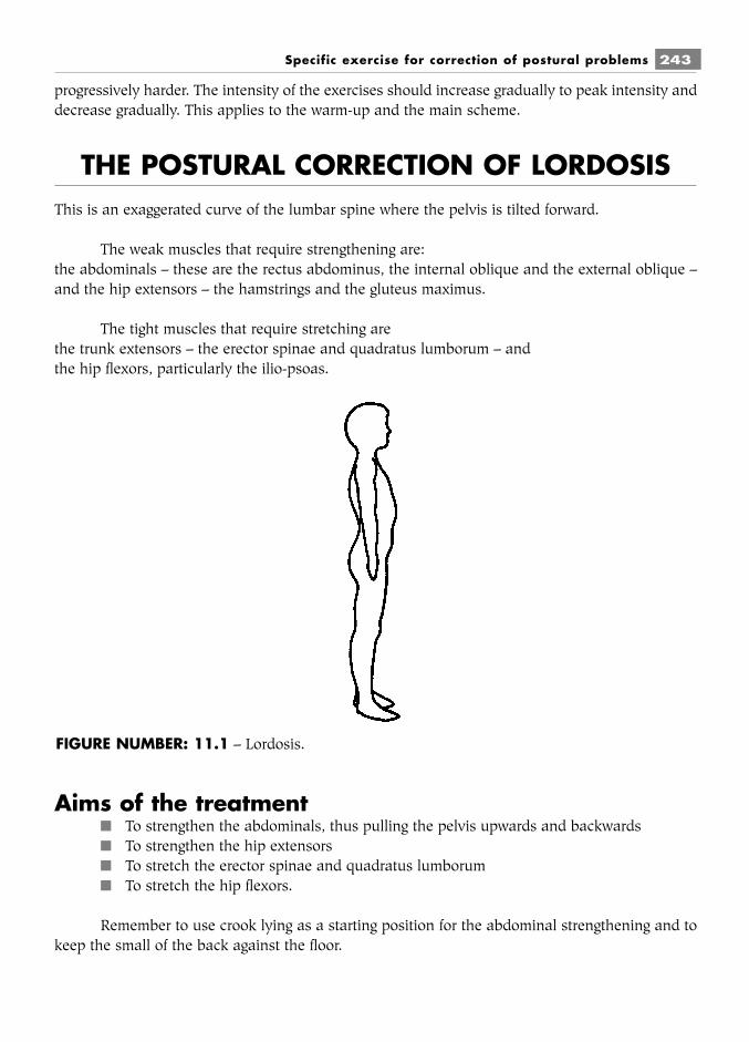
progressively harder. The intensity of the exercises should increase gradually to peak intensity anddecrease gradually. This applies to the warm-up and the main scheme.
THE POSTURAL CORRECTION OF LORDOSISThis is an exaggerated curve of the lumbar spine where the pelvis is tilted forward.
The weak muscles that require strengthening are:the abdominals – these are the rectus abdominus, the internal oblique and the external oblique –and the hip extensors – the hamstrings and the gluteus maximus.
The tight muscles that require stretching arethe trunk extensors – the erector spinae and quadratus lumborum – andthe hip flexors, particularly the ilio-psoas.
Aims of the treatment■ To strengthen the abdominals, thus pulling the pelvis upwards and backwards■ To strengthen the hip extensors■ To stretch the erector spinae and quadratus lumborum■ To stretch the hip flexors.
Remember to use crook lying as a starting position for the abdominal strengthening and tokeep the small of the back against the floor.
Specific exercise for correction of postural problems 243
FIGURE NUMBER: 11.1 – Lordosis.
Exercises for the correction of lordosisRead the appropriate suggestions for strengthening and stretching.
Warm-up – include exercises for mobilising, pulse raising and shortstretch
Starting position Exercise
■ Crook lying tilt pelvis upwards pressinglumbar region into the floor,release.
■ Crook lying keeping the chin in, raise the headand shoulders to look at the knees.
■ Crook lying curl up. This is progressed bymoving the hand position to headrest and then stretching above thehead.
■ Bend crook lying twist the trunk, bringing alternateelbows to the opposite knee.
■ Yard crook lying drop the knees to the right andthen to the left.
■ Lying slide the right arm down the rightside and the left arm down the leftside.
■ Prone kneeling arch the back to stretch thelumbar spine, then return tohorizontal.
■ Prone kneeling stretch alternate legs out and liftno more than fifteen degreesabove horizontal.
THE POSTURAL CORRECTION OF KYPHOSISThis is an exaggerated curve of the thoracic region. The shoulders are usually rounded, the neckis shortened and held in extension and the chin pokes forward.
The weak muscles that require strengthening are:the middle fibres of the trapezius, the rhomboids and the erector spinae.
244 Body Fitness and Exercise 2E
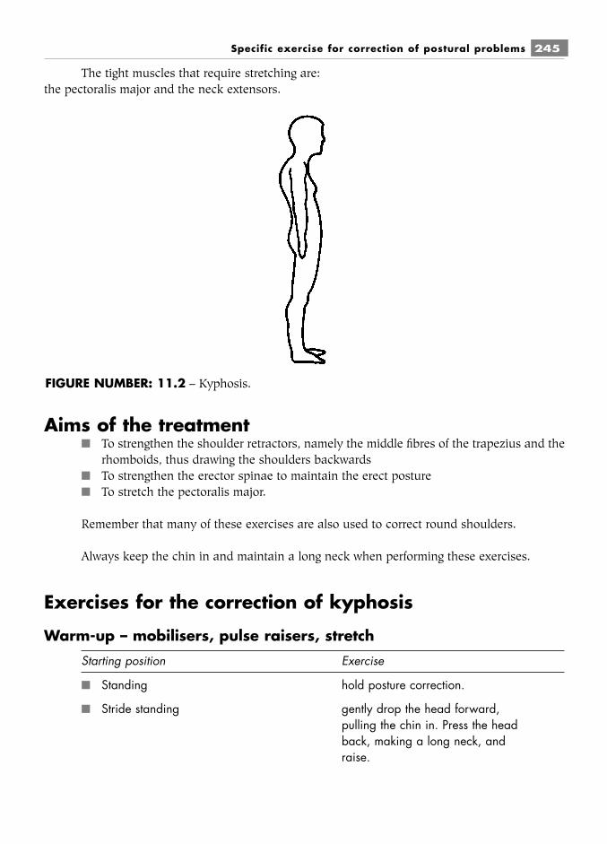
The tight muscles that require stretching are:the pectoralis major and the neck extensors.
Aims of the treatment■ To strengthen the shoulder retractors, namely the middle fibres of the trapezius and the
rhomboids, thus drawing the shoulders backwards■ To strengthen the erector spinae to maintain the erect posture■ To stretch the pectoralis major.
Remember that many of these exercises are also used to correct round shoulders.
Always keep the chin in and maintain a long neck when performing these exercises.
Exercises for the correction of kyphosis
Warm-up – mobilisers, pulse raisers, stretch
Starting position Exercise
■ Standing hold posture correction.
■ Stride standing gently drop the head forward,pulling the chin in. Press the headback, making a long neck, andraise.
Specific exercise for correction of postural problems 245
FIGURE NUMBER: 11.2 – Kyphosis.

■ Lax stoop sitting raise the trunk gradually from thebase of the spine, vertebra byvertebra.
■ Lax stoop sitting as above, against resistance fromthe therapist.
■ Stride standing circle the shoulders backwards.
■ Stride standing circle the arms backwards.
■ Stride standing pull the shoulders backwards.
■ Across bend stride standing pull the shoulders back andrelease. Then pull the elbows backand release. Then press the armsback and release.
■ Lax stoop stride standing slowly return to standing from thebase of the spine, vertebra byvertebra.
■ Stoop standing clasp the hands behind the back,pull the arms and shoulders upand back, hold for a count of tenand release.
■ Crook lying place a tightly rolled towellengthways along the spinebetween the scapulae, press theshoulders back towards the floor,hold for a count of ten andrelease.
■ Crook lying, arms out to side, as above.elbows at right angles, palmsfacing forward
■ Prone lying keeping the chin in, pull theshoulders back and raise the headand shoulders
■ Prone lying, pull on the hands and pull thehands clasped behind the back shoulders off the floor.
■ Prone lying, keeping the chin in, pull thehands clasped behind back shoulders back, lifting the head and
shoulders off the floor.
■ Wing prone lying keeping the chin in, lift the headand shoulders off the floor, pullingthe shoulders back.
246 Body Fitness and Exercise 2E

■ Wing prone lying as above, but against the resistanceof the therapist.
■ Prone lying with the arms abducted, elbowsbent and palms to the floor, lift thearms and head and shoulders.
■ Yard prone lying as above.
■ Sitting place one hand behind the neckand the other behind the back,and try to clasp hands or link witha strap. Pull downwards, bringingthe upper arm nearer the head,hold for a count of ten andrelease.
■ Sitting use a bar that is shoulder-widthlong. Hold the bar at the ends, liftit upwards above the head andthen lower it down behind thehead. Hold for a count of ten andlift up.
Kypho-lordosis is a combination of the previous two conditions. Select exercises from the twoschemes for this condition.
THE POSTURAL CORRECTION OF ROUNDSHOULDERS
In this condition the shoulders are protracted (drawn forward), the head is extended and the chinpokes forward.
This postural defect may be present without any kyphosis of the spine. However, if the spineis kyphotic the shoulders will also be rounded.
The weak muscles that require strengthening are:the middle fibres of the trapezius and the rhomboids.
The tight muscles that require stretching are:the pectorals and the neck extensors.
Specific exercise for correction of postural problems 247

Aims of the treatment■ To strengthen the shoulder retractors and draw the shoulders backwards■ To stretch the pectoralis major and the neck extensors.
For suitable exercises, refer to the scheme for kyphosis. The same exercises can be used forboth conditions.
You may wish to modify this slightly by selecting others from chapters 9 and this chapter orby adding your own.
THE POSTURAL CORRECTION OF SCOLIOSISThis is a lateral curvature of the spine, which may be a long C curve or an S curve. The conditionmay cause scapular deviation and slight unevenness in the levels of the shoulders and pelvic gir-dle, caused by muscle imbalance on the right and left sides of the spine. The spine must be care-fully examined. To make observation easier, run a finger downwards along the spinous processeswith slight pressure. The red line will show the extent and direction of the curve. If the conditionis postural, the curve will right itself in forward flexion; ask the client to bend over so that you cansee if this is so. If the condition does not correct with flexion it is a structural problem and shouldbe referred to a doctor.
The muscles that will require strengthening will be those on the outside of the curve.
The muscles that will require stretching will be those on the inside of the curve.
General back-strengthening exercises are usually effective in correcting this condition. It isfrequently found in adolescence, when pupils carry heavy school bags over the same shoulder orin the same hand each day. Suggest that the bag is carried in one hand to school and in the otheron the way home.
Aims of the treatmentTo restore balance to muscles of the back, thus reducing the deformity.
248 Body Fitness and Exercise 2E

Exercises for the correction of Scoliosis
Warm-up – mobilising, pulse raising and stretch
Starting position Exercise
■ Stride standing reach up into the air with the handon the concave side of the curve,where the muscles are tight, andreach towards the floor with the otherhand. Stretch, hold, relax, repeat.
■ Stride standing side flex the trunk towards theconvex side, where the musclesare stretched. Slide the hand downthe side and return.
■ Prone lying stretch the arm up along the flooron the concave side and slide thearm down the side of convexity.Hold, release and repeat.
■ Prone lying stretch the arm above the head onthe concave side and the oppositeleg along the floor.
■ Prone lying raise the opposite arm and leg as above.
■ Crook lying, rotate the arms to palms up andarms abducted and elbows at push back into the floor.right angles, palms to floor
■ Prone lying, raise arms backwards.arms abducted and elbows atright angles, palms to floor
■ Prone lying, arms to side keeping the chin in, lift the headand shoulders.
■ Prone lying, clasping hands keeping the chin in, lift the headbehind back and shoulders and pull down the
arms.
■ Prone lying, arms abducted, raise the arms, head and shoulders.elbows at right angles, palms to (Do not extend the head; keep itfloor in line with the body.)
■ Yard prone lying as above.■ Stretch prone lying as above.
Specific exercise for correction of postural problems 249

THE POSTURAL CORRECTION OF FLAT BACKThis is a condition where there is little or no lumbar curve, the back is flat in this region and thepelvis is tilted backwards. It is usually accompanied by kyphosis of the thoracic spine.
The muscles that require strengthening are:the back extensors, namely the erector spinae. (Sometimes the abdominals and gluteus maximusare weak.)
The muscles that require stretching are:the hamstrings.
Aims of the treatment■ To try to develop a normal lumbar curve by strengthening the erector spinae and glu-
teus maximus■ To maintain a correct pelvic tilt by ensuring the strength of the abdominals and stretch
of the hamstrings.
250 Body Fitness and Exercise 2E
FIGURE NUMBER: 11.3 – Flat back.
Exercises for the correction of flat back
Warm-up – mobilisers, pulse raisers and stretch
Starting position Exercise
■ Sitting lean forward, taking the pressurefrom the buttocks onto the thigh,then extend the back to create alumbar lordosis. Hold for a countof ten and release.
■ Prone lying raise alternate legs.
■ Prone lying raise both legs (allowed for thiscondition).
■ Prone kneeling arch and hollow the back.
■ Prone kneeling lift alternate legs up andbackwards, stretch, hold andreturn to the floor.
■ Lying with rolled towel under flex one knee, then extend the leglumbar spine up towards the ceiling. Keep the
opposite leg pressed hard downon the floor. Repeat with theother leg.
■ Long sitting rotate the pelvis forwards, thenlean slightly backwards. Hold andrelease.
■ Supine lying, legs up against pull alternate legs away from thea wall, with a towel under wall, keeping the knee straight andlumbar spine the pelvis on the floor. Hold and
release.
THE CORRECTION OF FLABBY UPPER ARMSThis is due to poor muscle tone in the triceps, which is the extensor muscle of the elbow. Fattydeposits in this area also contribute to the problem.
The muscle that requires strengthening is the triceps. If the client is overweight, he or shewill need aerobic work to reduce the percentage of body fat.
Specific exercise for correction of postural problems 251
Aims of the treatment■ To increase the strength of the triceps muscle■ To reduce body fat if necessary.
Remember that the triceps extends the elbow joint; therefore, elbow extension must be themovement that is included in all exercises for improving this condition. Movement downwards isassisted by gravitational pull and uses the biceps, working eccentrically, to control the movement,therefore triceps is not working. Movements using the triceps must therefore be horizontal orupwards.
Exercises for the correction of flabby upper arms
Warm-up – mobilisers, pulse raisers and stretchIf the client needs to lose weight, include an aerobic section here for 20 minutes.
Starting position Exercise
■ Across bend sitting or stride stretch alternate arms outstanding sideways and back; stretch both
arms out sideways and back.
■ Bend stride standing punch forward; punch a pillow orpunch bag.
■ Stride standing place the hands against thetherapist’s hands, with elbowsbent. Push alternate arms straightagainst the therapist’s resistance.
■ Bend stride standing or sitting stretch the arm vertically upwards.Repeat with progressive weights.
■ Stride standing, arms bent, hands push away from the wall.against a wall, leaning forwards
■ Prone kneeling bend and straighten the elbows.
■ Prone lying place the hands under theshoulders and push up.
■ Prone lying press-ups.
■ Lean sitting holding weights holding the weight in the hand atshoulder level, extend the armbackwards and upwards.
252 Body Fitness and Exercise 2E

■ Crook lying holding the weight in the hands atshoulder level, push the weightsvertically upward and then lower.The weights can be increased forprogression.
THE CORRECTION OF WINGED SCAPULAThis is a condition where the medial border and inferior angle of the scapula move back away fromthe chest wall. It is due to weakness of the serratus anterior and the lower fibres of the trapezius.
Muscles that require strengthening are:the serratus anterior and the lower fibres of the trapezius.
Aims of the treatment■ To strengthen those muscles which hold the scapula against the chest wall.
Remember that the serratus anterior is used powerfully in all punching movements. It helpsthe trapezius to swing the scapula laterally during arm abduction and arm swinging, and it workspowerfully to hold the scapula in position when the weight of the body is taken on the hands, asin prone kneeling, push-ups and press-ups.
The exercises for strengthening this muscle are the same as those for strengthening the tri-ceps.
THE CORRECTION OF FLABBY BUTTOCKSThese are caused by poor muscle tone in the hip extensors, primarily the gluteus maximus. Thehip abductors also contribute to the problem, namely the gluteus medius, gluteus minimus andtensor fascia lata.
The muscles that require strengthening are:the hip extensors, i.e. the gluteus maximus, the hamstrings, and the hip abductors.
The muscles that require stretching are:the hip flexors, primarily the ilio-psoas.
Aims of the treatment■ To increase the strength of the hip extensors and abductors■ To stretch the hip flexors.
Specific exercise for correction of postural problems 253

Remember that the hip extensors will extend the fully flexed hip backwards until the leg isin line with the body and for a further fifteen degrees. At this point further movement is preventedby the structure and ligaments of the hip joint and by the tension in the flexors. The hip extensorswill also pull the forward-flexed trunk upwards, working with origin and insertion reversed.
Exercises for the correction of flabby buttocks
Warm-up – mobilisers, pulse raisers, stretch
Starting position Exercise
■ High sitting tighten the buttocks.
■ High sitting press the thighs downwards into the seatand rotate them outwards.
■ Forward stoop sitting raise the trunk upwards against theresistance of the therapist.
■ Stoop stride standing, hands on raise the trunk to standing.legs
■ Crook lying tighten the buttocks and raise the pelvis.
■ Prone kneeling stretch alternate legs out behind.Attach ankle weights forprogression. (Caution: do not archthe back or lift the leg more thanfifteen degrees above horizontal.)
■ Side lying swing the upper leg forward and back.
■ Side lying push back against resistance fromthe therapist or springs.
■ Side lying raise and lower the upper leg,using weights for progression.
■ Prone lying bending the knee, raise alternatelegs off the floor.
■ Prone lying raise alternate straight legs off the floor(Caution: extend the leg fifteen degreesonly and keep the hips on the floor.)Use weights for progression.
■ Prone lying on a couch with one lift leg backwards and upwardsleg over the edge fifteen degrees from horizontal.
Use weights for progression.Repeat with the other leg.
254 Body Fitness and Exercise 2E

Specific exercise for correction of postural problems 255
TASKSWork with a partner. Practise a role play with one of you as the client and the otheras the therapist.■ Select one of the postural problems – the ‘client’ mimics this standing posture.■ Identify the weak muscles for strengthening and the tight muscles for stretching.■ Construct a scheme of exercises to correct the problem.■ Teach the exercises to the client.

This page intentionally left blank
PART CSafety
Considerations,Assessment and
exercise guidelines

This page intentionally left blank

Chapter 12General exercises
WARM-UPPerforming a warm-up routine prior to any active performance is an important safety factor. Thewarm-up is designed to prepare the body systems for more intense activity. It allows the systemsto make gradual adaptations in preparation for increased demand. This is very important for thecardiovascular system as it cannot adapt quickly and a change from a sedentary, relaxed state tohigh level activity will impose a stress on the heart. A warm-up should last for 15–20 minutes butenvironmental temperature should be considered. On cold days a longer warm-up routine will berequired. It should be long enough to produce a warm feeling and induce light sweating. This is agood indication that the body temperature has been raised sufficiently to begin the stretch routine.The intensity of the movements should increase gradually and decrease gradually. To bring aboutthe desired adaptations the warm-up should include:
pulse raisers are any aerobic activity which will raise the heart rate gradually. They mustbegin with low level, slow activity which gradually increases in intensity.
mobilising exercises which improve the range of movement at the joints. Initially the jointsare moved through middle range. As the warm-up continues the range is gradually increasedthrough to full range.
stretch/flexibility are performed to improve the extensibility and elasticity of muscles,ligaments and tendons. This may limit or prevent injury as these tissues are more able to accom-modate any excessive forces imposed upon them.
After these activities, athletes and sports people include specific movements or routines rel-evant to the performances e.g. tennis players practise the serve etc.
Warm-up is performed for both physiological and psychological effects.
Physiological effects of warm-up■ Increase in heart rate and dilation of capillaries which will increase blood flow to the
active muscles.■ Increased blood flow to active muscles through blood shunting (the volume of blood to
the viscera is reduced and redirected to active muscles). This will increase the deliveryof glycogen and oxygen to the muscles and removal of metabolic waste from themuscles.
■ Increased body and muscle temperature.■ Increased enzyme activity and increased metabolic rate due to temperature rise will
ensure a continuing supply of energy.■ Increased elasticity and extensibility of warm muscles will improve speed and force of
contraction.■ Increased extensibility of connective tissue components, ligaments and tendons will
reduce the risk of injury.■ Increased speed of nerve impulse transmission which will improve skill componants.■ Increased secretion of synovial fluid will improve the function of joints.■ Increased hormonal response. One example is glucagon which increases blood glucose
levels.
Psychological effects■ Aids mental preparation through visualisation and imagery of the following perfor-
mance.■ Increases arousal levels – there is an optimum level of arousal which will improve per-
formance but above this level performance will be adversely affected.■ Practise of the activities related to performance may increase confidence.
Examples of warm-up exercisesWarm-up exercises may be selected from the following:
■ Alternate heel raising■ Stepping, step-kicks■ Walking around the room■ Brisk walking around the room■ Marching on the spot■ Marching around the room■ Knee bend and grasp with hands■ Skipping
260 Body Fitness and Exercise 2E
■ Alternate leg swinging forwards and backwards■ Alternate leg swinging sideways■ Hip circling■ Pelvic tilting forwards and backwards■ Hip and trunk rotation clockwise and anti-clockwise■ Trunk side bends (caution: keep arms to sides)■ Shoulder shrugging■ Shoulder circling backwards and forwards■ Arm swinging across body, chest press, shoulder press■ Arm circling, beginning with small circles and increasing the range, or upward rowing■ Elbow bending and extending with backward arm swing■ Alternate arm and trunk upward stretch■ Neck flexion, extension (do not hyper extend), side flexion and rotation■ Combine leg and arm movements.
Cool-down/warm-downThis is performed at the end of a performance or exercise session. It reverses the processes thatoccur during warm-up and allows the body systems to gradually return to a balanced state. Againcool-down is performed for 15–20 minutes. Some of the performance activities can be repeated butat a low level of intensity. Active movements of the large muscle groups of the legs must also beincluded. The final part will be stretch movements of all the previously used muscles. Rest anddeep breathing may be added to finish.
Effects of cool-down■ To keep the heart rate and blood flow elevated. This blood flow will flush the metabolic
waste products out of the muscles which will help reduce the pain and stiffness oftenexperienced following exercise.
■ To maintain muscle contraction. If the exercises stopped suddenly, the heart is nolonger helped by the contracting muscles – this will increase the demand on the cardio-vascular system and heart rate will rise.
■ To maintain the pumping action of the contracting muscles to prevent blood pooling inthe legs due to the dilation of the blood vessel and the capillaries. This pooling of bloodin the legs results in a reduction in blood flow to the brain, which may cause dizzinessand fainting.
■ Stretching after exercise is important as it has been shown that this also reduces‘delayed onset muscle soreness’ (DOMS).
General exercises 261
LEARNAlways begin the routine with easy, gentle exercises, progress to the more difficultand then end with gentle exercises

COOL-DOWN EXERCISES
Examples of cool-down exercises■ Continue with some movements, from main activities but at lower intensity and pace.■ Skipping■ Jogging■ Walking■ Stepping■ Knee bends to chest
Complete cool-down with some stretch movements.■ Spinal rotation stretch■ Knee-hug hamstring stretch■ Shoulder rotations■ Gastrocnemius stretch
In prone lying:■ Push-up back extension■ Knee-bend quadriceps stretch
In long sitting■ Gastrocnemius stretch■ Hamstring stretch■ Spinal rotation stretch
End the class with deep breathing in the lying position and get up slowly.
MOBILITY EXERCISES
Neck mobilityNote: keep the shoulders relaxed throughout.
Starting position Exercise
■ Stride standing or sitting tuck the chin in and drop the headforwards, then lift the chin andtake the head backwards slightly. (Caution: do not hyper-extend.)
262 Body Fitness and Exercise 2E

■ Stride standing or sitting looking straight ahead, take thehead sideways, the ear towardsthe shoulder, first to the right andthen to the left.
■ Stride standing or sitting keeping the chin in, turn the headtowards the right shoulder andthen the left shoulder.
■ Stride standing or sitting tuck the chin in, drop the headforwards and keep the chin on thechest. Then turn the head to theright and then the left.
Do not circle the head round and round or hyper extend as this can damage the cervicaljoints and cause pressure on the spinal nerves in this area.
Shoulder mobilityStarting position Exercise
■ Stride standing or sitting pull the shoulders back and relax.Lift the shoulders up and down.
■ Stride standing or sitting circle the shoulders forward,upwards, backwards and down,then the other way.
■ Stride standing swing the arms forwards and upabove the head, then down andbackwards.
■ Stride standing swing the arms sideways and up tocross above the head, then downto cross behind the back.
General exercises 263
FIGURE NUMBER: 12.1

■ Stride standing keep the elbows straight androtate the arms medially andlaterally (turn in and out).
■ Stride standing circle the arms backwardsalternately and then together.
■ Stride standing swing the arms backwardsalternately and then together.
■ Stride standing raise the arms sideways to clapabove the head and then lowerthem to clap the sides.
■ Stride standing place the right hand behind theneck and the left arm behind theback, trying to touch hands, thenchange hands.
■ Stride standing or sitting reach forward, keeping the arms atshoulder level, cross the arms andclasp the hands, lift the armsstraight above the head and pushbackwards behind the head.
■ Yard stride standing bend the elbows with the palmsfacing forward, drop the arms sothat the palms face backwards androtate the arms up and down.
Combinations of the above movements can be used and performed to music. Towels, barsand dumb-bells can be used to add variety and interest.
■ Prone kneeling on elbows (not resting the chin on one hand, takehands) the other arm under the body and
stretch it down the outside ofopposite leg, then take it out andlift it out to the side. Repeat withthe other arm.
Trunk mobilityStarting position Exercise
■ Stride standing, hands on thighs keeping the chin in, curl the trunkforwards while sliding the handsdown the thighs.
264 Body Fitness and Exercise 2E

■ Stride standing, hands on buttocks extend the trunk backwards whilesliding the hands down thebuttocks.
■ Stride standing, hands to sides bend the trunk to the right andthen to the left while sliding thehands down the sides.
■ Crook lying pressing the lower back into thefloor, bend alternate legs onto the chest.
■ Crook lying bend both legs onto the chest.
■ Crook lying bend both legs onto the chest andlift the head and shoulderstowards the knees, clasping theknees with the hands.
■ Crook lying keep the shoulders against thefloor. Tip both knees to the rightand then to the left. Keep theknees together.
■ Supine lying stretch the right arm down theright side and the left arm downthe left side.
■ Prone kneeling with back flat arch the back upwards, pull in thestomach, lower the back andhollow it gently.
■ Prone kneeling with back flat bend the right knee onto thechest, arch the back and kick outbehind. Repeat with the left leg.(Caution: perform this exercisecarefully, lifting the leg only fifteendegrees above horizontal.)
General exercises 265
FIGURE NUMBER: 12.2

■ Prone kneeling stretch the opposite arm and legoutwards and upwards. Repeatwith the other side.
■ Supine lying lift the right leg to a right angle tothe body. Lower the leg towardsthe floor on the left side of thebody, twisting the trunk gently.Repeat with the other leg.
■ Prone lying lift the opposite arm and legupwards. Repeat on the otherside.
Hip mobilityStarting position Exercise
■ Support standing raise one leg and swing it forwardsand backwards. Keep the toespointing forwards, do not movethe trunk and keep the hipforwards. Repeat with the otherleg.
■ Support standing raise one leg out sideways andswing it sideways and back acrossthe other leg. Keep the toepointing forwards and do notmove the trunk. Repeat with theother leg.
■ Support standing raise one leg and circle it around.Repeat with the other leg.
■ Prone kneeling bend the knee up to the chest andmove out and up. Repeat with theother leg. (Caution: this should beperformed carefully, lifting the legonly fifteen degrees abovehorizontal.)
■ Supine lying part the legs and bring themtogether.
■ Crook lying drop the knees outwards andbring them together.
266 Body Fitness and Exercise 2E

■ Supine lying bend the knees onto the chest andstraighten them into the air,keeping them at 90°to the body.Open and cross the legs in ascissor movement. Bend the kneesto the chest to lower them.
■ Standing run on the spot, lifting the kneeshigher and higher.
■ Standing star jumping – jump feet intostride standing position and back.(Caution: do not abduct too far.)Only for the young and agile.
■ Walk standing bring back foot forwards and takeforward foot back by jumping.
Knee mobilityStarting position Exercise
■ High sitting on a chair or the swing the lower legs up and downend of a couch from bent to straight knee.
■ Prone lying on a couch, with bend alternate knees, bringing heelfeet over the end to buttock, and straighten.
■ Prone lying on a couch, with bend one knee and cross thefeet over the end other leg over the back of the
bent leg. Now push with the backleg to increase the bend in thefront leg. This is useful if there islimited range in the knee joint.Repeat with the other leg.
■ Supine lying bend one knee onto the chest.Kick it out straight and lower it. Ifbending is limited, grasp the kneewith hands and pull it into thebody. Repeat with the other leg.(Caution: DO NOT perform thisexercise with both legs together.)
■ Supine lying bend the knees onto the chest andstraighten them into the air. Cyclethe legs in the air.
General exercises 267
■ Support standing hold onto a wall bar with thehands at shoulder height. Bend theknees and hips down to thesquatting position, hold andstraighten. (Caution: do not takethe buttocks below the knees.)Only for young and agile.
Foot mobilityStarting position Exercise
■ Sitting with feet on floor ■ push the toes into the floor(do not let them curl);
■ raise the toes away from thefloor;
■ spread the toes outwardsand together;
■ move the big toes towardseach other;
■ pick up a pencil with thetoes.
■ Sitting with feet on floor and a Keeping the heel firmly pressed ontotowel or strap lengthways under towel, use the toes to pull the towelfoot towards the heel.
■ Sitting with feet on floor and Keeping the heel on the ground,a towel or strap widthways under move the towel to one side andfoot then the other.
■ Sitting with hands over top of toes push down with the hand andlift the toes up against the resistance.
■ Sitting with hands under the toes push up with the hand and pushdown with the toes against the resistance.
■ Sitting with one leg across the other turn the foot in and out.at the knee
■ Sitting with one leg across the other pull the foot up and down.at the knee
■ Sitting with one leg across the other circle the foot ten times one wayat the knee and ten times the opposite way.
■ Long sitting turn the feet inwards and pressthe inner arches together.
268 Body Fitness and Exercise 2E

Additional exercises:■ Walk around, moving the weight from the heel to the outer border to the ball of the foot.■ Walk in a straight line, moving the weight from the heel to the outer border to the ball
of the foot.■ Walk around on the toes.■ Walk around on the heels.■ Walk normally.■ Skip or jog, changing direction.■ Hop on one leg, then the other (only for young and agile).■ Jump forwards, backwards and sideways.■ Jump to stride and back, jump to walk and back.
THE BENEFITS OF EXERCISEResearch has shown that regular exercise is beneficial for all age groups. We have seen from ourstudy of the body systems how they make physiological adaptations in response to exercise oroverload. The extent of these changes will depend on the type of exercise and on the systems sub-jected to stress. Appropriate exercises regularly practised will improve physical fitness, promotehealth and reduce the risk of developing many diseases. Even moderate activities willimprove health and the quality of life. Activities need not be specific exercise. Gardening,walking, vacuuming, dancing or any activity which expends energy will result in improvementproviding it is done for 30 mins, 5–6 times/week. The greater the energy expenditure thegreater the benefits. The beneficial effects derived from exercise are both physiological and psycho-logical.
Physiological benefits:■ improvement in cardio-vascular function, i.e. heart and circulation;■ improvement in respiratory function, i.e. lungs and breathing;■ improvement in muscle tone, strength and stamina, which in turn improves posture and
body contours (shape);■ improvement in the flexibility and tensile strength of tendons and ligaments. This will
increase the range of movement at joints and reduce the likelihood of trauma;■ improvement in the condition of joints. Exercise increases the production of synovial
fluid, which lubricates and nourishes the cartilage;■ improvement in bone density, which will combat osteoporosis;■ improvement in neuro-muscular co-ordination, which improves skills such as rhythm,
balance, timing, reaction time and co-ordination;
General exercises 269
TASKWork as a group. Each member of the group must devise and teach five mobilityexercises for one named joint. Each person should take a different joint.
■ increase in metabolic rate, with reduction in fat reserves. Muscle tissue has a high meta-bolic rate, therefore the more muscle tissue on the body, the higher the metabolic rate.This will increase demand for fuel and thus reduce fat stores;
■ lowering of blood cholesterol levels;■ reduction in trauma and pain due to improved posture, strength and flexibility.
The psychological benefits are:■ feelings of well-being, achievement and euphoria;■ an increase in self-confidence and self-esteem;■ reduction in stress levels;■ promotion of relaxation and sleep.
Health-related benefits■ Reduces the risk of developing high blood pressure.■ Helps to reduce high blood pressure in those who suffer from it.■ Reduces the risk of developing heart disease.■ Reduces the risk of developing diabetes.■ Reduces the risk of developing vascular disease such as atherosclerosis (which is nar-
rowing and hardening of the arteries, caused by the build up of fatty plaques on thevessel walls).
■ Helps to reduce body weight and improve body shape.■ Helps to reduce cholesterol levels.■ Reduces the risk of developing colon cancer.■ Helps to build stronger bones thus reducing the risk of developing osteoporosis.■ Helps to prevent joint problems.■ Maintains strength and mobility therefore improving quality of life.■ Reduces anxiety and depression.
To achieve these benefits, exercises must be carefully selected and accurately and carefullyperformed. Exercise should always be specific to the individual, and the degree of ease or difficultyshould be appropriate for the level of fitness. Inappropriate exercises that are too difficult or arecasually and excessively performed can result in damage and pain.
Damaging effects of inappropriate exercise■ muscle strain, tears and soreness;■ ligamentous sprains, overstretching and tears;■ joint stresses;■ bone stresses;■ inflammation of tendons, bursae and joint capsules, namely tendonitis, bursitis and
capsulitis;■ pain, which adversely affects daily activities, relaxation and sleep. Pain may also pro-
duce feelings of tension, stress, depression, disappointment and low self-esteem.
270 Body Fitness and Exercise 2E

IMPORTANT POINTS TO REMEMBER■ To improve fitness, the principle of overload and progression must be applied. The
intensity of exercise must be greater than that normally encountered and must becomeprogressively harder. Repeating the same exercise a set number of times will maintain alevel of fitness but will not result in significant improvement. The variables that are usedto increase overload are intensity, duration and frequency.
■ Different types of exercise stress different body systems and produce different effects.Cardio-respiratory or aerobic endurance is a fundamental requirement of fitness foreveryone. Other components should be specific to the sport or performance and meetthe needs of the individual.
■ Muscle flexibility and the range of joint movement will improve through regular stretch-ing exercises.
■ Muscle strength and bulk will increase through strengthening exercises, i.e. regularlyworking the muscle against progressive resistance. The degree of tension developed ina muscle will be directly proportional to the overload. Fewer repetitions at maximumoverload will increase muscle strength.
■ Muscle endurance, i.e. the capacity of a muscle to repeat an activity without fatigue, willimprove if the muscle is made to perform repetitive movements. High repetitions withlow resistance will increase muscle endurance.
■ Speed will increase if movements and activities are practised at increasing pace.■ Skill, agility, balance and co-ordination develop through repeated practice of an activ-
ity, increasing the difficulty by altering the base, the centre of gravity and the pace.■ Cardio-respiratory endurance will improve in response to regular practice of aerobic
activities such as jogging, walking, cycling, swimming and aerobic exercise classes. Theexercises must be slow and steady and increase very gradually in intensity. This allowsthe heart, blood vessels and lungs to deliver sufficient oxygen to the muscles for thecomplete breakdown of glycogen. If the exercises become too fast or intense, thenanaerobic metabolism takes over.
■ To deliver aerobic endurance, the heart rate must be raised, but it must not be raisedbeyond 80–90 per cent of an individual’s maximum heart rate. Remember: MHR is cal-culated as 220 minus the person’s age. Initially, the pulse rate should be raised to only60 per cent of MHR, progressing to 70 per cent and then to 80–90 per cent. The elevatedheart rate must be maintained for fifteen to twenty minutes per session. Exercise shouldbe repeated three to four times per week on alternate days to allow time for recovery.
■ To develop anaerobic systems, short sharp bursts of activity must be practised. Theseshould be explosive and dynamic activities such as squash, fast sprints, shot putting,etc.
■ Spot reduction of fat through exercise is not possible. Exercising a specific area of thebody will improve muscle tone in that area, but will not disperse covering fat. To reducebody fat, calories consumed (intake) must be less than calories used (output). Only thenis fat removed from all over the body and broken down for energy. Long durationaerobic exercises are high calorie burners, use fat for energy and are the best way of
General exercises 271

reducing body fat. Strengthening exercises are low calorie burners, so lots of curl-upswill not remove fat from the abdomen, although they will improve the strength ofabdominal muscles.
■ Weight is often gained rather than lost when people take up exercise, even though fatis reduced. This is because muscle tissue is heavier than fat. Losing weight is not anindication of fitness: it is the ratio of fat to fat-free tissue that is important. More muscleand less fat will result in an increase in fitness and better body shape.
■ Muscle tissue has a high metabolic rate. Therefore having more muscle tissue gives anindividual a higher metabolic rate, which means that calories are burnt up more quickly.This makes it easier to lose weight. Through regular exercise people who burn caloriesslowly can become high calorie burners, thus reducing fat stores more rapidly.
■ Muscles will not change to fat if exercising stops, as is commonly supposed. However,muscle strength will be reduced and the muscles will feel softer. Muscle and fat are com-pletely different tissues. Fat is stored on the body as a reserve of energy if calorific intakeis greater than calorific output.
■ The warm-up is a very important part of all training and exercise routines. It enables theenergy systems to increase their rate of work gradually. It warms muscles, whichimproves their contractability and flexibility. It improves the flexibility of connectivetissue components and reduces the likelihood of injury. The warm-up should includemobilising exercises, pulse-raising exercises and simple static stretch.
■ The cool-down or warm-down is equally important, as it allows the system to slow downgradually and aids the removal of the waste products lactic acid and carbon dioxide.The cool-down enables the body to return to a stable state, i.e. homeostasis is re-established. Cool-down will include less strenuous or gentle activity and stretching ofthe muscles used in the training.
■ Exercises should never stop suddenly, nor should one stand still after vigorous exercise,as fainting and dizziness may result. During vigorous exercise the blood vessels supply-ing the muscles dilate, which increases the load on the heart, but the contractingmuscles assist in pumping the blood around the body. If exercise stops suddenly, thevessels are still dilated and blood pools in the legs due to gravitational pull. Thisdeprives the brain of oxygen, causing dizziness and faintness. The pressure on the heartis increased as it attempts to maintain blood supply. The cool-down must include gentleactivity such as jogging or walking around, followed by stretching and breathing exer-cises that are performed while lying down.
Exercise and asthmaAsthma sufferers may exercise with caution, but must not over-exert. One must always bear inmind that physical exertion can sometimes trigger an asthmatic attack. This is referred to asexercise-induced asthma. If breathing difficulties are experienced for longer than normal, i.e. overfive minutes, and there is obvious distress, then medical advice should be sought quickly.Asthmatics should always have bronchodilators to hand, and must learn to recognise distresssignals and take appropriate steps to control the condition.
272 Body Fitness and Exercise 2E

Chapter 13Safety and hygiene factors
related to exercise
SAFETYThe safety of clients and other staff is of paramount importance and is the responsibility of the ther-apist in charge. Every precaution should be taken to create conditions and consider factors whichwill avoid injury.
The factors which contribute to injuries are:■ Inadequate demonstration, unclear commands or poor tuition.■ Activities that are too difficult and inappropriate to age and fitness levels.■ Excessive overload or too rapid an increase of overload.■ Failure to warm-up and cool-down adequately.■ Inadequate rest during the exercise session.■ Inadequate recovery time between sessions.■ Ignoring contra-indications and proceeding with exercise.■ Ignoring signs of stress while exercising and not stopping.■ Incomplete recovery from previous injury.■ Faulty equipment.■ Improper use of equipment.

■ Equipment left lying around in the working area.■ Insufficient space and overcrowding.■ Inappropriate footwear and clothing.■ Intensive competition among participants.■ Poor exercise technique and poor body alignment.■ Inappropriate starting positions.■ Performing potentially damaging exercises.■ Dehydration.
This is a long list but you may be able to think of other factors. The following guidelines willhelp you to take the precautions necessary to avoid injury.
CONTRA-INDICATIONS TO EXERCISEAll clients should have a thorough consultation before embarking on an exercise programme. Anyof the following conditions would indicate that the client should not exercise. Unfamiliar condi-tions may be highlighted during consultation. If in doubt, always seek medical advice.
Common contra-indications are:■ any recent injuries: these would include fractures, strains, sprains, ruptures or tears. It
is sometimes desirable to maintain fitness in other parts of the body while the injuredpart is immobilised. The exercises for other body parts must be carefully planned andperformed, ensuring that no stress is placed on the injured part and surroundingtissues;
■ heart conditions or any history of heart disease. Appropriate exercise regimes are under-taken following heart attacks and surgery, but these should be medically directed orsupervised;
■ high blood pressure: generally, if the blood pressure is controlled by drugs, exercise isallowed, but check with the client’s doctor. Relaxation can help hypertensive clients, butisometrics should never be performed;
■ any acute fevers, such as influenza, glandular fever, the common cold, etc;■ any infections, such as throat infections, measles, chicken pox, etc;■ any inflammatory joint conditions, such as arthritis. Rheumatoid arthritis is a systemic
condition in which joints become hot, swollen and stiff. Osteoarthritis is a condition ofwear of the joints, the cartilage wears thin, making movement very painful;
■ neurological disorders, such as strokes, multiple sclerosis, etc. – exercises for these con-ditions must be medically supervised;
■ undiagnosed illness: seek a doctor’s advice;■ musculo-skeletal problems, such as joint or back pain;■ pain and soreness in muscles caused by trauma or injury as opposed to delayed onset of
muscle stiffness;■ pregnancy, medical consent must be sought and gentle exercises only should be given.
During the first three months of pregnancy, particular care must be taken. The fit client
274 Body Fitness and Exercise 2E

who has exercised regularly may continue with low-impact, low-intensity work. Weightsshould not be used, nor should exercises that increase intra-abdominal pressure be per-formed.
■ after eating a heavy meal or under the influence of alcohol;■ if overtired or exhausted;■ if under the influence of pain-killing drugs;■ if there has been any past difficulty with exercise.■ Diabetics must obtain medical advice and approval. Low or moderate intensity exercise
is normally recommended.
Be particularly aware of increasing problems with age. To be safe, all those over 40 shouldhave a medical check-up before starting an exercise programme.
Those who are at greatest risk are:■ obese people;■ those with a history of heart problems in the immediate family;■ hypertensives;■ diabetics – a doctor’s referral is important, especially if the client is on insulin;■ those with a history of lung problems, such as asthma, bronchitis or emphysema;■ smokers.
Refer these people for a check-up before commencing exercise programmes.
SAFETY FACTORS TO OBSERVE WHILEEXERCISING
The premises■ The room should be warm, well ventilated and without draughts. It must not be too hot.■ There must be good, even lighting, with no pools of light or dark corners.■ Lights should be shielded with guards, particularly if games are played.■ The floor should be firm, smooth and non-slip, and preferably sprung.■ There must be sufficient space for everyone to move freely, with no overcrowding.■ The room should be clean and uncluttered; all apparatus not in use should be stored
neatly away from the working area.■ Apparatus should be in good condition; there should be no rough edges or sharp pro-
truding parts that could cause injury.■ There must be a sufficient number of well-marked fire exits.■ A well-stocked first-aid box should be clearly visible and accessible.■ Shower and toilet facilities should be available.■ Drinking water should be available but kept away from the working area.
Safety and hygiene factors related to exercise 275

■ Water and fluids must be kept away from the working area, as spillages make the floorslippery and dangerous.
■ There should be no eating or drinking in the working area.■ Exercises should be supervised by qualified instructors at all times. Accurate demon-
stration and instruction must be given.■ Protective mats should be available for floor exercises, one per client.■ Mirrors should be available to check body alignment and to correct the performance of
activities.
The client■ Suitable clothing that will allow free, unrestricted movement must be worn. Cotton is
the best fabric, as it allows easy absorption of perspiration. Cotton vests or T-shirts andshorts with elasticated waists, or cotton bodies, are all suitable.
Some athletes wear track suits and leg-warmers to maintain or raise body tempera-ture during the warm-up and stretch routines.
■ Footwear must be chosen with care to suit the activity. Well-constructed shoes should bebought from a reputable manufacturer. Footwear for exercise should be light and comfort-able and offer good lateral support. The toe box should have sufficient height, breadth andlength to prevent the toes rubbing. The inner sole should absorb shock and the outer soleshould be pliable, durable and non-slip. The tongue should be padded to protect thedorsum of the foot and the heel tab should not be too high and should not press on the heel.
Socks alone should never be used for exercise because of the danger of slipping, butthey should be worn with shoes to reduce friction.
■ Hair should be tied back off the face. Hair combs, slides and pins should be avoided.■ Jewellery should be removed.■ Check for contra-indications: if in doubt, seek a doctor’s advice.■ Clients must not exercise after a heavy meal, nor under the influence of alcohol. Allow
at least two hours after eating.■ Clients must not exercise if pain-killing drugs have been taken.■ All equipment to be used must be fully demonstrated, its effect explained and safety
factors highlighted.■ Exercise or training must be specific to the individual. Clients must work at their own
pace and level, must not exceed their target rate■ Clients should not be encouraged or allowed to compete against each other■ The different levels of fitness and the age range of those present must always be care-
fully considered when giving group exercise. Individuals must rest when tired and mustnot exceed their maximum heart rate during performance.
■ Exercises should not cause pain – clients must be advised to stop exercising if pain isexperienced.
■ Select safe, stable starting positions.■ Exercises must be clearly explained and demonstrated accurately and any precautions
must be stressed. The client must fully understand the exercise and be aware of poten-tial hazards.
276 Body Fitness and Exercise 2E
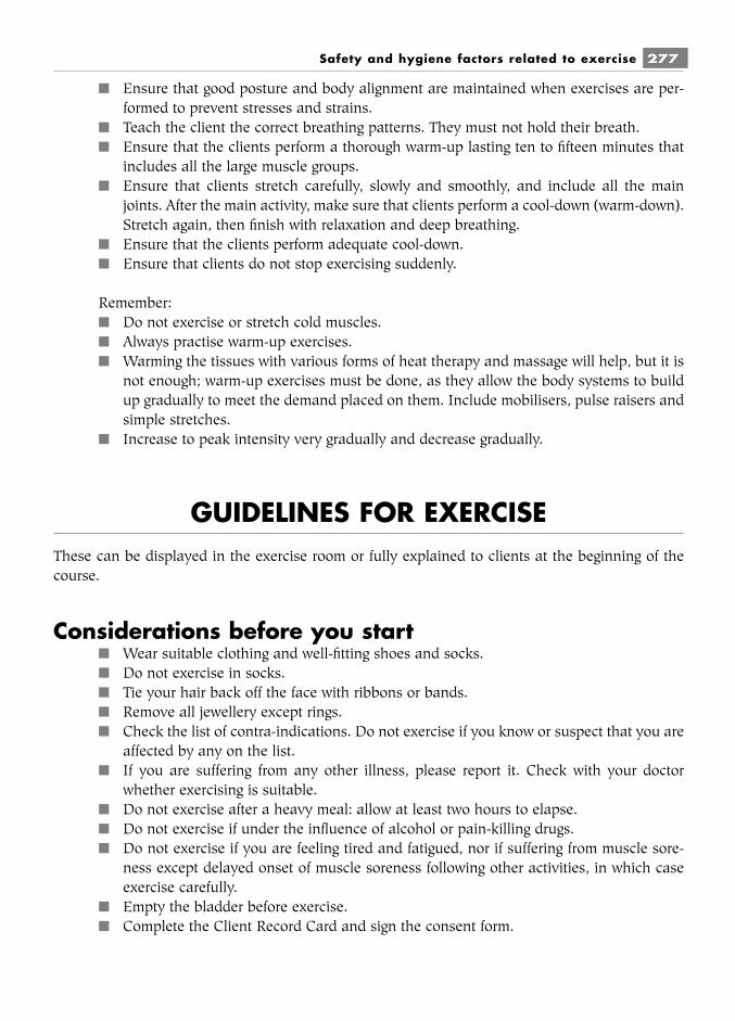
■ Ensure that good posture and body alignment are maintained when exercises are per-formed to prevent stresses and strains.
■ Teach the client the correct breathing patterns. They must not hold their breath.■ Ensure that the clients perform a thorough warm-up lasting ten to fifteen minutes that
includes all the large muscle groups.■ Ensure that clients stretch carefully, slowly and smoothly, and include all the main
joints. After the main activity, make sure that clients perform a cool-down (warm-down).Stretch again, then finish with relaxation and deep breathing.
■ Ensure that the clients perform adequate cool-down.■ Ensure that clients do not stop exercising suddenly.
Remember:■ Do not exercise or stretch cold muscles.■ Always practise warm-up exercises.■ Warming the tissues with various forms of heat therapy and massage will help, but it is
not enough; warm-up exercises must be done, as they allow the body systems to buildup gradually to meet the demand placed on them. Include mobilisers, pulse raisers andsimple stretches.
■ Increase to peak intensity very gradually and decrease gradually.
GUIDELINES FOR EXERCISEThese can be displayed in the exercise room or fully explained to clients at the beginning of thecourse.
Considerations before you start■ Wear suitable clothing and well-fitting shoes and socks.■ Do not exercise in socks.■ Tie your hair back off the face with ribbons or bands.■ Remove all jewellery except rings.■ Check the list of contra-indications. Do not exercise if you know or suspect that you are
affected by any on the list.■ If you are suffering from any other illness, please report it. Check with your doctor
whether exercising is suitable.■ Do not exercise after a heavy meal: allow at least two hours to elapse.■ Do not exercise if under the influence of alcohol or pain-killing drugs.■ Do not exercise if you are feeling tired and fatigued, nor if suffering from muscle sore-
ness except delayed onset of muscle soreness following other activities, in which caseexercise carefully.
■ Empty the bladder before exercise.■ Complete the Client Record Card and sign the consent form.
Safety and hygiene factors related to exercise 277
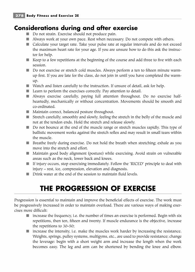
Considerations during and after exercise■ Do not strain. Exercise should not produce pain.■ Always work at your own pace. Rest when necessary. Do not compete with others.■ Calculate your target rate. Take your pulse rate at regular intervals and do not exceed
the maximum heart rate for your age. If you are unsure how to do this ask the instruc-tor for help.
■ Keep to a few repetitions at the beginning of the course and add three to five with eachsession.
■ Do not exercise or stretch cold muscles. Always perform a ten to fifteen minute warm-up first. If you are late for the class, do not join in until you have completed the warm-up.
■ Watch and listen carefully to the instruction. If unsure of detail, ask for help.■ Learn to perform the exercises correctly. Pay attention to detail.■ Always exercise carefully, paying full attention throughout. Do no exercise half-
heartedly, mechanically or without concentration. Movements should be smooth andco-ordinated.
■ Maintain correct, balanced posture throughout.■ Stretch carefully, smoothly and slowly, feeling the stretch in the belly of the muscle and
not at the tendon ends. Hold the stretch and release slowly.■ Do not bounce at the end of the muscle range or stretch muscles rapidly. This type of
ballistic movement works against the stretch reflex and may result in small tears withinthe muscle.
■ Breathe freely during exercise. Do not hold the breath when stretching; exhale as youmove into the stretch and effort.
■ Maintain good body alignment (posture) while exercising. Avoid strain on vulnerableareas such as the neck, lower back and knees.
■ If injury occurs, stop exercising immediately. Follow the ‘RICED’ principle to deal withinjury – rest, ice, compression, elevation and diagnosis.
■ Drink water at the end of the session to maintain fluid levels.
THE PROGRESSION OF EXERCISEProgression is essential to maintain and improve the beneficial effects of exercise. The work mustbe progressively increased in order to maintain overload. There are various ways of making exer-cises more difficult:
■ increase the frequency, i.e. the number of times an exercise is performed. Begin with sixrepetitions, then ten, fifteen and twenty. If muscle endurance is the objective, increasethe repetitions to 30–50;
■ increase the intensity, i.e. make the muscles work harder by increasing the resistance.Weights, springs, pulley systems, multigyms, etc., are used to provide resistance; changethe leverage: begin with a short weight arm and increase the length when the workbecomes easy. The leg and arm can be shortened by bending the knee and elbow.
278 Body Fitness and Exercise 2E
Progression is achieved by straightening the limb, then holding a pole or dumb-bells.The leverage of curl-ups is increased by moving the arm position from the side to acrossthe chest, then putting the hands on the shoulders, etc. Leverage and weight can becombined for progression.
■ Increase the number of sessions, e.g. from twice to three times per week or more. Ensurethat there is sufficient rest time for the body to recover fully, otherwise there is risk ofdamage.
■ To improve cardio-respiratory endurance, increase the duration of the exercise, i.e. thetime of each session and/or the length of the training programme.
■ Reduce the stability of the starting position. Exercising becomes more difficult as thestarting position becomes less stable. This will improve the skill components of balance,co-ordination and agility.
■ Change the speed at which an exercise is performed. Exercises are easier at naturalspeed, which varies with the individual. Exercises become more difficult if the speed isincreased or decreased.
POTENTIALLY DAMAGING EXERCISES THATSHOULD BE AVOIDED
There are a number of exercises that produce excessive stress on vulnerable areas of the body,such as the neck, the lower back and the knees, and that may result in injury. Many of these exer-cises form part of certain training programmes and routines for specific sports. When they are per-formed by very fit trained athletes in controlled situations the risk of injury is greatly reduced. Theyshould not be performed by unfit individuals, nor included in general exercise or group exerciseswhere individual supervision and control is impossible. The ability to evaluate the safety and effec-tiveness of an exercise is an important part of an instructor’s role. All exercise videos, exercisebooks and magazine articles should be carefully studied, and each exercise must be analysed andchecked for safety, as many of these hazardous exercises are often included. New forms of exer-cise routines or ‘crazes’ require particular caution. Because the human body is designed to performa certain finite number of movements through specific ranges, the so-called ‘new’ exercises mustbe versions of the old. Claims made for the results are often exaggerated, and so must be carefullyassessed: are they realistic and achievable?
When assessing the safety and effectiveness of an exercise it is useful to ask the followingquestions:
1 Will the exercise work the appropriate body part?2 Will the exercise move the selected muscle and joint through the correct range?3 Will the movement be a controlled, free movement, not forced or ballistic?4 Is the exercise appropriate for the client’s level of fitness?5 Could the exercise over-stress the moving joints or other body parts, causing
damage?6 Is this the most suitable exercise for achieving the set goal?
Safety and hygiene factors related to exercise 279

7 Will the exercise or the starting position put stress on any of the following vulnerableareas: the cervical region (the neck); the lumbar region (the lower back); the knees?
The following section deals with vulnerable areas of the body, potentially damaging exer-cises are highlighted and safe alternatives are given where possible.
The seven vertebrae of the cervical regionThis is the region of greatest spinal mobility. The first two cervical vertebrae – the atlas and axis –allow rotation of the head. The normal movements of the cervical region are:
■ flexion – dropping the head forward, chin on chest;■ extension – tilting the head backwards – and hyper-extension – taking it beyond exten-
sion to look at the ceiling;■ side flexion (lateral flexion) – dropping the head sideways, ear towards shoulder;■ rotation – turning the head to the right and left, looking towards the shoulder;■ circumduction – a combination of the above;
Movements of the chin also affect the cervical region;■ protraction of the chin – pushing the chin forward;■ retraction of the chin – pulling the chin back and in.
Before commencing exercises for the cervical spine, select a stable starting position such assitting or stride standing. If the client is very tense, the lying position can be used. Make sure thehead is in a good position: erect, with ear lobes level, eyes looking straight ahead and shouldersrelaxed.
SAFE EXERCISES
Starting position Exercise
■ Sitting drop chin onto chest, return toupright position.
■ Sitting take right ear down towards rightshoulder and back, then left eartowards left shoulder and back.
■ Sitting turn the head to the right to looktowards the right shoulder, repeatto left.
For all these exercises, it is important to keep the chin in and the head up.
280 Body Fitness and Exercise 2E
DAMAGING EXERCISESNOT RECOMMENDED
■ Sitting hyper-extension – dropping the head backto look at the ceiling.
■ Sitting circling head around on theshoulders.
These two exercises are not recommended as they produce excessive pressure and com-pression of the cervical discs. These may rupture and protrude into the inter-vertebral fora-mina, resulting in pressure on and damage to the nerves. This will cause pain, numbnessand pins and needles over the shoulder and down the arm.
With age the cervical region is susceptible to wear and tear, with erosion of the cartilage andbone. This may result in inflammation of surrounding structures, with pain and stiffness of theneck. These exercises will exacerbate this condition. They should not be done in general exerciseclasses, by those over 30 years old or by anyone suffering from headaches, neck pain or shoulderand arm pain, numbness or pins and needles.
Safety and hygiene factors related to exercise 281
FIGURE NUMBER: 13.1
FIGURE NUMBER: 13.2
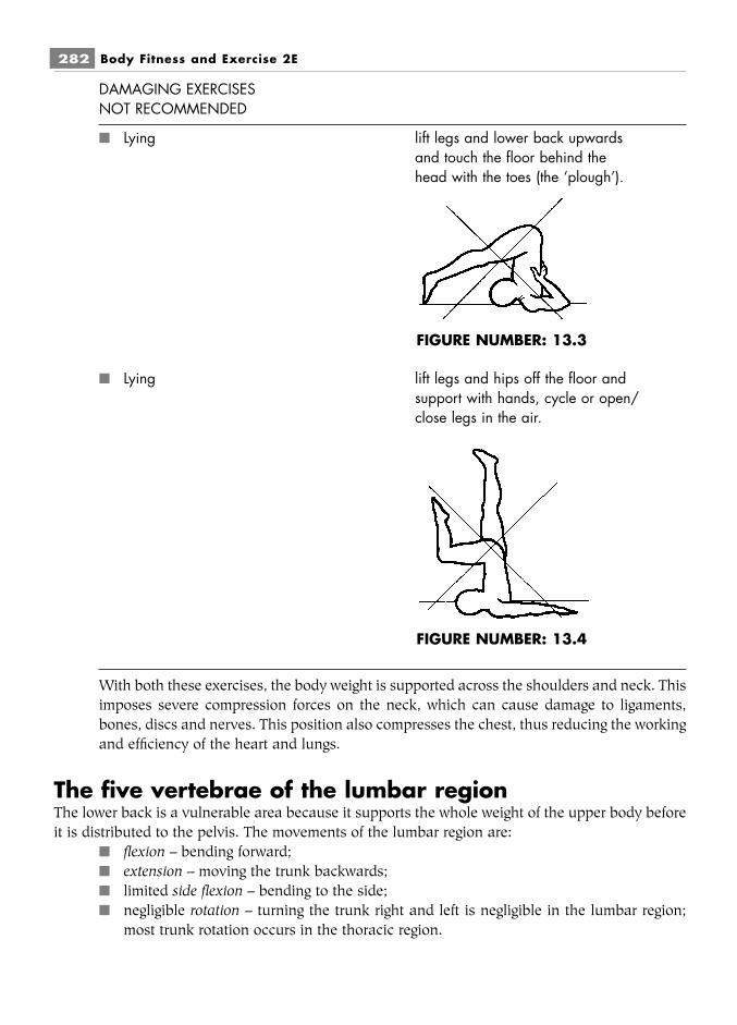
DAMAGING EXERCISESNOT RECOMMENDED
■ Lying lift legs and lower back upwardsand touch the floor behind thehead with the toes (the ‘plough’).
■ Lying lift legs and hips off the floor andsupport with hands, cycle or open/close legs in the air.
With both these exercises, the body weight is supported across the shoulders and neck. Thisimposes severe compression forces on the neck, which can cause damage to ligaments,bones, discs and nerves. This position also compresses the chest, thus reducing the workingand efficiency of the heart and lungs.
The five vertebrae of the lumbar regionThe lower back is a vulnerable area because it supports the whole weight of the upper body beforeit is distributed to the pelvis. The movements of the lumbar region are:
■ flexion – bending forward;■ extension – moving the trunk backwards;■ limited side flexion – bending to the side;■ negligible rotation – turning the trunk right and left is negligible in the lumbar region;
most trunk rotation occurs in the thoracic region.
282 Body Fitness and Exercise 2E
FIGURE NUMBER: 13.3
FIGURE NUMBER: 13.4
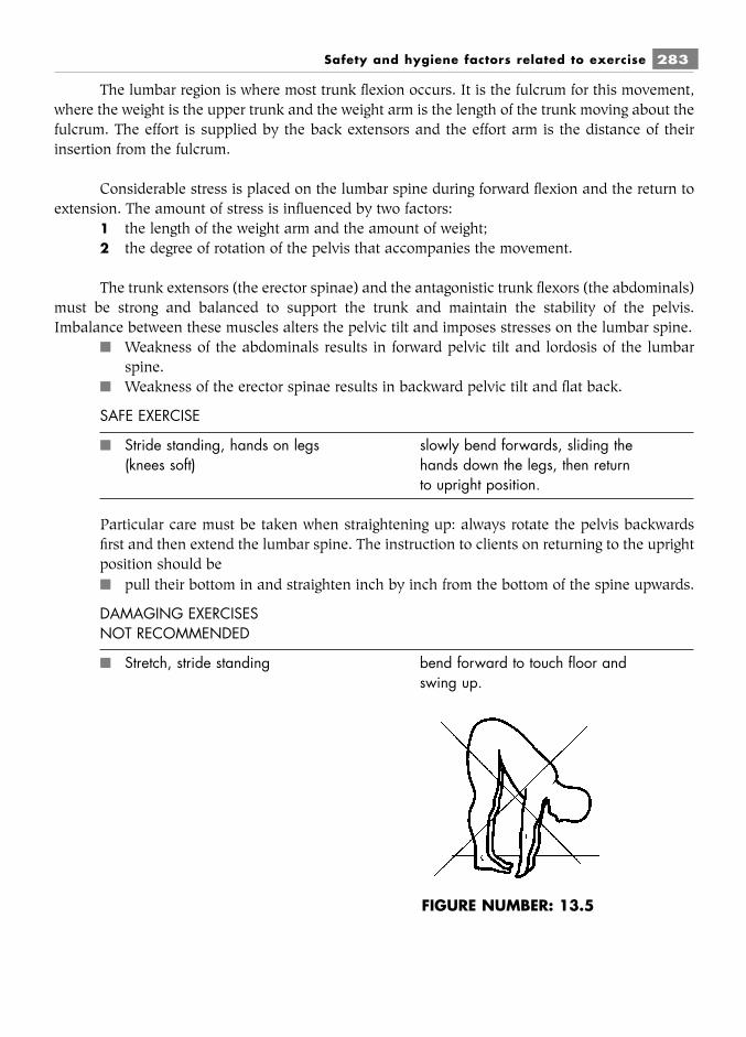
The lumbar region is where most trunk flexion occurs. It is the fulcrum for this movement,where the weight is the upper trunk and the weight arm is the length of the trunk moving about thefulcrum. The effort is supplied by the back extensors and the effort arm is the distance of theirinsertion from the fulcrum.
Considerable stress is placed on the lumbar spine during forward flexion and the return toextension. The amount of stress is influenced by two factors:
1 the length of the weight arm and the amount of weight;2 the degree of rotation of the pelvis that accompanies the movement.
The trunk extensors (the erector spinae) and the antagonistic trunk flexors (the abdominals)must be strong and balanced to support the trunk and maintain the stability of the pelvis.Imbalance between these muscles alters the pelvic tilt and imposes stresses on the lumbar spine.
■ Weakness of the abdominals results in forward pelvic tilt and lordosis of the lumbarspine.
■ Weakness of the erector spinae results in backward pelvic tilt and flat back.
SAFE EXERCISE
■ Stride standing, hands on legs slowly bend forwards, sliding the(knees soft) hands down the legs, then return
to upright position.
Particular care must be taken when straightening up: always rotate the pelvis backwardsfirst and then extend the lumbar spine. The instruction to clients on returning to the uprightposition should be■ pull their bottom in and straighten inch by inch from the bottom of the spine upwards.
DAMAGING EXERCISESNOT RECOMMENDED
■ Stretch, stride standing bend forward to touch floor andswing up.
Safety and hygiene factors related to exercise 283
FIGURE NUMBER: 13.5

■ Stretch, stride standing bend to touch opposite foot withhand.
■ Stride standing holding a Twist right and left.pole across back of shoulders
In these positions, with the arms stretched above the head, the weight arm is lengthenedand there is a far greater load in front of the fulcrum (the lumbar spine). This increases thestress and compression on the discs, and damage can occur to ligaments, discs, cartilage orbones.
Punching the air with the hands in forward flexion imposes the same stress and is not rec-ommended. Rotating the trunk to touch the opposite foot increases the compression forces stillfurther.
Ballistic-type bouncing at the end of the range of forward flexion in order to stretch the ham-strings must be avoided. This exerts excessive pressure on the lower back and may produce micro-tears and damage to the hamstrings. Since the hamstrings are not relaxed in this position but arecontracting eccentrically, stretching is ineffective and can cause damage.
SAFE EXERCISE
■ Stride standing trunk side flexion to the right andleft, slide hand down side.
The same principle of leverage applies to this exercise, which will be safe if the arms arekept down to the side, keeping the weight arm as short as possible.
284 Body Fitness and Exercise 2E
FIGURE NUMBER: 13.6

DAMAGING EXERCISESNOT RECOMMENDED
■ Stride standing swing left arm into the air and sideflex laterally to the right, thenreturn and swing right arm up andside flex laterally to the left.
The arm is stretched up into the air, increasing the length of the weight arm. The increasedleverage imposes stresses on the lumbar joints and discs, causing damage. Again, ballisticbounding at the end of the range will make the exercise even more hazardous.
DAMAGING EXERCISESNOT RECOMMENDED
■ Side lying lift both legs upwards.
The trunk side flexors will strain to lift the legs. This stresses the lower back. Twisting of thetrunk while straining to lift the legs causes further damage.
Safety and hygiene factors related to exercise 285
FIGURE NUMBER: 13.7
FIGURE NUMBER: 13.8
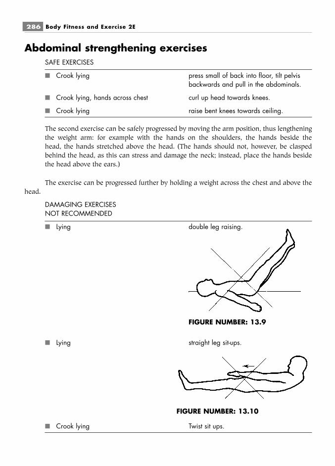
Abdominal strengthening exercisesSAFE EXERCISES
■ Crook lying press small of back into floor, tilt pelvisbackwards and pull in the abdominals.
■ Crook lying, hands across chest curl up head towards knees.
■ Crook lying raise bent knees towards ceiling.
The second exercise can be safely progressed by moving the arm position, thus lengtheningthe weight arm: for example with the hands on the shoulders, the hands beside thehead, the hands stretched above the head. (The hands should not, however, be claspedbehind the head, as this can stress and damage the neck; instead, place the hands besidethe head above the ears.)
The exercise can be progressed further by holding a weight across the chest and above thehead.
DAMAGING EXERCISESNOT RECOMMENDED
■ Lying double leg raising.
■ Lying straight leg sit-ups.
■ Crook lying Twist sit ups.
286 Body Fitness and Exercise 2E
FIGURE NUMBER: 13.9
FIGURE NUMBER: 13.10

Both these exercises impose stress and can cause many problems. They are not effective instrengthening the abdominal muscles, as can be seen by analysing the movement.Moving joint: hip jointDirection of movement: flexion coming up, extension going downPrime movers: ilio-psoas (hip flexor)Muscle work: concentric coming up, eccentric going down.
The psoas is a short muscle passing from the lumbar vertebrae to the lesser trochanter ofthe femur. It works with the iliacus and both muscles may be named ilio-psoas. As the psoas liftsthe legs or the trunk it pulls on its origin on the lumbar spine, imposing stresses and strains in thisregion. The psoas is working at tremendous mechanical disadvantage, as it is made to lift a longweight arm with a large weight (that of the legs). The back arches and strain is felt in the lumbarspine.
With this exercise, the psoas becomes stronger and tighter. This is undesirable, because atight posas pulls and tilts the pelvis forwards, resulting in lordosis due to muscle imbalance.
The abdominal muscles will be working statically in outer range in an attempt to keep thepelvis level. This type of muscle work is extremely difficult to maintain and can only be done byvery strong muscles; weaker muscles will be strained.
Static work of the abdominals increases intra-abdominal pressure, which will push on thepelvic organs and stretch the pelvic floor. The muscles of the pelvic floor may already be weakenedin post-natal women and older age groups. Strength must be maintained in the pelvic floor toprevent incontinence.
Back strengthening exercisesSAFE EXERCISES
■ Prone lying alternate single leg raising.
■ Prone lying alternate arm raising.
■ Prone lying opposite arm and leg raising.
Safety and hygiene factors related to exercise 287

DAMAGING EXERCISESNOT RECOMMENDED
■ Prone lying double leg raising.
■ Prone lying double leg, arm and trunk raising.
■ Prone lying trunk extension, touching feet with hands.
Raising both legs or, worse, raising both legs and arms, imposes severe compression andstress on the lumbar spine.
288 Body Fitness and Exercise 2E
FIGURE NUMBER: 13.11
FIGURE NUMBER: 13.12
FIGURE NUMBER: 13.13

Gluteal strengthening exercisesSAFE EXERCISES
■ Prone lying raise alternate legs off the floorand lower (keep hips in contactwith the floor and raise legs onlyfifteen degrees from floor).
■ Prone kneeling straighten alternate legs backwards(lifting no more than fifteendegrees from horizontal) and lower.
NOT RECOMMENDED
■ Prone kneeling bend knee onto chest and kick out behind.
Raising the leg more than fifteen degrees above the horizontal can stress the lumbar spine.Also, this exercise uses the hip flexor (ilio-psoas) to bend the knee towards the chest (thisdoes not usually require strengthening). This exercise is frequently performed in a swingingmanner, where the movement is not controlled and is likely to cause damage.
The knee jointThe stability of the knee is maintained by several strong ligaments (the medial and lateral liga-ments and the cruciate ligaments), by powerful muscles (the quadriceps and hamstrings) and bythe fascia of the high (the fascia lata).
The movements of the knee are:■ flexion – bending the knee;■ extension – straightening the knee;■ a slight amount of rotation in flexion.
Safety and hygiene factors related to exercise 289
FIGURE NUMBER: 13.14

SAFE EXERCISE
■ Long sitting bend right knee and rotate hipoutwards, drop knee onto floorand place foot against left thigh.Place hands on either side of leftleg and gently slide hands downleft leg forwards. Repeat onopposite side.
DAMAGING EXERCISESNOT RECOMMENDED
■ Long sitting bend right knee, turn legbackwards, slide hands along leftleg to touch toes (hurdler’sstretch).
■ Long sitting forward flexion, bringing headdown onto knees.
■ Standing one leg propped up at rightangles, bend forwards sliding handsdown leg.
290 Body Fitness and Exercise 2E
FIGURE NUMBER: 13.15
FIGURE NUMBER: 13.16
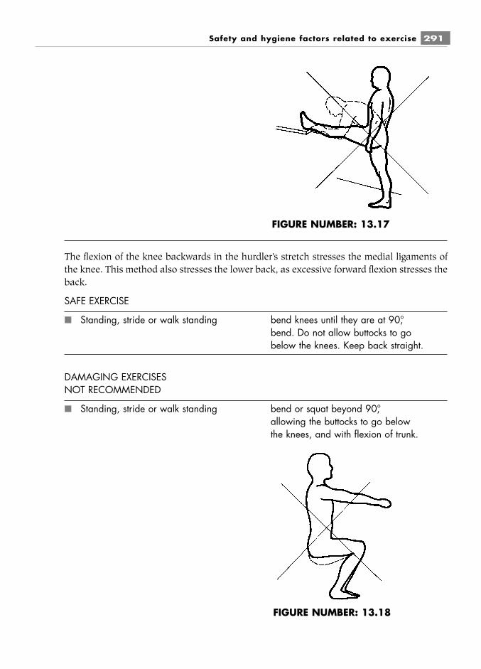
The flexion of the knee backwards in the hurdler’s stretch stresses the medial ligaments ofthe knee. This method also stresses the lower back, as excessive forward flexion stresses theback.
SAFE EXERCISE
■ Standing, stride or walk standing bend knees until they are at 90°,bend. Do not allow buttocks to gobelow the knees. Keep back straight.
DAMAGING EXERCISESNOT RECOMMENDED
■ Standing, stride or walk standing bend or squat beyond 90°,allowing the buttocks to go belowthe knees, and with flexion of trunk.
Safety and hygiene factors related to exercise 291
FIGURE NUMBER: 13.17
FIGURE NUMBER: 13.18
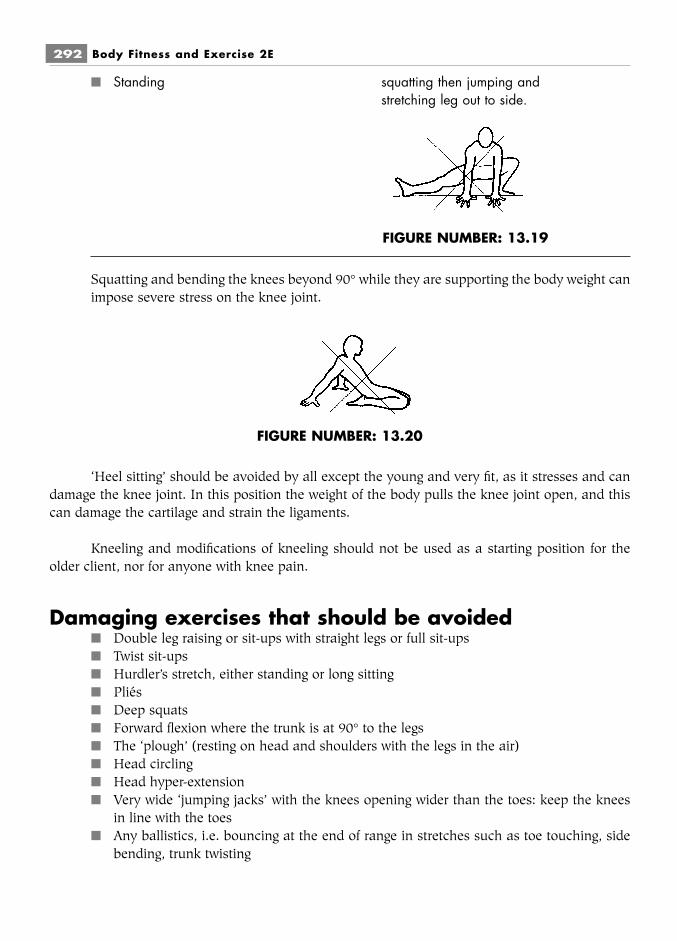
■ Standing squatting then jumping andstretching leg out to side.
Squatting and bending the knees beyond 90° while they are supporting the body weight canimpose severe stress on the knee joint.
‘Heel sitting’ should be avoided by all except the young and very fit, as it stresses and candamage the knee joint. In this position the weight of the body pulls the knee joint open, and thiscan damage the cartilage and strain the ligaments.
Kneeling and modifications of kneeling should not be used as a starting position for theolder client, nor for anyone with knee pain.
Damaging exercises that should be avoided■ Double leg raising or sit-ups with straight legs or full sit-ups■ Twist sit-ups■ Hurdler’s stretch, either standing or long sitting■ Pliés■ Deep squats■ Forward flexion where the trunk is at 90° to the legs■ The ‘plough’ (resting on head and shoulders with the legs in the air)■ Head circling■ Head hyper-extension■ Very wide ‘jumping jacks’ with the knees opening wider than the toes: keep the knees
in line with the toes■ Any ballistics, i.e. bouncing at the end of range in stretches such as toe touching, side
bending, trunk twisting
292 Body Fitness and Exercise 2E
FIGURE NUMBER: 13.19
FIGURE NUMBER: 13.20
■ Moving from heel sitting up to kneeling, except for the young and athletic (i.e. thosewith strong quadriceps). It should not be performed by anyone with knee problems northose over 50. The same applies to exercises using kneeling as a starting position.
■ Back hyper-extension.■ Vigorous kickback in prone kneeling■ Hyper extending spine in crook lying for gluteal strength■ Deep lunges■ Stride standing holding pole behind shoulder trunk twist.
CLIENT ASSESSMENTThe assessment of a client prior to exercise has developed considerably over the last few years. Inaddition to obtaining information regarding health, medical history and taking measurements ofheight, weight, fat distribution and muscle tone, it is now desirable to assess fitness by measuringthe pulse rate, blood pressure, lung capacity, muscle strength, muscle endurance, flexibility andbody composition.
Accurate assessment is important for the following reasons:■ It provides information on the client’s past and present state of health. This will high-
light any contra-indications or any conditions where caution is necessary.■ It provides information on the client’s lifestyle, activities, athleticism and motivation.■ It establishes the figure type, i.e. endomorph, ectomorph or mesomorph. This facilitates
the planning and setting of realistic, achievable goals.■ It identifies postural problems and muscle imbalance, so that specific strategies to
restore balance can be planned.■ It establishes the current level of fitness, which provides a starting point for the exercise
programme.■ It provides the information necessary for setting objectives and planning safe, effective
exercise that will not place the client at risk.■ It provides a record of data and the starting point from which future improvement can
be measured.
Safety and hygiene factors related to exercise 293
TASKWork with a partner.■ Teach your partner safe head and neck movements.■ Teach your partner safe trunk forward flexion, side flexion and back extension
exercises.■ Explain why the following movements should not be performed:
a head circling and hyper-extensionb double leg raising and straight leg sit-upsc deep squatting with the bottom lower than the knees.
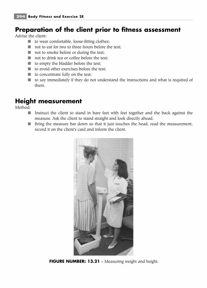
Preparation of the client prior to fitness assessmentAdvise the client:
■ to wear comfortable, loose-fitting clothes;■ not to eat for two to three hours before the test;■ not to smoke before or during the test;■ not to drink tea or coffee before the test;■ to empty the bladder before the test;■ to avoid other exercises before the test;■ to concentrate fully on the test;■ to say immediately if they do not understand the instructions and what is required of
them.
Height measurementMethod:
■ Instruct the client to stand in bare feet with feet together and the back against themeasure. Ask the client to stand straight and look directly ahead.
■ Bring the measure bar down so that it just touches the head, read the measurement,record it on the client’s card and inform the client.
294 Body Fitness and Exercise 2E
FIGURE NUMBER: 13.21 – Measuring weight and height.

Weight measurementMethod:
■ Instruct the client to wear minimum clothing (record this to ensure that the same cloth-ing is worn each time the weight is taken), with bare feet. Ask the client to stand still inthe centre of the weighing machine and to look directly ahead.
■ Read the weight, record it on the client’s card and inform the client.
Body measurementMeasurements to be recorded:
■ Bust/chest■ Waist■ Hips■ Upper thigh■ Lower thigh■ Upper arms
Method:■ Select a tape measure that is in good condition, not frayed or stretched. Always ensure
that the tape is level on the body part and not twisted. Do not pull the tapemeasure. Use the nearest prominent bony point as a marker. This will ensure that thetape will be placed at the same level each time. Thin elastic can be used to indicate thelevel.
■ Instruct the client to remove all clothing except pants (very self-conscious womenmay keep a bra on, but they should wear the same bra each time the measure-ments are taken), stand in bare feet and maintain a good posture, with the arms to theside.
Bust or chest■ Bring the tape around the back, under the armpit and around the nipple line.■ Record the measurement and inform the client.
Waist■ Give a circle of narrow elastic to the client and ask her to place this at the narrowest
part, i.e. her natural waistline. Measure just above the elastic. For males, measure at thelevel of the navel.
■ Record the measurement and inform the client.
Hips■ Place the tape around the widest part of the hips and measure. Then measure the dis-
tance of the tape from the greater trochanter: this will ensure that the tape is placed atthe same level next time.
■ Record the measurement and inform the client.
Safety and hygiene factors related to exercise 295
Right and left upper thigh■ Again, use a circle of narrow elastic. Place this around the widest part of the thigh.
Measure the distance from the elastic to the top of the patella.■ Place the tape around the leg just above the elastic.■ Record the measurement and inform the client.
Right and left lower thigh■ Use a circle of narrow elastic and place it two to three inches above the top of the
patella.■ Place the tape around the leg just above the elastic.■ Record the measurement and inform the client.
Right and left upper arms■ Place a circle of narrow elastic around the widest part of the upper arm. Measure the
distance from this to the olecranon process.■ Place the tape around the arm just above the elastic.■ Record the measurement and inform the client.
Midriff■ For women whose objective is to lose body weight, measurement of the midriff is
necessary. Measure around the midriff two to three inches below the xiphoid pro-cess.
Calf■ For those wishing to build up the calf muscle, measure around the wide part of the calf
and note the distance from the tape to the lateral malleolus. Use the same distance nexttime.
Testing for muscle strengthMuscle strength is measured by how much weight the muscle is able to move. This is tested usingweights or a grip test or by pulling against machines.
Weight lift■ Select a suitable stable starting position.■ Select an appropriate weight and check that it is secure.■ Isolate the movement to the muscle being tested.■ Ask the client to lift the weight smoothly to the full inner range three times. If an extra
lift is possible, the weight must be increased.■ Ask the client to rest for one to two minutes and repeat the lift with extra weight. The
weight that is lifted smoothly two or three times indicates the strength.■ Record the weight.
296 Body Fitness and Exercise 2E

Grip test■ Make sure the client is holding the grip comfortably in the hand. In the standing posi-
tion the client lifts the arm above the head, lowers the arm and squeezes as hard as pos-sible. Repeat three times.
■ Record the highest reading.
There are a variety of machines on the market designed for testing strength. Read the manu-facturer’s instructions very carefully, and test a colleague to ensure that you fully understand theprocedure.
Exercises such as push-ups and sit-ups are sometimes used to give an indication of fitness,but these are not measurable.
Muscle toneIt is possible to obtain some indication of muscle strength by applying manual resistance to muscleaction and feeling the degree of tone within the muscle. This will only provide a rough guide, as itis not possible to quantify the strength but only to categorise it into poor, moderate, good, verygood or excellent. Muscles that are easily tested in this way are the biceps and triceps, the abdom-inals, gluteus maximus and the hip abductors and adductors.
Method:■ Position the client in crook lying. This position can be maintained throughout and
avoids moving the client unnecessarily.■ One hand must be placed over the working muscle to feel the tone, while the other hand
is used to resist the movement.
Biceps strength■ The client bends the elbow to the mid-point of the range.■ Place one hand over the biceps on the anterior aspect of the upper arm. Grasp the wrist
with the other.■ Instruct the client to bend the elbow while you stop the movement. Feel the increased
tone with the hand placed over the muscle. Is the strength you are feeling poor,moderate, good, very good or excellent? This value judgement becomes easier withpractice.
■ Record the result.
Triceps strength■ With one hand, cover the triceps on the posterior aspect of the upper arm. Keep the
other hand around the wrist.■ Instruct the client to straighten the elbow against resistance. Feel the increased tone
with the hand placed over the muscle, and assess the strength.■ Record the result.
Safety and hygiene factors related to exercise 297

Abdominal strength (particularly rectus abdominus)■ Place one hand over the abdominals.■ Instruct the client to perform a curl-up (lifting head and shoulders with chin on chest).
If this is done with ease, the free hand can be placed over the sternum and resistancegiven to the curl-up.
■ Feel the increased tone with the hand placed on the abdominals, and assess thestrength.
■ Record the result.
Gluteus maximus strength■ Instruct the client to lift his or her bottom up off the bed and tighten the buttocks. (A
sandbag weight can be placed over the pelvis to provide resistance.)■ Place a hand over the gluteus maximus.■ Feel the increased tone and judge the strength.■ Record the result.
Abductor strength■ Straighten the client’s leg.■ Place one hand over the abductors on the outer aspect of the thigh above the greater
trochanter.■ Place the other hand under the ankle to cup it.■ Instruct the client to ‘push out’ towards you. Resist the movement, using the hand at
the ankle to push inwards.■ Feel the increased tone in the abductors and judge the strength.■ Record the result.
Adductor strength■ Keep the client’s leg straight and pulled outwards.■ Keep the hand under the ankle.■ Place the other hand over the adductors on the inner aspect of the thigh (upper third).■ Instruct the client to pull the leg inwards towards the other leg. Resist the movement,
using the hand at the ankle to pull outwards.■ Feel the increased tone in the adductors and judge the strength.■ Record the result.
Cardio-respiratory enduranceThis may be tested using a treadmill or bicycle ergometer. If this specialised equipment is not avail-able, the step test can be used.
Three-minute step testEquipment:
■ Twelve-inch step
298 Body Fitness and Exercise 2E
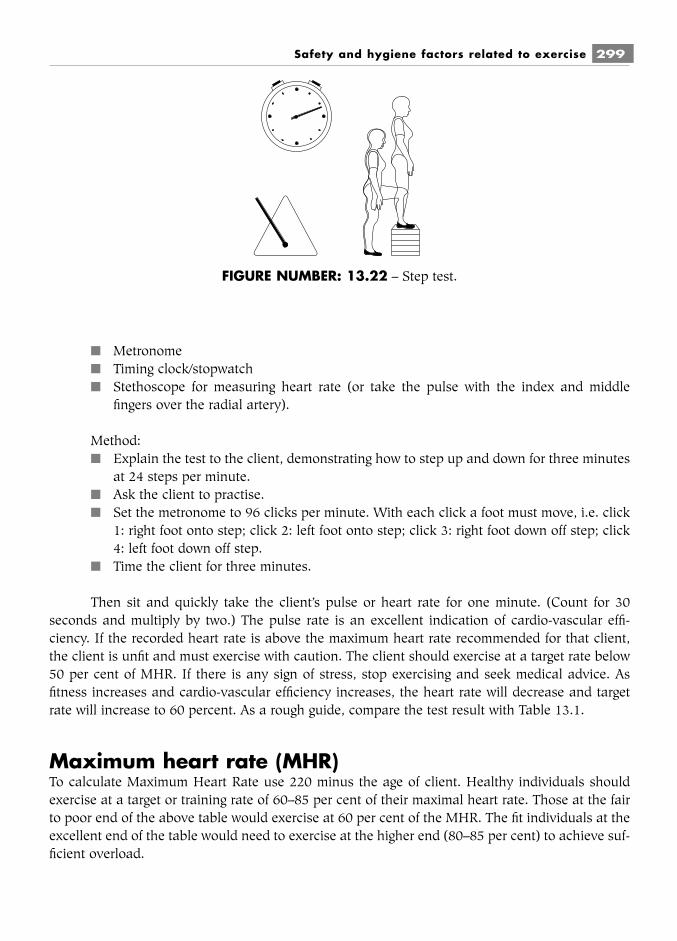
■ Metronome■ Timing clock/stopwatch■ Stethoscope for measuring heart rate (or take the pulse with the index and middle
fingers over the radial artery).
Method:■ Explain the test to the client, demonstrating how to step up and down for three minutes
at 24 steps per minute.■ Ask the client to practise.■ Set the metronome to 96 clicks per minute. With each click a foot must move, i.e. click
1: right foot onto step; click 2: left foot onto step; click 3: right foot down off step; click4: left foot down off step.
■ Time the client for three minutes.
Then sit and quickly take the client’s pulse or heart rate for one minute. (Count for 30seconds and multiply by two.) The pulse rate is an excellent indication of cardio-vascular effi-ciency. If the recorded heart rate is above the maximum heart rate recommended for that client,the client is unfit and must exercise with caution. The client should exercise at a target rate below50 per cent of MHR. If there is any sign of stress, stop exercising and seek medical advice. Asfitness increases and cardio-vascular efficiency increases, the heart rate will decrease and targetrate will increase to 60 percent. As a rough guide, compare the test result with Table 13.1.
Maximum heart rate (MHR)To calculate Maximum Heart Rate use 220 minus the age of client. Healthy individuals shouldexercise at a target or training rate of 60–85 per cent of their maximal heart rate. Those at the fairto poor end of the above table would exercise at 60 per cent of the MHR. The fit individuals at theexcellent end of the table would need to exercise at the higher end (80–85 per cent) to achieve suf-ficient overload.
Safety and hygiene factors related to exercise 299
FIGURE NUMBER: 13.22 – Step test.

Body compositionEquipment:
■ Skin-fold callipers.
Method:■ Identify the locations of the skin folds (see Table 13.2).■ Hold the callipers in the dominant hand.■ Pinch the skin fold with the thumb and forefinger of the other hand.■ Hold the callipers perpendicular to the skin fold and place the pads very near the thumb
and forefinger.■ Close or release the callipers, depending on type.
300 Body Fitness and Exercise 2E
Table 13.1The range of heart rates after three minutes’ stepping
Men aged 20–46 Women aged 20–46
Excellent 181–901 179–841
Good 199–102 190–971
Above average 103–112 106–109
Average 120–121 118–119
Below average 123–125 122–124
Fair 127–130 129–134
Poor 136–138 137–145
(Adapted from YMCA Y’s Way to Fitness.)
FIGURE NUMBER: 13.23 – Skin fold callipers.
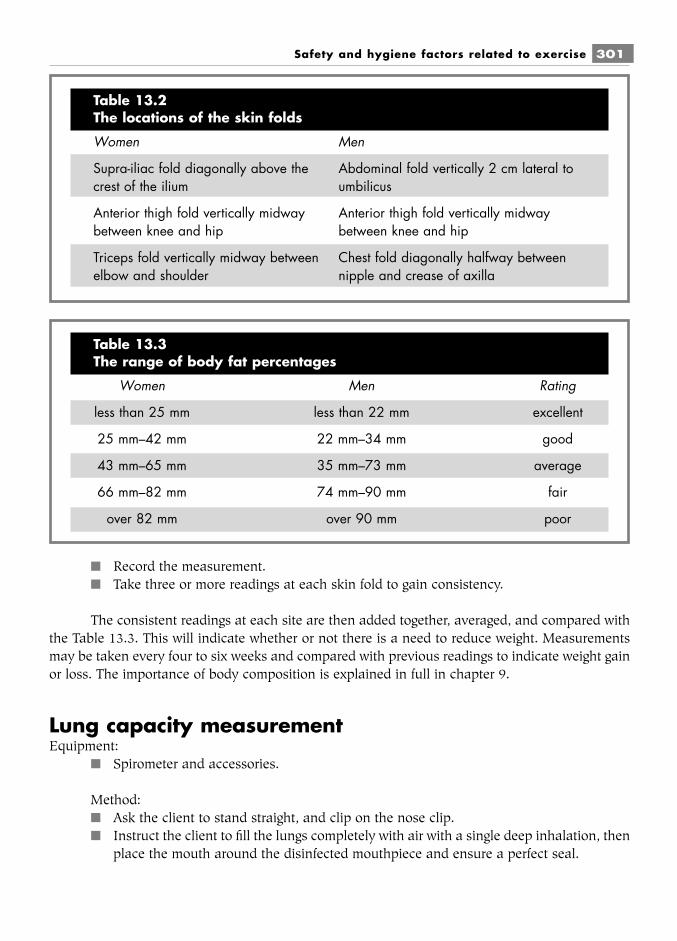
■ Record the measurement.■ Take three or more readings at each skin fold to gain consistency.
The consistent readings at each site are then added together, averaged, and compared withthe Table 13.3. This will indicate whether or not there is a need to reduce weight. Measurementsmay be taken every four to six weeks and compared with previous readings to indicate weight gainor loss. The importance of body composition is explained in full in chapter 9.
Lung capacity measurementEquipment:
■ Spirometer and accessories.
Method:■ Ask the client to stand straight, and clip on the nose clip.■ Instruct the client to fill the lungs completely with air with a single deep inhalation, then
place the mouth around the disinfected mouthpiece and ensure a perfect seal.
Safety and hygiene factors related to exercise 301
Table 13.2The locations of the skin folds
Women Men
Supra-iliac fold diagonally above the Abdominal fold vertically 2 cm lateral tocrest of the ilium umbilicus
Anterior thigh fold vertically midway Anterior thigh fold vertically midwaybetween knee and hip between knee and hip
Triceps fold vertically midway between Chest fold diagonally halfway betweenelbow and shoulder nipple and crease of axilla
Table 13.3The range of body fat percentages
Women Men Rating
less than 25 mm less than 22 mm excellent
25 mm–42 mm 22 mm–34 mm good
43 mm–65 mm 35 mm–73 mm average
66 mm–82 mm 74 mm–90 mm fair
over 82 mm over 90 mm poor
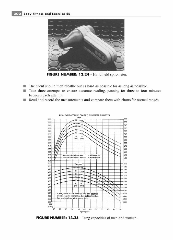
■ The client should then breathe out as hard as possible for as long as possible.■ Take three attempts to ensure accurate reading, pausing for three to four minutes
between each attempt.■ Read and record the measurements and compare them with charts for normal ranges.
302 Body Fitness and Exercise 2E
FIGURE NUMBER: 13.24 – Hand held spirometer.
FIGURE NUMBER: 13.25 – Lung capacities of men and women.

Blood pressureEquipment:
■ A sphygmomanometer is used to measure blood pressure. The modern models usuallyfound in fitness centres are automatic and give a digital readout. These differ from themodels used medically, where a stethoscope is used over the radial artery at the elbow,the sounds during systole and diastole are listened to and the corresponding pressurelevel for each is read.
Method:■ Normal blood pressure for a young resting adult is about mm Hg but this
varies considerably with activity, emotion and age. Blood pressure tends to rise as weget older. Systolic pressure in adults at rest averages 110–150 mm Hg, diastolic 60–65mm Hg. Anyone giving repeated systolic pressure readings of more than of 140 mm Hgshould consult a doctor for a check-up before exercising.
FlexibilityIt is possible to measure lower back and hamstring flexibility for the fit client.
Equipment:■ A box or stool with a tape attached
120 (systolic)
80 (diastolic)
Safety and hygiene factors related to exercise 303
FIGURE NUMBER: 13.26 – A sphygmomanometer.
FIGURE NUMBER: 13.27 – Flexibility test. (Seat and reach.)

Method:■ The client should sit with legs stretched out and feet against the box or stool, and with
arms stretched forward.■ Ask the client to breathe out, reach forward and slide the hands along the stool. Read
the distance at the middle finger. If the box has a slider, the client should push thisforward with the hands and the reading is taken.
■ Measure for three attempts and record the best result.
304 Body Fitness and Exercise 2E
TASKWork with a partner. Practise the different assessment on each other.
FIGURE NUMBER: 13.28 – Blood pressure range.

Chapter 14Exercise classes
Exercise classes provide many different forms of exercise, for example general keep fit, aerobic,dance, step, weight loss, line dancing, relaxation, etc. Although they differ in the type of exerciseoffered, certain basic principles apply to all.
It is desirable, though not always possible, to organise classes so that there is parity withinthe groups, for example similar fitness levels, similar age groups and similar desired outcomes. Itis very important to recognise and allow for individual differences within a group: different intel-lectual levels, fitness levels, body shapes, health, previous experience, lifestyles, age and motiva-tion.
Each class member must be instructed to work at his or her own pace, and heart rate mustbe used to monitor intensity. No-one should exceed their maximum heart rate – see page 146.
Remember: beginners must work at 60 per cent of the maximum heart rate and increase to80 per cent as fitness develops. All members must practice taking the radial pulse for fifteenseconds and multiplying by four. They must continue to monitor the pulse at intervals throughout theclass. Members must stop exercising if the target heart rate is exceeded or if there are any signs ofstress.
LEARNTo work out a client’s maximum heart rate (MHR) subtract their age from 220. Heartrate must stay below this when exercising. The target rate will be 60–90 percent of this.

LEARNING NEW SKILLSThe theories of learning apply to both individual and class teaching. Members attend in order toacquire new skills, which will aid the achievement of set goals or objectives. These goals will varyfrom person to person. In order to help the members of a class to achieve these goals, the teachermust organise the lessons so that optimum learning and performance can take place.
The teaching of a skill requires demonstration (by the teacher) followed by practice by themembers, but before this can occur it is essential to prepare adequately the environment where thelearning is to take place.
As previously outlined, the environment must be warm, well ventilated, free from distrac-tions and external noise and safe. The floor must be firm, smooth and, preferably, sprung. Allequipment must be checked and organised before the commencement of the class so that thesequence of the lesson is not broken, as interruption of any kind interferes with the learningprocess. Members must be acknowledged and greeted in a friendly and reassuring way. Thisensures that members feel physically and psychologically ‘safe’.
Learning a new exercise is a very complex process. It is useful to consider the nature of askill and how it may be defined.
■ Curzon (1976) defines a skill as ‘a series of learned acts requiring simultaneous orsequential co-ordination’.
■ The UK Department of Employment (Curzon, 1976) defines a skill as ‘an organised andco-ordinated pattern of mental and physical activity’.
The essential features of a skilled performance are:■ accuracy of timing;■ anticipation of movement;■ economy of effort;■ grace and precision of movement;■ the overall flow of the movement.
To enable members of the class to attain this level, the teacher must analyse each skill andbreak it down into a sequence of separate movements, which are then joined together to make thewhole performance. The acquisition of a skill requires the use of both receptor and effector pro-cesses, in other words effective co-ordination of mind and muscle. There are many theories on thelearning of motor skills.
One theory suggests that the learner must pass through three overlapping stages: (Fitts andPosner, 1967)
Stage 1■ The cognitive phase – this involves knowledge of the skill and understanding of what is
required. The learner analyses the task and tries to understand what is to be done and
306 Body Fitness and Exercise 2E
how to do it. He or she knows when errors are made but is unsure how to correct them.At this stage co-ordination is poor, concentration levels high, cues and correction mustbe given frequently and common errors pointed out. At this stage the skill can be brokendown into simple parts to make learning easier.
Stage 2■ The associative or motor phase – as a result of practice, errors are gradually eliminated.
Correct patterns are established and, although errors are still made, they are correctedwith minimal prompting. Movement is more consistent and effective, the learner canconsider finer detail and can link sequences together to produce skilled performance.The learner can now recognise and correct his/her mistakes. Few cues are given at thisstage as too much feedback makes the learner too dependent.
Stage 3■ The autonomous phase – skills are performed automatically and require little thought.
Errors have been eliminated, speed and accuracy increased and the effects of stressreduced. The motor skill is now highly developed.
FeedbackIt is the instructor’s role to guide members of the class through these stages and to provide contin-uous feedback throughout. They must be informed of their current progress and told how toimprove it. Millar distinguishes between two forms of feedback:
■ Action feedback, which provides knowledge of current progress. Actions are corrected asthey are performed. The instructor must therefore provide spoken cues during the perfor-mance of the exercise, such as ‘eyes front’, ‘hold the head up’, ‘shoulders down’, ‘kneeshigher’, ‘do not stretch too far’, ‘do not lift too high’, ‘curl back slowly from below’, etc.
■ Learning feedback, which provides information that enables the student to improve nexttime. This might include cues such as ‘that was a good attempt, but some people did notkeep the back straight’, ‘well done, but keep those tummies in’, ‘well done, but watch thatyou roll the pelvis backwards before you come up’ or ‘I obviously didn’t explain that exer-cise clearly’, followed by further explanation and demonstration. In this way, memberscan correct their performance and will move quickly into the autonomous phase.
Feedback must therefore be given as each exercise or part of an exercise is being performed,and also at the end. This enables the learner to discriminate between correct and incorrect patternsat an early stage and thus avoids incorrect patterns being reinforced.
Many corrective statements and cues will be required for new members of a class or whenmembers are learning a new exercise, but as the skill is mastered fewer cues are needed. Positivevalue statements such as ‘good’, ‘that’s better’, ‘well done’ or ‘great’ will encourage members andincrease motivation. It is important always to encourage and never to put anyone down, nor drawattention to poor performance or embarrass the learner. If only one or two members are not per-forming correctly, give a general corrective statement, or catch their eye and say ‘watch me’, orexplain the correction quietly and privately at the end of the class.
Exercise classes 307
Feedback is essential as it provides knowledge of correct or incorrect performance, rein-forces the correct response and increases motivation.
MOTIVATIONMotivation may be defined as the drive within an individual to take an appropriate course of actionin order to satisfy a need.
Motivation heightens performance. The good instructor, teacher or coach is able to stimu-late the student’s own motivation. The instructor can ensure that the class is well organised andadministered and that the teaching style is enthusiastic, positive, concerned, caring, knowledge-able and safe. A high level of expertise must be demonstrated at all times, both in theoreticalknowledge and in practical demonstration.
Motivation is greatly enhanced by encouraging and promoting feelings of satisfaction, achieve-ment, recognition, responsibility, advancement, personal growth and success, enjoyment and fun.
However, poor, unpleasant surroundings, lack of organisation and safety, feelings of dis-satisfaction, failure and embarrassment, lack of interest and unrealistic goals that are not achiev-able are all demotivators and must be avoided.
Setting objectives or goalsThis is a very important part of planning for group or individual exercises. The members’ aspira-tions and the possibilities of realising them must be fully discussed. When members are involvedin setting the objectives they know where they are going and can better help themselves to getthere, so motivation is increased. The objective may be to:
■ increase cardio-respiratory fitness;■ improve muscle strength;■ improve flexibility;■ reduce fatty deposits;■ improve body shape;■ improve posture;■ correct figure faults;■ improve speed and skill.
Objectives must be realistic and they must be achievable within a limited time scale. Arounded five foot tall endomorph will never be tall and slender – that goal would be unrealistic –but to lose fat and improve body shape would be achievable and realistic. The setting of long-termgoals and short-term goals, which are continually monitored through regular assessment, willincrease motivation. Once the objectives have been agreed, planning the strategy can begin.Consider the age and ability of participants. Organise groups into beginners, intermediate oradvanced. Lesson plans for each session should be prepared: these are necessary as a record of
308 Body Fitness and Exercise 2E
work and show progression. Consider the type of exercises required to meet objectives. Considerintensity, frequency and duration.
Each lesson plan should include:■ the objectives;■ the sequence of activities;■ the time allowed for each part of the sequence;■ the music, if used;■ the equipment necessary;■ any comments or notes.
An example can be found in Table 14.1
Objective:■ To correct lordosis and improve muscle balance■ To reduce weight.
THE ORGANISATION OF A CLASS■ Prepare the room before the members arrive.■ Ensure that the lighting and ventilation are adequate.■ Check that the floor is firm, clean and smooth.■ If mats are to be used, make sure there is one for each person.■ Locate the first-aid box and note all the exits.■ Check that the area is clear of equipment or apparatus or anything that may be a safety
hazard.■ Select all required equipment or apparatus.■ Check that the equipment and apparatus is in sound working order and safe to use.
Ensure that there is enough to go round.■ Arrange all the equipment neatly at one end of the room well away from the working
area.■ Select the music tapes or records and stack them in order of use.■ Check that the music centre or player is working.■ Provide a large clock with a second hand to be used when monitoring maximum heart
rate.■ Shower and change into appropriate clothes. Remember that you set the standard for
the class. Wear clean, unrestricting, absorbent, smart clothing and suitable footwear(see chapter 13).
■ Tie the hair back and remove jewellery.
When the members arrive:■ Greet the members warmly, using their names where possible; at the very least, make
eye contact or wave to show each member that they are recognised.
Exercise classes 309

310 Body Fitness and Exercise 2E
Table 14.1Sample lesson plan
Activity Time Equipment Music Comments
Discussion of previous 5–10 minutes – – explain that theseweek’s class including objectives require differentany problems, and exercises:outline today’s exercises – ■ to reduce weight –
aerobics;■ to correct lordosis –
strengtheningabdominals andstretching backextensors
Warm-up: alternate heel 10 minutes – any suitable begin slowly and increaseraising, walking on the music for pace. Work on largespot and around the age group muscle groups. Includeroom, marching with or time of exercises for mobilisingknees high, pelvic tilt, year and pulse raising.circle pelvis, twist trunk,shoulder circling, armcircling, chest press
Stretch: erector spinae, 5 minutes rolled – slow gentle static stretch,hip flexors (select from towel hold for a count of ten, chapter 11) repeat
Aerobic section 15 minutes step theme music, work to target heart rate.faster tempo Take pulse every five
minutes, stop if abovetarget, work harder if toolow (see chapter 7)
Strengthening exercises 10 minutes medicine – use crook lying curl-upsball, sand for the abdominal musclesbag and leg extension for the
gluteus maximus and ham-strings. Caution: do notextend the lower back,keep the hips on the floor
Cool-down: select from 10 minutes – any music keep moving – do notchapter 12 with a slow stand still
tempo
Stretch
Discussion: feedback to 5 minutes – – Allow the clients time toand from class or longer ask questions and clarifyparticipants if time allows uncertainties. Give them
positive feedback. Leaveon a ‘high’ note.
■ Make a point of greeting and speaking to new members and introduce them to othermembers.
■ Point out toilets, exits, first-aid box, safety procedures.■ Register all participants.■ Carry out a consultation or assessment of each new member and ensure that he or she
has read the instructions (see chapter 13) and signed the consent form.■ Check that members are wearing suitable unrestricting clothing and suitable footwear.■ Check that hair is tied back and jewellery is removed.■ Begin the class on time.
During the class:■ Step confidently in front of the class, speak clearly and make sure that those at the back
can hear. Use the voice to good effect: change the tone to govern speed, rhythm, inten-sity and effort.
■ Remember that you are the role model for the class. Therefore, develop a warm, friendly,enthusiastic, positive approach. Good posture and an alert and efficient manner will setthe tone of the class. Do not fidget or develop irritating mannerisms such as tossing thehair or rubbing the leg.
■ When demonstrating an exercise, ensure that your performance is as accurate andperfect as possible. Poor demonstration means poor performance and ineffective exer-cise, which may also impose stresses on the body, causing strain and injury.
■ Become an educator. Outline the objectives. Explain the reasons for and the effects ofeach exercise. Explain the health benefits of exercise (see chapter 12).
■ Take time to explain and teach new exercises.■ Stress that each individual must work at his or her own pace and target heart rate. He
or she must not exceed the maximum heart rate. New members should work at 60 percent of MHR working up to 80–85 per cent (see chapter 13). Make sure that eachmember can calculate this and know how to take their pulse.
■ Following the demonstration, give clear, simple commands and corrective cues, partic-ularly in the early stages.
■ Break complicated exercises down into manageable ‘chunks’. Teach one part at a timeuntil each is well executed and then perform them together as a whole.
■ Give encouragement to enhance motivation, i.e. ‘well done’, ‘that’s good’, ‘much better’,‘that’s great’, ‘a great effort’, etc.
■ Watch for signs of stress.■ Tell the client to stop exercising if there are signs of stress such as profuse sweating,
breathlessness, tightness in the chest, pain in the chest or arms, pain in the back or anyjoints, faintness or dizziness, headache, nausea or a heart rate above the maximum.
■ Members must not be made to feel that they are in competition with others, nor underpressure to keep up with the instructor. If possible, organise classes into easy (begin-ners), intermediate and advanced to accommodate fitness levels, or organise them intoage groups, in order to maintain some parity within the groups. Allow time for discus-sion before the session commences. This may cover any problems experiencedduring or after the previous class, any minor injuries or joint problems experienced or
Exercise classes 311

any interesting new information for discussion. Explain the importance of maintaininggood posture and give advice on protecting the back, neck and knees.
■ Explain the importance of breathing normally and not holding the breath. Instructmembers to breathe in before an effort or on release and to breathe out on the effort.
■ Give reasons for the importance of the warm-up (see chapter 12). Emphasise that evenif members turn up late they must not join the class until they have completed the ten-minute warm-up. They must do this at the edge or back of the class and then join in theother exercises. Explain the importance of stretching and the associated hazards (seechapter 9).
■ Explain the importance of the cool-down in allowing the body to return slowly to thebalanced state. Explain why cool-down exercises should be performed while continu-ously moving around or end with lying down for rest and breathing.
■ Allow time at the end for participants to ask questions and clarify any uncertain points.■ Give feedback regarding the session■ Outline the activities for the next and future sessions – remind them of goals.■ Clear away all equipment and leave the room in a clean and tidy state.
AEROBIC CLASSThe American College of Sports Medicine defines aerobic activity as follows:
‘Aerobic activity is that requiring continuous, rhythmic use of large muscle groups at 60–90per cent of the maximum heart rate and 50–85 per cent of maximum oxygen uptake for 20–60minutes at least three times per week.’
The main effects of aerobic exercises are:■ an increase in cardio-respiratory fitness;■ a reduction of fatty deposits, with resultant weight loss;■ an increase in muscular endurance;■ the maintenance of bone mass.
Types of aerobic classThere are various activities designed to keep the body in continuous rhythmic motion and the listis continually growing. We now have high, moderate and low impact aerobics, step, dance andwater aerobics.
High-impact aerobics, where both feet leave the ground, are no longer recommended asthey place members at risk of injury due to continuous jarring.
Low impact, where one foot always stays in contact with the ground and the knees areslightly bent, are safer. Moderate impact will include some high impact, but consists mainly of low-impact work. It is more desirable to concentrate on duration and intensity than on impact.
312 Body Fitness and Exercise 2E
Duration is increased by performing more repetitions. Intensity is increased by using longerand higher steps: from jogging to marching to high stepping. The arm position can change fromwaist level or at the sides to shoulder level and above the head. Light ankle and wrist weights canbe used. Fitter members can exaggerate their movements, taking bigger steps and travelling more.
Energy expenditure will depend on the intensity and duration of the exercises. Low inten-sity long duration movements will utilise the aerobic energy system, this means glycogen and fat.The higher, faster and longer the movements, the greater will be the energy expenditure. Howeverif the exercises are too intense and fast the anaerobic system will switch in. This will use only gly-cogen and result in an accumulation of lactic acid.
As with all classes, it is important to begin gradually. This allows the body to adapt toincreased demand. Exercises should begin with low-intensity work, build up to peak intensity anddecrease gradually.
■ The heart must adapt to maintain adequate blood supply to the working muscles.■ Blood flow will be diverted from the organs to the muscles.■ The respiratory rate must increase gradually to ensure adequate ventilation. A too-rapid
increase in breathing will result in hyper-ventilation and side cramps.
Plan the class as outlined on page 309. Always include a warm-up, stretching, main coreexercises and a cool-down. Relaxation and breathing exercises may also be included if appropri-ate.
Selecting musicSelecting music is an important part of the planning. The music will set the ‘mood’ of the class, itprovides timing for the exercises and it keeps the class working together. It helps to provide inter-est, fun and enjoyment, and also increases motivation. When selecting music, consider the agerange and lifestyle of the clients where possible; for example, younger clients will enjoy contempo-rary pop music, while older clients may prefer 1960s music, big bands, folk, country and western,gospel or square dance music. Consider also the time of year, for example Christmas music orsummer songs. If the class is mixed, use a variety of music, with something to suit everyone. Askclass members if they have favourite records or tapes that they would like to use.
Planning exercises to musicThe choreography, that is the planning of the movements and sequences of music, must be donewell in advance. This is difficult at first, but becomes easier with practice. Watch exercise videos,
Exercise classes 313
LEARNInstruct members to go from standing to lying by going down on the hands andknees, rolling onto their side and then onto the back, with the reverse procedure forstanding up again.)
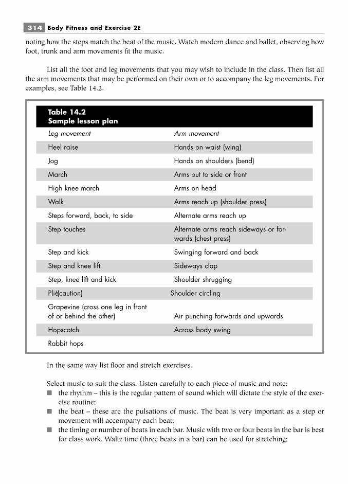
noting how the steps match the beat of the music. Watch modern dance and ballet, observing howfoot, trunk and arm movements fit the music.
List all the foot and leg movements that you may wish to include in the class. Then list allthe arm movements that may be performed on their own or to accompany the leg movements. Forexamples, see Table 14.2.
In the same way list floor and stretch exercises.
Select music to suit the class. Listen carefully to each piece of music and note:■ the rhythm – this is the regular pattern of sound which will dictate the style of the exer-
cise routine;■ the beat – these are the pulsations of music. The beat is very important as a step or
movement will accompany each beat;■ the timing or number of beats in each bar. Music with two or four beats in the bar is best
for class work. Waltz time (three beats in a bar) can be used for stretching;
314 Body Fitness and Exercise 2E
Table 14.2Sample lesson plan
Leg movement Arm movement
Heel raise Hands on waist (wing)
Jog Hands on shoulders (bend)
March Arms out to side or front
High knee march Arms on head
Walk Arms reach up (shoulder press)
Steps forward, back, to side Alternate arms reach up
Step touches Alternate arms reach sideways or for-wards (chest press)
Step and kick Swinging forward and back
Step and knee lift Sideways clap
Step, knee lift and kick Shoulder shrugging
Plié (caution) Shoulder circling
Grapevine (cross one leg in frontof or behind the other) Air punching forwards and upwards
Hopscotch Across body swing
Rabbit hops

■ the tempo – the rate at which the music is played. The American book Aerobics Dance-Exercise suggests that slow tempos of 100–120 beats per minute are suitable for warm-up and cool-down, and under 100 beats per minute for stretch and floor exercises, whilefaster tempos of 130–160 should be used for aerobics and dance.
Having listened to the music, select from the list of exercises suitable movements and pat-terns to fit the music. Practise these thoroughly yourself and record each movement and series ofmovements. Movements or patterns are usually repeated four to six times. Plan movements for thewarm-up, stretch, main core – building up the intensity and then easing down again – and cool-down. Note cues for good posture, breathing and accurate movements.
Exercise classes 315
TASK 1Work with a partner.■ Prepare a first lesson plan for a 30-year-old client who wants to lose weight and
improve the strength of the abdominals.■ Explain the reasons for your choice of exercises to the client.
TASK 2Work with a partner.■ Prepare a working area for an aerobics class.■ Instruct a client on how to lie down on the floor and come up again.■ Teach a client correct breathing, using the diaphragm and lower ribs.
TASK 3■ Plan an aerobics lesson for a group of 30–45-year-olds who are moderately fit.■ Discuss the reasoning behind your selection of exercises.
TASK 4Work in a group.■ Teach any six exercises to the group without music.

316 Body Fitness and Exercise 2E
TASK 5Work in a group.■ Select a piece of suitable music and plan a sequence of movements to fit the
music which may be used in an aerobics class.■ Teach these exercises to a small group.Remember to:■ listen to the music first;■ demonstrate the movement correctly;■ break the movement down into parts;■ teach each part thoroughly, with and without music,■ link all the parts together;■ practise the entire sequence.
Chapter 15First Aid for sports injuries
Attempting to diagnose or treat any injury without specialist medical training is dangerous prac-tice. The rate and success of recovery will depend on accurate diagnosis, followed by careful reha-bilitation. Inappropriate treatment can cause further damage and permanently impair function.However, knowing what action to take immediately before medical attention is available canreduce the extent of tissue damage. The trainer is often the first person on the scene followinginjury and must make rapid and crucial decisions based on knowledge and experience. Everyoneconnected with sport should be familiar with the principles of first aid, as immediate action maybe needed.
Prevention of injuryEvery precaution should be taken to prevent the occurrence of injury. Factors which contribute toinjuries are:
■ Inadequate or inappropriate training: plan well-designed exercise schemes showinggradual progression, give accurate instruction.
■ Inadequate warm-up and cool-down.■ Faulty equipment and poor surfaces: select a suitable venue with appropriate facilities
and good surfaces, check equipment.■ Improper use of equipment.■ Inappropriate footwear or clothing.■ Unsuitable weather conditions.■ Incomplete recovery following a previous injury: ensure adequate rest, relaxation and
recovery time.■ Activities inappropriate to age and fitness levels: ensure appropriate fitness assessment
prior to undertaking sport or exercise and also on return after injury.

■ Intensive competition, resulting in risk taking.■ Dehydration and poor nutrition.■ Exercising when there are contra-indications.■ Poor exercise technique: maintain good body alignment throughout, practise correct
technique and breathing patterns.
Immediate assessment of injuryIt is vitally important to assess the situation as soon as injury occurs. The injured person shouldnot be moved until a preliminary examination has been carried out.
BreathingWatch for chest movement or check nose and mouth for air flow. If there is no rise and fall of thechest, and you feel no movement of air, begin mouth to mouth resuscitation.
HeartbeatFeel for pulse: radial pulse at wrist, carotid pulse at throat behind wind pipe. If there is no pulse,begin cardiac compression.
Broken bonesIf for any reason fractures are suspected, do not move the casualty more than is absolutely neces-sary. This is particularly important if there is damage to the spine. Moving a casualty with damageto their spine can result in permanent paralysis.
BleedingAny profuse bleeding should be stemmed by applying firm even pressure directly over the area,preferably over a sterile dressing. Protect yourself from blood contamination.
Other injuriesLook for wounds, cuts, abrasions, signs of joint damage, e.g., pain, ligament sprains, muscle andtendon strain and tears.
Immediate treatmentThe quicker the treatment is administered following injury the greater the chance of speedy fullrecovery. Treatment should start immediately where possible and certainly within 24 hours.
Treatment is generally aimed to:■ preserve life;■ promote healing;■ return body to normal function.
More specific aims are:■ to prevent further damage;
318 Body Fitness and Exercise 2E

■ to reduce the inflammatory response;■ to reduce pain, swelling and stiffness;■ to gradually stretch, mobilise and strengthen the affected tissues;■ to maintain full strength and condition of unaffected body parts.
Fast action limits damage: for immediate action, think ‘RICED’:■ R – rest and immobilisation to prevent further damage;■ I – apply ice immediately for vasoconstriction and to prevent secondary damage;■ C – compression to the area to reduce swelling;■ E – elevation (using gravity to assist drainage from the area);■ D – diagnosis by a doctor either on site, in a surgery or in hospital.
RestFurther damage to an injured part can be prevented by resting and immobilising the part. The cas-ualty should be moved only if absolutely necessary, to facilitate breathing, to remove from the fieldof play or to prevent further injury. The injured part should be rested and supported correctly usingsplints, tubular, stocking or crepe bandages as necessary. These give firmer support if a layer ofcotton wool is wrapped around the area before applying the bandage. If leg fractures are suspected,the good leg can be used as a splint by tying the two legs together, or long strips of wood can beused and bound to the leg until medical treatment is available. A stretcher will then be needed totransport the casualty.
The casualty should rest for 24–48 hours after injury and should not put weight on aninjured leg. Elbow or axillary crutches should be used, or support can be given by a person oneither side with the casualty hopping on the good leg.
If the arm is seriously injured, a sling should be used for support. A triangular sling is placedaround the lower arm and supporting the elbow. The long end is taken over the opposite shoulderand the short end over the injured side, the two ends are tied at the back of the neck. If the injuryis below the elbow, the forearm should be supported upwards to assist drainage.
Crutch walkingMeasure the crutches carefully. There should be a space the width of three fingers (i.e. 6 cm)between the top of the crutch and the axilla (arm pit), otherwise pressure will damage the nervesin the region. The hand rest should be level with the crease of the wrist or the styloid process.
Two point walking should be used when both crutches are moved forward together: the cas-ualty pushes on the hand rest, straighten the elbows and hops to the crutches – do not hop throughthe crutches as this can result in loss of balance and falling backwards. Do not place the crutchestoo far forward as they will slip. The rhythm will be:
■ lift and move crutches forward;■ push on hands;■ straighten elbows;■ hop to the crutches.
First Aid for sports injuries 319

For going upstairs, put foot first, then crutches; for coming downstairs, put crutches first,then foot. Elbow crutches are used in a similar way, but do not give as much support.
IceIce should be placed over the injured area as soon as possible. This will reduce the metabolic rateand oxygen requirement of the cells around the periphery of the injury. These cells would notreceive oxygen because of the damage to blood vessels and would die resulting in secondary injury.Cold will also reduce internal bleeding and swelling as the blood vessels constrict, reducing fluidexudate and bruising.
Care must be taken when applying ice to the area, as there is a risk of producing ice burnsif the ice comes into contact with the skin over a prolonged period. The area should be coveredwith oil for ice cube massage, or a tea towel should be used between the skin and the ice whenusing ice packs.
There are various ways of applying ice:1 Stroke the oiled skin with an ice cube, keeping the ice moving over the area slowly.2 Ice cubes can be shattered and placed in a towel, which is wrapped around the injury
over a tea towel.3 Freezer packs or even frozen food such as a packet of peas can be used; place them over
a tea towel covering the area, and hold in place by another towel. These packs are veryuseful as they can be refrozen and reused.
4 Ankle and wrist injuries can be immersed in iced water in a bucket or bowl. The part is heldin the water for as long as is tolerable, is removed for a new minutes and then re-immersed.
Ice should be applied for at least 10–15 minutes, increasing to 30 minutes, unless the skinis sensitive and the area feels uncomfortable. The skin should turn colour: pink for pale skin butdarker for dark skin.
Ice should be applied every two to three hours, initially working down to three times a dayas healing progresses and the swelling subsides.
Note: Heat should never be used in the acute stage of injury as it increases metabolic rate,and produces vasodilation, which increases blood flow and swelling. Heat may be used afterhealing has taken place (usually in 6–12 days), but only after the bruising turns yellow.
CompressionThis means applying pressure to the area, which helps to limit the bleeding into the tissues. Anelasticated tubular or crepe bandage may be used. Additional pressure can be applied if a layer ofcotton wool is applied to the area before bandaging. Do not use non-elasticated bandages on arecent injury; the strapping needs some stretch to allow for swelling.
The strapping must not be too tight, as it will restrict the circulation. If the swellingincreases, the pressure under the strapping will increase, and this will produce further restriction
320 Body Fitness and Exercise 2E
and damage. Check the swelling and the colour of the skin and nails beyond the strapping;white/grey skin and blue nails indicate that the strapping is too tight, in which case the bandageshould be released.
ElevationThe injured part should be supported in elevation whenever possible. Gravity will then assist thedrainage of any fluid exudate away from the area. This will help to reduce the pressure within thetissues and the pain around the damaged area.
DiagnosisAccurate diagnosis is crucial for maximum recovery. Seek medical advice as quickly as possible ifthere is doubt about the injury, and for any of the following conditions:
■ Head injuries■ Headache, nausea, vomiting or dizziness following head injuries■ Pains in the neck or symptoms down the arms such as tingling or numbness■ Pains in the back and down the legs, or numbness■ Breathing difficulties or pains in the chest■ Fracture or suspected fracture■ Dislocation of a joint, or severe injury to a joint or ligament■ Profuse bleeding and deep or large wounds■ Severe muscle and tendon injuries■ Abdominal or groin pain■ Eye injuries
Summary of precautions■ Do not move a casualty with injuries to the spine.■ Do not move a casualty with fractured bones, more than is absolutely necessary.■ Apply bandages and strapping firmly, but not too tightly as too much pressure may
restrict the circulation.■ Check the limbs beyond the strapping for cold, white or blue colouration which indi-
cates lack of circulation – loosen the strap immediately if this is the case.■ Do not apply heat in any form to the injured area, i.e. do not use heat lamps, hot packs,
hot baths, showers, ultra-sound, diathermy, hot towels or any liniments.■ Do not massage the injured area.■ Do not exercise through the pain or use electrical stimulation.■ Do not allow the injured person to drink alcohol.
Types of injuryInjuries can be divided into two categories:
1 Acute injuries: Traumatic injuries which happen suddenly due to some external force orinternal stress; these produce sudden pain, swelling, bruising or wounds.
2 Chronic injuries: Repetitive strain injuries or overuse injuries occur slowly and becomeprogressively worse over a period of time. Pain and swelling is usually of gradual onsetbut persists over a long period of time.
First Aid for sports injuries 321
Skin and subcutaneous tissues injuriesSharp objects, equipment or apparatus, the playing surface, etc, may cause injuries to the skin andunderlying tissue. These include cuts, abrasions, infections, contusions, blisters.
When dealing with blood spill injuries, protect yourself from contact with blood. Wearrubber surgical gloves if available, or place the contact hand in a plastic bag. This is important pro-cedure as many life-threatening viruses are transmitted through blood contamination.
CutsCuts should be thoroughly cleaned and all dirt or debris removed. They should be washed orswabbed with clean water and/or antiseptic solutions, then covered with a sterile dressing. Swabfrom the centre outwards and use a clean area each time the wound is touched. Cover with a steriledressing and bandage in position.
Small cuts can be covered with plasters but again clean carefully, dry and apply thegauze centre over the cut. For stab type cuts, draw the edges together with butterfly plasters. Ifthere is extensive bleeding from a wound, cover with a sterile dressing and apply pressure with acotton wool pad; bandage and elevate the area. Seek medical treatment as soon as possible.Large cuts over 2 cm long and gaping will require stitching – refer at once to a CasualtyDepartment.
AbrasionsThese are caused by friction or scraping of the skin, and are usually superficial. Clean and treat ascuts.
InfectionsCuts and abrasions can become infected as a result of dirt and micro-organisms penetrating theskin or hair follicles. Infections may result in boils or carbuncles which may require antibiotics toprevent them spreading to underlying tissues. Refer to a doctor if infection occurs.
ContusionsContusion is caused by a direct blow, and results in bleeding into the tissues. Apply ice as directedover the contusion to reduce bleeding and swelling. Do not use heat or massage, as these willincrease bleeding.
BlistersBlisters are the result of pressure or friction, and prevention is better than cure. If blisters havedeveloped, they should be left intact as the skin acts as a barrier to infection. A blister can be pro-tected from pressure by surrounding it with a piece of plastic foam with a hole in the middle, thenprotected with adhesive plaster and left to heal. Large blisters or blood blisters which cause paindue to the build-up of pressure can be carefully punctured. Use a sterile needle and puncture twotiny holes in the blister. Gently squeeze out the fluid and cover with a non-adhesive sterile dress-ing and then plaster.
322 Body Fitness and Exercise 2E
If the area is raw, clean it with water and apply an antiseptic gel or plastic skin. Use sterileequipment to avoid infection.
Muscle injuriesInjuries to muscles may be strains, partial tears or complete tears, and haematoma.
StrainsStrains will damage and result in micro tears within some fibres of the muscle. The symptoms ofpain and stiffness are slow in onset and usually mild. Active movement or passive stretching willcause pain around the damaged area.
Partial tearsThese result in tearing and disruption of some fibres within the muscle. The symptoms are feltimmediately with severe pain and tenderness, especially when attempting to contract the muscle.
Complete tearsThese involve the tearing of all muscle fibres, and the two ends of the muscle contract away fromeach other. Pain, swelling and tenderness is very severe and there will be complete loss of func-tion. This type of injury may require surgery – refer quickly to hospital.
Muscular haematomaDirect impact injuries will result in muscle rupture and bleeding. Bleeding may occur within amuscle (intramuscular) or between muscles (intermuscular). It is important to diagnose theseinjuries accurately as recovery from intermuscular haematoma should be relatively quick andcomplete. However, complications can arise following intramuscular haematoma due to intra-compartmental pressure; muscle function may be absent and recovery may be slow and incom-plete.
Initially, muscle injuries should be treated with RICE as soon as possible and continued for48–72 hours. Any vigorous movements, stretching, heat and massage must be avoided initially, ascomplications may result.
Tendon injuriesTendons attach muscles to bone. Tendon injuries are either ruptures (tears) or inflammation (ten-dinitis).
TearsTendons usually tear at their weakest point, i.e. where they join the muscle at the musculo-tendinous junction. They may be partial tears, when some fibres are torn or complete tears whenthe tendon is severed. Pain may be mild or severe.
Complete tears may require surgery and the best results are obtained if surgery is performedimmediately, before the two ends shrink and move apart. Pain is immediate, sharp and severe. Itfeels like a sharp blow or a ‘snap’ sensation in the area; the ‘snap’ can sometimes be heard.
First Aid for sports injuries 323

TendinitisInflammation of a tendon (tendinitis) and inflammation of the tendon in its sheath (tenosynovitis)are very common problems. They are usually caused by repetitive stress or overuse, but can becaused by awkward movements such as landing awkwardly or mis-hitting a ball. The pain is nig-gling and comes on gradually. It is worse when the tendon is moved and may progress until move-ments are impossible. Because the blood supply to tendons is poor, they can take a very long timeto heal: up to 12 weeks or even longer.
LigamentsLigaments attach bone to bone; they support and stabilise joints. Ligaments are damaged whenjoints are forced into abnormal positions. Ligaments may be sprained, partially or completely torn.
SprainedThis occurs when a few fibres are torn, producing pain and swelling. These heal quickly with littledisruption of joint movement.
Partial tearsMany fibres are torn because of greater stress. These produce severe pain and swelling, and thejoint will be unstable.
Complete tearsThese are very severe, producing extreme pain and swelling; the joint will be quite unstable andmay dislocate. Torn ligaments may heal well without surgery but others require suturing (a stitchor stitches for closing a wound, or joining two or more structures).
Injuries to ligaments result in bruising, tenderness and swelling around the affected joint,and the healing process may take over six weeks. The joint should be supported during this timewith some form of strapping. Severe tears may require a brace or plaster cast.
MenisciThese are discs of cartilage found in certain joints such as the knee, where the medial and lateralmenisci lie on the upper surface of the condyles of the tibia. These may tear due to excessive forcesduring rotation or extreme flexion, causing acute pain and swelling. The knee may lock if a part ofthe discs becomes dislodged as it may interfere with the function of the knee. Surgery may berequired to remove part of the cartilage but some tears heal without surgery.
BursaeThese are sacs of fluid which reduce friction between moving parts of a joint. They may lie betweentendons and bones to allow smooth movement of the tendon over the bone. They usually becomeinflamed because of overuse or repetitive trauma.
Inflammation of a bursa is known as bursitis: it produces pain and swelling in the area ofthe bursa and radiates pain around it. It may heal with rest or may require a cortisone injection tohelp it settle. Very occasionally surgery is required for a chronic, persistently painful bursa.
324 Body Fitness and Exercise 2E
Bone fracturesA fracture is a break in a bone. It may be classified as transverse, oblique, spiral or comminuted.Fractures may be simple or compound:
■ A simple fracture is a clean break in the bone with the skin intact.■ A compound fracture involves more complex breaks of the bone and perforation of the
skin.Bones fracture due to excessive force applied to the bone. Stress or fatigue fractures occur as aresult of overuse, when repetitive muscle contraction pulls on the bone. This causes repeatedminor stress and damage which does not have time to heal. Fractures require immobilisation toreduce the displacement and prevent movement, thus allowing time for the fracture to heal.Fractures of the upper limb usually heal in six to eight weeks, providing there are no complicationssuch as inadequate blood supply. However, fractures of the lower limb take 12 to 14 weeks.Fractures heal more quickly in children than adults.
Treatment of soft tissue injuriesThese are general guidelines; treatment will vary depending on the type and extent of the injury.Only those with specialist knowledge should attempt to treat sports injuries.
1 Acute phase: for immediate treatment, apply RICED; ice applications should be usedevery hour and then every two to three hours.
Slow static movements within the limit of the pain can be practised two to three timesa day. These isometric movements must be performed slowly, and must stop before painis felt. Start with three contractions only, then build up gradually to five, seven and ten,providing there is no deterioration in the condition.
2 Sub-acute phase (24–48 hours): continue with the above routine. Once static move-ments can be carried out without pain, add gentle active movements within the painfreerange. When inner and middle range is pain-free, move into outer range; it is importantto maintain flexibility.
No definite timescale can be set for these stages, they will depend on the extent of the injuryand the speed of recovery.
Rehabilitation
Stage 1This will begin as soon as possible after severe pain and muscle spasm eases. It is a non-weightbearing phase; ice or other modalities may be used. Free active exercises must be practised four tosix times per day. Little and often is the best format – the number must increase each time.Eccentric work may be easier initially. Static stretch exercises should also be included to improveflexibility. During this phase, the unaffected parts should be exercised to maintain fitness levels,but care must be taken not to stress the injury.
First Aid for sports injuries 325

Stage 2This is a partial weight-bearing phase, which will begin when there is little swelling and no pain innearly full range of movement. Heat can now be used, or other modalities. Muscles are exercisedagainst light resistance with a build-up of repetitions. Partial weight-bearing exercises are prac-tised; pain must be the guide – if it hurts, stop.
Stage 3This is the weight-bearing phase. Strength training, flexibility and co-ordination work are included,in preparation for the return to normal activities.
Stage 4This is a return to full function. This phase must be planned to cover all the activities that the clientwill encounter on return to normal situation, whether simply coping with daily living or heavytraining. Much encouragement is required in this stage as movements which caused the injuriesmust be introduced.
It is important not to progress too quickly through each phase as injuries may recur if ath-letes are over anxious to return to their sport. Fitness tests must be undertaken to ensure adequatefitness levels before resuming sporting or normal activities.
CryotherapyCryotherapy (cold or ice therapy) is the first line of action in the treatment of sports injury. It iseffective, simple, easy to use and inexpensive. It is beneficial in the immediate (acute) post-traumatic phase, and also through the rehabilitation phases. As it is easy to apply with little dangeror complication, the athlete can continue the treatment at home. Ice must be applied as soon aspossible to the injured part, within 5–15 minutes. Any delay will result in secondary damage whichwill increase the extent of the injury. This will prolong the rate of recovery and limit the return tofull normal function.
Methods of ice application1 Ice cubes which are slowly moved over the area; sometimes referred to as ice massage.2 Crushed ice placed in a plastic or towelling bag; place over the area with a thin towel in
between.3 Gel freezer packs which are kept frozen until required and applied to the area as above.
These are convenient to use and can be refrozen and reused.4 Packs of frozen food such as peas applied to the area as above; these can be refrozen
and reused (but must not be eaten after defrosting).5 Chemical packs which become cold when struck hard to mix the chemicals. These packs
are not as effective as other methods as the temperature is not as low. There is also adanger of chemical burns, should the chemicals leak. Manufacturers are continuallyworking on improvements.
326 Body Fitness and Exercise 2E

6 Ice-water in a pan or bucket into which the part is immersed. Most suitable for ankleand wrist injuries. To ensure a sufficiently cold temperature, ice must float on thesurface throughout the treatment.
7 Cold aerosol sprays are not as effective as other methods as they produce superficialcooling only. They do have the advantage of being very convenient, easy to carryaround and quick to use.
Physiological effects of cooling the tissuesCertain changes will be produced in the tissues as a result of cold application – these are knownas the physiological effects. These effects include:
■ a decrease in metabolic rate in the area;■ a decrease in the circulation due to vasoconstriction in the area;■ local anaesthesia with a reduction in pain;■ a decrease in muscle spasm;■ a decrease in inflammatory response;■ a decrease in the flexibility of ligaments and tendons.
Decrease in metabolic rateThis is the main reason for applying ice to the area immediately following injury. Cold reduces themetabolic rate of cells and consequently their oxygen requirement. If cold therapy is not given, thecells around the periphery of the damaged area will require oxygen to meet metabolic demands. Ifthis demand for oxygen cannot be met because of damage to the blood vessels in the area, the cellswill die. This happens within 10–15 minutes following injury and continues for around 12 hours.This will increase the amount of tissue damage and the size of the injury; it is known as ‘secon-dary hypoxic injury’.
By decreasing the cells’ metabolic rate and need for oxygen, these cells may survive untilcirculation is restored, thus limiting the extent of the injury.
Note: The application of heat will have the opposite effect. Heating the tissues will increasemetabolic rate and the demand for oxygen, and will cause greater secondary damage. Heat musttherefore not be used to treat immediate acute injury.
Decrease in circulationCold therapy produces vasoconstriction, which will reduce blood flow. Less bleeding into thetissues will facilitate quicker healing. However, the blood clotting mechanism will be activatedimmediately following injury; this will also prevent blood loss and will have occurred before cold
First Aid for sports injuries 327
LEARNDo not place ice directly on to the skin, which may result in ice burns. Alwaysseparate the ice from the skin using a towel.

packs can be applied. In addition, the constriction of capillaries in response to cold will reducefluid exudate and tissue swelling.
Reduction of painCold induces anaesthesia – the sensory receptors in the skin are inhibited and sensation isreduced but not totally absent. Initially (one minute or so after applying the ice) pain is increased.A dull radiating pain is felt which may increase for a while but will eventually pass, giving way toa prickling sensation and then numbness.
The decrease in pain will reduce the attendant muscle spasm and limit the pain-spasm-paincycle which occurs after injury. This analgesic effect may mask the extent and seriousness of theinjury, therefore the athlete or sportsperson must not return to the activity immediately followingice treatment.
Decrease in muscle spasmIt is thought that the reduction in muscle spasm is brought about by the anaesthesia, and becausecold decreases nervous transmission and depresses muscle spindle sensitivity and reflex mecha-nisms. After injury, the body’s protective mechanism increases muscle tone to prevent furtherdamage to the tissues. Tight muscles act as a splint preventing movement and hence furtherdamage. If this spasm is inhibited in the early stages, careful active movements can be performed,improving recovery rate. In the rehabilitative stages, less muscle spasm allows greater flexibilityand increased range of movement which facilitates a speedier return to full function.
Decreased inflammatory responseIt is thought that cold decreases the inflammatory response because it reduces the effects of hista-mine.
Decrease flexibility of ligaments and tendonsThe flexibility of connective tissue decreases after cold application; ligaments and tendons are notas elastic. It is therefore important to stop cold therapy when introducing flexibility exercises. Atthis stage, when healing is progressing satisfactorily and there is no risk of further bleeding, someform of heat should be given. This may commence 72 hours or so after injury, but the time willdepend on the rate of healing and the extent of the injury. If in doubt, continue with ice.
Uses of cryotherapy1 Cold therapy may be used to treat soft tissue injuries in the acute, subacute, and reha-
bilitative stages.2 In the acute stage, ice is applied for 15 minutes every 1–2 hours. Little and often is the
best guide. It is most effective at this stage because it slows down the metabolic rate,reduces bleeding and fluid exudate thus limiting further tissue damage.
3 In the sub acute and rehabilitative stages, the time of application is increased to 20–30minutes, 3–4 times a day. Its main use in these stages is to relieve pain and musclespasm, and thus facilitate early active movement.
4 To treat overuse injuries.
328 Body Fitness and Exercise 2E

Contra-indications■ Open bleeding wounds.■ Deficient circulation in the part.■ Lack of skin sensation.■ Hypersensitivity to cold.■ Extreme pain during application.
Factors which affect the rate of coolingThe application of cold to the area conducts heat away from the superficial and deep tissues,resulting in a decrease in temperature. There will be a rapid and immediate reduction in surfacetemperature, but the temperature of deeper tissues will decrease more slowly and will continue todecrease for some time after the ice is removed. There are many factors which affect the rate ofcooling. These include:
1 The area of the part in contact with the ice.2 The length of time the part is in contact with the ice.3 The difference in temperature between the part and the ice.4 The rate at which the body regenerates heat.5 The rate at which heat is conducted away from the ice.
Ice packs and gel packs used for 30 minutes produce similar cooling effects, but researchindicates that immersion in ice water for an equal length of time produces more intense coolingand slower rewarming. This may relate to the larger surface area being treated.
Chemical packs are less effective at reducing temperature than the other methods, and coldsprays produce superficial, temporary cooling only.
TreatmentRemember the RICE routine (rest, ice, compression, elevation).
Ice must be applied as soon as possible, within 5–10 minutes of the injury occurring.Immediate appropriate treatment will increase effectiveness and considerably shorten the recoveryperiod. Ice should be applied as soon as an initial assessment of the injury has been made.
The method of application will depend on availability and convenience. Chipped or crushedice in a bag, gel packs from a refrigerator, or ice cubes are suitable for most injuries; ice water in abucket or bowl is suitable for ankle, calf, wrist, forearm and elbow injuries.
If ice is placed in direct contact with the skin it can stick and long-term application canproduce ice burns. It is therefore essential to oil the skin lightly before stroking with an ice cube,and to place a thin layer of cold wet towel or bandage on the skin when using an ice pack, i.e.between the skin and the ice pack. The part may be immersed in iced water without protection, butif pain is intense the part must be removed from the water for 15–30 seconds and then reimmersed.
First Aid for sports injuries 329
Rest is important, as any movement may produce further damage increasing the extent ofthe injury. Compression may be applied and the part elevated if possible to assist drainage.
Technique using ice pack or gel pack1 Prepare the ice pack by placing approximately 1 kg of crushed ice in a plastic or towel-
ling bag; alternatively, pile the ice onto the towel and fold the ends over. If using a coldpack, remove from the freezer just before use.
2 Ensure that the client is comfortable, well supported and in a suitable position toreceive the treatment.
3 Remove any clothing or jewellery from the area.4 Explain the treatment to the client, highlighting the beneficial effects and the impor-
tance of regular timed application. Explain that pain may be felt or increase initially, butthat this will give way to pins and needles and then numbness. If pain continues removethe ice.
5 Wring out a towel in cold or iced water and place over the injured part. Place the cold packover this and wrap a double layer of towelling around the part to hold the pack in place. Ifintense pain develops, the ice must be removed for 15–30 seconds and then reapplied.
6 Compression should be applied and the part elevated and rested.7 Treat for 15 minutes initially, increasing to 30 minutes in the subacute stage, but this
will depend on client tolerance. Pale skin should be red, dark skin will be darker.8 Repeat the procedure every two hours for the first 24 hours after injury, or as often as
possible.9 Exercise as explained below.
Technique using iced water1 Fill the container to three-quarters full with cold water and ice. Ensure that there is ice
floating in the water throughout the treatment.2 Position the client in a comfortable position; remove all clothing and jewellery around
the injury.3 Explain the treatment to the client as in point 4 above.4 Immerse the part in the water and reassure that pain is to be expected initially but that
it will subside. If the pain is intolerable, lift the part out of the water for 15–30 secondsthen reimmerse.
5 Keep immersing the part until it is numb and red; aim for 20 minutes.6 Remove the part from the water and dry gently.7 Exercise as explained below.
Technique with ice cube massage1 Place a supply of ice cubes in a container.2 Position the client in a comfortable, well-supported position; elevate the part if possible.3 Remove all clothing and jewellery from the area.4 Explain the procedure to the client.5 Spread a thin layer of oil over the area.6 Hold the ice cube with folded tissue or lint.
330 Body Fitness and Exercise 2E

7 Move the ice slowly over the part, moving up and down in straight lines; overlap theprevious stroke. Work over and around the injured part. The ice will melt, so ensure thatthere is a towel under the part to absorb the water.
8 Continue working in this way for 20 minutes or so, until the part is red and numb.9 Dry the area gently.
10 Exercise as explained below.
Technique with cold spraysThese aerosol sprays are not as effective as other methods, and are generally used for convenienceon the field of play. The part is uncovered and sprayed at a certain distance for a few seconds. Itis important that these sprays are used according to manufacturer’s directions, as their mode ofapplication may vary. If incorrectly applied they can cause ice burns.
Exercises following cryotherapyDuring the first 24–48 hours, ice application is followed by slow, gentle isometric exercises. Thesestatic muscle contractions are performed within the pain-free range.
As healing progresses, isotonic movements are performed. The client is instructed to movethe joint slowly through to the point just before pain is felt, to hold for a moment and return. Allpossible joint movements must be practised in this way. Great care must be taken during this sub-acute stage since movement can disturb the healing process and increase secondary damage.
Remember: movements must not produce pain. All movements must be within the limit ofpain.
Example: Treatment to injured ankle joint, where injuries are usually to the lateral or medialligaments.
■ Apply cold therapy for up to 30 minutes, then remove.■ Static exercise instruction – ‘I’m going to hold your foot firmly and I want you to pull as
hard as you can against my hand, stop if there is any pain’. The resistance against themovement must be even, and great enough to produce tension within the muscle but toprevent any movement. Initially the resistance is applied to dorsi flexion (hand apply-ing resistance on the dorsum of the foot), and is then applied to plantar flexion (handon the sole of the foot). When these contractions are easy to perform, inversion andeversion are added. Exercise away from the injury first, i.e. for lateral ligament injury,perform static inversion first. When this is easy, carefully perform static eversion (thismay not be possible initially). Each contraction is held for five to six seconds.
Remember: tension must be developed within the pain-free limit.■ After 48–72 hours or so, depending on the severity of the injury, ice treatment is fol-
lowed by isotonic exercises.■ Instruction for isotonic movement – ‘Pull the foot up slowly towards you (dorsi flexion);
stop when you feel any pain; hold; now move the foot slowly down away from you.’(plantar flexion). Repeat for inversion and eversion and then progress to circumduction.Perform three movements of each initially, increasing by two with each application.
First Aid for sports injuries 331

Practice Tasks
Explain how and why you would teach a client to breathe correctly (refer to page 42):
Devise a record card that you would use during the initial consultation and assessment of eachclient, prior to their exercise classes (refer to chapter 13):
Study a variety of exercise videos, exercise books and magazine articles and complete the follow-ing tasks.

List any exercises that you would consider unsafe:
Devise your own set of warm-up exercises:
Devise a set of aerobic exercises:
Devise a set of cool-down exercises:
Practice Tasks 333
QUESTIONS1. List the methods by which ice may be applied.2. Discuss the physiological effects of cooling the tissues.3. List the contra-indications to cold therapy.4. List the factors which affect the rate of cooling.5. Explain the importance of applying oil to the skin prior to ice massage.6. State how frequently ice should be administered during the first 24 hours.

References and furtherreading
Alter, Michael J. (1988). Science of Sketching, Human Kinetics Books. An excellent book for anyonerequiring any information on flexibility work.
American Council on Exercise (1991). Aerobic Dance-Exercise Instructors Manual. A useful sourceof detailed information for anyone leading aerobic classes.
Beashel, Paul, and Taylor, John (1988) Sport Examined, Macmillan Education.Cross, Gibbs and Gray (1991), The Sporting Body, Sydney: McGraw Hill Book Co.Curzon, L.B. (1976), Teaching in Further Education, London: Cassell.Daniels, Lucille, and Worthington, Catherine (1977), Therapeutic Exercise, WB Saunders Co.
Comprehensive information on correct and maintenance of posture and body alignment.Davies, Kimmel and Anly (1988), Physical Education Theory & Practice, Macmillan Company
(Australia). Covers the detail of exercise theory. Interesting additional reading.Grisogono, Vivian (1984), Sports Injuries, London: John Murray. A good self-help guide on the
avoidance and treatment of sports injuries.Hazeldine, R. (1993). Fitness for Sport, The Crowood Press. Excellent, clearly explained informa-
tion for individuals training for or teaching the theory of fitness. Good examples of exercise pro-grammes, circuits, etc., for achieving set goals.
Kennedy, Legel and Sagamore (1992), Anatomy of an Exercise Class, Human Kinetics Books. Givesdetailed information on the degrees of movement possible at body joints. Excellent informationon analysis of exercises.
Luby, Sue, and St Onge, Richard A. (1986), Body Sense, Faber & Faber Inc. A well-illustrated bookclearly showing the hazards of poor postural alignment. Covers the fundamentals of breathing,posture and alignment, stretching techniques and other useful topics.
Sharkey, B.J. (1990), Physiology of Fitness, Human Kinetics Books. Gives detailed information onthe physiological aspects of exercise.
Smith, B. (1993), Advanced Fitness Teachers Manual, Ludoe Publications (LoughboroughUniversity). Gives specific information on training for the components of fitness.
St George, Francine (1990), The Muscle Fitness Book, The Crowood Press. Good guidelines on safe,effective exercise for specific sporting activities.
Time-Life Books (1990), Cross Training – Ultimate Fitness. Interesting reading for those interestedin the concept of cross training. A good chapter on ‘Eating for Performance’ gives recipes for suit-able foods.
Aabdomen
exercises 178, 243, 286–87,298
weakness 237, 240abduction 24, 49abductor muscles 298abrasions, treating 322acceleration 126, 208achilles tendon 65acromio-clavicular joint 45actin 67acute injuries 321adaptation, of body systems 3adductor muscles 49, 64f, 176,
197, 298adenosine triphosphate (ATP) see
ATP energy; ATP-PC systemadipocytes 14adipose tissue 13f, 14taerobic
capacity (see cardio-respiratoryendurance)
energy systems 9, 73, 74, 75,76t, 145, 148–49, 313
exercise 14t, 73, 76, 224,312–16
Aerobics Dance-Exercise 315age
exercise 275, 281flexibility 184–85, 190joint range 54posture 235
agonists and antagonists(muscles) 50, 112, 113,119
alactic ATP-PC system 73–75,74, 76t, 77, 80–81, 145,148, 158
alcohol 275alveoli 102f, 103American College of Sports
Medicine 312amino acids 216anabolism 8anaerobic
energy systems 73, 74, 76t, 77,145, 148–49, 271, 313
exercise 77glycolysis 9respiration 8
anatomical position 22–27anatomy 5angina pectoris 91ankle
joint 56, 56fmovement 49strengthening 168
annulus fibrosus 39antioxidants 220aponeuroses 71appendicular skeleton 28, 30t, 46arches, foot 57–58, 57fareolar tissue 12f, 13tarrangement, of exercises 152,
166arteries 90f, 91, 92articular cartilage see hyaline
cartilagearticulation see jointsasphyxia 103asthma, and exercise 272ATP energy 6, 9, 80–81, 98, 213ATP-PC system 73–75, 76t, 77,
80–81, 145, 148, 158atria (sing. atrium) 90–91, 95–96autonomic nervous system 18,
83–84, 94axes of movement 23–24axial skeleton 28, 29
Bback
extensor muscles 179, 250problems 180, 235, 250–51strengthening 179–80, 287–88stretching 202–4
balance 233–34ball and socket joints 52t, 54–55,
58, 59fballistic
bouncing 284, 285stretching 186–87, 190
basal metabolic rate (BMR) 211base, and stability 122, 123f,
124, 133beta-carotene 217biceps muscles 204, 297
brachii 64ffemoris 65f, 172, 196
bleeding, treating 318blisters, treating 322–23
blood 15t, 93–94, 100contact with 322flow 90, 91functions 94muscles 72–73pressure 96–97, 98, 100, 274,
303shunting 93, 98, 150supply 91vessels 92–93, 99, 100
bodycomposition 143, 164, 224–25,
299–300fat 166planes 22–23, 23fstructures 25, 26t, 27–28surfaces 25, 25f, 26t, 27–28systems 3, 5, 19–20
Body Mass Index (BMI) 224–25bones
breaks 318cell types 26fractures 325marrow 24osseous tissue 15tskeleton 28–46structure 25–27types 27
brachioradialis 65fbrain 84fbreasts, and posture 237breathing
injury assessment 318mechanism 42–43safety 277see also lungs; ventilation
bronchi 102f, 103bronchioles 102fbursae 53bursitis 324buttocks 173, 200–201, 238,
253–54
Ccalcaneus (foot bone) 33fcalcium 45, 219calories 211–12cancellous bone 15t, 25, 28cancer 215, 217, 218, 220capillaries 93, 102fcapitate (hand bone) 36f
Index
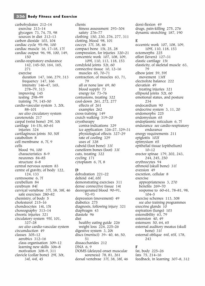
carbohydrates 212–14exercise 213–14glycogen 73, 74, 75, 98sources in diet 212–13
carbon dioxide 103, 104cardiac cycle 95–96, 100cardiac muscle 16, 17–18, 17fcardiac output 96, 98, 100, 149,
150cardio-respiratory endurance
142, 145–50, 164, 165, 271
exerciseduration 147, 166, 279, 313frequency 147, 166intensity 146–47, 165,
278–79, 313improving 145testing 298–99training 79, 145–50
cardio-vascular system 3, 20t,88–101
see also circulatory systemcarotenoids 217carpal (wrist bone) 29f, 30tcartilage 14–15t, 60–61
injuries 324cartilaginous joints 50, 50fcatabolism 8cell membrane 6, 7f, 9cells
blood 94, 100characteristics 8–9neurones 84–85structure 6–8
central nervous system 83centre of gravity, of body 122,
124, 133centrosome 6, 7fcerebellum 84cerebrum 84fcervical vertebrae 37f, 38, 38f, 46
safe exercises 280–82chemistry, of body 5cholesterol 215–16chondrocytes 14t, 15tchoreography 313–14chronic injuries 321circulatory system 95f, 101,
327–28see also cardio-vascular system
circumduction 49classes 305–12
aerobics 312–16class organisation 309–12learning new skills 306–8motivation 308–9, 311
clavicle (collar bone) 29f, 30t,34f, 44f, 45
clientsfitness assessment 293–304safety 276–77
clothing 150, 230, 276, 277, 311clotting, blood 98, 101coccyx 37f, 38, 46compact bone 15t, 25, 28compression, for injuries 320–21concentric work 107, 108, 109,
109f, 110, 113, 118, 153condyloid joints 52t, 60connective tissue 10, 12–16
muscles 65, 70–71contraction, of muscles 63, 71,
79all or none law 69, 80blood supply 73energy for 73–76
contusions, treating 322cool-down 261, 272, 277
effects of 261examples 262
cross-training 149crutch walking 319–20cryotherapy
contra-indications 329ice application 326–27, 329–31physiological effects 327–29rate of cooling 329uses of 328
cuboid (foot bone) 33fcuneiform bones (foot) 33fcuts, treating 322cycling 171cytoplasm 6, 7f, 8
Ddehydration 221–22deltoid 64f, 65fdemonstrating exercises 311dense connective tissue 14tdeoxygenated blood 90–91,
92–93depression (movement) 49diabetics 275diagnosis, following injury 321diaphragm 43diastole 96diet
healthy eating guide 226weight loss 224, 225–26
digestive system 3, 20tdiscs (menisci) 39– 40, 46, 50,
51dissaccharides 212DNA 6, 9DOMS (delayed onset muscular
soreness) 78, 81, 261dorsal vertebrae 37f, 38, 38f, 46
dorsi-flexion 49drugs, pain-killing 275, 276dynamic stretching 187, 190
Eeccentric work 107, 108, 109,
109f, 110, 118, 153ectomorphs 225effort (levers) 127–31elastic cartilage 15telasticity, of skeletal muscle 63,
79elbow joint 59, 59f
movement 130felectrolyte balance 222elevation 49
treating injuries 321ellipsoid joints 52t, 60emotional states, and posture
234endocardium 90endocrine system 3, 11, 20endomorphs 225endomysium 65endoplasmic reticulum 6, 7fendurance see cardio-respiratory
enduranceenergy requirements 211epiglottis 102fepimysium 65epithelial tissue (epithelium)
10–12erector spinae 179, 202, 243,
244, 245, 250erythrocytes 94ethmoid (skull bone) 31feversion 49excretion, cellular 8exercise
appropriateness 3, 270benefits 269–70response to 60–61, 78–81, 98,
104–5exercise schemes 113, 308
see also training programmesexocrine glands 10expiration (lungs) 103extensibility 63, 79extension 40, 49extensors 50, 64, 65external auditory meatus (skull
bone) 31fexternal oblique 64f, 65f, 178,
243
Ffat, body 225–26fats 75, 214–16feedback, in learning 307–8, 312
336 Body Fitness and Exercise
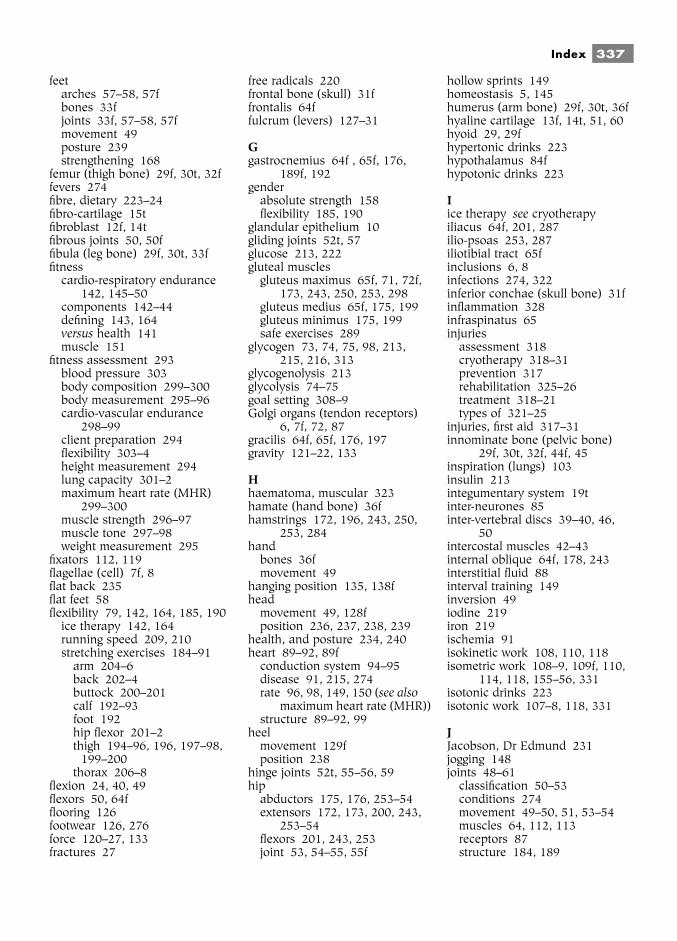
feetarches 57–58, 57fbones 33fjoints 33f, 57–58, 57fmovement 49posture 239strengthening 168
femur (thigh bone) 29f, 30t, 32ffevers 274fibre, dietary 223–24fibro-cartilage 15tfibroblast 12f, 14tfibrous joints 50, 50ffibula (leg bone) 29f, 30t, 33ffitness
cardio-respiratory endurance142, 145–50
components 142–44defining 143, 164versus health 141muscle 151
fitness assessment 293blood pressure 303body composition 299–300body measurement 295–96cardio-vascular endurance
298–99client preparation 294flexibility 303–4height measurement 294lung capacity 301–2maximum heart rate (MHR)
299–300muscle strength 296–97muscle tone 297–98weight measurement 295
fixators 112, 119flagellae (cell) 7f, 8flat back 235flat feet 58flexibility 79, 142, 164, 185, 190
ice therapy 142, 164running speed 209, 210stretching exercises 184–91
arm 204–6back 202–4buttock 200–201calf 192–93foot 192hip flexor 201–2thigh 194–96, 196, 197–98,
199–200thorax 206–8
flexion 24, 40, 49flexors 50, 64fflooring 126footwear 126, 276force 120–27, 133fractures 27
free radicals 220frontal bone (skull) 31ffrontalis 64ffulcrum (levers) 127–31
Ggastrocnemius 64f , 65f, 176,
189f, 192gender
absolute strength 158flexibility 185, 190
glandular epithelium 10gliding joints 52t, 57glucose 213, 222gluteal muscles
gluteus maximus 65f, 71, 72f,173, 243, 250, 253, 298
gluteus medius 65f, 175, 199gluteus minimus 175, 199safe exercises 289
glycogen 73, 74, 75, 98, 213,215, 216, 313
glycogenolysis 213glycolysis 74–75goal setting 308–9Golgi organs (tendon receptors)
6, 7f, 72, 87gracilis 64f, 65f, 176, 197gravity 121–22, 133
Hhaematoma, muscular 323hamate (hand bone) 36fhamstrings 172, 196, 243, 250,
253, 284hand
bones 36fmovement 49
hanging position 135, 138fhead
movement 49, 128fposition 236, 237, 238, 239
health, and posture 234, 240heart 89–92, 89f
conduction system 94–95disease 91, 215, 274rate 96, 98, 149, 150 (see also
maximum heart rate (MHR))structure 89–92, 99
heelmovement 129fposition 238
hinge joints 52t, 55–56, 59hip
abductors 175, 176, 253–54extensors 172, 173, 200, 243,
253–54flexors 201, 243, 253joint 53, 54–55, 55f
hollow sprints 149homeostasis 5, 145humerus (arm bone) 29f, 30t, 36fhyaline cartilage 13f, 14t, 51, 60hyoid 29, 29fhypertonic drinks 223hypothalamus 84fhypotonic drinks 223
Iice therapy see cryotherapyiliacus 64f, 201, 287ilio-psoas 253, 287iliotibial tract 65finclusions 6, 8infections 274, 322inferior conchae (skull bone) 31finflammation 328infraspinatus 65injuries
assessment 318cryotherapy 318–31prevention 317rehabilitation 325–26treatment 318–21types of 321–25
injuries, first aid 317–31innominate bone (pelvic bone)
29f, 30t, 32f, 44f, 45inspiration (lungs) 103insulin 213integumentary system 19tinter-neurones 85inter-vertebral discs 39–40, 46,
50intercostal muscles 42–43internal oblique 64f, 178, 243interstitial fluid 88interval training 149inversion 49iodine 219iron 219ischemia 91isokinetic work 108, 110, 118isometric work 108–9, 109f, 110,
114, 118, 155–56, 331isotonic drinks 223isotonic work 107–8, 118, 331
JJacobson, Dr Edmund 231jogging 148joints 48–61
classification 50–53conditions 274movement 49–50, 51, 53–54muscles 64, 112, 113receptors 87structure 184, 189
Index 337

Kkinesiology 120knee
extensors 169flexors 172joint 55, 56fand posture 235, 238, 239safe exercises 289–92
kneeling position 135, 136fKrebs cycle 75kypho-lordosis 41, 240, 247kyphosis 41, 235, 237, 240,
244–47, 250
Llacrimal (skull bone) 31flactic acid system 74–75, 77,
209larynx 102–3, 102flateral rotators 176latissimus dorsi 65f, 204, 205learning new skills 306–8leg joints 54–58lesson plans 310t, 314tleucocytes 94levers 127–34
speed 209, 210ligaments 26, 53–54
ankle joint 57damaged 324exercise 60foot joints 58knee joint 55
line of gravity, of body 122, 123f,124, 133, 235f
lipids 8lipoproteins 215liquids 221–23lordosis 41, 235, 238, 240,
243–44lumbar curve 250–51lumbar vertebrae 37f, 38, 46
safe exercises 282–85, 288,289
lunate (hand bone) 36flungs 103–4lying position 135, 136flymphatic system 20tlysosomes 6, 7f
Mmacronutrients 212magnesium 219malar bone (skull) 31fmandible (skull bone) 31fmasseter 64fmastoid process (skull bone)
31fmaxilla (skull bone) 31f
maximum heart rate (MHR)146–47, 150, 165–66, 271,299–300, 305, 311
medial rotators 175medulla oblongata 84fmelanin 8menisci 324mesomorphs 225metabolic systems 8–9, 73–76,
76t, 81, 272metabolites 88metacarpal (hand bone) 29f, 30t,
36fmetatarsal (foot bone) 29f, 30t,
33fMHR see maximum heart ratemicronutrients 212minerals 218–19
requirement for 219–20mitochondria 6, 7f, 9mitosis 8mobility exercises 262–69
foot 268–69hip 266–67knee 267–68neck 262–63shoulder 263–64trunk 264–66
monosaccharides 212motor neurones 68, 85motor skill 210motor units 68, 151, 158movement
classification 117–18range 110–11, 118–19
musclesendurance 142, 162–63, 164,
167, 271fatigue 73, 78, 81fibres 65, 66–67, 66f, 80,
86and gravity 121–22, 133group action 112–13, 119imbalance 233–34,
236–38injuries 323soreness 78, 81, 156, 274spindles 72, 86strength 142, 151–62, 164,
166–67system 3, 20ttissue 10, 16–18tone 72, 80, 297–98work 107–19, 113–16, 119,
131–32see also skeletal muscle
myocardium 90myofibrils 66f, 67, 158myofilaments 67
Nnasal bones 31fnavicular (foot bone) 33fneck
extensors 245, 247, 248posture 239
nervous system 3, 20t, 83–88nervous tissue 10, 18neuroglia 18neurological disorders 274neuromuscular function 68–69,
87neurones 18, 68, 84–85, 85fNewton’s Laws of Motions
124–27nose 101, 102fnucleolus 7fnucleus, cell 6, 7f, 9nucleus pulposus 39nutrition 143, 164, 210–26
Oobesity 226objectives, setting 308–9occipital (skull bone) 31foccipitalis 65forbicularis oculi 64forbicularis oris 64forganelles 6, 7f, 9organs 19osteoporosis 45, 219overload 144, 145, 151, 152,
165, 166, 271overweight 224–25oxygen 73, 74, 75, 76, 77–78, 81
blood 90–91, 92
Ppain receptors 86parietal (skull bone) 31fpassive movements 117passive stretching 187patella (knee cap) 29f, 30t, 237pectineus 64f, 176, 197pectoral girdle see shoulder girdlepectorals 247
pectoralis major 64f, 206, 245,248
pelvic girdle 44–45, 44f, 46pelvis 239pericardium 89perimysium 65periosteum 26peripheral nervous system 83peristalsis 16, 17peroneus longus 64phalanges 29f, 30t, 33f, 36fpharynx 102, 102fphosphagen system 9
338 Body Fitness and Exercise
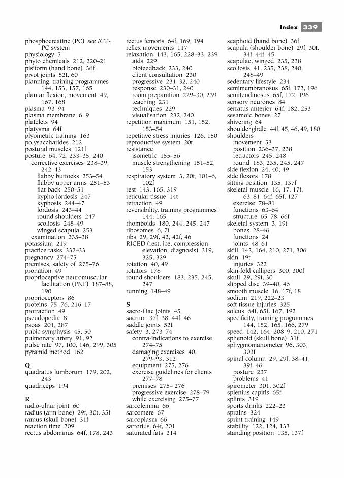
phosphocreatine (PC) see ATP-PC system
physiology 5phyto chemicals 212, 220–21pisiform (hand bone) 36fpivot joints 52t, 60planning, training programmes
144, 153, 157, 165plantar flexion, movement 49,
167, 168plasma 93–94plasma membrane 6, 9platelets 94platysma 64fplyometric training 163polysaccharides 212postural muscles 121fposture 64, 72, 233–35, 240
corrective exercises 238–39,242–43
flabby buttocks 253–54flabby upper arms 251–53flat back 250–51kypho-lordosis 247kyphosis 244–47lordosis 243–44round shoulders 247scoliosis 248–49winged scapula 253
examination 235–38potassium 219practice tasks 332–33pregnancy 274–75premises, safety of 275–76pronation 49proprioceptive neuromuscular
facilitation (PNF) 187–88,190
proprioceptors 86proteins 75, 76, 216–17protraction 49pseudopodia 8psoas 201, 287pubic symphysis 45, 50pulmonary artery 91, 92pulse rate 97, 100, 146, 299, 305pyramid method 162
Qquadratus lumborum 179, 202,
243quadriceps 194
Rradio-ulnar joint 60radius (arm bone) 29f, 30t, 35framus (skull bone) 31freaction time 209rectus abdominus 64f, 178, 243
rectus femoris 64f, 169, 194reflex movements 117relaxation 143, 165, 228–33, 239
aids 229biofeedback 233, 240client consultation 230progressive 231–32, 240response 230–31, 240room preparation 229–30, 239teaching 231techniques 229visualisation 232, 240
repetition maximum 151, 152,153–54
repetitive stress injuries 126, 150reproductive system 20tresistance
isometric 155–56muscle strengthening 151–52,
153respiratory system 3, 20t, 101–6,
102frest 143, 165, 319reticular tissue 14tretraction 49reversibility, training programmes
144, 165rhomboids 180, 244, 245, 247ribosomes 6, 7fribs 29, 29f, 42, 42f, 46RICED (rest, ice, compression,
elevation, diagnosis) 319,325, 329
rotation 40, 49rotators 178round shoulders 183, 235, 245,
247running 148–49
Ssacro-iliac joints 45sacrum 37f, 38, 44f, 46saddle joints 52tsafety 3, 273–74
contra-indications to exercise274–75
damaging exercises 40,279–93, 312
equipment 275, 276exercise guidelines for clients
277–78premises 275– 276progressive exercise 278–79while exercising 275–77
sarcolemma 66sarcomere 67sarcoplasm 66sartorius 64f, 201saturated fats 214
scaphoid (hand bone) 36fscapula (shoulder bone) 29f, 30t,
34f, 44f, 45scapulae, winged 235, 238scoliosis 41, 235, 238, 240,
248–49sedentary lifestyle 234semimembranosus 65f, 172, 196semitendinosus 65f, 172, 196sensory neurones 84serratus anterior 64f, 182, 253sesamoid bones 27shivering 64shoulder girdle 44f, 45, 46, 49, 180shoulders
movement 53position 236–37, 238retractors 245, 248round 183, 235, 245, 247
side flexion 24, 40, 49side flexors 178sitting position 135, 137fskeletal muscle 16, 17, 17f,
63–81, 64f, 65f, 127exercise 78–81functions 63–64structure 65–78, 66f
skeletal system 3, 19tbones 28–46functions 24joints 48–61
skill 142, 164, 210, 271, 306skin 19t
injuries 322skin-fold callipers 300, 300fskull 29, 29f, 30slipped disc 39–40, 46smooth muscle 16, 17f, 18sodium 219, 222–23soft tissue injuries 325soleus 64f, 65f, 167, 192specificity, training programmes
144, 152, 165, 166, 279speed 142, 164, 208–9, 210, 271sphenoid (skull bone) 31fsphygmomanometer 96, 303,
303fspinal column 29, 29f, 38–41,
39f, 46posture 237problems 41
spirometer 301, 302fsplenius capitis 65fsplints 319sports drinks 222–23sprains 324sprint training 149stability 122, 124, 133standing position 135, 137f
Index 339
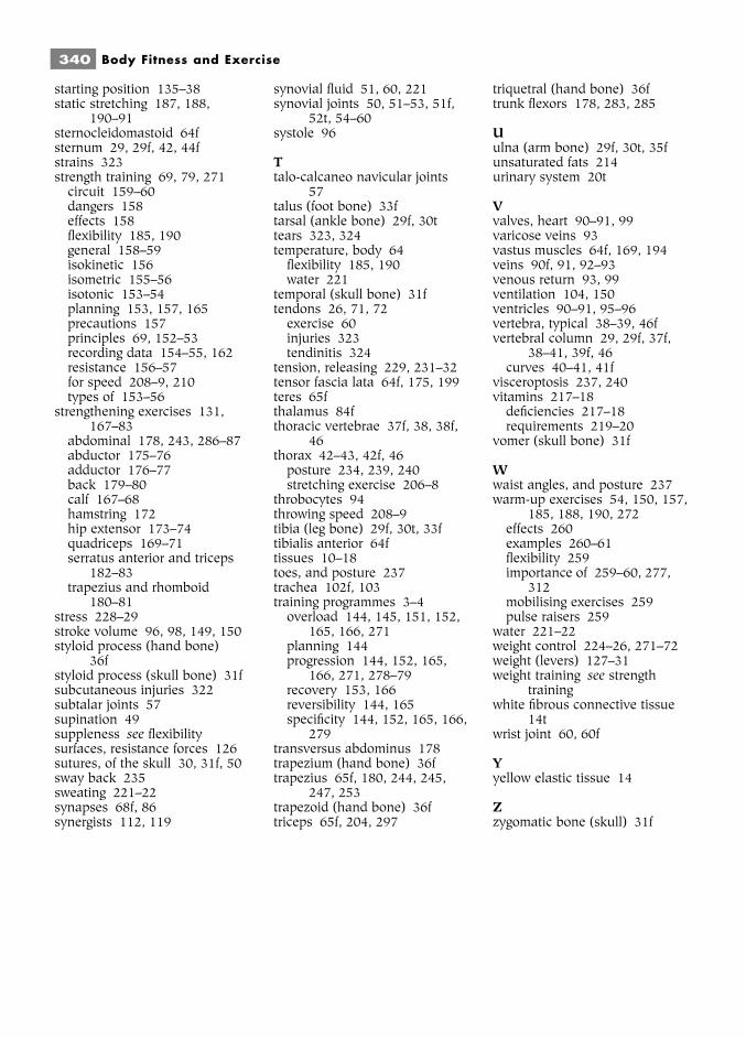
starting position 135–38static stretching 187, 188,
190–91sternocleidomastoid 64fsternum 29, 29f, 42, 44fstrains 323strength training 69, 79, 271
circuit 159–60dangers 158effects 158flexibility 185, 190general 158–59isokinetic 156isometric 155–56isotonic 153–54planning 153, 157, 165precautions 157principles 69, 152–53recording data 154–55, 162resistance 156–57for speed 208–9, 210types of 153–56
strengthening exercises 131,167–83
abdominal 178, 243, 286–87abductor 175–76adductor 176–77back 179–80calf 167–68hamstring 172hip extensor 173–74quadriceps 169–71serratus anterior and triceps
182–83trapezius and rhomboid
180–81stress 228–29stroke volume 96, 98, 149, 150styloid process (hand bone)
36fstyloid process (skull bone) 31fsubcutaneous injuries 322subtalar joints 57supination 49suppleness see flexibilitysurfaces, resistance forces 126sutures, of the skull 30, 31f, 50sway back 235sweating 221–22synapses 68f, 86synergists 112, 119
synovial fluid 51, 60, 221synovial joints 50, 51–53, 51f,
52t, 54–60systole 96
Ttalo-calcaneo navicular joints
57talus (foot bone) 33ftarsal (ankle bone) 29f, 30ttears 323, 324temperature, body 64
flexibility 185, 190water 221
temporal (skull bone) 31ftendons 26, 71, 72
exercise 60injuries 323tendinitis 324
tension, releasing 229, 231–32tensor fascia lata 64f, 175, 199teres 65fthalamus 84fthoracic vertebrae 37f, 38, 38f,
46thorax 42–43, 42f, 46
posture 234, 239, 240stretching exercise 206–8
throbocytes 94throwing speed 208–9tibia (leg bone) 29f, 30t, 33ftibialis anterior 64ftissues 10–18toes, and posture 237trachea 102f, 103training programmes 3–4
overload 144, 145, 151, 152,165, 166, 271
planning 144progression 144, 152, 165,
166, 271, 278–79recovery 153, 166reversibility 144, 165specificity 144, 152, 165, 166,
279transversus abdominus 178trapezium (hand bone) 36ftrapezius 65f, 180, 244, 245,
247, 253trapezoid (hand bone) 36ftriceps 65f, 204, 297
triquetral (hand bone) 36ftrunk flexors 178, 283, 285
Uulna (arm bone) 29f, 30t, 35funsaturated fats 214urinary system 20t
Vvalves, heart 90–91, 99varicose veins 93vastus muscles 64f, 169, 194veins 90f, 91, 92–93venous return 93, 99ventilation 104, 150ventricles 90–91, 95–96vertebra, typical 38–39, 46fvertebral column 29, 29f, 37f,
38–41, 39f, 46curves 40–41, 41f
visceroptosis 237, 240vitamins 217–18
deficiencies 217–18requirements 219–20
vomer (skull bone) 31f
Wwaist angles, and posture 237warm-up exercises 54, 150, 157,
185, 188, 190, 272effects 260examples 260–61flexibility 259importance of 259–60, 277,
312mobilising exercises 259pulse raisers 259
water 221–22weight control 224–26, 271–72weight (levers) 127–31weight training see strength
trainingwhite fibrous connective tissue
14twrist joint 60, 60f
Yyellow elastic tissue 14
Zzygomatic bone (skull) 31f
340 Body Fitness and Exercise